Uremic Toxins and the Lung Alveolar Capillary Barrier: A Narrative Review

 , ,
, ,
Abstract
1. Introduction
2. Materials and Methods
2.1. Search Strategy
2.2. Study Selection Criteria
2.3. Data Extraction
3. Results
3.1. UTs and Inflammation
3.2. UTs and Oxidative Stress
3.3. UT and Endothelial Cells
3.4. UTs and Epithelial Cells
3.5. UT and the Alveolar-Capillary Barrier
4. Discussion
5. Future Directions and Therapeutic Implications
Limitations
Supplementary Materials
Author Contributions
Funding
Institutional Review Board Statement
Informed Consent Statement
Data Availability Statement
Conflicts of Interest
Abbreviations
| ADMA | Asymmetric Dimethylarginine |
| AGEs | Advanced glycation end products |
| AhR | Aryl hydrocarbon receptor |
| ALI | Acute lung injury |
| AKI | Acute kidney injury |
| AQPs | Aquaporins |
| CKD | Chronic kidney disease |
| CRP | C-reactive protein |
| EMPs | Endothelial microparticles |
| ENaC | Epithelial sodium channel |
| eNOS | Endothelial nitric oxide synthase |
| EPC | Endothelial progenitor cell |
| ESKD | End-stage kidney disease |
| FGF-23 | Fibroblast growth factor |
| GSTM-1 | Glutathione S-transferase-1 |
| HD | Hemodialysis |
| IAA | Indole-3-Acetic acid |
| ICAM-1 | Intercellular adhesion molecule-1 |
| IFN-γ | Interferon gamma |
| IL-1 | Interleukin-1 |
| IL-6 | Interleukin-6 |
| IS | Indoxyl sulfate |
| JAMs | Junctional adhesion molecules |
| MAPK | Mitogen-activated protein kinases |
| MCP-1 | Monocyte chemoattractant protein-1 |
| MIP-1 | Macrophage inflammatory protein |
| NADPH oxidase | Nicotinamide adenine dinucleotide phosphate oxidase |
| Na+/K+/ATPase | Sodium-potassium ATPase |
| NF-κB | Nuclear factor kappa-light-chain-enhancer of activated B cells |
| NO | Nitric oxide |
| Nrf2 | Nuclear erythroid-related factor 2 |
| OATs | Organic anion transporters |
| PCS | p-Cresyl Sulfate |
| PD | Peritoneal dialysis |
| PECAM-1 | Platelet endothelial cell adhesion molecule-1 |
| PMVEC | Pulmonary microvascular endothelial cell |
| RAGE | Receptor for advanced glycation endproducts |
| RANK | Receptor activator of nuclear factor |
| RANKL | Receptor activator of nuclear factor ligand |
| RIR | Renal ischemia–reperfusion |
| ROS | Reactive oxygen species |
| RNS | Reactive nitrogen species |
| SDMA | Symmetric dimethylarginine |
| TGF-β | Transforming Growth Factor Beta |
| TJ | Tight Junction |
| TMAO | Trimethylamine N-oxide |
| TNF | Tumor necrosis factor |
| TNF-α | Tumor necrosis factor-alpha |
| ULI | Uremic lung injury |
| UT | Uremic toxins |
| VE-cadherin | Vascular endothelial cadherin |
| VEGF | Vascular endothelial growth factor |
| ZO-1 | Zonula occludens-1 |
References
- Duranton, F.; Cohen, G.; De Smet, R.; Rodriguez, M.; Jankowski, J.; Vanholder, R.; Argiles, A.; European Uremic Toxin Work Group. Normal and Pathologic Concentrations of Uremic Toxins. J. Am. Soc. Nephrol. JASN 2012, 23, 1258–1270. [Google Scholar] [CrossRef]
- Vanholder, R.; De Smet, R.; Glorieux, G.; Argilés, A.; Baurmeister, U.; Brunet, P.; Clark, W.; Cohen, G.; De Deyn, P.P.; Deppisch, R.; et al. Review on Uremic Toxins: Classification, Concentration, and Interindividual Variability. Kidney Int. 2003, 63, 1934–1943. [Google Scholar] [CrossRef]
- Glorieux, G.; Burtey, S.; Evenepoel, P.; Jankowski, J.; Koppe, L.; Masereeuw, R.; Vanholder, R. A Guide to Uraemic Toxicity. Nat. Rev. Nephrol. 2026, 22, 52–68. [Google Scholar] [CrossRef] [PubMed]
- Parivakkam Mani, A.; K, S.; Sundar, R.; Yadav, S. Pulmonary Manifestations at Different Stages in the Chronic Kidney Disease: An Observational Study. Cureus 2023, 15, e39235. [Google Scholar] [CrossRef]
- Kaysi, S.; Hamad, A.; Boulgheraif, A.; Bonkain, F.; Libertalis, M.; Ayyach, A.A.; Baz, M.; Farah, I.; Ibrahim, R.; Ateya, H.; et al. Lung Ultrasound Guided Management of Pulmonary Congestion in Hemodialysis: A Multi-Center Randomized Controlled Trial. Kidney360 2026, 7, 139–149. [Google Scholar] [CrossRef] [PubMed]
- Mukai, H.; Ming, P.; Lindholm, B.; Heimbürger, O.; Barany, P.; Stenvinkel, P.; Qureshi, A.R. Lung Dysfunction and Mortality in Patients with Chronic Kidney Disease. Kidney Blood Press. Res. 2018, 43, 522–535. [Google Scholar] [CrossRef]
- Mukai, H.; Ming, P.; Lindholm, B.; Heimbürger, O.; Barany, P.; Anderstam, B.; Stenvinkel, P.; Qureshi, A.R. Restrictive Lung Disorder Is Common in Patients with Kidney Failure and Associates with Protein-Energy Wasting, Inflammation and Cardiovascular Disease. PLoS ONE 2018, 13, e0195585. [Google Scholar] [CrossRef] [PubMed]
- de Souza Rezende, P.; Porcher Andrade, F.; Ferraro Dos Santos Borba, C.; Eidt Rovedder, P.M. Pulmonary Function, Muscle Strength, and Quality of Life Have Differed between Chronic Kidney Disease Patients and Healthy Individuals. Ther. Apher. Dial. 2022, 26, 337–344. [Google Scholar] [CrossRef]
- Gembillo, G.; Calimeri, S.; Tranchida, V.; Silipigni, S.; Vella, D.; Ferrara, D.; Spinella, C.; Santoro, D.; Visconti, L. Lung Dysfunction and Chronic Kidney Disease: A Complex Network of Multiple Interactions. J. Pers. Med. 2023, 13, 286. [Google Scholar] [CrossRef]
- Yang, F.; Chang, Y.; Zhang, C.; Xiong, Y.; Wang, X.; Ma, X.; Wang, Z.; Li, H.; Shimosawa, T.; Pei, L.; et al. UUO Induces Lung Fibrosis with Macrophage-Myofibroblast Transition in Rats. Int. Immunopharmacol. 2021, 93, 107396. [Google Scholar] [CrossRef]
- National Heart, Lung, and Blood Institute Acute Respiratory Distress Syndrome (ARDS) Clinical Trials Network; Wiedemann, H.P.; Wheeler, A.P.; Bernard, G.R.; Thompson, B.T.; Hayden, D.; deBoisblanc, B.; Connors, A.F.; Hite, R.D.; Harabin, A.L. Comparison of Two Fluid-Management Strategies in Acute Lung Injury. N. Engl. J. Med. 2006, 354, 2564–2575. [Google Scholar] [CrossRef] [PubMed]
- Kaysi, S.; Pacha, B.; Mesquita, M.; Collart, F.; Nortier, J. Pulmonary Congestion and Systemic Congestion in Hemodialysis: Dynamics and Correlations. Front. Nephrol. 2024, 4, 1336863. [Google Scholar] [CrossRef] [PubMed]
- Faubel, S.; Edelstein, C.L. Mechanisms and Mediators of Lung Injury after Acute Kidney Injury. Nat. Rev. Nephrol. 2016, 12, 48–60. [Google Scholar] [CrossRef] [PubMed]
- Assimon, M.M.; Wenger, J.B.; Wang, L.; Flythe, J.E. Ultrafiltration Rate and Mortality in Maintenance Hemodialysis Patients. Am. J. Kidney Dis. Off. J. Natl. Kidney Found. 2016, 68, 911–922. [Google Scholar] [CrossRef]
- Jing, Y.J.; Ni, J.W.; Ding, F.H.; Fang, Y.H.; Wang, X.Q.; Wang, H.B.; Chen, X.N.; Chen, N.; Zhan, W.W.; Lu, L.; et al. P-Cresyl Sulfate Is Associated with Carotid Arteriosclerosis in Hemodialysis Patients and Promotes Atherogenesis in apoE-/- Mice. Kidney Int. 2016, 89, 439–449. [Google Scholar] [CrossRef]
- Miyata, T.; van Ypersele de Strihou, C.; Kurokawa, K.; Baynes, J.W. Alterations in Nonenzymatic Biochemistry in Uremia: Origin and Significance of “Carbonyl Stress” in Long-Term Uremic Complications. Kidney Int. 1999, 55, 389–399. [Google Scholar] [CrossRef]
- Linden, E.; Cai, W.; He, J.C.; Xue, C.; Li, Z.; Winston, J.; Vlassara, H.; Uribarri, J. Endothelial Dysfunction in Patients with Chronic Kidney Disease Results from Advanced Glycation End Products (AGE)-Mediated Inhibition of Endothelial Nitric Oxide Synthase through RAGE Activation. Clin. J. Am. Soc. Nephrol. CJASN 2008, 3, 691–698. [Google Scholar] [CrossRef]
- Akiyama, Y.; Kikuchi, K.; Toyohara, T.; Mishima, E.; Suzuki, C.; Suzuki, T.; Nakayama, M.; Tomioka, Y.; Soga, T.; Abe, T. CE-MS-Based Identification of Uremic Solutes Specific to Hemodialysis Patients. Toxins 2021, 13, 324. [Google Scholar] [CrossRef]
- Ebersolt, M.; Santana Machado, T.; Mallmann, C.; Mc-Kay, N.; Dou, L.; Bouchouareb, D.; Brunet, P.; Burtey, S.; Sallée, M. Protein/Fiber Index Modulates Uremic Toxin Concentrations in Hemodialysis Patients. Toxins 2022, 14, 589. [Google Scholar] [CrossRef]
- Eiselt, J.; Rajdl, D.; Racek, J.; Siroká, R.; Trefil, L.; Opatrná, S. Asymmetric Dimethylarginine in Hemodialysis, Hemodiafiltration, and Peritoneal Dialysis. Artif. Organs 2010, 34, 420–425. [Google Scholar] [CrossRef]
- Klein, C.L.; Hoke, T.S.; Fang, W.-F.; Altmann, C.J.; Douglas, I.S.; Faubel, S. Interleukin-6 Mediates Lung Injury Following Ischemic Acute Kidney Injury or Bilateral Nephrectomy. Kidney Int. 2008, 74, 901–909. [Google Scholar] [CrossRef]
- Kramer, A.A.; Postler, G.; Salhab, K.F.; Mendez, C.; Carey, L.C.; Rabb, H. Renal Ischemia/Reperfusion Leads to Macrophage-Mediated Increase in Pulmonary Vascular Permeability. Kidney Int. 1999, 55, 2362–2367. [Google Scholar] [CrossRef]
- Rabb, H.; Wang, Z.; Nemoto, T.; Hotchkiss, J.; Yokota, N.; Soleimani, M. Acute Renal Failure Leads to Dysregulation of Lung Salt and Water Channels. Kidney Int. 2003, 63, 600–606. [Google Scholar] [CrossRef]
- Awad, A.S.; Rouse, M.; Huang, L.; Vergis, A.L.; Reutershan, J.; Cathro, H.P.; Linden, J.; Okusa, M.D. Compartmentalization of Neutrophils in the Kidney and Lung Following Acute Ischemic Kidney Injury. Kidney Int. 2009, 75, 689–698. [Google Scholar] [CrossRef]
- Abbasian, N.; Burton, J.O.; Herbert, K.E.; Tregunna, B.-E.; Brown, J.R.; Ghaderi-Najafabadi, M.; Brunskill, N.J.; Goodall, A.H.; Bevington, A. Hyperphosphatemia, Phosphoprotein Phosphatases, and Microparticle Release in Vascular Endothelial Cells. J. Am. Soc. Nephrol. JASN 2015, 26, 2152–2162. [Google Scholar] [CrossRef]
- Ahdieh, M.; Vandenbos, T.; Youakim, A. Lung Epithelial Barrier Function and Wound Healing Are Decreased by IL-4 and IL-13 and Enhanced by IFN-Gamma. Am. J. Physiol. Cell Physiol. 2001, 281, C2029–C2038. [Google Scholar] [CrossRef] [PubMed]
- Annuk, M.; Zilmer, M.; Lind, L.; Linde, T.; Fellström, B. Oxidative Stress and Endothelial Function in Chronic Renal Failure. J. Am. Soc. Nephrol. JASN 2001, 12, 2747–2752. [Google Scholar] [CrossRef] [PubMed]
- Bao, S.; Knoell, D.L. Zinc Modulates Cytokine-Induced Lung Epithelial Cell Barrier Permeability. Am. J. Physiol. Lung Cell. Mol. Physiol. 2006, 291, L1132–L1141. [Google Scholar] [CrossRef] [PubMed]
- Catar, R.; Moll, G.; Kamhieh-Milz, J.; Luecht, C.; Chen, L.; Zhao, H.; Ernst, L.; Willy, K.; Girndt, M.; Fiedler, R.; et al. Expanded Hemodialysis Therapy Ameliorates Uremia-Induced Systemic Microinflammation and Endothelial Dysfunction by Modulating VEGF, TNF-α and AP-1 Signaling. Front. Immunol. 2021, 12, 774052. [Google Scholar] [CrossRef]
- Chang, J.-F.; Liang, S.-S.; Thanasekaran, P.; Chang, H.-W.; Wen, L.-L.; Chen, C.-H.; Liou, J.-C.; Yeh, J.-C.; Liu, S.-H.; Dai, H.-M.; et al. Translational Medicine in Pulmonary-Renal Crosstalk: Therapeutic Targeting of p-Cresyl Sulfate Triggered Nonspecific ROS and Chemoattractants in Dyspneic Patients with Uremic Lung Injury. J. Clin. Med. 2018, 7, 266. [Google Scholar] [CrossRef]
- Chen, Q.; Yi, B.; Ma, J.; Ning, J.; Wu, L.; Ma, D.; Lu, K.; Gu, J. A2-Adrenoreceptor Modulated FAK Pathway Induced by Dexmedetomidine Attenuates Pulmonary Microvascular Hyper-Permeability Following Kidney Injury. Oncotarget 2016, 7, 55990–56001. [Google Scholar] [CrossRef] [PubMed]
- de Groot, K.; Bahlmann, F.H.; Sowa, J.; Koenig, J.; Menne, J.; Haller, H.; Fliser, D. Uremia Causes Endothelial Progenitor Cell Deficiency. Kidney Int. 2004, 66, 641–646. [Google Scholar] [CrossRef] [PubMed]
- Dou, L.; Bertrand, E.; Cerini, C.; Faure, V.; Sampol, J.; Vanholder, R.; Berland, Y.; Brunet, P. The Uremic Solutes P-Cresol and Indoxyl Sulfate Inhibit Endothelial Proliferation and Wound Repair. Kidney Int. 2004, 65, 442–451. [Google Scholar] [CrossRef]
- Dou, L.; Jourde-Chiche, N.; Faure, V.; Cerini, C.; Berland, Y.; Dignat-George, F.; Brunet, P. The Uremic Solute Indoxyl Sulfate Induces Oxidative Stress in Endothelial Cells. J. Thromb. Haemost. JTH 2007, 5, 1302–1308. [Google Scholar] [CrossRef] [PubMed]
- Dou, L.; Sallée, M.; Cerini, C.; Poitevin, S.; Gondouin, B.; Jourde-Chiche, N.; Fallague, K.; Brunet, P.; Calaf, R.; Dussol, B.; et al. The Cardiovascular Effect of the Uremic Solute Indole-3 Acetic Acid. J. Am. Soc. Nephrol. JASN 2015, 26, 876–887. [Google Scholar] [CrossRef]
- El-Gamal, D.; Rao, S.P.; Holzer, M.; Hallström, S.; Haybaeck, J.; Gauster, M.; Wadsack, C.; Kozina, A.; Frank, S.; Schicho, R.; et al. The Urea Decomposition Product Cyanate Promotes Endothelial Dysfunction. Kidney Int. 2014, 86, 923–931. [Google Scholar] [CrossRef]
- Favretto, G.; Souza, L.M.; Gregório, P.C.; Cunha, R.S.; Maciel, R.A.P.; Sassaki, G.L.; Toledo, M.G.; Pecoits-Filho, R.; Souza, W.M.; Stinghen, A.E.M. Role of Organic Anion Transporters in the Uptake of Protein-Bound Uremic Toxins by Human Endothelial Cells and Monocyte Chemoattractant Protein-1 Expression. J. Vasc. Res. 2017, 54, 170–179. [Google Scholar] [CrossRef]
- García-Jérez, A.; Luengo, A.; Carracedo, J.; Ramírez-Chamond, R.; Rodriguez-Puyol, D.; Rodriguez-Puyol, M.; Calleros, L. Effect of Uraemia on Endothelial Cell Damage Is Mediated by the Integrin Linked Kinase Pathway. J. Physiol. 2015, 593, 601–618; discussion 618. [Google Scholar] [CrossRef]
- Guo, Y.; DuVall, M.D.; Crow, J.P.; Matalon, S. Nitric Oxide Inhibits Na+ Absorption across Cultured Alveolar Type II Monolayers. Am. J. Physiol. 1998, 274, L369–L377. [Google Scholar] [CrossRef]
- Gupta, J.; Mitra, N.; Kanetsky, P.A.; Devaney, J.; Wing, M.R.; Reilly, M.; Shah, V.O.; Balakrishnan, V.S.; Guzman, N.J.; Girndt, M.; et al. Association between Albuminuria, Kidney Function, and Inflammatory Biomarker Profile in CKD in CRIC. Clin. J. Am. Soc. Nephrol. CJASN 2012, 7, 1938–1946. [Google Scholar] [CrossRef]
- Ito, S.; Osaka, M.; Edamatsu, T.; Itoh, Y.; Yoshida, M. Crucial Role of the Aryl Hydrocarbon Receptor (AhR) in Indoxyl Sulfate-Induced Vascular Inflammation. J. Atheroscler. Thromb. 2016, 23, 960–975. [Google Scholar] [CrossRef]
- Kim, H.Y.; Yoo, T.-H.; Hwang, Y.; Lee, G.H.; Kim, B.; Jang, J.; Yu, H.T.; Kim, M.C.; Cho, J.-Y.; Lee, C.J.; et al. Indoxyl Sulfate (IS)-Mediated Immune Dysfunction Provokes Endothelial Damage in Patients with End-Stage Renal Disease (ESRD). Sci. Rep. 2017, 7, 3057. [Google Scholar] [CrossRef] [PubMed]
- Komaru, Y.; Ning, L.; Lama, C.; Suresh, A.; Kefaloyianni, E.; Miller, M.J.; Kawana, S.; Shepherd, H.M.; Li, W.; Kreisel, D.; et al. Acute Kidney Injury Triggers Hypoxemia by Lung Intravascular Neutrophil Retention That Reduces Capillary Blood Flow. J. Clin. Investig. 2025, 135, e186705. [Google Scholar] [CrossRef]
- Li, S.; Xie, Y.; Yang, B.; Huang, S.; Zhang, Y.; Jia, Z.; Ding, G.; Zhang, A. MicroRNA-214 Targets COX-2 to Antagonize Indoxyl Sulfate (IS)-Induced Endothelial Cell Apoptosis. Apoptosis Int. J. Program. Cell Death 2020, 25, 92–104. [Google Scholar] [CrossRef] [PubMed]
- Li, R.; Zhang, X.; Xu, Y.; Feng, T. Vascular Endothelial Dysfunction Improvements in Patients with Uremia Using Pentoxifylline-Suppressing NLRP3 Expressions and HMGB1 Release. Shock 2024, 62, 746–754. [Google Scholar] [CrossRef]
- Ma, T.; Fukuda, N.; Song, Y.; Matthay, M.A.; Verkman, A.S. Lung Fluid Transport in Aquaporin-5 Knockout Mice. J. Clin. Investig. 2000, 105, 93–100. [Google Scholar] [CrossRef] [PubMed]
- Ma, T.; Liu, Z. Functions of Aquaporin 1 and α-Epithelial Na+ Channel in Rat Acute Lung Injury Induced by Acute Ischemic Kidney Injury. Int. Urol. Nephrol. 2013, 45, 1187–1196. [Google Scholar] [CrossRef]
- Martínez-Moreno, J.M.; Herencia, C.; de Oca, A.M.; Díaz-Tocados, J.M.; Vergara, N.; Gómez-Luna, M.J.; López-Argüello, S.D.; Camargo, A.; Peralbo-Santaella, E.; Rodríguez-Ortiz, M.E.; et al. High Phosphate Induces a Pro-Inflammatory Response by Vascular Smooth Muscle Cells and Modulation by Vitamin D Derivatives. Clin. Sci. 2017, 131, 1449–1463. [Google Scholar] [CrossRef]
- Masai, N.; Tatebe, J.; Yoshino, G.; Morita, T. Indoxyl Sulfate Stimulates Monocyte Chemoattractant Protein-1 Expression in Human Umbilical Vein Endothelial Cells by Inducing Oxidative Stress through Activation of the NADPH Oxidase-Nuclear Factor-κB Pathway. Circ. J. Off. J. Jpn. Circ. Soc. 2010, 74, 2216–2224. [Google Scholar] [CrossRef]
- Merino, A.; Portolés, J.; Selgas, R.; Ojeda, R.; Buendia, P.; Ocaña, J.; Bajo, M.A.; del Peso, G.; Carracedo, J.; Ramírez, R.; et al. Effect of Different Dialysis Modalities on Microinflammatory Status and Endothelial Damage. Clin. J. Am. Soc. Nephrol. CJASN 2010, 5, 227–234. [Google Scholar] [CrossRef]
- Nemmar, A.; Karaca, T.; Beegam, S.; Yuvaraju, P.; Yasin, J.; Ali, B.H. Lung Oxidative Stress, DNA Damage, Apoptosis, and Fibrosis in Adenine-Induced Chronic Kidney Disease in Mice. Front. Physiol. 2017, 8, 896. [Google Scholar] [CrossRef]
- Nowak, K.L.; Jovanovich, A.; Farmer-Bailey, H.; Bispham, N.; Struemph, T.; Malaczewski, M.; Wang, W.; Chonchol, M. Vascular Dysfunction, Oxidative Stress, and Inflammation in Chronic Kidney Disease. Kidney360 2020, 1, 501–509. [Google Scholar] [CrossRef]
- Peng, A.; Wu, T.; Zeng, C.; Rakheja, D.; Zhu, J.; Ye, T.; Hutcheson, J.; Vaziri, N.D.; Liu, Z.; Mohan, C.; et al. Adverse Effects of Simulated Hyper- and Hypo-Phosphatemia on Endothelial Cell Function and Viability. PLoS ONE 2011, 6, e23268. [Google Scholar] [CrossRef]
- Saum, K.; Campos, B.; Celdran-Bonafonte, D.; Nayak, L.; Sangwung, P.; Thakar, C.; Roy-Chaudhury, P.; Owens, A.P. Uremic Advanced Glycation End Products and Protein-Bound Solutes Induce Endothelial Dysfunction Through Suppression of Krüppel-Like Factor 2. J. Am. Heart Assoc. 2018, 7, e007566. [Google Scholar] [CrossRef]
- Shuto, E.; Taketani, Y.; Tanaka, R.; Harada, N.; Isshiki, M.; Sato, M.; Nashiki, K.; Amo, K.; Yamamoto, H.; Higashi, Y.; et al. Dietary Phosphorus Acutely Impairs Endothelial Function. J. Am. Soc. Nephrol. JASN 2009, 20, 1504–1512. [Google Scholar] [CrossRef]
- Six, I.; Okazaki, H.; Gross, P.; Cagnard, J.; Boudot, C.; Maizel, J.; Drueke, T.B.; Massy, Z.A. Direct, Acute Effects of Klotho and FGF23 on Vascular Smooth Muscle and Endothelium. PLoS ONE 2014, 9, e93423. [Google Scholar] [CrossRef]
- Vaziri, N.D.; Yuan, J.; Rahimi, A.; Ni, Z.; Said, H.; Subramanian, V.S. Disintegration of Colonic Epithelial Tight Junction in Uremia: A Likely Cause of CKD-Associated Inflammation. Nephrol. Dial. Transplant. Off. Publ. Eur. Dial. Transpl. Assoc.—Eur. Ren. Assoc. 2012, 27, 2686–2693. [Google Scholar] [CrossRef]
- Verkman, A.S.; Yang, B.; Song, Y.; Manley, G.T.; Ma, T. Role of Water Channels in Fluid Transport Studied by Phenotype Analysis of Aquaporin Knockout Mice. Exp. Physiol. 2000, 85, 233S–241S. [Google Scholar] [CrossRef] [PubMed]
- Ware, L.B.; Matthay, M.A. Alveolar Fluid Clearance Is Impaired in the Majority of Patients with Acute Lung Injury and the Acute Respiratory Distress Syndrome. Am. J. Respir. Crit. Care Med. 2001, 163, 1376–1383. [Google Scholar] [CrossRef] [PubMed]
- Watanabe, H.; Miyamoto, Y.; Enoki, Y.; Ishima, Y.; Kadowaki, D.; Kotani, S.; Nakajima, M.; Tanaka, M.; Matsushita, K.; Mori, Y.; et al. P-Cresyl Sulfate, a Uremic Toxin, Causes Vascular Endothelial and Smooth Muscle Cell Damages by Inducing Oxidative Stress. Pharmacol. Res. Perspect. 2015, 3, e00092. [Google Scholar] [CrossRef] [PubMed]
- Wu, S.-B.; Wang, Y.-D.; Zhang, W. Effects of uremia serum on monolayer permeability and F-actin in rat pulmonary microvascular endothelial cells. Zhonghua Yi Xue Za Zhi 2008, 88, 3374–3377. [Google Scholar] [PubMed]
- Yabuuchi, N.; Sagata, M.; Saigo, C.; Yoneda, G.; Yamamoto, Y.; Nomura, Y.; Nishi, K.; Fujino, R.; Jono, H.; Saito, H. Indoxyl Sulfate as a Mediator Involved in Dysregulation of Pulmonary Aquaporin-5 in Acute Lung Injury Caused by Acute Kidney Injury. Int. J. Mol. Sci. 2016, 18, 11. [Google Scholar] [CrossRef] [PubMed]
- Yang, K.; Nie, L.; Huang, Y.; Zhang, J.; Xiao, T.; Guan, X.; Zhao, J. Amelioration of Uremic Toxin Indoxyl Sulfate-Induced Endothelial Cell Dysfunction by Klotho Protein. Toxicol. Lett. 2012, 215, 77–83. [Google Scholar] [CrossRef] [PubMed]
- Yuan, J.; Guo, Q.; Qureshi, A.R.; Anderstam, B.; Eriksson, M.; Heimbürger, O.; Bárány, P.; Stenvinkel, P.; Lindholm, B. Circulating Vascular Endothelial Growth Factor (VEGF) and Its Soluble Receptor 1 (sVEGFR-1) Are Associated with Inflammation and Mortality in Incident Dialysis Patients. Nephrol. Dial. Transplant. Off. Publ. Eur. Dial. Transpl. Assoc.—Eur. Ren. Assoc. 2013, 28, 2356–2363. [Google Scholar] [CrossRef]
- Zarbock, A.; Schmolke, M.; Spieker, T.; Jurk, K.; Van Aken, H.; Singbartl, K. Acute Uremia but Not Renal Inflammation Attenuates Aseptic Acute Lung Injury: A Critical Role for Uremic Neutrophils. J. Am. Soc. Nephrol. JASN 2006, 17, 3124–3131. [Google Scholar] [CrossRef]
- Zhao, H.; Ning, J.; Lemaire, A.; Koumpa, F.-S.; Sun, J.J.; Fung, A.; Gu, J.; Yi, B.; Lu, K.; Ma, D. Necroptosis and Parthanatos Are Involved in Remote Lung Injury after Receiving Ischemic Renal Allografts in Rats. Kidney Int. 2015, 87, 738–748. [Google Scholar] [CrossRef]
- Zickler, D.; Luecht, C.; Willy, K.; Chen, L.; Witowski, J.; Girndt, M.; Fiedler, R.; Storr, M.; Kamhieh-Milz, J.; Schoon, J.; et al. Tumour Necrosis Factor-Alpha in Uraemic Serum Promotes Osteoblastic Transition and Calcification of Vascular Smooth Muscle Cells via Extracellular Signal-Regulated Kinases and Activator Protein 1/c-FOS-Mediated Induction of Interleukin 6 Expression. Nephrol. Dial. Transplant. Off. Publ. Eur. Dial. Transpl. Assoc.—Eur. Ren. Assoc. 2018, 33, 574–585. [Google Scholar] [CrossRef]
- Stenvinkel, P.; Ketteler, M.; Johnson, R.J.; Lindholm, B.; Pecoits-Filho, R.; Riella, M.; Heimbürger, O.; Cederholm, T.; Girndt, M. IL-10, IL-6, and TNF-Alpha: Central Factors in the Altered Cytokine Network of Uremia--the Good, the Bad, and the Ugly. Kidney Int. 2005, 67, 1216–1233. [Google Scholar] [CrossRef]
- Speeckaert, M.M.; Speeckaert, R.; Laute, M.; Vanholder, R.; Delanghe, J.R. Tumor Necrosis Factor Receptors: Biology and Therapeutic Potential in Kidney Diseases. Am. J. Nephrol. 2012, 36, 261–270. [Google Scholar] [CrossRef]
- Cobo, G.; Lindholm, B.; Stenvinkel, P. Chronic Inflammation in End-Stage Renal Disease and Dialysis. Nephrol. Dial. Transplant. Off. Publ. Eur. Dial. Transpl. Assoc.—Eur. Ren. Assoc. 2018, 33, iii35–iii40. [Google Scholar] [CrossRef]
- Meyer, T.W.; Hostetter, T.H. Uremia. N. Engl. J. Med. 2007, 357, 1316–1325. [Google Scholar] [CrossRef]
- Azevedo, C.A.B.; da Cunha, R.S.; Junho, C.V.C.; da Silva, J.V.; Moreno-Amaral, A.N.; de Moraes, T.P.; Carneiro-Ramos, M.S.; Stinghen, A.E.M. Extracellular Vesicles and Their Relationship with the Heart-Kidney Axis, Uremia and Peritoneal Dialysis. Toxins 2021, 13, 778. [Google Scholar] [CrossRef]
- Ridker, P.M.; Tuttle, K.R.; Perkovic, V.; Libby, P.; MacFadyen, J.G. Inflammation Drives Residual Risk in Chronic Kidney Disease: A CANTOS Substudy. Eur. Heart J. 2022, 43, 4832–4844. [Google Scholar] [CrossRef] [PubMed]
- Harlacher, E.; Wollenhaupt, J.; Baaten, C.C.F.M.J.; Noels, H. Impact of Uremic Toxins on Endothelial Dysfunction in Chronic Kidney Disease: A Systematic Review. Int. J. Mol. Sci. 2022, 23, 531. [Google Scholar] [CrossRef]
- Tumur, Z.; Shimizu, H.; Enomoto, A.; Miyazaki, H.; Niwa, T. Indoxyl Sulfate Upregulates Expression of ICAM-1 and MCP-1 by Oxidative Stress-Induced NF-kappaB Activation. Am. J. Nephrol. 2010, 31, 435–441. [Google Scholar] [CrossRef] [PubMed]
- Amdur, R.L.; Feldman, H.I.; Gupta, J.; Yang, W.; Kanetsky, P.; Shlipak, M.; Rahman, M.; Lash, J.P.; Townsend, R.R.; Ojo, A.; et al. Inflammation and Progression of CKD: The CRIC Study. Clin. J. Am. Soc. Nephrol. CJASN 2016, 11, 1546–1556. [Google Scholar] [CrossRef] [PubMed]
- Barnes, P.J. The Cytokine Network in Chronic Obstructive Pulmonary Disease. Am. J. Respir. Cell Mol. Biol. 2009, 41, 631–638. [Google Scholar] [CrossRef]
- Gutiérrez, O.M.; Mannstadt, M.; Isakova, T.; Rauh-Hain, J.A.; Tamez, H.; Shah, A.; Smith, K.; Lee, H.; Thadhani, R.; Jüppner, H.; et al. Fibroblast Growth Factor 23 and Mortality among Patients Undergoing Hemodialysis. N. Engl. J. Med. 2008, 359, 584–592. [Google Scholar] [CrossRef]
- Mesquita, M.; Demulder, A.; Damry, N.; Mélot, C.; Wittersheim, E.; Willems, D.; Dratwa, M.; Bergmann, P. Plasma Osteoprotegerin Is an Independent Risk Factor for Mortality and an Early Biomarker of Coronary Vascular Calcification in Chronic Kidney Disease. Clin. Chem. Lab. Med. 2009, 47, 339–346. [Google Scholar] [CrossRef]
- Castillo-Rodríguez, E.; Pizarro-Sánchez, S.; Sanz, A.B.; Ramos, A.M.; Sanchez-Niño, M.D.; Martin-Cleary, C.; Fernandez-Fernandez, B.; Ortiz, A. Inflammatory Cytokines as Uremic Toxins: “Ni Son Todos Los Que Estan, Ni Estan Todos Los Que Son.”. Toxins 2017, 9, 114. [Google Scholar] [CrossRef]
- Kooman, J.P.; Dekker, M.J.; Usvyat, L.A.; Kotanko, P.; van der Sande, F.M.; Schalkwijk, C.G.; Shiels, P.G.; Stenvinkel, P. Inflammation and Premature Aging in Advanced Chronic Kidney Disease. Am. J. Physiol. Renal Physiol. 2017, 313, F938–F950. [Google Scholar] [CrossRef]
- Chiang, C.-K.; Huang, J.-W.; Hsu, S.-P.; Ho, T.-I.; Pai, M.-F.; Peng, Y.-S.; Liu, S.-H.; Hung, K.-Y.; Tsai, T.-J. Plasma Interleukin-18 Levels in Hemodialysis Patients: Increased by Dialysis Process and Association with Interleukin-6 and Tumor Necrotic Factor-Alpha. Blood Purif. 2006, 24, 174–179. [Google Scholar] [CrossRef]
- Panichi, V.; Taccola, D.; Rizza, G.M.; Consani, C.; Ghiadoni, L.; Filippi, C.; Cristofani, R.; Panicucci, E.; Migliori, M.; Sidoti, A.; et al. Interleukin-8 Is a Powerful Prognostic Predictor of All-Cause and Cardiovascular Mortality in Dialytic Patients. Nephron Clin. Pract. 2006, 102, c51–c58. [Google Scholar] [CrossRef]
- Dias, G.F.; Tozoni, S.S.; Bohnen, G.; van Spitzenbergen, B.A.K.; Grobe, N.; Nakao, L.S.; Pecoits-Filho, R.; Kotanko, P.; Moreno-Amaral, A.N. Effect of Hypoxia and Uremia on Oxidative Stress on Erythrocytes from Hemodialysis Patients. Cell Biochem. Funct. 2022, 40, 856–864. [Google Scholar] [CrossRef]
- Cachofeiro, V.; Goicochea, M.; de Vinuesa, S.G.; Oubiña, P.; Lahera, V.; Luño, J. Oxidative Stress and Inflammation, a Link between Chronic Kidney Disease and Cardiovascular Disease. Kidney Int. Suppl. 2008, 74, S4–S9. [Google Scholar] [CrossRef]
- Ravarotto, V.; Simioni, F.; Pagnin, E.; Davis, P.A.; Calò, L.A. Oxidative Stress—Chronic Kidney Disease—Cardiovascular Disease: A Vicious Circle. Life Sci. 2018, 210, 125–131. [Google Scholar] [CrossRef] [PubMed]
- Wang, Y.; Gao, L. Inflammation and Cardiovascular Disease Associated With Hemodialysis for End-Stage Renal Disease. Front. Pharmacol. 2022, 13, 800950. [Google Scholar] [CrossRef]
- Martínez-Moreno, J.M.; Muñoz-Castañeda, J.R.; Herencia, C.; Oca, A.M.D.; Estepa, J.C.; Canalejo, R.; Rodríguez-Ortiz, M.E.; Perez-Martinez, P.; Aguilera-Tejero, E.; Canalejo, A.; et al. In Vascular Smooth Muscle Cells Paricalcitol Prevents Phosphate-Induced Wnt/β-Catenin Activation. Am. J. Physiol. Renal Physiol. 2012, 303, F1136–F1144. [Google Scholar] [CrossRef] [PubMed]
- Kharbanda, R.K.; Deanfield, J.E. Functions of the Healthy Endothelium. Coron. Artery Dis. 2001, 12, 485–491. [Google Scholar] [CrossRef] [PubMed]
- Zoccali, C. Endothelial Dysfunction in CKD: A New Player in Town? Nephrol. Dial. Transplant. Off. Publ. Eur. Dial. Transpl. Assoc.—Eur. Ren. Assoc. 2008, 23, 783–785. [Google Scholar] [CrossRef]
- Katz, S.D.; Hryniewicz, K.; Hriljac, I.; Balidemaj, K.; Dimayuga, C.; Hudaihed, A.; Yasskiy, A. Vascular Endothelial Dysfunction and Mortality Risk in Patients with Chronic Heart Failure. Circulation 2005, 111, 310–314. [Google Scholar] [CrossRef] [PubMed]
- Briet, M.; Bozec, E.; Laurent, S.; Fassot, C.; London, G.M.; Jacquot, C.; Froissart, M.; Houillier, P.; Boutouyrie, P. Arterial Stiffness and Enlargement in Mild-to-Moderate Chronic Kidney Disease. Kidney Int. 2006, 69, 350–357. [Google Scholar] [CrossRef]
- Lano, G.; Burtey, S.; Sallée, M. Indoxyl Sulfate, a Uremic Endotheliotoxin. Toxins 2020, 12, 229. [Google Scholar] [CrossRef]
- Miyamoto, Y.; Watanabe, H.; Noguchi, T.; Kotani, S.; Nakajima, M.; Kadowaki, D.; Otagiri, M.; Maruyama, T. Organic Anion Transporters Play an Important Role in the Uptake of P-Cresyl Sulfate, a Uremic Toxin, in the Kidney. Nephrol. Dial. Transplant. Off. Publ. Eur. Dial. Transpl. Assoc.—Eur. Ren. Assoc. 2011, 26, 2498–2502. [Google Scholar] [CrossRef]
- Catar, R.; Moll, G.; Hosp, I.; Simon, M.; Luecht, C.; Zhao, H.; Wu, D.; Chen, L.; Kamhieh-Milz, J.; Korybalska, K.; et al. Transcriptional Regulation of Thrombin-Induced Endothelial VEGF Induction and Proangiogenic Response. Cells 2021, 10, 910. [Google Scholar] [CrossRef]
- Domigan, C.K.; Warren, C.M.; Antanesian, V.; Happel, K.; Ziyad, S.; Lee, S.; Krall, A.; Duan, L.; Torres-Collado, A.X.; Castellani, L.W.; et al. Autocrine VEGF Maintains Endothelial Survival through Regulation of Metabolism and Autophagy. J. Cell Sci. 2015, 128, 2236–2248. [Google Scholar] [CrossRef] [PubMed]
- Belgore, F.M.; Blann, A.D.; Li-Saw-Hee, F.L.; Beevers, D.G.; Lip, G.Y. Plasma Levels of Vascular Endothelial Growth Factor and Its Soluble Receptor (SFlt-1) in Essential Hypertension. Am. J. Cardiol. 2001, 87, 805–807. [Google Scholar] [CrossRef]
- Farber, A.; Eberhardt, R.T. The Current State of Critical Limb Ischemia: A Systematic Review. JAMA Surg. 2016, 151, 1070–1077. [Google Scholar] [CrossRef]
- van Riemsdijk-van Overbeeke, I.C.; Baan, C.C.; Hesse, C.J.; Loonen, E.H.; Niesters, H.G.; Zietse, R.; Weimar, W. TNF-Alpha: mRNA, Plasma Protein Levels and Soluble Receptors in Patients on Chronic Hemodialysis, on CAPD and with End-Stage Renal Failure. Clin. Nephrol. 2000, 53, 115–123. [Google Scholar]
- El-Gamal, D.; Holzer, M.; Gauster, M.; Schicho, R.; Binder, V.; Konya, V.; Wadsack, C.; Schuligoi, R.; Heinemann, A.; Marsche, G. Cyanate Is a Novel Inducer of Endothelial Icam-1 Expression. Antioxid. Redox Signal. 2012, 16, 129–137. [Google Scholar] [CrossRef] [PubMed]
- Lander, H.M.; Grant, A.M.; Albrecht, T.; Hill, T.; Peters, C.J. Endothelial Cell Permeability and Adherens Junction Disruption Induced by Junín Virus Infection. Am. J. Trop. Med. Hyg. 2014, 90, 993–1002. [Google Scholar] [CrossRef] [PubMed]
- Jerotic, D.; Suvakov, S.; Matic, M.; Alqudah, A.; Grieve, D.J.; Pljesa-Ercegovac, M.; Savic-Radojevic, A.; Damjanovic, T.; Dimkovic, N.; McClements, L.; et al. GSTM1 Modulates Expression of Endothelial Adhesion Molecules in Uremic Milieu. Oxid. Med. Cell. Longev. 2021, 2021, 6678924. [Google Scholar] [CrossRef] [PubMed]
- Zhu, J.; Yang, K.; Jing, Y.; Du, R.; Zhu, Z.; Lu, L.; Zhang, R. The Effects of Low-Dose Nepsilon-(Carboxymethyl)Lysine (CML) and Nepsilon-(Carboxyethyl)Lysine (CEL), Two Main Glycation Free Adducts Considered as Potential Uremic Toxins, on Endothelial Progenitor Cell Function. Cardiovasc. Diabetol. 2012, 11, 90. [Google Scholar] [CrossRef]
- Meijers, B.K.I.; Bammens, B.; De Moor, B.; Verbeke, K.; Vanrenterghem, Y.; Evenepoel, P. Free P-Cresol Is Associated with Cardiovascular Disease in Hemodialysis Patients. Kidney Int. 2008, 73, 1174–1180. [Google Scholar] [CrossRef]
- Valdivielso, J.M.; Rodríguez-Puyol, D.; Pascual, J.; Barrios, C.; Bermúdez-López, M.; Sánchez-Niño, M.D.; Pérez-Fernández, M.; Ortiz, A. Atherosclerosis in Chronic Kidney Disease: More, Less, or Just Different? Arterioscler. Thromb. Vasc. Biol. 2019, 39, 1938–1966. [Google Scholar] [CrossRef]
- Suk Kang, J.; Son, S.S.; Lee, J.-H.; Lee, S.W.; Jeong, A.R.; Lee, E.S.; Cha, S.-K.; Chung, C.H.; Lee, E.Y. Protective Effects of Klotho on Palmitate-Induced Podocyte Injury in Diabetic Nephropathy. PLoS ONE 2021, 16, e0250666. [Google Scholar] [CrossRef]
- Izquierdo, M.C.; Perez-Gomez, M.V.; Sanchez-Niño, M.D.; Sanz, A.B.; Ruiz-Andres, O.; Poveda, J.; Moreno, J.A.; Egido, J.; Ortiz, A. Klotho, Phosphate and Inflammation/Ageing in Chronic Kidney Disease. Nephrol. Dial. Transplant. Off. Publ. Eur. Dial. Transpl. Assoc.—Eur. Ren. Assoc. 2012, 27, iv6–iv10. [Google Scholar] [CrossRef]
- Buendía, P.; Carracedo, J.; Soriano, S.; Madueño, J.A.; Ortiz, A.; Martín-Malo, A.; Aljama, P.; Ramírez, R. Klotho Prevents NFκB Translocation and Protects Endothelial Cell From Senescence Induced by Uremia. J. Gerontol. A. Biol. Sci. Med. Sci. 2015, 70, 1198–1209. [Google Scholar] [CrossRef]
- Sartori, C.; Matthay, M.A. Alveolar Epithelial Fluid Transport in Acute Lung Injury: New Insights. Eur. Respir. J. 2002, 20, 1299–1313. [Google Scholar] [CrossRef] [PubMed]
- Matalon, S.; Benos, D.J.; Jackson, R.M. Biophysical and Molecular Properties of Amiloride-Inhibitable Na+ Channels in Alveolar Epithelial Cells. Am. J. Physiol. 1996, 271, L1–L22. [Google Scholar] [CrossRef]
- Dobbs, L.G.; Gonzalez, R.; Matthay, M.A.; Carter, E.P.; Allen, L.; Verkman, A.S. Highly Water-Permeable Type I Alveolar Epithelial Cells Confer High Water Permeability between the Airspace and Vasculature in Rat Lung. Proc. Natl. Acad. Sci. USA 1998, 95, 2991–2996. [Google Scholar] [CrossRef]
- Staub, O.; Gautschi, I.; Ishikawa, T.; Breitschopf, K.; Ciechanover, A.; Schild, L.; Rotin, D. Regulation of Stability and Function of the Epithelial Na+ Channel (ENaC) by Ubiquitination. EMBO J. 1997, 16, 6325–6336. [Google Scholar] [CrossRef] [PubMed]
- Pittet, J.F.; Wiener-Kronish, J.P.; Serikov, V.; Matthay, M.A. Resistance of the Alveolar Epithelium to Injury from Septic Shock in Sheep. Am. J. Respir. Crit. Care Med. 1995, 151, 1093–1100. [Google Scholar] [CrossRef]
- Watanabe, H.; Miyamoto, Y.; Honda, D.; Tanaka, H.; Wu, Q.; Endo, M.; Noguchi, T.; Kadowaki, D.; Ishima, Y.; Kotani, S.; et al. P-Cresyl Sulfate Causes Renal Tubular Cell Damage by Inducing Oxidative Stress by Activation of NADPH Oxidase. Kidney Int. 2013, 83, 582–592. [Google Scholar] [CrossRef]
- Kabay, B.; Aytekin, F.O.; Aydin, C.; Ozer, A.; Kabay, N.; Tekin, K.; Sungurtekin, U.; Erdem, E.; Ozden, A. Interleukin-10 Gene Therapy Attenuates Pulmonary Tissue Injury Caused by Mesenteric Ischemia-Reperfusion in a Mouse Model. Tohoku J. Exp. Med. 2005, 207, 133–142. [Google Scholar] [CrossRef] [PubMed][Green Version]
- Guidot, D.M.; Modelska, K.; Lois, M.; Jain, L.; Moss, I.M.; Pittet, J.F.; Brown, L.A. Ethanol Ingestion via Glutathione Depletion Impairs Alveolar Epithelial Barrier Function in Rats. Am. J. Physiol. Lung Cell. Mol. Physiol. 2000, 279, L127–L135. [Google Scholar] [CrossRef] [PubMed]
- Wiener-Kronish, J.P.; Albertine, K.H.; Matthay, M.A. Differential Responses of the Endothelial and Epithelial Barriers of the Lung in Sheep to Escherichia Coli Endotoxin. J. Clin. Investig. 1991, 88, 864–875. [Google Scholar] [CrossRef]
- Hastings, R.H.; Folkesson, H.G.; Matthay, M.A. Mechanisms of Alveolar Protein Clearance in the Intact Lung. Am. J. Physiol. Lung Cell. Mol. Physiol. 2004, 286, L679–L689. [Google Scholar] [CrossRef]
- Alesutan, I.; Seifert, J.; Pakladok, T.; Rheinlaender, J.; Lebedeva, A.; Towhid, S.T.; Stournaras, C.; Voelkl, J.; Schäffer, T.E.; Lang, F. Chorein Sensitivity of Actin Polymerization, Cell Shape and Mechanical Stiffness of Vascular Endothelial Cells. Cell. Physiol. Biochem. Int. J. Exp. Cell. Physiol. Biochem. Pharmacol. 2013, 32, 728–742. [Google Scholar] [CrossRef]
- Matthay, M.A. Alveolar Fluid Clearance in Patients with ARDS: Does It Make a Difference? Chest 2002, 122, 340S–343S. [Google Scholar] [CrossRef]
- Wheeler, A.P.; Bernard, G.R. Acute Lung Injury and the Acute Respiratory Distress Syndrome: A Clinical Review. Lancet Lond. Engl. 2007, 369, 1553–1564. [Google Scholar] [CrossRef] [PubMed]
- Mazumder, M.K.; Giri, A.; Kumar, S.; Borah, A. A Highly Reproducible Mice Model of Chronic Kidney Disease: Evidences of Behavioural Abnormalities and Blood-Brain Barrier Disruption. Life Sci. 2016, 161, 27–36. [Google Scholar] [CrossRef]
- Bobot, M.; Thomas, L.; Moyon, A.; Fernandez, S.; McKay, N.; Balasse, L.; Garrigue, P.; Brige, P.; Chopinet, S.; Poitevin, S.; et al. Uremic Toxic Blood-Brain Barrier Disruption Mediated by AhR Activation Leads to Cognitive Impairment during Experimental Renal Dysfunction. J. Am. Soc. Nephrol. JASN 2020, 31, 1509–1521. [Google Scholar] [CrossRef]
- Liu, M.; Liang, Y.; Chigurupati, S.; Lathia, J.D.; Pletnikov, M.; Sun, Z.; Crow, M.; Ross, C.A.; Mattson, M.P.; Rabb, H. Acute Kidney Injury Leads to Inflammation and Functional Changes in the Brain. J. Am. Soc. Nephrol. JASN 2008, 19, 1360–1370. [Google Scholar] [CrossRef]
- Jing, W.; Jabbari, B.; Vaziri, N.D. Uremia Induces Upregulation of Cerebral Tissue Oxidative/Inflammatory Cascade, down-Regulation of Nrf2 Pathway and Disruption of Blood Brain Barrier. Am. J. Transl. Res. 2018, 10, 2137–2147. [Google Scholar]
- Matsuki, H.; Mandai, S.; Shiwaku, H.; Koide, T.; Takahashi, N.; Yanagi, T.; Inaba, S.; Ida, S.; Fujiki, T.; Mori, Y.; et al. Chronic Kidney Disease Causes Blood-Brain Barrier Breakdown via Urea-Activated Matrix Metalloproteinase-2 and Insolubility of Tau Protein. Aging 2023, 15, 10972–10995. [Google Scholar] [CrossRef]
- Vaziri, N.D.; Yuan, J.; Norris, K. Role of Urea in Intestinal Barrier Dysfunction and Disruption of Epithelial Tight Junction in Chronic Kidney Disease. Am. J. Nephrol. 2013, 37, 1–6. [Google Scholar] [CrossRef]
- Rapa, S.F.; Prisco, F.; Popolo, A.; Iovane, V.; Autore, G.; Di Iorio, B.R.; Dal Piaz, F.; Paciello, O.; Nishijima, F.; Marzocco, S. Pro-Inflammatory Effects of Indoxyl Sulfate in Mice: Impairment of Intestinal Homeostasis and Immune Response. Int. J. Mol. Sci. 2021, 22, 1135. [Google Scholar] [CrossRef]
- Turner, J.R. Intestinal Mucosal Barrier Function in Health and Disease. Nat. Rev. Immunol. 2009, 9, 799–809. [Google Scholar] [CrossRef] [PubMed]
- Nusrat, A.; Turner, J.R.; Madara, J.L. Molecular Physiology and Pathophysiology of Tight Junctions. IV. Regulation of Tight Junctions by Extracellular Stimuli: Nutrients, Cytokines, and Immune Cells. Am. J. Physiol. Gastrointest. Liver Physiol. 2000, 279, G851–G857. [Google Scholar] [CrossRef] [PubMed]
- Vaziri, N.D.; Goshtasbi, N.; Yuan, J.; Jellbauer, S.; Moradi, H.; Raffatellu, M.; Kalantar-Zadeh, K. Uremic Plasma Impairs Barrier Function and Depletes the Tight Junction Protein Constituents of Intestinal Epithelium. Am. J. Nephrol. 2012, 36, 438–443. [Google Scholar] [CrossRef]
- Shen, L.; Turner, J.R. Role of Epithelial Cells in Initiation and Propagation of Intestinal Inflammation. Eliminating the Static: Tight Junction Dynamics Exposed. Am. J. Physiol. Gastrointest. Liver Physiol. 2006, 290, G577–G582. [Google Scholar] [CrossRef]
- Feroze, U.; Kalantar-Zadeh, K.; Sterling, K.A.; Molnar, M.Z.; Noori, N.; Benner, D.; Shah, V.; Dwivedi, R.; Becker, K.; Kovesdy, C.P.; et al. Examining Associations of Circulating Endotoxin with Nutritional Status, Inflammation, and Mortality in Hemodialysis Patients. J. Ren. Nutr. Off. J. Counc. Ren. Nutr. Natl. Kidney Found. 2012, 22, 317–326. [Google Scholar] [CrossRef]
- Watson, C.J.; Hoare, C.J.; Garrod, D.R.; Carlson, G.L.; Warhurst, G. Interferon-Gamma Selectively Increases Epithelial Permeability to Large Molecules by Activating Different Populations of Paracellular Pores. J. Cell Sci. 2005, 118, 5221–5230. [Google Scholar] [CrossRef] [PubMed]
- Six, I.; Flissi, N.; Lenglet, G.; Louvet, L.; Kamel, S.; Gallet, M.; Massy, Z.A.; Liabeuf, S. Uremic Toxins and Vascular Dysfunction. Toxins 2020, 12, 404. [Google Scholar] [CrossRef]
- Jourde-Chiche, N.; Dou, L.; Cerini, C.; Dignat-George, F.; Brunet, P. Vascular Incompetence in Dialysis Patients--Protein-Bound Uremic Toxins and Endothelial Dysfunction. Semin. Dial. 2011, 24, 327–337. [Google Scholar] [CrossRef]
- Komaru, Y.; Bai, Y.Z.; Kreisel, D.; Herrlich, A. Interorgan Communication Networks in the Kidney-Lung Axis. Nat. Rev. Nephrol. 2024, 20, 120–136. [Google Scholar] [CrossRef]
- Basu, R.K.; Wheeler, D. Effects of Ischemic Acute Kidney Injury on Lung Water Balance: Nephrogenic Pulmonary Edema? Pulm. Med. 2011, 2011, 414253. [Google Scholar] [CrossRef] [PubMed]
- Romaldini, H.; Faro, S.; Stabile, C.; dos-Santos, M.L.; Ajzen, H.; Ratto, O.R. Pulmonary Gas Exchange and Ventilation during Hemodialysis. Braz. J. Med. Biol. Res. Rev. Bras. Pesqui. Medicas E Biol. 1982, 15, 395–404. [Google Scholar]
- Kerget, B.; Aksakal, A.; Doğdu, İ.E.; Sevinç, C.; Erkuş, E. Comparison of the Effects of Hemodialysis and Peritoneal Dialysis on Exhaled Nitric Oxide in Renal Replacement Therapy. Semin. Dial. 2025, 38, 194–200. [Google Scholar] [CrossRef] [PubMed]


| Uremic Toxin | Molecular Structure | Molecular Weight | Protein Bound Ratio |
|---|---|---|---|
| Urea |  | 60 Daltons | 0% |
| ADMA |  | 202 Daltons | 30% |
| SDMA |  | 202 Daltons | 9% |
| P-Cresyl Sulfate |  | 188 Daltons | 90–95% |
| Indoxyl Sulfate |  | 213 Daltons | 90% |
| B2 Microglobulin |  | 12,000 Daltons | It is a protein |
| Study | Year | Country | Kidney Injury | Experimental Model | Major Findings |
|---|---|---|---|---|---|
| Abbasian et al. [25] | 2015 | UK | Uremic toxins | Cells | Inorganic phosphate induced cellular stress in endothelial cells. |
| Ahdieh et al. [26] | 2001 | USA | Inflammatory factors | Cells | Interleukin IL-4 and IL-13 diminish the capacity of alveolar epithelial cells to maintain barrier function and repair wounds, whereas Interferon IFN-gamma promotes epithelial restitution by enhancing barrier function and wound healing. |
| Annuk et al. [27] | 2001 | Estonia | CKD | Humans | Impaired endothelium vasodilation function and oxidative stress are related to each other in patients with CKD. |
| Bao et al. [28] | 2006 | USA | Inflammatory factors | Cells | Tumor necrosis factor-alpha, interferon-gamma, and Fas receptor ligation accelerates caspase-3 activation, proteolysis of E-cadherin and beta-catenin, and cellular apoptosis, leading to increased paracellular leak across monolayers of both upper airway and alveolar lung epithelial cultures. |
| Carter et al. [29] | 2021 | Germany | CKD | Cells | Uremic toxins promote endothelial maladaptation, VEGF expression and aberrant angiogenesis |
| Chang et al. [30] | 2022 | Taiwan | CKD | Humans | CKD patients had higher medial arterial calcification. |
| Chen et al. [31] | 2016 | China | AKI | Mice/Cells | The permeability of pulmonary endothelial cells monolayer following exposure to serum from RI/R mice was increased significantly. |
| De Groot et al. [32] | 2004 | Germany | CKD | Humans | Renal patients had significantly fewer circulating bone marrow–derived endothelial progenitor cells (EPCs) than healthy subjects. |
| Dou et al. [33] | 2004 | France | Uremic toxins | Cells | P-Cresol and Indoxyl Sulfate decrease endothelial proliferation and wound repair. |
| Dou et al. [34] | 2007 | Fance | Uremic toxins | Cells | Indoxyl Sulfate enhances ROS production, increases NADPH oxidase activity, and decreases glutathione levels in endothelial cells. |
| Dou et al. [35] | 2015 | France | Uremic toxins | Cells | Indole-3 acetic acid (IAA) activated an inflammatory nongenomic aryl hydrocarbon receptor (AhR)/p38MAPK/NF-κB pathway that induced the proinflammatory enzyme cyclooxygenase-2. Additionally, IAA increased production of endothelial reactive oxygen species. |
| El-Gamal et al. [36] | 2012 | Austria | Uremic toxins | Cells/Mice | Cyanate compromises endothelial functionality in vitro and in vivo. |
| Favretto et al. [37] | 2017 | Brazil | Uremic toxins | Cells | Cell viability decreased after toxin treatment in a dose-dependent manner. Organic anion transporters (OATs) are involved in the uptake of uremic toxins |
| García-Jérez et al. [38] | 2015 | Spain | Uremic toxins | Cells | Integrin-linked kinase has a protective effect against endothelial cell damage induced by uremic toxins. |
| Guo et al. [39] | 1998 | USA | Nitric Oxide | Cells | Nitric oxide NO at noncytotoxic concentrations decreased Na+ absorption across cultured alveolar epithelial cells monolayers by inhibiting both the amiloride-sensitive Na+ channels and Na(+)-K(+)-ATPase through guanosine 3′,5′-cyclic monophosphate-independent mechanisms. |
| Gupta et al. [40] | 2012 | USA | CKD | Humans | Biomarkers of inflammation were inversely associated with measures of kidney function and positively with albuminuria. |
| Ito et al. [41] | 2016 | Japan | Uremic toxins | Mice | Aryl hydrocarbon receptor (AhR) mediates Indoxyl Sulfate-enhanced leukocyte–endothelial interactions through activator protein-1 (AP-1) transcriptional activity. |
| Kim et al. [42] | 2017 | China | Uremic toxins | Cells | Indoxyl Sulfate mediated immune dysfunction may cause vascular endothelial cell damage. |
| Komaru et al. [43] | 2025 | USA | AKI | Mice | Lung capillary neutrophil retention negatively affected oxygenation by causing a ventilation-perfusion mismatch, representing a driver of AKI-induced hypoxemia. |
| Kramer al. [22] | 1999 | USA | AKI | Rats | Increased pulmonary vascular permeability develops after isolated renal ischemia/reperfusion injury, and macrophage-derived products are mediators in this response. |
| Li et al. [44] | 2020 | China | Uremic toxins | Cells | MicroRNA-214 may protect endothelial cells from damage induced by Indoxyl Sulfate. |
| Li et al. [45] | 2024 | China | Uremic toxins | Cells | The expression of tight junction proteins ZO-1 and VE-cadherin was decreased in endothelial cells incubated with uremic toxins. |
| Ma et al. [46] | 2000 | USA | Transgenic | Mice | AQP5 is responsible for the majority of water transport across the apical membrane of alveolar epithelial cells. The unimpaired alveolar fluid clearance in AQP5-null mice indicates that high alveolar water permeability is not required for active fluid transport. |
| Ma et al. [47] | 2013 | China | AKI | Rat | The TNF-α and IL-6 levels increased significantly and the pulmonary expression of AQP1 and α-ENaC declined at the early stage of AKI. |
| Martínez-Moreno et al. [48] | 2017 | Spain | Uremic toxins | Cells | High phosphate (3.3 mmol/L) medium caused an increased expression of the pro-inflammatory mediators intercellular adhesion molecule 1 (ICAM-1), interleukins (ILs) IL-1β, IL-6, IL-8 and tumor necrosis factor α (TNF-α), as well as an increase in reactive oxygen/nitrogen species (ROS/RNS) production. This was accompanied by the activation of nuclear factor κ-light-chain-enhancer of activated B cells (NF-κB) signaling. |
| Masai et al. [49] | 2010 | Japan | Uremic toxins | Cells | Indoxyl Sulfate enhanced reactive oxygen species (ROS) production, induced the expression of MCP-1, and activated NF-κB. IS increases NADPH oxidase-derived ROS, which in turn, activates the MAPK/NF-κB pathway and leads to induction of MCP-1 |
| Merino et al. [50] | 2010 | Spain | CKD | Humans | Microinflammatory status is promoting the endothelial damage in dialysis patients. |
| Mukai et al. [7] | 2018 | Sweden | CKD | Humans | Lung dysfunction is a common complication in patients with advanced CKD |
| Nemmar et al. [51] | 2017 | UAE | CKD | Mice | CKD is accompanied by lung oxidative stress, DNA damage, apoptosis, and Nrf2 expression and fibrosis. |
| Nowak et al. [52] | 2020 | USA | CKD | Humans | Vascular oxidative stress is present in CKD. |
| Peng et al. [53] | 2011 | USA | Uremic toxins | Cells | Both hyperphosphatemia and hypophosphatemia decrease eNOS expression and NO production and are associated with endothelial cell death. |
| Rabb et al. [23] | 2003 | USA | AKI | Rats | Ischemic acute renal failure leads to down regulation of pulmonary ENaC, Na, K-ATPase and aquaporin-5, but not aquaporin-1. Since bilateral nephrectomy but not single kidney I/R injury also leads to lung changes, these changes are likely mediated by uremic toxins, rather than reperfusion products. |
| Saum et al. [54] | 2018 | USA | Uremic serum | Cells | Krüppel-like factor 2 (KLF2), a key regulator of endothelial function and activation, is suppressed in uremic milieu, which may exacerbate endothelial dysfunction. |
| Shuto et al. [55] | 2009 | Japan | Uremic toxins | Cells/Rats/ Humans | Phosphorus load increased production of endothelial cells reactive oxygen species and decreased nitric oxide production. Phosphorus loading inhibited endothelium-dependent vasodilation of rat aortic rings. The high dietary phosphorus load significantly decreased flow-mediated dilation. |
| Six et al. [56] | 2014 | France | Uremic toxins | Cells | Klotho deficiency is deleterious to vascular smooth muscle and endothelium whereas Klotho sufficiency is protective against the negative effects of phosphate and FGF23. |
| Vaziri et al. [57] | 2013 | USA | Uremic toxins | Cells | Exposure to uremic milieu damages the intestinal epithelial TJ and impairs its barrier function |
| Verkman et al. [58] | 2000 | USA | Transgenic | Mice | Deletion of AQP1 or AQP5, water channels in lung endothelia and epithelia, resulted in a 90% decrease in airspace-capillary water permeability. |
| Ware et al. [59] | 2001 | USA | ALI | Humans | Alveolar fluid clearance in patients with ALI is impaired |
| Watanabe et al. [60] | 2015 | Japan | Uremic toxins | Cells | p-Cresyl Sulfate enhances the production of reactive oxygen species ROS in vascular endothelial and smooth muscle cells. |
| Wu et al. [61] | 2008 | China | Uremic serum | Cells | Serum of uremia patients increased the permeability of pulmonary endothelial cells. |
| Yabuuchi et al. [62] | 2016 | Japan | AKI | Rat | AKI causes dysregulation of pulmonary AQP-5 expression, in which Indoxyl Sulfate could play a toxico-physiological role. |
| Yang et al. [63] | 2012 | China | Uremic toxins | Cells | The release of reactive oxygen species ROS and the expression of monocyte chemoattractant protein-1 (MCP-1) were enhanced while the cell viability and production of nitric oxide (NO) were inhibited by Indoxyl Sulphate, while the phosphorylation of p38MAPK and the nuclear translocation of NF-κB were increased. Klotho protein has the ability to ameliorate the IS-induced endothelial dysfunction, which may be partly through inhibiting the ROS/p38MAPK and downstream NF-κB signaling pathways. |
| Yuan et al. [64] | 2013 | Sweden | CKD | Humans | Vascular endothelial growth factor (VEGF) and its soluble receptor 1 (sVEGFR-1) are associated with biomarkers of inflammation. |
| Zarbock et al. [65] | 2006 | Germany | AKI | Mice | Pulmonary recruitment of uremic neutrophils was significantly attenuated compared with that of normal neutrophils in aseptic ALI. |
| Zhao et al. [66] | 2015 | UK | AKI | Rats | Renal graft injury triggered remote lung injury, likely through regulated necrosis. |
| Zickler et al. [67] | 2018 | Germany | CKD | Cells | Uremic serum contains higher levels of uremic toxins TNF-α and IL-6 that promotes vascular calcification through a signaling pathway involving TNF-α, IL-6 and the AP-1/c-FOS cytokine-signaling axis. |
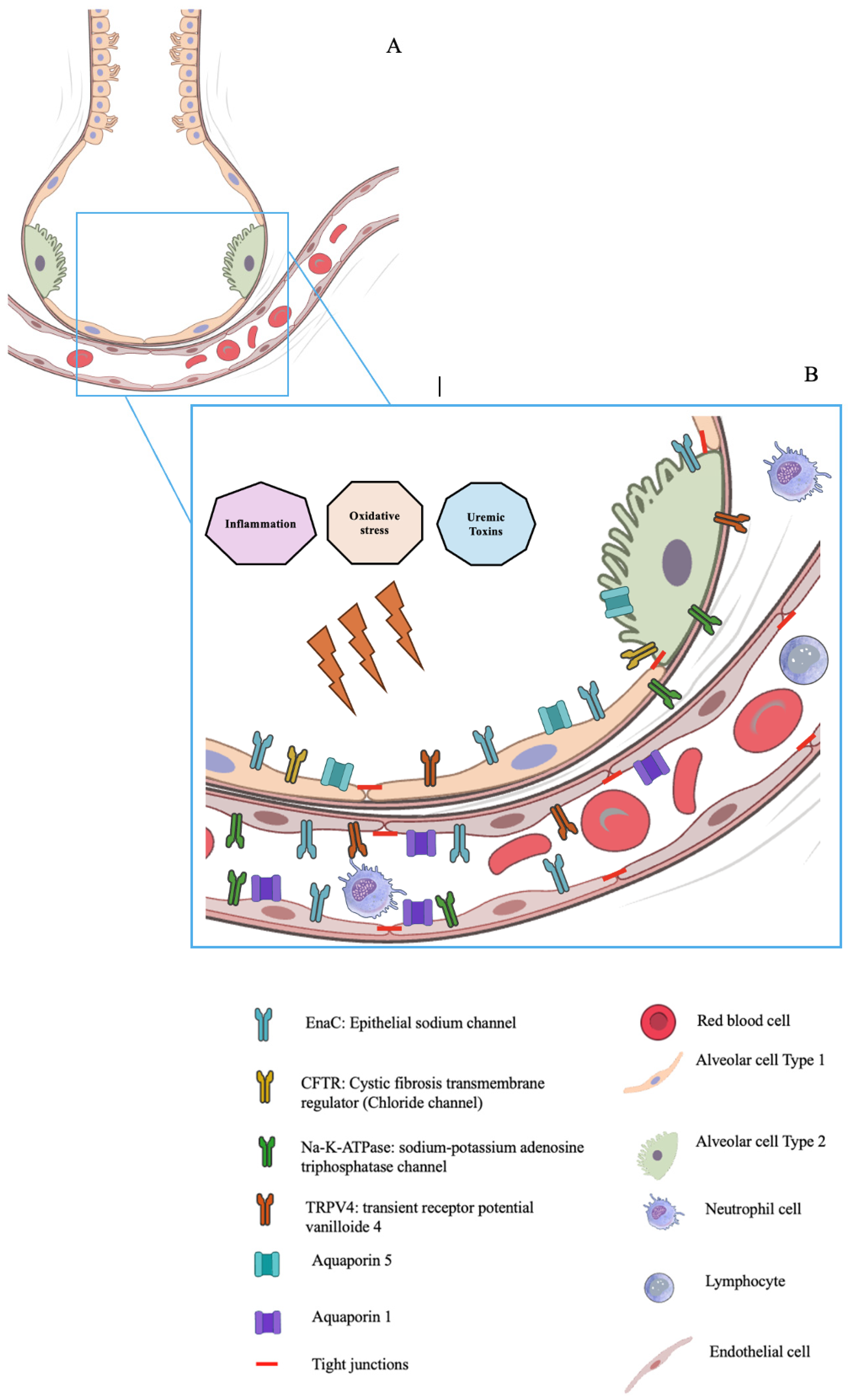
| Molecular Structure | Location | Function | Changes Due to UTs | Experimental Conditions | Experimental Model | Study |
|---|---|---|---|---|---|---|
| ENaC | Apical membrane of the alveolar epithelial cell | Participating in creating a sodium concentration gradient | Reduced | AKI | Rats | Rabb et al. [23] |
| Reduced | AKI | Rats | Ma et al. [47] | |||
| Na+/K+/ATPase | Basolateral membrane of the alveolar epithelial cell | Participating in creating a sodium concentration gradient | Reduced | AKI | Rats | Rabb et al. [23] |
| AQP5 | Apical membrane of the alveolar epithelial cell | Facilitate water movement through the alveolar epithelial cell | Reduced | AKI | Rats | Rabb et al. [23] |
| Reduced | AKI | Rats | Yabuuchi et al. [62] | |||
| AQP1 | Membrane of the pulmonary endothelial cell | Facilitate water movement through the pulmonary endothelial cell | Unchanged | AKI | Rats | Rabb et al. [23] |
| Reduced | AKI | Rats | Ma et al. [47] | |||
| TJ | Linking between cells | Regulate the iter-cellular fluid movements | Reduced | Uremic toxins | Cells | Vaziri et al. [57] |
Disclaimer/Publisher’s Note: The statements, opinions and data contained in all publications are solely those of the individual author(s) and contributor(s) and not of MDPI and/or the editor(s). MDPI and/or the editor(s) disclaim responsibility for any injury to people or property resulting from any ideas, methods, instructions or products referred to in the content. |
© 2026 by the authors. Licensee MDPI, Basel, Switzerland. This article is an open access article distributed under the terms and conditions of the Creative Commons Attribution (CC BY) license.
Share and Cite
Kaysi, S.; Taghavi, M.; El Mourabi, A.; Antoine, M.-H.; De Prez, E.; Nortier, J. Uremic Toxins and the Lung Alveolar Capillary Barrier: A Narrative Review. Toxins 2026, 18, 126. https://doi.org/10.3390/toxins18030126
Kaysi S, Taghavi M, El Mourabi A, Antoine M-H, De Prez E, Nortier J. Uremic Toxins and the Lung Alveolar Capillary Barrier: A Narrative Review. Toxins. 2026; 18(3):126. https://doi.org/10.3390/toxins18030126
Chicago/Turabian StyleKaysi, Saleh, Maxime Taghavi, Alissa El Mourabi, Marie-Hélène Antoine, Eric De Prez, and Joëlle Nortier. 2026. "Uremic Toxins and the Lung Alveolar Capillary Barrier: A Narrative Review" Toxins 18, no. 3: 126. https://doi.org/10.3390/toxins18030126
APA StyleKaysi, S., Taghavi, M., El Mourabi, A., Antoine, M.-H., De Prez, E., & Nortier, J. (2026). Uremic Toxins and the Lung Alveolar Capillary Barrier: A Narrative Review. Toxins, 18(3), 126. https://doi.org/10.3390/toxins18030126