Article

In Vitro Screening of the Antifungal and Antimycotoxin Effects of a Stilbenoids-Riche Grapevine Cane Extract on Fusarium graminearum, Aspergillus flavus and Penicillium expansum

1
Qualisud, Univ Montpellier, CIRAD, Institut Agro, IRD, Avignon Univ, Univ de La Réunion, 4 CIRAD, UMR QualiSud, F-34398 Montpellier, France
2
Antofénol, Place Eugène Bataillon, Bldg. 15, 3rd Floor, F-34095 Montpellier, France
3
UMR IATE, University of Montpellier, CIRAD, INRAE, Institut Agro, F-34398 Montpellier, France
4
CIRAD, UMR Qualisud, F-34398 Montpellier, France
*
Authors to whom correspondence should be addressed.
These authors contributed equally to this work.
Toxins 2025, 17(9), 454; https://doi.org/10.3390/toxins17090454
Submission received: 23 July 2025 / Revised: 27 August 2025 / Accepted: 2 September 2025 / Published: 9 September 2025
(This article belongs to the Special Issue Mycotoxins in Food Safety: Challenges and Biocontrol Strategies)

Abstract

Grapevine cane, an abundant viticultural by-product, contains high levels of stilbenoids and therefore holds promise as a natural antifugal and antimycotoxigenic agent. Produced by a microwave-assisted hydro-ethanolic extraction process, the grapevine cane extract (GCE) was tested for its activity against three mycotoxigenic fungi F. graminearum, A. flavus, and P. expansum. Dose-response assays were performed, based on radial growth and inhibition of specific mycotoxin production. For all fungi, growth inhibition IC50 values clustered between 1.0 and 5.0 g/L, while for specific toxin production, IC50 were lower (≈0.5 g/L) except for patulin, which increased in a dose-dependent manner in the presence of the extract. Specific experiments were designed to highlight the effect of the extracts at various stages of the fungal life cycle (e.g., spore germination, early mycelium, and established colonies). F. graminearum spores’ germination was strongly inhibited (5.0 to 15 g/L), while for other fungi, germination was only delayed. Interestingly, antifungal and especially antimycotoxigenic effects were shown to be persistent after exposure.
Key Contribution: A stilbenoid-rich extract from grapevine cane simultaneously inhibited fungal growth and suppressed DON- and AFB1-specific production by mycotoxigenic fungi, except for patulin, for which the opposite effect was observed. F. graminearum was the most sensitive, particularly at the germination stage, while A. flavus and P. expansum required higher doses to achieve comparable growth inhibition. Toxin inhibition was independent of the fungal developmental stage and persisted after extract removal.

1. Introduction

Filamentous fungi, including those producing mycotoxins, rank among the most significant threats to global crop production from both health and economic perspectives [1,2]. Some of the most reported species responsible for infecting crops and producing mycotoxins are Fusarium graminearum with type B trichothecenes (TCTBs), Aspergillus flavus with aflatoxins (AFLAs), and Penicillium expansum with patulin (PAT) [3,4,5]. TCTBs include deoxynivalenol (DON) and its acetylated forms 3-acetyl-4-deoxynivalenol and 15-acetyl-4-deoxynivalenol (3-ADON and 15-ADON) and AFLAs include aflatoxin B1 (AFB1)–B2 (AFB2)–G1 (AFG1)–G2 (AFG2). These mycotoxins encompass a broad range of toxic secondary metabolites known for their potential genotoxic and carcinogenic effects on human health [6,7]. Numerous factors contribute to fungal development and mycotoxin accumulation in crops—both before harvest and during storage or processing—such as climatic conditions, temperature, water activity (aw), nutrient availability, insect infestation, specific microorganism interactions, harvest timing, drying methods or inadequate storage practices [8,9,10]. Mycotoxin regulations for agricultural products have been estalished in many countries worldwide, with strict limits set specifically for each agricultural commodity [11,12,13]. Despite intensified fungal surveillance and mycotoxins control, the Food and Agriculture Organization (FAO) estimated that at least 25% of global crops contain mycotoxins above detectable levels, resulting in an annual loss of approximately one billion metric tons of agricultural products. In addition, according to the Rapid Alert System for Food and Feed (RASFF), mycotoxins are consistently identified as one of the top ten hazards in food products, particularly in cereals and nuts. The damage caused by fungi in the field and after harvest, combined with mycotoxin contaminations, significantly impacts agricultural production and the economy due to the rejection of contaminated commodities [14,15]. Several strategies have been developed to control fungal and mycotoxins contamination at both, pre-harvest and post-harvest stages [16]. Pre-harvest approaches focus on preventing fungal development and mycotoxins production in the field, while post-harvest approaches focus on monitoring toxins presence at key control points and mycotoxin decontamination [17,18,19]. Thus, the use of fungicides remains central to prevent the proliferation of mycotoxin-producing fungi in crops. However, even used in accordance with optimal agronomic practices, these plant protection products may negatively affect human health and the environment, and residues may persist on crops. In 2009, the authorization of all the active substances used in European Union for plant protection was reevaluated, leading to national action plans in order to reduce the impact of pesticides. Along with their detrimental effects, extensive use of pesticides have favoured the emergence of drug-resistant fungal strains [20].
In this context, the search for new active substances and eco-friendly fungicides has become a priority and the development of biopesticides appears to be an encouraging approach. Biopesticides solutions include macro-, microorganism, natural substances, and a chemical mediator [21]. Altogether, combined to each other, and associated to good agricultural practices, biopesticides represent a suitable alternative for crop protection. Plant-based extracts are promising due to their diverse antifungal compounds, lower toxicity to human health and the environment and their biodegradability. In addition, compared to single, purified molecules, natural plant extracts may offer synergistic benefits, as multiple active compounds can target various fungal physiological pathways, thereby boosting efficacy and limiting the emergence of resistant strains [22,23,24]. Beyond their direct phytochemical effects, plant-derived residues might be used as a substrate to grow biocontrol microorganisms that produces antifungal enzymes and detoxifies mycotoxins (AFB1, DON, fumonisins) or be co-applied with these microbes in an integrated biological control strategy, where plant phenolic compounds enhance that enzymatic activity [25,26]. Despite this potential, only a limited number of plant-derived extracts are commercially available. Fortunately, many plant species remain unscreened for antifungal properties, suggesting promising opportunities for discovering new molecules. Agricultural and forest byproducts represent an abundant source of raw materials with a significant potential for antifungal and antimycotoxin activities against toxigenic fungi [27,28].
Among agricultural residues, grapevine wood contains a wide variety of phenolic compounds including phenolic acids, tannins, flavonoids, and stilbenoids [29,30,31]. This large group of secondary metabolites produced via the phenylpropanoid pathways includes stilbenes which are especially abundant in the Vitaceae family (such as grapevines) and have been described as antifungal in bibliography [32,33]. Previous studies suggest that hydroxystilbene monomers (trans-resveratrol, piceatannol) and oligomers (trans-ε-viniferin, hopeaphenol, isohopeaphenol, and vitisin B) could play a role in limiting the fungal development [34,35,36]. These stilbenes are synthesized by Vitis vinifera in response to different stresses and can therefore be constitutively found in the canes and other woody tissues [37,38]. Traditionally, once grape leaves fall at the beginning of the dormant season, canes are pruned and either left on the soil or burned, representing an underutilized source of bioactive compounds. Therefore, with 2–5 tons of grapevine pruning residues per hectare per year, grapevine canes represent a large source of raw material in Europe and all around the world [39,40].
In the present study, we investigated the antifungal and antimycotoxigenic activity of GCE obtained via a hydro-ethanolic extraction process combined with microwave on three mycotoxigenic fungi, F. graminearum, A. flavus, and P. expansum. After characterization of the phenolic profile, the optimal concentration of extract required to inhibit fungal growth and supress the production of TCTB, AFLA, and PAT was evaluated. Then, we compared fungal sensitivity at early versus later physiological stages of development. Spores or established mycelial colonies were exposed to various concentrations of GCE, after which growth and mycotoxin production were monitored. Additionally, we tested whether the GCE acted as a germination or a mycelial inhibitor by transferring spores or colonies from treated to untreated media and checking for subsequent development. Therefore, the objectives of this work were to (1) demonstrate the extract’s biological activity, (2) elucidate the link between physiological stage and fungal sensitivity, and (3) confirm the promising potential of GCE as a natural antifungal and antimycotoxigenic agent.

2. Results

2.1. Grapevine Cane Extract as a Growth and Toxin Inhibitor of Mycotoxinogenic Fungi

The antifungal activity of GCE on fungal growth and mycotoxins production was evaluated by observing morphological changes, measuring the colony surface area, and dosing of mycotoxins in media supplemented with increasing extract concentrations (Figure 1).
For all fungi, hyphal growth and specific mycotoxin production on GCE-treated media were compared to both a negative control (no treatment) and a positive control (chemical fungicide at 1 g/L). In the presence of the chemical fungicide, fungal development and thus mycotoxins production were completely inhibited for all species and throughout the experimental period.

2.1.1. Growth and Mycotoxin Inhibition by GCE (Fusarium graminearum)

GCE induced a significant, dose-dependent inhibition, with a visible shift from red to white observed at day 6 in F. graminearum (Figure 1 and Figure S1).
At 0.5 g/L and 1.0 g/L, growth was delayed, resulting in a surface area 6% and 28% smaller than the control, respectively, on day 6. At 2.5 g/L, colony expansion was substantially decreased (−55% on day 6), and full plate coverage was delayed by three days. Consistently, the 5.0 g/L treatment fully suppressed F. graminearum development, with no visible colony formation (Figure 1 and Figure S1). Growth rate calculations between day 3 and day 6 confirmed this pattern: 13 cm2/day at 1.0 g/L and 8.5 cm2/day at 2.5 g/L, compared to 16.0 cm2/day for the untreated control.
Figure 1 also presents the specific DON production (µg/cm2 of mycelium) by F. graminearum. On day 3, all conditions exhibited a DON production < LOQ = 0.9 µg/cm2. By day 6, DON accumulation in the untreated control increased substantially to 3.7 µg/cm2 of mycelium. In the presence of GCE treatments, DON was systematically below the LOQ. Table S1 indicates that 15-ADON and 3-ADON followed a similar concentration-dependent suppression pattern, confirming a broad-spectrum inhibitory effect on the trichothecene.

2.1.2. Growth and Mycotoxin Inhibition by GCE (Aspergillus flavus)

The growth and aflatoxin B1 (AFB1) production of A. flavus were assessed over a 9-day incubation period. Untreated colonies displayed a characteristic green-yellowish pigmentation, reaching a surface area of approximately 47 cm2 and producing 2.5 µg/cm2 of AFB1 (Figure 1 and Figure S2). In GCE-treated cultures, pigmentation and colony morphology shifted in a dose-dependent manner. At 0.5 g/L, 1.0 g/L, and 2.5 g/L, colonies developed a circular aerial mycelium layer that was absent at lower concentrations, while at 5.0 g/L and 15.0 g/L, they became highly aerial and formed dome-shaped structures (Figure 1).
GCE concentrations of 0.5 g/L and 1.0 g/L had limited impact (~10% inhibition). At 2.5 g/L, 5.0 g/L, and 15.0 g/L, the colony surface areas were decreased by 48% and up to 78%. Higher concentrations (20.0 and 30.0 g/L) were tested but did not further enhance the inhibitory effect Growth rates were consistent with these measures: untreated colonies expanded at 7.1 cm2/day from day 6 to day 9 similar to colonies exposed to GCE at 0.5 g/L and 1.0 g/L, while rates decreased to 2.0 cm2/day and 1.7 cm2/day at 5.0 g/L and 15.0 g/L, respectively. Although GCE significantly delayed growth at its highest concentrations, complete inhibition was never observed.
Regarding mycotoxin production, on day 6, the untreated condition exhibited minimal AFB1 levels (0.003 µg/cm2), while low-to-moderate GCE concentrations (0.5 g/L–2.5 g/L) yielded higher AFB1 levels (0.1–0.3 µg/cm2), thought not significantly different from the control (Figure 1, Table S1). No AFB1 was detected at 5.0 g/L and 15.0 g/L, despite colony development. By day 9, while AFB1 accumulation in the control stabilized to 2.5 µg/cm2, all GCE supplemented cultures remained significantly lower (≤0.1 µg/cm2), with inhibition rates of ~97% (0.5 g/L), 98% (1.0 g/L), and 94% (2.5 g/L) (Figure 1). At 5.0 g/L and 15.0 g/L, AFB1 was not detectable. A. flavus predominantly synthesized AFB1, while other aflatoxins (AFB2, AFG1, AFG2) may have been detected at substantially lower concentrations, and when present, they exhibited a similar inhibitory response to that observed for AFB1 (Table S2) [41,42,43].

2.1.3. Growth and Mycotoxin Inhibition by GCE (Penicillium expansum)

The effect of GCE on P. expansum was monitored over a 9-day period. Under untreated conditions, colonies reached 22 cm2 by day 9. Treatment with the GCE resulted in a clear, dose-dependent inhibition of growth. At 0.5 g/L and 1.0 g/L, colony surface areas were reduced to 17.5 cm2 and 13.5 cm2 (21% and 40% inhibition, respectively). Higher concentrations (2.5, 5.0, and 15.0 g/L) resulted in more pronounced reductions of 69%, 92%, and 81%, respectively (Figure 1 and Figure S3). No additional inhibition beyond 15.0 g/L was observed. Growth rate analysis (day 3–9) reflected this pattern: 3.0 cm2/day for both the control and the 0.5 g/L treated condition, followed by decreases to 2.3, 1.2, 0.1, 0.9 cm2/day at 1.0, 2.5, 5.0, and 15.0 g/L, respectively. Overall, GCE induced between 21% and 91% growth inhibition, although complete growth suppression was not achieved under the tested conditions.
Patulin production was also influenced by GCE, as shown in Figure 1 and Table S1. At day 6, control cultures produced 2.0 µg/cm2 of patulin. Lower extract concentrations (0.5–1.0 g L) yielded similar or slightly elevated values (2.2, 2.4 µg/cm; +10% to +19%). However, higher concentrations (2.5, 5.0, and 15.0 g/L) resulted in significantly increased patulin levels from 159% to 329%. By day 9, patulin levels in the control stabilized to 1.4 µg/cm2. Lower GCE treatments (0.5 g/L, 1.0 g/L) kept specific patulin production relatively close to the control (1.4 and 1.6 µg/cm2), while higher concentrations (2.5, 5.0, 15.0 g/L) led to significant increases, from 237% to 587% (Figure 1, Table S1).

2.2. Inhibition Through Fungal Life Cycle: From Spores to Expanding Mycelium

To assess whether GCE impacts fungal development and mycotoxin production at different life-cycle stages, three complementary approaches were employed. (1) First, spores (macroconidia—F. graminearum, or conidia–A. flavus and P. expansum) were inoculated on porous membranes placed over culture media supplemented, or not, with GCE at various concentrations, to determine whether germination was blocked or delayed (Figure 2, Figure 3 and Figure 4A). (2) Next, to determine whether early exposure to GCE exerted a lasting effect on fungal growth and toxin production, or if normal development resumed once GCE was removed, germinated or non-germinated spores were transferred from treated to untreated media (Figure 2, Figure 3 and Figure 4B). (3) Finally, the capacity of GCE to inhibit active mycelial growth and ongoing toxin production, and thus exert a potential curative effect, was evaluated. Spores were first germinated and microscopic colonies were allowed to develop on untreated media before being transferred to GCE-supplemented plates (Figure 2, Figure 3 and Figure 4C). Together, these approaches provided insight into whether the antifungal and antimycotoxigenic activities of GCE were limited to early development stages or extended to mature, actively growing colonies.

2.2.1. Fusarium graminearum: An Early-Stage Sensitive Fungi: Germination Inhibition and Remanence of Antifungal Effect
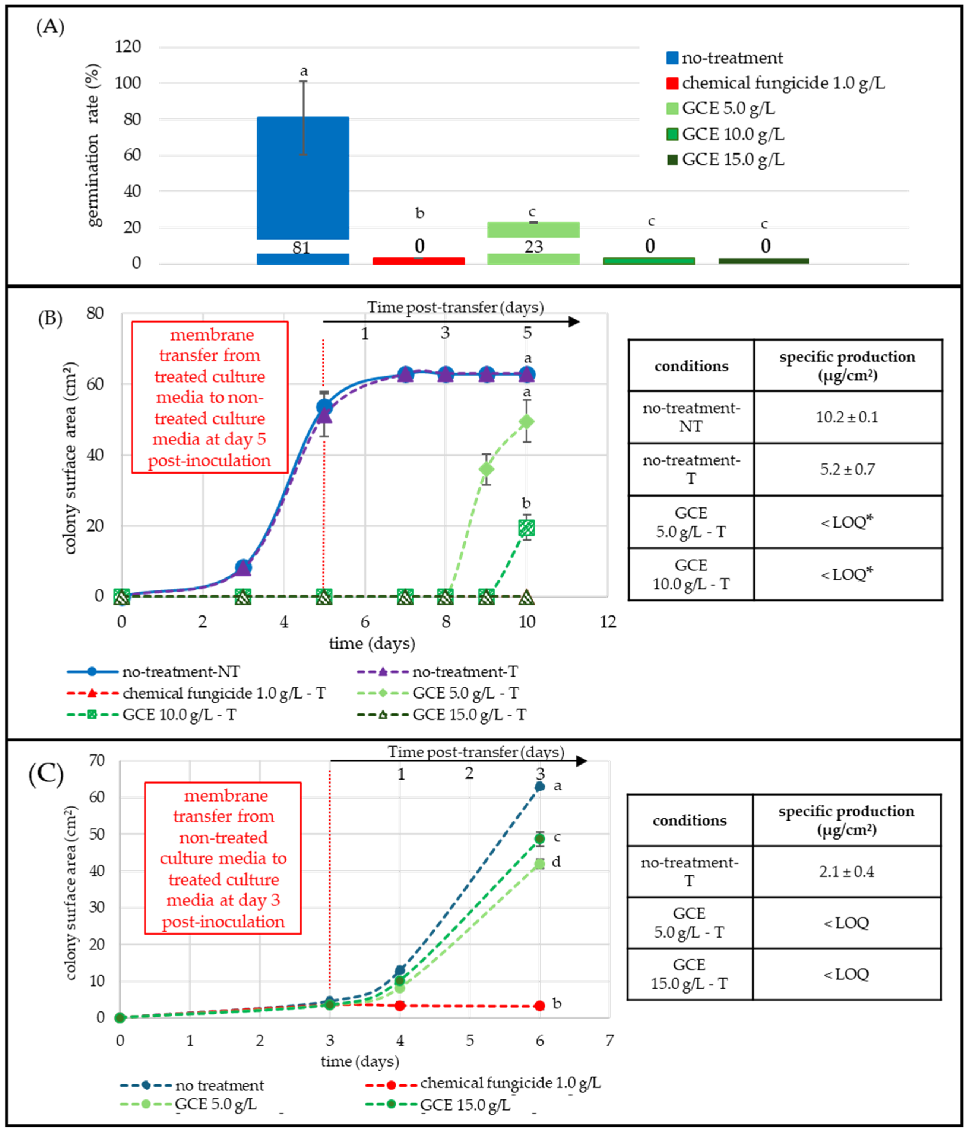
Figure 2A showed that a 12 h exposure at 25 °C to GCE (10.0 and 15.0 g/L) completely blocked the germ-tube emergence and reduced the germination rate from 81% (untreated control) to 23% at 5.0 g/L. The commercial fungicide (1.0 g/L) also completely inhibited germination.
Figure 2B quantified both growth recovery and toxin output following early exposure to GCE and subsequent transfer to fresh CYA medium. Spores were first incubated for 5 days on media containing 5.0, 10.0, and 15.0 g/L of GCE, after which the membranes were transferred to fresh non-supplemented plates (dashed vertical red line). Non-transferred (NT-blue) and transferred (T-purple, dashed) controls displayed identical growth curves, confirming that transfer did not limit expansion (≈53.0 cm2 on day 5 and 63.0 cm2 on day 10, respectively). Colonies exposed to 5.0 and 10.0 g/L did not develop before transfer, but resumed growth afterward, reaching 49.6 cm2 and 20.0 cm2 by day 10, which represents a 21% and 68% reduction in surface area, respectively (Figure 2). The 15.0 g/L treatment fully blocked fungal development. No development was noted until 15 days.
Figure 2. Effect of GCE on F. graminearum germination, growth, and DON-specific production (µg/cm2 of mycelium) at different developmental stages. All GCE treatments 5.0, 10.0, 15.0 g/L (green shades) were compared to non-treated (blue) and fungicide-treated (1 g/L, red) conditions. Each point represents the mean of three replicates. DON LOQ = 0.9 µg/cm2. (A) Spore germination assay: spores were inoculated on membranes placed over treated CYA medium; germination was evaluated microscopically (20×–40×) 12 h post-inoculation, (50 spores/image, three replicates). A spore was considered germinated when the germ tube exceeded twice the spore length. (B) Post-transfer assay: spores were first grown on GCE supplemented media, then transferred (T) to untreated media on day 5 (vertical red dashed line). Colony growth (cm2) and DON-specific production (µg/cm2) were monitored until day 10. (C) Mycelial-stage assay: Colonies were grown for 3 days on untreated media, then transferred to GCE-supplemented media. Growth and DON-specific production were measured until day 6. Statistical significance (p < 0.05) was assessed using the Kruskal-Wallis test, followed by Dunn’s post-hoc test. Different letters indicate significant differences in germination and growth; asterisks indicate differences in DON production. Curves at 0 are superimposed; the red series is present but coincides with other zero-valued conditions and is therefore not visible.
Toxins 17 00454 g002
Specific DON production corroborated these profiles: the untreated transferred control (T-purple, dashed) produced 5.2 ug/cm2, while early exposure to 5.0 g/L GCE reduced DON-specific production to 0.4 g/L (−92%), and 10.0 g/L kept DON below LOQ (Figure 2); 15-ADON followed the same pattern as DON, whereas 3-ADON stayed < LOQ under all conditions tested including controls (Table S3). Because the graph presents both, absolute and post-transfer timelines, colonies of equivalent physiological age can be compared. Control conditions (blue, purple) plotted on the absolute timeline had already reached their maximal diameter by the time the treated colonies began to expand. Thus, a five-day post-transferred colony (dashed—green shades) that had been inhibited by a 5.0 or 10.0 g/L GCE corresponded to an absolute age of ten days but still exhibited a five-day-old control equivalent (−3%) or smaller (−61%) surface area, respectively. DON levels from five-day post-transfer colonies were far lower (−85% inhibition) than the five-day-old control (≈50.0 cm2, 2.7 µg/cm2). Toxin production in treated culture did not catch up with untreated controls during the experiment.
Figure 2C addressed later physiological stages. Three-day-old colonies (≈3.0 cm2 in diameter), grown on non-treated medium until they reached a mycelial stage, were transferred to plates containing 5.0 or 15.0 g/L GCE, or to Imazalil-supplemented medium (1.0 g/L). The untreated, transferred control expanded rapidly to 63 cm2 by day 6, while colonies exposed to the fungicide control barely progressed (≈3 cm2). The extract slowed radial growth but did not fully stop it, with average final areas of 41.9 cm2 at 5.0 g/L (−42% vs. control) and 48.7 cm2 at 15.0 g/L (−23% vs. control) (Figure 2).
DON levels on day 6 revealed a stronger contrast between non-treated (blue) and treated (green shades) conditions. The untreated control accumulated 2.1 µg/cm2, while both extract treatments kept DON below LOQ. The same observation was made for 15-ADON and 3-ADON (Table S4).

2.2.2. Aspergillus flavus: Moderate Germination Inhibition with Sustained Mycelial Suppression and Remanence of Antifungal Effect

Figure 3A presents the impact of GCE on spore germination after 12 h. Under control conditions (no treatment, blue) 93% of spores germinated, while no germination was observed when treated with the chemical fungicide (1 g/L, red). GCE at 5.0 g/L and 10.0 g/L had minimal impact on germination, with rates of 89% and 92%, respectively. The 15 g/L concentration reduced the germination rate to 63%, only the highest dose significantly interfered with the germination of A. flavus spores.
Figure 3A tracked colony establishment following germination. A. flavus spores were incubated for 5 days on GCE-supplemented medium, after which the membranes were transferred to fresh, non-treated PDA (vertical dashed red line). Non-transferred (NT-blue) and transferred (T-purple, dashed) controls grew identically, both reaching 49.3 cm2 by day 11, indicating that the transfer itself did not restrict the growth. Colonies pre-exposed to 5.0, 10.0, and 15.0 g/L of GCE were partially inhibited, achieving final areas of 29.3 cm2 (−40% vs. control), 25.5 cm2 (−48% vs. control), and 30.5 cm2 (−38% vs. control), respectively (Figure 3). The three GCE treatments (5.0, 10.0, and 15.0 g/L) inhibited colony expansion to a similar extent, and their final surface areas did not differ significantly from one another. No fungal development was observed in the presence of the fungicide treatment.
Mycotoxin analysis revealed that early extract exposure reduced AFB1 accumulation. The transferred control (T-purple, dashed) produced 5.0 µg/cm2 of AFB1 by day 11, while colonies exposed to 5.0 and 15.0 g/L accumulated only 0.3 and 0.2 µg/cm2, respectively, corresponding to 94% and 96% reductions (Figure 3, Table S5). The 10 g/L concentration achieved a 70% reduction.
Figure 3C evaluated the response of A. flavus once colonies had reached the mycelial stage. Three-day-old colonies (≈3 cm2) grown on non-supplemented PDA were transferred (vertical dashed red line) to plates containing 5.0 or 15.0 g/L GCE, or to fungicide-supplemented media (1 g/L, dashed red). The untreated, transferred control (T, dashed blue) developed rapidly after transfer, with its area increasing from 2.8 cm2 to 39.7 cm2 within five days. Exposure to GCE slowed growth, resulting in final surface areas of 16.5 cm2 (−58%) at 5.0 g/L and 13.8 cm2 (−65%) at 15.0 g/L. The fungicide fully stopped the expansion of A. flavus colonies.
AFB1 quantification on day 8 revealed a pronounced inhibition effect. The transferred control (T, dashed blue) produced 1.4 µg/cm2, whereas colonies exposed to 5.0 g/L accumulated only 0.01 µg/cm2 (−99%), and 15.0 g/L kept AFB1 < LOQ (Figure 3, Table S6).
Figure 3. Effect of GCE on A. flavus germination, growth, and AFB1 production at different developmental stages. All GCE treatments (5.0, 10.0, 15.0 g/L; green shades) were compared to non-treated (blue) and fungicide-treated (1 g/L, red) conditions. Each point represents the mean of three replicates. AFB1 LOQ = 0.001 µg/cm2. Panel (A) Spore germination assay: spores were inoculated on membranes over GCE-treated PDA medium; germination was assessed microscopically (20×–40×) 12 h post-inoculation (50 spores/image, three replicates). Panel (B) Post-transfer assay: spores were pre-grown on extract-supplemented media and transferred (T) to untreated media on day 5 (vertical red dashed line). Colony growth (cm2) and AFB1 specific production (µg/cm2) were monitored until day 11. Panel (C) Mycelial-stage assay: colonies pre-formed for 3 days on untreated medium were transferred to GCE-supplemented media. Growth and AFB1 specific production were evaluated until day 8. Statistical significance (p < 0.05) was determined using the Kruskal-Wallis test, followed by Dunn’s post-hoc test. Different letters indicate significant differences in germination and growth; asterisks indicate differences in specific AFB1 production.
Toxins 17 00454 g003

2.2.3. Penicillium expansum: Moderate Germination Inhibition with Strong and Persistent Mycelial Suppression
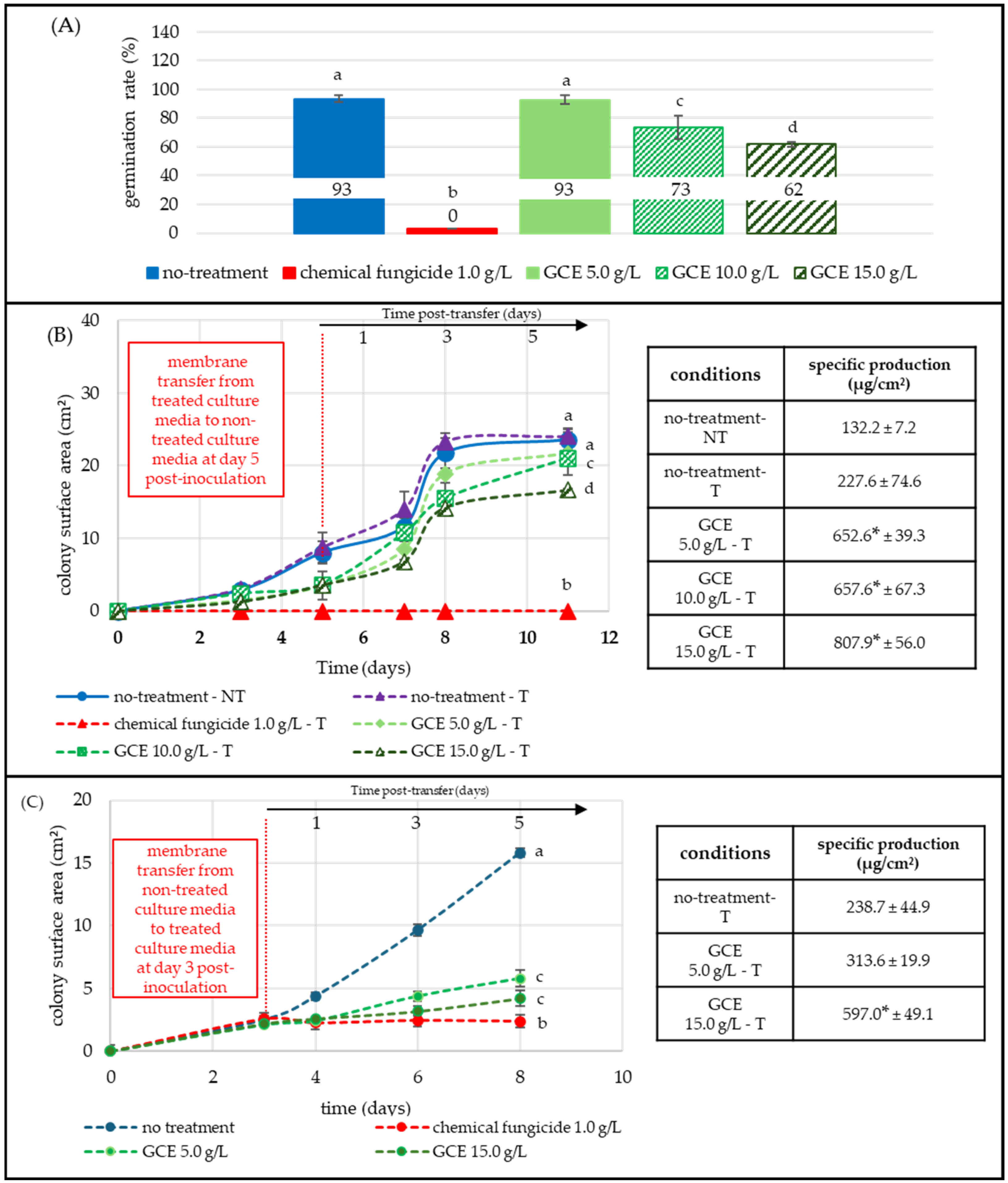
Figure 4A illustrates the P. expansum germination rate in the presence of GCE after 12 h of incubation at 25 °C. Under control condition, 93% of spores germinated, while the chemical fungicide (1 g/L, red) condition showed no germination. GCE at 5.0 g/L did not significantly affect the germination rate, while at 10.0 g/L and 15.0 g/L, germination decreased to 73% and 62%, respectively.
Figure 4. Effect of GCE on P. expansum germination, growth, and PAT production at different developmental stages. All GCE treatments 5.0, 10.0, 15.0 g/L (green shades) were compared to non-treated (blue) and fungicide-treated (1 g/L, red) conditions. Each point represents the mean of three replicates. PAT LOQ = 0.08 × 10⁻³ µg/cm2. Panel (A) Spore germination assay: spores were inoculated on membranes placed over treated PDA medium; germination was assessed microscopically (20×–40×) 12 h post-inoculation (50 spores/image, three replicates). Panel (B) Post-transfer assay: spores were first grown on extract-supplemented media and transferred (T) to untreated media on day 5 (vertical red dashed line). Colony growth (cm2) and PAT specific production (µg/cm2) were monitored until day 11. Panel (C) Mycelial-stage assay: 3-day-old colonies grown on untreated culture media were transferred to GCE-supplemented media; colony surface and patulin specific production were evaluated up to day 8. Statistical significance (p < 0.05) was determined using the Kruskal-Wallis test, followed by Dunn’s post-hoc test. Significant differences in germination and growth are shown with letters; significance in patulin specific production is indicated with an asterisk.
Toxins 17 00454 g004
Figure 4B followed colony development after five-day exposure to 5.0, 10.0, and 15.0 g/L GCE and subsequent transfer (vertical dashed red line) to untreated, fresh PDA medium. Colony growth and mycotoxin production were monitored until day 11. Non-transferred (NT-blue) and transferred (T-purple, dashed) controls expanded identically, both reaching ≈ 24 cm2 by day 11, confirming that the transfer step itself did not restrict growth. Pre-exposure to 5.0, 10.0, and 15.0 g/L GCE reduced final colony size to 21.7 cm2 (−10%), 21.0 cm 2 (−12%), and 16.6 cm2 (−31%), respectively (Figure 4).
In contrast, PAT-specific production increased in the presence of GCE. The transferred control (T-purple, dashed) produced 227.6 µg/cm2 of PAT, while the specific production in colonies initially exposed to GCE increased to 652.6 µg/cm2 (+65%) at 5.0 g/L, 657.6 µg/cm2 (+65%) at 10.0 g/L, and 807.9 µg/cm2 (+72%) at 15.0 g/L (Figure 4, Table S7).
Figure 4C assessed P. expansum growth and PAT production from colonies that had already reached a mycelial stage. Three-day-old colonies (≈2.5 cm2) grown on non-treated media were transferred (vertical dashed red line) to GCE-supplemented media, or to fungicide-supplemented medium. The untreated transferred control (T-purple, dashed) expanded to 15.8 cm2 by day 8, whereas 5.0 g/L and 15.0 g/L limited fungal growth to 5.8 cm2 (−63%) and 4.2 cm2 (73%), respectively. The fungicide blocked the fungal development.
Specific PAT production of the non-treated control reached 238.7 µg/cm2 after 8 days. In the presence of GCE, PAT-specific production increased to 313.6 µg/cm2 (+24%) at 5.0 g/L and 597.0 (+60%) at 15.0 g/L (Figure 4, Table S8).

3. Discussion

The current study shows that GCE, obtained by the hydro-ethanolic extraction process combined with microwaves treatment, affected both growth and mycotoxin production in all three targeted fungi, F. graminearum (BRFM 1967 strain), A. flavus (NRRL62477, E73 strain), and P. expansum (NRRL 35695 strain), but displayed species-specific patterns. We did not perform viability assays or determine MIC/MFC; results are therefore reported as endpoint-specific inhibition under the tested conditions, without any claim of lethality. Radial growth IC50 values clustered between 1.0 and 5.0 g/L for all models (Figure 1; Table 1). While GCE exhibited a pronounced inhibitory effect against F. graminearum, fully stopping growth at concentrations ranging from 2.5 g/L to 5.0 g/L, it also showed complete and remanent inhibition of germination at 15.0 g/L (Figure 2B; Table 1). No total inhibition of germination was observed for A. flavus and P. expansum, and mycelial growth was not abolished for any species under the tested conditions at the mycelial state (Figure 1; Table 1). Complete growth inhibition was not achieved, even at 30.0 g/L; however, a concentration of 15.0 g/L still suppressed growth by approximately 78% and 92%, for P. expansum and A. flavus, respectively, demonstrating a strong inhibitory effect. These concentrations were of the same order of magnitude as those reported for other aqueous plant extracts. For example, an aqueous maritime-pine sawdust extract and GCE suppressed F. graminearum growth by 60–89% at 3.7 g/L and by 50% at 11.3 g/L, respectively [27]. Other studies in the bibliography confirmed that similarly high extract doses are needed to inhibit these fungi: 7.5 g/L of Adenocalymma alliaceum extract reduced A. flavus growth by 51%, and 30.0 g/L of Piper nigrum extract suppressed P. expansum growth by 60% [44,45].
In addition to its antifungal effects, GCE significantly impacted mycotoxin production in all three fungi studied. DON and its acetylated derivatives produced by F. graminearum were strongly suppressed in a dose-dependent manner. At subinhibitory concentration (0.5 g/L), DON production decreased by 88% and dropped below 1 µg/cm2 at 1 g/L, even though mycelial growth persisted up to 5 g/L (Figure 1; Table 1). This result aligns with previous studies reporting TCTB inhibition by polyphenol-rich extracts, like grapevine derivatives (Table 2) [28,29,30,31,32,33,34,35,36,37,38,39,40,41,42,43,44,45,46]. Similarly, AFB1 levels in A. flavus dropped by ≥95% at doses that reduced radial growth by only 10% (Figure 1; Table 1). On day 6, low-to-moderate extract concentrations (0.5–2.5 g/L) transiently increased AFB1 levels, reaching a maximum at 1.0 g/L. This stimulatory effect may reflect stress-induced overactivation of the aflatoxin biosynthetic pathway. In contrast, the response of P. expansum to GCE differed markedly with a dose-dependent increase in patulin production, reaching a six-fold rise at 5.0 g/L despite a 91% inhibition of fungal growth (Figure 1; Table 1). This enhancement may also result from stress-induced activation of toxin biosynthesis. Across species, GCE inhibited mycotoxin production at lower concentrations than those required to inhibit growth, revealing a complex and species-specific dose-response pattern (Figure 1; Table 1).
F. graminearum was the most impacted fungus during the germination stage. In Figure 2A, the extract inhibited spore germination by 72–100% at moderate (5.0 g/L) and high (15.0 g/L) concentrations, suggesting interference with one or more early morphogenetic steps. In contrast, germination was only moderately inhibited at the highest concentrations of GCE for A. flavus and P. expansum, with 32% and 33% lower germination rate, respectively (Figure 3A and Figure 4A). These results were consistent with reports showing a global but species-dependent anti-germinative activity of polyphenols [28,29,30,31,32,33,34,35,36,37,38,39,40,41,42,43,44,45,46,47,48,49].
After the spores were transferred from treated to fresh, untreated medium (Figure 2B and Figure 3B), the expansion of F. graminearum and A. flavus colonies was either delayed or slowed down, while specific DON and AFB1 levels dropped by 92–100%. F. graminearum colonies previously exposed to 5.0 g/L GCE eventually recovered, reaching a surface area comparable to the 5-day, non-treated control. This confirms a concentration-dependent yet persistent antifungal effect, extending beyond direct contact between the fungus and GCE. The sustained reduction in both biomass, DON and AFB1, indicates a measurable remanent effect. At 15.0 g/L, no spore germination was observed, and F. graminearum spores did not recover after transfer to GCE-free medium over five days, indicating a strong, sustained inhibitory effect under the tested conditions. Further experiments are mandatory to further confirm any potential sporicidal effects. A. flavus growth inhibition was maximal at 5.0 g/L, higher doses (10.0 and 15.0 g/L) produced no additional inhibition, suggesting the maximal inhibitory threshold had already been reached. P. expansum responded differently. In Figure 4B, patulin production increased, increasing from 5.0 to 15.0 g/L despite progressive growth suppression, suggesting that the extract may have induced metabolic stress that triggered the patulin biosynthetic pathway. Growth resumed slowly after transfer to non-treated medium, yet patulin levels remained elevated, underscoring a dissociation between radial growth and toxin output.
When the GCE extract was applied to pre-grown mycelia, the impact on growth was more limited (Figure 2C, Figure 3C and Figure 4C). F. graminearum, which was sensitive during germination, became comparatively resilient once a mycelial colony was established, exhibiting only 22% growth inhibition, while A. flavus showed the opposite pattern: little inhibition during germination but a 62% reduction at the mycelial stage acting mainly as a growth inhibitor. DON and AFB1 remained undetectable at 5.0 and 15.0 g/L, respectively, showing that the extract permanently inhibits TCTB and AFLA biosynthesis even without complete growth inhibition. These results confirmed the extract’s ability to disrupt secondary metabolism even in mature colonies.
In Figure 4C, patulin continued to increase even when P. expansum colony development was limited, highlighting a clear decoupling of growth and toxin-specific production. Although P. expansum was more resistant than F. graminearum at the germination stage, it proved to be the most sensitive species once mycelia had formed, displaying a 69% inhibition of radial growth at 15.0 g/L.
Taken together, these stage-specific experiments emphasized both fungal-specific differences and common patterns. F. graminearum exhibited a tightly linked decline in growth and DON production, with DON dropping below the limit of quantification at doses that still allowed for some radial expansion. A. flavus responded more complexly: moderate GCE concentrations partially slowed growth, whereas higher concentrations completely suppressed AFB1. In contrast, P. expansum showed the opposite pattern, patulin levels rose sharply under extract-induced stress even as growth was strongly inhibited. Despite species-specific responses, two similarities emerged. First, intermediate extract doses completely blocked toxin formation in both F. graminearum and A. flavus. Second, growth and toxin suppression persisted after the extract was removed, showing that the effect was not fully reversible. The pronounced uncoupling of radial growth and toxin inhibition in these fungi suggests that GCE might primarily target secondary metabolism regulation, likely by down-regulating key genes such as TRI5 (F. graminearum) and aflR (A. flavus), rather than merely limiting biomass [50,51]. P. expansum, in contrast, strongly increased patulin accumulation when stressed by GCE, indicating an alternative stress-response mechanism that activates toxin biosynthesis, consistent with reports of patulin upregulation under sub-lethal oxidative stress [52].
The GCE used in this study was obtained from grapevine cane (Vitis vinifera). The biochemical composition of GCE has been described in detail over many years, consistently showing a high content of phenolic compounds, including stilbenes [53,54,55,56]. Trans-resveratrol, the best-known stilbene and precursor of many others, is the predominant monomer in this extract [32,33] (Table S9). In plants, trans-resveratrol and piceatannol can be further oligomerized, glycolyzed, methylated, isomerized, or isoprenylated, generating a wide spectrum of derivatives [56]. The same molecular diversity was evident in our extract (Table S9). Our microwave-assisted hydro-ethanolic extract contained 210 g of stilbenes per kg of GCE powder. This high proportion reflects the extraction process: microwaves hydro-ethanolic extraction followed by evaporation, concentration, and drying concentrates of the stilbenes in the final powder extract. Because composition influences bioactivity, the main constituents of the extract were summarized in Table 2 and Table S9.
Stilbenes are well-known phytoalexins that accumulate in grapevine tissues during fungal attack [38,39,40,41,42,43,44,45,46,47,48,49,50,51,52,53]. Pezet et al. (2003) demonstrated that their accumulation increases in lignified tissues infected by Plasmopara viticola, the causal agent of downy mildew [57]. This defensive response has since been confirmed in several studies, including those involving other ascomycete pathogens [34,35,36,37,38] (Table 3). In addition, stilbenes identified in our extract, such as trans-resveratrol, piceatannol, trans-ε-viniferin, and vitisin B, have antifungal and antimycotoxigenic activities that matched those reported for purified compounds at an equivalent concentration (Table 3). For example, trans-resveratrol, trans-ε-viniferin, and piceatannol inhibit Plasmopora viticola and Botrytis cinierea at ≈60–120 µM and ≈60–400 µM, respectively, while our extract contains 72.4–233.4 µM of resveratrol and 6.4–39.7 µM of piceatannol in the active concentration range from 1 to 5 g/L [34,35,36].
Tardif et al. (2025) compared cane, wood, and root grapevine extracts from several grapevine cultivars against F. graminearum; all fractions suppressed growth and TCTB production in a dose- and cultivar-dependent manner, although cane extracts were the least active [50]. In our study, the concentrations of trans-resveratrol, trans-ε-viniferin, and piceatannol delivered by a 2.5 g/L to 5.0 g/L cane extract matched those concentrations and abolished TCTB production while strongly inhibiting fungal growth. In A. flavus, trans-resveratrol and related phenolics down-regulate aflatoxin-cluster genes, yet this inhibition requires higher doses than those effective against F. graminearum. Similarly, stronger concentrations are needed to slow A. flavus mycelial growth, a difference that might be explained by its thicker, more hydrophobic conidial wall [58,59].
Consistent with these findings, the highest doses of GCE lowered AFB1 by 80% and strongly inhibited colony expansion. In contrast, P. expansum was only moderately inhibited, showing limited growth reduction and a dose-dependent increase in patulin, as previously reported [52]. Across all three fungi, toxin suppression occurred at extract doses below those required for substantial growth inhibition, a pattern documented for polyphenols [60,61,62]. Therefore, stilbenoids might primarily target the secondary-metabolite pathway and gene clusters (e.g., TRI5, aflR) rather than impacting cellular and metabolic processes involved in biomass accumulation.
Due to methodological differences (strain origin, extraction technique, culture conditions) cross-study comparisons (Table 2) are limited. While prior work often focused on single phenolic molecules (e.g., trans-resveratrol), the GCE is a mixture. Not all phenolic molecules mentioned in those studies were stilbenoids (e.g., non-stilbene classes like caffeic acid and quercetin). Such variability limits direct quantitative comparisons, yet data obtained with GCE and presented in this article were consistent with prior results: phenolics compounds alone or in a mixture (plant extract) can lower mycotoxin accumulation even when growth effects are modest; growth suppression did not always mirror toxin reduction; both GCE and single phenolic compound or other grapevine extracts appear to have consistent inhibitory action.
Stilbenes such as ampelopsin A, hopeaphenol, trans-resveratrol, trans-ε-viniferin, and trans-vitisin B were reported to disorganize the plasma membrane and organelle membranes of P. viticola and B. cinerea spores, an effect confirmed in several follow-up studies [49,50,51,52,53,54,55,56,57,58,59,60,61,62,63,64]. Phan et al. (2014) further demonstrated that, in fungi, flavonoids tend to rigidify membranes, whereas stilbenes increase fluidity [65]. Given the high content of trans-ε-viniferin (a resveratrol dimer) in our extract, and the greater antifungal efficacy reported for resveratrol oligomers, as their degree of polymerization and log P increases, disrupting the fungal cell wall and plasma membrane which control nutrient uptake, defence and permeability might be one of the underlying mechanisms of action of GCE [66,67].
Once inside the cell, phenolic compounds can disrupt both primary and secondary metabolism. For example, trans-resveratrol at much lower concentrations than those present in our extract down-regulated aflaA and aflaB involved in aflatoxin synthesis in A. flavus and simultaneously inhibited mycelial development and sporulation [51]. These reports supported our observation that toxin repression occurred at doses an order of magnitude below those required for strong growth inhibition. Finally, free diffusion of phenolic compounds inside the cell can disturb oxidative-stress regulation and mitochondrial function, altering the cellular ROS balance [68,69]. Although direct data on stilbene-induced metabolic and mitochondrial dysfunction remain limited, the combined evidence of gene down-regulation, ROS scavenging and ETC inhibition for related phenolics compounds supports a similar mode of action for stilbenes present in GCE [70,71]. Studies have shown that antioxidant phenolics compounds scavenge ROS and supress aflatoxin and trichothecene biosynthesis in A. flavus and F. graminearum [72,73]. Hydrogen bonding between phenolic hydroxyls and enzymes may amplify this disruption.
The extract shows promising in vitro activity, but turning GCE into a robust phytopharmaceutical product (P.P.P) will depend, in part, on its effectiveness on fields and on future formulation strategies. The efficacy of the extract has been tested and demonstrated on fields for apple-scab (Venturia inaequalis), which has been chosen as the representative use for the registration as P.P.P, following E.U. regulation 1107/2009 [74]. In this study, GCE was used as a powder, but liquid formulations are more suitable for field use. Indeed, powder extracts are no longer of interest due to higher risk of exposure for farmers and poor water solubility of active molecules such as phenolic compounds. Formulation approaches adapted from the cosmetics and pharmaceutical sectors can enhance solubility, stability, and prevent rainfastness [75]. Indeed, the amphiphilic nature of phenolic compounds such as stilbenes, governs adsorption, aggregation, complexation, and micellization, thereby shaping antifungal efficacy. Combining plant fractions that act on complementary cellular targets, then encapsulating and formulating them, should protect the actives, concentrate them on plant surfaces, and deliver adequate doses to their sites of action. Practical solutions include nanoemulsions (including Pickering systems), nanocrystals, phytosomes, or lipid nanoparticles [76,77,78,79]. For field applications, water-based sprays are preferred, but hydrophobic leaf surfaces promote runoff, incorporating carriers into aqueous solutions that turn into hydrogels (e.g., tannin- or polysaccharide-based can improve adhesion and rainfastness).

4. Conclusions

This study demonstrated that a stilbenoid-rich extract from grapevine cane, obtained through an industrial extraction process combining microwaves, ultrasound, and vacuum is a potential antifungal and antimycotoxigenic agent. In vitro Petri dish culture showed that low concentrations (as low as 0.5 g/L) suppressed TCTB and AFLA production by approximately 90%, while growth IC50 values ranged from 1.0 to 5.0 g/L. Although patulin production increased in the presence of GCE, its hyphal development was partially inhibited. These results might indicate that growth inhibition and mycotoxin suppression are uncoupled phenomena, each likely driven by distinct modes of action. Stage-specific assays revealed that spores were more vulnerable than established mycelia for F. graminearum, and that the extract retained partial activity even after removal, confirming true inhibitor remanence. Comparison with IC50 values from the bibliography indicated that the concentrations of trans-resveratrol, piceatannol, trans-ε-viniferin and vitisin B delivered by GCE were comparable to a certain extent. The results indicate that higher GCE concentrations are associated with both stronger antifungal and antimycotoxigenic effects, plausibly reflecting the concentration and molecular structures of its stilbenes, with potential synergistic, antagonistic, or agonistic interactions among components. Moreover, previous studies have identified structural and functional fungal targets that may explain species-dependent effects. To further elucidate its mechanisms of action, comparative studies on extract fractions and purified molecules are needed to determine the specific role of stilbenes in its antifungal and antimycotoxin activity. Moreover, the mode of action should be explored through the study of GCE impact on cellular oxidative state after exposure.

5. Materials and Methods

5.1. Grapevine Cane Extract (GCE)

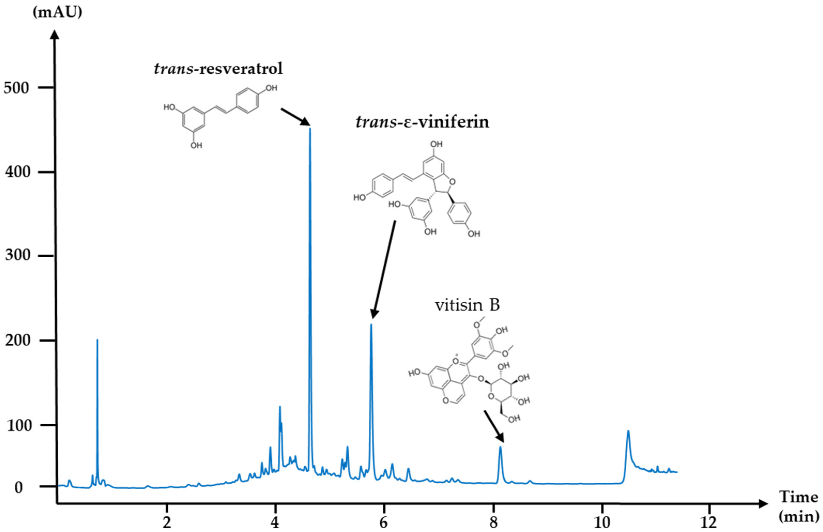
One-year old grapevine canes from Vitis vinifera, variety “Pinot noir”, were collected on the pruning season (2021) from several regions in France. Grapevine canes were cut into small segments of 10 to 20 cm, then crushed into smaller fragments (2 to 10 mm) using a conventional knives grinder equipped with sieves. The ground grapevine canes were washed for 1 h in hot water (90 °C) to clean the raw material and remove most of the sugars present in the wood; finally, the cleaning water was eliminated. The ground, humidified wood material was mixed with 30% (v/v) aqueous ethanol. Microwaves (1–100 kW) combined with ultrasound and vacuum, as described in Antofénol patent from 2020, were applied to the grapevine cane/EtOH/water mixture for 1 h, over 80 °C. The resulting liquid extract was filtered on a 20 μm filter, evaporated and spray-dried into a fine brown powder, which was packaged and stored in the dark at room temperature until further use. At the end of this process, a control was performed to check the biochemical composition of the extract by HPLC with a UV detector at 254 nm, as detailed in Table 4: HPLC mobile phase gradient and Figure 5: UHPLC-DAD chromatogram at 254 nm of the grapevine cane extract (V. vinifera) at 5 g/L solubilized in ethanol 50% (v/v). A single industrial eco-extract of Pinot Noir grapevine cane with a very reproducible phenolic composition was used for the entire experiment.
The grapevine cane extract was produced in batches, with a specific batch used for all experiments in this study. Following production, its biochemical profile was analyzed using UHPLC (Vanquish CORE, Thermo Fisher, Villebon-sur-Yvette, France) coupled with a UV-Vis DAD detector. The separation was performed on a C18 Accucore RP-MS column (100 × 4.6 mm, 2.6 µm, ThermoScientific).
The mobile phase consisted of 0.1% formic acid (v/v) (Sigma-Aldrich, Saint-Quentin-Fallavier, France, F0507-1L, CAS: 64-18-6) in water (solvent A) and 0.1% formic acid (v/v) in acetonitrile (FisherBioScientificVillebon-sur-Yvette, France, HPLC gradient grade, CAS: 75-05-8) (solvent B). A 5 g/L solution of the extract was prepared in 50% ethanol (v/v) to enhance the solubility of hydrophobic compounds, particularly stilbene oligomers. Analysis was performed at 254 nm to optimize stilbene detection. Three key compounds, trans-resveratrol, trans-ε-viniferin, and vitisin B, were specifically monitored as biomarkers of the extract.

5.2. Microorganisms, Storage Conditions, and Fungal Culture

5.2.1. Fusarium graminearum

F. graminearum strain CIRM-BRFM 1967 (CIRM, University of Aix-Marseille, Marseille, France) was selected for its documented high mycotoxin production; the same culture-collection strain (BRFM 1967) was used in [80,81]. On CYA and PDA (25 °C, 7 days), colonies grow rapidly forming a pale to red abundant aerial mycelium. Numerous macroconidia (large asexual spores) are produced: curved, multiseptated. Originally isolated from a wheat plant, it exhibits a deoxynivalenol (DON), 15-acetyldeoxynivalenol (15-ADON), and 3-acetyldeoxynivalenol (3-ADON) chemotype. The strain was maintained on potato dextrose agar (PDA; Biokar diagnostics, Beauvais, France) under paraffin oil at 4 °C. For spore production, the strain was inoculated on PDA agar and grown for 4 days. Mycelium plugs were then transferred to carboxymethyl cellulose liquid media (CMC) and incubated for 96 h in the dark, with agitation at 150 rpm, 25 °C. The CMC medium was prepared by dissolving the following components in 1 L of demineralized water: 0.5 g of magnesium sulfate heptahydrate (MgSO4.7H2O, Fluka honywell, Sigma Aldrish, Saint-Quentin-Fallavier, France, CAS: 10034-99-8), 1 g of ammonium nitrate (NH4NO3, Sigma-Aldrich, CAS: 6484-52-2), 1 g of potassium dihydrogen phosphate (Sigma-Aldrich, CAS: 7778-77-0), 1 g of yeast extract (powder) (BioKar diagnostics, Allonne, France, reference A1202 HA, 500 g), and 1 g of carboxymethyl cellulose (Sigma-Aldrich, CAS: 9004-32-4). After complete dissolution, the medium was sterilized by autoclaving at 121 °C for 15 min. The spore suspension was prepared by filtering the CMC culture medium through a 100 µm membrane (Sefar Nitrex, nylon filter, 100 µm, Dutscher, Issy-les-Moulineaux, France), followed by centrifugation to remove the supernatant. Spores were then resuspended in water. Spore density was determined with a Neubauer counting chamber.

5.2.2. Aspergillus flavus

The A. flavus E73/NRRL62477 strain, from UMR Toxalim (Toulouse, France), and initially isolated from spices was selected for its strong ability to produce aflatoxins (primarily B1) [41]. The strain was stored on PDA at 4 °C and cultured on the same medium at 25 °C for 10 days until sporulation. Spores were harvested by adding 1 mL of sterile water and scraping the surface of the culture to free the spores. The spore suspension density was further adjusted with a Neubauer counting chamber.

5.2.3. Penicillium expansum

The P. expansum NRRL 35695 strain (Northern Regional Research Laboratory, Peoria, IL, USA), from UMR Toxalim (Toulouse, Franc) primarily isolated from grape, was selected for its high patulin production [82,83]. The strain was stored on PDA at 4 °C and cultured on the media at 25 °C for 10 days until sporulation. Spores were harvested by adding 1 mL of sterile water and scraping the surface of the culture to release the spores. The spore suspension density was further adjusted with a Neubauer counting chamber.

5.3. Preparation of Culture Medium

PDA was prepared at a concentration of 39 g/L. To prepare the Czapek medium (CYA), 1 L of demineralized water is combined with 30 g sucrose (Sigma-Aldrich, for microbiology, ACS reagent, >99.0%, CAS: 57-50-1), 15 g agar (Sigma-Aldrich, Millipore, Saint-Quentin-Fallavier, France CAS: 9002-18-0), 5 g yeast extract (BioKar diagnostics, reference A1202 HA, 500 g), 1 g dipotassium hydrogen phosphate (Fluka, honywell, CAS: 7758-11-4), 0.3 g sodium nitrate (Acros organique, ThermoScientific, CAS: 7631-99-4), 0.05 g potassium chloride (Dominique Dutcher, Cat n°: P2035-500GR), and 0.05 g magnesium sulfate (Honeywell Fluka, CAS: 10034-99-8). Trace elements were prepared by dissolving 0.1 g of iron sulfate (Sigma Life science, Saint-Quentin-Fallavier, France, CAS: 7782-63-0), 0.1 g of zinc sulfate (Fluka chemika, CAS: 7446-20-0), and 0.05 g of copper sulfate (Sigma Life science, CAS: 7758-99-8) in 100 mL of demineralized water. Both culture mediums were sterilized by autoclaving at 120 °C for 15 min. After sterilization, 1 mL of the trace element solution was added to the CYA medium using a sterile syringe equipped with a 0.22 µm polyethersulfone (PES) filter, and the mixture was stirred to ensure homogeneity. Finally, 20 mL of PDA and CYA media were poured into Petri dishes.

5.4. In Vitro Evaluation of Antifungal Potential of GCE

5.4.1. GCE Incorporation to Culture Medium

Once the culture medium cooled down to 60 °C, it was mixed with the appropriate weighted quantities of GCE powder in falcon tubes. The mixture was then vortexed for 10 sec to ensure homogeneity. The culture medium/GCE mixture was poured into sterile Petri dishes for solidification at room temperature in sterile condition. The prepared Petri dishes were directly inoculated with the fungal strains. Inoculated plates were placed in an incubator set to 25 °C in the dark.

5.4.2. General Evaluation of Antifungal Activity

In Petri dishes, 10 µL of a spore suspension prepared at 500,000 spores/mL were inoculated. Culture media (PDA or CYA) were supplemented with the extract at 0.25–0.5–1.0–2.5–5.0–10.0–15.0 g/L (see the section on GCE incorporation to culture medium). Control conditions consisted of non-treated culture media and a chemical fungicide (Imazalil®, Janssen, France) at 1 g/L mixed with culture media. As described in previous work, fungal growth was monitored by measuring the colony surface from pictures, using ImageJ (1.54p, National Institutes of Health, Bethesda, MD, USA) through 6 to 10 days [41,42,43,44,45,46,47,48,49,50,51,52,53,54,55,56,57,58,59,60,61,62,63,64,65,66,67,68,69,70,71,72,73,74,75,76,77,78,79,80,81,82,83,84].

5.4.3. Sensitivity of the Fungal Targets at Various Physiological Stages

For these experiments, fungi were cultured on cellophane membranes (Le joint Français, Hutchinson) of 1.5 cm or 8 cm, sterilized under UV light for 30 min. Spore suspension was prepared at 500,000 spores/mL. Then, 10 µL of the spore solution was inoculated at the centre of each culture membrane. The Petri dishes were incubated at 25 °C in the dark.
Germination Inhibition
Membranes were placed on culture medium, either supplemented with the extract (0.5–2.5–5–10–15–20 g/L for F. graminearum and 0.5–5–10–15 g/L for P. expansum and A. flavus), a commercial fungicide at 1 g/L (positive control) or not supplemented (negative control). To monitor germination, three membranes were observed immediately after inoculation under microscope (Axiolab 5, Zeiss, reference 430037-9011-000) combined with a camera (Toupcam, E3ISPM05000KPA, 60N-C, 2/3”, 0.63×, 426113, Touptek photonics) at various magnifications (10×, 20×, 40×) in bright-field mode. This procedure was repeated every 6 h to monitor spore germination over time. The images were subsequently analyzed using ImageJ software to count the spores and calculate the ratio between the total number of spores and the number of germinated spores at different observation times under the various tested conditions. This ratio enabled the determination of the germination rate.
Remanence of Antifungal and Antimycotoxin Activity
Membranes were placed on culture medium, either supplemented with plant extract (0.5–2.5–5–10–15 g/L for F. graminearum and 0.5–5–10–15 g/L for P. expansum and A. flavus), with Imazalil, a commercial fungicide at 1 g/L (positive control) or without antifungal substances (negative control). Three days post-inoculation, six membranes per condition among nine, were transferred to non-treated culture media. The remaining nine membranes were either kept on their initial culture media (6 membranes) or used for mycotoxin analysis (3 membranes) (see section: In vitro evaluation of antimycotoxic potential of GCE). From the time of transfer until the end of the experiment, colony development was monitored for 10 days, with daily macroscopic observation and surface measurement using ImageJ software.
Mycelial Stage Inhibition
Membranes were placed on non-treated culture medium and incubated at 25 °C until macroscopic colonies of approximately 1 cm in diameter were visible. Then, membranes were transferred to culture media supplemented with GCE (5.0–10.0–15.0 g/L), a commercial fungicide (Imazalil) at 1 g/L (positive control) or non-treated (negative control). Colony development was monitored for 10 days, with daily photographs and surface measurement using ImageJ software.

5.5. In Vitro Evaluation of Antimycotoxigenic Potential of GCE

For each culture condition, mycotoxins extraction was performed at three different time points, typically on days 3, 6, and 9, or as indicated in the Section 2. One half of the agar from the Petri dish, along with the fungal colonies, were transferred into polypropylene (PP) containers (sampling polypropylene container, reference: 35317405, Cloup), and cut into small pieces before extraction with the appropriate solvent. The mass of agar was calculated by subtracting the container mass before and after sampling to adjust the volume of extraction solvent. All solvent volumes mentioned in this section correspond to a 10 g mass of sampled agar along with fungal colony. The containers were then placed on a shaking platform at 250 RPM for 20 min to facilitate the extraction process.

5.5.1. Trichothecenes (TCTBs) Extraction and LC-MS/MS Analysis

The TCTB extraction procedure and LC-MS/MS analysis were previously published and detailed by Pellan et al. 2020 and Dieye et al. 2024 [80,81,82,83,84]. A 30 mL solution of acetonitrile/water/acetic acid (79/20/1, v/v/v) was mixed with half of F. graminearum colonies and culture medium in PP containers before homogenization on the shaking platform. The resulting solution containing the extraction solvent and toxins was then diluted at a ratio of 1:50 with ultrapure water/acetic acid (99.5/0.5, v/v) and filtered through a 0.45 μm cellulose acetate filter into 2 mL amber vials for further LC-MS/MS analysis (Figure S4). TCTB detection and quantification were performed using an Ultra High-Performance Liquid Chromatography (UHPLC, Shimadzu, Tokyo, Japan) coupled with a mass spectrometer (8040, Shimadzu, Tokyo, Japan). LC separation was carried out using a Phenomenex Kinetex XB Column C18 (50 mm × 2 mm; 2.6 μm particles) at 50 °C, with an injection volume of 50 μL. The mobile phase consisted of 0.5% acetic acid in ultra-pure water (solvent A) and 0.5% acetic acid in isopropanol (HPLC MS grade, Sigma, St Louis, MO, USA), (solvent B) at a flow rate of 0.4 mL/min (Table S11). The mass spectrometer was operated in electrospray positive (ESI+) and negative (ESI) ionization mode, and two multiple reaction monitoring (MRM) transitions for each analyte were monitored for quantification (Q) and qualification (q). All data were analyzed using LabSolutions Software (v5.91/2017, Shimadzu, Tokyo, Japan, 2017).

5.5.2. Aflatoxins (AFLAs) Extraction and HPLC Analysis

AFLAs extraction and the HPLC analysis procedure were previously published and detailed by Campos-Avelar et al. (2021) [80]. Half of each Petri dish containing A. flavus colonies and agar medium was sampled and mixed with a 33 mL of methanol/formic acid (96/15, v/v) solution in PP containers. After homogenization, 2 mL of the mixture (solvent, agar, and fungi) was sampled and evaporated for 1 h at 60 °C using an evaporator concentrator (Concentrator plus/Vacufuge®plus, Eppendorf). Once evaporation was complete, the extracts were resolubilized in 2 mL of methanol/water (55/45, v/v), sonicated for 20 min, homogenized, and 1 mL was filtered through a 0.45 µm polytetrafluoroethylene (PTFE) filter into 2 mL amber vials for further HPLC analysis (Figure S5). AFLA detection and quantification were performed using HPLC coupled with a fluorescence detector (Shimadzu RF 20A, Japan) and post-column electrochemical derivatization (Kobra Cell™ R. Biopharm Rône Ltd., Glasgow, UK). A. flavus predominantly synthesizes AFB1, while other aflatoxins (AFB2, AFG1, AFG2) may be detected at substantially lower concentrations. When present, they exhibit a similar inhibitory response to that observed for AFB1 (Table S2). The operating conditions were as follows: injection volume of 100 µL; C18 reverse-phase HPLC column, Uptisphere type 5 ODB, ODS, 5 µm particle size, 5 ODB, 250 × 4.6 mm, with identical pre-column, thermostatically controlled at 40 °C; isocratic flow rate of 0.8 mL/min (water/methanol (55/45, v/v) with 119 mg KBr and 350 µL of 4 M nitric acid). Excitation wavelength was 362 nm and emission wavelength was 425 nm. Concentrations were calculated from an AFB1 calibration curve established using a standard (25 µg/mL; Biopharm Rhône Ltd., Glasgow, UK). Detection and quantification limits were established at 0.05 and 0.2 ng/mL, respectively.

5.5.3. Patulin (PAT) Extraction and HPLC Analysis

PAT extraction and the HPLC analysis procedure were detailed by Al Riachy [84]. A 25 mL solution of water/acetic acid (99.5/0.5, v/v) was mixed along with half of P. expansum colonies and culture media in PP containers before homogenization on a shaking platform. Then, 2 mL of the solution was sampled, homogenized, and filtered through a 0.45 μm cellulose acetate filter into 2 mL amber vials for HPLC analysis (Figure S6). PAT detection and quantification were performed using HPLC coupled with a UV-vis detector (Shimadzu RF 20A, Japan). Separation was achieved using a LiChrospher C18 column (250 × 4.6 mm, 5 µm) at a flow rate of 1 mL/min, at 35 °C with an injection volume of 100 µL. The mobile phase consisted of ultra-pure water (solvent A) and acetonitrile (solvent B). PAT was detected at 277 nm.

5.5.4. Quantification of Mycotoxins: Data Analysis

Raw data consisted of mycotoxin concentration values expressed in ng/mL (TCTB and AFLA) or µg/mL (PAT). These concentrations were used to calculate the total amount of mycotoxins produced and accumulated in each Petri dish (in ng) and the specific production (in ng/cm2), as detailed in Figure S4: TCTB concentration and specific production by F. graminearum, Table S10: Multiple reaction monitoring (MRM) parameters for the quantification of DON, 3-ADON, 15-ADON by LC-MS/MS, Table S11: Mobile phase gradient. Solvent (A): water + 0.5% acetic acid (v/v)/solvent (B): isopropanol + 0.5% formic acid (v/v) for TCTB analysis, Figure S5: AFLA concentration and specific production by A. flavus, and Figure S6: PAT concentration and specific production by P. expansum, as well as Table S12: HPLC mobile phase gradient. Water (solvent A)/acetonitrile (solvent B) for patulin analysis.

Supplementary Materials

The following supporting information can be downloaded at: https://www.mdpi.com/article/10.3390/toxins17090454/s1. Figure S1: F. graminearum kinetic growth on CYA medium supplemented with grapevine cane extract directly incorporated into the agar; Figure S2: A. flavus kinetic growth on PDA medium supplemented with grapevine cane extract directly incorporated into the agar; Figure S3: Penicillium expansum kinetic growth on PDA medium supplemented with grapevine cane extract directly incorporated into the agar; Table S1: Quantification of mycotoxins produced by F. graminearum, A. flavus, and P. expansum on culture media supplemented with grapevine cane extract; Table S2: Quantification of aflatoxins produced by Aspergillus flavus on culture media supplemented with grapevine cane extract; Table S3: Quantification of trichothecenes (DON, 15-ADON, and 3-ADON) produced by F. graminearum on CYA supplemented with cane extract; Table S4: Quantification of trichothecenes (DON, 15-ADON, and 3-ADON) produced by F. graminearum at a mycelial stage on CYA supplemented with cane extract; Table S5: Quantification of aflatoxin B1 (AFB1) produced by A. flavus on PDA supplemented with cane extract; Table S6: Quantification of aflatoxin B1 (AFB1) produced by A. flavus at a mycelial stage on PDA supplemented with cane extract; Table S7: Quantification of patulin (PAT) produced by P. expansum on PDA supplemented with cane extract; Table S8: Quantification of patulin (PAT) produced by P. expansum at a mycelial stage on PDA supplemented with cane extract; Table S9: Main stilbenoid compounds identified in the grapevine cane extract; Figure S4: Quantification of TCTB concentration and specific production by F. graminearum; Table S10: Multiple reaction monitoring (MRM) parameters for the quantification of DON, 3-ADON, 15-ADON by LC-MS/MS; Table S11: Mobile phase gradient. solvent (A): water + 0.5% acetic acid (v/v)/solvent (B): isopropanol + 0.5% formic acid (v/v) for TCTB analysis; Figure S5: Quantification of AFLA concentration and specific production by A. flavus; Figure S6: Quantification of PAT concentration and specific production by P. expansum; Table S12: Mobile phase gradient. Water (solvent A)/acetonitrile (solvent B) for patulin analysis.

Author Contributions

Conceptualization, D.A., A.C.d.l.N., L.P.R.B., C.C. and C.S.; methodology, D.A., A.C.d.l.N., L.P.R.B. and C.C.; validation, D.A., A.C.d.l.N., L.P.R.B., C.C. and C.S.; formal analysis, D.A., E.C.; investigation, D.A., E.C., A.C. and C.P.; resources, F.R.; data curation, D.A.; writing—original draft preparation, D.A.; writing—review and editing, D.A., A.C.d.l.N., L.P.R.B., C.S., C.C., A.F., C.P. and F.R.; visualization, D.A.; supervision, A.C.d.l.N., L.P.R.B., C.S., C.C. and A.F.; project administration, A.C.d.l.N., C.C. and C.S.; funding acquisition, D.A., C.S. and F.R. All authors have read and agreed to the published version of the manuscript.

Funding

This research was funded by the Association Nationale de la Recherche et de la Technologie (ANRT) through a CIFRE fellowship, grant number 2022/0363, in collaboration with Antofénol under the same CIFRE contract. The APC was funded by a CIFRE PhD fellowship (ANRT, France), using funds allocated to the project.

Institutional Review Board Statement

Not applicable.

Informed Consent Statement

Not applicable.

Data Availability Statement

The original contributions presented in this study are included in the article and Supplementary Materials. Further inquiries can be directed to the corresponding authors.

Acknowledgments

We acknowledge Olivier Puel from INRAE’s UMR Toxalim in Toulouse for providing the A. flavus E73 and P. expansum NRRL 35695 strains. We thank Charlie poss for his helps with the mycotoxins analyses performed on the CIRAD platform. During the preparation of this manuscript, the authors used ChatGPT (OpenAI, model GPT-4o, May 2025) to improve English grammar and rephrase selected sentences. The authors reviewed and edited all AI-generated content and take full responsibility for it.

Conflicts of Interest

D.A. is the Ph.D. student funded by ANRT CIFRE fellowship hosted by Antofénol SAS; C.C., E.C. and A.C. are employees of Antofénol SAS; F.R. is the CEO of and holds the patent covering the microwave-assisted hydro-ethanolic extraction technology used to obtain the grapevine cane extract studied. The grapevine cane extract investigated in this work was produced by Antofénol SAS.

References

  1. Dean, R.; Van Kan, J.A.; Pretorius, Z.A.; Hammond-Kosack, K.E.; Di Pietro, A.; Spanu, P.D.; Rudd, J.J.; Dickman, M.; Kahmann, R.; Ellis, J.; et al. The Top 10 fungal pathogens in molecular plant pathology. Mol. Plant Pathol. 2012, 13, 414–430. [Google Scholar] [CrossRef]
  2. Luo, S.; Du, H.; Kebede, H.; Liu, Y.; Xing, F. Contamination status of major mycotoxins in agricultural product and food stuff in Europe. Food Control 2021, 127, 108120. [Google Scholar] [CrossRef]
  3. Patriarca, A.; Pinto, V.F. Prevalence of mycotoxins in foods and decontamination. Curr. Opin. Food Sci. 2017, 14, 50–60. [Google Scholar] [CrossRef]
  4. Pitt, J.I.; Miller, J.D. A concise history of mycotoxin research. J. Agric. Food Chem. 2017, 65, 7021–7033. [Google Scholar] [CrossRef] [PubMed]
  5. Ahmed, O.S.; Tardif, C.; Rouger, C.; Atanasova, V.; Richard-Forget, F.; Waffo-Téguo, P. Naturally occurring phenolic compounds as promising antimycotoxin agents: Where are we now? Compr. Rev. Food Sci. Food Saf. 2022, 21, 1161–1197. [Google Scholar] [CrossRef] [PubMed]
  6. IARC. IARC Monographs on the Identification of Carcinogenic Hazards to Human; International Agency for Research on Cancer: Lyon, France, 2012; Volume 1–124. [Google Scholar]
  7. Marques, M.M.; de Gonzalez, A.B.; Beland, F.A.; Browne, P.; Demers, P.A.; Lachenmeier, D.W.; Bahadori, T.; Barupal, D.K.; Belpoggi, F.; Comba, P.; et al. Advisory Group recommendations on priorities for the IARC Monographs. Lancet Oncol. 2019, 20, 763–764. [Google Scholar] [CrossRef]
  8. Gonçalves, A.; Gkrillas, A.; Dorne, J.Á.; Dall’Asta, C.; Palumbo, R.; Lima, N.; Battilani, P.; Venâncio, A.; Giorni, V.P. Pre-and postharvest strategies to minimize mycotoxin contamination in the rice food chain. Compr. Rev. Food Sci. Food Saf. 2019, 18, 441–454. [Google Scholar] [CrossRef]
  9. Mannaa, M.; Kim, K.D. Influence of temperature and water activity on deleterious fungi and mycotoxin production during grain storage. Mycobiology 2017, 45, 240–254. [Google Scholar] [CrossRef]
  10. Manu, N.; Osekre, E.A.; Opit, G.P.; Arthur, F.H.; Mbata, G.; Armstrong, P.; Danso, J.; McNeill, S.; Campbell, J. Moisture content, insect pests and mycotoxin levels of maize on farms in Tamale environs in the northern region of Ghana. J. Stored Prod. Res. 2019, 83, 153–160. [Google Scholar] [CrossRef]
  11. European Commission. Commission Regulation (EU) 2023/915 of 25 April 2023 on Maximum Levels for Certain Contaminants in Food and Repealing Regulation (EC) No 1881/2006. Official Journal of the European Union L 119 (5 May 2023), pp. 103–157. CELEX: 32023R0915. EUR-Lex (ELI). Available online: https://eur-lex.europa.eu/eli/reg/2023/915/oj (accessed on 20 July 2025).
  12. European Commission. Commission Regulation (EU) 2021/1399 of 24 August 2021 Amending Regulation (EC) No 1881/2006 as Regards Maximum Levels of Ergot Sclerotia and Ergot Alkaloids in Certain Foodstuffs (Text with EEA Relevance). Official Journal of the European Union L 301 (25 Aug 2021), pp. 1–5. CELEX: 32021R1399. EUR-Lex (ELI). Available online: https://eur-lex.europa.eu/eli/reg/2021/1399/oj (accessed on 20 July 2025).
  13. Mahdjoubi, C.K.; Arroyo-Manzanares, N.; Hamini-Kadar, N.; García-Campaña, A.M.; Mebrouk, K.; Gámiz-Gracia, L. Multimycotoxin occurrence and exposure assessment approach in foodstuffs from Algeria. Toxins 2020, 12, 194. [Google Scholar] [CrossRef]
  14. Taniwaki, M.H.; Pitt, J.I.; Copetti, M.V.; Teixeira, A.A.; Iamanaka, B.T. Understanding mycotoxin contamination across the food chain in Brazil: Challenges and opportunities. Toxins 2019, 11, 411. [Google Scholar] [CrossRef]
  15. Eskola, M.; Kos, G.; Elliott, C.T.; Hajšlová, J.; Mayar, S.; Krska, R. Worldwide contamination of food-crops with mycotoxins: Validity of the widely cited ‘FAO estimate’of 25%. Crit. Rev. Food Sci. Nutr. 2020, 60, 2773–2789. [Google Scholar] [CrossRef]
  16. Conte, G.; Fontanelli, M.; Galli, F.; Cotrozzi, L.; Pagni, L.; Pellegrini, E. Mycotoxins in feed and food and the role of ozone in their detoxification and degradation: An update. Toxins 2020, 12, 486. [Google Scholar] [CrossRef]
  17. Focker, M.; Van Der Fels-Klerx, H.J.; Magan, N.; Edwards, S.G.; Grahovac, M.; Bagi, F.; Budakov, D.; Suman, M.; Schatzmayr, G.; Krska, R.; et al. The impact of management practices to prevent and control mycotoxins in the European food supply chain: MyToolBox project results. World Mycotoxin J. 2021, 14, 139–154. [Google Scholar] [CrossRef]
  18. Nada, S.; Nikola, T.; Bozidar, U.; Ilija, D.; Andreja, R. Prevention and practical strategies to control mycotoxins in the wheat and maize chain. Food Control 2022, 136, 108855. [Google Scholar] [CrossRef]
  19. Agriopoulou, S.; Stamatelopoulou, E.; Varzakas, T. Advances in occurrence, importance, and mycotoxin control strategies: Prevention and detoxification in foods. Foods 2020, 9, 137. [Google Scholar] [CrossRef]
  20. Brent, K.J.; Hollomon, D.W. Fungicide resistance the assessment of risk. FRAC Monogr. 1998, 2, 1–48. [Google Scholar]
  21. Amichot, M.; Bertrand, C.; Chauvel, B.; Corio-Costet, M.F.; Martin-Laurent, F.; Le Perchec, S.; Mamy, L. Natural products for biocontrol: Review of their fate in the environment and impacts on biodiversity. Environ. Sci. Pollut. Res. 2025, 32, 2857–2892. [Google Scholar] [CrossRef] [PubMed]
  22. Valette, N.; Perrot, T.; Sormani, R.; Gelhaye, E.; Morel-Rouhier, M. Antifungal activities of wood extractives. Fungal Biol. Rev. 2017, 31, 113–123. [Google Scholar] [CrossRef]
  23. Han, J.W.; Shim, S.H.; Jang, K.S.; Choi, Y.H.; Dang, Q.L.; Kim, H.; Choi, G.J. In vivo assessment of plant extracts for control of plant diseases: A sesquiterpene ketolactone isolated from Curcuma zedoaria suppresses wheat leaf rust. J. Environ. Sci. Health Part B 2018, 53, 135–140. [Google Scholar] [CrossRef]
  24. Choi, N.H.; Choi, G.J.; Min, B.S.; Jang, K.S.; Choi, Y.H.; Kang, M.S.; Park, M.; Choi, J.; Bae, B.; Kim, J.C. Effects of neolignans from the stem bark of Magnolia obovata on plant pathogenic fungi. J. Appl. Microbiol. 2009, 106, 2057–2063. [Google Scholar] [CrossRef]
  25. Bulgari, D.; Alias, C.; Peron, G.; Ribaudo, G.; Gianoncelli, A.; Savino, S.; Boureghda, H.; Bouznad, Z.; Monti, E.; Gobbi, E. Solid-state fermentation of Trichoderma spp.: A new way to valorize the agricultural digestate and produce value-added bioproducts. J. Agric. Food Chem. 2023, 71, 3994–4004. [Google Scholar] [CrossRef] [PubMed]
  26. Bouassida, M.; Mnif, I.; Hammami, I.; Triki, M.A.; Ghribi, D. Bacillus subtilis SPB1 lipopeptide biosurfactant: Antibacterial efficiency against the phytopathogenic bacteria Agrobacterium tumefaciens and compared production in submerged and solid state fermentation systems. Food Sci. Biotechnol. 2023, 32, 1595–1609. [Google Scholar] [CrossRef]
  27. Montibus, M.; Vitrac, X.; Coma, V.; Loron, A.; Pinson-Gadais, L.; Ferrer, N.; Verdal-Bonnin, M.-N.; Gabaston, J.; Waffo-Téguo, P.; Richard-Forget, F.; et al. Screening of wood/forest and vine by-products as sources of new drugs for sustainable strategies to control Fusarium graminearum and the production of mycotoxins. Molecules 2021, 26, 405. [Google Scholar] [CrossRef]
  28. Mattio, L.; Catinella, G.; Iriti, M.; Vallone, L. Inhibitory activity of stilbenes against filamentous fungi. Ital. J. Food Saf. 2021, 10, 8461. [Google Scholar] [CrossRef]
  29. Goufo, P.; Singh, R.K.; Cortez, I. A reference list of phenolic compounds (including stilbenes) in grapevine (Vitis vinifera L.) roots, woods, canes, stems, and leaves. Antioxidants 2020, 9, 398. [Google Scholar] [CrossRef]
  30. Pawlus, A.D.; Waffo-Téguo, P.; Shaver, J.; Mérillon, J.M. Stilbenoid chemistry from wine and the genus Vitis, a review. Oeno One 2012, 46, 57–111. [Google Scholar] [CrossRef]
  31. Jeandet, P.; Delaunois, B.; Conreux, A.; Donnez, D.; Nuzzo, V.; Cordelier, S.; Clément, C.; Courot, E. Biosynthesis, metabolism, molecular engineering, and biological functions of stilbene phytoalexins in plants. Biofactors 2010, 36, 331–341. [Google Scholar] [CrossRef] [PubMed]
  32. Vogt, T. Phenylpropanoid biosynthesis. Mol. Plant 2010, 3, 2–20. [Google Scholar] [CrossRef]
  33. Riviere, C.; Pawlus, A.D.; Merillon, J.M. Natural stilbenoids: Distribution in the plant kingdom and chemotaxonomic interest in Vitaceae. Nat. Prod. Rep. 2012, 29, 1317–1333. [Google Scholar] [CrossRef] [PubMed]
  34. Lambert, C.; Bisson, J.; Waffo-Téguo, P.; Papastamoulis, Y.; Richard, T.; Corio-Costet, M.F.; Mérillon, J.-M.; Cluzet, S. Phenolics and their antifungal role in grapevine wood decay: Focus on the Botryosphaeriaceae family. J. Agric. Food Chem. 2012, 60, 11859–11868. [Google Scholar] [CrossRef]
  35. Xu, D.; Deng, Y.; Han, T.; Jiang, L.; Xi, P.; Wang, Q.; Jiang, Z.; Gao, L. In vitro and in vivo effectiveness of phenolic compounds for the control of postharvest gray mold of table grapes. Postharvest Biol. Technol. 2018, 139, 106–114. [Google Scholar] [CrossRef]
  36. Gabaston, J.; Cantos-Villar, E.; Biais, B.; Waffo-Teguo, P.; Renouf, E.; Corio-Costet, M.F.; Richard, T.; Mérillon, J.M. Stilbenes from Vitis vinifera L. waste: A sustainable tool for controlling Plasmopara viticola. J. Agric. Food Chem. 2017, 65, 2711–2718. [Google Scholar] [CrossRef]
  37. Pezet, R.; Gindro, K.; Viret, O.; Spring, J.L. Glycosylation and oxidative dimerization of resveratrol are respectively associated to sensitivity and resistance of grapevine cultivars to downy mildew. Physiol. Mol. Plant Pathol. 2004, 65, 297–303. [Google Scholar] [CrossRef]
  38. Bavaresco, L.; Petegolli, D.; Cantu, E.; Fregoni, M.; Chiusa, G.; Trevisan, M. Elicitation and accumulation of stilbene phytoalexins in grapevine berries infected by Botrytis cinerea. Vitis 1997, 36, 77–83. [Google Scholar]
  39. Anđelini, D.; Cvitan, D.; Prelac, M.; Pasković, I.; Černe, M.; Nemet, I.; Major, N.; Ban, S.G.; Užila, Z.; Ferri, T.Z.; et al. Biochar from Grapevine-Pruning Residues Is Affected by Grapevine Rootstock and Pyrolysis Temperature. Sustainability 2023, 15, 4851. [Google Scholar] [CrossRef]
  40. Guerrero, R.F.; Biais, B.; Richard, T.; Puertas, B.; Waffo-Teguo, P.; Merillon, J.M.; Cantos-Villar, E. Grapevine cane’s waste is a source of bioactive stilbenes. Ind. Crops Prod. 2016, 94, 884–892. [Google Scholar] [CrossRef]
  41. Campos-Avelar, I.; Colas de la Noue, A.; Durand, N.; Cazals, G.; Martinez, V.; Strub, C.; Fontana, A.; Schorr-Galindo, S. Aspergillus flavus growth inhibition and aflatoxin B1 decontamination by Streptomyces isolates and their metabolites. Toxins 2021, 13, 340. [Google Scholar] [CrossRef]
  42. Baranyi, N.; Despot, D.J.; Palágyi, A.; Kiss, N.; Kocsubé, S.; Szekeres, A.; Kecskeméti, A.; Bencsik, O.; Vágvölgyi, C.; Klarić, M.Š.; et al. Identification of Aspergillus species in Central Europe able to produce G-type aflatoxins. Acta Biol. Hung. 2015, 66, 339–347. [Google Scholar] [CrossRef]
  43. Varga, J.; Frisvad, J.C.; Samson, R. Two new aflatoxin producing species, and an overview of Aspergillus section Flavi. Stud. Mycol. 2011, 69, 57–80. [Google Scholar] [CrossRef]
  44. Shukla, R.; Kumar, A.; Prasad, C.S.; Srivastava, B.; Dubey, N.K. Antimycotic and antiaflatoxigenic potency of Adenocalymma alliaceum Miers. on fungi causing biodeterioration of food commodities and raw herbal drugs. Int. Biodeterior. Biodegrad. 2008, 62, 348–351. [Google Scholar] [CrossRef]
  45. Zabka, M.; Pavela, R.; Slezakova, L. Antifungal effect of Pimenta dioica essential oil against dangerous pathogenic and toxinogenic fungi. Ind. Crops Prod. 2009, 30, 250–253. [Google Scholar] [CrossRef]
  46. Gauthier, L.; Bonnin-Verdal, M.N.; Marchegay, G.; Pinson-Gadais, L.; Ducos, C.; Richard-Forget, F.; Atanasova-Penichon, V. Fungal biotransformation of chlorogenic and caffeic acids by Fusarium graminearum: New insights in the contribution of phenolic acids to resistance to deoxynivalenol accumulation in cereals. Int. J. Food Microbiol. 2016, 221, 61–68. [Google Scholar] [CrossRef]
  47. Perret, C.; Pezet, R.; Tabacchi, R. Qualitative analysis of grapevine tannins by mass spectrometry and their inhibitory effect on stilbene oxidase of Botrytis cinerea. Chimia 2003, 57, 607. [Google Scholar] [CrossRef]
  48. Alonso-Villaverde, V.; Voinesco, F.; Viret, O.; Spring, J.L.; Gindro, K. The effectiveness of stilbenes in resistant Vitaceae: Ultrastructural and biochemical events during Plasmopara viticola infection process. Plant Physiol. Biochem. 2011, 49, 265–274. [Google Scholar] [CrossRef]
  49. Schnee, S.; Queiroz, E.F.; Voinesco, F.; Marcourt, L.; Dubuis, P.H.; Wolfender, J.L.; Gindro, K. Vitis vinifera canes, a new source of antifungal compounds against Plasmopara viticola, Erysiphe necator, and Botrytis cinerea. J. Agric. Food Chem. 2013, 61, 5459–5467. [Google Scholar] [CrossRef]
  50. Tardif, C.; Rouger, C.; Miranda, J.; Ahmed, O.S.; Richard-Forget, F.; Atanasova, V.; Waffo-Teguo, P. Targeting of Antifungal Metabolites from Grapevine Byproducts by UPLC–HRMS/MS Approaches Using Bioactivity-Based Molecular Networking. J. Agric. Food Chem. 2024, 72, 9621–9636. [Google Scholar] [CrossRef]
  51. Wang, H.; Lei, Y.; Yan, L.; Cheng, K.; Dai, X.; Wan, L.; Guo, W.; Cheng, L.; Liao, B. Deep sequencing analysis of transcriptomes in Aspergillus flavus in response to resveratrol. BMC Microbiol. 2015, 15, 182. [Google Scholar] [CrossRef]
  52. Sanzani, S.M.; De Girolamo, A.; Schena, L.; Solfrizzo, M.; Ippolito, A.; Visconti, A. Control of Penicillium expansum and patulin accumulation on apples by quercetin and umbelliferone. Eur. Food Res. Technol. 2009, 228, 381–389. [Google Scholar] [CrossRef]
  53. Loupit, G.; Prigent, S.; Franc, C.; De Revel, G.; Richard, T.; Cookson, S.J.; Fonayet, J.V. Polyphenol profiles of just pruned grapevine canes from wild Vitis accessions and Vitis vinifera cultivars. J. Agric. Food Chem. 2020, 68, 13397–13407. [Google Scholar] [CrossRef] [PubMed]
  54. Besrukow, P.; Irmler, J.; Schmid, J.; Stoll, M.; Winterhalter, P.; Schweiggert, R.; Will, F. Variability of constitutive stilbenoid levels and profiles in grape cane (Vitis vinifera L.) depending upon variety and clone, location in the vineyard, pruning time, and vintage. J. Agric. Food Chem. 2022, 70, 4342–4352. [Google Scholar] [CrossRef]
  55. Lambert, C.; Richard, T.; Renouf, E.; Bisson, J.; Waffo-Téguo, P.; Bordenave, L.; Ollat, N.; Mérillon, J.-M.; Cluzet, S. Comparative analyses of stilbenoids in canes of major Vitis vinifera L. cultivars. J. Agric. Food Chem. 2013, 61, 11392–11399. [Google Scholar] [CrossRef]
  56. Chong, J.; Poutaraud, A.; Hugueney, P. Metabolism and roles of stilbenes in plants. Plant Sci. 2009, 177, 143–155. [Google Scholar] [CrossRef]
  57. Pezet, R.; Perret, C.; Jean-Denis, J.B.; Tabacchi, R.; Gindro, K.; Viret, O. δ-Viniferin, a resveratrol dehydrodimer: One of the major stilbenes synthesized by stressed grapevine leaves. J. Agric. Food Chem. 2003, 51, 5488–5492. [Google Scholar] [CrossRef]
  58. Garcia-Rubio, R.; de Oliveira, H.C.; Rivera, J.; Trevijano-Contador, N. The fungal cell wall: Candida, Cryptococcus, and Aspergillus species. Front. Microbiol. 2020, 10, 2993. [Google Scholar] [CrossRef]
  59. Gow, N.A.; Latge, J.P.; Munro, C.A. The fungal cell wall: Structure, biosynthesis, and function. Microbiol. Spectr. 2017, 5, 10–1128. [Google Scholar] [CrossRef] [PubMed]
  60. Atanasova-Penichon, V.; Bernillon, S.; Marchegay, G.; Lornac, A.; Pinson-Gadais, L.; Ponts, N.; Zehraoui, E.; Barreau, C.; Richard-Forget, F. Bioguided isolation, characterization, and biotransformation by Fusarium verticillioides of maize kernel compounds that inhibit fumonisin production. Mol. Plant-Microbe Interact. 2014, 27, 1148–1158. [Google Scholar] [CrossRef]
  61. Schöneberg, T.; Kibler, K.; Sulyok, M.; Musa, T.; Bucheli, T.D.; Mascher, F.; Bertossa, M.; Voegele, R.T.; Vogelgsang, S. Can plant phenolic compounds reduce Fusarium growth and mycotoxin production in cereals? Food Addit. Contam. Part A 2018, 35, 2455–2470. [Google Scholar] [CrossRef] [PubMed]
  62. Ferruz, E.; Atanasova-Pénichon, V.; Bonnin-Verdal, M.N.; Marchegay, G.; Pinson-Gadais, L.; Ducos, C.; Lorán, S.; Ariño, A.; Barreau, C.; Richard-Forget, F. Effects of phenolic acids on the growth and production of T-2 and HT-2 toxins by Fusarium langsethiae and F. sporotrichioides. Molecules 2016, 21, 449. [Google Scholar] [CrossRef] [PubMed]
  63. Adrian, M.; Jeandet, P. Effects of resveratrol on the ultrastructure of Botrytis cinerea conidia and biological significance in plant/pathogen interactions. Fitoterapia 2012, 83, 1345–1350. [Google Scholar] [CrossRef]
  64. Pezet, R.; Gindro, K.; Viret, O.; Richter, H. Effects of resveratrol, viniferins and pterostilbene on Plasmopara viticola zoospore mobility and disease development. Vitis 2004, 43, 145–148. [Google Scholar]
  65. Phan, H.T.; Yoda, T.; Chahal, B.; Morita, M.; Takagi, M.; Mun’delanji, C.V. Structure-dependent interactions of polyphenols with a biomimetic membrane system. Biochim. Biophys. Acta (BBA)-Biomembr. 2014, 1838, 2670–2677. [Google Scholar] [CrossRef]
  66. Pryce, R.J.; Langcake, P. α-Viniferin: An antifungal resveratrol trimer from grapevines. Phytochemistry 1977, 16, 1452–1454. [Google Scholar] [CrossRef]
  67. Pont, V.; Pezet, R. Relation between the chemical structure and the biological activity of hydroxystilbenes against Botrytis cinerea. J. Phytopathol. 1990, 130, 1–8. [Google Scholar] [CrossRef]
  68. Crespo-Sempere, A.; Selma-Lázaro, C.; Palumbo, J.D.; González-Candelas, L.; Martínez-Culebras, P.V. Effect of oxidant stressors and phenolic antioxidants on the ochratoxigenic fungus Aspergillus carbonarius. J. Sci. Food Agric. 2016, 96, 169–177. [Google Scholar] [CrossRef]
  69. Palumbo, J.D.; O’Keeffe, T.L.; Mahoney, N.E. Inhibition of ochratoxin A production and growth of Aspergillus species by phenolic antioxidant compounds. Mycopathologia 2007, 164, 241–248. [Google Scholar] [CrossRef]
  70. Wang, L.; Wang, D.; Yuan, S.; Feng, X.; Wang, M. Transcriptomic insights into the antifungal effects of magnolol on the growth and mycotoxin production of Alternaria alternata. Toxins 2020, 12, 665. [Google Scholar] [CrossRef]
  71. Zhao, X.; Zhi, Q.Q.; Li, J.Y.; Keller, N.P.; He, Z.M. The antioxidant gallic acid inhibits aflatoxin formation in Aspergillus flavus by modulating transcription factors FarB and CreA. Toxins 2018, 10, 270. [Google Scholar] [CrossRef] [PubMed]
  72. Ponts, N.; Couedelo, L.; Pinson-Gadais, L.; Verdal-Bonnin, M.N.; Barreau, C.; Richard-Forget, F. Fusarium response to oxidative stress by H2O2 is trichothecene chemotype-dependent. FEMS Microbiol. Lett. 2009, 293, 255–262. [Google Scholar] [CrossRef] [PubMed]
  73. Tian, F.; Lee, S.Y.; Woo, S.Y.; Chun, H.S. Alternative oxidase: A potential target for controlling aflatoxin contamination and propagation of Aspergillus flavus. Front. Microbiol. 2020, 11, 419. [Google Scholar] [CrossRef] [PubMed]
  74. Nguyen, T.N.; Cayzac, C.; Maguet, J.L.; Bidel, L.P.; Jay-Allemand, C.; Lebrun, A.; Giger, A.; Aznar, D.; Gauthier, L.; Rolet, F. Field sustainable management of apple scab disease using cane extract of Vitis vinifera. EPPO Bull. 2025, 55, 297–304. [Google Scholar] [CrossRef]
  75. Borges, D.F.; Lopes, E.A.; Moraes, A.R.F.; Soares, M.S.; Visôtto, L.E.; Oliveira, C.R.; Valente, V.M.M. Formulation of botanicals for the control of plant-pathogens: A review. Crop Prot. 2018, 110, 135–140. [Google Scholar] [CrossRef]
  76. Hadji, T.K.A.I.; Senanayake, N.S.; Dharmasena, A. Formulation of botanical extracts as nanoemulsion: Towards sustainable pest management in agriculture—A review. Trop. Agric. 2023, 171, 1–22. [Google Scholar] [CrossRef]
  77. Jeandet, P.; Sobarzo-Sánchez, E.; Uddin, S.; Bru, R.; Clément, C.; Jacquard, C.; Khayatkashani, M.; Batiha, G.E.-S.; Khan, H.; Morkunas, I.; et al. Resveratrol and cyclodextrins, an easy alliance: Applications in nanomedicine, green chemistry and biotechnology. Biotechnol. Adv. 2021, 53, 107844. [Google Scholar] [CrossRef] [PubMed]
  78. Silva, A.F.; Monteiro, M.; Resende, D.; Braga, S.S.; Coimbra, M.A.; Silva, A.M.; Cardoso, S.M. Inclusion complex of resveratrol with γ-cyclodextrin as a functional ingredient for lemon juices. Foods 2020, 10, 16. [Google Scholar] [CrossRef] [PubMed]
  79. Besrukow, P.; Orbach, A.; Schweiggert, R. Modulating the Photostability of (E)-Resveratrol in Grape Cane Extract Formulations. ACS Food Sci. Technol. 2024, 4, 625–632. [Google Scholar] [CrossRef]
  80. Pellan, L.; Durand, N.; Martinez, V.; Fontana, A.; Schorr-Galindo, S.; Strub, C. Commercial biocontrol agents reveal contrasting comportments against two mycotoxigenic fungi in cereals: Fusarium graminearum and Fusarium verticillioides. Toxins 2020, 12, 152. [Google Scholar] [CrossRef]
  81. Pellan, L.; Dieye, C.A.T.; Durand, N.; Fontana, A.; Strub, C.; Schorr-Galindo, S. Biocontrol agents: Toolbox for the screening of weapons against mycotoxigenic Fusarium. J. Fungi 2021, 7, 446. [Google Scholar] [CrossRef]
  82. Al Riachy, R.; Strub, C.; Durand, N.; Guibert, B.; Guichard, H.; Constancias, F.; Chochois, V.; Lopez-Lauri, F.; Fontana, A.; Schorr-Galindo, S. Microbiome status of cider-apples, from orchard to processing, with a special focus on Penicillium expansum occurrence and patulin contamination. J. Fungi 2021, 7, 244. [Google Scholar] [CrossRef]
  83. Al Riachy, R.; Strub, C.; Durand, N.; Chochois, V.; Lopez-Lauri, F.; Fontana, A.; Schorr-Galindo, S. The influence of long-term storage on the epiphytic microbiome of postharvest apples and on Penicillium expansum occurrence and patulin accumulation. Toxins 2024, 16, 102. [Google Scholar] [CrossRef]
  84. Dieye, C.A.T.; Durand, N.; Schorr-Galindo, S.; Strub, C.; Fontana, A. Impacts of abiotic factors on the growth of three commercial biological control agents, on the growth and mycotoxinogenesis of Fusarium graminearum and on their interaction. J. Sci. Food Agric. 2024, 104, 932–941. [Google Scholar] [CrossRef] [PubMed]
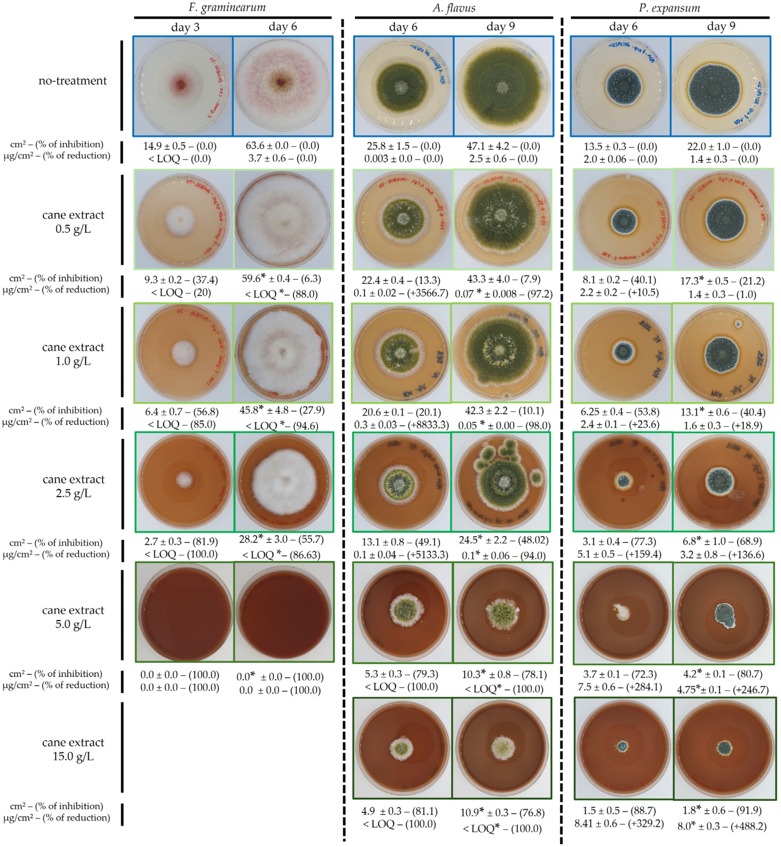
Figure 1. Effect of GCE on fungal growth and mycotoxin production. Fungal strains (F. graminearum, A. flavus, and P. expansum) were cultivated on CYA or PDA media supplemented with increasing concentrations of GCE (0.5 to 5 g/L for F. graminearum; and 0.5 to 15 g/L for A. flavus and P. expansum). Culture media color reflects the GCE supplementation. A commercial chemical fungicide (Imazalil 1.0 g/L) was included as a positive control; no fungal growth was observed in its presence for any of the tested species. Each column pair corresponds to a fungal species observed at two time points: F. graminearum (Day 3 and Day 6), A. flavus and P. expansum (Day 6 and Day 9). Below each condition, the following are mentioned: the fungal colony surface area (cm2), and the percentage of growth inhibition, along with the specific mycotoxin production (µg/cm2 of mycelium), and the percentage of reduction, relative to the untreated control. Limit of quantification (LOQ): DON = 0.9 µg/cm2, AFB1 = 0.001 µg/cm2, PAT = 0.08 × 10⁻³ µg/cm2. Asterisks (*) indicate conditions that differed significantly from the untreated control (p < 0.05; Dunn’s post-hoc test). Images are representative of three independent replicates.
Toxins 17 00454 g001
Figure 5. UHPLC-DAD chromatogram at 254 nm of the grapevine cane extract (V. vinifera) at 5 g/L solubilized in ethanol 50% (v/v). The analysis was set using a C18 column in reverse phase mode, the elution was performed at a flow rate of 1.6 mL/min with a volume of injection of 5 µL.
Toxins 17 00454 g005
Table 1. Summary of the activity of GCE against F. graminearum, A. flavus and P. expansum. IC50 and IC100 values (g/L) were reported for radial growth and toxin production; italic ranges indicate the corresponding inhibition level relative to the untreated control (%). For spores, we reported the minimal dose at which no germination was observed under the tested conditions. “Mycelium/colony” indicates the physiological stage at which the fungus was treated, and the related data shown are the maximal growth inhibition measured after transfer to fresh medium; “Remanence” graded the growth and toxin recovery five days after transfer to fresh untreated media (low < 40%, moderate 40–70%, strong > 70%). “Pro-patulin” denotes a stimulatory effect on patulin production; Mycotoxins LOQ are DON = 0.9 µg/cm2, AFB1 = 0.001 µg/cm2, PAT = 0.08 × 10⁻³ µg/cm2, below the analytical limit of quantification. All values were averaged from three independent assays.
FungiIC50 (g/L)IC100 (g/L)Spores/GerminationMycelium/ColonyRemanence
GrowthToxinsGrowthToxinsInhibitionSporicidalFungicideToxinsGrowthToxins
F. graminearum2.5
≈55%
<0.5
≈88%
2.5–5.0
≈55–100%
1.0–2.5
≈94–87%
5.0 and <15.0
≈72–100%
15.0not
observed
15.0 g/L ≈ 22%
<5.0
<LOQ
moderatestrong
A. flavus2.5–5.0
≈48–77%
<0.5
≈98%
>30.0
15.0 g/L ≈ 78%
2.5–5.0
≈94–100%
>15.0
≈68%
not
observed
15.0 g/L ≈ 68%
not
observed
15.0 g/L ≈ 62%
<5.0
≈99%
moderatestrong
P. expansum1.0–2.5
≈40–69%
pro-patulin>30.0
15.0 g/L ≈ 92%
pro-patulin10.0
≈21%
not
observed
15.0 g/L ≈ 33%
not
observed
15.0 g/L ≈ 66%
pro-patulinlowpro-patulin
Table 2. Grapevine cane extract content of the main biochemical families. Results are expressed as % w/w (weight/weight), indicating that 1% of a given compound corresponds to 1 g of that compound per 100 g of powder cane extract. This unit represents the relative mass fraction of each family of compound within the total extract. * Indicates analytes measured with a method of limited specificity; interfering compounds may affect these values, so quantification is approximate.
AnalyteContent
[% w/w]
SD
[% w/w]
Detection Mode
Water4.591.40Kar-Fisher-titration method
monosaccharides1.240.81HPLC-RID
disaccharides0.700.68HPLC-RID
polysaccharides14.560.38HPLC-RID
lignin *70.91NAHPLC-UV-vis
polyphenols *36.901.37folin-Ciocalteu
hydrolysable
tannins *
3.980.19absorbance
condensed tannins *0.410.07absorbance
flavonoids6.34NAULPC-MS, spectrometry
stilbenoids21.202.56UPLC-DAD
Table 3. Growth- and toxin-inhibition IC50 values (µM) of the present GCE and of selected grapevine- and plant-derived stilbenoids in the literature. IC50 values are expressed in micromolar (µM) and refer to the concentration that reduced radial growth (“growth” column) or mycotoxin production (“toxin” column) by 50% for F. graminearum, A. flavus, and P. expansum. Stilbenes concentrations from GCE were obtained by HPLC dosage, while bibliographic values were re-calculated from the original data when necessary. <LOQ indicates below the analytical limit of quantification; “-“ not determined. All reported IC50 values from the bibliography have a corresponding percentage of inhibition and are reported in parentheses. The reference number in the right column matches the bibliography.
ExtractsCompoundsF. graminearum IC50 (µM)ExtractsCompoundsA. flavus IC50 (µM)ExtractsCompoundsP. expansum IC50 (µM)Reference
GrowthToxins GrowthToxins GrowthToxins
Grapevine cane extract (GCE)
IC50 growth:
2.5 g/L
IC50 toxine:
<0.5 g/L
piceid25.51.3Grapevine cane
extract (GCE)
IC50 growth:
2.5–5.0 g/L
IC50 toxine:
<0.5 g/L
piceid25.5–61.61.3Grapevine cane
extract (GCE)
IC50 growth:
1.0–2.5 g/L
IC50 toxine:
<30.0 g/L
piceid5.3–25.5
piceatanol13.5<LOQpiceatanol13.5–39.7<LOQpiceatanol6.4–13.5
trans-resveratrol138.916.4trans-resveratrol138.9–233.416.4trans-resveratrol72.4–138.9
trans-ε-viniferin53.210.8trans-ε-viniferin53.3–94.310.8trans-ε-viniferin23.9–53.3
vitisin B<LOQ<LOQvitisin B<LOQ–3.8<LOQvitisin B<LOQ
Pure compound in water: 0.1 g/Lpiceatanol>410.0
(≈no inhibition)
Pure compound in water: 0.1 g/Lpiceatanol>410.0
(≈23% inhibition)
Pure compound in water: 0.1 g/Lpiceatanol>410.0
(≈no inhibition)
[28]
Sauvignon blanc canes in
H2O/EtOH,
95.5/0.5, v/v
IC50: >0.1 g/L
piceatanol>8.18.1Pure compound in water: 0.003 g/Ltrans-resveratrol13.2
(≈no inhibition)
13.2
(≈47% inhibition)
Pure compound in phosphate buffer/NaoH,
9:1, v/v): 5.0 g/L
trans-resveratrol21,900
(≈7.2% inhibition)
21,900
(+40%)
[50,51,52]
trans-resveratrol>43.243.2
trans-ε-viniferin>110.9
(<50% inhibition)
110.9
(<36% inhibition)
Tannat canes in H2O/EtOH,
95.5:0.5, v/v
IC50: >0.09 g/L
piceatanol4.96.9 [50]
trans-resveratrol31.043.6
trans-ε-viniferin61.2
(50% inhibition)
86.0
(69% inhibition)
Merlot woods in H2O/EtOH,
95.5/0.5, v/v
IC50: 0.07 g/L
trans-resveratrol73.7139.5 [50]
trans-ε-viniferin49.8
(50% inhibition)
94.4
(81% inhibition)
Tannat wood in H2O/EtOH,
95.5/0.5, v/v
IC50: 0.07 g/L
trans-resveratrol16.828.8 [50]
trans-ε-viniferin44.7
(50% inhibition)
76.6
(80% inhibition)
Table 4. HPLC mobile phase gradient. Solvent (A): water + 0.1% formic acid (v/v)/solvent (B): acetonitrile + 0.1% formic acid (v/v) for GCE analysis.
Time (min)Solvent B (%)
0.05
3.030
9.240
9.4100
10.5100
10.75
11.35
Disclaimer/Publisher’s Note: The statements, opinions and data contained in all publications are solely those of the individual author(s) and contributor(s) and not of MDPI and/or the editor(s). MDPI and/or the editor(s) disclaim responsibility for any injury to people or property resulting from any ideas, methods, instructions or products referred to in the content.