Article

Efficacy of Entomopathogenic Fungal Formulations against Elasmolomus pallens (Dallas) (Hemiptera: Rhyparochromidae) and Their Extracellular Enzymatic Activities

by
Fredrick Fidelis Umaru
1,2 and
Khanom Simarani
1,*
1
Division of Microbiology, Faculty of Science, Institute of Biological Sciences, University of Malaya, Kuala Lumpur 50603, Malaysia
2
Department of Biological Sciences, Faculty of Science, Taraba State University, Jalingo 660213, Nigeria
*
Author to whom correspondence should be addressed.
Toxins 2022, 14(9), 584; https://doi.org/10.3390/toxins14090584
Submission received: 30 June 2022 / Revised: 7 August 2022 / Accepted: 14 August 2022 / Published: 25 August 2022
(This article belongs to the Special Issue Insecticidal Toxins: Application and Assessment)

Abstract

:
Elasmolomus pallens are post-harvest insect pests of peanuts that are becoming resistant to chemical insecticides. In this, we study evaluated the effect of conidial formulations on entomopathogenic fungi against E. pallens to reduce the adverse effects. Fungal conidia were formulated and applied on sterile filter papers at varying concentrations (1 × 10⁴–1 × 10⁸ conidia mL−1) inside plastic containers. The test insects were exposed and maintained in a relative humidity of 80 ± 10% for 10 d at room temperature (25 ± 2 °C). Mortality was recorded every 24 h. Dose–response bioassay (LC50 and LC90) values for Aspergillus flavus formulated in oil were 1.95 × 10⁶ and 3.66 × 10⁹ conidia/mL, whereas formulations in Tween 80 had 9.36 × 10⁷ and 6.50 × 10⁹ conidia/mL. However, oil-formulated Metarhizium anisopliae had 3.92 × 10⁶ and 2.57 × 10⁸ conidia/mL, with 6.85 × 10⁶ and 5.37 × 10⁸, for formulations in Tween 80. A. flavus had LT50 values of 3.3 and 6.6 days, whereas M. anisopliae had LT50 values of 3.6 and 5.7 d. Maximum protease, chitinase, and lipase activities of 2.51, 0.98, and 3.22 U/mL, respectively, were recorded for A. flavus, whereas values of 2.43, 0.93, and 3.46 were recorded for M. anisopliae. The investigated pathogens demonstrate potential against E. pallens; therefore, their applicability under field conditions requires further investigation.
Key Contributions: Conidia formulation in oil exhibits enhanced virulence of entomopathogenic fungi. Median lethal times for A. flavus were 3.3 & 6.6 day; while M. anisopliae; 3.6 & 5.7 day. The fungi isolates are good cuticle-degrading enzymes, virulence against the bug.

Graphical Abstract

1. Introduction

Insects from the order Hemiptera, commonly described as ‘true bugs,’ are well-recognized as economically important pests in commercial crops [1]. The test insects, Elasmolomus pallens (Hemiptera: Rhyparochromidae), are post-harvest insect pests [2], the family members of which are diverse and comprise the most prominent Lygaeoidea [3]. Elasmolomus pallens are distributed across various global climatic zones, including the tropical, subtropical, and temperate climatic zones. The insects’ distribution is linked to their appetite for the legume Arachis hypogea L. [4]. Adults and nymphs are a cause of significant economic loss of peanuts in Sub-Saharan Africa, attacking entire pods using their piercing rostrum. Peanut pods are usually attacked by insects in the field during harvest and storage. When such infestations occur, the seeds become shriveled, resulting in increased fatty acid content and a rancid flavor of the oil [5].
The menace of E. pallens as a pest of economic concern is often tackled using classical biological and chemical control methods. With classical biological methods, predators such as Cephalonomia (a bethylid wasp) and the Reduviidae Coranus pallidus are employed as valuable predators of the eggs, nymphs, and adult insects [4]. With the chemical control method, 0.5% lindane and 2% malathion are applied on peanut stalks to prevent infestation [4]. However, the tendencies for this pest species to attack crop plants are evident, and the negative impacts of synthetic chemicals on biodiversity, the environment, and health are significant. Therefore, lindane is no longer in use. It has been asserted that commonly used synthetic insecticides, such as organophosphates, promote harmful environmental hazards [6].
A report by Sharma et al. [7] reveals that the chemical method of control has produced resistance among insect populations, toxic effects on the environment, and traces of insecticide residues in food and food products. The continuous use of chemical insecticides on hemipteran bugs may lead to resistance, considerably impacting food security and safety, with negative effects on natural enemies [8]. It is essential to search for alternative environmentally friendly biological control measures, considering the lethal effects of chemical pesticides on the environment [9].
Entomopathogenic fungi are naturally pathogenic to insect hosts, harmless to humans, good regulators of the insect population in the natural environment, and impose no adverse effects on the environment [10]. Entomopathogenic fungi vary in their specificity to natural hosts that they infect and are often referred to as specialist or generalist pathogens [11].
Unlike pathogens such as protozoa, bacteria, and viruses, which a host must ingest to initiate infection, entomopathogenic fungi have well-developed mechanisms that exploit nutrients on the insect cuticle, aiding in host colonization and infection. This makes entomopathogenic fungi a choice pathogen for the biological control of sucking E. pallens [12]. There are existing reports on the use of entomopathogenic fungi for the biological control of insects from the order Hemiptera. However, there is currently a lack of reports on their application as biological control agents of E. pallens. Although A. flavus is perceived as a harmful pathogen to humans and animals, given its ability to infest and produce aflatoxins on agricultural grain products [13], several reports have emphasized and described it as a ubiquitous mold, and not all of its strains can produce aflatoxins [14,15]. Additionally, atoxigenic A. flavus strains are known to be used for the biological control of toxigenic strains in crop plantations via competitive bio-exclusion to prevent aflatoxin contamination in grains [16].
Furthermore, proper formulations containing the required carriers are essential for host infection during large-scale applications of fungal entomopathogens [17]. Studies demonstrated that oil-based formulations enhanced the adhesion of conidia, conferred protection from ultraviolet radiation, and increased the virulence of entomopathogenic fungi on insect hosts compared to aqueous formulations [17].
In nature, some isolates of entomopathogenic fungi (EPF) possess the capacity to kill their host faster than others. The ability to secrete hydrolytic cuticle-degrading enzymes confers the property of virulence or nonvirulence. To eliminate their hosts, they must breach the epicuticle via specific and non-specific events [18]. The synthesis of a cocktail of extracellular hydrolytic cuticle-degrading enzymes, including proteases, chitinases, lipases [19], catalase, and phospholipase C [20] by EPF forms the underlying basis for the infection of insects, aiding in the degradation of host cuticle and penetration of the hemocoel [21,22]. Some of these hydrolytic cuticle-degrading enzymes (CDEs) are determinants of virulence for EPF [23] and work in concert to breach the proteo-chitin matrix to enable penetration into the hemolymph and when exiting to the surface of the cadaver for conidiation [24].
Previous studies by Dong et al. [25] and García-Munguía et al. [26] reported that entomopathogenic fungi conidia applied on filter paper were highly effective at infecting and killing the mosquito Aedes aegypti via horizontal propagule transmission during sexual activities. However, the question of the virulence of formulated propagules applied on filter paper surface to which E. pallens is exposed remains to be answered. Therefore, it is essential to establish and validate the novelty of horizontal transmission of entomopathogenic fungal propagules against insects such as E. pallens, which belong to a completely different order. Hence, the aim of this study was to determine the virulence of the formulated entomopathogenic fungi against E. pallens when applied on sterile filter paper surfaces.

2. Results

2.1. Fungal Isolates

The colony morphology of A. flavus showed dense sporulation on the culture medium. The colony color appeared dirty green, with septate hyphae, naturally radiate phialides (7.5–12 × 3.5–4.2 µm), and classically globose-to-subglobose vesicles (19–45 µm). The conidial size ranged between 3.5–5.2 × 2.2–2.8 µm. M. anisopliae produced dark-herbage green colony colors, with cylindrical and podgy phialides. Its spores were colorless and ellipsoidal with a rounded apex and slightly truncate base, whereas the conidia size ranged between 4.8–6.1 × 2.2–3.6 µm, respectively.

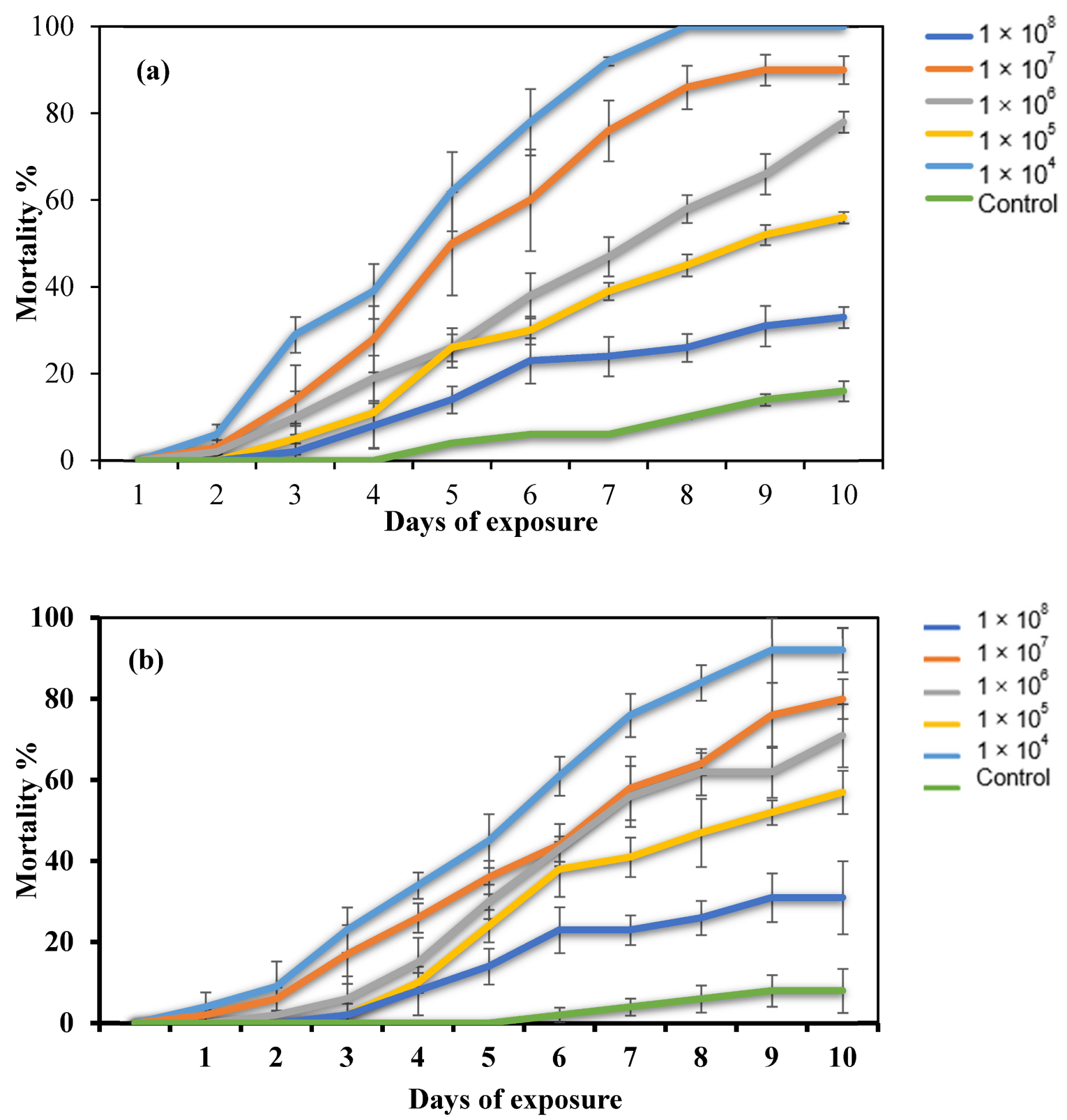
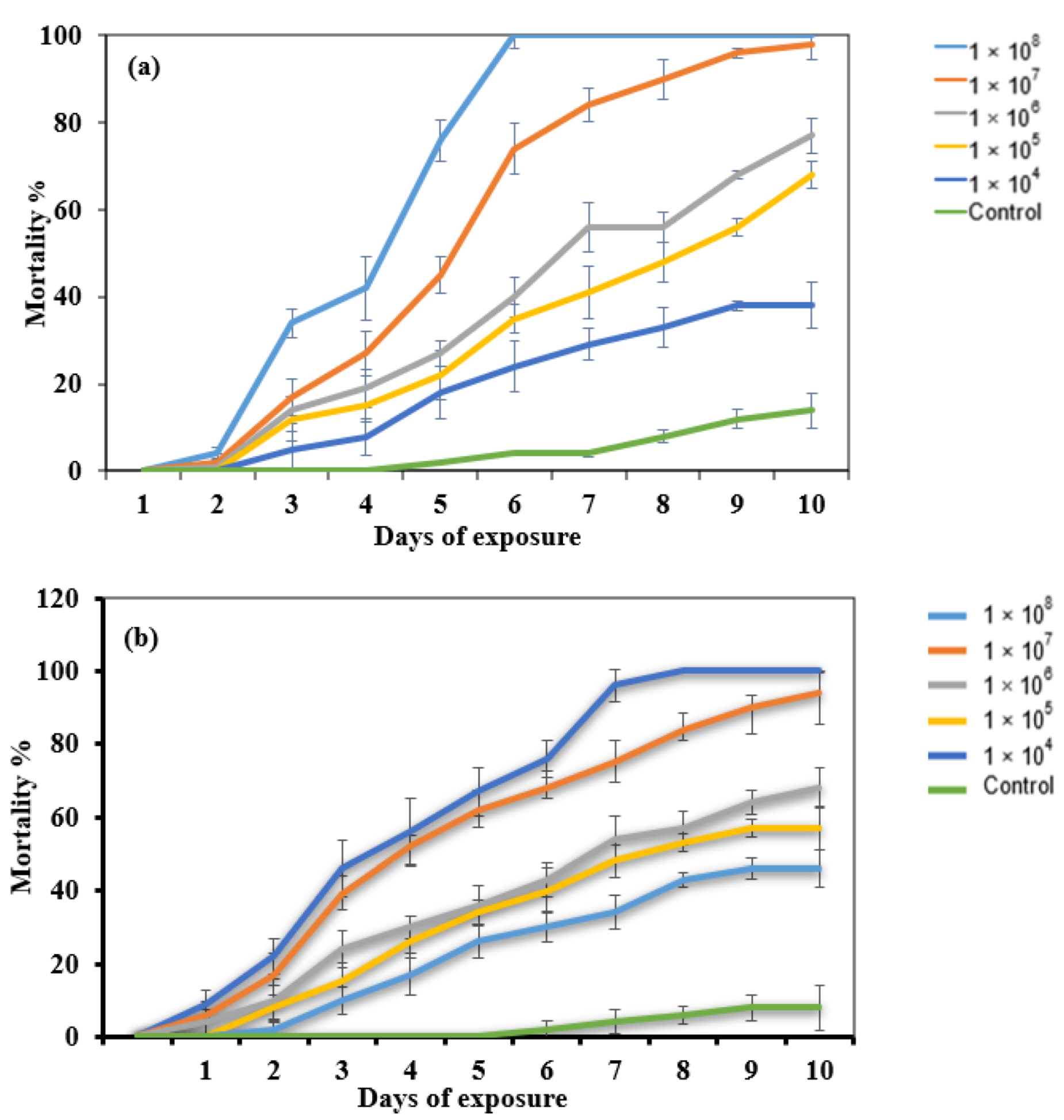
2.2. Dose–Response Bioassay

Mortality increased in the dose–response bioassay with increased conidial concentration (Table 1). The two fungal isolates generated significantly different mortality rates against E. pallens (F5, 60 = 5.745; p < 0.05). Additionally, oil-formulated conidia showed enhanced virulence compared to the formulation in the surfactant, Tween 80 (Table 1). The highest conidial concentration (1 × 10⁸ conidia/mL) of A. flavus isolates formulated in oil achieved 100% cumulative mortality after 8 days. In contrast, the same conidial concentration formulated in Tween 80 achieved 92% cumulative mortality after 9 d of treatment (Figure 1). The mortality rate was substantially higher in the conidial concentrations used compared with the control (F = 4.394; df = 5, 60; p < 0.05). The insect mortalities in the control were less than 10% after 10 d.
The dose–response bioassay involving M. anisopliae showed effects on the bugs’ mortalities based on the formulation type. M. anisopliae conidial concentration (1 × 10⁸ conidia/mL) formulated in oil achieved a 100% cumulative mortality in 6 d after treatment, whereas a 100% cumulative mortality for the same conidial concentration formulated in 0.05% Tween 80 was achieved after 8 d of treatment. This shows variations in the efficacies of the fungal conidial formulations. The mortality of the bugs was faster for the oil-formulated conidia compared to the formulations in Tween 80, demonstrating the enhanced efficacy of oil-formulated conidia. Significant mortalities of E. pallens due to M. anisopliae were observed (F5, 60 = 4.363; p < 0.05), whereas the mortalities in the control never exceeded 10% (Figure 2). Therefore, it is possible that control mortality could become zero or be set at zero when Abbott’s correction results in negative mortality. In the case of this study, no negative mortality was obtained.
The infected bugs showed visible mycosis characterized by the development of fungal mycelia on the integument while still alive. In this stage, the insects became sluggish, losing the ability to copulate and leading to the onset of death. Dead bug cadavers cultured on damp sterile filter paper developed fungal mycelial characteristics of the isolates used in the study (Figure 3).
Under the bioassay conditions, the mean mortalities of the bugs exposed to individual conidial concentrations of the isolates were compared (Table 1). Mean mortality values after 10 d of inoculation were proportional to the fungal conidial concentrations (108 and 107) used for each isolate. The mean mortality values for oil-formulated conidia were observed to be higher (F = 4.882; df = 5,60; p < 0.05) compared to conidia formulated in Tween 80. This shows that the virulence of the fungal pathogens was enhanced when formulated in oil relative to that of pathogens formulated in Tween 80.
Mortalities 10 d after inoculation differed significantly depending on the dose (108–104 and control) for both isolates formulated in oil (A. flavus BAMF2a F = 5.60; df = 5, 60; p < 0.05; M. anisopliae F = 8.825; df = 5, 60; p < 0.05) and 0.05% Tween 80 (A. flavus isolate BAMF2a F = 4.3934; df = 5, 60; p < 0.05; M. anisopliae F = 4.363; df = 5, 60; p < 0.05) (Table 1). The results show that the mortalities observed in the bioassay were dose-dependent.
The bioassay experiment determined the lethal concentrations (LC50 and LC90) and lethal time (LT50 and LT90) for the fungal isolates. We observed that the LC and LT values were lower for fungal conidia formulated in oil than in Tween 80 (Table 2 and Table 3). However, comparisons between the two formulations indicate that conidia formulated in oil showed increased virulence relative to that formulated in Tween 80. The A. flavus isolate formulated in oil sometimes performed better than M. anisopliae. The comparison was made by calculating the 95% confidence interval (CI) for each population. If the CIs do not overlap, a significant difference exists. In contrast, if CIs overlap, we concluded that no significant difference existed. The CIs do not overlap; hence, they are different.
M. anisopliae formulated in oil killed the bugs at an LC50 and LC90 of 3.92 × 10⁶ and 5.37 × 10⁸ conidia/mL (Table 2), respectively, producing LT50 and LT90 values of 3.6 and 5.7 d, respectively (Table 3). These values were found to be lower compared to results obtained for fungal conidia formulated in Tween 80.

2.3. Enzyme Assay

The enzyme activity of the isolates was observed to increase with days of culture, with activity later decreasing (Table 4). Maximum protease activity for A. flavus isolates (2.51 U/mL) was obtained on the 8th day, whereas M. anisopliae (2.43 U/mL) was observed on the 6th day. For the chitinase enzyme, maximum activity (0.98 U/mL) was recorded for the A. flavus isolate on the 8th day, and M. anisopliae also exhibited maximum chitinase activity (0.93 U/mL) on the 8th. Nevertheless, maximum lipase activity (3.22 U/mL) was obtained for A. flavus isolate on the 6th day compared to that obtained for M. anisopliae (3.46 U/mL).

2.4. Correlation between Rates of Mortality and Enzyme Activities

The mortality rate data of E. pallens were analyzed in correlation with the enzymatic activity of the fungal pathogens (Table 5). The correlation coefficient (r) achieved for the mortality of E. pallens and cuticle-degrading enzymes (protease, chitinase, and lipase) exhibited positive correlations for the two isolates.

3. Discussion

Entomopathogenic fungi are valuable alternatives to conventional synthetic insecticides and are broadly applied to effectively manage agroecosystem pests [27]. Compared to other microbial pathogens, they confer several advantages when applied for pest control [28]. Although insects have established myriads of defenses against infective pathogens, pathogens have likewise coevolved to counter these defenses [27].
The present study confirms that the two fungal isolates of A. flavus isolates and M. anisopliae are potential biocontrol agents for the management of the seed bug E. pallens. The mortalities of the bugs were found to be proportional to the conidial concentrations used in the bioassay involving the two fungal isolates. Conidia of both isolates formulated in oil showed increased virulence relative to those formulated in Tween 80 surfactant, as evidenced by the mortality rates and the number of positive cadavers confirmed upon culture. Oil-based formulations of biopesticides have, over the years, been found to enhance the adherence of pathogenic fungal propagules to insect cuticle, promote the spread of the propagules over the insect body, penetration of the insect integument, protection from the effect of ultraviolet (UV) light radiation, and enhanced infectivity of the propagules, even under low moisture (humidity) conditions [9,29,30]. Furthermore, vegetable-oil-based formulations, compared to water-based formulations, have been reported to demonstrate increased virulence against various hosts, including Bemisia tabaci [31], arid locust, Schistocerca gregoria under low humidity [32], and ticks [29,33,34].
The efficacy of A. flavus was previously studied against mango seed weevil, Sternochetus mangiferae (Coleoptera: Curculionidae), producing mortality rates of 80% at a conidial concentration of 6.8 × 10⁷ conidia/mL. Aspergillus species have been reported to kill the tropical locust Zonocerus variegatus [35] and cause infection among many insect populations. However, it is unknown whether it is host-specific. Previous studies have reported the effectiveness of A. flavus against a wide range of insects [27,36,37]. A. flavus was also tested against Galleria mellonella, killing 100% of insects after 48 h when injected with 3 × 10³ conidia/mL of the fungus. In contrast, Aspergillus fumigatus and Aspergillus nidulans lacked parasitic attributes [38].
We observed that M. anisopliae formulated in oil achieved a 100% cumulative mortality in 6 d compared to the 8 d required for the Tween 80 formulation. This showed an increase in the virulence of the fungus by achieving a faster killing rate due to its formulation in oil. Santi et al. [17] reported formulations containing M. anisopliae conidia in 5% and 10% soybean oil as the most effective virulent against the cotton seed bug Dysdercus peruvianus. Peng and Xia [39] reported an increase in the virulence of an oil-based formulation of M. anisopliae var. acridium against grasshoppers and locusts by reducing the dependence on high humidity for germination, maintaining the thermotolerance and UV tolerance of the fungal conidia.
Although conidial viability was higher for A. flavus isolate than M. anisopliae, we found no significant difference in the mortality rates of the bugs between the two isolates (p > 0.05). However, in a previous study employing dipping bioassay involving the same isolates, a significant difference (p < 0.05) was observed in bug mortality [40]. The formulation of the fungal conidia in peanut oil was observed to have enhanced the virulence of both A. flavus isolate and M. anisopliae against E. pallens, as evidenced by their increased killing rates. Although Aspergillus species are described as facultative generalists [41], the virulence of certain species and strains against insect hosts can be enhanced to match promising biological control agents such as M. anisopliae when formulated in non-evaporative diluents, such as peanut oil.
Furthermore, the LC50 values obtained for both isolates were comparable, indicating no difference in virulence between them. In a previous study, M. anisopliae isolate FRM515 was considered and selected as the most virulent against the brown-winged green bug, Plautia stali, at an LT50 value of 3.9 days. Ihara et al. [42] reported a similar LC50 value for M. anisopliae against adult citrus mealybug, Planococcus citri. There was no difference in LT50 values between the two isolates. This shows how peanut oil formulation could impact the virulence of the isolates, especially A. flavus, which is less often reported to demonstrate virulence against arthropods. Balogun and Fagade [35] explored an LT50 of 4 d for Aspergillus niger and 5 d for M. anisopliae against Zonocerus variegatus. Therefore, it is noteworthy in this study that fungal conidia formulated in peanut oil showed no significant difference in their virulence against E. pallens compared to the formulation in Tween 80. This implies that oil can enhance the effectiveness of these entomopathogens in terms of killing insects.
However, the major impediment to fungal penetration is the host’s cuticle, which must be broken down to ease penetration. Fungal pathogens secrete a certain class of cuticle-degrading enzymes (protease, chitinase, and lipase) to help dissolve the host integument, thereby overcoming the barrier [43]. In a previous study, El-Sayed et al. [44] established that successful penetration and infection of the host cuticle depends on the combined outcome of enzymatic degradation and mechanical pressure exerted by the infecting fungus. For example, the lipolytic activity of CDEs has been proposed as a ‘virulence index,’ which could be harnessed for selection of effective strains from the population [45]. Recent understanding of the entomo-pathogenesis of fungi reveals that the CDE system of entomopathogenic fungi is distinctive and of considerable importance as a potential standard for improving mycoinsecticides [18].
In this study, the maximum protease activity found for both A. flavus and M. anisopliae correlated positively with the mortality rates of E. pallens. For A. flavus, protease activity was highest (2.28 U/mL) on the 8th day before decreasing. A similar observation was reported by Grewal et al. [46], observed a positive correlation between enzymatic activity and the mortality rate of Spodoptera litura larvae exposed to different doses of Metarhizium rileyi. In the present study, we recorded the highest protease activity (2.43 U/mL) for M. anisopliae on the 6th day, which is similar to that observed by Saleem and Ibrahim, [47], who described maximum protease activity of M. anisopliae (2.97 U/mL), Beauveria bassiana (2.61 U/mL) and Akanthomyces lecanii (2.49 U/mL) to be highest on the 6th day after culture. For chitinase, the maximum activity for both isolates was highest on the 8th day of culture, in agreement with the findings of Grewal et al. [48], whereas lipase enzyme maximum activity was highest for M. anisopliae (3.46 U/mL) on the 6th day of culture, and A. flavus exhibited maximum activity (3.22 U/mL) on the 8th day. A similar trend was observed by Grewal et al. [46] but contrasted by Supakdamrongkul et al. [48], who recorded the highest lipase activity of 20.8 U/mL by the 8th day from Nomuraea rileyi. It can be inferred from this study that the variations in maximum enzyme activities could be attributed to isolate and species types. Because arthropod integument mainly contains proteins, lipids, and chitin, the enzyme activity results obtained in this study support the ecological and physiological importance of CDEs, as previously reported [46]. Studies on the mortality of E. pallens and the ability of fungal isolates to produce CDE activity showed positive correlations when analyzed. This is in agreement with previous studies that reported positive correlations between mortality and CDE activity of entomopathogenic fungi [23,46,49], indicating that specific enzymes in the enzyme system may serve as key determinants of virulence.

4. Materials and Methods

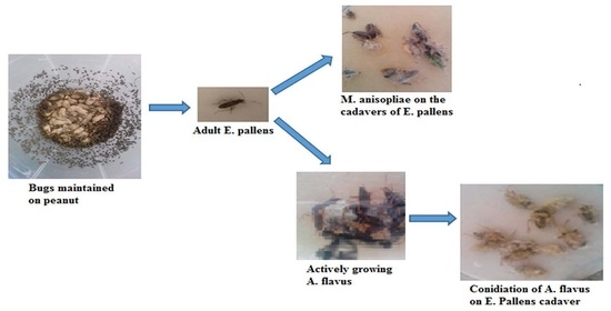
4.1. E. pallens Collection and Maintenance

Adult E. pallens were hand-picked from under heaps of groundnut haulms in the field. The bugs were kept in a plastic container and sent to the lab. The bugs were maintained in the lab according to the method described in [40]. Adult bugs were collected for the experiment during peanut harvest between August and October 2018. E. pallens were identified using the molecular method [50].

4.2. Fungal Isolates

Aspergillus flavus isolate (accession number MF319893.1) (registered as ITS sequence of A. flavus in the NCBI databank) originally isolated from the cadavers of E. pallens was obtained from the Biotechnology and Applied Microbiology Laboratory of the University of Malaya, Kuala Lumpur, Malaysia. Metarhizium anisopliae previously reported by Balogun and Fagade [35] (originally from cadavers of Zonocerus variegatus) was collected from the Environmental Microbiology and Biotechnology Laboratory, Department of Microbiology, University of Ibadan (Ibadan, Nigeria). The purity of both fungi was checked before culturing. Pure cultures were maintained on PDA.

4.3. Fungal Conidia Preparation

Fungal conidia were harvested from 2-week-old culture according to the modified method of [51] by gently scrapping the culture plate with a sterile spatula to avoid dispersing the conidia into the agar. Conidia were harvested into separate sterile 50 mL centrifuge tubes, each containing 20 mL (10 mL of 5% peanut oil and 10 mL of 0.05% (v/v) Tween 80 (Sigma Chemical Co., St. Louis, MO, USA)) solution. The conidia were vigorously vortexed for 1 min to homogenize before sieving. The conidial homogenate suspensions were sieved through four layers of sterile cheesecloth into sterile centrifuge tubes and kept for further use. Conidial concentrations were estimated using a Neubauer hemocytometer under a Leica DM500 bright-field microscope (Leica Microsystems, Wetzlar, Germany) [52]. Five concentrations from 104 to 108 conidia/mL were prepared in tenfold dilutions for the bioassay. Conidial viability was determined before each bioassay by plating 100 µL aliquot of diluted conidia on three PDA plates. Three plates were prepared for each isolate with three replications and incubated at 25 °C for 24 h under 14:10 light/dark regimes. Germination was observed to be ≥90 under the microscope at 40× by counting 300 conidia (100 conidia per field). Viability was indicated for conidia that produced germ tubes half their size.

4.4. Dose–Response Bioassay

The dose–response bioassay was conducted using fungal pathogens on sterile filter papers according to the method described in [25] with minor modifications. For each conidial preparation, 1 mL of the suspension was pipetted onto a moist sterile filter paper (65 mm, Whatman No. 1) inside a plastic container (65 × 45 mm). Sterile cotton dipped in sterile demineralized water was added to the container to afford ≥80% relative humidity (RH) at room temperature. The bioassay tested 10 adult bugs against individual conidial concentrations inside a sterile container, with five replications for each concentration. The bugs were introduced into plastic containers containing sterile filter paper treated with 1 mL peanut oil, and Tween 80 served control. Mortality was scored at an interval of 24 h for 10 d in replicate using independent batches of bugs and conidial inoculum. Furthermore, to confirm whether the bugs’ mortality was caused by the fungal pathogen used in the bioassay, the cadavers were sterilized on the surface for 5 min using 1% (v/v) NaClO solution. The sterilized cadavers were washed 3 times in sterilized demineralized water and subjected to 5–7 d incubation at 25 ± 2 °C on sterilized wet filter paper in Petri plates to stimulate fungal growth and examined under a bright-field microscope (Leica DM500, Leica Microsystems, Wetzlar, Germany) at 400×.

4.5. Production of Cuticle-Degrading Enzymes

Synthesis and determination of the activity of extracellular cuticle-degrading enzymes: protease, lipase, and chitinase were studied from the isolates of A. flavus and M. anisopliae based on the method described in [53] with minor modifications. A 200 µL conidial suspension of each fungal isolate (containing 1 × 10⁸ conidia/mL) was inoculated in 100 mL of Sabouraud dextrose broth (into a 100 mL flask). Inoculated flasks were incubated on a rotary shaker at 28 °C and 150 rpm for 10 d. Enzyme assays were performed on alternate days for 10 d. The cultures were filtered using cheesecloth to remove the mycelia before centrifugation at 13,000 rpm for 10 min at 4 °C. The supernatant, the crude enzyme source, was used for the enzyme activity assay.

4.6. Enzyme Assays

According to the method, the protease activity was determined by employing casein as a substrate [48]. The reaction mixture (2 mL) comprised an aliquot of diluted enzyme solution, 10 mg casein, and Na2CO3 buffer (200 µM) at pH 9.7. Enzyme activity was assayed at 37 °C for 20 min and stopped by adding 3 mL trichloroacetic acid (TCA) (2.6 mL of 5% TCA and 0.4 mL of 3.3 N HCl). The absorbance of the reaction mixture was measured at 280 nm. One enzyme unit was defined as the amount of enzyme that catalyzes the release of 1 µmol of tyrosine per min under the assay conditions.
Chitinase activity was determined using a colloidal chitin substrate based on the method adopted in [54], with some modifications. Analytical-grade chitin (20 g) was suspended in 200 mL concentrated HCl and stirred for 3 min at 40 °C. Distilled water (2 L) adjusted to 5 °C was gradually added to the chitin suspension for precipitation as a colloidal suspension. The precipitated colloidal material was obtained using coarse filter paper and washed in distilled water to attain a pH of 4.0. The chitinase activity was determined using the technique described in [46]. The assay reacting mixture containing 250 µL of 0.5% (w/v) colloidal chitin, 250 µL of 0.2 acetate buffer (pH 4), and 500 µL enzyme suspension was incubated at 37 °C for 2 h. After centrifuging the resulting mixture, 0.8 M boric acid (100 µL) was mixed with the supernatant (500 µL), and the solution was adjusted to a pH of 10.2 using 1 M KOH. The solution was subjected to heat for 3 min. The resulting mixture was cooled down, and 3 mL of p-dimethyl amino benzaldehyde (DMAB) solution (1 g of DMAB dissolved in 100 mL glacial CH3COOH comprising 1% v/v HCl) was added before incubating at 37 °C for 20 min. Absorbance was measured at 585 nm against a water blank. A unit of enzyme activity was expressed as 1 µmol/min of N-acetylglucosamine.
Lipase activity was determined by employing gum acacia and olive oil as substrates based on the method adopted in [46]. Substrate preparation involved mixing olive oil (50 mL) and gum acacia (50 mL, 10% w/v at 1:1 ratio). The lipase enzyme reaction mixture comprised a 5 mL substrate, 2 mL phosphate buffer (50 mM, pH 6.8), and 1 mL enzyme suspension. The reaction mixture was subjected to incubation for 1 h at 37 °C. This was followed by intermittent shaking and the inclusion of 4 mL ethanol/acetone (1:1 v/v), comprising phenolphthalein (0.09%), as an indicator to end the reaction. Lipase activity was measured by titrating the final mixture against NaOH (50 mM) solution (to estimate the free fatty acids discharged throughout the reaction). A unit of lipase enzyme activity was defined as 1 µmol fatty acids liberated per mL per min. All enzyme assays were performed in triplicate for all isolates.

4.7. Statistical Analyses

The experiments were performed in 5 replicates for each isolate, and the entire experiment was repeated 3 times. The mortality data were adjusted using Abbott’s formula [55]; the normality of the data was checked with a skewness–kurtosis test and analyzed using IBM SPSS Statistics 21.0 software (Long Beach, CA, USA). Analysis of variance (ANOVA) was used to make multiple comparisons. An F test was employed to measure the significant differences between treatments, and the means were compared using the least significant difference (LSD). The median lethal concentrations (LC50 and LC90) needed to kill 50% and 90% of the bugs and their respective confidence intervals (C. I.), as well as the median lethal time (LT50 and LT90), were determined with the aid of probit analysis [56]. The correlation between mortality and enzyme activity was analyzed with statistical significance set at the level of 0.05 for all tests.

5. Conclusions

The fungal conidial formulation demonstrated outstanding potential for controlling E. pallens under laboratory conditions. The oil-based conidial formulation showed enhanced virulence of both fungal isolates, implying that either of the fungi can be used as a biocontrol agent for bugs. The mortality of the bugs was observed to increase with an increase in conidial concentrations. The effectiveness of the formulated conidia at infecting and killing bugs when applied on filter papers makes these fungi potent for field applications, with a considerable possibility of success. Furthermore, the cuticle-degrading enzyme activity showed positive correlations with the mortality rates of E. pallens in interaction with varying concentrations of the fungal conidia. Exploiting the potential of the two fungi via inundative field applications and optimization of their enzyme production capacity will undoubtedly improve their commercial acceptance and use as biological control agents.

Author Contributions

We confirm that this manuscript is original and has not been published elsewhere, nor is it under consideration by any journal. F.F.U. conceived the study, conducted the experiments, and analyzed the results, whereas K.S. provided intellectual input. Both authors contributed equally to the drafting of the manuscript and approved of its publication in this referred journal. All authors have read and agreed to the published version of the manuscript.

Funding

The authors acknowledge the support of the University of Malaya Center of Research Grant Management (PPGP), Grant number PG343 2016A (12 July 2016).

Institutional Review Board Statement

Not applicable.

Informed Consent Statement

Not applicable.

Data Availability Statement

The data presented in this study are available in this article and Graphical Abstract.

Acknowledgments

We appreciate the effort of Obasola E. Fagade of the Environmental Microbiology and Biotechnology Laboratory, Department of Microbiology, University of Ibadan, Ibadan, Nigeria, for donating the M. anisopliae expended for this study.

Conflicts of Interest

The authors declare no competing interest concerning this work.

References

  1. Chandra, K.; Kushwaha, S.; Sambath, S.; Biswas, B. Distribution and Diversity of Hemiptera Fauna of Veerangana Durgavati Wildlife Sanctuary, Damoh, Madhya Pradesh (India). Biol. Forum-Int. J. 2012, 4, 68–74. [Google Scholar]
  2. Weirauch, C.; Schuh, R.T. Systematics and evolution of Heteroptera: 25 years of progress. Annu. Rev. Entomol. 2011, 56, 487–510. [Google Scholar] [CrossRef] [PubMed]
  3. Gad, S.K.M.S.M. Taxonomic review of family Rhyparochromidae (Hemiptera: Lygaeoidea) from Egypt. Egypt. J. Exp. Biol. 2013, 9, 33–60. [Google Scholar]
  4. Schaefer, C.W.; Panizzi, A.R. Heteroptera of Economic Importance; CRC Press: Boca Raton, FL, USA, 2000. [Google Scholar]
  5. Ranga Rao, G.; Rameshwar Rao, V.; Nigam, S. Post-Harvest Insect Pests of Groundnut and Their Management; International Crops Research Institute for the Semi-Arid Tropics: Patancheru, India, 2010. [Google Scholar]
  6. Senthil-Nathan, S. Natural pesticide research. Physiol. Mol. Plant Pathol. 2018, 101, 1–2. [Google Scholar] [CrossRef]
  7. Sharma, H.; Pampapathy, G.; Dwivedi, S.; Reddy, L. Mechanisms and diversity of resistance to insect pests in wild relatives of groundnut. J. Econ. Entomol. 2003, 96, 1886–1897. [Google Scholar] [CrossRef] [PubMed]
  8. Lu, L.; Cheng, B.; Du, D.; Hu, X.; Peng, A.; Pu, Z.; Zhang, X.; Huang, Z.; Chen, G. Morphological, molecular and virulence characterization of three Lencanicillium species infecting Asian citrus psyllids in Huangyan citrus groves. J. Invertebr. Pathol. 2015, 125, 45–55. [Google Scholar] [CrossRef]
  9. Carrillo, D.; Dunlap, C.; Avery, P.; Navarrete, J.; Duncan, R.; Jackson, M.; Behle, R.; Cave, R.; Crane, J.; Rooney, A. Entomopathogenic fungi as biological control agents for the vector of the laurel wilt disease, the redbay ambrosia beetle, Xyleborus glabratus (Coleoptera: Curculionidae). Biol. Control 2015, 81, 44–50. [Google Scholar] [CrossRef]
  10. Roy, H.E.; Vega, F.E.; Chandler, D.; Goettel, M.S.; Pell, J.; Wajnberg, E. The Ecology of Fungal Entomopathogens; Springer: Cham, Switzerland, 2010. [Google Scholar]
  11. Boomsma, J.J.; Jensen, A.B.; Meyling, N.V.; Eilenberg, J. Evolutionary interaction networks of insect pathogenic fungi. Annu. Rev. Entomol. 2014, 59, 467–485. [Google Scholar] [CrossRef]
  12. Lacey, L.; Grzywacz, D.; Shapiro-Ilan, D.; Frutos, R.; Brownbridge, M.; Goettel, M. Insect pathogens as biological control agents: Back to the future. J. Invertebr. Pathol. 2015, 132, 1–41. [Google Scholar] [CrossRef]
  13. Amaike, S.; Keller, N.P. Aspergillus flavus. Annu. Rev. Phytopathol. 2011, 49, 107–133. [Google Scholar] [CrossRef]
  14. Mamo, F.T.; Shang, B.; Selvaraj, J.N.; Wang, Y.; Liu, Y. Isolation and characterization of Aspergillus flavus strains in China. J. Microbiol. 2018, 56, 119–127. [Google Scholar] [CrossRef] [PubMed]
  15. Frisvad, J.C.; Hubka, V.; Ezekiel, C.; Hong, S.-B.; Nováková, A.; Chen, A.; Arzanlou, M.; Larsen, T.; Sklenář, F.; Mahakarnchanakul, W. Taxonomy of Aspergillus section Flavi and their production of aflatoxins, ochratoxins and other mycotoxins. Stud. Mycol. 2019, 93, 1–63. [Google Scholar] [CrossRef] [PubMed]
  16. Humphrey, J. Food safety, trade, standards and the integration of smallholders into value chains: A review of the literature. IFAD Res. Ser. 2017, 11, 1–72. [Google Scholar]
  17. Santi, L.; e Silva, L.A.D.; da Silva, W.O.B.; Corrêa, A.P.F.; Rangel, D.E.N.; Carlini, C.R.; Schrank, A.; Vainstein, M.H. Virulence of the entomopathogenic fungus Metarhizium anisopliae using soybean oil formulation for control of the cotton stainer bug, Dysdercus peruvianus. World J. Microbiol. Biotechnol. 2011, 27, 2297–2303. [Google Scholar] [CrossRef]
  18. Mustafa, U.; Kaur, G. Extracellular enzyme production in Metarhizium anisopliae isolates. Folia Microbiol. 2009, 54, 499–504. [Google Scholar] [CrossRef]
  19. Santi, L.; da Silva, W.O.B.; Berger, M.; Guimarães, J.A.; Schrank, A.; Vainstein, M.H. Conidial surface proteins of Metarhizium anisopliae: Source of activities related with toxic effects, host penetration and pathogenesis. Toxicon 2010, 55, 874–880. [Google Scholar] [CrossRef]
  20. Schrank, A.; Vainstein, M.H. Metarhizium anisopliae enzymes and toxins. Toxicon 2010, 56, 1267–1274. [Google Scholar] [CrossRef]
  21. Hussain, A.; Tian, M.-Y.; He, Y.-R.; Bland, J.M.; Gu, W.-X. Behavioral and electrophysiological responses of Coptotermes formosanus Shiraki towards entomopathogenic fungal volatiles. Biol. Control 2010, 55, 166–173. [Google Scholar] [CrossRef]
  22. Hussain, A.; Rizwan-ul-Haq, M.; Al-Ayedh, H.; AlJabr, A.M. Susceptibility and immune defence mechanisms of Rhynchophorus ferrugineus (Olivier)(Coleoptera: Curculionidae) against entomopathogenic fungal infections. Int. J. Mol. Sci. 2016, 17, 1518. [Google Scholar] [CrossRef]
  23. Nunes, A.R.F.; Martins, J.N.; Furlaneto, M.C.; Barros, N.M.d. Production of cuticle-degrading proteases by Nomuraea rileyi and its virulence against Anticarsia gemmatalis. Ciênc. Rural 2010, 40, 1853–1859. [Google Scholar] [CrossRef]
  24. Butt, T.; Coates, C.; Dubovskiy, I.; Ratcliffe, N. Entomopathogenic fungi: New insights into host–pathogen interactions. Adv. Genet. 2016, 94, 307–364. [Google Scholar] [PubMed]
  25. Dong, Y.; Morton, J.C.; Ramirez, J.L.; Souza-Neto, J.A.; Dimopoulos, G. The entomopathogenic fungus Beauveria bassiana activate toll and JAK-STAT pathway-controlled effector genes and anti-dengue activity in Aedes aegypti. Insect Biochem. Mol. Biol. 2012, 42, 126–132. [Google Scholar] [CrossRef] [PubMed]
  26. García-Munguía, A.M.; Garza-Hernández, J.A.; Rebollar-Tellez, E.A.; Rodríguez-Pérez, M.A.; Reyes-Villanueva, F. Transmission of Beauveria bassiana from male to female Aedes aegypti mosquitoes. Parasites Vectors 2011, 4, 24. [Google Scholar] [CrossRef] [PubMed] [Green Version]
  27. Karthi, S.; Vaideki, K.; Shivakumar, M.S.; Ponsankar, A.; Thanigaivel, A.; Chellappandian, M.; Vasantha-Srinivasan, P.; Muthu-Pandian, C.K.; Hunter, W.B.; Senthil-Nathan, S. Effect of Aspergillus flavus on the mortality and activity of antioxidant enzymes of Spodoptera litura Fab.(Lepidoptera: Noctuidae) larvae. Pestic. Biochem. Physiol. 2018, 149, 54–60. [Google Scholar] [CrossRef] [PubMed]
  28. Zimmermann, G. Review on safety of the entomopathogenic fungus Metarhizium anisopliae. Biocontrol Sci. Technol. 2007, 17, 879–920. [Google Scholar] [CrossRef]
  29. Polar, P.; Kairo, M.T.; Moore, D.; Pegram, R.; John, S.-A. Comparison of water, oils and emulsifiable adjuvant oils as formulating agents for Metarhizium anisopliae for use in control of Boophilus microplus. Mycopathologia 2005, 160, 151–157. [Google Scholar] [CrossRef]
  30. Inglis, D.; Jaronski, S.T.; Wraight, S.P.; Beattie, A.; Watson, D.; Stevens, M.; Rae, D. Use of spray oils with entomopathogens. In Spray Oils Beyond 2000: Sustainable Pest and Disease Management; University of Western Sydney: Sydney, Australia, 2002; pp. 302–312. [Google Scholar]
  31. Batta, Y. Production and testing of novel formulations of the entomopathogenic fungus Metarhizium anisopliae (Metschinkoff) Sorokin (Deuteromycotina: Hyphomycetes). Crop Prot. 2003, 22, 415–422. [Google Scholar] [CrossRef]
  32. Lomer, C.; Bateman, R.; Johnson, D.; Langewald, J.; Thomas, M. Biological control of locusts and grasshoppers. Annu. Rev. Entomol. 2001, 46, 667–702. [Google Scholar] [CrossRef]
  33. Kaaya, G.P.; Hassan, S. Entomogenous fungi as promising biopesticides for tick control. Exp. Appl. Acarol. 2000, 24, 913–926. [Google Scholar] [CrossRef]
  34. Camargo, M.G.; Golo, P.S.; Angelo, I.C.; Perinotto, W.M.; Sá, F.A.; Quinelato, S.; Bittencourt, V.R. Effect of oil-based formulations of acaripathogenic fungi to control Rhipicephalus microplus ticks under laboratory conditions. Vet. Parasitol. 2012, 188, 140–147. [Google Scholar] [CrossRef]
  35. Balogun, S.; Fagade, O. Entomopathogenic fungi in population of Zonocerus variegatus (l) in Ibadan, south west, Nigeria. Afr. J. Biotechnol. 2004, 3, 382–386. [Google Scholar]
  36. Scully, L.R.; Bidochka, M.J. Serial passage of the opportunistic pathogen Aspergillus flavus through an insect host yields decreased saprobic capacity. Can. J. Microbiol. 2005, 51, 185–189. [Google Scholar] [CrossRef] [PubMed]
  37. Seye, F.; Bawin, T.; Boukraa, S.; Zimmer, J.-Y.; Ndiaye, M.; Delvigne, F.; Francis, F. Effect of entomopathogenic Aspergillus strains against the pea aphid, Acyrthosiphon pisum (Hemiptera: Aphididae). Appl. Entomol. Zool. 2014, 49, 453–458. [Google Scholar] [CrossRef]
  38. Leger, R.J.S.; Screen, S.E.; Shams-Pirzadeh, B. Lack of Host Specialization inAspergillus flavus. Appl. Environ. Microbiol. 2000, 66, 320–324. [Google Scholar] [CrossRef] [PubMed] [Green Version]
  39. Peng, G.; Xia, Y. The mechanism of the mycoinsecticide diluent on the efficacy of the oil formulation of insecticidal fungus. BioControl 2011, 56, 893–902. [Google Scholar] [CrossRef]
  40. Umaru, F.F.; Simarani, K. Evaluation of the Potential of Fungal Biopesticides for the Biological Control of the Seed Bug, Elasmolomus pallens (Dallas)(Hemiptera: Rhyparochromidae). Insects 2020, 11, 277. [Google Scholar] [CrossRef]
  41. Sandhu, S.S.; Sharma, A.K.; Beniwal, V.; Goel, G.; Batra, P.; Kumar, A.; Jaglan, S.; Sharma, A.K.; Malhotra, S. Myco-biocontrol of insect pests: Factors involved, mechanism, and regulation. J. Pathog. 2012, 2012, 126819. [Google Scholar] [CrossRef]
  42. Ihara, F.; Yaginuma, K.; Kobayashi, N.; Mishiro, K.; Sato, T. Screening of entomopathogenic fungi against the brown-winged green bug, Plautia stali Scott (Hemiptera: Pentatomidae). Appl. Entomol. Zool. 2001, 36, 495–500. [Google Scholar] [CrossRef]
  43. De Moraes, C.K.; Schrank, A.; Vainstein, M.H. Regulation of extracellular chitinases and proteases in the entomopathogen and acaricide Metarhizium anisopliae. Curr. Microbiol. 2003, 46, 0205–0210. [Google Scholar] [CrossRef]
  44. El-Sayed, G.; Coudron, T.; Ignoffo, C.; Riba, G. Chitinolytic activity and virulence associated with native and mutant isolates of an entomopathogenic fungus, Nomuraea rileyi. J. Invertebr. Pathol. 1989, 54, 394–403. [Google Scholar] [CrossRef]
  45. Beys da silva, W.O.; Santi, L.; Corrêa, A.P.F.; Silva, L.A.D.; Bresciani, F.R.; Schrank, A.; Vainstein, M.H. The entomopathogen Metarhizium anisopliae can modulate the secretion of lipolytic enzymes in response to different substrates including components of arthropod cuticle. Fungal Biol. 2010, 114, 911–916. [Google Scholar] [CrossRef] [PubMed]
  46. Grewal, G.K.; Joshi, N.; Suneja, Y. Pathogenicity of Metarhizium rileyi (Farlow) Kepler, S.A. Rehner and Humber isolates against Spodoptera litura (Fabricius) and their extracellular enzymatic activities. Egypt. J. Biol. Pest Control 2021, 31, 59. [Google Scholar] [CrossRef]
  47. Saleem, A.-R.; Ibrahim, R.A. Assessment of the virulence and proteolytic activity of three native entomopathogenic fungi against the larvae of Oryctes agamemnon (Burmeister)(Coleoptera: Scarabaeidae). Egypt. J. Biol. Pest Control 2019, 29, 21. [Google Scholar] [CrossRef]
  48. Supakdamrongkul, P.; Bhumiratana, A.; Wiwat, C. Characterization of an extracellular lipase from the biocontrol fungus, Nomuraea rileyi MJ, and its toxicity toward Spodoptera litura. J. Invertebr. Pathol. 2010, 105, 228–235. [Google Scholar] [CrossRef]
  49. Bidochka, M.J.; Khachatourians, G.G. Protein hydrolysis in grasshopper cuticles by entomopathogenic fungal extracellular proteases. J. Invertebr. Pathol. 1994, 63, 7–13. [Google Scholar] [CrossRef]
  50. Tembe, S.; Shouche, Y.; Ghate, H. DNA barcoding of Pentatomomorpha bugs (Hemiptera: Heteroptera) from Western Ghats of India. Meta Gene 2014, 2, 737–745. [Google Scholar] [CrossRef]
  51. Quinelato, S.; Golo, P.S.; Perinotto, W.M.; Sá, F.A.; Camargo, M.G.; Angelo, I.C.; Moraes, A.M.; Bittencourt, V.R. Virulence potential of Metarhizium anisopliae sl isolates on Rhipicephalus (Boophilus) microplus larvae. Vet. Parasitol. 2012, 190, 556–565. [Google Scholar] [CrossRef]
  52. Jaber, L.R.; Enkerli, J. Effect of seed treatment duration on growth and colonization of Vicia faba by endophytic Beauveria bassiana and Metarhizium brunneum. Biol. Control 2016, 103, 187–195. [Google Scholar] [CrossRef]
  53. Kim, J.C.; Lee, M.R.; Kim, S.; Lee, S.J.; Park, S.E.; Nai, Y.-S.; Lee, G.S.; Shin, T.Y.; Kim, J.S. Tenebrio molitor-mediated entomopathogenic fungal library construction for pest management. J. Asia-Pac. Entomol. 2018, 21, 196–204. [Google Scholar] [CrossRef]
  54. Kang, S.C.; Park, S.; Lee, D.G. Purification and characterization of a novel chitinase from the entomopathogenic fungus, Metarhizium anisopliae. J. Invertebr. Pathol. 1999, 73, 276–281. [Google Scholar] [CrossRef]
  55. Abbott, W. A method of computing the effectiveness of an insecticide. J. Econ. Entomol. 1925, 18, 265–267. [Google Scholar] [CrossRef]
  56. Throne, J.E.; Weaver, D.K.; Chew, V.; Baker, J.E. Probit analysis of correlated data: Multiple observations over time at one concentration. J. Econ. Entomol. 1995, 88, 1510–1512. [Google Scholar] [CrossRef]
Figure 1. Mortality of E. pallens exposed to different concentrations of A. flavus isolate. (a) Formulation in oil and (b) formulation in 0.05% Tween 80. Mortality data were corrected using Abbott’s formula.
Toxins 14 00584 g001
Figure 2. Mortality of E. pallens exposed to different conidia of M. anisopliae. (a) Formulation in oil and (b) formulation in 0.05% Tween 80. Mortality data were corrected using Abbott’s formula.
Toxins 14 00584 g002
Figure 3. Mycotic infection of E. pallens results in dead and fungal development on the dead cadavers. (a) Infected bug showing fungal mycelia growing on its surface, (b) A. flavus mycelia growing on the cadavers of E. pallens, and (c) M. anisopliae mycelia growing on cadavers of E. pallens.
Toxins 14 00584 g003
Table 1. Mean mortality of E. pallens evaluated after 10 d of exposure to different formulations of entomopathogenic fungi conidia applied on sterile filter paper in the bioassay.
Oil-Formulated ConidiaTween 80 Formulation
Conidia/mLMean ± S.E.
A. flavusM. ansopliaeA. flavusM. ansopliae
10854.56 a ± 10.9271.45 a ± 10.8852.64 a ± 11.7058.91 a ± 12.84
10745.91 a ±10.1959.73 ab± 10.3839.27 ab ± 10.2353.18 a ± 12.03
10643.46 a ± 7.0649.64 bc ± 8.9230.56 bc ± 9.7748.00 ab ± 10.35
10536.27 ab ± 7.0639.18 bcd ± 7.8124.56 bc ± 8.3635.82 bc ± 9.28
10431.09 bc ± 5.7629.27 cd ± 7.3020.00 bc ± 6.9531.46 cb ± 8.80
Control1.27 d ± 0.621.09 e ± 0.491.09 d ± 0.632.18 d ± 0.95
Skewness and kurtosis: ± 0.141 & ± 0.808
Mean values in the same column followed by the same letters are not significantly different based on probit analysis and confidence intervals.
Table 2. Lethal concentration (LC50 and LC90) values for A. flavus isolate and M. anisopliae conidia formulated in oil and Tween 80 applied against E. pallens.
Fungal Conidia Formulated in OilFungal Conidia Formulated in Tween 80
Isolates
A. flavusM. anisopliaeA. flavusM. anisopliae
LC50 (conidia/mL)1.95 × 1063.92 × 1069.36 × 1076.85 × 106
95% fiducial limits (lower–upper)2.17 × 10⁴–1.12 × 1071.86×104–7.23 × 1061.37 × 107–3.22 × 1081.33 × 106–1.55 × 10⁶
LC90 (conidia/mL)3.66 × 1095.37 × 1086.50 × 1092.57 × 108
95% fiducial limits (lower–upper)8.56 × 10⁸–11.16 × 1091.78 × 108–1.45 × 1094.16 × 109–1.35 × 10121.15 × 108–1.24 × 10⁹
Toxicity regression equationY = 4.13 + 0.71 × XY = 4.82 + 0.82 × XY = 4.72 + 1.02 × XY = 5.21 + 0.99 × X
Slope ± standard error0.39 ± 0.090.60 ± 0.160.48 ± 0.190.43 ± 0.09
X: enzyme activity.
Table 3. Lethal time of the virulence of fungal pathogens against E. pallens.
Fungal Conidia Formulated in OilFungal Conidia Formulated in Tween 80
Isolates
A. flavusM. anisopliaeA. flavusM. anisopliae
LT50 (days)/95% fiducial limits (lower–upper)3.3 (1.3–4.1)3.6 (1.3–4.2)5.0 (4.1–5.5)3.8 (3.1–4.2)
LT90 (days)/95% fiducial limits (lower–upper)6.6 (5.9–8.1)5.4 (5.3–6.3)8.8 (7.6–12.1)5.7 (5.1–6.1)
Toxicity regression equationY = 0.25 + 1.45 × XY = 0.63 + 1.66 × XY = 1.45 + 0.11 × XY = 2.97 + 1.62 × X
Slope ± standard error0.34 ± 0.110.56 ± 0.160.44 ± 0.190.49 ± 0.09
Skewness and kurtosis: ± 0.143 ± 0.81.
Table 4. Cuticle-degrading enzyme activity of Aspergillus flavus isolates and Metarhizium anisopliae.
Enzyme Activity on Alternate Days (U/mL) (Mean ± S.E.)
IsolateEnzyme2nd Day4th Day6th Day8th Day10th DayMean
A. flavusProtease0.49 ± 0.14 a0.99 ± 0.20 ab1.79 ± 0.43 bc2.51 ± 0.34 bc2.02 ± 0.45 cd1.56
Chitinase0.63 ± 0.22 ab0.87 ± 0.03 ba0.95 ± 0.65 bc0.98 ± 0.36 bc0.37 ± 0.32 da0.76
Lipase1.26 ± 0.42 ab2.51 ± 0.02 bc3.22 ± 0.14 cb2.44 ±0.56 cb1.11 ± 0.65 dac2.18
M. anisopliaeProtease0.68 ± 0.32 a0.94 ± 0.16 ba2.43 ± 0.28 cb2.12 ±0.12 cb1.87 ± 0.42 abd1.61
Chitinase0.52 ± 0.18 ab0.89 ± 0.12 bc0.92 ± 0.16 cb0.93 ±0.45 cb0.85 ± 0.54 ad0.82
Lipase1.09 ± 0.31 a1.59 ± 0.12 bc3.46 ± 0.23 cb2.78 ± 0.43 cb1.21 ± 0.41 abd2.02
Mean values in the same column followed by same letters are not significantly different based on probit analysis and confidence intervals.
Table 5. Correlation between percent mortality and enzymatic activities of the pathogens.
EntomopathogenProteaseChitinaseLipase(n)
A. flavusr = 0.96, p = 0.03r = 0.85, p = 0.19r = 0.99, p = 0.00627
M. anisopliaer = 0.87, p = 0.16r = 0.91, p = 0.06r = 0.97, p = 0.0427
Publisher’s Note: MDPI stays neutral with regard to jurisdictional claims in published maps and institutional affiliations.

