Abstract
In India, polyvalent antivenom is the mainstay treatment for snakebite envenoming. Due to batch-to-batch variation in antivenom production, manufacturers have to estimate its efficacy at each stage of IgG purification using the median effective dose which involves 100–120 mice for each batch. There is an urgent need to replace the excessive use of animals in snake antivenom production using in vitro alternatives. We tested the efficacy of a single batch of polyvalent antivenom from VINS bioproducts limited on Echis carinatus venom collected from three different locations—Tamil Nadu (ECVTN), Goa (ECVGO) and Rajasthan (ECVRAJ)—using different in vitro assays. Firstly, size-exclusion chromatography (SEC-HPLC) was used to quantify antivenom–venom complexes to assess the binding efficiency of the antivenom. Secondly, clotting, proteolytic and PLA2 activity assays were performed to quantify the ability of the antivenom to neutralize venom effects. The use of both binding and functional assays allowed us to measure the efficacy of the antivenom, as they represent multiple impacts of snake envenomation. The response from the assays was recorded for different antivenom–venom ratios and the dose–response curves were plotted. Based on the parameters that explained the curves, the efficacy scores (ES) of antivenom were computed. The binding assay revealed that ECVTN had more antivenom–venom complexes formed compared to the other venoms. The capacity of antivenom to neutralize proteolytic and PLA2 effects was lowest against ECVRAJ. The mean efficacy score of antivenom against ECVTN was the greatest, which was expected, as ECVTN is mainly used by antivenom manufacturers. These findings pave a way for the development of in vitro alternatives in antivenom efficacy assessment.
Keywords:
3Rs; envenomation; in vitro assays; median effective dose; polyvalent antivenom; SEC-HPLC; snakebites; vipers Key Contribution:
By normalizing the scale of measurements of the neutralization capacity of the Indian polyvalent antivenom using different in vitro assays, we were able to arrive at an efficacy score for Echis carinatus venoms that could be used to predict the ED50. This approach captures the variation in venom toxins shown by the snake species; and it paves the way to replace use of mice and implement 3Rs in antivenom production.
1. Introduction
Snakebite envenomation is an important public health concern in India that impacts impoverished sections of our society. Polyvalent antivenom raised against the venom of “The Indian Big Four”—Spectacled Cobra, Common Krait, Russell’s Viper and Saw-Scaled Viper—is used as therapy for snakebite envenomation. As per the WHO guidelines, antivenom manufacturers have to rely on stringent quality checks by measuring the potency of every antivenom batch at various stages of production [1,2]. The gold standard for measuring the potency of antivenom is the median effective dose (ED50), which requires approximately 100–120 mice per batch. This quality testing step not only induces suffering in the mice but also adds cost to the production as manufacturers have to either maintain a mice facility that complies with CPCSEA guidelines or outsource it.
In recent years, developing in vitro assays for testing antivenom efficacy has gained momentum in the field of toxinology [1,3]. For instance, in vitro activities such as the PLA2, cytotoxicity and procoagulant effects of Bothrops asper venom were shown to correlate with the in vivo lethality assay [4,5]. On the other hand, antivenom efficacy tested using ELISA has shown poor correlation with the in vivo ED50 and were inconclusive [6,7]. In vitro assays can be categorized into three types: (1) binding assays that measure the ability of antivenom to form complexes with venom toxins, such as ELISA [8,9,10] and SEC-HPLC [11,12]; (2) functional assays that assess the neutralization potential of antivenom against specific venom effects, such as coagulation, phospholipase A2 (PLA2) toxicity, proteolytic activity, L-amino acid oxidase toxicity, and hyaluronidase activity [13,14]; (3) cellular assays that measure the extent of cytotoxicity by venoms and its prevention by antivenom [5]. This assay is useful in measuring antivenom neutralization against three-finger toxins that bind to cellular receptors. Depending on snake venom composition, a combination of various in vitro assays could be used as an alternative to animal testing. Additionally, the third-generation antivenomics approach was used to estimate the immunological profile of antivenom and measure the total binding capacity of antibodies that could also be used as a predictor of the potency of antivenom [11,15].
Echis carinatus is among the “Big Four” snakes in India. Their bites are considered fatal and require urgent medical attention. Venom-induced consumption coagulopathy (VICC), bleeding of gums, pain and inflammation at the bite site are common symptoms [16,17]. Echis carinatus venom is composed of 6–15 toxin families [18] and their proportions vary within geographically distinct populations [19]. Approximately 70% of venom is composed of three toxin families—phospholipase A2 (PLA2), snake venom metalloproteinases (SVMP), and snake C-type lectins (Snaclecs) [19]. The symptomatology of E. carinatus bite correlates well with the toxin composition [18]. Viperidae PLA2 is known to cause muscle necrosis and induces inflammation at the bite site [20]. The hemorrhagic activity is predominantly due to SVMPs hydrolyzing the basement membrane of endothelial cells in capillary blood vessels. Moreover, disruption of cell adhesion and the apoptotic cycle is influenced by the metalloproteinase domain of SVMPs [21]. Snaclecs are either directly or indirectly responsible for hemorrhage and coagulopathy [22].
In this study, we estimated antivenom efficacy against venom collected from Tamil Nadu (ECVTN), Rajasthan (ECVRAJ), and Goa (ECVGO) using relevant in vitro assays that were selected based on toxin composition and symptomatology of the bite. We quantified antivenom–venom complexes using size-exclusion chromatography to determine binding efficacy and three functional assays—a plasma clotting assay, a non-specific proteolytic assay and a PLA2 assay for neutralization efficacy. The efficacy score (ES) for the antivenom was computed by fitting antibody response from different in vitro assays to the logistic/hyperbolic curve. The efficacy score brings all the different in vitro assays to same scale of scale of measurement. It can be further used to predict the ED50, and replace experiments performed on mice during antivenom production.
2. Methodology
2.1. Venoms and Antivenoms
Echis carinatus venom (ECV) from Tamil Nadu (ECVTN) was procured from Irula cooperative society. The venoms from Goa (ECVGO) (n = 3) and Rajasthan (ECVRAJ) (n = 1) were extracted from wild snakes, as mentioned in Bhatia and Vasudevan 2020. We procured lyophilized antivenom (Batch no. 01AS20055; expiry 07/24) from VINS Bioproducts Ltd., Hyderabad, India with a protein percentage of 5.84% w/v (584 mg/vial). The potency was calculated using mice neutralization assay at manufacturer’s facility and it was estimated that 1 mL of antivenom could neutralize 0.566 mg of Echis carinatus venom. Antivenom and ECV were reconstituted in 1X PBS buffer and concentrations were estimated using the absorbance at 280 nm (1 ABS unit = 1 mg/mL) from a Nanodrop® ND-1000 spectrophotometer in triplicates.
2.2. Evaluating Antivenom–Venom Complexes Using SE-HPLC
Different dilutions of antivenom were prepared in 100 µL PBS (1500, 1000, 500, 250, 125, 62.5, and 31.25 µg). Reaction mixtures were prepared by incubating antivenom dilutions with 10 µg venom samples for 30 min at 37 °C and the total volume was made up to 200 µL. The samples were loaded on HPLC using Protein-Pak™ 300 SW column (7.5 X 300 mm, Waters Part No. WAT080013) and followed isocratic elution with an 1X PBS elution buffer for 20 min to generate reaction profiles. The profiles were generated in triplicates for each dilution. The method for measuring antivenom–venom interaction based on changes in elution profiles was previously reported [23]. Elution profiles of antivenom and venoms were generated separately and summated to obtain ‘null’ profiles. Two regions—Zone 1 (Z1) and Zone 2 (Z2)—were chosen based on the changes in the elution profiles of reaction mixtures when compared to null profiles. The area under the peak attributed to antivenom–venom complexes (AUCZ1) was estimated by subtracting the Z1 peak of the reaction profile from the null profile for every antivenom–venom ratio [11,23]. Changes in the AUCZ1 for every antivenom dilution were evaluated using a hyperbolic dose–response equation and binding parameters—the AUCmax and the EC50—were estimated. The AUCmax refers to the asymptotic maximum of the AUCZ1 and the EC50 refers to the antivenom–venom ratio at which the AUCZ1 was half the AUCmax. The percentage of maximum bound antivenom–venom complexes was estimated by dividing the AUCmax by the total area of the chromatogram. Since the quantity of venom used was negligible compared to antivenom at a higher antivenom–venom ratio, we assumed the Z1 peak area contributed entirely by F(ab)2 fragments in antivenom. The valency of F(ab)2 molecules (MW: 110,000 Da) involved in the antivenom–venom complex formation was calculated using the following formulae:
MWAg = average molecular weight of toxin for each venom group estimated by densitometry analysis of SDS-PAGE previously reported [19].
NAg = amount of venom.
MWAb = molecular weight of F(ab)2 molecules.
NAb = maximum amount of F(ab)2 molecules bound.
2.3. Preclinical Assays for Testing the Efficacy of Antivenom
2.3.1. In Vitro Coagulation Assay
Blood was collected in heparinized tubes from two healthy donors. The standard procedure for plasma extraction was followed as approved by the CCMB internal ethical committee (IEC protocol No. IEC70/2019). The venoms were serially diluted to five dilutions—10, 5, 2.5, 1.25 and 0.65 µg—and added to 200 µL of heparinized plasma from two donors separately; and clotting time (CT) was measured in triplicates. Minimum clotting dose (MCD), the venom amount at which a clot is formed at 60 s was estimated [24]. Different dilutions of antivenom were added to the challenge dose (2MCD) in the following ratio—100:1, 50:1, 25:1, 12.5:1, and 6.25:1—and incubated at 37 °C for 30 min. Fold change in the CT for antivenom–venom dilutions were estimated by dividing the CT of antivenom–venom dilutions with the CT of the challenge dose. The effective dose of antivenom was recorded as the amount at which a 3-fold change in the CT was observed [25]. We considered 100% neutralization by antivenom as a 3-fold change in the CT for calculating the percentage reduction (%R) in clotting activity.
2.3.2. Proteolytic Activity Using Azocasein as a Substrate
The proteolytic activity of venom samples and its neutralization by antivenom were assayed using Azocasein as a substrate [26]. Aliquots for different concentrations of proteinase K were prepared in 50 µL using 10-fold serial dilution to plot the calibration curve. The proteolytic activity of venom at 2 different dilutions (20 and 10 µg) was estimated. For each venom dilution, readings were taken in duplicates (n = 2). Different antivenom–venom ratios (168:1, 84:1, 42:1, 21:1, 11.5:1, and 5.75:1) were prepared by incubating different amounts of antivenom in 20 µg venom for 30 min at 37 °C. The samples were mixed with 50 µL of 10% Azocasein and incubated at 37 °C for 1 h. The undigested Azocasein was precipitated using 130 µL 10% TCA (Merck, Darmstadt, Germany) and pelleted at 10,000 g for 10 min. The supernatant was transferred in triplicate to a 96-well plate containing 200 µL of 0.5 M NaOH. The OD was recorded at 440 nm using the Multiskan spectrum (ThermoFisher Scientific, Waltham, MA, USA) in duplicates and average OD was considered. The experiment was independently performed in triplicates for ECVTN and ECVGO and in duplicates for ECVRAJ. The proteolytic activity of venom groups and different antivenom–venom ratios were measured using the proteinase K calibration curve. The following equation was used to measure the percentage reduction in the proteolytic activity by antivenom:
where Pv is the proteolytic activity at 20 µg venom samples, is the proteolytic activity of ith antivenom–venom dilution and the %Ri is the percentage reduction in proteolytic activity for ith dilution.
2.3.3. Phospholipase A2 Activity Using EnzChek™ PLA2 Assay Kit
The PLA2 activity of venoms was estimated using the EnzChek™ PLA2 assay kit (ThermoFisher Scientific, Waltham, MA, USA) by following the manufacturer’s protocol. A lipid cocktail was prepared by adding 30 μL each of 10 mM dioleoyl phosphatidylcholine (DOPC), 10 mM dioleoyl phosphatidylglycerol (DOPG), and red/green BODIPY® PC-A2. Liposomes used as PLA2 substrates were formed by adding 90 µL of lipid cocktail slowly to 9 mL PLA2 buffer. Venoms at 5 and 2.5 µg were used to estimate the PLA2 activity. For each dilution, readings were taken in duplicates (n = 2). Different amounts of antivenom were serially diluted and incubated with 5 µg venom for 30 min at 37 °C, to prepare the following antivenom–venom ratios: 100:1, 50:1, 25:1, 12.5:1 and 6.25:1. The samples for positive control, venoms and antivenom–venom dilutions were added to the 96-well opaque plates along with 50 µL PLA2 substrate and the relative fluorescence unit (RFU) was recorded in duplicates after 10 min incubation in Spectramax ID3 (Molecular Devices, San Jose, CA, USA) using the emission at 515 nm and excitation at 480 nm [27]. The experiment was conducted in triplicates for ECVTN and ECVGO and in duplicates in ECVRAJ. The PLA2 activity of venom samples and antivenom–venom dilutions were measured using the calibration curve of positive control. The decrease in the RFU was used for estimating the percentage reduction (%R) in the PLA2 activity by antivenom using the formula described in Section 2.3.2.
2.4. Estimating the Efficacy of Antivenom
The efficacy score for the binding assay was measured by dividing the AUCmax with the EC50 described in Section 2.2. The %R was plotted against the log of antivenom–venom ratios for all the functional assays and fitted to the dose–response curve. The efficacy scores for functional assays were estimated by dividing the %Rmax with the IC50, where the %Rmax represents the maximum reduction in activity by antivenom and the IC50 is the antivenom–venom ratio at which the %Rmax is half. The average efficacy score (ESav) was estimated by calculating the mean value of all the individual efficacy scores obtained from in vitro assays.
2.5. Statistical Analyses
All statistical analyses were performed in the licensed version of GraphPad prism 9. For Section 2.2, linear regression was used to arrive at the relationship between the AUC and amount of antivenom. Two-way ANOVA was used to test differences in the AUCZ1 across venom groups. For the clotting assay, MCDs and the effective dose (ED) of the antivenom were interpolated by fitting a semi-log line to respective plots. One-way ANOVA was performed to test differences among venom groups and neutralization by antivenom in functional assays. The efficacy score was estimated for each replicate (n = 3). The standard deviation of the average ES was computed by taking the square root of the mean variance. The efficacy scores for the venom groups were tested for differences using two-way ANOVA, and a pair-wise post hoc test was performed using a two-tailed Tukey HSD test.
3. Results
3.1. Evaluation of Antivenom–Venom Complexes Using SEC-HPLC
The total peak area for different dilutions of antivenom showed a positive correlation with amount of antivenom (F(1,46) = 1162, p < 0.0001, R2 = 0.962) (Figure 1A,B). In the reaction profiles, we observed that 90–95% of antibodies remained unbound and eluted in Zone 2 (Figure 1C). We observed antivenom–venom complexes eluting out in Zone 1, with maximum bound F(ab)2 in the range of 7–13% against ECVTN, 3–8% against ECVGO and 5–8% against ECVRAJ. In the reaction mixtures, with the increase in antivenom–venom ratios, we observed mass transfer from Zone 2 to Zone 1 (Figure 1C,D).
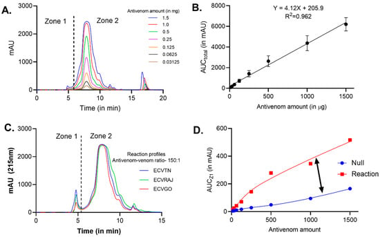
Figure 1.
Size-exclusion chromatography of antivenom and antivenom–venom complexes. (A) Overlay of elution profiles for different amounts of antivenom—0.31, 0.62, 0.125, 0.25, 0.5, 1 and 1.5 mg. (B) Calibration curve for the antivenom amount versus the AUCTotal—area under the curve for the entire chromatogram. The points represent the mean ± SD (n = 3). (C) Overlay of reaction profiles for three venom groups—ECVTN, ECVGO and ECVRAJ—at the antivenom–venom ratio of 150:1. A total of 1.5 mg of VINS antivenom (Batch No. 01AS20055) was incubated with a fixed amount of venom (10 µg) for 30 min at 37 °C before loading on the SEC column. Zone 1 is the area where antivenom–venom complexes are eluted and Zone 2 comprises unbound venom toxins and F(ab)2 molecules. (D) Relationship between the Zone 1 area of null and reaction profiles and the difference is represented by a double-headed arrow.
The peak areas for antivenom–venom complexes (AUCZ1) were significantly different in the venoms (F(2,40) = 117.6, p < 0.0001). The binding parameters estimated by fitting the hyperbolic curve are reported in Supplementary Table S1. The AUCmax was highest for ECVTN at 600.1 (95% CI, 463 to 897) followed by ECVRAJ at 467.4 (95% CI, 399.1 to 569.5) and ECVGO at 262.4 (95% CI, 187.1 to 456.2) (Figure 2).
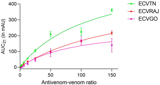
Figure 2.
The binding curve for E. carinatus venom groups is plotted as a graph of different antivenom–venom ratios (150:1, 100:1, 50:1, 25:1, 12.5:1, 6.25:1, 3.125:1 and 1.5:1) and the AUCZ1. The AUCZ1 was estimated by subtracting the Z1 peak area of the reaction profile from the null profile for each of ECVTN, ECVRAJ and ECVGO—E. carinatus venom from Tamil Nadu, Rajasthan and Goa, respectively. Each data point represents the mean ± SD from three independent experiments. Binding parameters—the AUCmax and the EC50—are reported with 95% CI in Supplementary Table S1. Two-way ANOVA showed that venom toxin binding to antivenom was significantly different (F(2,40) = 117.6, p < 0.0001).
Using the equation from the calibration curve (Figure 1B), the maximum amount of immune complex was estimated to be: 95.7 µg for ECVTN, 13.4 µg for ECVGO, and 62.3 µg for ECVRAJ. Using the formulae described in Section 2.2, the valency of antivenom–venom binding was estimated to be 4.0 for ECVTN, 0.67 for ECVGO and 1.89 for ECVRAJ (Supplementary Table S2).
3.2. Preclinical Assays
3.2.1. Coagulant Activity
The clotting times (CT) for venom and antivenom–venom dilutions are reported in Supplementary Tables S3 and S4. We observed significant differences in their CT across different venom groups (F = 356.6, p < 0.0001) (Figure 3A). ECVRAJ had the highest MCD (2.13 ± 0.12 µg), followed by ECVTN (1.57 ± 0.08 µg) and ECVGO (0.58 ± 0.09 µg) (Figure 3B). Since the MCDs were different, amounts of antivenom taken were different to maintain the same antivenom–venom ratios. For all the venom groups, a steep increase in the fold change was observed in antivenom–venom ratios < 25:1 (Figure 3C). We observed that antivenom was able to completely neutralize the clotting effects of ECVRAJ venom at a 100:1 dilution as we could not see clot formation even after 30 min of incubation. The effective dose (ED) is considered as the amount of antivenom that increases the CT by 3-fold, and in the venom groups, it differed significantly (F = 34.01, p < 0.0001). The lowest ED was for ECVGO (127.4 ± 7.5 µg), followed by ECVRAJ (141.2 ± 3.8 µg) and ECVTN (209 ± 29.2 µg).
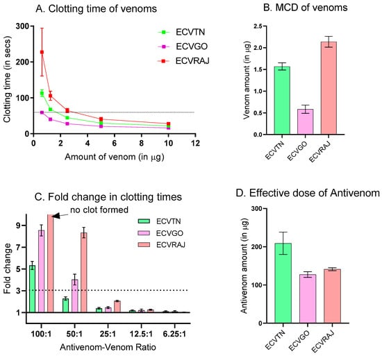
Figure 3.
(A) Plasma clotting times plotted against the amount of E. carinatus venoms (n = 6; p < 0.0001). Clotting times were estimated from the plasma of two healthy donors and readings were taken in triplicate for each plasma. The dashed line corresponds to the clotting time of 60 s. (B) The minimum clotting dose (MCD), defined as the amount of venom required to form plasma clots in 60 s, is represented as a bar plot for all venom groups. One-way ANOVA was performed to show that MCDs were significantly different (p < 0.0001). (C) Fold change in clotting times for different antivenom–venom ratios for each venom group. Double the MCD of each venom group was taken as the challenge dose and incubated with increasing amounts of antivenom for 30 min at 37 °C before testing for residual activity (n = 6). (D) The effective dose of antivenom is defined as the amount of antivenom required to increase the clotting time by 3-fold for ECVTN, ECVGO, and ECVRAJ E. carinatus venom from Tamil Nadu, Goa and Rajasthan. The symbols and bars represent the mean ± SD (n = 6; p < 0.0001). The neutralization of clotting activity by antivenom was shown to be significantly different against venom groups measured using one-way ANOVA (F = 34.01, p < 0.0001).
3.2.2. Proteolytic Activity
We observed significant differences in the activity of venom groups (F = 491.8, p < 0.0001), with ECVRAJ showing the greatest activity at 512.4 U/mg, followed by ECVTN (335.2 U/mg) and ECVGO (160.7 U/mg) (Figure 4A). The decrease in activity represented as the %R is presented in Supplementary Table S5. At the highest antivenom–venom ratio of 168:1, we observed the %R saturation at 31 ± 10% for ECVRAJ compared to 74.3 ± 1.4% for ECVTN and 68.9 ± 4.1% for ECVGO (Figure 4B).
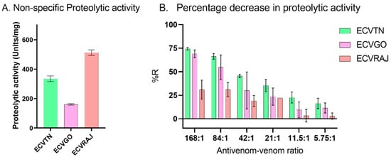
Figure 4.
(A) Non-specific proteolytic activity (n = 4) was measured using Azocasein as a substrate at 10 µg (n = 2) and 20 µg (n = 2) of ECVGO, ECVRAJ and ECVTN–E. carinatus venoms from Goa, Rajasthan and Tamil Nadu, respectively. Enzymatic activity (units/mg) was estimated from the standard curve of proteinase K (n = 4). One-way ANOVA showed proteolytic activity to be significantly different across venom groups (p < 0.0001). (B) Percentage reduction (%R) in the proteolytic activity of 20 µg venom was measured after preincubation (60 min, 37 °C) with different amounts of antivenom. The bar plots are represented as the mean ± SD for ECVTN (n = 3), ECVGO (n = 3) and ECVRAJ (n = 2).
3.2.3. Phospholipase A2 Activity
There was a significant difference in the venom groups for PLA2 activity (F = 15.93; p = 0.0011). The activity was in the following order: ECVRAJ (161.3 U/mg) > ECVGO (126.7 U/mg) > ECVTN (85 U/mg) (Figure 5A). We observed a 100% reduction in PLA2 activity against ECVTN and ECVGO by the antivenom at a 100:1 ratio (Figure 5B, Supplementary Table S6). For ECVRAJ, the reduction in the activity remained unchanged with an increasing antivenom–venom ratio by approximately 20–40%.
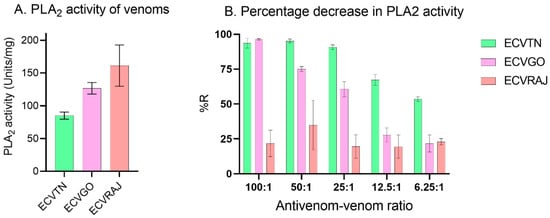
Figure 5.
(A) PLA2 activity (n = 4) for E. carinatus venoms at 5 µg (n = 2) and 2.5 µg (n = 2) was measured using a commercial EnzChek PLA2 assay kit. ECVGO, ECVRAJ and ECVTN E. carinatus venoms were from Goa, Rajasthan and Tamil Nadu, respectively. Enzymatic activity was calculated as described in Section 2.3.3 and expressed in units/mg (n = 4). PLA2 activity was significantly different, as shown by one-way ANOVA (p < 0.001). (B) Percentage reduction (%R) in the PLA2 activity of E. carinatus venom (5 µg) after preincubation (30 min, 37 °C) with different amounts of antivenom. The bars represent the mean ± SD for ECVTN (n = 3), ECVGO (n = 3) and ECVRAJ (n = 2).
3.2.4. Estimating the Antivenom Efficacy Score (ESav)
The logistic dose–response curve was plotted by taking the log of the antivenom–venom ratio as the x-axis and the %R as the y-axis (Figure 6). The curve fitting parameters including the %Rmax (the asymptotic maxima for the percentage reduction) and the IC50 (the antivenom–venom ratio at which 50% Rmax is attained) are presented in Supplementary Table S7. The efficacy scores for antivenom against venom from different locations are presented in Table 1.

Figure 6.
Dose–response curves for the percentage reduction in the proteolytic, PLA2 and clotting activities of E. carinatus venoms at different antivenom–venom ratios (expressed on a log10 scale). The curves were constructed based on the percentage reduction captured in Figure 3, Figure 4 and Figure 5. See the corresponding figures and legends for the amounts of venom and antivenom and the preincubation conditions used in each case. ECVGO, ECVRAJ and ECVTN E. carinatus venoms from Goa, Rajasthan and Tamil Nadu, respectively. The points represent the mean ± SD.

Table 1.
Polyvalent antivenom efficacy score for ECVGO, ECVRAJ and ECVTN E. carinatus venoms from Goa, Rajasthan and Tamil Nadu, respectively, obtained from in vitro assays. The AUCmax and the EC50 are binding parameters for the hyperbolic curve and refer to the maximum area under the curve and the antivenom venom ratio at which the AUC is half the maximum value, respectively. The %Rmax and the IC50 are parameters of the sigmoidal curve fit and refer to the maximum % reduction in venom effects and the antivenom–venom ratio at which the %R is half the maximum value, respectively. The column values represent the mean ± SD (n = 3). Two-way ANOVA showed that the efficacy scores were significantly different for both venom groups and in vitro assays (F(6,30) = 19.62; p < 0.001).
The efficacy scores for antivenom across venom groups and assays were significantly different (F(6,30) = 19.62, p < 0.001; Table 1). Pairwise directional differences were significant (one-tailed Tukey HSD test p < 0.025), suggesting that ECVTN showed higher efficacy compared to the other venoms. The scores for different assays were not consistent (F(3,30) = 9.234, p < 0.001). For instance, antivenom showed better efficacy against ECVTN for the PLA2 and binding assays but not for the other assays. The PLA2 assay for ECVRAJ did not fit the sigmoidal curve as the %R remained at approximately 20–40%, indicating poor neutralization.
4. Discussion
Our study quantified the efficacy of antivenom using a suite of relevant in vitro assays against E. carinatus venom from different locations. The Indian polyvalent antivenom is pepsin-refined containing IgG and F(ab)2 fragments, which can neutralize the toxins either by direct inhibition, where it blocks the catalytic site, rendering toxin inactive; or by indirect inhibition, where it binds elsewhere and is cleared by other immune cells [1]. These modes of inhibition can be captured by quantifying antivenom–venom complexes using either ELISA or SEC-HPLC, although estimating binding through immunoassays alone was inconclusive in the past as several reports claimed poor correlation [6,28]. This could be because toxins that are low in toxicity could be highly immunogenic or vice versa. For instance, in the case of neurotoxic venoms, three-finger toxins (3FTx) are highly neurotoxic but show low immunogenicity due to their small size [29]. To tackle this, some research groups have attempted to isolate the toxic component of venoms and quantified antibodies against them with better correlation [30]. However, this method for testing antivenom efficacy could be difficult to adopt by antivenom manufacturers as their routine procedure, since it involves isolating relevant toxins from the crude venoms.
In this study, we have used a different in vitro approach for assessing antivenom by complementing toxin-binding the characteristics of F(ab)2 molecules with its capacity to neutralize different venom effects. This opens up opportunities to integrate them into the routine testing activities in antivenom production. We calculated the mean value of all the individual efficacy scores obtained from in vitro assays to arrive at an efficacy score (Table 1). This score integrates the results obtained from different in vitro assays by bringing them to the same scale of measurement and is therefore termed as the average efficacy score (ESav). The parameters of the non-linear dose–response curves were used for this purpose. The AUCmax and the %Rmax define the asymptotic maxima of the non-linear curves, indicating maximum antivenom–venom binding and neutralization, respectively. This parameter was used in the numerator to compute the efficacy score because its value is directly proportional to the efficacy. The EC50 and the IC50 indicated the antivenom–venom ratio required to achieve half of the AUCmax and the 50% Rmax, respectively. This parameter denotes the affinity of antivenom towards venom and was used in the denominator since it was inversely related to efficacy. This computation approach could be applied to any other assay that shows a hyperbolic or logistic curve to different antivenom–venom ratios.
In our previous report, we showed that the maximum percentage of antivenom bound was less than 7%, which is consistent with the current findings [15]. The effective F(ab)2 molecules for ECV were lower than the standard (~20%), which is correlated with a good outcome for in vivo neutralization [31]. This could be among the reasons for the use of a high number of vials for snake envenoming treatment in India. The antivenom showed higher binding for ECVTN toxins than toxins of the other venoms, as indicated by the AUCmax (Figure 2). The valency of antivenom–venom binding, indicating the number of F(ab)2 molecules bound per toxin molecule, was also higher for ECVTN than for the other venoms. Venom immunization in equines generate polyclonal antibodies, which could bind to multiple epitopes on a single toxin [32]. High valency of the antivenom might increase the chances of neutralization, either by the complement system removing antibody-bound toxins or by macrophages ingesting it [33].
Different functional assays were assessed to evaluate the ability of antivenom to neutralize the toxicity of different venoms. The assays were selected keeping in mind E. carinatus venom composition and symptomatology. For instance, venom-induced consumption coagulopathy (VICC) is the most common clinical syndrome, often complicated by life-threatening hemorrhage [34,35]. We measured this by an in vitro clotting assay and observed that antivenom was able to neutralize all the venoms. Although the %Rmax was similar for all venom groups, the efficacy score against clotting activity was highest for ECVRAJ due to its low IC50.
Non-specific proteolysis by snake venom metalloproteinases (SVMP) and serine proteases (SP) and cytotoxicity by phospholipase A2 (PLA2) have a variety of other pathophysiological effects including inflammation and necrosis in the bite site [20,36]. The neutralization efficacy of antivenom measured using these functional assays was limited to the direct mode of inhibition and was measured as the percentage reduction (%R) in proteolytic and PLA2 activity. Among the venom groups, ECVRAJ showed a poor %R for proteolytic and PLA2 assays, indicating the absence of sufficient F(ab)2 molecules in antivenom to block the catalytic sites of toxins involved in these assays. Since the antivenom did not reduce the PLA2 effects of ECVRAJ, we were not able to fit a logistic curve and estimate the parameters. In the proteolytic assay, although the %Rmax for ECVGO was higher than that for ECVRAJ, the efficacy score was low due to the high IC50. The mice neutralization assay tested at the manufacturer’s facility indicated neutralization of 566 µg of E. carinatus venom (IRULA) per ml of antivenom. The concentration of antivenom given as 58.4 mg/mL suggests that ~103 parts of antivenom are required to neutralize 1 part of E. carinatus venom. We observed complete neutralization for the PLA2 assay at 25:1 and for the coagulation assay at between 50:1 and 100:1. This suggests that antibody neutralization capacity could be overestimated for in vitro assays.
The binding and neutralization by F(ab)2 molecules were significantly different across venom groups (Figure 6) and this provided insights into the variability of the efficacy scores. This could be used to draw a correlation with the ED50. The average efficacy score (ESav) of antivenom was the highest for ECVTN, and this was supported by the fact that venoms procured from this region were used for preparing antivenom by most manufacturers. We acknowledge that without data on the ED50 from mice experiments, replacement with in vitro assays would not be feasible. We could not perform these experiments due to limited access to the venoms. Venoms are a limiting resource in India due to the restrictions on accessing venomous snake species. We were able to perform the entire study with <1 mg of venom from the three regions. Future refinement of in vitro assays will also have important implications for the optimal use of venoms. Although we could not capture all the in vitro effects of E. carinatus venom, such as fibrinogenolysis, L-amino acid oxidase, and hyluronidase activity, through this study, we provided a novel framework to arrive at an efficacy score. Future studies could estimate the efficacy scores using different in vitro assays, and quantify the median effective dose in mice. The ESav quantified in this study might be a useful predictor of the ED50, as it accounted for the diverse effects of the venom.
Supplementary Materials
The following supporting information can be downloaded at: https://www.mdpi.com/article/10.3390/toxins14070481/s1, Table S1: Binding parameters for hyperbolic curve fitted AUC vs Antivenom-venom amounts plot (Figure 2) are listed above where AUCmax is the asymptotic maxima for AUCZ1 and EC50 is antivenom-venom ratio at which AUCZ1 was half the AUCmax, Table S2: Number of F(ab)2 molecules per venom toxin is estimated using the formula described in methodology Section 2.2. Average molecular weight of toxin for each venom group was estimated by densitometry analysis of SDS-PAGE bands reported in (Bhatia and Vasudevan, 2020). Molecular weight of F(ab)2 molecules was taken as 110 kDa, Table S3: Clotting times (CT) for different amounts of venoms. Clotting times were estimated from the plasma of two healthy donors and readings were taken in triplicate for each plasma, Table S4: Fold change in the clotting times (CT) for different antivenom-venom ratios are represented in the table above. It is calculated by dividing CT of antivenom-venom dilution with CT of challenge dose of venom (2*MCD). The readings were taking in triplicates for the plasma from 2 volunteers, Table S5: Percentage decrease in proteolytic activity referred as %R were estimated using formula described in methodology Section 2.3.2. The table represents %R (n = 3 for ECVTN and ECVGO; n = 2 for ECVRAJ) for different antivenom-venom ratios, Table S6: Percentage decrease in PLA2 activity (%R) calculated by the formula described in methodology Section 2.3.2. at different antivenom-venom ratio is represented for each venom group (n = 3 for ECVTN and ECVGO; n = 2 for ECVRAJ), Table S7: Parameters for sigmoidal curve fitted to the %R vs Log(antivenom-venom ratio) plot are represented in the table. %Rmax is maximum reduction in the activity and IC50 is antivenom-venom ratio at which %Rmax is half.
Author Contributions
Conceptualization, S.B., A.B. and K.V.; data curation, S.B. and A.B.; formal analysis, S.B. and A.B.; funding acquisition, K.V.; investigation, S.B. and A.B.; methodology, S.B. and K.V.; project administration, K.V.; resources, K.V.; software, K.V.; supervision, K.V.; validation, S.B. and A.B.; visualization, S.B. and A.B.; writing—original draft, S.B. and A.B. All authors have read and agreed to the published version of the manuscript.
Funding
The provided financial and technical support for this study. The Council for Scientific and Industrial Research, India provided a graduate student scholarship to S.B. for conducting his research. The SERB-Department of Science and Technology provided support to K.V. and a fellowship to A.B. for this study through grant EMR/2017/005515.
Institutional Review Board Statement
The study was conducted in accordance with the Institutional Ethical Committee (IEC), and approved by the IEC of CSIR-Centre for Cellular and Molecular Biology (IEC-70/2019 dated 30 April 2019) for studies involving humans.
Informed Consent Statement
Informed consent was obtained from all the subjects involved in the study.
Data Availability Statement
All data associated with the study are provided in the supplementary files. Data from this study has not been archived in public repositories.
Acknowledgments
We thank the CCMB Institutional Ethical Committee for providing permission (IEC-70/2019) to collect blood samples. Forest departments of Goa and Rajasthan provided permission to collect ECV and extended logistic support. The Irula Cooperative society provided us with the pooled ECV used in this study. V Krishna Kumari provided valuable assistance for RP-HPLC. We thank VINS bioproducts for providing us with antivenom vials and relevant information. We thank the anonymous reviewers of this manuscript for their comments, which have helped in improving it.
Conflicts of Interest
The authors declare no conflict of interest.
References
- Gutiérrez, J.M.; Vargas, M.; Segura, A.; Herrera, M.; Villalta, M.; Solano, G.; Sánchez, A.; Herrera, C.; León, G. In Vitro Tests for Assessing the Neutralizing Ability of Snake Antivenoms: Toward the 3Rs Principles. Front. Immunol. 2021, 11, 617429. [Google Scholar] [CrossRef]
- WHO. Guidelines for the Production, Control and Regulation of Snake Antivenom Immunoglobulins; Replacement of Annex 2 of WHO Technical Report Series, No. 964; WHO: Geneva, Switzerland, 2018. [Google Scholar]
- Sells, P.G. Animal experimentation in snake venom research and in vitro alternatives. Toxicon 2003, 42, 115–133. [Google Scholar] [CrossRef]
- Chacón, F.; Oviedo, A.; Escalante, T.; Solano, G.; Rucavado, A.; Gutiérrez, J.M. The lethality test used for estimating the potency of antivenoms against Bothrops asper snake venom: Pathophysiological mechanisms, prophylactic analgesia, and a surrogate in vitro assay. Toxicon 2015, 93, 41–50. [Google Scholar] [CrossRef] [PubMed]
- Lopes-De-Souza, L.; Costal-Oliveira, F.; Stransky, S.; de Freitas, C.F.; Guerra-Duarte, C.; Braga, V.M.; Chávez-Olórtegui, C. Development of a cell-based in vitro assay as a possible alternative for determining bothropic antivenom potency. Toxicon 2019, 170, 68–76. [Google Scholar] [CrossRef]
- Barbosa, C.F.; Rodrigues, R.J.; Olortegui, C.C.; Sanchez, E.F.; Heneine, L.G. Determination of the neutralizing potency of horse antivenom against bothropic and crotalic venoms by indirect enzyme immunoassay. Braz. J. Med. Biol. Res. 1995, 28, 1077–1080. [Google Scholar] [PubMed]
- Maria, W.S.; Cambuy, M.O.; Costa, J.O.; Velarde, D.T.; Chávez-Olórtegui, C. Neutralizing potency of horse antibothropic antivenom. Correlation between in vivo and in vitro methods. Toxicon 1998, 36, 1433–1439. [Google Scholar] [CrossRef]
- Rial, A.; Morais, V.; Rossi, S.; Massaldi, H. A new ELISA for determination of potency in snake antivenoms. Toxicon 2006, 48, 462–466. [Google Scholar] [CrossRef]
- Rungsiwongse, J.; Ratanabanangkoon, K. Development of an ELISA to assess the potency of horse therapeutic antivenom against Thai cobra venom. J. Immunol. Methods 1991, 136, 37–43. [Google Scholar] [CrossRef]
- Theakston, R.; Lloyd-Jones, M.J.; Reid, H. Micro-Elisa for Detecting and Assaying Snake Venom and Venom-Antibody. Lancet 1977, 310, 639–641. [Google Scholar] [CrossRef]
- Pla, D.; Rodríguez, Y.; Calvete, J.J. Third Generation Antivenomics: Pushing the Limits of the In Vitro Preclinical Assessment of Antivenoms. Toxins 2017, 9, 158. [Google Scholar] [CrossRef]
- Sanny, C.G. Antibody–antigen binding study using size-exclusion liquid chromatography. J. Chromatogr. B 2002, 768, 75–80. [Google Scholar] [CrossRef]
- Senji Laxme, R.R.S.; Khochare, S.; Attarde, S.; Suranse, V.; Iyer, A.; Casewell, N.R.; Whitaker, R.; Martin, G.; Sunagar, K. Biogeographic venom variation in Russell’s viper (Daboia russelii) and the preclinical inefficacy of antivenom therapy in snakebite hotspots. PLoS Negl. Trop. Dis. 2021, 15, e0009247. [Google Scholar] [CrossRef] [PubMed]
- Senji Laxme, R.R.; Khochare, S.; De Souza, H.F.; Ahuja, B.; Suranse, V.; Martin, G.; Whitaker, R.; Sunagar, K. Beyond the ‘big four’: Venom profiling of the medically important yet neglected Indian snakes reveals disturbing antivenom deficiencies. PLoS Negl. Trop. Dis. 2019, 13, e0007899. [Google Scholar] [CrossRef]
- Bhatia, S.; Blotra, A.; Vasudevan, K. Immunorecognition capacity of Indian polyvalent antivenom against venom toxins from two populations of Echis carinatus. Toxicon 2021, 201, 148–154. [Google Scholar] [CrossRef]
- Bawaskar, H.; Bawaskar, P. Profile of snakebite envenoming in western Maharashtra, India. Trans. R. Soc. Trop. Med. Hyg. 2002, 96, 79–84. [Google Scholar] [CrossRef]
- Punde, D.P. Management of snake-bite in rural Maharashtra: A 10-year experience. Natl. Med. J. India 2005, 18, 71–75. [Google Scholar]
- Patra, A.; Kalita, B.; Chanda, A.; Mukherjee, A.K. Proteomics and antivenomics of Echis carinatus carinatus venom: Correlation with pharmacological properties and pathophysiology of envenomation. Sci. Rep. 2017, 7, 17119. [Google Scholar] [CrossRef]
- Bhatia, S.; Vasudevan, K. Comparative proteomics of geographically distinct saw-scaled viper (Echis carinatus) venoms from India. Toxicon X 2020, 7, 100048. [Google Scholar] [CrossRef]
- Kini, R.M. Excitement ahead: Structure, function and mechanism of snake venom phospholipase A2 enzymes. Toxicon 2003, 42, 827–840. [Google Scholar] [CrossRef]
- Gutiérrez, J.M.; Escalante, T.; Rucavado, A.; Herrera, C. Hemorrhage Caused by Snake Venom Metalloproteinases: A Journey of Discovery and Understanding. Toxins 2016, 8, 93. [Google Scholar] [CrossRef]
- White, J. Snake venoms and coagulopathy. Toxicon 2005, 45, 951–967. [Google Scholar] [CrossRef] [PubMed]
- Sanny, C.G. In vitro evaluation of total venom–antivenin immune complex formation and binding parameters relevant to antivenin protection against venom toxicity and lethality based on size-exclusion high-performance liquid chromatography. Toxicon 2011, 57, 871–881. [Google Scholar] [CrossRef] [PubMed]
- Theakston, R.D.; A Reid, H. Development of simple standard assay procedures for the characterization of snake venom. Bull. World Health Organ. 1983, 61, 949–956. [Google Scholar]
- Pornmuttakun, D.; Ratanabanangkoon, K. Development of an in vitro potency assay for antivenom against Malayan pit viper (Calloselasma rhodostoma). Toxicon 2014, 77, 1–5. [Google Scholar] [CrossRef]
- Caldas, C.; Cherqui, A.; Pereira, A.; Simões, N. Purification and Characterization of an Extracellular Protease from Xenorhabdus nematophila Involved in Insect Immunosuppression. Appl. Environ. Microbiol. 2002, 68, 1297–1304. [Google Scholar] [CrossRef]
- Fonteh, A.N.; Chiang, J.; Cipolla, M.; Hale, J.; Diallo, F.; Chirino, A.; Arakaki, X.; Harrington, M.G. Alterations in cerebrospinal fluid glycerophospholipids and phospholipase A2 activity in Alzheimer’s disease. J. Lipid Res. 2013, 54, 2884–2897. [Google Scholar] [CrossRef]
- Alape-Girón, A.; Miranda-Arrieta, K.; Cortes-Bratti, X.; Stiles, B.G.; Gutiérrez, J. A comparison of in vitro methods for assessing the potency of therapeutic antisera against the venom of the coral snake Micrurus nigrocinctus. Toxicon 1997, 35, 573–581. [Google Scholar] [CrossRef][Green Version]
- Liu, B.S.; Jiang, B.R.; Hu, K.C.; Liu, C.H.; Hsieh, W.C.; Lin, M.H.; Sung, W.C. Development of a Broad-Spectrum Antiserum against Cobra Venoms Using Recombinant Three-Finger Toxins. Toxins 2021, 13, 556. [Google Scholar] [CrossRef]
- Khaing, E.M.; Hurtado, P.R.; Hurtado, E.; Zaw, A.; White, J.; Warrell, D.A.; Alfred, S.; Mahmood, M.A.; Peh, C.A. Development of an ELISA assay to determine neutralising capacity of horse serum following immunisation with Daboia siamensis venom in Myanmar. Toxicon 2018, 151, 163–168. [Google Scholar] [CrossRef]
- Calvete, J.J.; Sanz, L.; Pla, D.; Lomonte, B.; Gutiérrez, J.M. Omics Meets Biology: Application to the Design and Preclinical Assessment of Antivenoms. Toxins 2014, 6, 3388–3405. [Google Scholar] [CrossRef]
- Sapsutthipas, S.; Leong, P.K.; Akesowan, S.; Pratanaphon, R.; Tan, N.H.; Ratanabanangkoon, K. Effective Equine Immunization Protocol for Production of Potent Poly-specific Antisera against Calloselasma rhodostoma, Cryptelytrops albolabris and Daboia siamensis. PLoS Negl. Trop. Dis. 2015, 9, e0003609. [Google Scholar] [CrossRef] [PubMed]
- Laustsen, A.H.; Maria Gutiérrez, J.; Knudsen, C.; Johansen, K.H.; Bermúdez-Méndez, E.; Cerni, F.A.; Jürgensen, J.A.; Ledsgaard, L.; Martos-Esteban, A.; Øhlenschlæger, M.; et al. Pros and cons of different therapeutic antibody formats for recombinant antivenom development. Toxicon 2018, 146, 151–175. [Google Scholar] [CrossRef]
- Berling, I.; Isbister, G. Hematologic Effects and Complications of Snake Envenoming. Transfus. Med. Rev. 2015, 29, 82–89. [Google Scholar] [CrossRef]
- Maduwage, K.; Isbister, G.K. Current Treatment for Venom-Induced Consumption Coagulopathy Resulting from Snakebite. PLoS Negl. Trop. Dis. 2014, 8, e3220. [Google Scholar] [CrossRef]
- Guerranti, R.; Cortelazzo, A.; Hope-Onyekwere, N.S.; Furlani, E.; Cerutti, H.; Puglia, M.; Bini, L.; Leoncini, R. In vitro effects of Echis carinatus venom on the human plasma proteome. Proteomics 2010, 10, 3712–3722. [Google Scholar] [CrossRef]
Publisher’s Note: MDPI stays neutral with regard to jurisdictional claims in published maps and institutional affiliations. |
© 2022 by the authors. Licensee MDPI, Basel, Switzerland. This article is an open access article distributed under the terms and conditions of the Creative Commons Attribution (CC BY) license (https://creativecommons.org/licenses/by/4.0/).