Article

Daphnia’s Adaptive Molecular Responses to the Cyanobacterial Neurotoxin Anatoxin-α Are Maternally Transferred

by
Anke Schwarzenberger
* and
Dominik Martin-Creuzburg
Limnological Institute, University Konstanz, Mainaustraße 252, 78464 Konstanz, Germany
*
Author to whom correspondence should be addressed.
Toxins 2021, 13(5), 326; https://doi.org/10.3390/toxins13050326
Submission received: 31 March 2021 / Revised: 27 April 2021 / Accepted: 29 April 2021 / Published: 30 April 2021
(This article belongs to the Section Marine and Freshwater Toxins)

Abstract

:
Cyanobacterial blooms are an omnipresent and well-known result of eutrophication and climate change in aquatic systems. Cyanobacteria produce a plethora of toxic secondary metabolites that affect humans, animals and ecosystems. Many cyanotoxins primarily affect the grazers of phytoplankton, e.g., Daphnia. The neurotoxin anatoxin-α has been reported world-wide; despite its potency, anatoxin-α and its effects on Daphnia have not been thoroughly investigated. Here, we investigated the effects of the anatoxin-α-producing Tychonema on life-history parameters and gene expression of nicotine-acetylcholine receptors (NAR), the direct targets of anatoxin-α, using several D. magna clones. We used juvenile somatic growth rates as a measure of fitness and analyzed gene expression by qPCR. Exposure to 100% Tychonema reduced the clones’ growth rates and caused an up-regulation of NAR gene expression. When 50% of the food consisted of Tychonema, none of the clones were reduced in growth and only one of them showed an increase in NAR gene expression. We demonstrate that this increased NAR gene expression can be maternally transferred and that offspring from experienced mothers show a higher growth rate when treated with 50% Tychonema compared with control offspring. However, the addition of further (anthropogenic) stressors might impair Daphnia’s adaptive responses to anatoxin-α. Especially the presence of certain pollutants (i.e., neonicotinoids), which also target NARs, might reduce Daphnia’s capability to cope with anatoxin-α.
Key Contribution: Daphnia can cope with dietary anatoxin-α if the percentage of the neurotox-in-producing cyanobacterium is not too high. Furthermore, the up-regulation of NAR gene ex-pression is maternally transferred and leads to higher fitness in the offspring of experienced mothers.

1. Introduction

Over the last decades, cyanobacterial blooms have increased in frequency in freshwater ecosystems because of the combined effects of eutrophication, global warming, and low riverflows due to drought conditions [1]. Cyanobacteria are a risk to the environment and human health because they produce a variety of toxins [2,3]. One exceptionally dangerous toxin for humans, livestock and wildlife is the Very Fast Death Factor [4,5]—the neurotoxic alkaloid anatoxin-α. This toxin acts very quickly on the nervous system and an antidote is unknown. Anatoxin-α has been observed in natural lake blooms globally [6,7,8,9,10,11,12]. Furthermore, it is produced by different cyanobacterial species, e.g., Tychonema [12], Anabaena [7,13,14], Pseudoanabaena, Planktothrix [7] and Aphanizomenon [15]. Therefore, the management of cyanobacterial blooms with anatoxin-α is crucial because this toxin is a concern for human, animal and ecosystem health [6], and because anatoxin-α has already been found in dietary supplements containing cyanobacteria [16].
Anatoxin-α is probably transferred through the food-web via the ingestion of zooplankton such as Daphnia and affects both fish and zooplankton [8,15]. Furthermore, anatoxin-α can accumulate in the tissues of fish [8]. Representatives of the genus Daphnia occupy a central position in freshwater food webs. They exert a significant grazing pressure on phytoplankton, including cyanobacteria, and serve as food for zooplanktivorous fish and invertebrate predators. Especially at high temperatures, population growth rates of D. pulex have been shown to be negatively affected by the ingestion of an anatoxin-α-producing cyanobacterium [14]. However, anatoxin-α has been observed to have a positive effect on D. dentifera by preventing infection by the fungal parasite Metschnikowia [13].
In vertebrates, anatoxin-α inhibits the nicotine-acetylcholine receptors (NARs) [4,17,18]. This can result in the permanent stimulation of muscles, which in turn leads to paralysis and possibly death due to respiratory arrest [19]. It is not known whether Daphnia‘s NARs are also affected by anatoxin-α, and whether or not Daphnia possess the potential to adapt to anatoxin-α by specific molecular responses. Molecular adaptations and responses to other cyanobacterial toxins with other modes of action have already been observed in Daphnia: One example of these are microcystins, which inhibit protein phosphatases II of Daphnia in vitro [20]. Daphnia respond to this toxin type by manifold means such as: (a) Higher activity of the detoxifying enzyme gluthathion-S-transferase [21], (b) up-regulation of certain transporters, most likely in order to export microcystins from the cells [22,23,24], and (c) increased activity of malate dehydrogenase [21]. In the case of dietary protease inhibitors (PIs), D. magna has been demonstrated to increase protease gene expression and activity [25,26]. Furthermore, Daphnia populations can locally adapt to PIs [27]. This adaptation is a result of the positive selection of the respective protease type [28], production protease isoforms that are different from those produced by sensitive populations, and a change in protease gene copy number which leads to increased protease activity and higher gene expression.
Furthermore, it has been shown that if Daphnia mothers ingest toxic cyanobacteria, their offspring show higher fitness (i.e., higher growth or survival) than offspring from unexposed mothers [29,30,31]. Those positive maternal effects on offspring fitness were shown to be due to the transfer of the specific molecular responses from mothers to their offspring (microcystins: higher activity of glutathione S-transferase and malate dehydrogenase [21]; protease inhibitors: increase in protease gene expression [30]). In the case of anatoxin-α, it is not known whether similar maternal effects can increase offspring fitness in Daphnia, and whether a putative specific response, e.g., increased NAR gene expression, is maternally transferred.
The aim of this study was to investigate the effects of anatoxin-α on D. magna and the capability of D. magna to respond and adapt to this toxin type by increasing NAR gene expression. For this, we measured the juvenile somatic growth rates of three different D. magna clones grown on different concentrations of the anatoxin-α-producing cyanobacterium T. bourrellyi and on a control food (the green alga Scenedesmus obliquus), we also quantified their NAR gene expression. Furthermore, we treated the F0 generation of one D. magna clone with two different concentrations of T. bourrellyi and measured the maternal and offspring gene expression and fitness of the exposed and naïve F1 offspring in maternal effect experiments.

2. Results

2.1. Clones

2.1.1. Somatic Growth Rates of the Three D. magna Clones

The clones achieved similar growth rates both on the control food (100% S. obliquus) and on the mixed diet containing 50% of T. bourrellyi; growth rates were significantly reduced only on the 100% T. bourrellyi diet (Tukey’s HSD after one-way ANOVA: FR-LR-6-1: F2,6 = 1175.2; p < 0.001; Binnensee: F2,6 = 1116.7; p < 0.001; MA-ES-3: F2,6 = 291.5; p = 0.002; Figure 1). The culture’s anatoxin-α concentration in the experiments was 161.8 ng per mg carbon.

2.1.2. Gene Expression

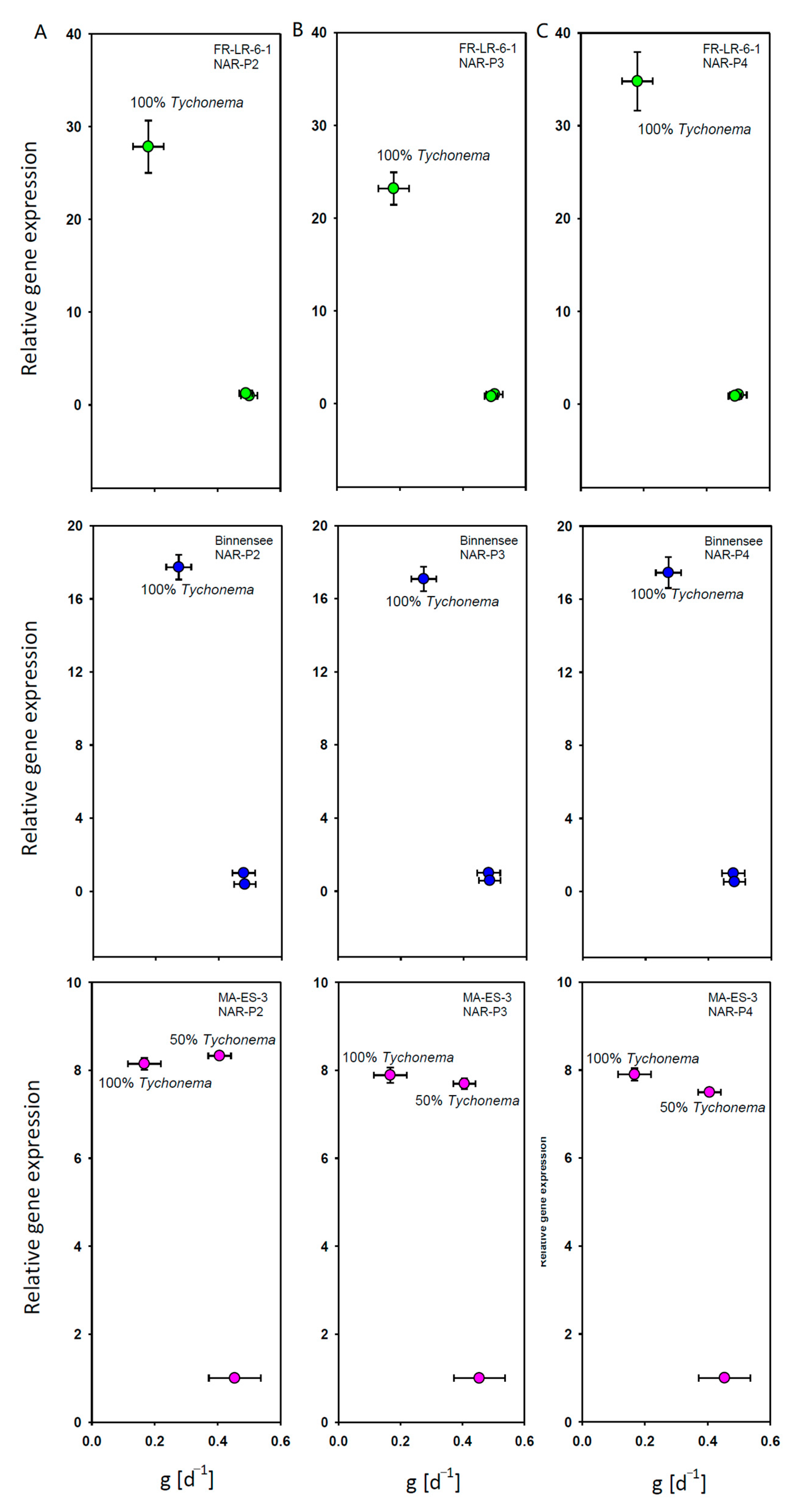
In the clones FR-LR6-1 and Binnensee, both the gene expression of the three nicotine-acetylcholine receptor genes and the juvenile somatic growth rates were similar between the control and 50% T. bourrellyi. In the treatment with 100% T. bourrellyi, an increase in gene expression was accompanied by a lower somatic growth rate (Figure 2A,B). In contrast, somatic growth in clone MA-ES-3 was similar on the control food and 50% T. bourrellyi, but the gene expression already increased on 50% T. bourrellyi (Figure 2C).

2.2. Maternal Effects

2.2.1. First Experiment: 50% T. bourrellyi

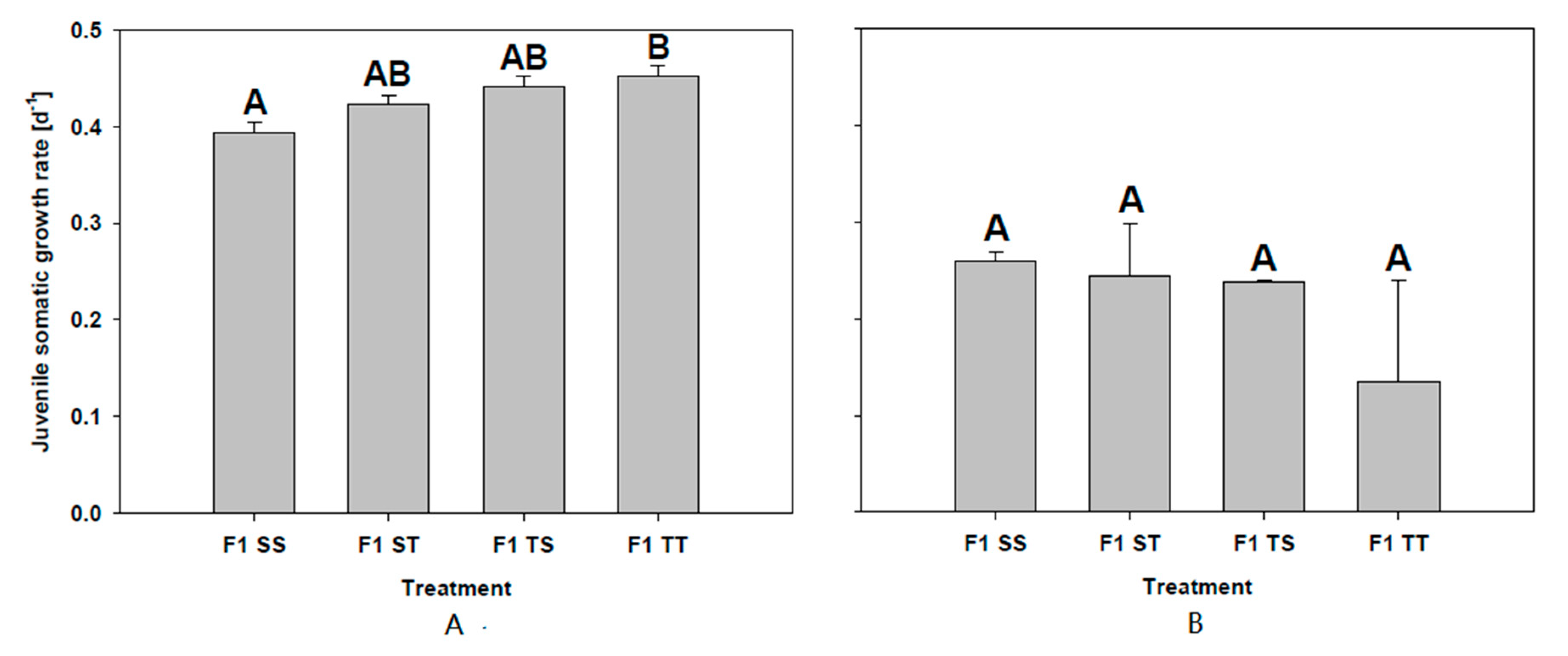
No difference in the juvenile somatic growth rates was observed between animals grown on the control food or on 50% T. bourrellyi. However, a significantly higher growth rate was found in F1 TT in comparison to F1 SS (Tukey’s HSD after one-way ANOVA F3,8 = 8320.70; p = 0.015; Figure 3A). Neither clutch size nor age at first reproduction differed between the treatments of the two F1 groups (clutch size; F1SS: 14.33 ± 1.15; F1ST: 20 ± 7.81; F1TS: 14.66 ± 1.15; F1TT: 16.67 ± 7.37; one-way ANOVA F3,8 = 901.04; p = 0.726; age at first reproduction: F1SS: 10 ± 0; F1ST: 9.67 ± 0.58; F1TS: 10 ± 0; F1TT: 9.33 ± 1.16); one-way ANOVA F3,8 = 12241.42; p = 0.556).
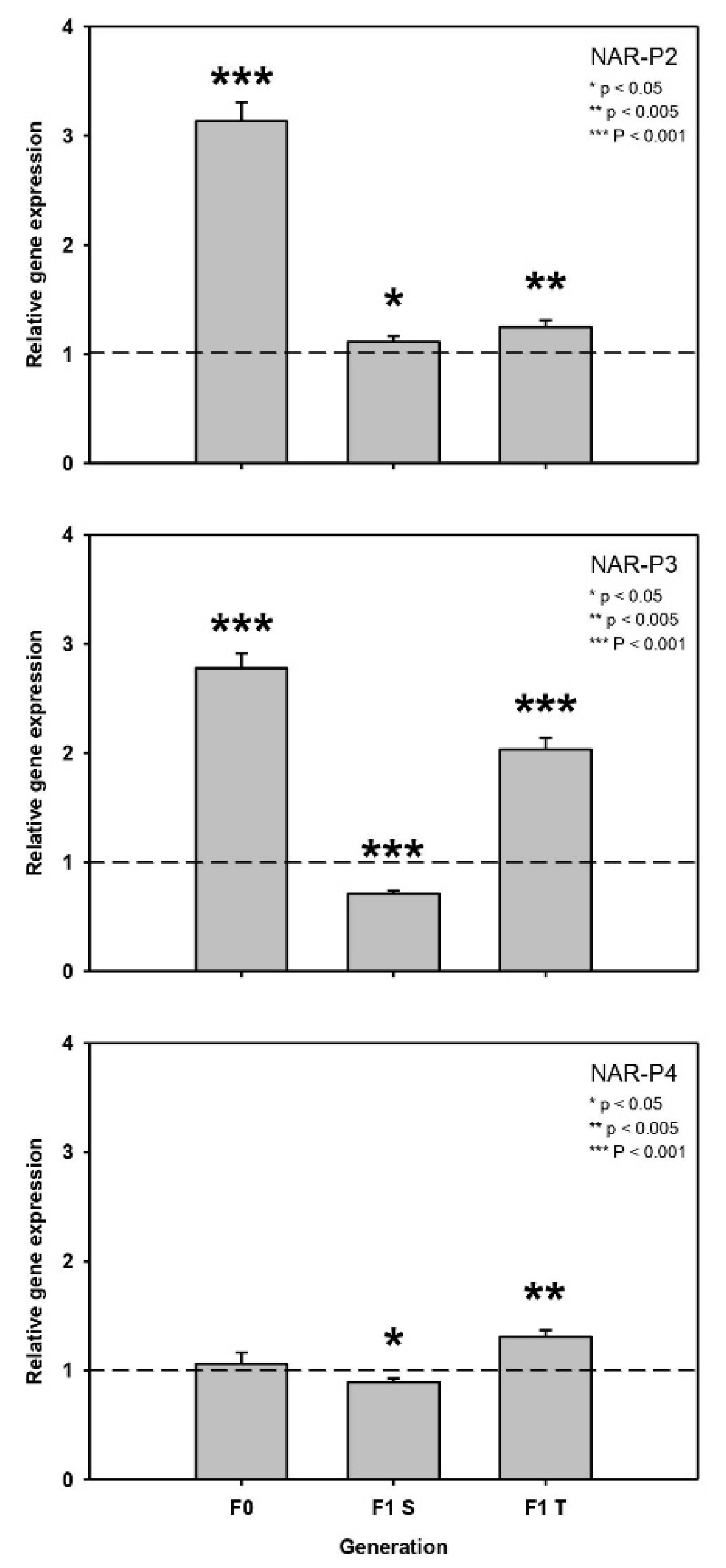
The genes NAR-P2 and NAR-P3 were significantly up-regulated on T. bourellyi, both in the F0 and F1 T generations (after six days), but Nar-P2 was up-regulated and Nar-P3 down-regulated in F1 S (NAR-P2: F0: t = 16.44, p < 0.001; F1S: t = 3.02, p = 0.039; F1T: t = 6.67, p = 0.003; NAR-P3: F0: t = 21.62, p < 0.001; F1 S: t = -11.87, p < 0.001; F1 T: t = 16.77, p < 0.001; Figure 4). NAR-P4 was not regulated in F0, was slightly down-regulated in F1 S and was up-regulated in F1 T (NAR-P4: F0: t = 0.85, p = 0.445; F1 S: t = -3.97, p = 0.017; F1 T: t = 8.298, p = 0.001; Figure 4).

2.2.2. Second Experiment: 100% T. bourrellyi

Because of the high mortality in F1 ST and F1 TT (F1SS: 0; F1ST: 73.33 ± 11.55%; F1TS: 0; F1TT: 90 ± 52.92%), the experiment was terminated on day four and the juvenile somatic growth rates were compared; no difference was found between the treatments and the offspring groups (one-way ANOVA F3,7 = 64.94; p = 0.174; Figure 3B).

3. Discussion

Anatoxin-α has been shown to induce behavioural changes, inhibit heart rate and alter oxygen consumption in D. magna [32]. Claska and Gilbert [3] reported reduced fecundity of D. pulex after the ingestion of an anatoxin-α-producing cyanobacterium. Furthermore, the growth rates and survival of D. similis were negatively affected [33]. Similarly, our tested D. magna clones which had ingested T. bourellyi were strongly and significantly reduced in growth when fed with 100% but not with 50% of the cyanobacterium. Although other unknown cyanotoxins might be present in T. bourellyi and might affect Daphnia, we feel confident that the negative effect on juvenile somatic growth rates was likely because of the presence of anatoxin-α. This is because (i) our T. bourellyi strain does not produce microcystins [12], and (ii) when we fed another D. magna clone with an aging T. bourellyi culture in which anatoxin-α was not detected via ELISA, we found no growth rate reduction on 100% T. bourellyi (Supplementary Figure S1).
It has been demonstrated that organisms can respond to anatoxin-α exposure by increasing the activity of detoxifying enzymes sucj as peroxidases and glutathione S-transferases [34]. However, until now it was unclear whether organisms can also respond or even adapt to anatoxin-α by changes in the production of the direct targets of anatoxin-α, i.e., NARs [16,17,35]. It has been shown that D. magna can respond to other cyanotoxins by increasing the gene expression and activity of the cyanotoxins’ direct targets (cf. protease inhibitors and dietary proteases [26]). We found that the gene expression of three NARs increased when the three D. magna clones were fed with 100% T. bourellyi. In MA-ES-3, this increase in gene expression was already observed on 50% T. bourellyi, which was accompanied by a similar growth rate as on the control food. This suggests that D. magna can actually respond and probably adapt to anatoxin-α by increasing target gene expression.
It is advantageous for parents and offspring to show the same phenotype if they share the same risk of exposure to the same deleterious environment. Adaptive phenotypic responses can be maternally transferred to the offspring, affecting the offspring’s development and ultimately its fitness [36]. A maternal effect is adaptive if the offspring’s fitness increases. The generation time of Daphnia is typically shorter than the exposure to cyanobacteria. Therefore, the maternal transfer of a certain phenotype is beneficial. For cyanobacterial protease inhibitors it has been shown that the mothers’ (F0 generation) increase in gene expression was also transferred to their offspring (F1 generation), and that this transfer of gene expression was accompanied by a higher fitness of the offspring of experienced mothers [30]. We hypothesized that such a maternal effect could also be found in clone MA-ES-3 in response to anatoxin-α. In fact, we found that in the experiment with 50% T. bourellyi the higher gene expression of NAR-P2 and NAR-P3 in the experienced mothers was also observed as increased gene expression in F1 TT in comparison to F1 TS. A higher gene expression of NAR-P2 was also observed in the offspring of naïve mothers, i.e., F1 ST in comparison to F1 SS; however this increase was clearly lower than in the offspring of experienced mothers. Furthermore, a gene expression increase of NAR-P4 was observed in F1 TT in comparison to siblings grown on the control food, although the increase was neither observed in their experienced mothers nor in the offspring of naïve mothers (F1 ST). Although the growth rate, clutch size, and age until the first reproduction of the offspring of both naïve and experienced mothers was not different on thecontrol food, we found that the F1 TT offspring of experienced mothers showed a higher growth rate than the F1 SS offspring of naïve mothers. Presumably, the effects of anatoxin-α on the growth of the F1 TT offspring were suppressed by the increase in NAR gene expression. Furthermore, it is possible that with this elimination of toxin stress, the offspring benefitted from the mixed diet by showing an even higher growth than the F1 SS offspring. This seems likely, since nutritional upgrading by non-toxic bacteria has already been described [37]. We concluded that the maternal transfer of increased gene expression led to a higher fitness in the F1 generation of experienced mothers.
We wanted to test whether this effect was more apparent when the offspring was stressed with even higher concentrations of T. bourellyi (100%). The experienced mothers were still grown on 50% T. bourellyi in order to ensure a similar maternal transfer of increased gene expression as in the first experiment. We found that the juvenile somatic growth rates of all types of offspring were similar. However, the experiment was ended already after four days because the mortality of the offspring grown on T. bourellyi was very high (both in F1 ST and F1 TT). Therefore, no fitness effect of the maternal transfer of increased gene expression was observed in the experiment with 100% T. bourellyi.
In conclusion, increased NAR gene expression is maternally transferred and leads to higher fitness in the offspring of experienced mothers if the percentage of T. bourellyi is not too high. It remains to be tested whether the presence of several other cyanotoxins within a bloom or the presence of additional stressors (e.g., anthropogenic stressors such as pharmaceuticals or light pollution) can reduce the positive effect of this maternal transfer. Particularly the presence of pollutants that also affect nicotine-acetylcholine receptors of arthropods (e.g., neonicotinoids [38]) might reduce Daphnia’s capability to cope with anatoxin-α.

4. Materials and Methods

4.1. Cultures

Experiments were conducted with an anatoxin-α producing strain of T. bourrellyi which was isolated from Lake Mjøsa, Norway [10]. T. bourrellyi was grown at 20 °C in 150 mL Cyano medium [39] at constant illumination (100 μmol quanta m−2 s−1) for seven days prior to the experiments. As the concentration of anatoxin-α decreases in aging cultures of T. bourrellyi, we always verified that anatoxin-α was present in the culture in two technical duplicates at day four of the experiments. For each replicate, 6 mL of the culture was centrifuged, and the pellet was resuspended with 500 µL Cyano medium and mixed with 500 µL 60% ethanol. The mix was diluted 1:100 with Millipore water, and its anatoxin-α content was determined with an ELISA kit (Anatoxin- α (VFDF) RBA, 96-test; Sension, Augsburg, Germany) as according to the manufacturer’s instructions and by making use of the calibration curve from the kit. Pure Cyano medium without T. bourellyi served as blank and was treated in the same way. The anatoxin-α content (in ng) was related to the carbon (C) concentration of the tested T. bourellyi culture via a previously determined carbon-light extinction regression. For this, one milliliter per each of six T. bourellyi dilutions was filtrated on a glass-fiber filter and dried at 50 °C for 24 h. Subsequently the carbon content (in mg C) of each filter was measured with a C/N analyzer. The extinction of each dilution at 470 nm was plotted against its carbon concentration and a linear regression line was drawn. From this regression line, the anatoxin-α concentration of each culture was calculated in relation to its carbon content (ng anatoxin-α per mg C).
The single-cell green alga Scenedesmus obliquus (SAG 276-3a, Culture Collection of Algae, University of Göttingen, Göttingen, Germany) was used as a control food. S. obliquus was cultured semi-continuously (dilution rate: 0.2 d−1) in Cyano medium at constant illumination in aerated 5 L flasks. Food suspensions were prepared by centrifugation and resuspension in fresh medium. The carbon concentrations of the S. obliquus and T. bourrellyi food suspensions were estimated from carbon–light extinction regressions established prior to the experiment.
Three Daphnia magna clones (‘Binnensee’ (isolated in Germany [40]), ‘MA-ES-3’ (isolated in Morocco; courtesy of D. Ebert, University of Basel, Basel, Switzerland) and ‘FR-LR6-1’ (isolated in France; courtesy of D. Ebert, University of Basel, Basel, Switzerland) were used in the experiments. They were cultivated at a day-night cycle of 16:8 h at 20 °C for several generations before the actual experiments were started: Seven individuals were kept in jars containing 250 mL filtered (<0.2 μm) and aerated Lake Constance water and saturating amounts of the green alga S. obliquus as food. The animals were fed daily and transferred into fresh medium every other day.

4.2. Experiment Set-Ups

4.2.1. Clone Experiments

Ten newborns per D. magna clone were used for the determination of dry mass at the start of the experiments. Seven individuals per individual D. magna clones were grown in 200 mL filtrated and aerated Lake Constance water on 2 mg C L−1 of either 100% S. obliquus, 100% T. bourrellyi, or 50% S. obliquus and 50% T. bourrellyi for four days at 20 °C at a day-night cycle of 16:8 h. The experiment was run in biological triplicates. At the end of the experiment, five animals per replicate were used for dry mass determination; the remaining two were stored at −80 °C for subsequent RNA extraction. The juvenile somatic growth rates were calculated by making use of the dry mass at the start and at the end of the experiments as according to Schwarzenberger et al. (2020) [24].

4.2.2. Maternal Effect Experiments

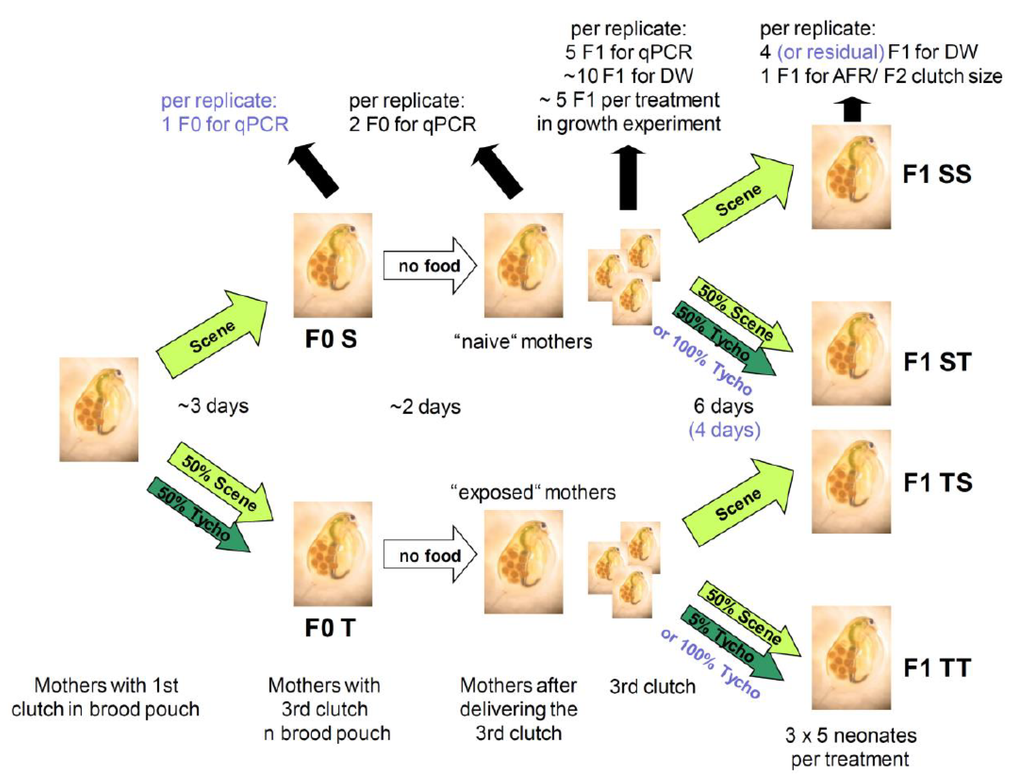
Two maternal effect experiments were conducted according to the following set-up (Figure 5): Seven newborns of clone MA-ES-3 per six 250 mL glasses were fed S. obliquus ad libitum until the eggs of the first clutch were deposited into their brood pouches. The medium was exchanged every other day. Subsequently, three of the replicates were reared on 2 mg C L−1 of 100% S. obliquus, whereas the other three replicates were fed 50% S. obliquus and 50% T. bourellyi until the second clutch was released. In the second experiment, at this point in time one F0 mother per replicate was frozen previous to RNA extraction. The two different F0 generations (F0S: naïve and F0T: experienced mothers) with the third clutch eggs deposited into the brood pouches were then put into a food-free medium. Thus, we ensured that the developing juveniles in the brood pouches had never ingested any food and that all effects we expected to find were exclusively due to maternal transfer. When the third clutch was released, two F0 individuals (first experiment only) and five F1 neonates per replicate per treatment were frozen for subsequent RNA extraction. Furthermore, ten F1 neonates per replicate were dried for the determination of the start weight for the growth rates. In the subsequent growth experiment, five F1 neonates per replicate were grown on 2 mg C L−1 of either 100% S. obliquus, or 50% S. obliquus and 50% T. bourellyi (second experiment: 100% T. bourellyi) for six days (second experiment: four days) in 100 mL glasses. Four of the F1 neonates (second experiment: all surviving neonates) were dried for the end weight of the growth rates. One individual per replicate was cultivated until its first clutch was released, and the number of the F2 offspring was counted. Not enough individuals survived for the determination of F2 clutch size in the second experiment.

4.3. Gene Expression

For RNA extraction, we used the NucleoSpin® RNA Kit (Macherey-Nagel) as according to the manufacturer’s instructions. The RNA concentrations and integrities were determined with the Thermo Scientific™ NanoDrop™ 2000 Spectrometer (ThermoFisher, Waltham, MA, USA). RNA was reverse transcribed with the High-Capacity cDNA Reverse Transcription Kit (Applied Biosystems™). Following the protocol of Schwarzenberger et al. (2009) [41], qPCR was performed with the 7500 Fast and 7500 Real-Time PCR System (Applied Biosystems™). Each reaction contained 5 ng cDNA (10 ng of RNA equivalent) and 1 μL of a primer pair (either cyclophilin, ubc [42], NAR-P2, NAR-P3, or NAR-P3 (Table 1). From a set of ten primers, cyclophilin and ubc were chosen as controls as they proved to be the most stable between the treatments. The results were analyzed with the 7500 Software v2.3 (Applied Biosystems™). Gene expression on 100% S. obliquus served as a calibrator and was always set to 1.

4.4. Statistics

The relative gene expression of the single-clone experiments, juvenile somatic growth rates, clutch size and age at first reproduction were analysed via one-way ANOVA and Tukey’s HSD post-hoc tests after verifying homogenous variances. When necessary, data were transformed before the analyses (log2: clutch size and age at first reproduction in the first maternal effects experiment; x2: juvenile somatic growth rates in the second maternal effects experiment). The relative gene expressions on S. obliquus and T. bourellyi were compared using t-tests in the maternal effects experiments. ANOVAs and t-tests were calculated with the program STATISTICA (StatSoft, Inc. 2011, version 10.0, Tulsa, OK, USA).

Supplementary Materials

The following are available online at https://www.mdpi.com/article/10.3390/toxins13050326/s1, Figure S1: Juvenile somatic growth rate.

Author Contributions

A.S. and D.M.-C. designed the project. All experiments and analyzes were conducted by A.S. D.M.-C. contributed to the manuscript that was written by A.S. All authors have read and agreed to the published version of the manuscript.

Funding

This project was funded by a grant to AS by the university’s Young Scholar Fund for Excellent Postdocs.

Data Availability Statement

All data will be provided by the authors upon request.

Acknowledgments

The authors would like to thank Patrick Bartolin, Ricarda Cremer and Katja Nafe for their assistance with the experiments. Thanks also to Laura Epp, Peter Kroth, Bernard Lepetit, and Lutz Becks for access to their laboratory infrastructure and Daniel Dietrich for providing the T. bourrellyi culture and information concerning the ELISA kit. Thanks to Frederic Bartlett for English corrections.

Conflicts of Interest

The authors declare no conflict of interest.

References

  1. Paerl:, H.W.; Huisman, J. Climate change: A catalyst for global expansion of harmful cyanobacterial blooms. Environ. Microbiol. Rep. 2009, 1, 27–37. [Google Scholar] [CrossRef] [PubMed]
  2. Carmichael, W.W. The toxins of cyanobacteria. Sci. Am. 1994, 270, 64–72. [Google Scholar] [CrossRef]
  3. Gademann, K.; Portmann, C. Secondary metabolites from cyanobacteria: Complex structures and powerful bioactivities. Curr. Org. Chem. 2008, 12, 326–341. [Google Scholar] [CrossRef]
  4. U.S. EPA (United States Environmental Protection Agency). 2015 Health Effects Support Document for the Cyanobacterial Toxin Anatoxin-a. EPA 820R15104, Washington, DC; June 2015; USEPA: Washington, DC, USA, 2015. Available online: http://water.epa.gov/drink/standards/hascience.cfm (accessed on 2 February 2021).
  5. Gorham, P.R.; McLachlan, J.; Hammer, U.T.; Sivonen, W.K. Isolation and culture of toxic strains of Anabaena flos-aquae. Mitt. Int. Ver. Limnol. 1964, 15, 796e804. [Google Scholar]
  6. Christensen, V.G.; Khan, E. Freshwater neurotoxins and concerns for human, animal, and ecosystem health: A review of anatoxin-a and saxitoxin. Sci. Total Environ. 2020, 736, 139515. [Google Scholar] [CrossRef]
  7. Graham, J.L.; Loftin, K.A.; Meyer, M.T.; Ziegler, A.C. Cyanotoxin mixtures and taste-and-odor compounds in cyanobacterial blooms from the midwestern United States. Environ. Sci. Technol. 2010, 44, 7361–7368. [Google Scholar] [CrossRef]
  8. Pawlik-Skowrońska, B.; Kalinowska, R.; Skowroński, T. Cyanotoxin diversity and food web bioaccumulation in a reservoir with decreasing phosphorus concentrations and perennial cyanobacterial blooms. Harmful Algae 2013, 28, 118–125. [Google Scholar] [CrossRef]
  9. Bumke-Vogt, C.; Mailahn, W.; Chorus, I. Anatoxin-a and neurotoxic cyanobacteria in German lakes and reservoirs. Environ. Toxicol. 1999, 14, 117–125. [Google Scholar] [CrossRef]
  10. Osswald, J.; Carvalho, A.P.; Claro, J.; Vasconcelos, V. Effects of cyanobacterial extracts containing anatoxin-a and of pure anatoxin-a on early developmental stages of carp. Ecotoxicol. Environ. Saf. 2009, 72, 473–478. [Google Scholar] [CrossRef]
  11. Cerasino, L.; Salmaso, N. Diversity and distribution of cyanobacterial toxins in the Italian subalpine lacustrine district. Oceanol. Hydrobiol. Stud. 2012, 41, 54e63. [Google Scholar] [CrossRef]
  12. Shams, S.; Capelli, C.; Cerasino, L.; Ballot, A.; Dietrich, D.R.; Sivonen, K.; Salmaso, N. Anatoxin-a producing Tychonema (Cyanobacteria) in European waterbodies. Water Res. 2015, 69, 68–79. [Google Scholar] [CrossRef] [PubMed] [Green Version]
  13. Sánchez, K.F.; Huntley, N.; Duffy, M.A.; Hunter, M.D. Toxins or medicines? Phytoplankton diets mediate host and parasite fitness in a freshwater system. Proc. R. Soc. B 2019, 286, 20182231. [Google Scholar] [CrossRef] [PubMed] [Green Version]
  14. Claska, M.E.; Gilbert, J.J. The effect of temperature on the response of Daphnia to toxic cyanobacteria. Freshw. Ecol. 1998, 39, 221–232. [Google Scholar] [CrossRef]
  15. Ballot, A.; Fastner, J.; Lantz, M.; Wiedner, C. First report of anatoxin-a-producing cyanobacterium Aphanizomenon issatchenkoi in northeastern Germany. Toxicon 2010, 56, 964–971. [Google Scholar] [CrossRef] [PubMed]
  16. Rellán, S.; Osswald, J.; Saker, M.; Gago-Martinez, A.; Vasconcelos, V. First detection of anatoxin-a in human and animal dietary supplements containing cyanobacteria. Food Chem. Toxicol. 2009, 47, 2189–2195. [Google Scholar] [CrossRef]
  17. Thomas, P.; Stephens, M.; Wilkie, G.; Amar, M.; Lunt, G.G.; Whiting, P.; Gallagher, T.; Pereira, E.; Alkondon, M.; Albuquerque, E.X.; et al. (+)-Anatoxin-a is a potent agonist at neuronal nicotinic acetylcholine receptors. J. Neurochem. 1993, 60, 2308–2311. [Google Scholar] [CrossRef]
  18. Monserrat, J.M.; Yunes, J.S.; Bianchini, A. Effects of Anabaena spiroides (cynobacteria) aqueous extracts on the acetylcholineesterade activity of aquatic species. Environ. Toxicol. Chem. 2001, 20, 1228–1235. [Google Scholar] [CrossRef]
  19. Fawell, J.K.; Mitchell, R.E.; Hill, R.E.; Everett, D.J. The toxicity of cyanobacterial toxins in the mouse: II anatoxin-a. Hum. Exp. Toxicol. 1999, 18, 168–173. [Google Scholar] [CrossRef] [PubMed]
  20. Pflugmacher, S.; Wiegand, C.; Oberemm, A.; Beattie, K.A.; Krause, E.; Codd, G.A.; Steinberg, C.E.W. Identification of an enzymatically formed glutathione conjugate of the cyanobacterial hepatotoxin microcystin-LR: The first step of detoxification. Biochim. Biophys. Acta 1998, 1425, 527–533. [Google Scholar] [CrossRef]
  21. Ortiz-Rodriguez, R.; Dao, T.S.; Wiegand, C. Transgenerational effects of microcystin-LR on Daphnia magna. J. Exp. Biol. 2012, 215, 2795–2805. [Google Scholar] [CrossRef] [Green Version]
  22. Schwarzenberger, A.; Sadler, T.; Motameny, S.; Ben-Khalifa, K.; Frommolt, P.; Altmüller, J.; Konrad, K.; Von Elert, E. Deciphering the genetic basis of microcystin tolerance. BMC Genom. 2014, 15, 776. [Google Scholar] [CrossRef] [Green Version]
  23. Sadler, T.; Von Elert, E. Physiological interaction of Daphnia and Microcystis with regard to cyanobacterial secondary metabolites. Aquat. Toxicol. 2014, 156, 96–105. [Google Scholar] [CrossRef] [PubMed]
  24. Schwarzenberger, A.; Kurmayer, R.; Martin-Creuzburg, D. Toward disentangling the multiple nutritional constraints imposed by Planktothrix: The significance of harmful secondary metabolites and sterol limitation. Front. Microbiol. 2020, 11, 586120. [Google Scholar] [CrossRef] [PubMed]
  25. Schwarzenberger, A.; Zitt, A.; Kroth, P.; Mueller, S.; Von Elert, E. Gene expression and activity of digestive proteases in Daphnia: Effects of cyanobacterial protease inhibitors. BMC Physiol. 2010, 10, 1–5. [Google Scholar] [CrossRef] [PubMed] [Green Version]
  26. Schwarzenberger, A.; Kuster, C.J.; Von Elert, E. Molecular mechanisms of tolerance to cyanobacterial protease inhibitors revealed by clonal differences in Daphnia magna. Mol. Ecol. 2012, 12, 4898–4911. [Google Scholar] [CrossRef] [PubMed]
  27. Blom, J.F.; Baumann, H.; Codd, G.A.; Jüttner, F. Sensitivity and adaptation of aquatic organisms to oscillapeptin J and [D-Asp3,(E)-Dhb7]microcystin-RR. Arch. Fuer Hydrobiol. 2006, 167, 547–559. [Google Scholar] [CrossRef]
  28. Schwarzenberger, A.; Hasselmann, M.; Von Elert, E. Positive selection of digestive proteases in Daphnia: A mechanism for local adaptation to cyanobacterial protease inhibitors. Mol. Ecol. 2020, 29, 912–919. [Google Scholar] [CrossRef] [Green Version]
  29. Gustafsson, S.; Rengefors, K.; Hansson, L.A. Increased consumer fitness following transfer of toxin tolerance to offspring via maternal effects. Ecology 2005, 86, 2561–2567. [Google Scholar] [CrossRef] [Green Version]
  30. Schwarzenberger, A.; Von Elert, E. Cyanobacterial protease inhibitors lead to maternal transfer of increased protease gene expression in Daphnia. Oecologia 2013, 172, 11–20. [Google Scholar] [CrossRef] [PubMed]
  31. Radersma, R.; Hegg, A.; Noble, D.W.A.; Uller, T. Timing of maternal exposure to toxic cyanobacteria and offspring fitness in Daphnia magna: Implications for the evolution of anticipatory maternal effects. Ecol. Evol. 2018, 8, 12727–12736. [Google Scholar] [CrossRef] [Green Version]
  32. Bownik, A.; Pawlik-Skowrońska, B. Early indicators of behavioral and physiological disturbances in Daphnia magna (Cladocera) induced by cyanobacterial neurotoxin anatoxin-a. Sci. Total Environ. 2019, 695, 133913. [Google Scholar] [CrossRef] [PubMed]
  33. Abreu, F.Q.D.; Ferrão Filho, A.D.S. Effects of an Anatoxin-a (s)-producing strain of Anabaena spiroides (Cyanobacteria) on the survivorship and somatic growth of two Daphnia similis clones. J. Environ. Prot. 2013, 4, 12–18. [Google Scholar] [CrossRef] [Green Version]
  34. Mitrovic, S.M.; Pflugmacher, S.; James, K.J.; Furey, A. Anatoxin-a elicits an increase in peroxidase and glutathione S-transferase activity in aquatic plants. Aquat. Toxicol. 2004, 68, 185–192. [Google Scholar] [CrossRef] [PubMed]
  35. Osswald, J.; Azevedo, J.; Vasconcelos, V.; Guilhermino, L. Experimental determination of the bioconcentration factors for anatoxin-a in juvenile rain-bow trout (Oncorhynchus mykiss). Proc. Int. Acad. Ecol. Environ. Sci. 2011, 1, 77–86. [Google Scholar]
  36. Mousseau, T.A.; Fox, C.W. The adaptive significance of maternal effects. Trends Ecol. Evol. 1998, 13, 403–407. [Google Scholar] [CrossRef]
  37. Freese, H.M.; Martin-Creuzburg, D. Food quality of mixed bacteria–algae diets for Daphnia magna. Hydrobiologia 2013, 715, 63–76. [Google Scholar] [CrossRef] [Green Version]
  38. Perry, T.; Heckel, D.G.; McKenzie, J.A.; Batterham, P. Mutations in Dα1 or Dβ2 nicotinic acetylcholine receptor subunits can confer resistance to neonicotinoids in Drosophila melanogaster. Insect Biochem. Mol. Biol. 2008, 38, 520–528. [Google Scholar] [CrossRef]
  39. Von Elert, E.; Jüttner, F. Phosphorus limitation not light controls the exudation of allelopathic compounds by Trichormus doliolum. Limnol. Oceanogr. 1997, 42, 1796–1802. [Google Scholar] [CrossRef] [Green Version]
  40. Lampert, W.; Rothhaupt, K.O. Alternating dynamics of rotifers and Daphnia magna in a shallow lake. Arch. Hydrobiol. 1992, 120, 447–456. [Google Scholar]
  41. Schwarzenberger, A.; Courts, C.; Von Elert, E. Target gene approaches: Gene expression in Daphnia magna exposed to predator-borne kairomones or to microcystin-producing and microcystin-free Microcystis aeruginosa. BMC Genom. 2009, 10, 527. [Google Scholar] [CrossRef] [Green Version]
  42. Heckmann, L.H.; Connon, R.; Hutchinson, T.H.; Maund, S.J.; Sibly, R.M.; Callaghan, A. Expression of target and reference genes in Daphnia magna exposed to ibuprofen. BMC Genom. 2006, 7, 175–182. [Google Scholar] [CrossRef] [PubMed] [Green Version]
Figure 1. Juvenile somatic growth rates (mean + SD) of three D. magna clones (‘Binnensee’, ‘MA-ES-3’, ‘FR-LR6-1’) grown either on 100% S. obliquus, 100% T. bourrellyi (Tycho) or a mixture of 50% S. obliquus with 50% T. bourrellyi for four days. Different letters indicate significant differences between treatments in a single clone (p < 0.05).
Toxins 13 00326 g001
Figure 2. Relative expression of three nicotine-acetylcholine receptor genes (A) NAR-P2, (B) NAR-P3, and (C) NAR-P4 as a function of juvenile somatic growth rates (g [d−1]) of the three D. magna clones when grown on 100% S. obliquus, 100% T. bourrellyi or a mixture of 50% S. obliquus with 50% T. bourrellyi.
Toxins 13 00326 g002
Figure 3. Juvenile somatic growth rates (mean + SD) of the F1 generation of the maternal effect experiments (clone MA-ES-3) with either 50% S. obliquus and 50% T. bourrellyi (A) or 100% T. bourrellyi (B) as cyanobacterial maternal and offspring food treatment. Different letters indicate significant differences between offspring treatments (p < 0.05).
Toxins 13 00326 g003
Figure 4. Relative expression (mean + SD) of three nicotine-acetylcholine receptor genes (NAR-P2, NAR-P3, and NAR-P4) of the F0 and F1 generations from the first maternal effect experiment (clone MA-ES-3 with 50% S. obliquus and 50% T. bourrellyi as maternal and offspring food treatment). Asterisks indicate significant differences (t-tests, p < 0.05) between control and 50% T. bourellyi. Gene expression on control food served as a calibrator and was always set to 1 (dashed line in the graphs).
Toxins 13 00326 g004
Figure 5. Experiment set-ups for the two maternal effect experiments (black: first experiment with 50% T. bourrellyi; blue in brackets: second experiment with 100% T. bourrellyi). T/Tycho = T. bourrellyi, S/Scene = S. obliquus, DW = dry weight, AFR = age at first reproduction.
Toxins 13 00326 g005
Table 1. Primer names, gene names (according to wfleabase.org), primer sequences, amplicon length, and gene accession numbers (acc #; wfleabase.org) of three nicotine-acetylcholine receptor genes. Their melting temperature was always 60 °C.
Primer NameGene NamePrimer Forward (5′–3′)Primer Reverse (5′–3′)Lengthacc #
NAR-P2Neuronal nicotinic acetyl-choline subunitGGCGCAGACCTCTCTTCTACGGTTGTGATGCCGAGAGTCA125 bpJGI_V11_55424
Nar-P3Neuronal nicotinic acetylcholine subunitATTTGCTTGGTGTCGTTCGCAGAATGATCCGGCGCATGAT107 bpJGI_V11_66098
NAR-P4Nicotinic acetylcholine receptor subunit beta 1CACAACCACACGAAACCCACGAAGACCAAGACGCACAGGA129 bpJGI_V11_321681
Publisher’s Note: MDPI stays neutral with regard to jurisdictional claims in published maps and institutional affiliations.