Co-Occurrence of Tetrodotoxin and Saxitoxins and Their Intra-Body Distribution in the Pufferfish Canthigaster valentini

 ,
,
Abstract
1. Introduction
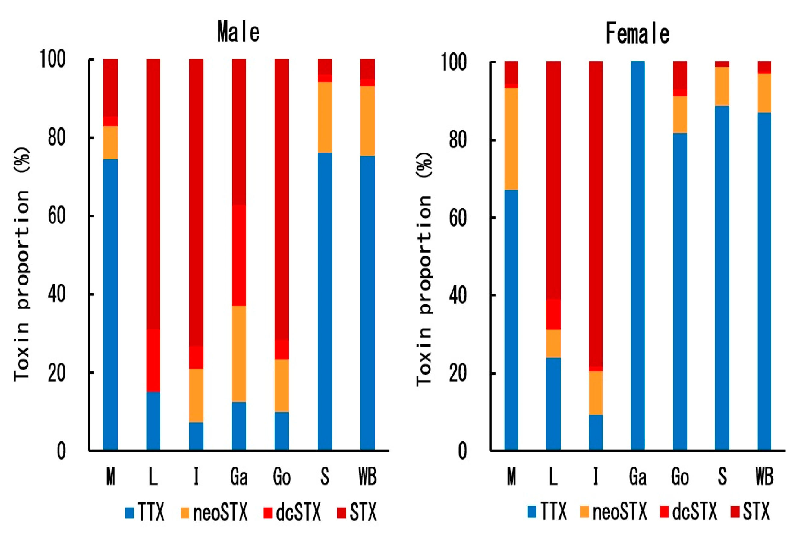
2. Results
3. Discussion
4. Materials and Methods
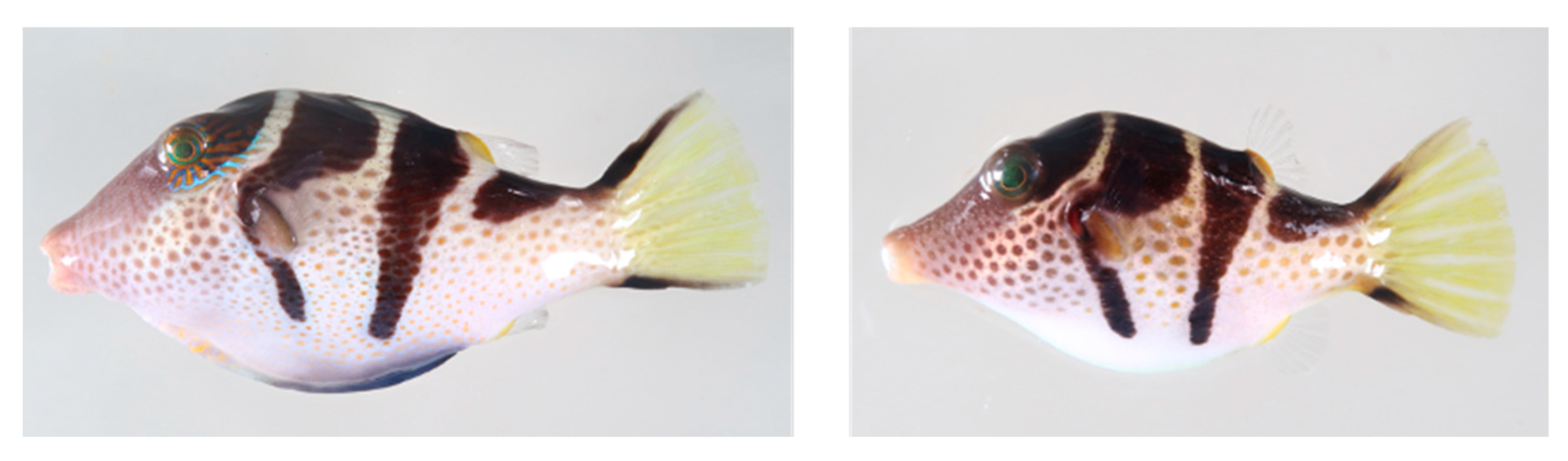
4.1. Pufferfish Specimens
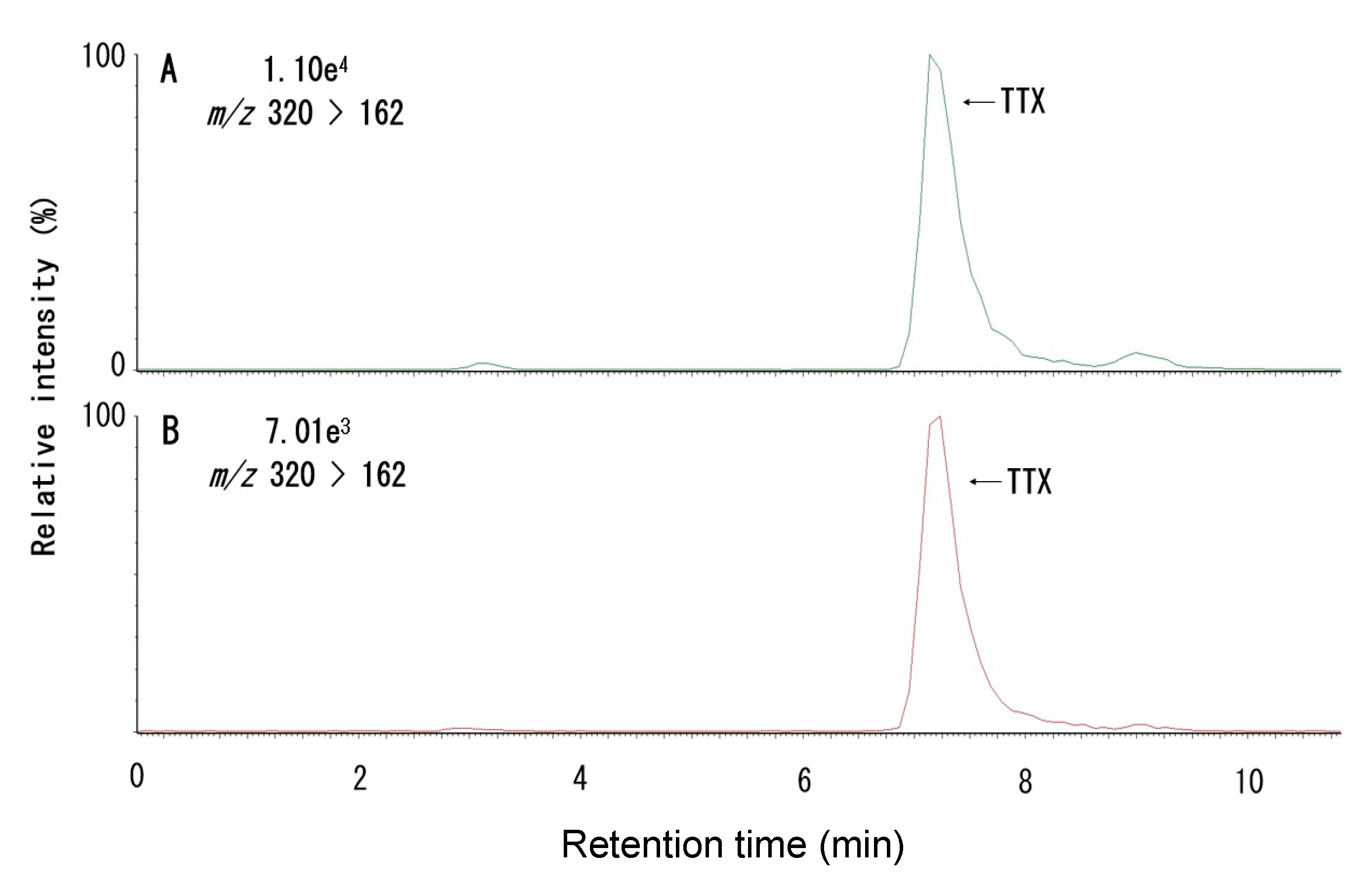
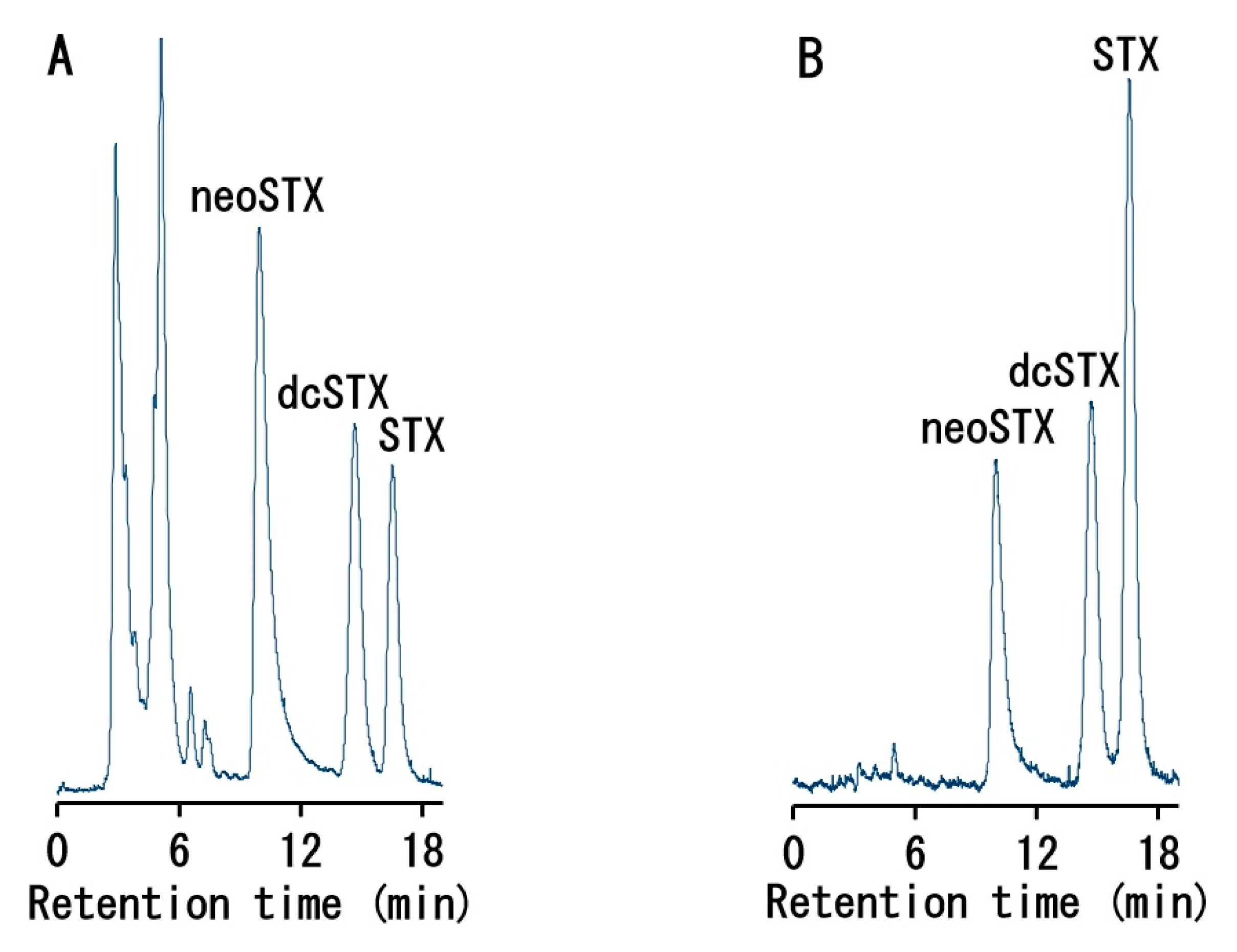
4.2. Toxin Quantification
4.3. Mouse Bioassay
4.4. Ethical Approval
Author Contributions
Funding
Conflicts of Interest
Appendix A


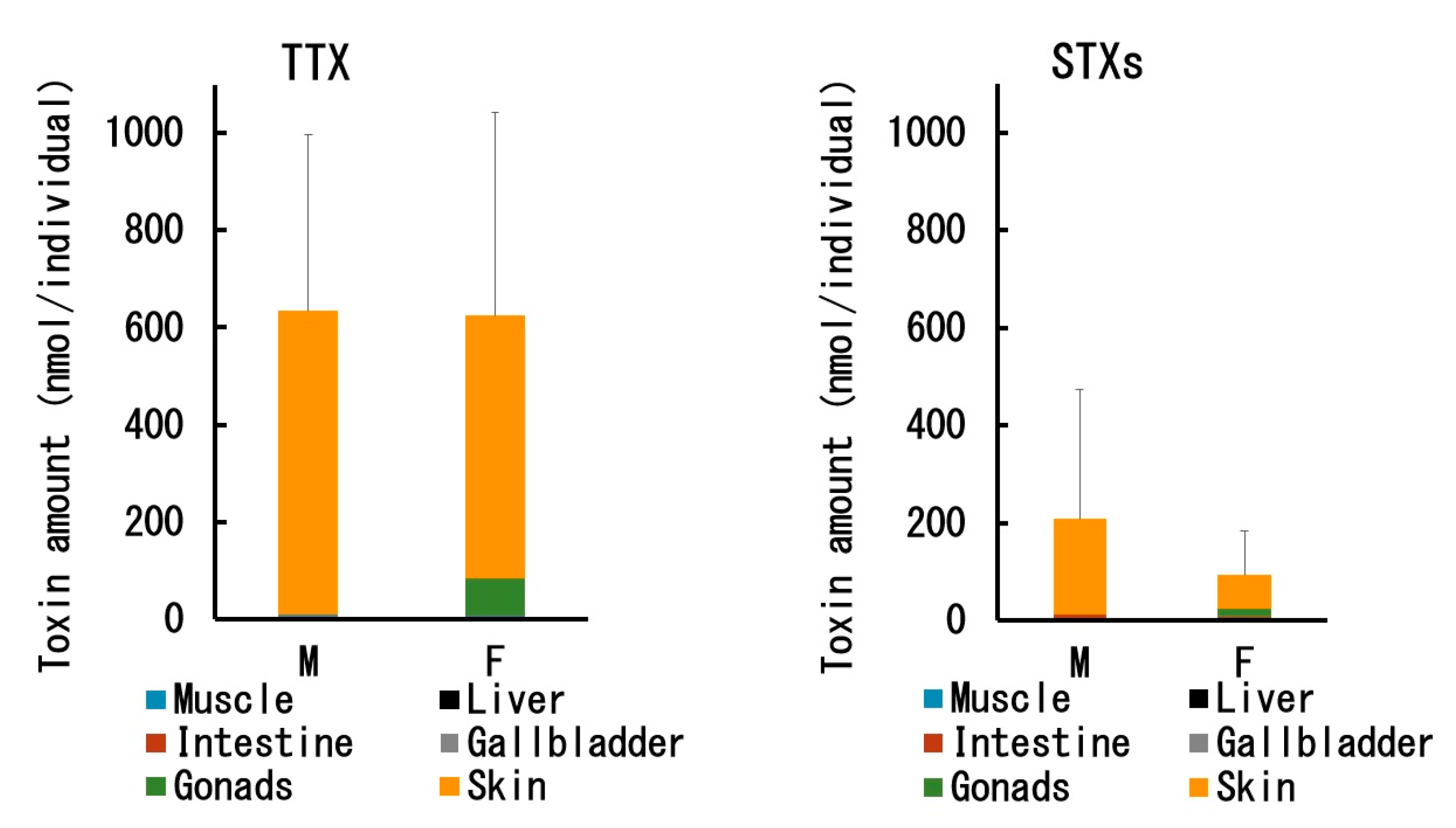
| Sex | Specimen No. | TTX Concentration (nmol/g) | |||||
|---|---|---|---|---|---|---|---|
| Muscle | Liver | Intestine | Gallbladder | Gonads | Skin | ||
| ♂ | 1 | 1.9 | 0.6 | 0.6 | 0.2 | 0.3 | 75 |
| 2 | 3.4 | 1.1 | 1.5 | 0.8 | 0.8 | 180 | |
| 3 | 1.5 | 0.1 | 0.2 | 1.3 | 0.4 | 151 | |
| 4 | 1.5 | 0.1 | 0.01 | 0.4 | 0.2 | 84 | |
| Mean ± SD | 2.0 ± 0.9 | 0.5 ± 0.5 | 0.6 ± 0.7 | 0.7 ± 0.5 | 0.4 ± 0.3 | 123 ± 51 | |
| ♀ | 5 | 4.0 | 3.5 | 2.0 | 0.8 | 648 | 420 |
| 6 | 2.4 | 0.2 | 0.2 | 1.8 | 165 | 108 | |
| 7 | 3.1 | 0.3 | 0.4 | 1.2 | 414 | 239 | |
| 8 | 4.3 | 0.6 | 0.7 | 1.2 | 455 | 361 | |
| 9 | 3.0 | 0.6 | 0.2 | 2.9 | 1.1 | 157 | |
| 10 | 10.4 | 2.1 | 1.9 | 11.5 | 1005 | 684 | |
| Mean ± SD | 4.5 ± 3.0 | 1.2 ± 1.3 | 0.9 ± 0.8 | 3.2 ± 4.1 | 448 ± 355 | 328 ± 211 | |
| Sex | Specimen No. | STX Concentration (nmol/g) | |||||
|---|---|---|---|---|---|---|---|
| Muscle | Liver | Intestine | Gallbladder 1 | Gonads 2 | Skin | ||
| ♂ | 1 | 1.0 | 5.5 | 8.8 | 5.3 | 1.2 | 78 |
| 2 | 0.3 | 3.6 | 8.5 | 1.9 | - | 11 | |
| 3 | 0.9 | 4.6 | 7.7 | 1.4 | 9.5 | 10 | |
| 4 | 0.8 | 2.6 | 6.3 | 0 | 1.5 | 21 | |
| Mean ± SD | 0.7 ± 0.3 | 4.1 ± 1.2 | 7.8 ± 1.1 | 2.1 ± 2.2 | 4.7 ± 4.0 | 30 ± 33 | |
| ♀ | 5 | 1.5 | 3.1 | 9.0 | 0 | 74 | 40 |
| 6 | 0.7 | 2.2 | 7.5 | 0 | 12 | 4 | |
| 7 | 0.3 | 5.0 | 6.7 | 0 | 108 | 13 | |
| 8 | 4.8 | 7.0 | 6.9 | 0 | 162 | 94 | |
| 9 | 0.9 | 0.4 | 7.3 | 0 | 14 | 21 | |
| 10 | 0.6 | 6.0 | 6.7 | 0 | 20 | 10 | |
| Mean ± SD | 1.5 ± 1.7 | 3.9 ± 2.5 | 7.4 ± 0.8 | 0 | 65 ± 61 | 30 ± 34 | |
References
- Noguchi, T.; Arakawa, O. Tetrodotoxin – distribution and accumulation in aquatic organisms, and cases of human intoxication. Mar. Drugs 2008, 6, 220–242. [Google Scholar] [CrossRef] [PubMed]
- Tani, T. Nihonsan Fugu no Chudokugakuteki Kenkyu (Toxicological Studies on Japanese Puffer); Teikoku: Tosho, Tokyo, 1945. [Google Scholar]
- Noguchi, T.; Ebesu, J.S.M. Puffer poisoning: Epidemiology and treatment. J. Toxicol. Toxin Rev. 2001, 20, 1–10. [Google Scholar] [CrossRef]
- Toda, M.; Uneyama, C.; Toyofuku, H.; Morikawa, K. Trends of poisonings caused by natural toxins in Japan, 1989–2011. Food Hyg. Saf. Sci. 2012, 53, 105–120. [Google Scholar] [CrossRef]
- EFSA Panel on Contaminants in the Food Chain. Risks for public health related to the presence of tetrodotoxin (TTX) and TTX analogues in marine bivalves and gastropods. EFSA J. 2017, 15, 4752. [Google Scholar]
- Ikeda, K.; Emoto, Y.; Tatsuno, R.; Wang, J.J.; Ngy, L.; Taniyama, S.; Takatani, T.; Arakawa, O. Maturation-associated changes in toxicity of the pufferfish Takifugu poecilonotus. Toxicon 2010, 55, 289–297. [Google Scholar] [CrossRef]
- Wang, J.; Araki, T.; Tatsuno, R.; Nina, S.; Ikeda, K.; Hamasaki, M.; Sakakura, Y.; Takatani, T.; Arakawa, O. Transfer profile of intramuscularly administered tetrodotoxin to artificial hybrid specimens of pufferfish, Takifugu rubripes and Takifugu niphobles. Toxicon 2011, 58, 565–569. [Google Scholar] [CrossRef]
- Tatsuno, R.; Shikina, M.; Shirai, Y.; Wang, J.; Soyano, K.; Nishihara, G.N.; Takatani, T.; Arakawa, O. Change in the transfer profile of orally administered tetrodotoxin to non-toxic cultured pufferfish Takifugu rubripes depending of its development stage. Toxicon 2013, 65, 76–80. [Google Scholar] [CrossRef]
- Gao, W.; Kanahara, Y.; Tatsuno, R.; Soyano, K.; Nishihara, G.N.; Urata, C.; Takatani, T.; Arakawa, O. Maturation-associated changes in internal distribution and intra-ovarian microdistribution of tetrodotoxin in the pufferfish Takifugu pardalis. Fish. Sci. 2018, 84, 723–732. [Google Scholar] [CrossRef]
- Kungsuwan, A.; Arakawa, O.; Promdet, M.; Onoue, Y. Occurrence of paralytic shellfish poisons in Thai freshwater puffers. Toxicon 1997, 35, 1341–1346. [Google Scholar] [CrossRef]
- Sato, S.; Kodama, M.; Ogata, T.; Saitanu, K.; Furuya, M.; Hirayama, K.; Kamimura, K. Saxitoxin as a toxic principle of a freshwater puffer, Tetraodon fangi, in Thailand. Toxicon 1997, 35, 137–140. [Google Scholar] [CrossRef]
- Zaman, L.; Arakawa, O.; Shimosu, A.; Onoue, Y. Occurrence of paralytic shellfish poison in Bangladeshi freshwater puffers. Toxicon 1997, 35, 423–431. [Google Scholar] [CrossRef]
- Ngy, L.; Tada, K.; Yu, C.F.; Takatani, T.; Arakawa, O. Occurrence of paralytic shellfish toxins in Cambodian Mekong pufferfish Tetraodon turgidus: Selective toxin accumulation in the skin. Toxicon 2008, 51, 280–288. [Google Scholar] [CrossRef] [PubMed]
- Cusick, K.D.; Sayler, G.S. An overview on the marine neurotoxin, saxitoxin: Genetics, molecular targets, methods of detection and ecological functions. Mar. Drugs 2013, 11, 991–1018. [Google Scholar] [CrossRef] [PubMed]
- World Health Organization & Food and Agriculture Organization of the United Nations. Toxicity Equivalence Factors for Marine Biotoxins Associated with Bivalve Molluscs; World Health Organization: Geneva, Switzerland, 2016. [Google Scholar]
- Kodama, M.; Ogata, T.; Noguchi, T.; Maruyama, J.; Hashimoto, K. Occurrence of saxitoxin and other toxins in the liver of pufferfish Takifugu Pardalis. Toxicon 1983, 21, 897–900. [Google Scholar] [CrossRef]
- Nakamura, M.; Oshima, Y.; Yasumoto, T. Occurrence of saxitoxin in puffer fish. Toxicon 1984, 22, 381–385. [Google Scholar] [CrossRef]
- Jang, J.; Yotsu-Yamashita, M. Distribution of tetrodotoxin, saxitoxin, and their analogs among tissues of the puffer fish Fugu pardalis. Toxicon 2006, 48, 980–987. [Google Scholar] [CrossRef]
- Sato, S.; Ogata, T.; Borja, V.; Gonzales, C.; Fukuyo, Y.; Kodama, M. Frequent occurrence of paralytic shellfish poisoning toxins as dominant toxins in marine puffer from tropical water. Toxicon 2000, 38, 1101–1109. [Google Scholar] [CrossRef]
- Nakashima, K.; Arakawa, O.; Taniyama, S.; Nonaka, M.; Takatani, T.; Yamamori, K.; Fuchi, Y.; Noguchi, T. Occurrence of saxitoxins as a major toxin in the ovary of a marine puffer Arothron firmamentum. Toxicon 2004, 43, 207–212. [Google Scholar] [CrossRef]
- Caley, M.J.; Schluter, D. Predators favour mimicry in a tropical reef fish. Proc. R. Soc. Lond. B 2003, 270, 667–672. [Google Scholar] [CrossRef]
- Hwang, D.F.; Kao, C.Y.; Yang, H.C.; Jeng, S.S.; Noguchi, T.; Hashimoto, K. Toxicity of puffer in Taiwan. Nippon Suisan Gakkaishi 1992, 58, 1541–1547. [Google Scholar] [CrossRef]
- Nakatani, T.; Shimizu, M.; Yamano, T. The contents and composition of tetrodotoxin and paralytic shellfish poisoning toxins in marine pufferfish Canthigaster rivulata. J. Food Hyg. Soc. Japan 2016, 57, 51–56. [Google Scholar] [CrossRef][Green Version]
- Barrientos, R.G.; Hernández-Mora, G.; Alegre, F.; Field, T.; Flewelling, L.; McGrath, S.; Deeds, J.; Chacón, Y.S.; Arrieta, K.R.; Vargas, E.C.; et al. Saxitoxin poisoning in green turtles (Chelonia mydas) linked to scavenging on mass mortality of Caribbean sharpnose puffer fish (Canthigaster rostrata-Tetraodontidae). Front. Vet. Sci. 2019, 6, 466. [Google Scholar] [CrossRef] [PubMed]
- Gladstone, W. Role of female territoriality in social and mating systems of Canthigaster valentini (Pisces: Tetraodontidae): Evidence from field experiments. Mar. Biol. 1987, 96, 185–191. [Google Scholar] [CrossRef]
- Gao, W.; Kanahara, Y.; Yamada, M.; Tatsuno, R.; Yoshikawa, H.; Doi, H.; Takatani, T.; Arakawa, O. Contrasting toxin selectivity between the marine pufferfish Takifugu pardalis and the freshwater pufferfish Pao suvattii. Toxins 2019, 11, 470. [Google Scholar] [CrossRef]
- Oshima, Y. Post-column devivatization HPLC method for analysis of PSP. J. AOAC Inter. 1995, 78, 528–532. [Google Scholar] [CrossRef]
- Japan Food Hygiene Association. 1. Pufferfish toxin. In Standard Methods of Analysis in Food Safety Regulation; Japan Food Hygiene Association: Tokyo, Japan, 2015; pp. 813–820. [Google Scholar]
- Yotsu-Yamashita, M. Chemistry of puffer fish toxin. J. Toxicol. Toxin Rev. 2001, 20, 51–66. [Google Scholar] [CrossRef]
- Jang, J.H.; Yotsu-Yamashita, M. 6,11-Dideoxytetrodotoxin from the puffer fish, Fugu pardalis. Toxicon 2007, 50, 947–951. [Google Scholar] [CrossRef] [PubMed]
- Yotsu-Yamashita, M.; Abe, Y.; Kudo, Y.; Ritson-Williams, R.; Paul, V.J.; Konoki, K.; Cho, Y.; Adachi, M.; Imazu, T.; Nishikawa, T.; et al. First identification of 5,11-dideoxytetrodotoxin in marine animals, and characterization of major fragment ions of tetrodotoxin and its analogs by high resolution ESI-MS/MS. Mar. Drugs 2013, 11, 2799–2813. [Google Scholar] [CrossRef]
- Kudo, Y.; Finn, J.; Fukushima, K.; Sakugawa, S.; Cho, Y.; Konoki, K.; Yotus-Yamashita, M. Isolation of 6-deoxytetrodotoxin from the pufferfish, Takifugu pardalis, and a comparison of the effects of the C-6 and C-11 hydroxy groups of tetrodotoxin on its activity. J. Nat. Prod. 2014, 77, 1000–1004. [Google Scholar] [CrossRef]
- Khora, S.S.; Yasumoto, T. Isolation of 11-oxotetrodotoxin from the puffer Arothron nigropunctatus. Tetrahedron Lett. 1989, 30, 4393–4394. [Google Scholar] [CrossRef]
- Wu, B.Q.; Yang, L.; Kao, C.Y.; Levinson, S.R.; Yotsu-Yamashita, M.; Yasumoto, T. 11-Oxo-tetrodotoxin and a specifically labeled 3H-tetrodotoxin. Toxicon 1996, 34, 407–416. [Google Scholar] [CrossRef]
- Arakawa, O.; Noguchi, T.; Shida, Y.; Onoue, Y. Occurrence of 11-oxotetrodotoxin and 11-nortetrodotoxin-6(R)-ol in a xanthid crab Atergatis floridus collected at Kojima, Ishigaki Island. Fish. Sci. 1994, 60, 769–771. [Google Scholar] [CrossRef]
- Taniyama, S.; Isami, Y.; Matsumoto, T.; Nagashima, Y.; Takatani, T.; Arakawa, O. Toxicity and toxin profile of tetrodotoxin detected in the scavenging gastropod Nassarius (Alectrion) glans “kinshibai”. J. Food Hyg. Soc. Japan 2009, 50, 22–28. [Google Scholar] [CrossRef] [PubMed]
- Zaman, L.; Arakawa, O.; Shimosu, A.; Shida, Y.; Onoue, Y. Occurrence of a methyl derivative of saxitoxin in Bangladeshi freshwater puffers. Toxicon 1998, 36, 627–630. [Google Scholar] [CrossRef]
- Kodama, M.; Sato, S.; Ogata, T.; Suzuki, Y.; Kaneko, T.; Aida, K. Tetrodotoxin secreting glands in the skin of puffer fishes. Toxicon 1986, 24, 819–829. [Google Scholar] [CrossRef]
- Mahmud, Y.; Okada, K.; Takatani, T.; Kawatsu, K.; Hamano, Y.; Arakawa, O.; Noguchi, T. Intra-tissue distribution of tetrodotoxin in two marine puffers Takifugu vermicularis and Chelonodon patoca. Toxicon 2003, 41, 13–18. [Google Scholar] [CrossRef]
- Kodama, M.; Ogata, T.; Sato, S. External secretion of tetrodotoxin from puffer fishes stimulated by electric shock. Mar. Biol. 1985, 87, 199–203. [Google Scholar] [CrossRef]
- Saito, T.; Noguchi, T.; Harada, T.; Murata, O.; Hashimoto, K. Tetrodotoxin as a biological defense agent for puffers. Bull. Jpn. Soc. Sci. Fish. 1985, 51, 1175–1180. [Google Scholar] [CrossRef]
- Itoi, S.; Yoshikawa, S.; Asahina, K.; Suzuki, M.; Ishizuka, K.; Takimoto, N.; Mitsuoka, R.; Yokoyama, N.; Detake, A.; Takayanagi, C.; et al. Larval pufferfish protected by maternal tetrodotoxin. Toxicon 2014, 78, 35–40. [Google Scholar] [CrossRef]
- Yamamori, K.; Nakamura, M.; Matsui, T.; Hara, T. Gustatory responses to tetrodotoxin and saxitoxin in fish: A possible mechanism for avoiding marine toxins. Can. J. Fish. Aqua. Sci. 1988, 45, 2182–2186. [Google Scholar] [CrossRef]
- Nagashima, Y.; Ohta, A.; Yin, X.; Ishizaki, S.; Matsumoto, T.; Doi, H.; Ishibashi, T. Difference in uptake of tetrodotoxin and saxitoxins into liver tissue slices among pufferfish, boxfish and porcupinefish. Mar. Drugs 2018, 16, 17. [Google Scholar] [CrossRef] [PubMed]
- Gao, W.; Yamada, M.; Ohki, R.; Nagashima, Y.; Tatsuno, R.; Ikeda, K.; Kawatsu, K.; Takatani, T.; Arakawa, O. Evaluation of the tetrodotoxin uptake ability of pufferfish Takifugu rubripes tissues according to age using an in vitro tissue slice incubation method. Toxicon 2020, 174, 8–12. [Google Scholar] [CrossRef] [PubMed]
- Ikeda, K.; Murakami, Y.; Emoto, Y.; Ngy, L.; Taniyama, S.; Yagi, M.; Takatani, T.; Arakawa, O. Transfer profile of intramuscularly administered tetrodotoxin to non-toxic cultured specimens of the pufferfish Takifugu rubripes. Toxicon 2009, 53, 99–103. [Google Scholar] [CrossRef]
- Wang, J.; Araki, T.; Tatsuno, R.; Nina, S.; Ikeda, K.; Takatani, T.; Arakawa, O. Transfer profile of orally and intramuscularly administered tetrodotoxin to artificial hybrid specimens of the pufferfish Takifugu rubripes and Takifugu porphyreus. Food Hyg. Saf. Sci. 2012, 55, 33–38. [Google Scholar] [CrossRef][Green Version]
- Yotsu-Yamashita, M.; Sugimoto, A.; Terakawa, T.; Shoji, Y.; Miyazawa, T.; Yasumoto, T. Purification, characterization, and cDNA cloning of a novel soluble saxitoxin and tetrodotoxin binding protein from plasma of the puffer fish, Fugu pardalis. Eur. J. Biochem. 2001, 268, 5937–5946. [Google Scholar] [CrossRef]
- Yotsu-Yamashita, M.; Yamaki, H.; Okoshi, N.; Araki, N. Distribution of homologous proteins to puffer fish saxitoxin and tetrodotoxin binding protein in the plasma of puffer fish and among the tissues of Fugu pardalis examined by Western blot analysis. Toxicon 2010, 55, 1119–1124. [Google Scholar] [CrossRef]
- Tatsuno, R.; Yamaguchi, K.; Takatani, T.; Arakawa, O. RT-PCR- and MALDI-TOF mass spectrometry-based identification and discrimination of isoforms homologous to pufferfish saxitoxin- and tetrodotoxin-binding protein in the plasma of non-toxic cultured pufferfish (Takifugu rubripes). Biosci. Biotechnol. Biochem. 2013, 77, 208–212. [Google Scholar] [CrossRef]
- Yotsu-Yamashita, M.; Nagaoka, Y.; Muramoto, K.; Cho, Y.; Konoki, K. Pufferfish saxitoxin and tetrodotoxin binding protein (PSTBP) analogues in the blood plasma of the pufferfish Arothron nigropunctatus, A. hispidus, A. manilensis, and Chelonodon patoca. Mar. Drugs 2018, 16, 224. [Google Scholar] [CrossRef]
- Arakawa, O.; Noguchi, T.; Shida, Y.; Onoue, Y. Occurrence of carbamoyl-N-hydroxy derivatives of saxitoxin and neosaxitoxin in a xanthid crab Zosimus aeneus. Toxicon 1994, 32, 175–183. [Google Scholar] [CrossRef]


| Sex | Specimen No. | Standard Body Length (mm) | Body Weight (g) | Gonadosomatic Index 1 |
|---|---|---|---|---|
| ♂ | 1 | 78.1 | 32.1 | 0.19 |
| 2 | 75.3 | 25.1 | 0.05 | |
| 3 | 63.3 | 13.5 | 0.13 | |
| 4 | 69.5 | 17.4 | 0.17 | |
| Mean ± SD | 71.8 ± 6.8 | 22.0 ± 8.3 | 0.13 ± 0.06 | |
| ♀ | 5 | 58.1 | 11.4 | 2.28 |
| 6 | 52.4 | 6.8 | 1.46 | |
| 7 | 67.9 | 17.5 | 2.03 | |
| 8 | 60.9 | 13.5 | 1.90 | |
| 9 | 42.9 | 4.1 | 0.35 | |
| 10 | 31.1 | 1.6 | 0.51 | |
| Mean ± SD | 52.2 ± 13.3 | 9.1 ± 6.0 | 1.4 ± 0.8 |
| Method | Toxicity (MU/g) | |||
|---|---|---|---|---|
| Specimen No. 5 | Specimen No. 8 | |||
| Ovary | Skin | Ovary | Skin | |
| Mouse Bioassay | 1094 | 720 | 1223 | 840 |
| Instrumental Analyses | 1100 | 702 | 1031 | 738 |
© 2020 by the authors. Licensee MDPI, Basel, Switzerland. This article is an open access article distributed under the terms and conditions of the Creative Commons Attribution (CC BY) license (http://creativecommons.org/licenses/by/4.0/).
Share and Cite
Zhu, H.; Sonoyama, T.; Yamada, M.; Gao, W.; Tatsuno, R.; Takatani, T.; Arakawa, O. Co-Occurrence of Tetrodotoxin and Saxitoxins and Their Intra-Body Distribution in the Pufferfish Canthigaster valentini. Toxins 2020, 12, 436. https://doi.org/10.3390/toxins12070436
Zhu H, Sonoyama T, Yamada M, Gao W, Tatsuno R, Takatani T, Arakawa O. Co-Occurrence of Tetrodotoxin and Saxitoxins and Their Intra-Body Distribution in the Pufferfish Canthigaster valentini. Toxins. 2020; 12(7):436. https://doi.org/10.3390/toxins12070436
Chicago/Turabian StyleZhu, Hongchen, Takayuki Sonoyama, Misako Yamada, Wei Gao, Ryohei Tatsuno, Tomohiro Takatani, and Osamu Arakawa. 2020. "Co-Occurrence of Tetrodotoxin and Saxitoxins and Their Intra-Body Distribution in the Pufferfish Canthigaster valentini" Toxins 12, no. 7: 436. https://doi.org/10.3390/toxins12070436
APA StyleZhu, H., Sonoyama, T., Yamada, M., Gao, W., Tatsuno, R., Takatani, T., & Arakawa, O. (2020). Co-Occurrence of Tetrodotoxin and Saxitoxins and Their Intra-Body Distribution in the Pufferfish Canthigaster valentini. Toxins, 12(7), 436. https://doi.org/10.3390/toxins12070436