Article

Reduced Membrane-Bound Alkaline Phosphatase Does Not Affect Binding of Vip3Aa in a Heliothis virescens Resistant Colony

by
Daniel Pinos
1,
Maissa Chakroun
1,†,
Anabel Millán-Leiva
1,
Juan Luis Jurat-Fuentes
2,
Denis J. Wright
3,
Patricia Hernández-Martínez
1 and
Juan Ferré
1,*
1
Department of Genetics, Instituto de Biotecnología y Biomedicina (BIOTECMED), Universitat de València, 46100 Burjassot, Spain
2
Department of Entomology and Plant Pathology, University of Tennessee, Knoxville, TN 37996, USA
3
Department of Life Sciences, Imperial College London, Silwood Park Campus, Ascot, Berks SL5 7PY, UK
*
Author to whom correspondence should be addressed.
Current address: Centre de Biotechnologie de Sfax, Sfax, Tunisia.
Toxins 2020, 12(6), 409; https://doi.org/10.3390/toxins12060409
Submission received: 7 May 2020 / Revised: 29 May 2020 / Accepted: 17 June 2020 / Published: 19 June 2020

Abstract

:
The Vip3Aa insecticidal protein from Bacillus thuringiensis (Bt) is produced by specific transgenic corn and cotton varieties for efficient control of target lepidopteran pests. The main threat to this technology is the evolution of resistance in targeted insect pests and understanding the mechanistic basis of resistance is crucial to deploy the most appropriate strategies for resistance management. In this work, we tested whether alteration of membrane receptors in the insect midgut might explain the >2000-fold Vip3Aa resistance phenotype in a laboratory-selected colony of Heliothis virescens (Vip-Sel). Binding of 125I-labeled Vip3Aa to brush border membrane vesicles (BBMV) from 3rd instar larvae from Vip-Sel was not significantly different from binding in the reference susceptible colony. Interestingly, BBMV from Vip-Sel larvae showed dramatically reduced levels of membrane-bound alkaline phosphatase (mALP) activity, which was further confirmed by a strong downregulation of the membrane-bound alkaline phosphatase 1 (HvmALP1) gene. However, the involvement of HvmALP1 as a receptor for the Vip3Aa protein was not supported by results from ligand blotting and viability assays with insect cells expressing HvmALP1.
Key Contribution: The biochemical characterization of a Vip3Aa-resistant colony of H. virescens shows that binding to receptors in the midgut is not affected and discards the role of mALP as a Vip3Aa receptor. This study suggests that Vip3A resistance may occur through mechanisms other than those commonly found for Cry proteins.

1. Introduction

The polyphagous pest Heliothis virescens (L.) (Lepidoptera: Noctuidae) is well known for producing substantial economic losses, particularly in cotton production, due to its ability to evolve resistance to different synthetic control products such as methyl parathion or pyrethroids [1,2]. As an alternative approach, genetically modified crops expressing Cry and Vip3A insecticidal protein genes from Bacillus thuringiensis (Bt crops) were introduced in 1996 for the control of this and other pests. However, extensive use threatens their effectiveness and cases of field-evolved practical resistance have already been reported for some lepidopteran and coleopteran pests [3].
Gene pyramiding has been proposed as an effective strategy for insect resistance management in Bt crops [4]. This approach consists of combined production of distinct insecticidal Bt proteins in the same plant, and its success heavily relies on the expressed insecticidal proteins having distinct mode of action, commonly defined as not sharing binding sites in target tissues [5,6].
Although the mechanism of action and receptors for Cry proteins have been widely studied [7], little is known about the biochemical mechanisms that underlie the action of Vip3A proteins. Several studies have shown that Vip3A proteins do not share binding sites with Cry1 or Cry2 proteins, yet their damage to the midgut epithelium resembles Cry action [8,9,10,11]. Supported by the lack of shared binding sites, transgenic corn and cotton varieties pyramided with Cry1, Cry2, and Vip3A genes are currently commercialized in several countries.
Knowledge of the biochemical and genetic factors involved in resistance is crucial to design management practices that delay the appearance of resistance and allow its rapid detection and ways to overcome it. The genetic potential to evolve resistance to Vip3A has already been shown in some laboratory-selected insect species such as H. virescens [12], Spodoptera litura [13], Helicoverpa armigera and Helicoverpa punctigera [14], Spodoptera frugiperda [15,16], and Helicoverpa zea [17]. However, the biochemical basis of resistance to Vip3A has only been studied in a laboratory-selected colony of H. armigera, for which alteration of binding sites was not the cause of resistance [18].
In the present study, we aimed to determine the biochemical basis of >2000-fold resistance to Vip3A in a H. virescens colony (Vip-Sel). In a previous study with this colony, resistance was shown to be polygenic, conferring little cross-resistance to Cry1Ab and no cross-resistance to Cry1Ac [19]. A transcriptomic analysis detected significant differences in gene expression compared to a susceptible strain, with 420 over-expressed and 1569 under-expressed genes in Vip-Sel [20]. Results herein support that Vip3Aa binding is not significantly altered in Vip-Sel compared to susceptible H. virescens and that membrane bound alkaline phosphatase (mALP) is not involved in Vip3Aa binding.

2. Results

2.1. Vip3Aa Binding to Midgut Brush Border Membrane Vesicles (BBMV)

In testing whether binding of Vip3Aa was altered in larvae from the Vip3A-resistant (Vip-Sel) compared to the reference susceptible (Vip-Unsel) colony, we measured binding of radiolabeled Vip3Aa to BBMV from the two colonies. Binding analyses showed specific Vip3Aa binding for BBMV from both colonies, with similar homologous competition curves (Figure 1а). A high percentage (35–40% of the input labeled toxin) of non-specific binding, i.e., not blocked by high concentrations of unlabeled Vip3Aa competitor, was detected, as previously reported [11,18]. The Kd and Rt values estimated from the competition curves (Table 1) indicated that Vip3Aa binds with low affinity to a high number of binding sites in BBMV from H. virescens. No major differences were found for these equilibrium binding parameters between the two H. virescens colonies, suggesting that binding alteration is not mechanistically related to Vip3Aa resistance in Vip-Sel.

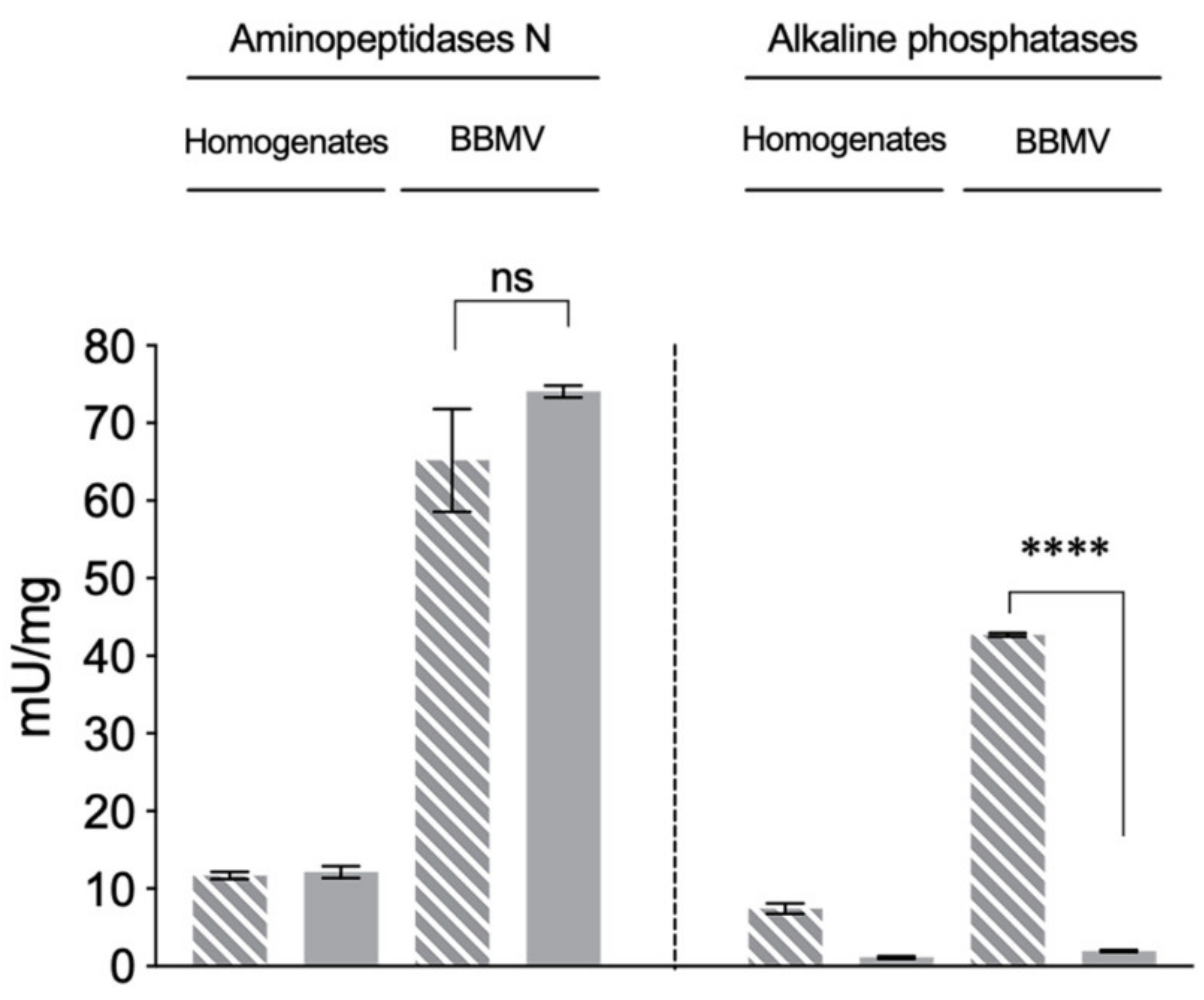
2.2. Reduced ALP Levels in the Vip3Aa-Resistant H. virescens Colony

During the evaluation of BBMV quality, we determined and compared the specific activities of alkaline phosphatase (ALP) and aminopeptidase-N (APN) as brush border membrane marker enzymes in midgut homogenates and BBMV preparations from Vip-Unsel and Vip-Sel colonies (Figure 2). The specific APN activity in midgut homogenates from both colonies was around 12 mU/mg, while in the BBMV preparations it was around 70 mU/mg, indicating an enrichment of APN activity of around 5.8 folds. Importantly, no significant differences (Student’s t-test, p > 0.05) in APN activity were observed between the midgut homogenates or BBMV from Vip-Unsel and Vip-Sel colonies. In agreement with the 5.8-fold enrichment value from APN activity comparisons, specific ALP activity was 7.44 mU/mg in midgut homogenates and 42.5 mU/mg in the BBMV from the Vip-Unsel colony. In contrast, dramatically reduced ALP activity was detected in both midgut homogenate (1.15 mU/mg) and BBMV (1.88 mU/mg) samples from the Vip-Sel colony. While unexpected, this observation is in line with reports of reduced ALP levels in Cry1-resistant lepidopteran species, including H. virescens [21,22,23,24]. Consequently, we further explored the extremely reduced ALP activity in Vip-Sel to determine whether it was due to a loss of enzymatic function or reduced gene expression.
Electrophoretic comparison of BBMV proteins from the two H. virescens colonies showed a protein band of ~66 kDa for the Vip-Unsel colony that was almost imperceptible in the BBMV from the Vip-Sel colony (Figure 3a). Western blotting indicated the presence of ALP in the ~66-kDa protein band, and confirmed the highly reduced levels of this protein in the Vip-Sel colony (Figure 3b). The composition of the ~66-kDa protein band and its relative abundance in the two H. virescens colonies were determined by liquid chromatography coupled to mass spectrometry (LC-MS) analysis. The spectra for the most abundant protein detected and identified in the ~66-kDa band matched to membrane-bound alkaline phosphatase (mALP) from H. virescens (Genbank Accession No. ABR88230). According to the exponentially modified protein abundance index (emPAI) expressing the proportional protein content in a protein mixture, the abundance ratio of mALP between Vip-Unsel and Vip-Sel was 22.7 folds.
To test if the reduced mALP protein levels in Vip-Sel were controlled at the transcriptional level, we performed real-time quantitative PCR (RT-qPCR) with mRNA extracted from total RNA from the two colonies. Transcript levels for two H. virescens mALP genes, HvmALP1 (Accession No. FJ416470.1) and HvmALP2 (Accession No. FJ416471.1), were analyzed. Compared to insects from the Vip-Unsel colony, larvae from the Vip-Sel colony had significant (p-value < 0.05) nine-fold downregulation of the HvmALP1 gene, while transcript levels for HvmALP2 were not different between colonies (Figure 3c). These results support that reduced ALP enzyme activity in BBMV from Vip-Sel compared to Vip-Unsel is due to reduced expression of HvmALP1 in the Vip-Sel colony.

2.3. Functional Role of HvmALP1 in Vip3Aa Binding

Since H. virescens ALP was proposed to play a role in binding of Cry1 proteins to the midgut membrane [25], we used ligand blotting to test whether mALP was involved in Vip3Aa binding. Binding of Vip3Aa to blots of resolved BBMV proteins was detected with anti-Vip3Aa antisera. No differences in the Vip3Aa-binding band pattern were detected between both colonies, in agreement with the binding results with radiolabeled Vip3Aa. However, no Vip3Aa binding was observed at the mALP position (~66 kDa) (Figure 1b).
To further discard mALP as a functional Vip3Aa receptor, we cloned and transiently expressed the HvmALP1 gene in cultured (Sf21) insect cells and performed cell viability tests after challenge with Vip3Aa. Transfection was successful, as transfected cells showed ~5-fold increased specific ALP activity compared with non-transfected cells or cells transfected with the empty plasmid (Figure 4a). However, after a challenge with Vip3Aa, the viability of transfected cells was not significantly different (Student’s t-test; p > 0.05) from that of the control cells (Figure 4b), confirming that mALP does not serve as a functional receptor for Vip3Aa during the toxicity process.

3. Discussion

The use of resistant insect strains isolated from the field or selected in the laboratory has been a powerful tool to understand the biochemical and genetic bases of resistance to Bt insecticidal proteins. Many studies have found that the alteration of membrane receptors is a common mechanism conferring high levels of resistance to Cry proteins [26,27,28]. In the case of Cry1 proteins, an important body of literature identifies aminopeptidase N, ABC transporters, cadherins and membrane alkaline phosphatases as main receptors, and identifies their alterations in association with resistance [29,30]. In contrast, three candidate receptors have been proposed for Vip3A proteins, including the Spodoptera spp. ribosomal protein S2 [31], the fibroblast growth factor receptor-like protein [32] and the scavenger receptor class C-like protein [33], yet their role in resistance has not been established.
In the present work, we aimed to determine whether alteration of membrane receptors was the basis for the observed 2040-fold resistance to Vip3Aa in the Vip-Sel colony of H. virescens. Results from binding assays with BBMV and radiolabeled Vip3Aa did not detect significant differences between the susceptible and resistant colonies, suggesting no involvement of binding site alteration in resistance. This conclusion was further supported by results from ligand blotting, where no differences between the binding patterns of Vip3Aa to BBMV proteins from the two colonies were observed. Similar results were reported for a laboratory-selected Vip3A-resistant colony of H. armigera [18], suggesting that high levels and narrow spectrum of Vip3A resistance may develop by mechanisms other than alteration of Vip3Aa binding sites.
Even though differences in binding were not found, a dramatic reduction in the ALP enzymatic activity was detected in midgut samples from the resistant compared to susceptible colony. Western blotting and RT-qPCR analyses showed that the decreased activity was due to a reduction in the amount of mALP protein, which was controlled at the transcriptional level, in agreement with a previous study [20]. Downregulation or reduced levels of mALP in the midgut membrane have been observed as a common phenomenon in resistance to Cry1Ac in H. virescens [25], Helicoverpa zea [21], Plutella xylostella [22], and Helicoverpa armigera [24]; to Cry1F in S. frugiperda [23]; to Cry1C in Spodoptera litura [34]; and even in Aedes aegypti resistant to Bt subsp. israeliensis (Bti) [35]. The fact that Cry1Ac and Cry1C do not share binding sites [36] suggests that the role of ALP downregulation in resistance may not be related to reduced Cry binding, but may represent a physiological response to resistance. In agreement with this hypothesis, susceptibility of Sf21 cells expressing HvmALP1 was not significantly different to Vip3Aa, supporting that ALP is not a functional receptor for Vip3Aa in H. virescens. In addition, in a Cry1Ac-resistant strain of P. xylostella, altered expression of different genes (including the PxmALP) was reported to be trans-regulated by upregulation of a mitogen-activated protein kinase, which was linked to resistance [22]. Similar trans-regulation of genes involved in resistance to Bt has also been observed for APN in Trichoplusia ni resistant to Cry1Ac [37] and Ostrinia nubilalis resistant to Cry1Ab [38], and for both APN and an ABCC transporter in Bombyx mori resistant to Cry1Ab [39]. Further research should test the involvement of this control mechanism in downregulation of mALP in Vip-Sel and other Bt-resistant colonies.
The two studies so far focused on the underlying mechanism of resistance to Vip3Aa proteins share a similar feature in that in vitro binding is not reduced [18] (and the present work). According to these results, mechanisms other than binding site alteration seem to be responsible for conferring specific and high-level resistance to Vip3A. This contrasts with the fact that the alteration of membrane receptors is a common mechanism conferring high levels of resistance to Cry proteins. Better knowledge of the mode of action of Vip3A proteins will help shed light on the biochemical basis of resistance to these proteins.

4. Conclusions

The results herein show lack of significant Vip3Aa binding alterations in a resistant colony of H. virescens. These observations are in contrast to most cases of high levels of resistance to Cry proteins for which decreased binding is commonly detected. In addition, this study provides evidence of downregulation of membrane bound alkaline phosphatase (mALP) in the Vip3Aa-resistant colony, although results do not support involvement of mALP as a receptor for the Vip3A protein.

5. Materials and Methods

5.1. Insects

Two colonies of H. virescens originating from the same field population collected in Arkansas (USA) were used in this study: Vip-Sel (Vip3Aa-resistant) and Vip-Unsel (Vip3Aa susceptible). The process of selection of the Vip-Sel colony with Vip3Aa has been previously described [12,19]. After 13 generations of selection, the LC50 of the Vip-Sel colony was 2300 µg/mL, representing a 2040-fold resistance ratio relative to the control Vip-Unsel colony. Both colonies were reared at the Imperial College London, Silwood Park campus (UK), and frozen larvae were sent for analysis to the Universitat de València (Spain).

5.2. BBMV Preparation and Enzyme Activity Assays

Brush border membrane vesicles (BBMV) from 3rd instar H. virescens larval midguts from Vip-Sel and Vip-Unsel colonies were prepared according to the differential magnesium precipitation method [40]. Isolated BBMV were flash frozen in liquid nitrogen and kept at −80 °C until used. The protein concentration of the BBMV preparations was determined by the method of Bradford using bovine serum albumin (BSA) as a standard [41].
Alkaline phosphatase (ALP) and leucine aminopeptidase (APN) activities were used as brush border membrane enzymatic markers to determine the quality of the BBMV preparations [42]. Specific ALP activity was determined by chromogenic detection of p-nitrophenyl phosphate (pNPP) substrate hydrolysis into p-nitrophenol, and specific APN activity was detected by hydrolysis of L-leu-p-nitroanilide substrate into p-nitroanilide. In both cases, chromogenic variation was measured on 1 µg of either BBMV or midgut homogenate at 405 nm (Infinite m200, Tecan, Mannedorf, Switzerland). Two different batches of BBMV were used and all enzymatic activity assays were performed in triplicate. Means values for enzyme activities from Vip-Unsel and Vip-Sel were compared by Student’s t-test at a 5% level of significance.
For measuring specific ALP enzymatic activity in cultured Sf21 cells, a 1.6-mL suspension of each cell type (non-transfected, transfected with empty plasmid, and transfected with plasmid with HvmALP1) was used. Culture cells were centrifuged, washed twice with 300 µL of phosphate buffered saline (PBS) and then resuspended in 50 µL of PBS. Protein concentration was determined by the method of Bradford and specific ALP activity measured as above.

5.3. Vip3Aa Protein Expression and Purification

The Vip3Aa16 (Vip3Aa) protein (NCBI Accession No. AAW65132) was overexpressed in recombinant Escherichia coli BL21 carrying the vip3Aa16 gene. Protein expression and lysis was carried out following the conditions described elsewhere [43]. Soluble Vip3Aa in the cell lysate was purified by two different methodologies. For binding and cell viability assays, Vip3Aa was partially purified by isoelectric point precipitation (IPP), activated with trypsin treatment and further purified by anion-exchange chromatography, as previously described [11]. For ligand assays, affinity chromatography purification was carried out using a HiTrap chelating HP column (GE Healthcare, Uppsala, Sweden) and then activated with trypsin, as described [11].

5.4. Vip3Aa Labeling and Binding Experiments

Purified Vip3Aa activated protein (25 µg) was labeled with 0.5 mCi of 125I using the chloramine T method [11]. The labeled protein was separated from the excess of free 125I in a PD10 desalting column (GE Healthcare, Uppsala, Sweden) and the purity of the 125I-labeled Vip3Aa was checked by autoradiography. The specific activity of the labeled protein was 2.2 mCi/mg.
Binding assays were performed as described elsewhere [11]. Prior to being used, BBMV were centrifuged and resuspended in binding buffer (20 mM Tris, 150 mM NaCl, 1 mM MnCl2, pH 7.4, 0,04% Blocking reagent from Sigma Aldrich, St. Louis, MO, USA). Competition binding experiments were conducted by incubating 1.4 µg of BBMV protein with 0.65 nM 125I-Vip3Aa in a final volume of 0.1 mL of binding buffer for 90 min at 25 °C in the presence of increasing amounts of unlabeled Vip3Aa. After incubation, samples were centrifuged at 16,000× g for 10 min and the pellet was washed once with 500 µL of ice-cold binding buffer. Radioactivity retained in the pellet was measured in a model 2480 WIZARD2 gamma counter. Data from the competition experiments were analyzed to determine equilibrium binding parameters, dissociation constant (Kd), and concentration of binding sites (Rt) using the LIGAND software [44].

5.5. Western and Ligand Blotting

For the detection of ALP proteins in BBMV by Western blotting, BBMV (20 µg) were suspended in ice-cold PBS and heat denatured before separation on a SDS–10% PAGE gel. The resolved BBMV proteins were transferred to a nitrocellulose filter (Protran 0.45 µm NC, GE Healthcare, Uppsala, Sweden) using a BioRad Mini Trans-Blot system (Bio-Rad, Hercules, CA, USA) at 4 °C in blotting buffer (39 mM Glycine, 48 mM Tris-HCl, 0.037% SDS, 10% methanol, pH 8.5) for 1 h at constant voltage (100 V). After transfer, the nitrocellulose filter was blocked in blocking buffer (PBS, 0.1% Tween 20, 5% skimmed milk powder) overnight at 4 °C. After blocking and washing with PBST (PBS, 0.1% Tween 20) three times (5 min each), incubation with primary antibody against the membrane-bound form of ALP from Anopheles gambiae (generously provided by M. Adang, University of Georgia, USA) was performed for 90 min at a 1:5000 dilution at room temperature (RT). The membrane was then washed with PBST three times for 5 min each and then incubated with secondary antibody (goat anti-rabbit conjugated to horseradish peroxidase (HRP) at a 1:10,000 dilution) for 1 h at RT. After being washed with PBST three times for 5 min each, the membrane was developed using enhanced chemiluminescence (ECL Prime Western Blotting detection reagent, GE Healthcare, Uppsala, Sweden) in an ImageQuant LAS 4000 (GE Healthcare, Uppsala, Sweden), according to the manufacturer’s instructions.
Ligand blotting for the detection of BBMV proteins binding Vip3Aa protein was performed with BBMV proteins resolved and immobilized as described above for Western blotting. The nitrocellulose membrane was blocked for 1 h at 4 °C in blocking buffer (5% skimmed milk), and after three washes for 5 min each with PBST buffer, it was incubated overnight at 4 °C with blocking buffer (1% skimmed milk) supplemented with affinity chromatography-purified Vip3Aa at a final concentration of 4 µg/mL. After washing with PBST three times for 5 min each, the membrane was incubated with primary antibody against Vip3Aa at a 1:5000 dilution for 1 h at RT. After three washing steps with PBST (5 min each), membranes were incubated with secondary antibody (goat anti-rabbit conjugated to HRP) for 1 h at RT. To visualize the marker, Precision Protein™ Streptactin-HRP conjugate (Bio-Rad, St. Louis, MO, USA) was used following the manufacturer’s instructions. Upon washing three times (5 min each) with PBST, the membrane was developed as described for Western blotting.

5.6. Proteomic Analysis

After resolving BBMV proteins from Vip-Sel and Vip-Unsel colonies by SDS–10% PAGE, the gel was stained with Coomassie blue (Thermo Scientific™, Waltham, MA, USA). The band corresponding to the expected molecular weight of ALP (~66 kDa) was cut out and subjected to analysis by nano-electron spray ionization (nano-ESI) followed by tandem mass spectrometry (qQTOF) in a 5600 TripleTOF (AB Sciex, Madrid, Spain) system. Results were analyzed with ProteinPilot v5.0 software and the relative amount of the proteins detected was estimated using the exponentially modified protein abundance index (emPAI) as described elsewhere [45].

5.7. RT-qPCR

Relative expression levels for HvmALP1 and HvmALP2 isoforms (accession numbers FJ416470 and FJ416471, respectively) were determined by reverse transcription quantitative polymerase-chain reaction (RT-qPCR). For this purpose, total RNA of dissected midguts from both colonies (Vip-Unsel and Vip-Sel) was isolated using RNAzol (MRC Inc., Cincinnati, OH, USA) according to the manufacturer’s protocol. Each RNA (1 µg) was reverse-transcribed to cDNA using random hexamers and oligo (dT) by following the instructions provided in the Prime-Script RT Reagent Kit (Perfect Real Time from TaKaRa Bio Inc., Otsu Shiga, Japan). RT-qPCR was carried out in a StepOnePlus Real-Time PCR system (Applied Biosystems, Foster City, CA, USA). Reactions were performed using 5× HOT FIREpol EVAGreen qPCR Mix Plus (ROX) from Solis BioDyne (Tartu, Estonia) in a total reaction volume of 25 µL. Specific primers for HvmALP1, HvmALP2 and Rps18 (endogenous control) genes were as described elsewhere [24]. The REST MCS software was used for gene expression analysis [46].

5.8. Expression Vector Construction

The full-length HvmALP1 transcript was amplified from cDNA of H. virescens larvae and cloned into pET30a as described elsewhere [47]. Purified plasmid DNA was digested with EcoRI and NotI to excise the full-length sequence and ligate it in frame into EcoRI-NotI sites of the pIZT/V5His vector (Thermo Scientific™, Waltham, MA, USA), to generate the pIZT/V5His/HvmALP1 construct. Ligation products were transformed into E. coli strain DH5α and transformants checked for correct insertion by sequencing (University of Tennessee Sequencing Facility, Knoxville, TN, USA). Purified plasmid was used to transform E. coli strain DH10β and liquid cultures of LB medium supplemented with Zeocin (25 µg/mL) were used to amplify the vector. To purify the plasmids for transfection, the NucleoSpin® Plasmid kit (Macherey-Nagel, Düren, Germany) was used. Double digestion with EcoRI and NotI (which cleaved the full-length HvmALP1 insert) and 1% agarose gel electrophoresis were performed to check plasmid and/or insert integrity. The concentration of plasmid DNA was measured with a Thermo Scientific™ Nanodrop™ 2000 Spectrophotometer.

5.9. Transient Expression of HvmALP1 in Sf21 Cells

Cultured Sf21 insect cells, originally derived from S. frugiperda, were maintained in 25 cm2 tissue culture flasks (Nunc T25 flasks, Thermo Scientific™, Waltham, MA, USA) at 25 °C with 4 mL of Gibco® Grace’s Medium (1×) (Life Technologies™, Paisley, UK) supplemented with 10% heat-inactivated fetal bovine serum (FBS).
For transient expression, cells were seeded on 12-well plates with the same medium without FBS at ca. 70% confluency and transfected with 0.5 µg of the pIZT/V5His/HvmALP1 or pIZT/V5His plasmid using Cellfectin® II Reagent (Thermo Scientific™, Waltham, MA, USA), following manufacturer’s instructions. Five hours post-transfection, the medium was replaced with fresh medium containing 10% FBS. After 24 h, cells were examined using a confocal microscope (Olympus, FV1000MPE, Tokyo, Japan) equipped with the appropriate filter for green fluorescent protein (GFP) detection as transfection marker. The enzymatic activity of alkaline phosphatase was then measured as explained above.

5.10. Cell Viability Assays

Viability of transfected Sf21 cells exposed for 24 h to Vip3Aa was measured using the MTT (3-[4,5-dimethylthiazol-2-yl]-2,5-diphenyltetrazolium bromide) assay. Preliminary assays were performed to determine a final Vip3A concentration of 300 µg/mL as resulting in ~50% loss of viability in the control cell line (data not shown). Briefly, cells (100 µL per well) were transferred to 96-well ELISA plates (flat bottom) and incubated at 25 °C for at least 45 min. Then, 10 µL of trypsin-activated Vip3Aa toxin was added to each well (300 µg/mL final concentration). As negative and positive controls, 10 µL of Tris buffer (Tris 20 mM, NaCl 150 mM, pH 9) and 10 µL of 2% Triton X-100 were used, respectively. After 24 h of incubation at 25 °C, cell viability was assessed by applying 20 µL of CellTiter 96® AQueous One Solution Reagent (Promega, Madison WI, USA) to each well and incubating for 2 h at 25 °C. Absorbance was measured at 490 nm (Infinite m200, Tecan, Mannedorf, Switzerland). The percentage of viable cells was obtained as described elsewhere [48]. Mean values in the transfected cells against the non-transfected cells were compared by Student’s t-test at 5% level of significance.

Author Contributions

Conceptualization, P.H.-M. and J.F.; Formal analysis, D.P., M.C., and A.M.-L.; Funding acquisition, J.L.J.-F. and J.F.; Methodology, D.P. and M.C.; Resources, J.L.J.-F. and D.J.W.; Supervision, P.H.-M. and J.F.; Writing—original draft, D.P.; and Writing—review and editing, J.L.J.-F., D.J.W., P.H.-M., and J.F. All authors have read and agreed to the published version of the manuscript.

Funding

This research was supported by the Spanish Ministry of Science, Innovation and Universities (grant no. RTI2018-095204-B-C21). D.P. is recipient of a PhD grant from the Spanish Ministry of Science, Innovation and Universities (grant ref. FPU15/05652). Support for JLJ-F was provided by the NC246 Multistate Hatch project from the USDA National Institute for Food and Agriculture (NIFA). The proteomic analysis was performed at the Proteomics Facility of the Servei Central de Suport a la Investigació Experimental (SCSIE) at the Universitat de València (Valencia, Spain) that belongs to ProteoRed, PRB2-ISCII, supported by grant PT13/0001, of the PE I+D+I 2013-2016, funded by ISCII and FEDERPT13/0001.

Conflicts of Interest

The authors declare no conflict of interest. The funders had no role in the design of the study; in the collection, analyses, or interpretation of data; in the writing of the manuscript, or in the decision to publish the results.

References

  1. Wolfenbarger, D.A.; Bodegas, P.R.; Flores, R. Development of resistance in Heliothis spp. in the Americas, Australia, Africa, and Asia. Bull. Entomol. Soc. Am. 1981, 27, 181–185. [Google Scholar] [CrossRef]
  2. Terán-Vargas, A.P. Insecticide resistance of tobacco budworm in Southern Tamaulipas Mexico. In Proceedings of the Beltwide Cotton Conference, Nashville, TN, USA, 9–12 January 1996; National Cotton Council of America: Memphis, TN, USA; pp. 784–786. [Google Scholar]
  3. Tabashnik, B.E.; Carrière, Y. Global patterns of resistance to Bt crops highlighting Pink Bollworm in the United States, China, and India. J. Econ. Entomol. 2019, 112, 2513–2523. [Google Scholar] [CrossRef]
  4. Roush, R.T. Bt-transgenic crops: Just another pretty insecticide or a chance for a new start in resistance management? Pestic. Sci. 1997, 51, 328–334. [Google Scholar] [CrossRef]
  5. Moar, W.J.; Anilkumar, K.J. Plant science: The power of the pyramid. Science 2007, 318, 1561–1562. [Google Scholar] [CrossRef] [Green Version]
  6. Carrière, Y.; Crickmore, N.; Tabashnik, B.E. Optimizing pyramided transgenic Bt crops for sustainable pest management. Nat. Biotechnol. 2015, 33, 161–168. [Google Scholar] [CrossRef]
  7. Adang, M.; Crickmore, N.; Jurat-Fuentes, J.L. Diversity of Bacillus thuringiensis crystal toxins and mechanism of action. In Advances in Insect Physiology; Dhadialla, T.S., Gill, S., Eds.; Academic Press: San Diego, CA, USA, 2014; Volume 47, pp. 39–87. [Google Scholar] [CrossRef]
  8. Lee, M.K.; Miles, P.; Chen, J. Brush border membrane binding properties of Bacillus thuringiensis Vip3A toxin to Heliothis virescens and Helicoverpa zea midguts. Biochem. Biophys. Res. Commun. 2006, 339, 1043–1047. [Google Scholar] [CrossRef]
  9. Sena, J.A.D.; Hernández-Rodríguez, C.S.; Ferré, J. Interaction of Bacillus thuringiensis Cry1 and Vip3A proteins with Spodoptera frugiperda midgut binding sites. Appl. Environ. Microbiol. 2009, 75, 2236–2237. [Google Scholar] [CrossRef] [Green Version]
  10. Gouffon, C.; Van Vliet, A.; Van Rie, J.; Jansens, S.; Jurat-Fuentes, J.L. Binding sites for Bacillus thuringiensis Cry2Ae toxin on heliothine brush border membrane vesicles are not shared with Cry1A, Cry1F, or Vip3A toxin. Appl. Environ. Microbiol. 2011, 77, 3182–3188. [Google Scholar] [CrossRef] [Green Version]
  11. Chakroun, M.; Ferré, J. In vivo and in vitro binding of Vip3Aa to Spodoptera frugiperda midgut and characterization of binding sites by 125I radiolabeling. Appl. Environ. Microbiol. 2014, 80, 6258–6265. [Google Scholar] [CrossRef] [Green Version]
  12. Pickett, B. Studies on Resistance to Vegetative (Vip3A) and Crystal (Cry1A) Insecticide Toxin of Bacillus thuringiensis in Heliothis virescens (Fabricius). Ph.D. Thesis, Imperial College London, London, UK, 2009. [Google Scholar]
  13. Barkhade, U.P.; Thakare, A.S. Protease mediated resistance mechanism to Cry1C and Vip3A in Spodoptera litura. Egypt Acad. J. Biol. Sci. 2010, 3, 43–50. [Google Scholar] [CrossRef]
  14. Mahon, R.J.; Downes, S.J.; James, B. Vip3A resistance alleles exist at high levels in Australian targets before release of cotton expressing this toxin. PLoS ONE 2012, 7, e39192. [Google Scholar] [CrossRef] [Green Version]
  15. Bernardi, O.; Bernardi, D.; Horikoshi, R.J.; Okuma, D.M.; Miraldo, L.L.; Fatoretto, J.; Medeiros, F.C.; Burd, T.; Omoto, C. Selection and characterization of resistance to the Vip3Aa20 protein from Bacillus thuringiensis in Spodoptera frugiperda. Pest. Manag. Sci. 2016, 72, 1794–1802. [Google Scholar] [CrossRef]
  16. Yang, F.; Morsello, S.; Head, G.P.; Sansone, C.; Huang, F.; Gilreath, R.T.; Kerns, D.L. F2 screen, inheritance and cross-resistance of field-derived Vip3A resistance in Spodoptera frugiperda (Lepidoptera: Noctuidae) collected from Louisiana, USA. Pest. Manag. Sci. 2018, 74, 1769–1778. [Google Scholar] [CrossRef]
  17. Yang, F.; Santiago-González, J.C.; Little, N.; Reisig, D.; Payne, G.; Ferreira Dos Santos, R.; Jurat-Fuentes, J.L.; Kurtz, R.; Kerns, D.L. First documentation of major Vip3Aa resistance alleles in field populations of Helicoverpa zea (Boddie) (Lepidoptera: Noctuidae) in Texas, USA. Sci. Rep. 2020, 10, 5867. [Google Scholar] [CrossRef] [Green Version]
  18. Chakroun, M.; Banyuls, N.; Walsh, T.; Downes, S.; James, B.; Ferré, J. Characterization of the resistance to Vip3Aa in Helicoverpa armigera from Australia and the role of midgut processing and receptor binding. Sci. Rep. 2016, 6, 24311. [Google Scholar] [CrossRef]
  19. Pickett, B.R.; Gulzar, A.; Ferré, J.; Wright, D.J. Bacillus thuringiensis Vip3Aa toxin resistance in Heliothis virescens (Lepidoptera: Noctuidae). Appl. Environ. Microbiol. 2017, 83, e03506–e03516. [Google Scholar] [CrossRef] [Green Version]
  20. Ayra-Pardo, C.; Ochagavía, M.E.; Raymond, B.; Gulzar, A.; Rodríguez-Cabrera, L.; Rodríguez de la Nova, C.; Morán-Bertot, I.; Terauchi, R.; Yoshida, K.; Matsumura, H.; et al. HT-SuperSAGE of the gut tissue of a Vip3Aa-resistant Heliothis virescens (Lepidoptera: Noctuidae) strain provides insights into the basis of resistance. Insect Sci. 2019, 26, 479–498. [Google Scholar] [CrossRef]
  21. Caccia, S.; Moar, W.J.; Chandrashekhar, J.; Oppert, C.; Anilkumar, K.J.; Jurat-Fuentes, J.L.; Ferré, J. Association of Cry1Ac toxin resistance in Helicoverpa zea (Boddie) with increased alkaline phosphatase levels in the midgut lumen. Appl. Environ. Microbiol. 2012, 78, 5690–5698. [Google Scholar] [CrossRef] [Green Version]
  22. Guo, Z.; Kang, S.; Chen, D.; Wu, Q.; Wang, S.; Xie, W.; Zhu, X.; Baxter, S.W.; Zhou, X.; Jurat-Fuentes, J.L.; et al. MAPK signaling pathway alters expression of midgut ALP and ABCC genes and causes resistance to Bacillus thuringiensis Cry1Ac toxin in diamondback moth. PLoS Genet. 2015, 11, e1005124. [Google Scholar] [CrossRef] [Green Version]
  23. Jakka, S.R.K.; Gong, L.; Hasler, J.; Banerjee, R.; Sheets, J.J.; Narva, K.; Blanco, C.A.; Jurat-Fuentes, J.L. Field-evolved Mode 1 resistance of the fall armyworm to transgenic Cry1Fa-expressing corn associated with reduced Cry1Fa toxin binding and midgut alkaline phosphatase expression. Appl. Environ. Microbiol. 2016, 82, 1023–1034. [Google Scholar] [CrossRef] [Green Version]
  24. Jurat-Fuentes, J.L.; Karumbaiah, L.; Jakka, S.R.K.; Ning, C.; Liu, C.; Wu, K.; Jackson, J.; Gould, F.; Blanco, C.A.; Portilla, M.; et al. Reduced levels of membrane-bound alkaline phosphatase are common to lepidopteran strains resistant to Cry toxins from Bacillus thuringiensis. PLoS ONE 2011, 6, e17606. [Google Scholar] [CrossRef] [Green Version]
  25. Jurat-Fuentes, J.L.; Adang, M.J. Characterization of a Cry1Ac-receptor alkaline phosphatase in susceptible and resistant Heliothis virescens larvae. Eur. J. Biochem. 2004, 271, 3127–3135. [Google Scholar] [CrossRef]
  26. Ferré, J.; Van Rie, J. Biochemistry and genetics of insect resistance to Bacillus thuringiensis. Annu. Rev. Entomol. 2002, 47, 501–533. [Google Scholar] [CrossRef]
  27. Ferré, J.; Van Rie, J.; MacIntosh, S.C. Insecticidal genetically modified crops and insect resistance management (IRM). In Integration of Insect-Resistant Genetically Modified Crops Within IPM Programs; Romeis, J., Shelton, A.M., Kennedy, G.G., Eds.; Springer Science Business Media BV: Dordrecht, The Netherlands, 2008; pp. 41–86. [Google Scholar] [CrossRef]
  28. Peterson, B.; Bezuidenhout, C.C.; Van den Berg, J. An overview of mechanisms of Cry toxin resistance in lepidopteran insects. J. Econ. Entomol. 2017, 110, 362–377. [Google Scholar] [CrossRef]
  29. Pigott, C.R.; Ellar, D.J. Role of receptors in Bacillus thuringiensis crystal toxin activity. Microbiol. Mol. Biol. Rev. 2007, 71, 255–281. [Google Scholar] [CrossRef] [Green Version]
  30. Sato, R.; Adegawa, S.; Li, X.; Tanaka, S.; Endo, H. Function and role of ATP-Binding Cassette Transporters as receptors for 3D-Cry toxins. Toxins 2019, 11, 124. [Google Scholar] [CrossRef] [Green Version]
  31. Singh, G.; Sachdev, B.; Sharma, N.; Seth, R.; Bhatnagar, R.K. Interaction of Bacillus thuringiensis vegetative insecticidal protein with Ribosomal S2 protein triggers larvicidal activity in Spodoptera frugiperda. Appl. Environ. Microbiol. 2010, 76, 7202–7209. [Google Scholar] [CrossRef] [Green Version]
  32. Jiang, K.; Hou, X.; Han, L.; Tan, T.; Cao, Z.; Cai, J. Fibroblast growth factor receptor, a novel receptor for vegetative insecticidal protein Vip3Aa. Toxins 2018, 10, 546. [Google Scholar] [CrossRef] [Green Version]
  33. Jiang, K.; Hou, X.; Tan, T.; Cao, Z.; Mei, S.; Yan, B.; Chang, J.; Han, L.; Zhao, D.; Cai, J. Scavenger receptor-C acts as a receptor for Bacillus thuringiensis vegetative insecticidal protein Vip3Aa and mediates the internalization of Vip3Aa via endocytosis. PLoS Pathog. 2018, 14, e1007347. [Google Scholar] [CrossRef]
  34. Gong, L.; Wang, H.; Qi, J.; Han, L.; Hu, M.; Jurat-Fuentes, J.L. Homologs to Cry toxin receptor genes in a de novo transcriptome and their altered expression in resistant Spodoptera litura larvae. J. Invertebr. Pathol. 2015, 129, 1–6. [Google Scholar] [CrossRef]
  35. Tetreau, G.; Bayyareddy, K.; Jones, C.M.; Stalinski, R.; Riaz, M.A.; Paris, M.; David, J.; Adang, M.J.; Després, L. Larval midgut modifications associated with Bti resistance in the yellow fever mosquito using proteomic and transcriptomic apaproaches. BMC Genom. 2012, 13, 248. [Google Scholar] [CrossRef] [Green Version]
  36. Jakka, S.R.K.; Ferré, J.; Jurat-Fuentes, J.L. Cry toxin binding sites and their use in strategies to delay resistance evolution. In Bt Resistance—Characterization and Strategies for GM Crops Producing Bacillus thuringiensis Toxins; Soberón, M., Gao, Y., Bravo, A., Eds.; CAB International: Boston, MA, USA, 2015; pp. 138–149. [Google Scholar] [CrossRef]
  37. Tiewsiri, K.; Wang, P. Differential alteration of two aminopeptidases N associated with resistance to Bacillus thuringiensis toxin Cry1Ac in cabbage looper. Proc. Natl. Acad. Sci. USA 2011, 108, 14037–14042. [Google Scholar] [CrossRef] [Green Version]
  38. Coates, B.S.; Sumerford, D.V.; Siegfried, B.D.; Hellmich, R.L.; Abel, C.A. Unlinked genetic loci control the reduced transcription of aminopeptidase N 1 and 3 in the European corn borer and determine tolerance to Bacillus thuringiensis Cry1Ab toxin. Insect Biochem. Mol. Biol. 2013, 43, 1152–1160. [Google Scholar] [CrossRef] [Green Version]
  39. Chen, Y.; Li, M.; Islam, I.; You, L.; Wang, Y.; Li, Z.; Ling, L.; Zeng, B.; Xu, J.; Huang, Y.; et al. Allelic-specific expression in relation to Bombyx mori resistance to Bt toxin. Insect Biochem. Mol. Biol. 2014, 54, 53–60. [Google Scholar] [CrossRef]
  40. Wolfersberger, M.; Luethy, P.; Maurer, A.; Parenti, P.; Sacchi, F.V.; Giordana, B.; Hanozet, G.M. Preparation and partial characterization of amino acid transporting brush border membrane vesicles from the larval midgut of the cabbage butterfly (Pieris brassicae). Comp. Biochem. Physiol. 1987, 86, 301–308. [Google Scholar] [CrossRef]
  41. Bradford, M.M. A rapid and sensitive method for the quantitation of microgram quantities of protein utilizing the principle of protein-dye binding. Anal. Biochem. 1976, 72, 248–254. [Google Scholar] [CrossRef]
  42. Hernández, C.S.; Rodrigo, A.; Ferré, J. Lyophilization of lepidopteran midguts: A preserving method for Bacillus thuringiensis toxin binding studies. J. Invertebr. Pathol. 2004, 85, 182–187. [Google Scholar] [CrossRef]
  43. Abdelkefi-Mesrati, L.; Rouis, S.; Sellami, S.; Jaoua, S. Prays oleae midgut putative receptor of Bacillus thuringiensis vegetative insecticidal protein Vip3LB differs from that of Cry1Ac toxin. Mol. Biol. 2009, 43, 15–19. [Google Scholar] [CrossRef]
  44. Munson, P.J.; Rodbard, D. Ligand: A versatile computerized approach for characterization of ligand-binding systems. Anal. Biochem. 1980, 107, 220–239. [Google Scholar] [CrossRef]
  45. Ishihama, Y.; Oda, Y.; Tabata, T.; Sato, T.; Nagasu, T.; Rappsilber, J.; Mann, M. Exponentially modified protein abundance index (emPAI) for estimation of absolute protein amount in proteomics by the number of sequenced peptides per protein. Mol. Cell. Proteom. 2005, 4, 1265–1272. [Google Scholar] [CrossRef] [Green Version]
  46. Pfaffl, M.W.; Horgan, G.W.; Dempfle, L. Relative expression software tool (REST) for group-wise comparison and statistical analysis of relative expression results in real-time PCR. Nucleic Acids Res. 2002, 30, e36. [Google Scholar] [CrossRef]
  47. Perera, O.P.; Willis, J.D.; Adang, M.J.; Jurat-Fuentes, J.L. Cloning and characterization of the Cry1Ac-binding alkaline phosphatase (HvALP) from Heliothis virescens. Insect Biochem. Mol. Biol. 2009, 39, 294–302. [Google Scholar] [CrossRef]
  48. Martínez-Solís, M.; Pinos, D.; Endo, H.; Portugal, L.; Sato, R.; Ferré, J.; Herrero, S.; Hernández-Martínez, P. Role of Bacillus thuringiensis Cry1A toxins domains in the binding to the ABCC2 receptor from Spodoptera exigua. Insect Biochem. Mol. Biol. 2018, 101, 47–56. [Google Scholar] [CrossRef]
Figure 1. Analysis of 125I-Vip3Aa binding to BBMV from susceptible (Vip-Unsel) and resistant (Vip-Sel) colonies of H. virescens. (a) Homologous competition binding assays of BBMV from the two colonies with 125I-Vip3Aa, using increasing concentrations of unlabeled Vip3Aa as a competitor. Each data point represents the mean of two replicates performed in technical duplicates (±SEM). (b) Ligand blot of BBMV proteins from Vip-Unsel and Vip-Sel colonies probed with Vip3Aa. Lane M, protein molecular weight marker (in kDa) (Precision Plus Protein ™ Dual Color Standards, Bio-Rad, St. Louis, MO, USA). The black arrow indicates expected molecular weight of mALP (ca. 66 kDa).
Toxins 12 00409 g001
Figure 2. Enzymatic activities in homogenates and BBMV from the two colonies of H. virescens (dashed-grey bars: Vip-Unsel; grey bars: Vip-Sel). Each bar represents the mean of three replicates (±SEM). Asterisks represent significant difference (Student’s t-test, **** p < 0.0001).
Toxins 12 00409 g002
Figure 3. Analysis of membrane ALP levels in the susceptible (Vip-Unsel) and resistant (Vip-Sel) colonies of H. virescens. (a) Protein gel electrophoresis (SDS–PAGE) of BBMV from the two colonies. (b) Western blot performed with anti-ALP antibody against BBMV from the two colonies. The black arrow indicates mALP (ca. 66 kDa). Lane M, protein marker (molecular weight in kDa). (c) Membrane ALP expression levels in Vip-Sel colony using transcript levels in Vip-Unsel colony as a reference. Fold changes calculated by REST-MCS Software. Bars represent the mean of three independent experiments (±SD, * p < 0.05).
Toxins 12 00409 g003
Figure 4. Specific ALP enzymatic activity and viability assays of Sf21 cells producing the HvmALP1 isoform. (a) Alkaline phosphatase enzymatic activity on non-transfected cells (empty bars), cells transfected with empty plasmid (grey bars, and plasmid with HvmALP1 (dashed-grey bars). (b) Cell viability after 24 h of Vip3Aa intoxication (300 µg/mL final concentration) on the same three cell types. Each value represents the mean (±SEM). Means were compared by Student’s t-test (** p < 0.01).
Toxins 12 00409 g004
Table 1. Equilibrium Kd (dissociation constant) and Rt (concentration of binding sites) binding parameters estimated from Vip3Aa homologous competition assays with BBMV from resistant (Vip-Sel) and susceptible (Vip-Unsel) H. virescens insects.
StrainMean ± SEM 1
Kd (nM)Rt (pmol/mg) 2
Susceptible138 ± 18443 ± 66
Resistant161 ± 34443 ± 109 1
1 Values are the mean of two replicates. 2 Values are expressed in picomoles per milligram of BBMV protein.

