Article

Extract from Syringa vulgaris L. Flowers—A Special Emphasis on Its Biological Activity: Evaluation of Antioxidant Properties and Modulation of Coagulation Process in Human Plasma In Vitro

by
Natalia Sławińska
1,
Jerzy Żuchowski
2,
Barbara Moniuszko-Szajwaj
2,
Bartosz Skalski
3 and
Beata Olas
1,*
1
Department of General Biochemistry, Faculty of Biology and Environmental Protection, University of Lodz, 90-236 Łódź, Poland
2
Department of Phytochemistry, Institute of Soil Science and Plant Cultivation—State Research Institute, Czartoryskich 8, 24-100 Puławy, Poland
3
Department of Plant Physiology and Biochemistry, Faculty of Biology and Environmental Protection, University of Łódź, 90-237 Łódź, Poland
*
Author to whom correspondence should be addressed.
Nutrients 2026, 18(7), 1022; https://doi.org/10.3390/nu18071022
Submission received: 23 February 2026 / Revised: 19 March 2026 / Accepted: 20 March 2026 / Published: 24 March 2026
(This article belongs to the Section Phytochemicals and Human Health)

Abstract

Background/Objectives: Syringa vulgaris L. (common lilac) is one of the most popular ornamental plant species. Through the ages, many parts of S. vulgaris, including fruits, flowers, leaves, and branches, have been used in folk medicine due to their beneficial biological activity. Lilac flowers are the basis of many supplements available on the market. Moreover, its petals and flowers are edible and are an aromatic ingredient in preserves and desserts. However, the data about the antioxidant properties of various parts of S. vulgaris is limited only to the in vitro antioxidant capacity of the extracts—so far, the effect of S. vulgaris flower extract on the parameters of oxidative stress in biological materials, including plasma, has not been demonstrated. Therefore, the aim of our study was to investigate the protective effects of the extract from S. vulgaris L. flowers against oxidative stress in human plasma, and its influence on the coagulation process in vitro. Methods: We measured the levels of three parameters of oxidative stress in human plasma treated with H2O2/Fe2+ (the donor of hydroxyl radicals): lipid peroxidation (based on the level of thiobarbituric acid reactive substances (TBARS)), protein carbonylation, and thiol oxidation. Ascorbic acid (vitamin C) was used as a reference antioxidant. In addition, we studied the effect of the extract on three coagulation parameters of human plasma-activated partial thromboplastin time (APTT), prothrombin time (PT), and thrombin time (TT). We also compared the biological properties of the extract from S. vulgaris flowers with the properties of a phenolic extract from Taraxacum officinalis (dandelion) flowers, as they have proven antioxidant activity in both in vitro and in vivo models and can modulate hemostasis in vitro. Results: Our UHPLC-HRMS analyses of S. vulgaris extract led to a tentative identification of 50 compounds, mainly phenolics and secoiridoids. For the first time, the present study demonstrated that the extract from S. vulgaris flowers (at the concentrations of 1–50 µg/mL) significantly reduced plasma lipid peroxidation and protein carbonylation induced by H2O2/Fe2+. Moreover, the concentrations of 1–25 µg/mL significantly reduced the oxidation of thiol groups in plasma treated with H2O2/Fe2+. The anticoagulant tests also demonstrated that S. vulgaris flowers extract, at physiologically relevant concentrations (1–50 µg/mL), did not affect blood clotting times in vitro, suggesting that it is hemostatically safe. Conclusions: Despite the differences in composition, the extracts from lilac flowers and dandelion flowers exhibited similar protective effects against oxidative damage to human plasma components. However, the extract from S. vulgaris flowers had a stronger inhibitory effect on lipid peroxidation than the extract from dandelion flowers.

1. Introduction

Various diseases, including cardiovascular diseases (CVDs) or cancers, are linked with the presence of oxidative stress. Oxidative stress can also modulate different elements of hemostasis, such as blood platelet function, or the coagulation process. On the other hand, research shows that phytochemicals can reduce the negative impact of oxidative stress on the organism and regulate hemostasis [1].
Syringa vulgaris L. (common lilac) is one of the most popular ornamental plant species. Through the ages, many parts of S. vulgaris (including fruits, flowers, leaves, and branches) have been used in folk medicine due to their antimicrobial, immunomodulating, and anti-inflammatory activities [2,3,4,5]. Lilac petals and flowers are edible—they can be added to various preserves and desserts as an aromatic additive. Popular products include honey, syrups, vinegars, teas, and infusions. Lilac flowers can also be used as an edible decoration in cakes or salads. However, data on the antioxidant properties of various parts of S. vulgaris are limited to measurements of their in vitro antioxidant capacity with methods such as ferric-reducing antioxidant power (FRAP), 2,2-diphenyl-picrylhydrazyl (DPPH•), and cupric ion reducing antioxidant capacity (CUPRAC) [3]. So far, the effect of S. vulgaris flower extract on oxidative stress parameters in biological samples, including plasma, has not been demonstrated. Therefore, the aim of our study was to investigate the protective effects of the extract from S. vulgaris L. flowers against oxidative stress in human plasma, and its influence on the coagulation process in vitro. We measured the levels of three parameters of oxidative stress in human plasma treated with H2O2/Fe2+ (the donor of hydroxyl radicals): lipid peroxidation (based on the level of thiobarbituric acid reactive substances (TBARS)), protein carbonylation, and thiol oxidation. Ascorbic acid (vitamin C) was used as a reference compound. In addition, we studied the effect of the extract on three coagulation parameters of human plasma-activated partial thromboplastin time (APTT), prothrombin time (PT), and thrombin time (TT). Importantly, we used concentrations below 50 µg/mL, which are usually achievable through oral administration.
We also compared the biological properties of the extract from S. vulgaris flowers with the properties of a phenolic extract from Taraxacum officinalis (dandelion) flowers. Dandelion is non-toxic and edible, which has led to its use in the food industry worldwide; its flowers are used to produce herbal teas, syrup, or wine. Dandelion flower extracts can be added to various food products, including cakes, desserts, puddings, and jellies, as flavoring agents [6]. Our earlier experiments demonstrated that T. officinalis flower extract possesses antioxidant activity not only in in vitro model, but also in vivo (in Wistar rats). Moreover, studies show that dandelion extract can also modulate hemostasis [7,8].

2. Materials and Methods

2.1. Chemicals

Methanol (isocratic grade), formic acid (LC-MS grade), acetonitrile (LC-MS grade), ammonium formate (for LC-MS), hexane, n-butanol, 5,5′-dithio-bis-(2-nitrobenzoic acid) (DTNB), thiobarbituric acid (TBA), guanidine hydrochloride, hydrogen peroxide, and sodium dodecyl sulfate (SDS) were acquired from Merck (Darmstadt, Germany). Trichloroacetic acid (TCA), EDTA, NaCl, ethanol, and ethyl acetate were from POCH (Gliwice, Poland). All coagulation times reagents were acquired from Diagon (Budapest, Hungary). All other reagents were acquired from commercial suppliers, including POCH (Gliwice, Poland), Chempur (Piekary Śląskie, Poland), and Merck (Darmstadt, Germany).

2.2. Plant Material

Inflorescences of the common lilac (Syringa vulgaris L.) were collected on 1 June 2021 in the village Łęka, Lublin Voivodeship, Poland (51°27′ N, 21°54′ E). Flowers were manually separated from the stalks, frozen at −18 °C, and freeze-dried (Gamma 2-16 LSC, Christ, Osterode am Harz, Germany). The dry flowers were powdered using a laboratory mill (ZM200, Retsch, Haan, Germany). A voucher specimen labeled 1/06/2021 has been deposited at the Institute of Soil Science and Plant Cultivation (Puławy, Poland).
Dandelion flowers were obtained from a farm in Rzeszów, Poland (50.114175 N, 21.911738 E) at the time of dandelion flowering (29 April 2015). Subsequently, plant material was freeze-dried, powdered, and used for extraction.

2.3. Preparation of S. vulgaris Extract

A portion of the milled flowers (80 g) was extracted overnight, with 2 L of 80% methanol (v/v), at room temperature; the extraction was assisted by ultrasonication (15 min). The extract was filtered, and the residual plant material was subjected to two further extractions (15 min, with ultrasonication) with new portions (1.5 L) of the same solvent. The obtained extracts were pooled, and methanol was removed by rotary-evaporation. After freezing, the preparation was freeze-dried (Gamma 2-16 LSC, Christ), to yield 45.42 g of the crude lilac flower extract. In the next step, sugars and other highly polar compounds were removed by solid phase extraction. The extract (45.42 g) was dissolved in 1% methanol with 0.1% formic acid (in MilliQ water; v/v), and loaded onto a C18 column (10 × 5 cm; Cosmosil 140C18-Prep, 140 μm). Next, the column was washed with the same solvent, in order to remove highly polar compounds, while phenolics and other specialized metabolites were eluted with 85% methanol (v/v). The eluate was concentrated in a rotary evaporator, frozen, and freeze-dried, yielding 15.12 g of the purified extract.

2.4. Phytochemical Analysis of the Plant Extracts

The composition of the lilac extract was analysed by UHPLC-HRMS, using a Thermo UltiMate 3000RS (Thermo Fischer Scientific, Waltham, MA, USA) chromatographic system, coupled with a Bruker Impact II Q-TOF mass spectrometer (Bruker Daltonics GmbH, Bremen, Germany). The extract was chromatographed on an ACQUITY UPLC HSS C18 column (2.1 × 100 mm, 1.8 µm; Waters, Milford, MA, USA), at 40 °C; the injection volume was 2.5 µL. The mobile phase A was 0.1% formic acid and 10 mM ammonium formate in MilliQ water, the mobile phase B was 0.1 formic acid in acetonitrile. The flow rate was 0.400 mL min−1, the following elution program was applied: 0.0–1.0 min: 2% B, 1–27.0 min: a concave gradient 2–40% B (the Chromeleon gradient No. 6), 27.5–30.0: 90% B, 30.5–32.0 min: 2% B. MS analyses were performed in negative and positive ion mode. The following settings were applied in negative ion mode: capillary voltage was 3000 V, dry gas temperature was 200 °C, nebulizer pressure was 0.7 Bar; collision RF was 700 Vpp; transfer time was 90 μs; prepulse storage time was 10 μs. Depending on the m/z values of fragmented ions, collision energy was set automatically in the range from 7 to 105 eV. The scanning range was m/z 80–1500. The mass spectrometer settings for positive ion mode: capillary voltage was 4500 V, dry gas temperature was 200 °C, nebulizer pressure was 0.7 Bar; collision RF was 700 Vpp; transfer time was 87.5 μs; prepulse storage time was 10 μs. Collision energy was set automatically in the range from 9 to 50 eV. The scanning range was m/z 80–1500. Constituents of the extracts were tentatively identified on the basis of their HRMS spectra, calculated formulas, and literature data.
To enable the evaluation of the relative content of phenolic compounds, including aromatic-conjugated secoiridoids, and provide information about their UV spectra, the extract was additionally analysed using an ACQUITY Premier® UHPLC system (Waters Corporation, Milford, MA, USA), equipped with a PDA detector, and coupled with a Xevo TQ-XS triple quadrupole mass spectrometer (Waters Corporation, Milford, MA, USA). The sample was separated on an ACQUITY UPLC HSS C18 column (2.1 × 100 mm, 1.8 µm; Waters), at 40 °C; the injection volume was 1 µL. The mobile phase A was 0.1% formic acid in MilliQ water, the mobile phase B was 0.1 formic acid in acetonitrile. The flow rate was 0.400 mL min−1, the following elution program was applied: 0.0–1.0 min: 2% B, 1–27.0 min: a concave gradient 2–40% B (the MassLynx gradient No. 7), 27.2–29.0: 90% B, 29.2–32.0 min: 2% B. The mass spectrometer was operated in negative and positive ion scanning modes (range m/z 100–1500). The following settings were used in negative mode: capillary voltage 2.80 kV, cone voltage 35 V, source temperature 150 °C, desolvation temperature 600 °C, cone gas (N2) flow 150 L h−1, desolvation gas (N2) flow 1000 L h−1. In positive ion mode, the capillary voltage was 3.0 kV and the cone voltage was 40 V.
By applying LC-PDA-MS/MS technique 52 phenolic compounds, including 25 phenolic acids and their derivatives and 27 flavonoids, were characterized and tentatively identified in dandelion flowers. The chromatographic analyses showed that flavonoids were dominant compounds in the extract from dandelion flowers and their total amount equaled 633.71 mg/g of dry weight. luteolin was present in the highest amount among different flavonoids. This extract did not contain phenolic acids. More details about its isolation and phytochemical characteristics were previously described by Jędrejek et al. [7].

2.5. Preparation of Stock Solutions of the Plant Extracts and Ascorbic Acid for Bioassays

The extracts were dissolved in 50% DMSO and the pure compound-ascorbic acid in 75% DMSO. The final concentration of DMSO in the tested human plasma was below 0.5% (v/v) (for the extract) and 0.75% (v/v) (for the pure compound). DMSO is a universal solvent for many phytochemicals that does not affect the antioxidant or hemostatic properties of plasma at tested concentrations.

2.6. Blood Samples

Blood samples were collected from “Diagnostyka” blood collection center located at Brzechwy 7A Street (Łodź, Poland). An informed consent form was signed by all donors one day prior to blood collection. The donors did not smoke, take medication, or drink alcohol for two weeks before the experiment. Blood was anticoagulated with CPDA (citrate/phosphate/dextrose/adenine; 8.5:1; v/v; blood/CPDA). Full blood was centrifuged at 2800× g for 20 min at room temperature to obtain plasma. The research was conducted according to the guidelines of the Helsinki Declaration for Human Research, with the approval of Bioethics Committee at the University of Łódź (2/KBBN-UŁ/III/2014).

2.7. Lipid Peroxidation Measurement

To measure lipid peroxidation, a method based on thiobarbituric acid-reactive substances (TBARS) was used. First, the extract (or ascorbic acid) was added to plasma together with an oxidative stress inducer (4.7 mM H2O2/3.8 mM Fe2+/2.5 mM EDTA). The samples were incubated for 30 min at 37 °C. The final concentrations of the extract were 1–50 μg/mL. The negative control sample contained 0.9% NaCl instead of the extract. Next, equal amounts of 15% TCA and 0.37% TBA (both reagents dissolved in 0.25 M HCl) were added to all the samples; afterwards, they were incubated at 100 °C 15 min. After a brief cooling period, the samples were centrifuged at 10,000× g for 15 min at 18 °C. The supernatant was collected and the absorbance at 535 nm was measured with a SPECTROstar Nano Microplate Reader (BMG LABTECH, Ortenberg, Germany) in triplicate. For the blank sample, equal amounts of 0.9% NaCl, 15% TCA, and 0.37% TBA were used. The concentration of TBARS was calculated with a molar extinction coefficient (ε = 156,000 M−1 cm−1) [9].

2.8. Protein Carbonylation Measurement

A method based on 2,4-dinitrophenylhydrazine (DNPH) was used to measure protein carbonylation; this method was described by Bartosz [9] and Levine et al. [10]. First, the extract (or ascorbic acid) was added to plasma together with an oxidative stress inducer (4.7 mM H2O2/3.8 mM Fe2+/2.5 mM EDTA). The samples were incubated for 30 min at 37 °C. The final concentrations of the extract were 1–50 μg/mL. The negative control sample contained 0.9% NaCl instead of the extract. After the incubation, 40% TCA was added to all samples (on ice). The samples were subsequently incubated for 5 min and centrifuged for 5 min at 2500 rpm and 4 °C. The supernatant was discarded; 10 mM DNPH in 2 M HCl was added to the remaining pellets. After 1 h incubation (at room temperature in the dark) 40% TCA was added on ice; the samples were once more incubated for 5 min and centrifuged (5 min, 2500 rpm, 4 °C). After discarding the supernatant, 1.5 mL of 1:1 ethanol/ethyl acetate was added to the pellets on ice. The samples were vortexed for 5 min and centrifuged for 5 min, at 2500 rpm and 4 °C. This procedure was repeated twice. After discarding the supernatant for the last time, 1 mL 6 M guanidine hydrochloride in 2 M HCl was added; the samples were vortexed until the pellets were dissolved. The dissolved samples were pipetted onto a 96-well plate (each sample in triplicate) and absorbance was measured with a SPECTROstar Nano Microplate Reader (BMG LABTECH, Ortenberg, Germany) at 280 and 375 nm. The levels of carbonyl groups were calculated with a molar extinction coefficient (ε = 22,000 M−1 cm−1) and expressed as nmol carbonyl groups/mg of plasma protein.

2.9. Thiol Group Oxidation Measurement

The levels of thiol groups were assessed with a method based on Ellman’s reagent (5,5′-dithio-bis-(2-nitrobenzoic acid), DTNB). First, the extract (or ascorbic acid) was added to plasma together with an oxidative stress inducer (4.7 mM H2O2/3.8 mM Fe2+/2.5 mM EDTA). The samples were incubated for 30 min at 37 °C. The final concentrations of the extract were 1–50 μg/mL. The negative control sample contained 0.9% NaCl instead of the extract. After the incubation, 20 μL of the samples were added to a 96-well plate in triplicate. 20 μL of 10% SDS in 10 mM phosphate buffer (pH 8) and 160 μL of 10 mM phosphate buffer (pH 8) were added to all samples. After brief mixing, a SPECTROstar Nano Microplate Reader (BMG LABTECH, Ortenberg, Germany) was used to measure the absorbance of the samples at 412 nm and 280 nm. Next, 16.6 μL of 10 mM DTNB in 10 mM phosphate buffer (pH 8) was added to all the samples (16.6 μL of 10 mM phosphate buffer (pH 8) was added to the blank sample instead of DTNB). The plate was covered with parafilm, mixed, and incubated at 37 °C for 1 h; after that time, the absorbance was read again. The levels of thiol groups were calculated with a molar extinction coefficient (ε = 13,600 M−1 cm−1) and expressed as nmol thiol groups/mg of plasma protein [9].

2.10. Measurement of Prothrombin Time

The extract was added to plasma at the final concentrations of 1–50 μg/mL and incubated for 30 min at 37 °C. The negative control sample contained 0.9% NaCl instead of the extract. All samples were measured in duplicate. 50 μL of the samples were added to coagulometer cuvettes and incubated for 2 min at 37 °C. Next 100 μL of Dia-PT reagent was added. The coagulation time was recorded with a K-3002 Optic Coagulometer (Kselmed, Grudziądz, Poland). This method was described earlier by Sławińska et al. [11].

2.11. Measurement of Thrombin Time

The extract was added to plasma at the final concentrations of 1–50 μg/mL and incubated for 30 min at 37 °C. The negative control sample contained 0.9% NaCl instead of the extract. All samples were measured in duplicate. 50 μL of the samples were added to coagulometer cuvettes and incubated for 1 min at 37 °C. Next, 100 μL of thrombin (final concentration—5 U/mL) was added. The coagulation time was recorded with a K-3002 Optic Coagulometer (Kselmed, Grudziądz, Poland). This method was described earlier by Sławińska et al. [11].

2.12. Measurement of Activated Partial Thromboplastin Time

The extract was added to plasma at the final concentrations of 1–50 μg/mL and incubated for 30 min at 37 °C. The negative control sample contained 0.9% NaCl instead of the extract. All samples were measured in duplicate. 50 μL of the samples were added to coagulometer cuvettes. Next, 50 μL of Dia-PTT reagent (pre-heated to 37 °C) was added and the samples were incubated for 3 min at 37 °C. Afterwards, 50 μL of Dia-CaCl2 reagent was added to the cuvettes. The coagulation time was recorded with a K-3002 Optic Coagulometer (Kselmed, Grudziądz, Poland). This method was described earlier by Sławińska et al. [11].

2.13. Statistical Analysis

Statistical analysis was performed using Statistica 10 (StatSoft 13.3, TIBCO Software Inc., Palo Alto, CA, USA). Data distribution was evaluated with Shapiro-Wilk test, while variance homogeneity was assessed with Levene’s test. If the data had normal distribution and homogenous variance, the differences between groups were analyzed with a one-way ANOVA with Tukey’s post-hoc test; otherwise, Kruskal-Wallis test was applied. The results are expressed as means ± SD. The results were considered significant at p < 0.05. Dixon’s Q-test was used to eliminate uncertain data.

3. Results

3.1. Chemical Characteristic of the Plant Extract

The UHPLC-HRMS analyses allowed for the tentative identification of 50 compounds, mainly phenolic compounds and secoiridoids (Table 1). Putative verbascoside (acteoside) seemed to be the dominant phenolic constituent of the extract (Figure 1). Other phenylethanoid glycosides were present in distinctly smaller amounts, mainly putative echinacoside, hydroxyacteosides, forsythoside B, isoverbascoside, as well as tyrosol hexoside, and hydroxytyrosol hexoside. Aromatic-conjugated secoiridoids constituted another important group of the lilac phenolics, which comprised putative oleoacteoside (apparently the second most abundant phenolic constituent of the extract), as well as putative oleonuezhenide, nuezhenide, hydroxyoleoacteoside, oleuropein and ligustroside (Table 1, Figure 1). Flavonoids were present in low number and small amounts: deoxyhexosides-hexosides of kaempferol and quercetin (Table 1, Figure 1). Other phenolics included putative syringin, putative lariciresinol hexoside (lignan), several caffeates and coumarates of a hexaric acid, and a number of unidentified larger derivatives of caffeic and coumaric acid. Most (with the exception of syringin) seemed to occur in small amounts (Table 1, Figure 1). As regards the identified non-phenolic compounds, they were represented by terpenoid-conjugated secoiridoids (putative 2″-epi-frameroside), simple secoiridoids (putative 11-methyloleoside), and monoterpenoid glycosides. The lilac flower monoterpenoid glycosides were tentatively identified as lilac alcohol (or its isomers) hexosides, and lilac alcohol hexoside malonates. Some unidentified compounds were also detected (Table 1).

3.2. Biomarkers of Oxidative Stress in Plasma

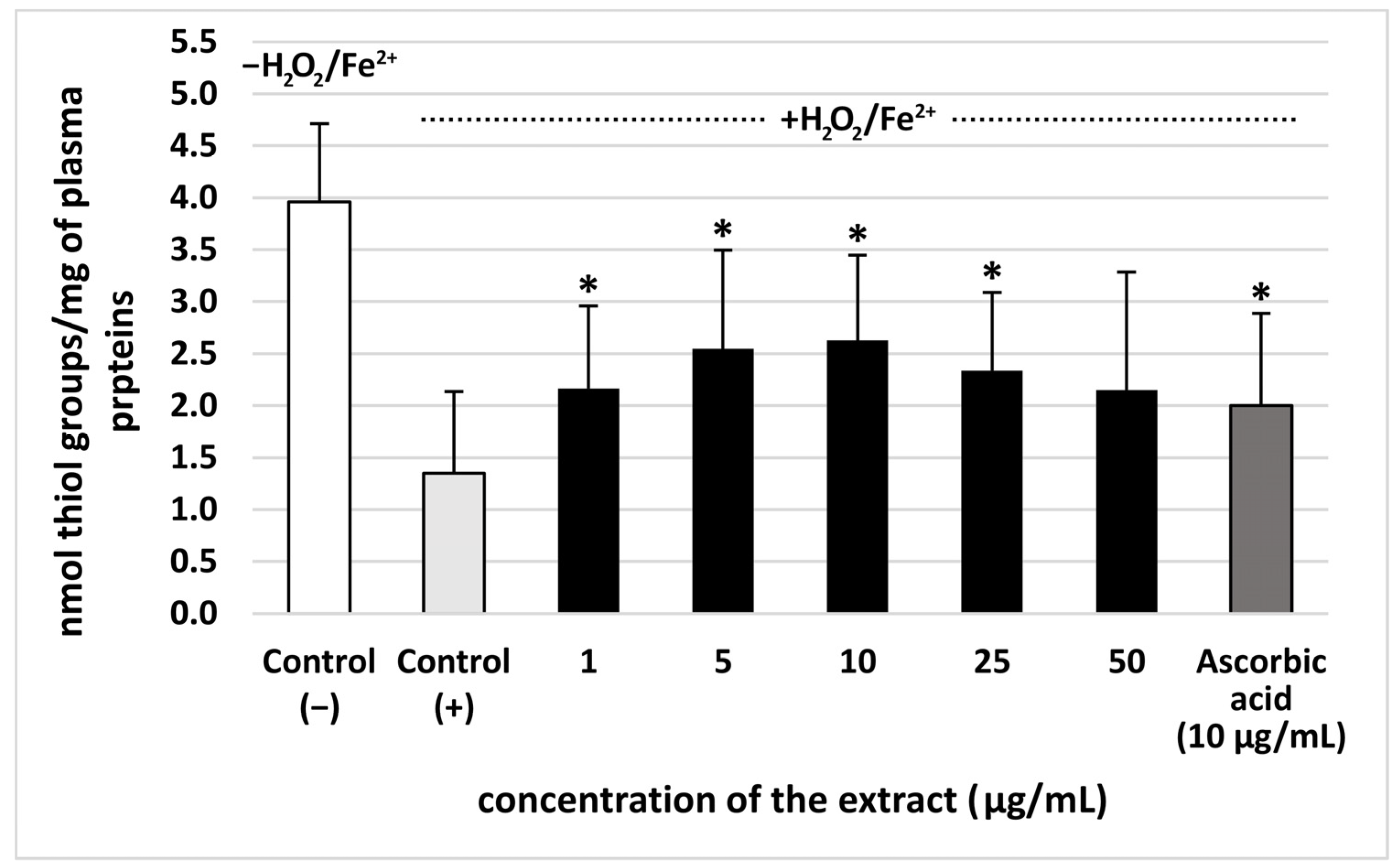
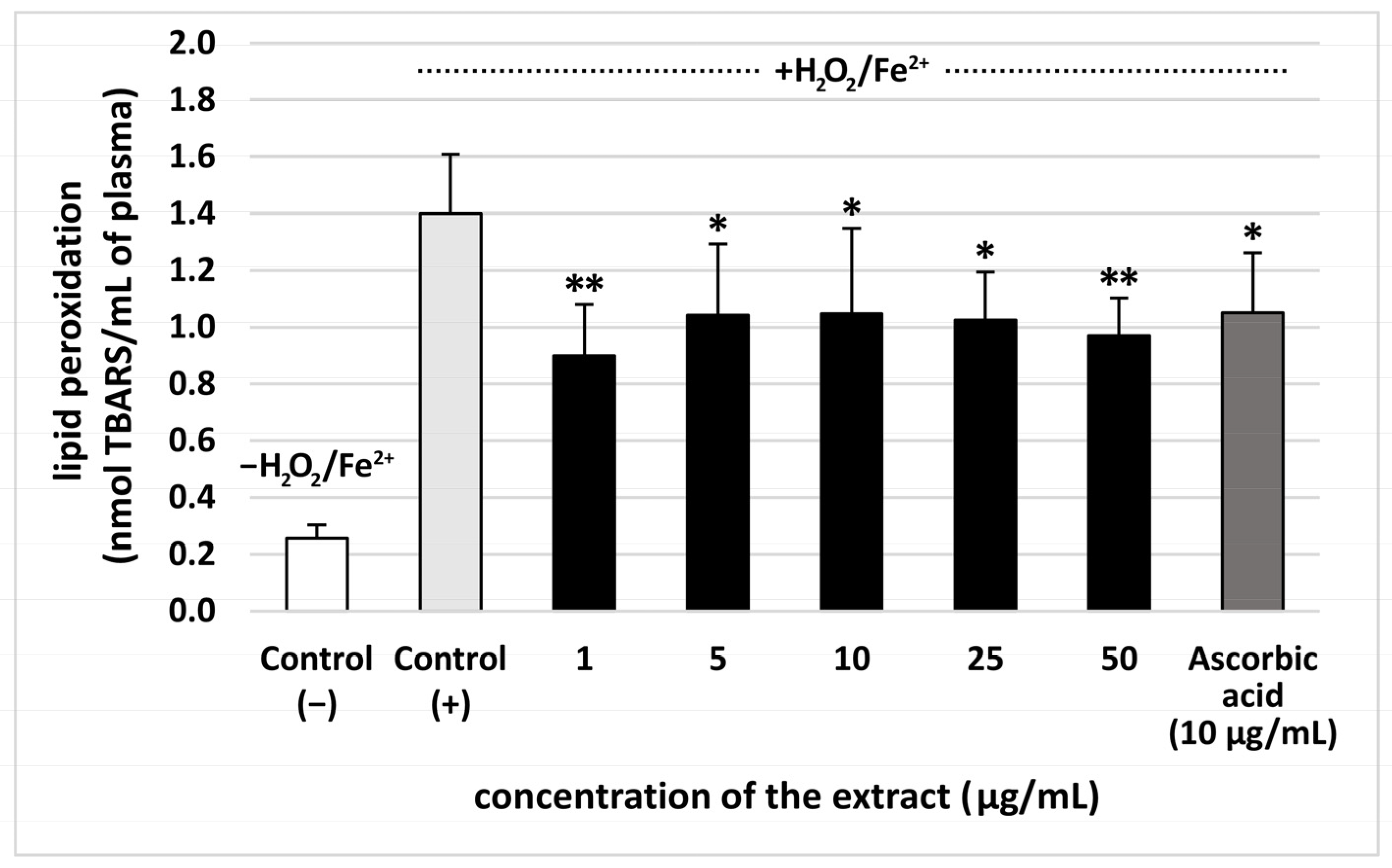
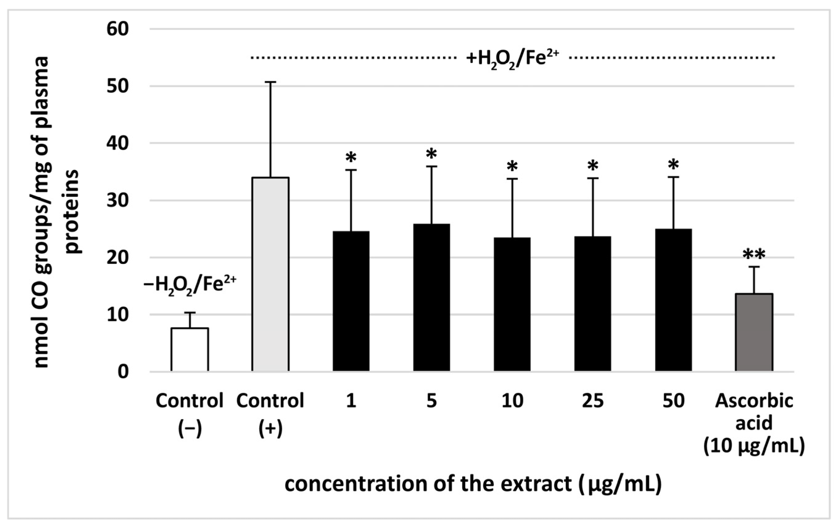
The extract from S. vulgaris flowers (at all used concentrations: 1–50 µg/mL) significantly reduced plasma lipid peroxidation induced by H2O2/Fe2+, however, this effect was not dose-dependent. The % of inhibition observed for the extract at the highest concentration–50 µg/mL—was approximately 35% (Figure 2). Moreover, all used concentrations (1–50 µg/mL) of the extract from S. vulgaris flowers decreased plasma protein carbonylation stimulated by H2O2/Fe2+ (the % of inhibition was about 35%) (Figure 3). The highest concentration of the extract (50 μg/mL) did not change the level of thiol groups in plasma treated with H2O2/Fe2+ (Figure 4). However, the lower concentrations of the extract from S. vulgaris flowers (1–25 µg/mL) significantly reduced thiol groups oxidation (Figure 4).
The effects of the extract from S. vulgaris flowers (10 µg/mL) on oxidative stress were compared with the extract from T. officinalis flowers (10 µg/mL) in Table 2. The dandelion extract, which was used as a positive control (10 µg/mL), demonstrated antioxidant properties, but the extract from S. vulgaris flowers had stronger inhibitory effect on lipid peroxidation. On the other hand, the % of inhibition of protein carbonylation for two tested extracts (from S. vulgaris flowers and T. officinalis flowers) was very similar (Table 2).

3.3. Coagulation Times (PT, TT, and APTT)

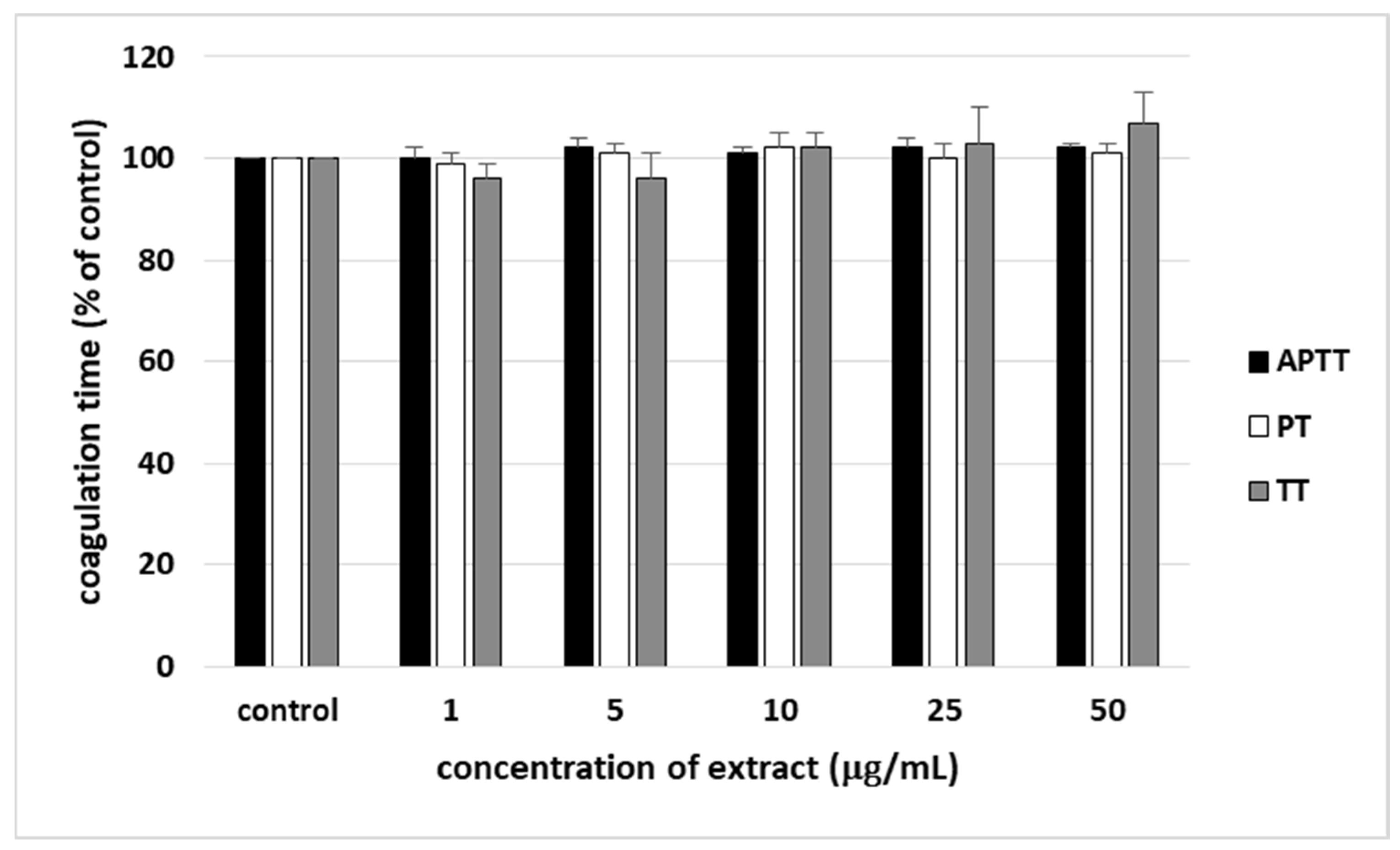
None of used concentrations of the extract from S. vulgaris flowers (1–25 µg/mL) significantly impacted the coagulation times measured in human plasma (Figure 5).

4. Discussion

The investigated lilac flower extract contained a broad array of specialized metabolites, belonging to different classes, mainly phenolic compounds (including phenylethanoid glycosides), and secoiridoids (Table 1, Figure 1). The presence of secoiridoids (frequently conjugated with a phenolic moiety) and phenylethanoid glycosides is a characteristic feature of plants from the Oleaceae family [12,13]. As shown by our UHPLC-MS analyses, the dominant phenolic constituent (also including aromatic-conjugated secoiridoids) of the extract from S. vulgaris flowers was putative verbascoside. Other phenylethanoid glycosides, such as putative hydroxyacteosides, echinacoside, forsythoide B, isoverbascoside, as well as hexosides of hydroxytyrosol and tyrosol were also present, but in much lower quantities. It seems that previous phytochemical research on S. vulgaris was focused mainly on the bark and leaves of the plant, while publications describing the flowers were scarce. Nevertheless, the above-mentioned compounds were reported in two earlier publications on the phytochemical composition of lilac flowers [5,14], while the presence of verbascoside and echinacoside was additionally confirmed by Hanganu et al. [3]. In addition, Toth et al. [14] also showed that the methanol extract from lilac flowers contained very high amounts of verbascoside, which seems to confirm our results. Apart from phenyetanoid glycosides, the lilac flower extract also contained three flavonoids, 3-O-hexosides-deoxyhexosides of quercetin and kaempferol. Two of them were most probably rutin (quercetin 3-O-rutinoside), and kaempferol 3-O-rutinose, as these compounds were previously isolated from S. vulgaris flowers [15]. Similar flavonoids were also detected in lilac flower extracts by other research teams [5,14]. Several caffeates and coumarates of a hexaric acid were also detected, which are most probably different caffeoylglucaric and p-coumaroylglucaric acids [2]. Such compounds were also detected in lilac flower extracts by LC-MS [5,14], as well as other Oleacea species, Ligustrum vulgare and Fraxinus excelsior [16,17]. Our lilac flower extract was also shown to contain significant amounts of putative syringin and trace quantities of putative lariciresinol hexoside. In the case of syringin, literature data are ambiguous. While S. vulgaris bark is a rich source of this substance [14,18,19], Tóth et al. [14] and Woźniak et al. [5] found only traces, or no syringin in the flower extracts. In contrast, Hanganu et al. [3] detected significant quantities of the compound in lilac flowers. It is quite possible that the content of syringin in S. vulgaris flowers may depend on the flower maturity state (the collection date), pedoclimatic conditions, and other factors. The detected lignan glycoside, putative lariciresinol hexoside is most probably (+)-lariciresinol 4-O-glucopyranoside, previously found in the bark of S. vulgaris [18,19]. Secoiridoids constitute another important group of the lilac specialized metabolites. Most of them are aromatic-conjugated secoiridoids, which may be regarded also as phenolic compounds. In the currently described extract, these compounds included oleoacteoside, oleonuezhenide (major compounds), as well as hydroxyoleoacteoside, nuezhenide, oleuropein and ligustroside. Simple secoiridoids (putative 11-methyloleoside), and terpenoid-conjugated secoiridoids (putative 2″-epi-frameroside) were also identified. All these compounds were previously detected in extracts from flowers of S. vulgaris [5,14]. In addition, several monoterpenoid hexosides, and putative malonylated monotorpenoid hexosides. The monoterpenoid hexosides are probably glucosides of lilac alcohols. Lilac alcohols (8 diastereoisomers) are among characteristic constituents of the lilac essential oil, and (βR,2R,5S)-lilac alcohol β-glucopyranoside was isolated from the flowers [2]. The tentatively-identified malonylated monoterponoid hexosides most probably have not been described before.
Our study is the first work devoted to a comprehensive assessment of biological effect of lilac flowers extract, employing in vitro experimental system related to the human plasma and blood physiology, which includes the coagulation process. The present study brings originality by offering, for the first time, the proof of the antioxidant activity of S. vulgaris flowers extract by using three assays—the level of TBARS (the final lipid peroxidation products), thiol groups, and carbonyl groups—in human plasma treated with H2O2/Fe2+ (the donor of •OH—one of the most aggressive reactive oxygen species generated in human). In addition, the inhibition of oxidative stress (especially lipid peroxidation and thiol oxidation) stimulated by H2O2/Fe2+ was comparable to or higher than the action of vitamin C (which is a potent water-soluble antioxidant) at 10 µg/mL.
Various secondary metabolites, including phenolic compounds, exhibit antioxidant activity within a defined therapeutic window, representing the exposure range in which they reduce oxidative stress without triggering prooxidant effects. At lower, physiologically compatible levels, they contribute to free radical scavenging and redox balance, as demonstrated in studies showing their role in mitigating oxidative stress. However, as exposure increases beyond the optimal range, different phenolic compounds begin to exhibit prooxidant effects. This dose-dependent duality underscores the need for careful characterization of safety margins when evaluating phenolic compounds as potential antioxidant agents [20]. Here, the tested extract from S. vulgaris flowers had the best antioxidant activity at the concentrations of 5 and 10 μg/mL, while the activity of 50 μg/mL extract was not significant, when we measured its effect on the level of thiol groups in plasma treated with H2O2/Fe2+. This indicates that S. vulgaris flower extract is more effective at preventing thiol groups oxidation at lower (5–10 μg/mL) concentrations.
The antioxidant mechanisms of lilac flower extract observed in human plasma might include scavenging oxidants (H2O2 and H2O2/Fe2+-derived •OH radicals). Our results are consistent with previous studies demonstrating the role of bioactive compounds from lilac flowers in protecting against oxidative stress. The antioxidant activity of lilac flowers (IC50 = 65.25 µg/mL) was noted by Tóth et al. [14] using the DPPH (2,2-diphenyl-1-picrylhydrazyl) bleaching assay. Results of Hanganu et al. [3] also demonstrated that the extract from S. vulgaris flowers has antioxidant potential, which was measured by three separate methods: DPPH, Ferric Reducing Antioxidant Power (FRAP), and Cupric Ion Reducing Antioxidant Capacity (CUPRAC) assay. Gąsecka et al. [21] noted that the scavenging capacity of the extract from lilac flowers is correlated with the total content of phenolic compounds, including flavonoids. These findings on the antioxidant potential of lilac flowers support the recommendation that S. vulgaris can be used as a supplement in managing atherosclerosis, as oxidative stress (particularly lipid peroxidation) is a key hallmark of this disease [22]. There are known supplements based on lilac flowers (e.g., Liliac PlantExtract produced in Romania) that are recommended in maintaining the function of the cardiovascular system by enhancing the elasticity of blood vessels. In addition, lilac flowers are a source of dietary fiber, which has cardioprotective potential [23].
Dudek et al. [2] observed that lilac flowers are a good source hydroxycinnamoyl derivatives and a secoiridoid glycoside with anti-inflammatory properties. These properties were observed in human neutrophils (in vitro) at the concentration of 50 µM. In addition, Woźniak et al. [5] found that neoleuropein—an especially active secoiridoid—decreased the production of various cytokines in human neutrophils through the inhibition of mitogen-activated protein kinase (MAP) kinases phosphorylation [5].
For the first time, our study provides a comparative analysis of lilac and dandelion flowers, demonstrating that S. vulgaris flowers, like T. officinale flowers, have antioxidant potential. Our results showed that the extract from lilac flowers exhibits stronger antioxidant activity, particularly in preventing plasma lipid peroxidation, than the phenolic extract from dandelion flowers. It seems probable that the differences in the chemical profiles of both extracts can explain the stronger action of the lilac extract, which contains not only various phenolic compounds, but also secoiroidoids—a diverse class of bioactive cyclopentane monoterpenoid derivatives formed by the cleavage of the cyclopentane ring, commonly found in plants of the Oleaceae family, including olives. Studies have found that secoiroidoids exhibit strong antioxidant, anti-inflammatory, neuroprotective, and anti-cancer properties.
It is known that oxidative stress can affect various elements of hemostasis, including the coagulation cascade, which is a complex process [24,25,26,27]. For example, the results of Wang et al. [27] demonstrated that the cross-linking, branching, and height distribution of formed fibrin is influenced by the oxidative stress of fibrinogen induced by H2O2. On the other hand, this process can be modulated by various plant extracts. Both phenolic compounds and other low molecular secondary metabolites may modulate not only the coagulation process, but also fibrinolysis and blood platelet activation [1,28]. For example, the anticoagulant effect of Rheum rhaponticum and Rheum rhabarbarum extracts is partly attributed to the inhibition of the coagulation factor Xa and thrombin activities [29].
Evaluation of the clotting times (PT, TT, and APTT) is a simple and rapid screening test that enables the assessment of the effects of plant extracts on coagulation. In the present study, the effects of lilac flower extract on these three coagulation times were measured in human plasma; the anticoagulant tests demonstrated that S. vulgaris flowers extract, at physiologically relevant concentrations (1–50 µg/mL), did not affect blood clotting times in vitro, suggesting that it is hemostatically safe.
Biological properties of the tested plant extracts (including their effects on oxidative stress stimulated by H2O2/Fe2+ and coagulation process) were examined in the concentration range of 1–50 µg/mL, which was chosen based on our previous study [7] and literature data, indicating that the physiologically achievable concentrations of phenolic compounds in plasma are mostly at levels ranging from nanomoles to a few micromoles per liter [29]. Although no data on secroiridoid bioavailability after S. vulgaris extract intake is available, some information on the concentrations of these compounds from other plants in plasma can be found in literature. Secoiridoides have low to moderate oral bioavailability. Despite this, their plasma concentration peaks quickly—after about 1 h in humans [23].

5. Conclusions

In conclusion, this study is the first to comprehensively evaluate S. vulgaris flowers extract, combining the physiological characterization with the assessment of its antioxidant properties. Using UHPLC-HRMS, 50 compounds, mainly phenolic compounds and secoiridoids were identified. The examined extract from lilac flowers displayed the ability to inhibit oxidative stress induced by H2O2/Fe2+. Lilac flowers extract provided a significant protective effect against the oxidation of plasma lipids and proteins, at the levels achievable for phenolic compounds in plasma after oral supplementation, and with the action comparable to vitamin C and the extract from dandelion flowers. Secoiridoids, which are found in S. vulgaris flowers, could also show antioxidant potential, but this data is available only in vitro, for secoiridoids isolated from other plant sources [30,31,32]. Therefore, their beneficial biological properties need to be further explored in vivo. In addition, according to recent studies, these compounds have also demonstrated therapeutic potential in various CVDs [31].
The antioxidant properties, including the protection of molecules from oxidative damage, might thus contribute to the beneficial effects of lilac flower remedies used in the prophylaxis and treatment of CVDs. However, further research is required to deepen the understanding of the molecular mechanisms of action of S. vulgaris flowers extract, and verify its antioxidant effects in vivo.

Author Contributions

Conceptualization, B.O.; methodology, N.S., J.Ż., B.S. and B.M.-S.; validation, N.S., J.Ż., and B.M.-S.; formal analysis, B.O. and J.Ż.; investigation, N.S., J.Ż., B.S. and B.M.-S.; writing—original draft preparation, B.O. and J.Ż.; writing—review and editing, B.O., N.S. and J.Ż.; visualization, B.O., N.S. and J.Ż.; supervision, B.O. All authors have read and agreed to the published version of the manuscript.

Funding

This research received no external funding.

Institutional Review Board Statement

The study was conducted in accordance with the Declaration of Helsinki, and approved by the Bioethics Committee at the University of Łódź (2/KBBN-UŁ/III/2014, approval date: 15 March 2014).

Informed Consent Statement

Informed consent was obtained from all subjects involved in the study.

Data Availability Statement

Dataset available on request from the authors.

Acknowledgments

The authors would like to thank Dariusz Jędrejek, PhD for performing UHPLC-HRMS analyses.

Conflicts of Interest

The authors declare no conflicts of interest.

Abbreviations

The following abbreviations are used in this manuscript:
APTTactivated partial thromboplastin time
CUPRACcupric ion reducing antioxidant capacity
CVDscardiovascular diseases
DPPH•2,2-diphenyl-picrylhydrazyl
FRAPferric-reducing antioxidant power
IC50half maximal inhibitory concentration
MAPmitogen-activated protein kinase
PTprothrombin time
ROSradical oxygen species
TBARSthiobarbituric acid reactive substances
TTthrombin time

References

  1. Bachmair, E.M.; Ostertag, L.M.; Zhang, X.; de Roos, B. Dietary manipulation of platelet function. Pharmacol. Ther. 2014, 144, 97–113. [Google Scholar] [CrossRef] [Scilit]
  2. Dudek, M.K.; Michalak, B.; Woźniak, M.; Czerwińska, M.E.; Filipek, A.; Granica, S.; Kiss, A.K. Hydroxycinnamoyl derivatives and secoiridoid glycoside derivatives from Syringa vulgaris flowers and their effects on the pro-inflammatory responses of human neutrophils. Fitoterapia 2017, 121, 194–205. [Google Scholar] [CrossRef] [Scilit] [PubMed]
  3. Hanganu, D.; Niculae, M.; Ielciu, I.; Olah, N.K.; Munteanu, M.; Burtescu, R.; Ștefan, R.; Olar, L.; Pall, E.; Andrei, S.; et al. Chemical profile, cytotoxic activity and oxidative stress reduction of different Syringa vulgaris L. Extracts. Molecules 2021, 26, 3104. [Google Scholar] [CrossRef] [Scilit] [PubMed]
  4. Jakubczyk, K.; Koprowska, K.; Gottschling, A.; Janda-Milczarek, K. Edible Flowers as a Source of Dietary Fibre (Total, Insoluble and Soluble) as a Potential Athlete’s Dietary Supplement. Nutrients 2022, 14, 2470. [Google Scholar] [CrossRef] [Scilit] [PubMed]
  5. Woźniak, M.; Michalak, B.; Wyszomierska, J.; Dudek, M.K.; Kiss, A.K. Effects of Phytochemically Characterized Extracts From Syringa vulgaris and Isolated Secoiridoids on Mediators of Inflammation in a Human Neutrophil Model. Front. Pharmacol. 2018, 9, 349. [Google Scholar] [CrossRef] [Scilit]
  6. Olas, B. New Perspectives on the Effect of Dandelion, Its Food Products and Other Preparations on the Cardiovascular System and Its Diseases. Nutrients 2022, 14, 1350. [Google Scholar] [CrossRef] [Scilit]
  7. Jędrejek, D.; Kontek, B.; Lis, B.; Stochmal, A.; Olas, B. Evaluation of antioxidant activity of phenolic fractions from the leaves and petals of dandelion in human plasma treated with H2O2 and H2O2/Fe. Chem. Biol. Interact. 2017, 262, 29–37. [Google Scholar] [CrossRef] [Scilit]
  8. Majewski, M.; Lis, B.; Juśkiewicz, J.; Ognik, K.; Jedrejek, D.; Stochmal, A.; Olas, B. The composition and vascular/antioxidant properties of Taraxacum officinale flower water syrup in a normal-fat diet using an obese rat model. J. Ethnopharmacol. 2021, 265, 113393. [Google Scholar] [CrossRef] [Scilit]
  9. Bartosz, G. Druga Twarz Tlenu; PWN: Warszawa, Poland, 2008. [Google Scholar]
  10. Levine, R.L.; Garland, D.; Oliver, C.N.; Amici, A.; Climent, I.; Lenz, A.G.; Ahn, B.W.; Shaltiel, S.; Stadtman, E.R. Determination of carbonyl content in oxidatively modified proteins. Methods Enzymol. 1990, 186, 464–478. [Google Scholar] [CrossRef] [Scilit]
  11. Sławińska, N.; Żuchowski, J.; Stochmal, A.; Olas, B. Extract from Sea Buckthorn Seeds—A Phytochemical, Antioxidant, and Hemostasis Study; Effect of Thermal Processing on Its Chemical Content and Biological Activity In Vitro. Nutrients 2023, 15, 686. [Google Scholar] [CrossRef] [Scilit]
  12. Huang, Y.-L.; Oppong, M.B.; Guo, Y.; Wang, L.-Z.; Fang, S.-M.; Deng, Y.-R.; Gao, X.-M. The Oleaceae family: A source of secoiridoids with multiple biological activities. Fitoterapia 2019, 136, 104155. [Google Scholar] [CrossRef] [Scilit]
  13. Jensen, S.; Franzyk, H.; Wallander, E. Chemotaxonomy of the Oleaceae: Iridoids as taxonomic markers. Phytochemistry 2002, 60, 213–231. [Google Scholar] [CrossRef] [Scilit]
  14. Tóth, G.; Barabás, C.; Tóth, A.; Kéry, Á.; Béni, S.; Boldizsár, I.; Varga, E.; Noszál, B. Characterization of antioxidant phenolics in Syringa vulgaris L. flowers and fruits by HPLC-DAD-ESI-MS. Biomed. Chromatogr. 2016, 30, 923–932. [Google Scholar] [CrossRef] [Scilit] [PubMed]
  15. Ahmad, M.; Salama, O. Flavonoids from violet flowers of Syringa vulgaris. Pak. J. Sci. Ind. Res. 1987, 30, 150–151. [Google Scholar]
  16. Czerwińska, M.; Granica, S.; Kiss, A. Effects of an Aqueous Extract from Leaves of Ligustrum vulgare on Mediators of Inflammation in a Human Neutrophils Model. Planta Med. 2013, 79, 924–932. [Google Scholar] [CrossRef] [Scilit] [PubMed]
  17. Kiss, A.K.; Michalak, B.; Patyra, A.; Majdan, M. UHPLC-DAD-ESI-MS/MS and HPTLC profiling of ash leaf samples from different commercial and natural sources and their in vitro effects on mediators of inflammation. Phytochem. Anal. 2020, 31, 57–67. [Google Scholar] [CrossRef] [Scilit]
  18. Kurkin, V.A. Phenylpropanoids from medicinal plants: Distribution, classification, structural analysis, and biological activity. Chem. Nat. Compd. 2003, 39, 123–153. [Google Scholar] [CrossRef] [Scilit]
  19. Varga, E.; Barabás, C.; Tóth, A.; Boldizsár, I.; Noszál, B.; Tóth, G. Phenolic composition, antioxidant and antinociceptive activities of Syringa vulgaris L. bark and leaf extracts. Nat. Prod. Res. 2019, 33, 1664–1669. [Google Scholar] [CrossRef] [Scilit]
  20. Gulcin, I. Antioxidants: A comprehensive review. Arch. Toxicol. 2025, 99, 1893–1997. [Google Scholar] [CrossRef] [Scilit]
  21. Gąsecka, M.; Krzymińska-Bródka, A.; Magdziak, Z.; Czuchaj, P.; Bykowska, J. Phenolic Compounds and Organic Acid Composition of Syringa vulgaris L. Flowers and Infusions. Molecules 2023, 28, 5159. [Google Scholar] [CrossRef] [Scilit]
  22. Gianazza, E.; Brioschi, M.; Martinez Fernandez, A.; Casalnuovo, F.; Altomare, A.; Aldini, G.; Banfi, C. Lipid Peroxidation in Atherosclerotic Cardiovascular Diseases. Antioxid. Redox Signal. 2021, 34, 49–98. [Google Scholar] [CrossRef] [Scilit] [PubMed]
  23. Peng, Z.; He, J.; Cheng, Y.; Xu, J.; Zhang, W. Biologically active secoiridoids: A comprehensive update. Med. Res. Rev. 2023, 43, 1201–1252. [Google Scholar] [CrossRef] [Scilit] [PubMed]
  24. Olas, B.; Wachowicz, B. Role of reactive nitrogen species in blood platelet functions. Platelets 2007, 18, 555–565. [Google Scholar] [CrossRef] [Scilit] [PubMed]
  25. Gao, L.; Liu, X.; Li, C.; Wang, Z.; Guo, T. A Rare Secoiridoid Dimer Derivative from Ligustri lucidi fructus. Rec. Nat. Prod. 2015, 9, 323–328. [Google Scholar]
  26. Pérez-Bonilla, M.; Salido, S.; van Beek, T.A.; Waard Pde Linares-Palomino, P.J.; Sánchez, A.; Altarejos, J. Isolation of antioxidative secoiridoids from olive wood (Olea europaea L.) guided by on-line HPLC–DAD–radical scavenging detection. Food Chem. 2011, 124, 36–41. [Google Scholar] [CrossRef] [Scilit]
  27. Wang, L.; Li, L.; Wang, H.; Liu, J. Study on the influence of oxidative stress on the fibrillization of fibrinogen. Biochem. J. 2016, 473, 4373–4384. [Google Scholar] [CrossRef] [Scilit]
  28. Kolodziejczyk-Czepas, J.; Czepas, J. Plant-Derived Compounds and Extracts as Modulators of Plasmin Activity—A Review. Molecules 2023, 28, 1677. [Google Scholar] [CrossRef] [Scilit]
  29. Liudvytska, O.; Ponczek, M.B.; Krzyżanowska-Kowalczyk, J.; Kowalczyk, M.; Balcerczyk, A.; Kolodziejczyk-Czepas, J. Effects of Rheum rhaponticum and Rheum rhabarbarum extracts on haemostatic activity of blood plasma components and endothelial cells in vitro. J. Ethnopharmacol. 2023, 315, 116562. [Google Scholar] [CrossRef] [Scilit]
  30. Manach, C.; Williamson, G.; Morand, C.; Scalbert, A.; Rémésy, C. Bioavailability and bioefficacy of polyphenols in humans. I. Review of 97 bioavailability studies. Am. J. Clin. Nutr. 2005, 81, 230S–242S. [Google Scholar] [CrossRef] [Scilit]
  31. Leong, X.Y.; Thanikachalam, P.V.; Pandey, M.; Ramamurthy, S. A systematic review of the protective role of swertiamarin in cardiac and metabolic diseases. Biomed. Pharmacother. 2016, 84, 1051–1060. [Google Scholar] [CrossRef] [Scilit]
  32. Lim, T.K. Edible Medicinal and Non Medicinal Plants; Springer: Dordrecht, The Netherlands, 2014. [Google Scholar] [CrossRef] [Scilit]
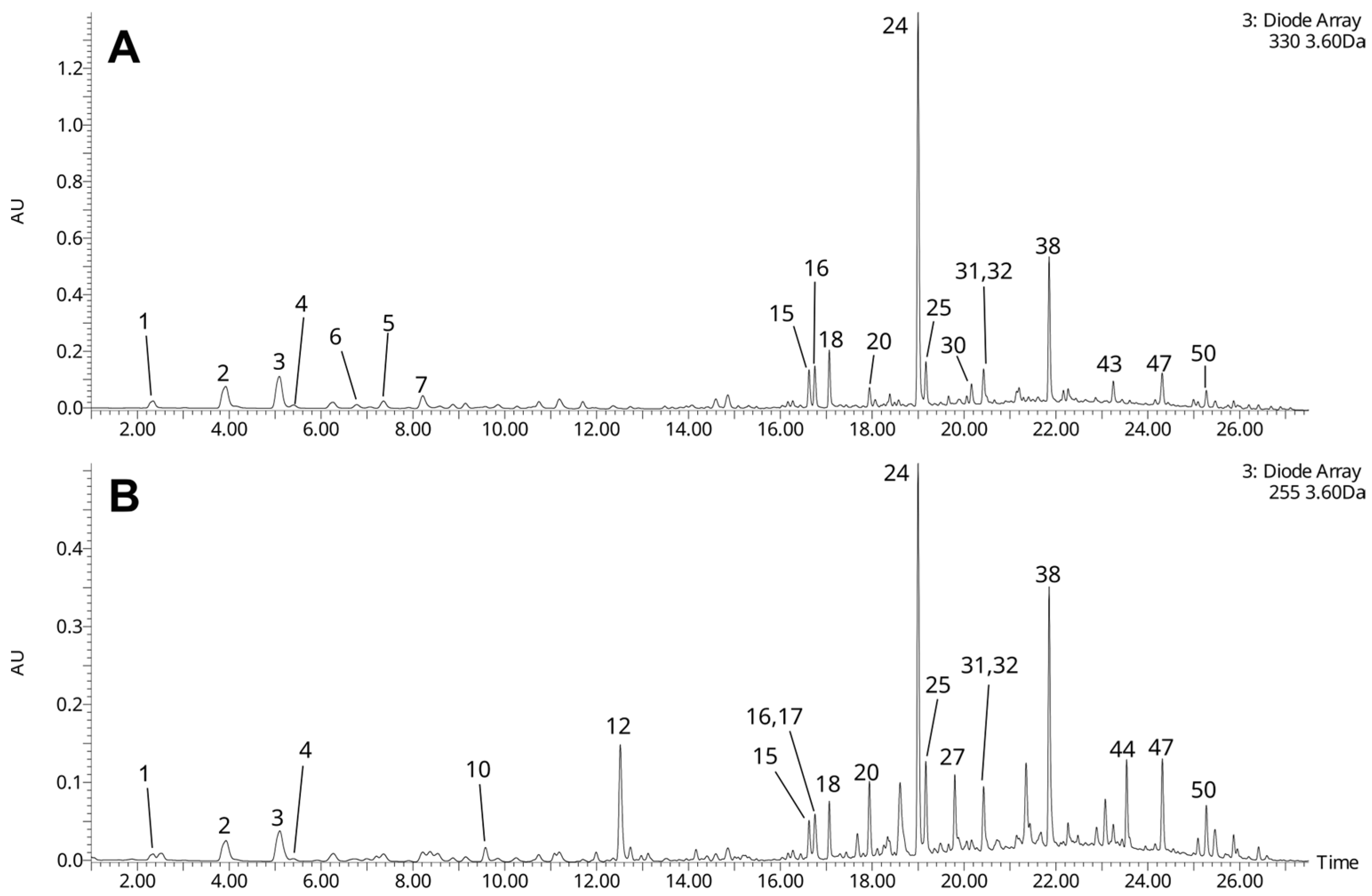
Figure 1. UHPLC-UV chromatograms of the lilac flower extract, registered at λ = 330 nm (A), and λ = 255 nm (B) (recorded using ACQUITY Premier® UHPLC system). Numbers of the chromatographic peaks correspond to numbers of compounds in Table 1.
Nutrients 18 01022 g001
Figure 2. Effect of the extract from S. vulgaris flowers (1–50 µg/mL) and ascorbic acid (10 µg/mL) on thiobarbituric acid reactive substances (TBARS) levels in human plasma treated with Fe2+/H2O2 (an oxidative stress inducer) (n = 7). Control (+) and test samples (1–50 μg/mL) were incubated for 30 min at 37 °C, with Fe2+/H2O2. The data are expressed as means ± SD. The results were considered significant at p < 0.05 (* p < 0.05, ** p < 0.01). The difference between control (−) and control (+) was statistically significant (p < 0.001).
Nutrients 18 01022 g002
Figure 3. The effect of the extract from S. vulgaris flowers (1–50 µg/mL) and ascorbic acid (10 µg/mL) on carbonyl (CO) groups level in human plasma treated with Fe2+/H2O2 (an oxidative stress inducer) (n = 7). Control (+) and test samples (1–50 μg/mL) were incubated (30 min, 37 °C) with Fe2+/H2O2. The data are expressed as means ± SD. The results were considered significant at p < 0.05 (* p < 0.05; ** p < 0.01). The difference between control (−) and control (+) was statistically significant (p < 0.001).
Nutrients 18 01022 g003
Figure 4. The effect of the extract from S. vulgaris flowers (1–50 µg/mL) and ascorbic acid (10 µg/mL) on thiol groups levels in human plasma treated with Fe2+/H2O2 (an oxidative stress inducer) (n = 7). Control (+) and test samples (1–50 μg/mL) were incubated (30 min, 37 °C) with Fe2+/H2O2. The data are expressed as means ± SD. The results were considered significant at p < 0.05 (* p < 0.05). The difference between control (−) and control (+) was statistically significant (p < 0.001).
Nutrients 18 01022 g004
Figure 5. The effect of the extract from S. vulgaris flowers (1–50 µg/mL) on the hemostatic parameters of human plasma: APTT, PT, and TT (n = 6). In the graphs, the coagulation time is expressed as a percentage of the control sample (plasma without the tested extract). The data are expressed as means ± SDs. The results were considered significant at p < 0.05.
Nutrients 18 01022 g005
Table 1. Tentative identification of constituents of the extract from flowers of S. vulgaris.
tRTypem/zMajor Fragment Ions (m/z)Error (ppm)mSigmaFormulaTentative Identification
11.85[M-H]371.0625209.0303 (100), 191.0211 (15), 179.0341 (8)−1.424.7C15H16O11caffeoylhexaric acid/isomer
22.39[M-H]371.0617209.0305 (100), 191.0188 (14), 179.0337 (2)0.82.2C15H16O11caffeoylhexaric acid/isomer
32.55[M-H]371.0626209.0305 (100), 191.0197 (13), 179.0393 (2)−1.77.1C15H16O11caffeoylhexaric acid/isomer
42.71[M-H]355.0673209.0307 (59), 191.0176 (100), 147.0281 (27)−0.71.8C15H16O10coumaroylhexaric acid/isomer
53.36[M-H]371.0614209.0308 (100), 191.0198 (13)1.65.1C15H16O11caffeoylhexaric acid/isomer
63.80[M-H]355.0665355.0671 (4), 209.0303 (99), 191.0203 (100), 163.0392 (9)1.64.8C15H16O10coumaroylhexaric acid/isomer
74.26[M-H]355.0665209.0300 (100), 191.0202 (300)1.58.6C15H16O10coumaroylhexaric acid/isomer
84.62[M+NH4]+332.1343332.1346 (100), 315.1084 (2), 279.0868 (4), 219.0651 (5), 180.0871 (11), 163.0607 (2), 153.0546 (4), 157.0499 (7), 127.0399 (3)−1.111.0C14H18O8unidentified hexoside
94.78[M-H]315.1091315.1088 (100), 135.0435 (2)−1.99.3C14H20O8hydroxytyrosol-Hex/isomer
[M+NH4]+334.1501334.1501 (100), 317.1241 (29), 281.1022 (14), 263.0917 (18), 221.0811 (7), 155.0704 (24), 137.0598 (31)−1.412.0
106.32[M+FA-H]345.1194345.1184 (4), 299.1135 (100), 179.0567 (6)−0.87.7C14H20O7tyrosol-Hex/isomer
118.20[M-H]403.1253371.0950 (11), 241.0721 (52), 223.0606 (49), 197.9821 (80), 179.0712 (83)−1.723.3C17H24O11oleoside 11-methyl ester hexoside/isomer
128.20[M+FA-H]417.1400209.0820 (100)−0.510.8C17H24O9syryngin/isomer
[M+NH4]+390.1762211.0965 (30), 193.0858 (100), 180.0867 (6), 161.0598 (45)−1.010.5
138.45[M+FA-H]315.1089315.1081 (38), 269.1028 (100)−1.313.2C13H18O6unidentified
[M+NH4]+288.1439288.1442 (9), 271.1178 (28), 253.1059 (100), 235.0955 (25), 217.0858 (25), 180.0865 (20), 161.0444 (17), 145.0496 (19), 127.0400 (6)0.95.1
1410.00[M+FA-H]447.1504401.1453 (100), 269.1029 69), 161.0447 (2)1.010.0C18H26O10unidentified
1512.23[M-H]639.1925639.1931 (100), 621.1823 (38), 529.1546 (8), 487.1446 (8), 477.1606 (5), 179.0353 (13), 161.0243 (28)0.99.6C29H36O16hydroxyverbascoside/iosomer
1612.39[M-H]639.1915639.1931 (100), 621.1829 (74), 529.1533 (6), 487.1461 912), 477.1601 (5), 459.1500 (13), 179.0349 (19), 161.0242 (38), 151.0384 (6), 133.0282 (12)2.524.8C29H36O16hydroxyverbascoside/isomer
1712.44[M+FA-H]461.1662415.1604 (100), 311.0980 (4), 283.1187 (6), 251.0766 (7), 221.0669 (3), 191.0561 (7), 179.0561 (7), 161.0443 (1), 149.0449 (15), 131.0329 (2)0.53.6C19H28O10unidentified
1812.95[M-H]785.2503785.2514 (100), 623.2197 (24), 179.0349 (1), 161.0235 (26), 133.0275 (8)0.822.9C35H46O20verbascoside-Hex (echinacoside)/isomer
[M+NH4]+804.2924625.2123 (4), 479.1553 (13), 471.1503 (15), 325.0925 (100), 309.0975 (3), 163.0394 (49)−0.43.2
1913.14[M+NH4]+350.2178350.2174 (4), 333.1913 (10), 315.1807 (26), 297.1706 (15), 279.1599 (7), 163.0604 (4), 153.1278 (58), 135.1171 (100)−1.311.8C16H28O7monoterpenoid hexoside (lilac alcohol hexoside ?)
2013.87[M-H]609.1459609.1453 (90), 300.0270 (69), 271.0240 (100), 255.0295 (39), 243.0294 (21)0.410.1C27H30O16quercetin-3-O-Hex-dHex (rutin ?)/isomer
2114.48[M-H]593.1506593.1516 (50), 284.0326 (90), 255.0290 (100), 227.0345 (42)1.012.9C27H30O15kaempferol-3-O-Hex-dHex/isomer
2214.54[M-H]521.2014359.1496 (17), 329.1391 (100)2.813.7C26H34O11lariciresinol-Hex
2314.80[M-H]755.2390755.2398 (100), 593.2086 (18), 161.0239 (20), 133.0286 (8)1.913.1C34H44O19forsythoside B/isomer
[M+NH4]+774.2824774.2819 (14), 625.2130 (4), 479.1557 (11), 471.1508 (19), 457.1353 (1), 325.0929 (100), 309.0982 (4), 181.0505 (2), 163.0396 (54)−1.110.2
2415.55[M-H]623.1973623.1974 (100), 461.1659 (10), 161.0239 (21), 133.0287 (7)1.42.6C29H36O15verbascoside/isomer
2515.71[M+H]+595.1657595.1656 (3), 449.1081 (14), 287.0553 (100)0.013.1C27H32O15kaempferol-3-O-Hex-dHex/isomer
2616.66[M+FA-H]377.1811377.1815 (53), 331.1752 (100), 161.0450 (2)1.51.4C16H28O7monoterpenoid hexoside (lilac alcohol hexoside ?)
2716.82[M+FA-H]731.2403685.2343 (25), 523.1818 (100), 453.1396 (95), 421.1500 (52), 299.1135 32), 223.0608 (18), 153.0188 (4)0.19.8C31H42O17nuezhenide/isomer
[M+NH4]+704.2764704.2763 (27), 525.1973 (15), 507.1867 (51), 387.1291 (42), 369.1184 (55), 295.0815 (18), 193.0496 (47), 165.0547 (100), 151.0391 (26)−0.510.3
2816.55[M-H]623.1968623.1974 (100), 461.1650 (10), 179.0338 (3), 161.0232 (19), 133.0283 (5)2.28.7C29H36O15verbascoside isomer
2917.28[M+NH4]+350.2173333.1909 (36), 171.1379 (100), 153.1273 (5), 135.1167 (8)0.27.3C16H28O7monoterpenoid hexoside (lilac alcohol hexoside ?)
3017.28[M-H]607.2025607.2022 (100), 461.1658 (47), 443.1581 (2), 315.1075 (2), 163.0386 (3), 145.0285 (23), 117.0337 (7)1.25.4C29H36O14deoxyverbascoside
3117.95[M+NH4]+1044.3551044.3541 (7), 847.2651 (5), 829.2542 (10), 677.2074 (14), 587.1760 (8), 505.1706 (14), 477.1394 (31), 371.1339 (21), 325.0920 (100), 225.0756 (24), 193.0495 (17), 165.0546 (56), 163.0390 (62)0.418C46H58O26hydroxyoleoacteoside
3218.08[M+NH4]+1190.41001190.4109 (14), 993.3212 (4), 831.2690 (12), 677.2068 (14), 589.1909 (4), 479.1545 (17), 371.1332 (15), 353.1228 (19), 325.0918 (100), 225.0755 (21), 193.0494 (13), 165.0544 (46), 163.0389 (56)2.823.4C52H68O30oleoechinacoside/isomer
3318.36[M+NH4]+436.2177436.2175 (8), 419.1908 (68), 401.1803 (4), 383.1707 (1), 249.0604 (2), 231.0498 (6), 171.1378 (100), 153.1273 (8), 135.1166 (11)0.04.5C19H30O10monoterpenoid hexoside-MaA/isomer
[2M-H]835.3594373.1853 (25), 331.1747 (100)1.38.9
3418.92[M+NH4]+436.2178436.2175 (10), 419.1911 (89), 401.1806 (4), 383.1700 (2), 249.0603 (2), 231.0498 (5), 171.1378 (100), 153.1272 (7), 135.1166 (10)−0.213.0C19H30O10monoterpenoid hexoside-MaA/isomer
3519.19[M-H]539.1756403.1248 (27), 377.1226 (69), 345.0974 (23), 307.0809 (100), 275.0913 (76), 223.0615 (24), 179.0561 (8), 149.0234 (8)2.77.8C25H32O13oleuropein/isomer
[M+NH4]+558.2184541.1920 (2), 379.1388 (54), 361.1282 (100), 347.1126 (16), 287.0916 (7), 225.0759 (8), 165.0547 (15), 137.0596 (69)−0.45.3
3619.45[M+NH4]+436.2178419.1912 (68), 401.1806 (2), 383.1700 (2), 339.1803 (2), 249.0604 (1), 231.0499 (3), 171.1378 (100), 153.1274 (5), 135.1166 (8)−0.218.6C19H30O10monoterpenoid hexoside-MaA/isomer
3719.77[M-H]677.2074633.2170 (100), 591.2060 (1), 487.1784 (5), 445.1683 (2), 163.0388 (3), 145.0279 (29), 117.0340 (10)2.017.4C32H38O16coumaric acid derivative
[M+NH4]+696.2493969.2494 (7), 679.2225 (1), 541.1550 (13), 533.1653 (20), 395.0972 (100), 377.0863 (4), 291.0862 (3), 165.0546 (4), 147.0440 (21)0.71.0
3820.25[M-H]1009.3158847.2812 (10), 745.2316 (8), 665.2068 (13), 623.1964 (21), 461.1651 (14), 315.1078 (4), 297.0971 (2), 179.0343 (3), 161.0235 (49), 133.0282 (15)3.63.0C46H58O25oleoacteoside/isomer
[M+NH4]+1028.36061028.3592 (13), 831.2699 (8), 695.2179 (4), 677.2072 (17), 507.1863 (6), 479.1549 (15), 371.1338 (15), 325.0921 (100), 275.0915 (15), 225.0758 (19), 193.0496 (14), 163.0391 (50), 151.0392 (9)0.08.8
3920.72[M+NH4]+460.1820460.1816 (58), 443.1552 (16), 425.1446 (6), 303.1234 (6), 266.0871 (10), 231.0501 (19), 195.1018 (100), 163.0757 (10)−1.510.7C20H26O11unidentified
4020.93[M+NH4]+1028.35951028.3591 (9), 831.2704 (6), 704.2258 (5), 685.2124 (74), 549.1604(57), 531.1497 (74), 307.0819 (35), 225.00758 (29), 193.0497 (33), 165.0458 (100), 163.0392 (97)1.010.3C46H58O25oleoacteoside/isomer
[M-H]1009.31701009.3166 (100), 873.2647 (11), 847.2780 (9), 745.2338 (13), 665.2057 (15), 623.1995 (14), 461.1639 (13), 403.1244 (4), 315.1065 (1), 297.1005 (4), 179.0343 (31), 161.0238 (66)2.516.5
4121.46[M-H]523.1812361.1276 (42), 291.0865 (100), 259.0965 (23)1.817.3C25H32O12ligstroside/isomer
[M+NH4]+542.2241525.1962 (2), 363.1442 (71), 345.1336 (100), 331.1180 (18), 225,225.0760 (7), 193.0499 (5), 165.0549 (9), 121.0650 (76)−1.73.4
4221.46[M-H]601.2122601.2116 (8), 403.1241 (7), 223.0600 (4), 197.0813 (100), 179.560 (1), 153.0910 (24)2.67.0C27H38O152″-epi-frameroside/isomer
[M+NH4]+620.2547441.1762 (39), 423.1654 (82), 391.1392 (25), 373.1291 (13), 331.1182 (100), 225.0762 (28), 199.0970 (37), 193.0499 (24), 165.0549 (50)0.332.7
4322.07[M+NH4]+610.2504593.2243 (1), 471.1508 (23), 447.1658 (7), 325.0927 (100), 309.0979 (7), 181.0.500 (3), 163.0394 (52)−1.711.4C29H36O13caffeic acid derivative
4422.89[M-H]1071.3541909.3030 (10), 839.2599 (42), 807.2696 (19), 685.2338 (63), 523.1813 (100), 453.1397 (49), 421.1494 (29), 403.1245 (28), 299.1135 (24), 223.0604 (35), 179.0558 (6)28.8C48H64O27oleonuezhenide/isomer
4523.04[M+NH4]+476.2498476.2500 (37), 459.2234 (99), 271.0817 (100), 253.0711 (17), 171.1383 (58), 145.0499 (17), 127.0401 (15)−1.69.3C22H34O10unidentified
4623.46[M+NH4]+476.2496476.2499 (19), 459.2233 (100), 271.0819 (91), 253.0714 (17), 171.1385 (91), 145.0499 (16), 127.0403 (14)−1.26.9C22H34O10unidentified
4723.73[M+NH4]+622.2498587.2126 (5), 471.1506 (17), 441.1552 (12), 325.0927 (100), 309.0979 (10), 279.1023 (9), 181.1119 (1), 163.0394 (58), 117.0700 (16)−0.54.4C30H36O13caffeic acid derivative
4823.73[M+NH4]+594.2546577.2293 (1), 455.1556 (25), 431.1711 (8), 309.0980 (100), 165.0551 (2), 147.0446 (29)−0.211.5C29H36O12coumaric acid derivative
4924.15[M+NH4]+680.2553680.2553 (8), 541.1561 (13), 517.1714 (21), 395.0981 (100), 377.0878 (4), 165.0551 (4), 147.0445 (19)−1.17.3C32H38O15coumaric acid derivative
5025.10[M-H]587.2124587.2120 (100), 441.1748 (6), 307.1035 (7), 163.0391 (4), 145.0287 (44), 117.0342 (15)1.713.8C30H36O12coumaric acid derivative
FA—formic acid; Hex—hexose; dHex—deoxyhexose; MaA—malonic acid.
Table 2. A comparison of the antioxidant activity of the extract from S. vulgaris flowers (10 µg/mL), and the extract from T. officinalis flowers (10 µg/mL) in human plasma treated with H2O2/Fe2+ (in vitro).
The Extract from S. vulgaris FlowersThe Extract from T. officinalis Flowers
% of inhibition of lipid peroxidation26.0 ± 15.1 (p < 0.05)18.6 ± 7.9 (p < 0.05)
% of inhibition of protein carbonylation27.7 ± 9.3 (p < 0.01)28.9 ± 10.1 (p < 0.05)
Disclaimer/Publisher’s Note: The statements, opinions and data contained in all publications are solely those of the individual author(s) and contributor(s) and not of MDPI and/or the editor(s). MDPI and/or the editor(s) disclaim responsibility for any injury to people or property resulting from any ideas, methods, instructions or products referred to in the content.