1. Introduction
Aging is a complex biological process characterized by the progressive decline in physiological functions and increased susceptibility to age-related diseases. The brain is particularly vulnerable to oxidative stress and inflammatory damage, attributed to its inherent physiological characteristics of high oxygen consumption and abundant unsaturated fatty acids. This renders it among the earliest involved and most severely damaged organs in the aging process [
1,
2]. Progressive cognitive decline is a hallmark feature of brain aging, with its core driving mechanism strongly linked to oxidative stress imbalance and sustained neuroinflammatory activation in brain tissue [
3]. On one hand, the excessive accumulation of reactive oxygen species (ROS) in the brain during aging induces oxidative damage to lipids, proteins, and DNA. Concurrently, the declining functionality of endogenous antioxidant systems such as superoxide dismutase (SOD) and glutathione peroxidase (GSH-Px) further exacerbates this oxidative cascade, forming a vicious cycle [
4]. On the other hand, the NF-κB signaling pathway remains persistently activated during brain aging, which drives the release of pro-inflammatory cytokines such as tumor necrosis factor-α (TNF-α) and interleukin-1β (IL-1β). This triggers neuroinflammation, accelerates neuronal apoptosis and synaptic dysfunction, and ultimately leads to cognitive impairment and other brain aging phenotypes [
5,
6].
The Nrf2/Keap1 pathway serves as the central antioxidant defense system in the brain. It helps maintain redox homeostasis and supports neuronal survival by regulating the expression of downstream antioxidant molecules, such as NAD(P)H quinone oxidoreductase 1 (NQO1) and heme oxygenase-1 (HO-1). Furthermore, the crosstalk between this pathway and the NF-κB inflammatory pathway directly modulates the balance of oxidative stress and neuroinflammation during brain aging. It thereby serves as a key molecular network regulating the progression of brain aging [
7,
8,
9]. Thus, combined interventions targeting antioxidant and anti-inflammatory processes in brain are regarded as an effective strategy to delay brain aging and improve cognitive function.
With the increasingly widespread application of natural products in anti-aging research, cyanidin-3-O-glucoside (C3G), one of the most widely distributed and abundant core monomers in anthocyanins, exhibits promising potential for intervening in brain aging. This is attributed to its excellent antioxidant and anti-inflammatory activities as well as favorable blood–brain barrier penetration potential [
10,
11]. Previous studies have confirmed that anthocyanins and their major component C3G exert preventive effects on various neurological disorders, such as cerebral ischemia, Alzheimer’s disease, Parkinson’s disease, multiple sclerosis, and glioblastoma [
11]. Furthermore, anthocyanins have been shown to ameliorate key pathological processes in aging, including neuronal apoptosis and necrosis, as well as learning and memory impairments [
12,
13,
14]. While anthocyanins have been shown to regulate both the Nrf2 and NF-κB pathways [
15,
16,
17], systematic evidence is still lacking for C3G. Specifically, its concurrent regulation of these pathways in the aging brain, the dose-dependency of these effects, and the integration of such molecular changes with improved behavioral outcomes remain to be fully established.
The D-galactose (D-gal)-induced aging mouse model has been widely used for the screening of anti-aging active substances and mechanism research, as it mimics key characteristics of natural aging, including enhanced oxidative stress, activated inflammatory responses, and organ dysfunction [
18,
19]. While this model cannot fully mimic the physiological evolutionary patterns of natural aging, it provides a reliable experimental tool for investigating the roles of oxidative stress and inflammation in aging-related cognitive decline. Based on this, the present study employed D-gal-induced aging mice as the model to systematically investigate the role of C3G in delaying brain aging. Specifically, behavioral tests were conducted to evaluate the protective effects of C3G on cognitive function. Additionally, histopathological analysis was performed to explore its impacts on hippocampal tissue. Furthermore, the levels of oxidative stress markers and inflammatory factors were determined to reveal the antioxidant and anti-inflammatory activities of C3G in the brain tissue. Finally, qRT-PCR and Western blot analyses were further used to elucidate the regulatory mechanisms of C3G on the Nrf2/Keap1 and NF-κB signaling pathways in hippocampal tissue. This study aims to systematically clarify the role and molecular mechanisms of C3G in delaying brain aging, thereby providing a theoretical foundation for its application in developing interventions against brain aging.
2. Materials and Methods
2.1. Materials
D-galactose (D-gal) was purchased from Beijing Biotopped Technology Co., Ltd. (Beijing, China). Cyanidin-3-O-glucoside (C3G) (purity = 94.59% as determined by HPLC) was obtained from Nanjing Jingzhu Biotechnology Co., Ltd. (Nanjing, China). Commercial kits for the determination of malondialdehyde (MDA), total antioxidant capacity (T-AOC), superoxide dismutase (SOD), Mn-SOD, reduced glutathione (GSH), and glutathione peroxidase (GSH-Px) in serum and brain tissue homogenates were all purchased from Nanjing Jiancheng Bioengineering Institute (Nanjing, China). Commercial kits for detecting inflammatory cytokines including interleukin-6 (IL-6), interleukin-1β (IL-1β), and tumor necrosis factor-α (TNF-α) in serum were acquired from MultiSciences Biotech Co., Ltd. (Hangzhou, China). Antibodies against Nrf2 (Cat No. 66504-1-Ig) and Keap1 (Cat No. 60027-1-Ig) were purchased from Proteintech Group, Inc. (Wuhan, China), while antibodies against IKKβ (Cat No. R1706-13) and NF-κB p65 (Cat No. ET1603-12) were obtained from Hangzhou Huabio Co., Ltd. (Hangzhou, China). All other chemical reagents used in this study were of analytical grade.
2.2. Animal Grouping and Intervention
Male C57BL/6J mice aged 6–8 weeks were purchased from Beijing Vital River Laboratory Animal Technology Co., Ltd. (Beijing, China) (Laboratory Animal Production License No.: SCXK (Jing) 2021-0006). All mice were housed under standard conditions (25 °C, 12 h light/dark cycle) with free access to food and water. All experimental protocols were approved by the Animal Experiment Ethics Committee of Beijing Technology and Business University (Approval No. Lunshen 2024 No. 163). After a 7-day acclimatization period, the mice were sorted by body weight and then allocated into different experimental groups using a snake-like randomization method to ensure comparable group mean weights. Ultimately, the mice were formally divided into four groups (n = 10 per group): normal control (NC) group, model group (500 mg/kg D-gal), low-dose C3G (C3G-L) intervention group, and high-dose C3G (C3G-H) intervention group. Mice in the NC group received a daily subcutaneous injection of normal saline (10 mL/kg), while those in the other groups received a daily subcutaneous injection of 5% (w/v) D-gal (500 mg/kg). Additionally, mice in the C3G-L and C3G-H groups were administered C3G via gavage at doses of 50 and 100 mg/kg, respectively. All treatments were performed once daily for 13 consecutive weeks.
2.3. Morris Water Maze (MWM) Assessment
The water maze experimental method referenced the research protocol by Qin et al. [
20], employing the Morris Water Maze (MWM) test to evaluate spatial memory and long-term memory capabilities in mice. The maze (Model XR-XM101, Shanghai Xinruan Information Technology Co., Ltd., Shanghai, China) consisted of a circular pool (120 cm in diameter, 50 cm in height) filled with water, with a hidden platform (10 cm in diameter, 30 cm in height) submerged 1–2 cm under the surface. Experimental parameters were set as follows: 60 s swimming duration, 15 s platform residence time, “mouse experiment mode” selected, and the red boundary box adjusted to accurately frame the pool and platform. Distinct black-and-white markers of different shapes were placed on the four walls of the pool (North, South, East, West) to provide visual cues for spatial navigation. Notably, rodents achieve learning saturation with minimal individual variability on day 5 of consecutive training [
21]. Accordingly, the MWM test was initiated at week 12 post-treatment and lasted 6 days, with days 1–5 devoted to acquisition training and day 6 to the spatial probe test. Acquisition training was conducted 1 h after daily gavage administration, with 4 training trials per mouse per day. Blinding was adopted throughout the experiment, and the personnel responsible for behavioral testing and data recording were unaware of the animal grouping information. For each trial, the mouse was placed into the water facing the pool wall from a predetermined quadrant, and the time taken to locate and mount the hidden platform (escape latency) was recorded. If a mouse found and remained on the platform for 15 s within the 60 s trial, its actual latency was recorded. Otherwise, the escape latency was recorded as 60 s, and the mouse was guided to the platform and allowed to remain there for 15 s to reinforce spatial memory. The interval between consecutive trials was 30–60 s. On day 6, the hidden platform was removed for the spatial probe test, which serves as an independent assessment of memory consolidation ability. Each mouse was released into the quadrant opposite the original platform location and allowed to swim freely for 60 s. The crossing frequency over the former platform location and the time spent exploring the target quadrant were recorded.
2.4. Sample Collection and Preparation
After the intervention period, mice were fasted for 12 h and then anesthetized via isoflurane inhalation. Following blood collection, mice were euthanized by cervical dislocation, and brain tissues were immediately dissected. Blood samples were centrifuged at 3500 rpm and 4 °C, for 15 min to separate serum, which was stored at −20 °C, for subsequent detection of inflammatory cytokines and antioxidant indices. The isolated brain tissues were rinsed with pre-cooled normal saline (0.9%, w/v), and surface moisture was blotted dry with sterile filter paper before being divided into three parts. The hippocampal region was rapidly dissected. One was fixed in 10% neutral buffered formalin for histopathological analysis, and the other was wrapped in aluminum foil and stored at −80 °C for subsequent qRT-PCR and Western blot analyses. The remaining brain tissue was homogenized in ice-cold saline (1:9, w/v) to prepare a 10% (w/v) tissue homogenate. The homogenate was centrifuged at 5000× g for 10 min at 4 °C, and the supernatant was collected and stored at −80 °C for antioxidant index determination.
2.5. Histopathological Evaluation of Hippocampal Tissue
Hippocampal tissue was fixed in 10% neutral buffered formalin solution for 24 h, followed by sequential tissue dehydration, xylene clearing, and paraffin embedding. Serial sections (5 μm thick) were cut and stained with Hematoxylin–eosin (H&E). Pathomorphological changes in the hippocampal tissue were observed and analyzed using a PANNORAMIC DESK/MIDI/250/1000 whole-slide scanner (3DHISTECH Ltd., Budapest, Hungary). The operators and analysts were unaware of the grouping information of the experimental animals to eliminate subjective bias in the evaluation process.
2.6. Determination of Antioxidant Indices
Quantitative determination of malondialdehyde (MDA), total antioxidant capacity (T-AOC), superoxide dismutase (SOD), manganese superoxide dismutase (Mn-SOD), reduced glutathione (GSH), and glutathione peroxidase (GSH-Px) levels was performed in serum and brain tissue homogenates. All aforementioned indices were measured using corresponding commercial kits, strictly following the manufacturers’ instructions. For brain tissue homogenate samples, all indices were calibrated based on the content per milligram of protein to standardize the results.
2.7. Detection of Inflammatory Cytokines
The serum concentrations of interleukin-6 (IL-6), interleukin-1β (IL-1β), and tumor necrosis factor-α (TNF-α) were quantified using mouse-specific sandwich enzyme-linked immunosorbent assay (ELISA) kits, strictly following the manufacturer’s protocols.
2.8. Quantitative Real Time PCR (qRT-PCR) Analysis
Total RNA was extracted from hippocampal tissue using the High-Purity Total RNA Rapid Extraction Kit (Tiangen Biotech, Beijing, China), strictly following the manufacturer’s instructions. All RNA samples were quantified using a Thermo Scientific NanoDrop
TM 2000 spectrophotometer (Thermo Fisher Scientific Inc., Waltham, MA, USA), and RNA integrity was verified via 1.5% agarose gel electrophoresis. Subsequently, 1 μg of the extracted RNA was reverse-transcribed into cDNA using the ReverTra Ace
® qPCR RT Master Mix (Toyobo Co. Ltd., Life Science Department, Osaka, Japan). qRT-PCR amplification and detection were conducted in Low-Profile PCR tubes using a Bio-Rad CFX96 Real-Time PCR System (Bio-Rad Laboratories, Inc., Hercules, CA, USA). Each 20 μL reaction system contained: 10 μL of SYBR
® Green Real-Time PCR Master Mix (Toyobo Co. Ltd., Life Science Department, Osaka, Japan), 10 μM of each forward and reverse primer, 1 μL of cDNA, and PCR-grade water to bring the volume to 20 μL. The thermal cycling protocol was set as follows: an initial polymerase activation step at 95 °C for 3 min, followed by 40 cycles of denaturation (94 °C for 30 s), annealing (60 °C for 40 s), and extension (72 °C for 1 min). Fluorescence was measured at the end of each annealing step to monitor amplification in real time. The specificity of the PCR products was confirmed by analyzing dissociation curves. Gene expression levels were normalized to the reference gene glyceraldehyde-3-phosphate dehydrogenase (GAPDH). Relative mRNA expression was calculated using the 2
−ΔΔCt method with 6 independent biological replicates performed for each experimental group. The primer sequences are provided in
Table 1. The amplification efficiency for each primer pair was determined using a standard curve and was confirmed to be between 90% and 110%. The specificity of amplification was verified by sequencing the PCR products, which matched the expected target sequences in GenBank.
2.9. Western Blot Analysis
Approximately 20 mg of hippocampal tissue was homogenized thoroughly on ice in RIPA lysis buffer containing 1% protease inhibitor. After homogenization, the mixture was centrifuged at 4 °C, and the supernatant was collected. Protein concentration was determined using the BCA assay. Samples were mixed with 4× loading buffer at a 3:1 ratio, boiled for 5 min, and then cooled to room temperature for immediate use or stored at −80 °C. Proteins were separated by SDS-PAGE using a 10% separating gel and a 5% stacking gel, followed by transfer onto a PVDF membrane. The membrane was blocked with 5% non-fat milk at room temperature for 1 h and then incubated overnight at 4 °C with the following primary antibodies: Nrf2 (1:2000), Keap1 (1:2000), IKKβ (1:1000), NF-κB p65 (1:1000), and rabbit anti-β-actin (1:50,000). Subsequently, the membranes were incubated with species-appropriate HRP-conjugated secondary antibodies at a dilution of 1:20,000 for 1 h at room temperature. Protein bands were visualized using ECL chemiluminescent substrate and detected with a chemiluminescence imaging system. β-actin was used as the internal reference, and protein expression levels were normalized to the band intensity of β-actin. Quantitative analysis of band optical density was performed using ImageJ software (version 1.8.0).
2.10. Animal Inclusion/Exclusion Criteria and Sample Selection
The inclusion criteria required animals to be in good health, well-acclimated to the environment, and in a stable condition, with 10 animals initially allocated per group. Animals were excluded if they died unexpectedly during model establishment or exhibited significant abnormal behavioral responses during testing (e.g., frequent floating, immobility against the wall, or motor impairments). Consequently, behavioral and certain biochemical analyses were performed with n = 8 per group. For qRT-PCR, sample selection was based on predefined RNA quality control criteria. Only samples with A260/A280 ratios between 1.8–2.1, A260/A230 ratios > 2.0, and no evident degradation upon agarose gel electrophoresis were included, resulting in n = 6 per group for this analysis. For Western blot, due to the constraints of gel electrophoresis lanes and sample processing capacity, a stratified random sampling method was employed to select n = 3 representative samples per group. All sample exclusions and selections were conducted based on objective, pre-established criteria, independently of subsequent experimental outcomes, thereby avoiding selection bias. Sample sizes for each analysis are clearly indicated in the corresponding figure legends.
2.11. Statistical Analysis
All data are presented as mean ± standard deviation (SD). Normality and homogeneity of variance were confirmed via the Shapiro–Wilk and Levene’s tests, respectively, prior to parametric analyses. Specifically, repeated-measures ANOVA with Bonferroni post hoc multiple comparisons was used to assess time-course data (escape latency and swimming speed) during Morris water maze acquisition training. One-way ANOVA was performed, followed by Dunnett’s test to compare Western blot band gray values, while Duncan’s multiple range test was used for all other datasets. All statistical analyses were performed using SPSS 27.0, with statistical significance defined as p < 0.05.
4. Discussion
This study employed a D-gal-induced aging mouse model to systematically investigate the improving effects of C3G on aging-related cognitive impairment and its underlying molecular mechanisms. In this study, male mice were employed for model construction, with the aim of controlling confounding variability associated with sex differences and the estrous cycle of females in the stage of initial mechanistic inquiry. Collectively, our results demonstrate that C3G dose-dependently improved D-gal-induced cognitive dysfunction and hippocampal histopathological damage. Importantly, these protective effects were linked to the concurrent modulation of two key pathways, specifically involving the activation of the Nrf2/Keap1 antioxidant pathway and the suppression of the NF-κB inflammatory pathway in the hippocampal tissue. The conclusions of this study are derived from a male accelerated aging model, which is artificially induced and characterized by rapid, targeted pathological changes that are distinct from the slow, systemic degeneration observed in natural aging. Since elevated oxidative stress and chronic inflammation are shared core mechanisms between accelerated and physiological aging, the ability of C3G to regulate these pathways suggests its potential as an intervention for mitigating cognitive decline during natural aging. However, it should be emphasized that the accelerated aging model cannot fully recapitulate the systemic, progressive hallmarks of natural aging. Future studies are needed to validate the effects of C3G on inflammation and cognition in female animals, and more crucially, in natural aging models, to comprehensively assess the generalizability of its potential applications.
Decline in spatial learning and memory is a hallmark phenotype of aging-related cognitive impairment. As the core brain region mediating memory encoding and retrieval, the structural integrity and functional activity of the hippocampal CA1, CA3 and DG subregions directly determine the body’s spatial cognitive ability [
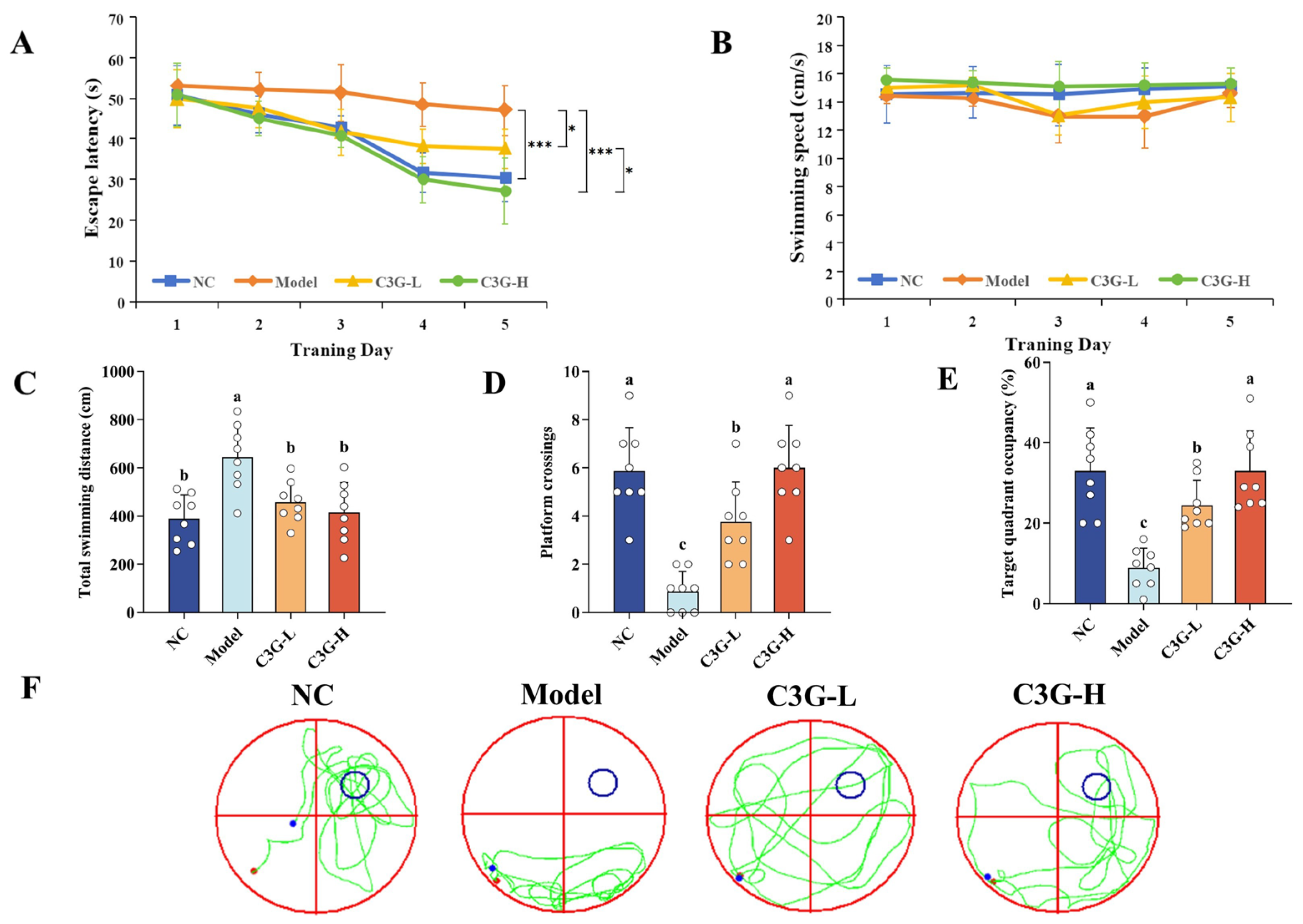
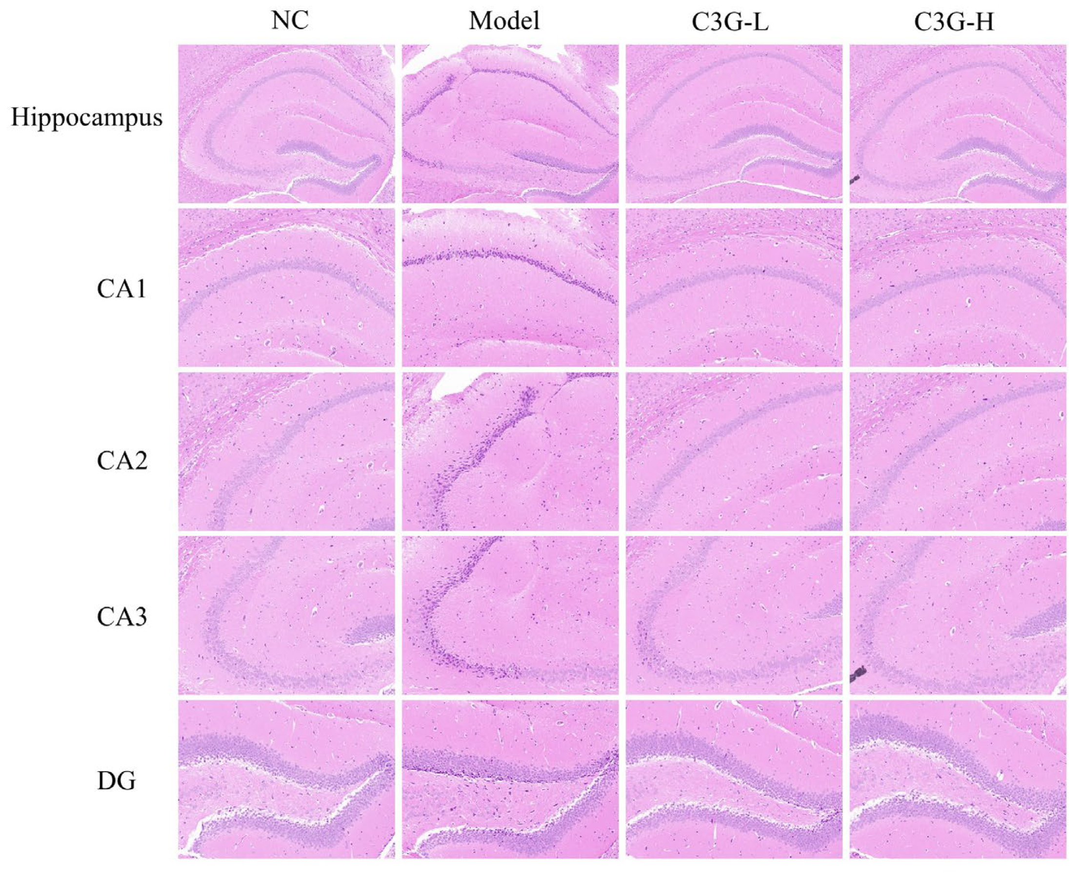
22]. In this study, D-gal treatment prolonged the escape latency of mice in the Morris water maze, reduced the percentage of time spent in the target quadrant and the number of crossings over the former platform location. Concurrently, histopathological examination revealed nuclear pyknosis, disordered arrangement, and blurred nuclear-cytoplasmic boundaries in the hippocampal CA1, CA3, and DG regions. These observations are consistent with the established model characteristics of D-gal-induced brain aging and cognitive impairment, which involve oxidative stress, neuroinflammation, and neuronal damage [
23]. C3G intervention significantly alleviated the aforementioned behavioral and histological abnormalities. The high-dose intervention was particularly effective, not only elevating cognitive function to near-normal levels but also largely preserving the structure and number of hippocampal neurons. This finding aligns with previous studies demonstrating the protective effects of anthocyanins on cognitive function in animal models of aging and neurodegenerative diseases [
12,
24,
25]. These results suggest that C3G can effectively alleviate D-gal-induced age-related cognitive impairment in mice by mitigating hippocampal neuronal damage. This effect may be attributed to the ability of C3G to cross the blood–brain barrier (BBB) and accumulate in brain tissue [
26]. However, the present study did not perform quantitative analysis of the actual exposure levels of C3G in the brain. To further clarify C3G’s neuroprotective role and underlying mechanisms, future studies should include systematic pharmacokinetic evaluations, as well as in vivo electrophysiology and assessments of synaptic plasticity markers in aging models.
Oxidative stress imbalance is one of the core mechanisms underlying D-gal-induced aging. Owing to its high oxygen demand and substantial lipid content, the brain is a susceptible target organ for oxidative damage [
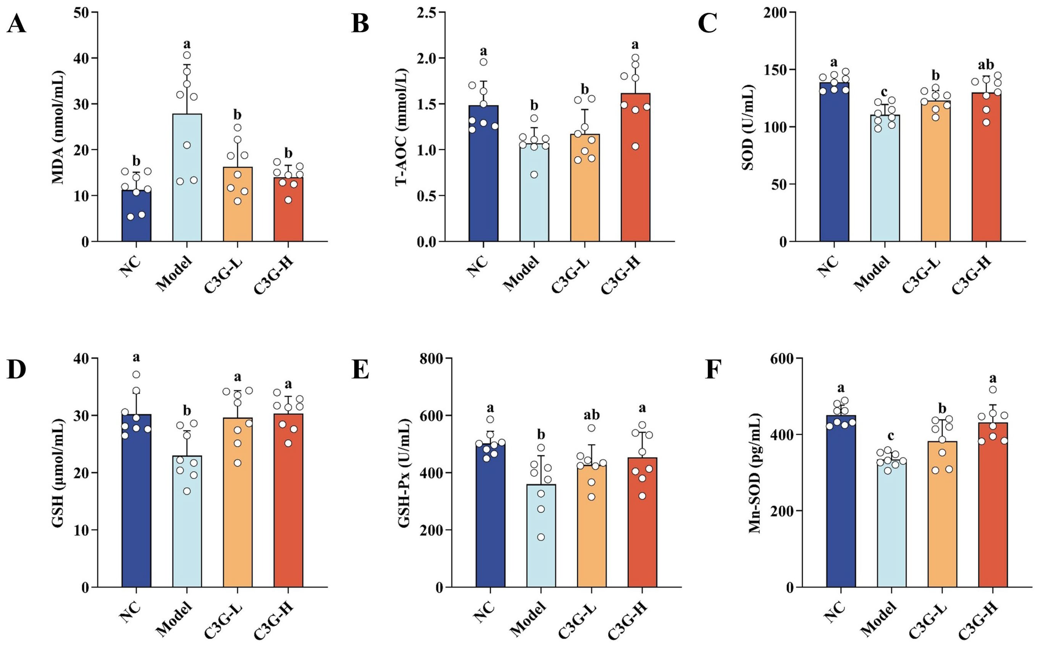
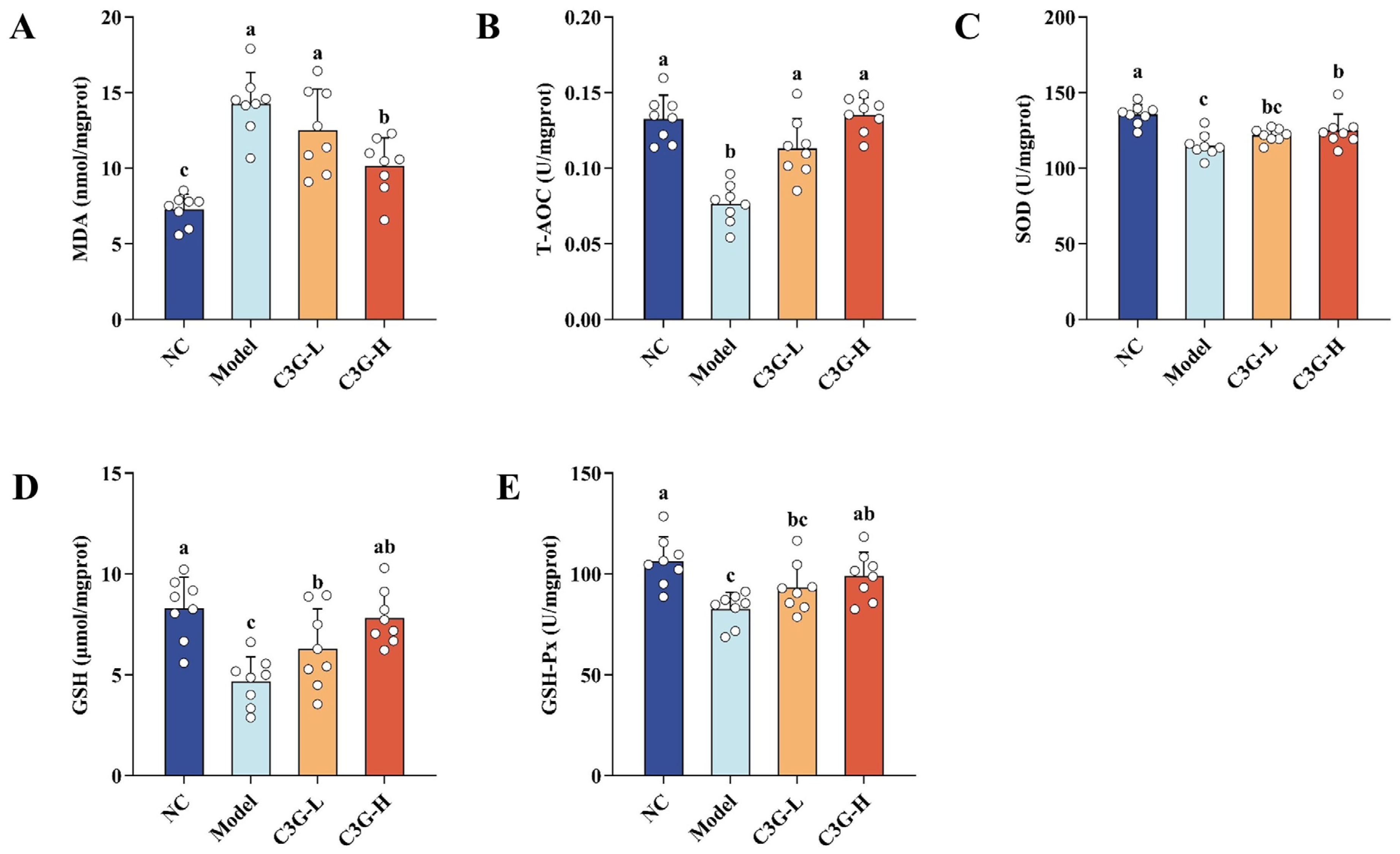
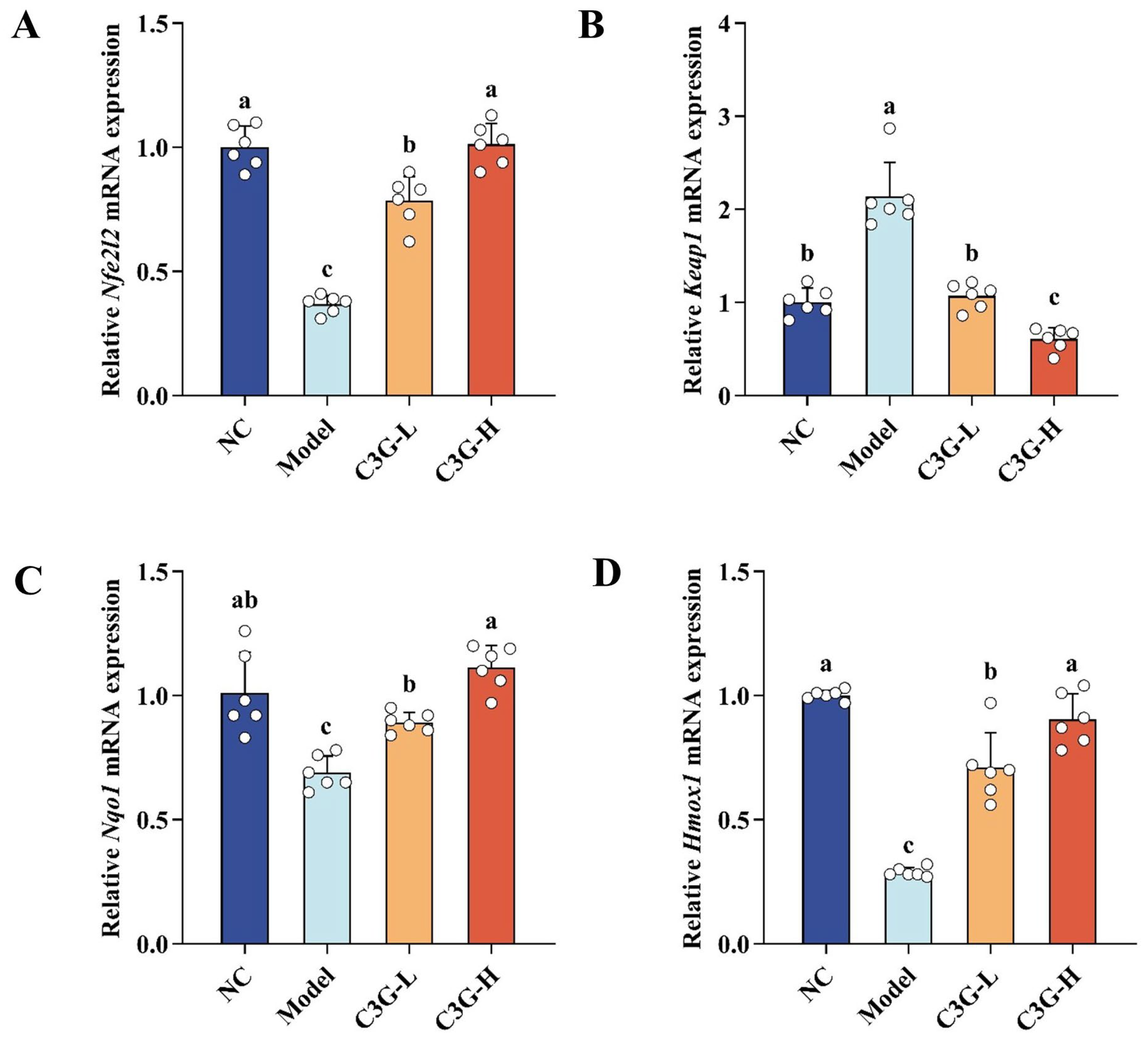
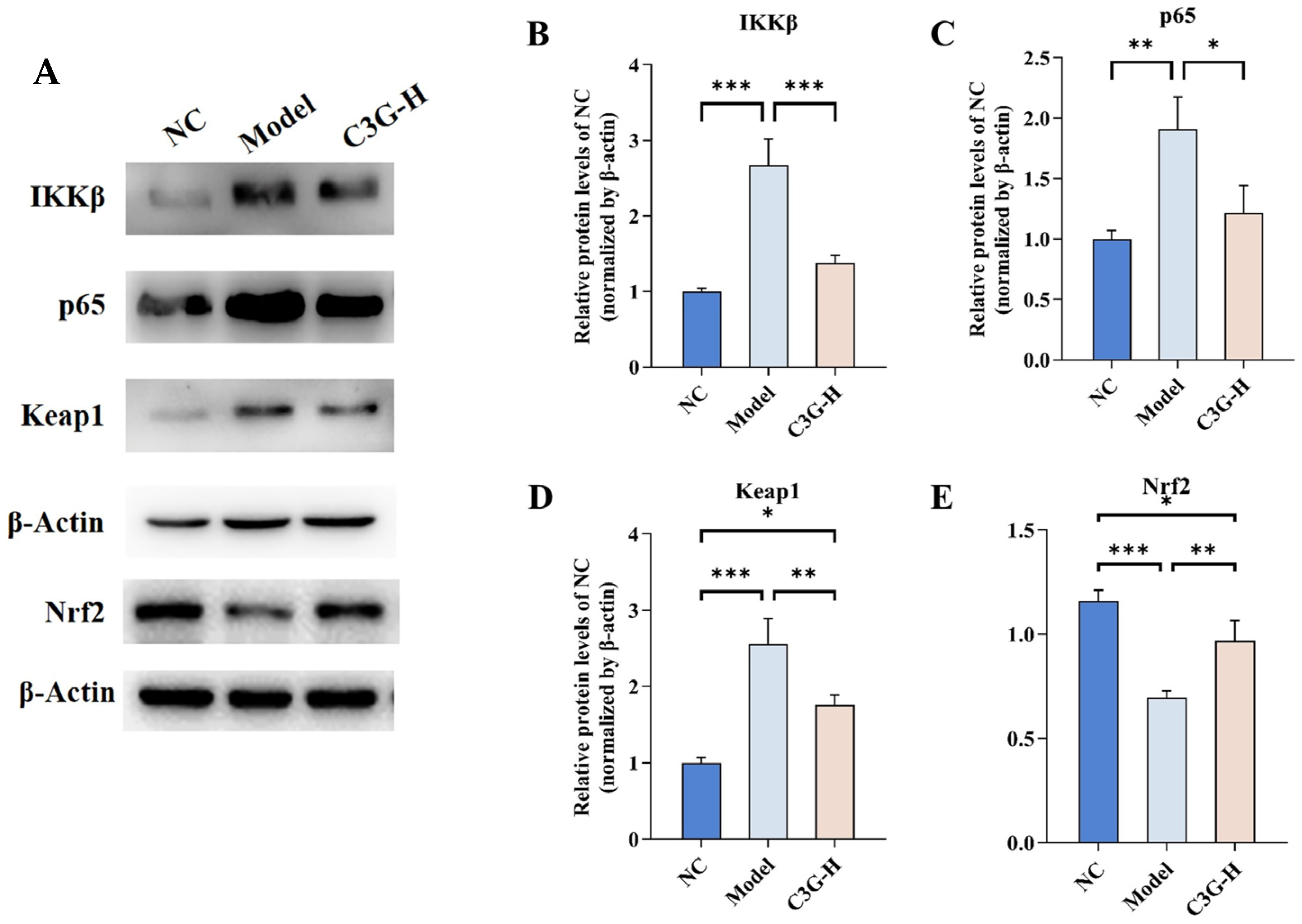
27]. Our study found that D-gal treatment significantly inhibited the activities of antioxidant enzymes such as SOD and GSH-Px in the serum and non-hippocampal brain tissue of mice, and increased the level of malondialdehyde (MDA), a lipid peroxidation product. Meanwhile, D-gal treatment was associated with downregulated mRNA and protein expression of Nrf2, as well as mRNA expression of its downstream target genes
Nqo1 and
Hmox1, whereas it upregulated mRNA and protein levels of the negative regulator Keap1 in the hippocampal tissue. In contrast, C3G treatment elevated antioxidant enzyme activities and reduced MDA content in a dose-dependent manner. In the C3G-H group, it was found to suppress Keap1 expression and increase Nrf2 expression, findings that are consistent with Nrf2 pathway activation. These observations support the ability of C3G to enhance the endogenous antioxidant defense system. These results are consistent with the findings reported by Chen et al. [
28], who confirmed that anthocyanins can alleviate oxidative stress in the organism by activating the Nrf2/Keap1 pathway. The alterations suggest that the Nrf2/Keap1 pathway may contribute to the protective effects of C3G against oxidative damage and neuronal injury. Furthermore, our previous study demonstrated that C3G could bind to key amino acid residues (e.g., Arg415, Ser602) of Keap1, thereby disrupting Keap1-Nrf2 interaction and stabilizing Nrf2 for nuclear translocation [
29]. Notably, the induced expression of Nrf2 downstream target gene
Hmox1 is crucial. HO-1 exhibits direct antioxidant activity, and its catalytic products (e.g., carbon monoxide and biliverdin) also possess anti-inflammatory and anti-apoptotic properties [
30]. This feature further links antioxidant activity closely with anti-inflammatory and neuroprotective effects. Notably, as behavioral assessments focused on hippocampus-dependent functions, hippocampal tissues were prioritized for histopathological and molecular analyses to link molecular changes to behavioral phenotypes. Due to limited tissue availability, non-hippocampal samples (including cortex) were used for oxidative stress marker detection. Although the cortex contributes to cognition, its biochemical measures do not fully align with behavioral outcomes. Future studies should validate oxidative changes directly in the hippocampus and clarify its interplay with the cortex.
Chronic neuroinflammation is a key driver of aging-related cognitive impairment. Aberrant activation of the NF-κB pathway represents the core mechanism underlying neuroinflammation [
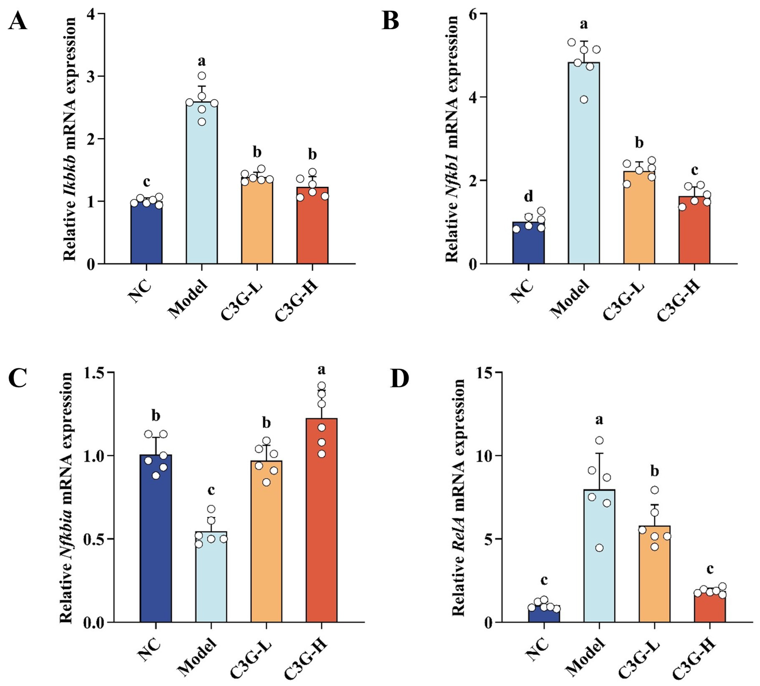
31]. In the present study, we observed that D-gal-induced aging in mice was associated with upregulated mRNA expression of key NF-κB pathway-related genes (
Ikbkb,
Nfkb1,
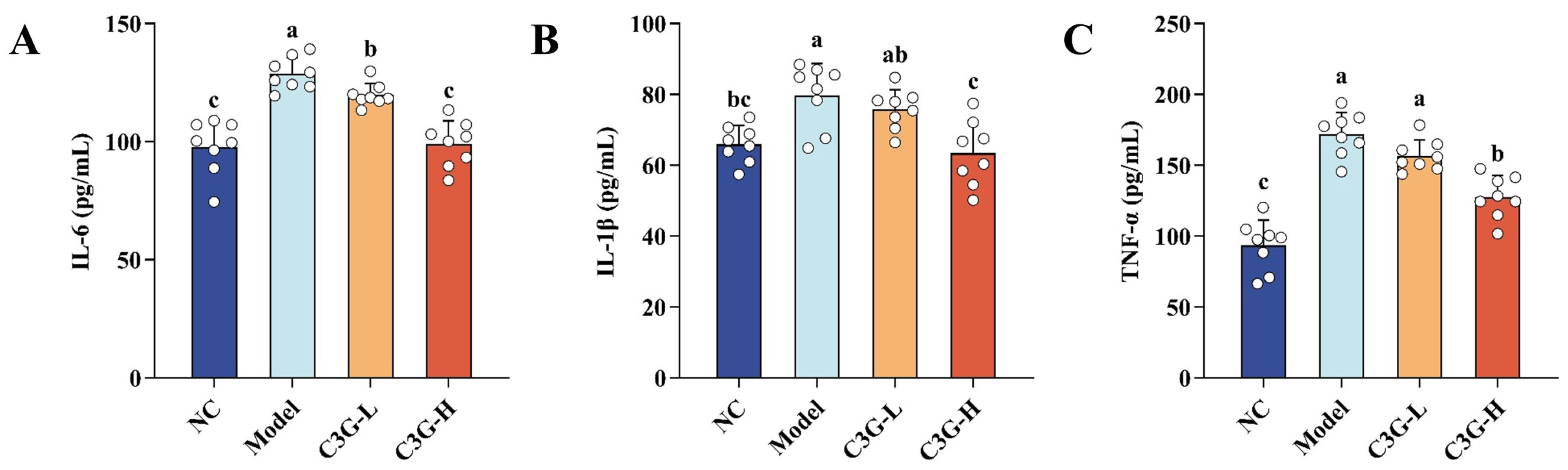
RelA) and increased protein levels of their encoded products IKKβ and p65, as well as downregulated mRNA expression of Ikbα in hippocampal tissue. Meanwhile, serum levels of pro-inflammatory cytokines such as TNF-α, IL-1β, and IL-6 were significantly elevated. These observations are consistent with a state of concurrent systemic inflammation and neuroinflammation in D-gal-induced aging mice. Notably, C3G dose-dependently downregulated mRNA expression of the key genes (
Ikbkb,
Nfkb1,
RelA) in hippocampal tissue and suppressed the release of pro-inflammatory cytokines. Moreover, C3G administration at the high dose significantly decreased protein levels of IKKβ and p65. Together, these observations contributed to the alleviation of neuroinflammation. These findings are consistent with previous studies reporting that C3G and other anthocyanins may alleviate inflammation by regulating the NF-κB pathway, as reflected by decreased levels of IL-6 and TNF-α in models of neurodegenerative disease and aging-related inflammation [
11,
32,
33]. Therefore, the downregulation of the upstream kinase IKKβ and the downstream p65 by C3G observed in this study is associated with attenuated inflammatory signaling in this pathway. Additionally, this dose-dependent regulatory pattern is consistent with the principle proposed by that natural bioactive components exert local regulation at low doses and comprehensive intervention on inflammatory pathways at high doses [
34]. It further supports the specificity and efficacy of C3G’s anti-inflammatory effects.
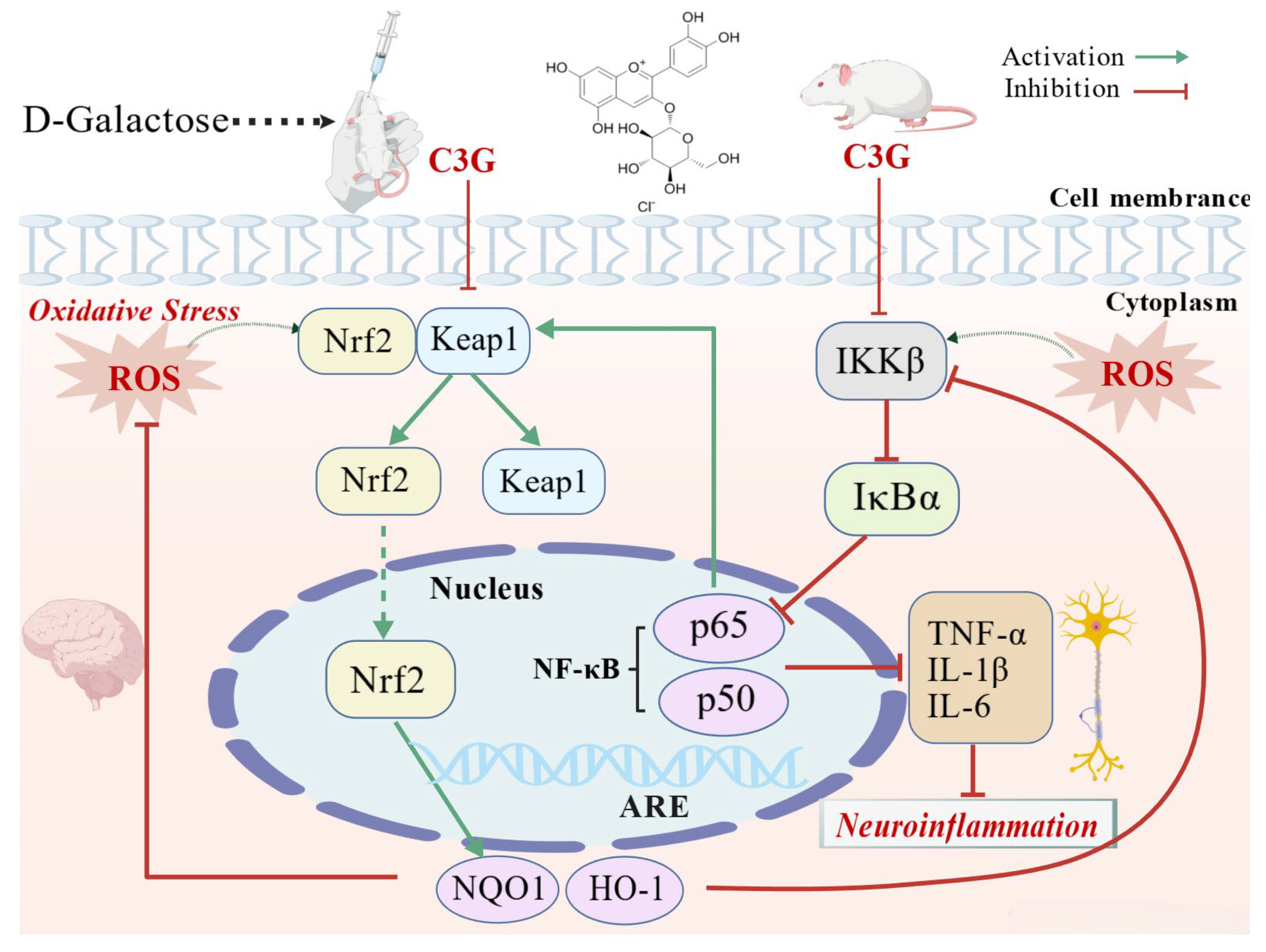
This study supports a potential working model by which C3G may counteract brain aging through the integration of Nrf2/NF-κB pathway regulation, along with hippocampal structural protection, and behavioral function recovery (
Figure 9). On one hand, Nrf2-related expression changes elevate antioxidant levels such as GSH and establish a reductive cellular microenvironment. By scavenging ROS, these changes indirectly inhibit IKKβ activity and block the ROS–inflammation vicious cycle [
35,
36]. Meanwhile, previous studies have indicated that Nrf2 directly competes with p65 for the essential transcriptional coactivators CBP/p300, thereby physically inhibiting NF-κB’s transcriptional activity [
37,
38]. These dual effects collectively alleviate oxidative stress and inflammatory damage, reduce hippocampal neuronal and synaptic damage, and thereby preserve the structural integrity of hippocampal tissue. On the other hand, C3G downregulates NF-κB pathway-related components, which may in turn reduce pro-inflammatory cytokine release and suppress NADPH oxidase activation, thereby mitigating the oxidative burst associated with excessive ROS production during inflammation [
35]. Such regulation may help preserve hippocampal structural stability and create a favorable cellular microenvironment for sustained antioxidant defense. These effects may, in turn, contribute to the behavioral improvements in cognitive function. These findings expand our understanding of C3G’s nutritional functions and offer new insights into dietary strategies against aging-related cognitive impairment. Future studies examining nuclear translocation of these key proteins and the crosstalk between Nrf2 and NF-κB pathways will help clarify the underlying mechanisms in the aging brain.




 ,
,