Intensive Care Unit Acquired Weakness as a Modifiable Organ Dysfunction? A Narrative Review of Evolving Diagnostic and Therapeutic Concepts


Abstract
1. Background
2. Methods
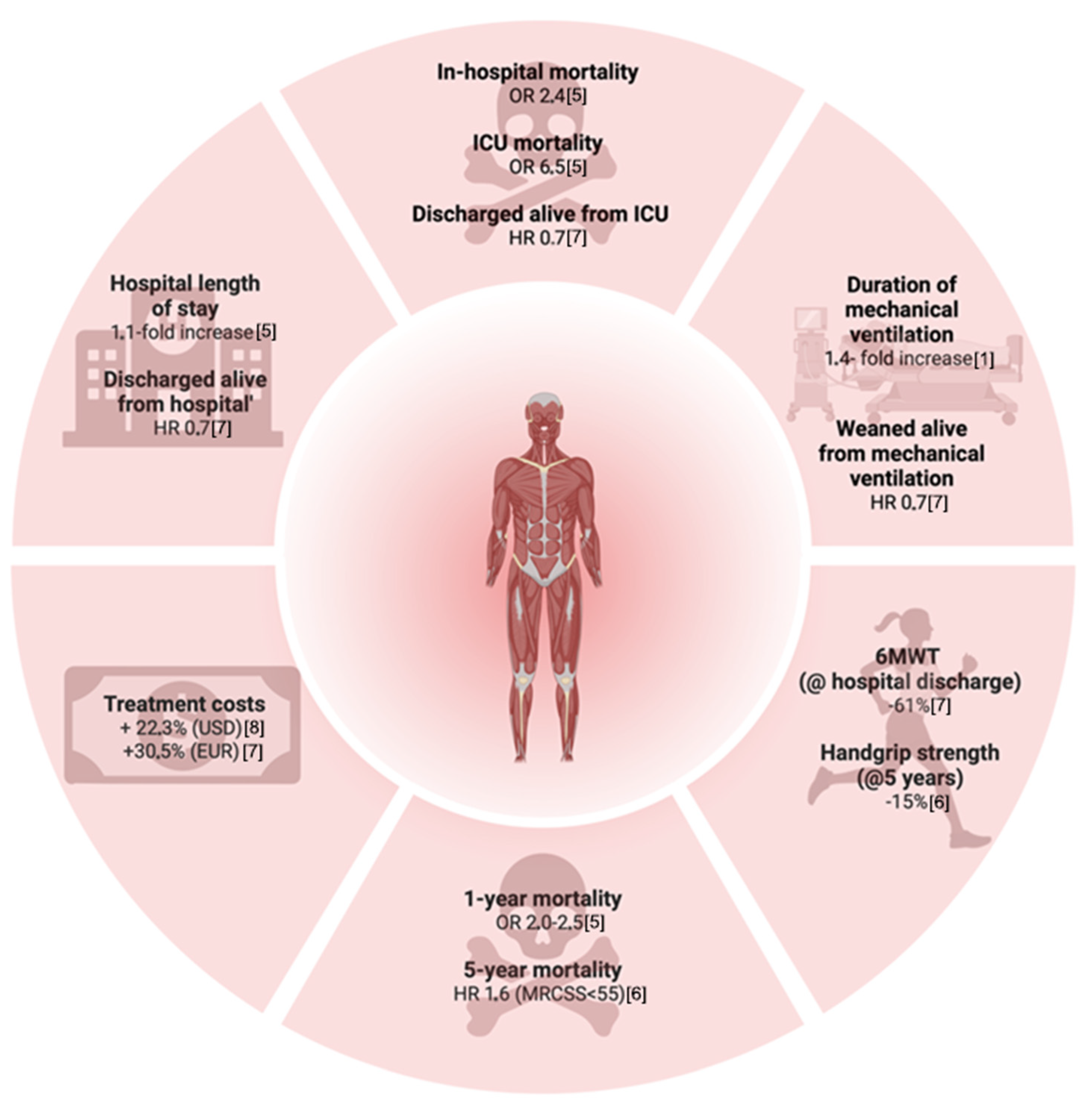
3. Clinical, Neurotopographic, and Pathophysiological Dimensions of ICUAW
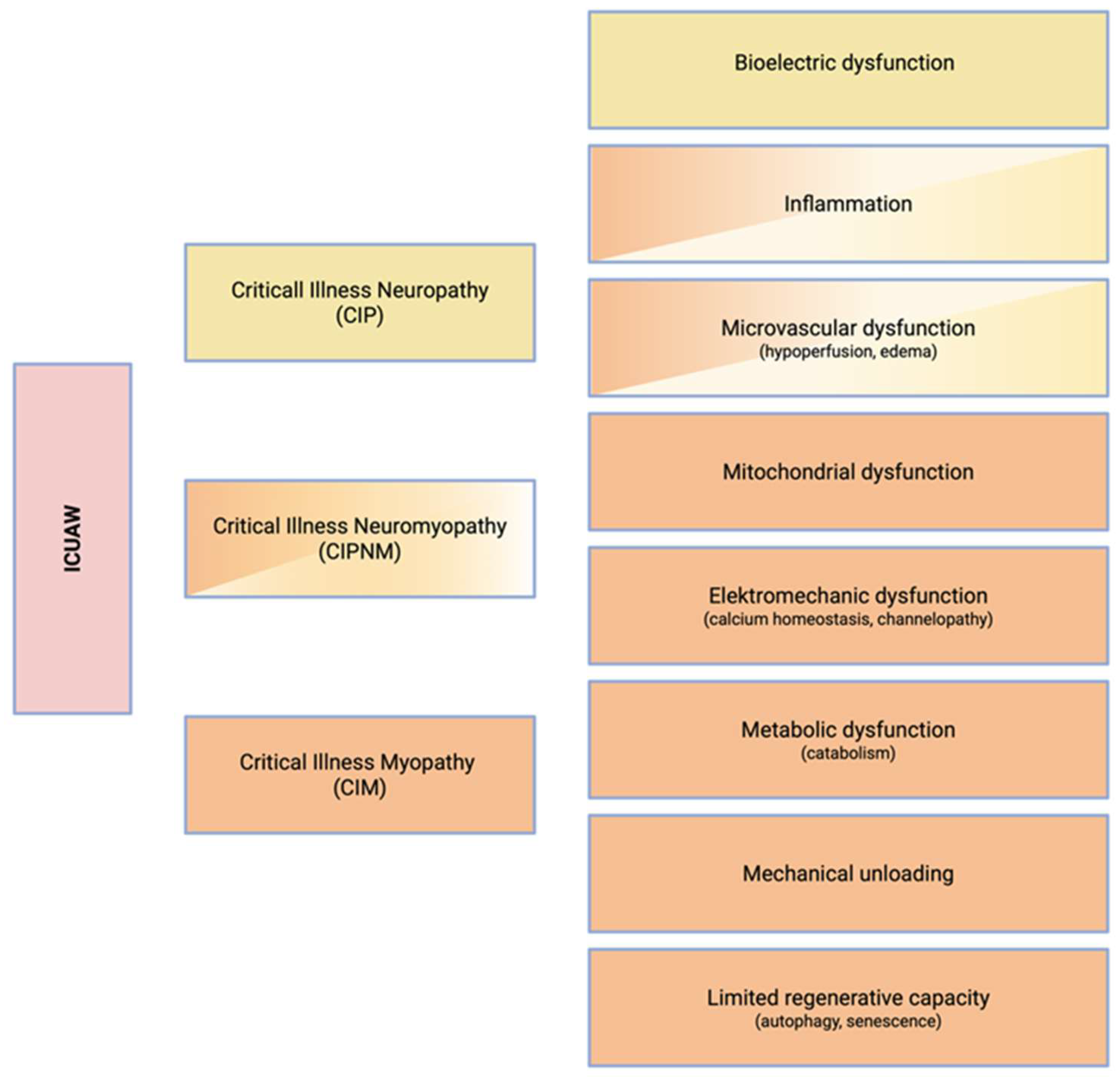
4. Pathophysiology
4.1. Myopathy
4.2. Neuropathy
5. Diagnosis
5.1. Clinical Assessment
5.2. Electrodiagnostic Testing
5.3. Imaging
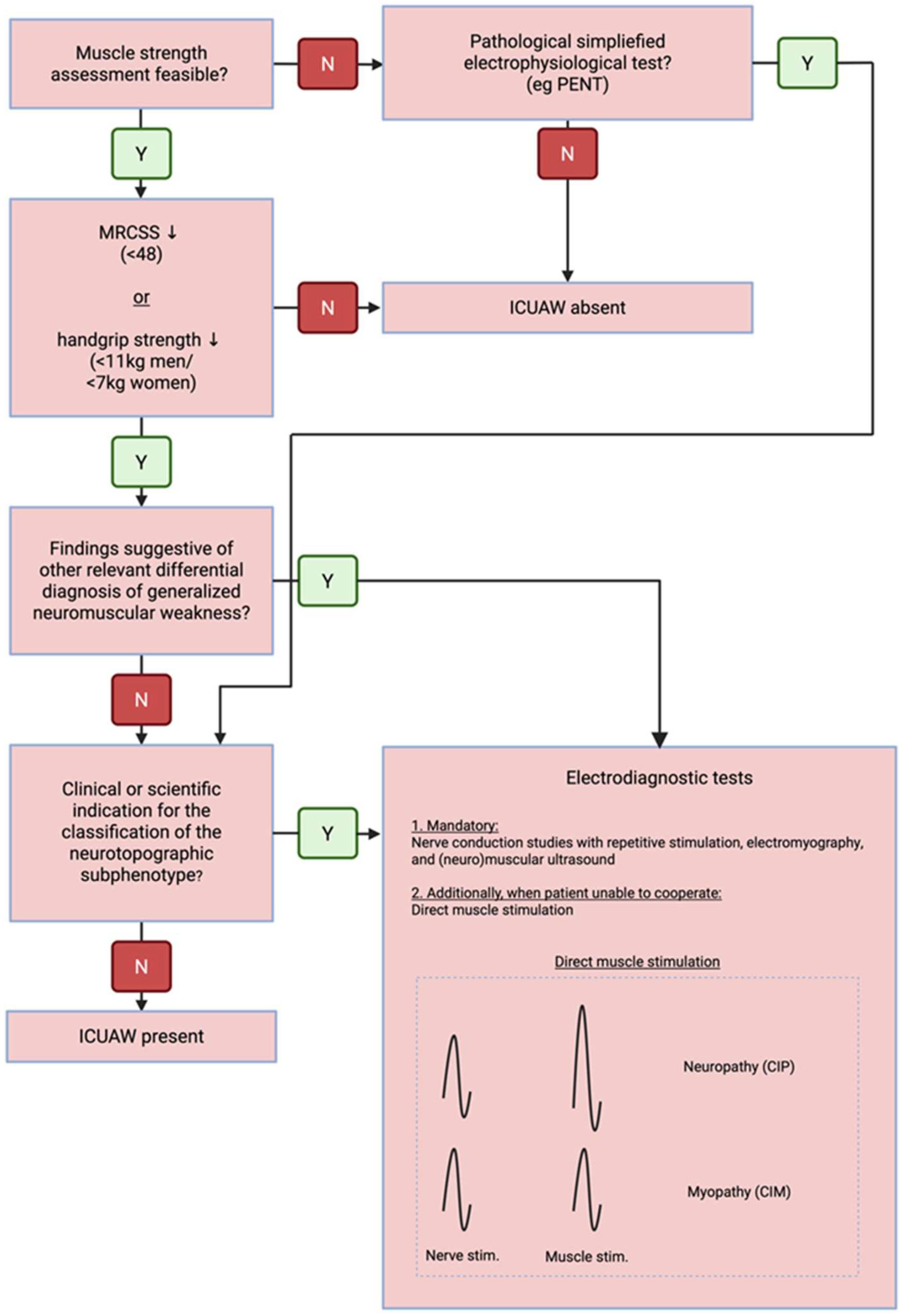
5.4. Diagnostic Algorithm
- Establishment of the syndromic diagnosis of diffuse neuromuscular dysfunction in the context of a preceding critical illness, based on:
- Clinical findings—symmetric muscle weakness
- Imaging—diffuse muscle atrophy and reduced muscle quality
- Electrophysiology—impairment of neuromuscular excitation
- Exclusion of relevant differential diagnoses
- Monitoring of neuromuscular dysfunction to support prognostication, prevent secondary complications, and guide therapeutic interventions
- Phenotypic subclassification using advanced diagnostics, reserved for selected cases where clinically indicated
6. Prevention and Treatment
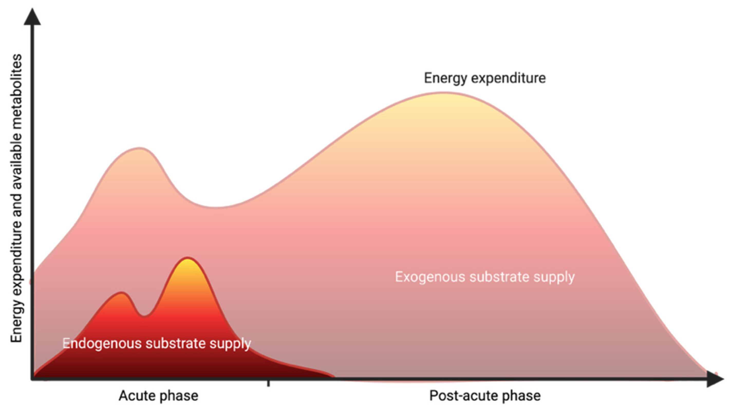
6.1. Nutrition and Metabolism
6.2. Drug Exposure
6.3. Mobilization and Neuromuscular Electrical Stimulation
7. Conclusions
Author Contributions
Funding
Institutional Review Board Statement
Informed Consent Statement
Data Availability Statement
Conflicts of Interest
References
- Fan, E.; Cheek, F.; Chlan, L.; Gosselink, R.; Hart, N.; Herridge, M.S.; Hopkins, R.O.; Hough, C.L.; Kress, J.P.; Latronico, N.; et al. An Official American Thoracic Society Clinical Practice Guideline: The Diagnosis of Intensive Care Unit–Acquired Weakness in Adults. Am. J. Respir. Crit. Care Med. 2014, 190, 1437–1446. [Google Scholar] [CrossRef] [Scilit]
- Tepper, M. Incidence and Onset of Critical Illness Polyneuropathy in Patients with Septic Shock. Neth. J. Med. 2000, 56, 211–214. [Google Scholar] [CrossRef] [Scilit]
- Coakley, J.H.; Nagendran, K.; Yarwood, G.D.; Honavar, M.; Hinds, C.J. Patterns of Neurophysiological Abnormality in Prolonged Critical Illness. Intensive Care Med. 1998, 24, 801–807. [Google Scholar] [CrossRef] [Scilit]
- Pierre, A.; Favory, R.; Bourel, C.; Howsam, M.; Romien, R.; Lancel, S.; Preau, S. Muscle Weakness after Critical Illness: Unravelling Biological Mechanisms and Clinical Hurdles. Crit. Care 2025, 29, 248. [Google Scholar] [CrossRef] [Scilit]
- Hermans, G.; Van Mechelen, H.; Bruyninckx, F.; Vanhullebusch, T.; Clerckx, B.; Meersseman, P.; Debaveye, Y.; Casaer, M.P.; Wilmer, A.; Wouters, P.J.; et al. Predictive Value for Weakness and 1-Year Mortality of Screening Electrophysiology Tests in the ICU. Intensive Care Med. 2015, 41, 2138–2148. [Google Scholar] [CrossRef] [Scilit]
- Van Aerde, N.; Meersseman, P.; Debaveye, Y.; Wilmer, A.; Gunst, J.; Casaer, M.P.; Bruyninckx, F.; Wouters, P.J.; Gosselink, R.; Van den Berghe, G.; et al. Five-Year Impact of ICU-Acquired Neuromuscular Complications: A Prospective, Observational Study. Intensive Care Med. 2020, 46, 1184–1193. [Google Scholar] [CrossRef] [Scilit]
- Hermans, G.; Van Mechelen, H.; Clerckx, B.; Vanhullebusch, T.; Mesotten, D.; Wilmer, A.; Casaer, M.P.; Meersseman, P.; Debaveye, Y.; Van Cromphaut, S.; et al. Acute Outcomes and 1-Year Mortality of Intensive Care Unit-Acquired Weakness: A Cohort Study and Propensity-Matched Analysis. Am. J. Respir. Crit. Care Med. 2014, 190, 410–420. [Google Scholar] [CrossRef] [Scilit]
- Kelmenson, D.A.; Held, N.; Allen, R.R.; Quan, D.; Burnham, E.L.; Clark, B.J.; Ho, P.M.; Kiser, T.H.; Vandivier, R.W.; Moss, M. Outcomes of ICU Patients with a Discharge Diagnosis of Critical Illness Polyneuromyopathy: A Propensity-Matched Analysis. Crit. Care Med. 2017, 45, 2055–2060. [Google Scholar] [CrossRef] [Scilit]
- Renner, C.; Jeitziner, M.-M.; Albert, M.; Brinkmann, S.; Diserens, K.; Dzialowski, I.; Heidler, M.-D.; Lück, M.; Nusser-Müller-Busch, R.; Sandor, P.S.; et al. Guideline on Multimodal Rehabilitation for Patients with Post-Intensive Care Syndrome. Crit. Care 2023, 27, 301. [Google Scholar] [CrossRef] [Scilit]
- Nakanishi, N. Intensive Care Unit-Acquired Muscle Atrophy and Weakness in Critical Illness: A Review of Long-Term Recovery Strategies. Acute Crit. Care 2025, 40, 361–372. [Google Scholar] [CrossRef] [Scilit]
- Chen, J.; Huang, M. Intensive Care Unit-Acquired Weakness: Recent Insights. J. Intensive Med. 2024, 4, 73–80. [Google Scholar] [CrossRef] [Scilit]
- Petrucci, M.; Gemma, S.; Carbone, L.; Piccioni, A.; Della Polla, D.A.; Simeoni, B.; Franceschi, F.; Covino, M. ICU-Acquired Weakness: From Pathophysiology to Management in Critical Care. Emerg. Care Med. 2025, 2, 4. [Google Scholar] [CrossRef] [Scilit]
- Vanhorebeek, I.; Latronico, N.; Van den Berghe, G. ICU-Acquired Weakness. Intensive Care Med. 2020, 46, 637–653. [Google Scholar] [CrossRef] [Scilit]
- Friedrich, O.; Reid, M.B.; Van den Berghe, G.; Vanhorebeek, I.; Hermans, G.; Rich, M.M.; Larsson, L. The Sick and the Weak: Neuropathies/Myopathies in the Critically Ill. Physiol. Rev. 2015, 95, 1025–1109. [Google Scholar] [CrossRef] [Scilit]
- Latronico, N.; Herridge, M.; Hopkins, R.O.; Angus, D.; Hart, N.; Hermans, G.; Iwashyna, T.; Arabi, Y.; Citerio, G.; Wesley Ely, E.; et al. The ICM Research Agenda on Intensive Care Unit-Acquired Weakness. Intensive Care Med. 2017, 43, 1270–1281. [Google Scholar] [CrossRef] [Scilit]
- Le Stang, V.; Latronico, N.; Dres, M.; Bertoni, M. Critical Illness-Associated Limb and Diaphragmatic Weakness. Curr. Opin. Crit. Care 2024, 30, 121–130. [Google Scholar] [CrossRef] [Scilit]
- Latronico, N.; Bolton, C.F. Critical Illness Polyneuropathy and Myopathy: A Major Cause of Muscle Weakness and Paralysis. Lancet Neurol. 2011, 10, 931–941. [Google Scholar] [CrossRef] [Scilit]
- Van den Berghe, G. On the Neuroendocrinopathy of Critical Illness. Perspectives for Feeding and Novel Treatments. Am. J. Respir. Crit. Care Med. 2016, 194, 1337–1348. [Google Scholar] [CrossRef] [Scilit]
- dos Santos, C.; Hussain, S.N.A.; Mathur, S.; Picard, M.; Herridge, M.; Correa, J.; Bain, A.; Guo, Y.; Advani, A.; Advani, S.L.; et al. Mechanisms of Chronic Muscle Wasting and Dysfunction after an Intensive Care Unit Stay. A Pilot Study. Am. J. Respir. Crit. Care Med. 2016, 194, 821–830. [Google Scholar] [CrossRef] [Scilit]
- Vana, P.G.; LaPorte, H.M.; Wong, Y.M.; Kennedy, R.H.; Gamelli, R.L.; Majetschak, M. Proteasome Inhibition After Burn Injury. J. Burn Care Res. 2016, 37, 207–215. [Google Scholar] [CrossRef] [Scilit]
- Yin, D.; Lin, D.; Xie, Y.; Gong, A.; Jiang, P.; Wu, J. Neuregulin-1β Alleviates Sepsis-Induced Skeletal Muscle Atrophy by Inhibiting Autophagy via AKT/MTOR Signaling Pathway in Rats. Shock 2022, 57, 397–407. [Google Scholar] [CrossRef] [Scilit]
- Rivera, J.C.; Abrigo, J.; Tacchi, F.; Simon, F.; Brandan, E.; Santos, R.A.; Bader, M.; Chiong, M.; Cabello-Verrugio, C. Angiotensin-(1-7) Prevents Lipopolysaccharide-Induced Autophagy via the Mas Receptor in Skeletal Muscle. Int. J. Mol. Sci. 2020, 21, 9344. [Google Scholar] [CrossRef] [Scilit]
- Hermans, G.; Casaer, M.P.; Clerckx, B.; Güiza, F.; Vanhullebusch, T.; Derde, S.; Meersseman, P.; Derese, I.; Mesotten, D.; Wouters, P.J.; et al. Effect of Tolerating Macronutrient Deficit on the Development of Intensive-Care Unit Acquired Weakness: A Subanalysis of the EPaNIC Trial. Lancet Respir. Med. 2013, 1, 621–629. [Google Scholar] [CrossRef] [Scilit]
- Vanhorebeek, I.; Casaer, M.; Gunst, J. Nutrition and Autophagy Deficiency in Critical Illness. Curr. Opin. Crit. Care 2023, 29, 306–314. [Google Scholar] [CrossRef] [Scilit]
- Reignier, J.; Plantefeve, G.; Mira, J.-P.; Argaud, L.; Asfar, P.; Aissaoui, N.; Badie, J.; Botoc, N.-V.; Brisard, L.; Bui, H.-N.; et al. Low versus Standard Calorie and Protein Feeding in Ventilated Adults with Shock: A Randomised, Controlled, Multicentre, Open-Label, Parallel-Group Trial (NUTRIREA-3). Lancet Respir. Med. 2023, 11, 602–612. [Google Scholar] [CrossRef] [Scilit]
- Reignier, J.; Boisramé-Helms, J.; Brisard, L.; Lascarrou, J.B.; Ait Hssain, A.; Anguel, N.; Argaud, L.; Asehnoune, K.; Asfar, P.; Bellec, F.; et al. Enteral versus Parenteral Early Nutrition in Ventilated Adults with Shock: A Randomised, Controlled, Multicentre, Open-Label, Parallel-Group Study (NUTRIREA-2). Lancet 2018, 391, 133–143. [Google Scholar] [CrossRef] [Scilit]
- Casaer, M.P.; Mesotten, D.; Hermans, G.; Wouters, P.J.; Schetz, M.; Meyfroidt, G.; Van Cromphaut, S.; Ingels, C.; Meersseman, P.; Muller, J.; et al. Early versus Late Parenteral Nutrition in Critically Ill Adults. N. Engl. J. Med. 2011, 365, 506–517. [Google Scholar] [CrossRef] [Scilit]
- Elke, G.; van Zanten, A.R.H.; Lemieux, M.; McCall, M.; Jeejeebhoy, K.N.; Kott, M.; Jiang, X.; Day, A.G.; Heyland, D.K. Enteral versus Parenteral Nutrition in Critically Ill Patients: An Updated Systematic Review and Meta-Analysis of Randomized Controlled Trials. Crit. Care 2016, 20, 117. [Google Scholar] [CrossRef] [Scilit]
- Schmidbauer, M.L.; Putz, T.; Gehri, L.; Ratkovic, L.; Maskos, A.; Zibold, J.; Bauchmüller, J.; Imhof, S.; Weig, T.; Wuehr, M.; et al. Accelerometer-Derived Movement Features as Predictive Biomarkers for Muscle Atrophy in Neurocritical Care: A Prospective Cohort Study. Crit. Care 2024, 28, 288. [Google Scholar] [CrossRef] [Scilit]
- Okada, Y.; Unoki, T.; Matsuishi, Y.; Egawa, Y.; Hayashida, K.; Inoue, S. Early versus Delayed Mobilization for In-Hospital Mortality and Health-Related Quality of Life among Critically Ill Patients: A Systematic Review and Meta-Analysis. J. Intensive Care 2019, 7, 57. [Google Scholar] [CrossRef] [Scilit]
- Owen, A.M.; Patel, S.P.; Smith, J.D.; Balasuriya, B.K.; Mori, S.F.; Hawk, G.S.; Stromberg, A.J.; Kuriyama, N.; Kaneki, M.; Rabchevsky, A.G.; et al. Chronic Muscle Weakness and Mitochondrial Dysfunction in the Absence of Sustained Atrophy in a Preclinical Sepsis Model. eLife 2019, 8, e49920. [Google Scholar] [CrossRef] [Scilit]
- Walsh, C.J.; Escudero King, C.; Gupta, M.; Plant, P.J.; Herridge, M.J.; Mathur, S.; Hu, P.; Correa, J.; Ahmed, S.; Bigot, A.; et al. MicroRNA Regulatory Networks Associated with Abnormal Muscle Repair in Survivors of Critical Illness. J. Cachexia Sarcopenia Muscle 2022, 13, 1262–1276. [Google Scholar] [CrossRef] [Scilit]
- Brealey, D.; Brand, M.; Hargreaves, I.; Heales, S.; Land, J.; Smolenski, R.; Davies, N.A.; Cooper, C.E.; Singer, M. Association between Mitochondrial Dysfunction and Severity and Outcome of Septic Shock. Lancet 2002, 360, 219–223. [Google Scholar] [CrossRef] [Scilit]
- Martín-Vicente, P.; López-Martínez, C.; Rioseras, B.; Albaiceta, G.M. Activation of Senescence in Critically Ill Patients: Mechanisms, Consequences and Therapeutic Opportunities. Ann. Intensive Care 2024, 14, 2. [Google Scholar] [CrossRef] [Scilit]
- Mayer, K.P.; Ismaeel, A.; Kalema, A.G.; Montgomery-Yates, A.A.; Soper, M.K.; Kern, P.A.; Starck, J.D.; Slone, S.A.; Morris, P.E.; Dupont-Versteegden, E.E.; et al. Persistent Fatigue, Weakness, and Aberrant Muscle Mitochondria in Survivors of Critical COVID-19. Crit. Care Explor. 2024, 6, e1164. [Google Scholar] [CrossRef] [Scilit]
- Bierbrauer, J.; Koch, S.; Olbricht, C.; Hamati, J.; Lodka, D.; Schneider, J.; Luther-Schröder, A.; Kleber, C.; Faust, K.; Wiesener, S.; et al. Early Type II Fiber Atrophy in Intensive Care Unit Patients with Nonexcitable Muscle Membrane. Crit. Care Med. 2012, 40, 647–650. [Google Scholar] [CrossRef] [Scilit]
- Donati, A.; Damiani, E.; Domizi, R.; Scorcella, C.; Carsetti, A.; Tondi, S.; Monaldi, V.; Adrario, E.; Romano, R.; Pelaia, P.; et al. Near-Infrared Spectroscopy for Assessing Tissue Oxygenation and Microvascular Reactivity in Critically Ill Patients: A Prospective Observational Study. Crit. Care 2016, 20, 311. [Google Scholar] [CrossRef] [Scilit]
- De Backer, D.; Donadello, K.; Sakr, Y.; Ospina-Tascon, G.; Salgado, D.; Scolletta, S.; Vincent, J.-L. Microcirculatory Alterations in Patients With Severe Sepsis. Crit. Care Med. 2013, 41, 791–799. [Google Scholar] [CrossRef] [Scilit]
- Mendelson, A.A.; Erickson, D.; Villar, R. The Role of the Microcirculation and Integrative Cardiovascular Physiology in the Pathogenesis of ICU-Acquired Weakness. Front. Physiol. 2023, 14, 1170429. [Google Scholar] [CrossRef] [Scilit]
- Walsh, C.J.; Batt, J.; Herridge, M.S.; Mathur, S.; Bader, G.D.; Hu, P.; dos Santos, C.C. Transcriptomic Analysis Reveals Abnormal Muscle Repair and Remodeling in Survivors of Critical Illness with Sustained Weakness. Sci. Rep. 2016, 6, 29334. [Google Scholar] [CrossRef] [Scilit]
- Novak, K.R.; Nardelli, P.; Cope, T.C.; Filatov, G.; Glass, J.D.; Khan, J.; Rich, M.M. Inactivation of Sodium Channels Underlies Reversible Neuropathy during Critical Illness in Rats. J. Clin. Investig. 2009, 119, 1150–1158. [Google Scholar] [CrossRef] [Scilit] [PubMed]
- Nardelli, P.; Khan, J.; Powers, R.; Cope, T.C.; Rich, M.M. Reduced Motoneuron Excitability in a Rat Model of Sepsis. J. Neurophysiol. 2013, 109, 1775–1781. [Google Scholar] [CrossRef] [Scilit] [PubMed]
- Z’graggen, W.J.; Lin, C.S.Y.; Howard, R.S.; Beale, R.J.; Bostock, H. Nerve Excitability Changes in Critical Illness Polyneuropathy. Brain 2006, 129, 2461–2470. [Google Scholar] [CrossRef] [Scilit] [PubMed]
- Latronico, N.; Rasulo, F.A.; Eikermann, M.; Piva, S. Critical Illness Weakness, Polyneuropathy and Myopathy: Diagnosis, Treatment, and Long-Term Outcomes. Crit. Care 2023, 27, 439. [Google Scholar] [CrossRef] [Scilit]
- De Jonghe, B.; Sharshar, T.; Lefaucheur, J.-P.; Authier, F.-J.; Durand-Zaleski, I.; Boussarsar, M.; Cerf, C.; Renaud, E.; Mesrati, F.; Carlet, J.; et al. Paresis Acquired in the Intensive Care Unit: A Prospective Multicenter Study. JAMA 2002, 288, 2859–2867. [Google Scholar] [CrossRef] [Scilit]
- Vanpee, G.; Hermans, G.; Segers, J.; Gosselink, R. Assessment of Limb Muscle Strength in Critically Ill Patients. Crit. Care Med. 2014, 42, 701–711. [Google Scholar] [CrossRef] [Scilit]
- Parry, S.M.; Berney, S.; Granger, C.L.; Dunlop, D.L.; Murphy, L.; El-Ansary, D.; Koopman, R.; Denehy, L. A New Two-Tier Strength Assessment Approach to the Diagnosis of Weakness in Intensive Care: An Observational Study. Crit. Care 2015, 19, 52. [Google Scholar] [CrossRef] [Scilit]
- Ali, N.A.; O’Brien, J.M.; Hoffmann, S.P.; Phillips, G.; Garland, A.; Finley, J.C.W.; Aimoosa, K.; Hejal, R.; Wolf, K.M.; Lemeshow, S.; et al. Acquired Weakness, Handgrip Strength, and Mortality in Critically III Patients. Am. J. Respir. Crit. Care Med. 2008, 178, 261–268. [Google Scholar] [CrossRef] [Scilit]
- Baldwin, C.E.; Paratz, J.D.; Bersten, A.D. Muscle Strength Assessment in Critically Ill Patients with Handheld Dynamometry: An Investigation of Reliability, Minimal Detectable Change, and Time to Peak Force Generation. J. Crit. Care 2013, 28, 77–86. [Google Scholar] [CrossRef] [Scilit]
- Zifko, U.A.; Zipko, H.T.; Bolton, C.F. Clinical and Electrophysiological Findings in Critical Illness Polyneuropathy. J. Neurol. Sci. 1998, 159, 186–193. [Google Scholar] [CrossRef] [Scilit]
- Latronico, N.; Nattino, G.; Guarneri, B.; Fagoni, N.; Amantini, A.; Bertolini, G. Validation of the Peroneal Nerve Test to Diagnose Critical Illness Polyneuropathy and Myopathy in the Intensive Care Unit: The Multicentre Italian CRIMYNE-2 Diagnostic Accuracy Study. F1000Research 2014, 3, 127. [Google Scholar] [CrossRef] [PubMed]
- Latronico, N.; Bertolini, G.; Guarneri, B.; Botteri, M.; Peli, E.; Andreoletti, S.; Bera, P.; Luciani, D.; Nardella, A.; Vittorielli, E.; et al. Simplified Electrophysiological Evaluation of Peripheral Nerves in Critically Ill Patients: The Italian Multi-Centre CRIMYNE Study. Crit. Care 2007, 11, R11. [Google Scholar] [CrossRef] [Scilit] [PubMed]
- Maskos, A.; Schmidbauer, M.L.; Kunst, S.; Rehms, R.; Putz, T.; Römer, S.; Iankova, V.; Dimitriadis, K. Diagnostic Utility of Temporal Muscle Thickness as a Monitoring Tool for Muscle Wasting in Neurocritical Care. Nutrients 2022, 14, 4498. [Google Scholar] [CrossRef] [Scilit] [PubMed]
- Dhar, S.; Oropello, J.; Morris, P.E. Assessing Skeletal Muscle Dysfunction in Sepsis Utilizing Muscle Ultrasound in Search for Pathways to Improve ICU Survivor’s Functional Outcomes. J. Crit. Care 2018, 47, 322–323. [Google Scholar] [CrossRef] [Scilit]
- Pillen, S.; van Dijk, J.P.; Weijers, G.; Raijmann, W.; de Korte, C.L.; Zwarts, M.J. Quantitative Gray-scale Analysis in Skeletal Muscle Ultrasound: A Comparison Study of Two Ultrasound Devices. Muscle Nerve 2009, 39, 781–786. [Google Scholar] [CrossRef] [Scilit]
- Yik, V.; Kok, S.S.X.; Chean, E.; Lam, Y.E.; Chua, W.T.; Tan, W.J.; Foo, F.J.; Ng, J.L.; Su, S.S.; Chong, C.X.Z.; et al. Diagnosing Sarcopenia with AI-Aided Ultrasound (DINOSAUR)—A Pilot Study. Nutrients 2024, 16, 2768. [Google Scholar] [CrossRef] [Scilit]
- Joskova, V.; Patkova, A.; Havel, E.; Najpaverova, S.; Uramova, D.; Kovarik, M.; Zadak, Z.; Hronek, M. Critical Evaluation of Muscle Mass Loss as a Prognostic Marker of Morbidity in Critically Ill Patients and Methods for Its Determination. J. Rehabil. Med. 2018, 50, 696–704. [Google Scholar] [CrossRef] [Scilit]
- de Man, A.M.E.; Gunst, J.; Reintam Blaser, A. Nutrition in the Intensive Care Unit: From the Acute Phase to Beyond. Intensive Care Med. 2024, 50, 1035–1048. [Google Scholar] [CrossRef] [Scilit]
- Stoppe, C.; Hill, A.; Christopher, K.B.; Kristof, A.S. Toward Precision in Nutrition Therapy. Crit. Care Med. 2024, 53, e429–e440. [Google Scholar] [CrossRef] [Scilit]
- Gehri, L.; Schmidbauer, M.L.; Putz, T.; Ratkovic, L.; Maskos, A.; Zeisberger, C.; Zibold, J.; Dimitriadis, K. Clinical Medicine Survey on Nutrition in Neurological Intensive Care Units (SONNIC)—A Cross-Sectional Survey among German-Speaking Neurointensivists on Medical Nutritional Therapy. J. Clin. Med. 2024, 13, 447. [Google Scholar] [CrossRef] [Scilit]
- Rice, T.W.; Wheeler, A.P.; Thompson, B.T.; Steingrub, J.; Hite, R.D.; Moss, M.; Morris, A.; Dong, N.; Rock, P. Initial Trophic vs. Full Enteral Feeding in Patients with Acute Lung Injury: The EDEN Randomized Trial. JAMA J. Am. Med. Assoc. 2012, 307, 795–803. [Google Scholar] [CrossRef] [Scilit]
- Arabi, Y.M.; Aldawood, A.S.; Haddad, S.H.; Al-Dorzi, H.M.; Tamim, H.M.; Jones, G.; Mehta, S.; McIntyre, L.; Solaiman, O.; Sakkijha, M.H.; et al. Permissive Underfeeding or Standard Enteral Feeding in Critically Ill Adults. N. Engl. J. Med. 2015, 372, 2398–2408. [Google Scholar] [CrossRef] [Scilit] [PubMed]
- Chapman, M.; Peake, S.L.; Bellomo, R.; Davies, A.; Deane, A.; Horowitz, M.; Hurford, S.; Lange, K.; Little, L.; Mackle, D.; et al. Energy-Dense versus Routine Enteral Nutrition in the Critically Ill. N. Engl. J. Med. 2018, 379, 1823–1834. [Google Scholar] [CrossRef] [Scilit] [PubMed]
- Harvey, S.E.; Parrott, F.; Harrison, D.A.; Bear, D.E.; Segaran, E.; Beale, R.; Bellingan, G.; Leonard, R.; Mythen, M.G.; Rowan, K.M. Trial of the Route of Early Nutritional Support in Critically Ill Adults. N. Engl. J. Med. 2014, 371, 1673–1684. [Google Scholar] [CrossRef] [Scilit]
- Fivez, T.; Kerklaan, D.; Mesotten, D.; Verbruggen, S.; Wouters, P.J.; Vanhorebeek, I.; Debaveye, Y.; Vlasselaers, D.; Desmet, L.; Casaer, M.P.; et al. Early versus Late Parenteral Nutrition in Critically Ill Children. N. Engl. J. Med. 2016, 374, 1111–1122. [Google Scholar] [CrossRef] [Scilit]
- Shepherd, S.J.; Newman, R.; Brett, S.J.; Griffith, D.M. Pharmacological Therapy for the Prevention and Treatment of Weakness after Critical Illness: A Systematic Review. Crit. Care Med. 2016, 44, 1198–1205. [Google Scholar] [CrossRef] [Scilit]
- Van den Berghe, G.; Wouters, P.; Weekers, F.; Verwaest, C.; Bruyninckx, F.; Schetz, M.; Vlasselaers, D.; Ferdinande, P.; Lauwers, P.; Bouillon, R. Intensive Insulin Therapy in Critically Ill Patients. N. Engl. J. Med. 2001, 345, 1359–1367. [Google Scholar] [CrossRef] [Scilit]
- Singer, P.; Blaser, A.R.; Berger, M.M.; Calder, P.C.; Casaer, M.; Hiesmayr, M.; Mayer, K.; Montejo-Gonzalez, J.C.; Pichard, C.; Preiser, J.C.; et al. ESPEN Practical and Partially Revised Guideline: Clinical Nutrition in the Intensive Care Unit. Clin. Nutr. 2023, 42, 1671–1689. [Google Scholar] [CrossRef] [Scilit]
- Reintam Blaser, A.; Starkopf, J.; Alhazzani, W.; Berger, M.M.; Casaer, M.P.; Deane, A.M.; Fruhwald, S.; Hiesmayr, M.; Ichai, C.; Jakob, S.M.; et al. Early Enteral Nutrition in Critically Ill Patients: ESICM Clinical Practice Guidelines. Intensive Care Med. 2017, 43, 380–398. [Google Scholar] [CrossRef] [Scilit]
- Elke, G.; Hartl, W.H.; Kreymann, K.G.; Adolph, M.; Felbinger, T.W.; Graf, T.; de Heer, G.; Heller, A.R.; Kampa, U.; Mayer, K.; et al. Clinical Nutrition in Critical Care Medicine—Guideline of the German Society for Nutritional Medicine (DGEM). Clin. Nutr. ESPEN 2019, 33, 220–275. [Google Scholar] [CrossRef] [Scilit]
- Zusman, O.; Kagan, I.; Bendavid, I.; Theilla, M.; Cohen, J.; Singer, P. Predictive Equations versus Measured Energy Expenditure by Indirect Calorimetry: A Retrospective Validation. Clin. Nutr. 2019, 38, 1206–1210. [Google Scholar] [CrossRef] [Scilit] [PubMed]
- Alberda, C.; Gramlich, L.; Jones, N.; Jeejeebhoy, K.; Day, A.G.; Dhaliwal, R.; Heyland, D.K. The Relationship between Nutritional Intake and Clinical Outcomes in Critically Ill Patients: Results of an International Multicenter Observational Study. Intensive Care Med. 2009, 35, 1728–1737. [Google Scholar] [CrossRef] [Scilit] [PubMed]
- Lew, C.; Wong, G.; Cheung, K.; Chua, A.; Chong, M.; Miller, M. Association between Malnutrition and 28-Day Mortality and Intensive Care Length-of-Stay in the Critically Ill: A Prospective Cohort Study. Nutrients 2017, 10, 10. [Google Scholar] [CrossRef] [Scilit] [PubMed]
- Villet, S.; Chiolero, R.L.; Bollmann, M.D.; Revelly, J.-P.; Cayeux RN, M.-C.; Delarue, J.; Berger, M.M. Negative Impact of Hypocaloric Feeding and Energy Balance on Clinical Outcome in ICU Patients. Clin. Nutr. 2005, 24, 502–509. [Google Scholar] [CrossRef] [Scilit]
- Bels, J.L.M.; Thiessen, S.; Van Gassel, R.J.J.; Beishuizen, A.; De, A.; Dekker, B.; Fraipont, V.; Lamote, S.; Ledoux, D.; Scheeren, C.; et al. Effect of High versus Standard Protein Provision on Functional Recovery in People with Critical Illness (PRECISe): An Investigator-Initiated, Double-Blinded, Multicentre, Parallel-Group, Randomised Controlled Trial in Belgium and the Netherlands. Lancet 2024, 404, 659–669. [Google Scholar] [CrossRef] [Scilit]
- Heyland, D.K.; Patel, J.; Compher, C.; Rice, T.W.; Bear, D.E.; Lee, Z.Y.; González, V.C.; O’Reilly, K.; Regala, R.; Wedemire, C.; et al. The Effect of Higher Protein Dosing in Critically Ill Patients with High Nutritional Risk (EFFORT Protein): An International, Multicentre, Pragmatic, Registry-Based Randomised Trial. Lancet 2023, 401, 568–576. [Google Scholar] [CrossRef] [Scilit]
- Summers, M.J.; Chapple, L.S.; Karahalios, A.; Bellomo, R.; Chapman, M.J.; Ferrie, S.; Finnis, M.E.; French, C.; Hurford, S.; Kakho, N.; et al. Augmented Enteral Protein During Critical Illness. JAMA 2025, 334, 319–328. [Google Scholar] [CrossRef] [Scilit]
- Lee, Z.Y.; Dresen, E.; Lew, C.C.H.; Bels, J.; Hill, A.; Hasan, M.S.; Ke, L.; van Zanten, A.; van de Poll, M.C.G.; Heyland, D.K.; et al. The Effects of Higher versus Lower Protein Delivery in Critically Ill Patients: An Updated Systematic Review and Meta-Analysis of Randomized Controlled Trials with Trial Sequential Analysis. Crit. Care 2024, 28, 15. [Google Scholar] [CrossRef] [Scilit]
- Gunst, J.; Debaveye, Y.; Güiza, F.; Dubois, J.; De Bruyn, A.; Dauwe, D.; De Troy, E.; Casaer, M.P.; De Vlieger, G.; Haghedooren, R.; et al. Tight Blood-Glucose Control without Early Parenteral Nutrition in the ICU. N. Engl. J. Med. 2023, 389, 1180–1190. [Google Scholar] [CrossRef] [Scilit]
- Vlasselaers, D.; Milants, I.; Desmet, L.; Wouters, P.J.; Vanhorebeek, I.; van den Heuvel, I.; Mesotten, D.; Casaer, M.P.; Meyfroidt, G.; Ingels, C.; et al. Intensive Insulin Therapy for Patients in Paediatric Intensive Care: A Prospective, Randomised Controlled Study. Lancet 2009, 373, 547–556. [Google Scholar] [CrossRef] [Scilit]
- Van den Berghe, G.; Wilmer, A.; Hermans, G.; Meersseman, W.; Wouters, P.J.; Milants, I.; Van Wijngaerden, E.; Bobbaers, H.; Bouillon, R. Intensive Insulin Therapy in the Medical ICU. N. Engl. J. Med. 2006, 354, 449–461. [Google Scholar] [CrossRef] [Scilit] [PubMed]
- ElSayed, N.A.; Aleppo, G.; Aroda, V.R.; Bannuru, R.R.; Brown, F.M.; Bruemmer, D.; Collins, B.S.; Hilliard, M.E.; Isaacs, D.; Johnson, E.L.; et al. 14. Children and Adolescents: Standards of Care in Diabetes-2023. Diabetes Care 2023, 46, S230–S253. [Google Scholar] [CrossRef] [Scilit] [PubMed]
- Jacobi, J.; Bircher, N.; Krinsley, J.; Agus, M.; Braithwaite, S.S.; Deutschman, C.; Freire, A.X.; Geehan, D.; Kohl, B.; Nasraway, S.A.; et al. Guidelines for the Use of an Insulin Infusion for the Management of Hyperglycemia in Critically Ill Patients. Crit. Care Med. 2012, 40, 3251–3276. [Google Scholar] [CrossRef] [Scilit] [PubMed]
- Evans, L.; Rhodes, A.; Alhazzani, W.; Antonelli, M.; Coopersmith, C.M.; French, C.; Machado, F.R.; Mcintyre, L.; Ostermann, M.; Prescott, H.C.; et al. Surviving Sepsis Campaign: International Guidelines for Management of Sepsis and Septic Shock 2021. Crit. Care Med. 2021, 49, e1063–e1143. [Google Scholar] [CrossRef] [Scilit]
- Hermans, G.; Wilmer, A.; Meersseman, W.; Milants, I.; Wouters, P.J.; Bobbaers, H.; Bruyninckx, F.; Van Den Berghe, G. Impact of Intensive Insulin Therapy on Neuromuscular Complications and Ventilator Dependency in the Medical Intensive Care Unit. Am. J. Respir. Crit. Care Med. 2007, 175, 480–489. [Google Scholar] [CrossRef] [Scilit]
- Rochwerg, B.; Oczkowski, S.J.; Siemieniuk, R.A.C.; Agoritsas, T.; Belley-Cote, E.; D’Aragon, F.; Duan, E.; English, S.; Gossack-Keenan, K.; Alghuroba, M.; et al. Corticosteroids in Sepsis: An Updated Systematic Review and Meta-Analysis. Crit. Care Med. 2018, 46, 1411–1420. [Google Scholar] [CrossRef] [Scilit]
- Yang, T.; Li, Z.; Jiang, L.; Xi, X. Corticosteroid Use and Intensive Care Unit-Acquired Weakness: A Systematic Review and Meta-Analysis. Crit. Care 2018, 22, 187. [Google Scholar] [CrossRef] [Scilit]
- Yang, T.; Li, Z.; Jiang, L.; Wang, Y.; Xi, X. Risk Factors for Intensive Care Unit-Acquired Weakness: A Systematic Review and Meta-Analysis. Acta Neurol. Scand. 2018, 138, 104–114. [Google Scholar] [CrossRef] [Scilit]
- Wieske, L.; Witteveen, E.; Verhamme, C.; Dettling-Ihnenfeldt, D.S.; van der Schaaf, M.; Schultz, M.J.; van Schaik, I.N.; Horn, J. Early Prediction of Intensive Care Unit–Acquired Weakness Using Easily Available Parameters: A Prospective Observational Study. PLoS ONE 2014, 9, e111259. [Google Scholar] [CrossRef] [Scilit]
- Wieske, L.; van Hest, R.M.; Witteveen, E.; Verhamme, C.; Schultz, M.J.; van Schaik, I.N.; Horn, J. Is Gentamicin Affecting the Neuromuscular System of Critically Ill Patients? Intensive Care Med. 2015, 41, 727–728. [Google Scholar] [CrossRef] [Scilit]
- Wolfe, K.S.; Patel, B.K.; MacKenzie, E.L.; Giovanni, S.P.; Pohlman, A.S.; Churpek, M.M.; Hall, J.B.; Kress, J.P. Impact of Vasoactive Medications on ICU-Acquired Weakness in Mechanically Ventilated Patients. Chest 2018, 154, 781–787. [Google Scholar] [CrossRef] [Scilit]
- Puthucheary, Z.; Rawal, J.; Ratnayake, G.; Harridge, S.; Montgomery, H.; Hart, N. Neuromuscular Blockade and Skeletal Muscle Weakness in Critically Ill Patients. Am. J. Respir. Crit. Care Med. 2012, 185, 911–917. [Google Scholar] [CrossRef] [Scilit]
- Papazian, L.; Forel, J.-M.; Gacouin, A.; Penot-Ragon, C.; Perrin, G.; Loundou, A.; Jaber, S.; Arnal, J.-M.; Perez, D.; Seghboyan, J.-M.; et al. Neuromuscular Blockers in Early Acute Respiratory Distress Syndrome. N. Engl. J. Med. 2010, 363, 1107–1116. [Google Scholar] [CrossRef] [Scilit]
- National Heart, Lung, and Blood Institute PETAL Clinical Trials Network. Early Neuromuscular Blockade in the Acute Respiratory Distress Syndrome. N. Engl. J. Med. 2019, 380, 1997–2008. [Google Scholar] [CrossRef] [Scilit]
- Nakanishi, N.; Yoshihiro, S.; Kawamura, Y.; Aikawa, G.; Shida, H.; Shimizu, M.; Fujinami, Y.; Matsuoka, A.; Watanabe, S.; Taito, S.; et al. Effect of Neuromuscular Electrical Stimulation in Patients With Critical Illness: An Updated Systematic Review and Meta-Analysis of Randomized Controlled Trials. Crit. Care Med. 2023, 51, 1386–1396. [Google Scholar] [CrossRef] [Scilit] [PubMed]
- The TEAM Study Investigators; The ANZICS Clinical Trials Group. Early Active Mobilization during Mechanical Ventilation in the ICU. N. Engl. J. Med. 2022, 387, 1747–1758. [CrossRef] [Scilit] [PubMed]
- Fuke, R.; Hifumi, T.; Kondo, Y.; Hatakeyama, J.; Takei, T.; Yamakawa, K.; Inoue, S.; Nishida, O. Early Rehabilitation to Prevent Postintensive Care Syndrome in Patients with Critical Illness: A Systematic Review and Meta-Analysis. BMJ Open 2018, 8, e019998. [Google Scholar] [CrossRef] [Scilit] [PubMed]
- Segers, J.; Vanhorebeek, I.; Langer, D.; Charususin, N.; Wei, W.; Frickx, B.; Demeyere, I.; Clerckx, B.; Casaer, M.; Derese, I.; et al. Early Neuromuscular Electrical Stimulation Reduces the Loss of Muscle Mass in Critically Ill Patients—A within Subject Randomized Controlled Trial. J. Crit. Care 2021, 62, 65–71. [Google Scholar] [CrossRef] [Scilit]
- Silva, P.E.; Babault, N.; Mazullo, J.B.; de Oliveira, T.P.; Lemos, B.L.; Carvalho, V.O.; Durigan, J.L.Q. Safety and Feasibility of a Neuromuscular Electrical Stimulation Chronaxie-Based Protocol in Critical Ill Patients: A Prospective Observational Study. J. Crit. Care 2017, 37, 141–148. [Google Scholar] [CrossRef] [Scilit]
- Schaller, S.J.; Anstey, M.; Blobner, M.; Edrich, T.; Grabitz, S.D.; Gradwohl-Matis, I.; Heim, M.; Houle, T.; Kurth, T.; Latronico, N.; et al. Early, Goal-Directed Mobilisation in the Surgical Intensive Care Unit: A Randomised Controlled Trial. Lancet 2016, 388, 1377–1388. [Google Scholar] [CrossRef] [Scilit]
- Patel, B.K.; Wolfe, K.S.; Patel, S.B.; Dugan, K.C.; Esbrook, C.L.; Pawlik, A.J.; Stulberg, M.; Kemple, C.; Teele, M.; Zeleny, E.; et al. Effect of Early Mobilisation on Long-Term Cognitive Impairment in Critical Illness in the USA: A Randomised Controlled Trial. Lancet Respir. Med. 2023, 11, 563–572. [Google Scholar] [CrossRef] [Scilit]
- Schweickert, W.D.; Pohlman, M.C.; Pohlman, A.S.; Nigos, C.; Pawlik, A.J.; Esbrook, C.L.; Spears, L.; Miller, M.; Franczyk, M.; Deprizio, D.; et al. Early Physical and Occupational Therapy in Mechanically Ventilated, Critically Ill Patients: A Randomised Controlled Trial. Lancet 2009, 373, 1874–1882. [Google Scholar] [CrossRef] [Scilit]
- Moss, M.; Nordon-Craft, A.; Malone, D.; Van Pelt, D.; Frankel, S.K.; Warner, M.L.; Kriekels, W.; McNulty, M.; Fairclough, D.L.; Schenkman, M. A Randomized Trial of an Intensive Physical Therapy Program for Patients with Acute Respiratory Failure. Am. J. Respir. Crit. Care Med. 2016, 193, 1101–1110. [Google Scholar] [CrossRef] [Scilit] [PubMed]
- Wright, S.E.; Thomas, K.; Watson, G.; Baker, C.; Bryant, A.; Chadwick, T.J.; Shen, J.; Wood, R.; Wilkinson, J.; Mansfield, L.; et al. Intensive versus Standard Physical Rehabilitation Therapy in the Critically Ill (EPICC): A Multicentre, Parallel-Group, Randomised Controlled Trial. Thorax 2018, 73, 213–221. [Google Scholar] [CrossRef] [Scilit] [PubMed]
- Jiroutková, K.; Duška, F.; Waldauf, P. Should New Data on Rehabilitation Interventions in Critically Ill Patients Change Clinical Practice? Updated Meta-Analysis of Randomized Controlled Trials. Crit. Care Med. 2024, 52, e299–e303. [Google Scholar] [CrossRef] [Scilit] [PubMed]
- Ruo Yu, L.; Jia Jia, W.; Meng Tian, W.; Tian Cha, H.; Ji Yong, J. Optimal Timing for Early Mobilization Initiatives in Intensive Care Unit Patients: A Systematic Review and Network Meta-Analysis. Intensive Crit. Care Nurs. 2024, 82, 103607. [Google Scholar] [CrossRef] [Scilit]
- Morris, P.E.; Goad, A.; Thompson, C.; Taylor, K.; Harry, B.; Passmore, L.; Ross, A.; Anderson, L.; Baker, S.; Sanchez, M.; et al. Early Intensive Care Unit Mobility Therapy in the Treatment of Acute Respiratory Failure. Crit. Care Med. 2008, 36, 2238–2243. [Google Scholar] [CrossRef] [Scilit]
- Schujmann, D.S.; Teixeira Gomes, T.; Lunardi, A.C.; Zoccoler Lamano, M.; Fragoso, A.; Pimentel, M.; Peso, C.N.; Araujo, P.; Fu, C. Impact of a Progressive Mobility Program on the Functional Status, Respiratory, and Muscular Systems of ICU Patients: A Randomized and Controlled Trial. Crit. Care Med. 2020, 48, 491–497. [Google Scholar] [CrossRef] [Scilit]
- Kho, M.E.; Berney, S.; Pastva, A.M.; Kelly, L.; Reid, J.C.; Burns, K.E.A.; Seely, A.J.; D’Aragon, F.; Rochwerg, B.; Ball, I.; et al. Early In-Bed Cycle Ergometry in Mechanically Ventilated Patients. NEJM Evid. 2024, 3, EVIDoa2400137. [Google Scholar] [CrossRef] [Scilit]
- Valenzuela, P.L.; Morales, J.S.; Pareja-Galeano, H.; Izquierdo, M.; Emanuele, E.; de la Villa, P.; Lucia, A. Physical Strategies to Prevent Disuse-Induced Functional Decline in the Elderly. Ageing Res. Rev. 2018, 47, 80–88. [Google Scholar] [CrossRef] [Scilit]
- Nydahl, P.; Sricharoenchai, T.; Chandra, S.; Kundt, F.S.; Huang, M.; Fischill, M.; Needham, D.M. Safety of Patient Mobilization and Rehabilitation in the Intensive Care Unit. Systematic Review with Meta-Analysis. Ann. Am. Thorac. Soc. 2017, 14, 766–777. [Google Scholar] [CrossRef] [Scilit]



| Modifiable Risk Factors | Non-Modifiable Risk Factors |
|---|---|
| Hyperglycemia * | Age |
| Catabolism/Underfeeding * | Female sex |
| Overfeeding * | Sarcopenia/Frailty |
| Vasopressors | Disease severity |
| Sedatives and neuromuscular blockers | |
| Aminoglycosides, vancomycin | |
| Inactivity * |
Disclaimer/Publisher’s Note: The statements, opinions and data contained in all publications are solely those of the individual author(s) and contributor(s) and not of MDPI and/or the editor(s). MDPI and/or the editor(s) disclaim responsibility for any injury to people or property resulting from any ideas, methods, instructions or products referred to in the content. |
© 2026 by the authors. Licensee MDPI, Basel, Switzerland. This article is an open access article distributed under the terms and conditions of the Creative Commons Attribution (CC BY) license.
Share and Cite
Schmidbauer, M.L.; Dimitriadis, K. Intensive Care Unit Acquired Weakness as a Modifiable Organ Dysfunction? A Narrative Review of Evolving Diagnostic and Therapeutic Concepts. Nutrients 2026, 18, 820. https://doi.org/10.3390/nu18050820
Schmidbauer ML, Dimitriadis K. Intensive Care Unit Acquired Weakness as a Modifiable Organ Dysfunction? A Narrative Review of Evolving Diagnostic and Therapeutic Concepts. Nutrients. 2026; 18(5):820. https://doi.org/10.3390/nu18050820
Chicago/Turabian StyleSchmidbauer, Moritz L., and Konstantinos Dimitriadis. 2026. "Intensive Care Unit Acquired Weakness as a Modifiable Organ Dysfunction? A Narrative Review of Evolving Diagnostic and Therapeutic Concepts" Nutrients 18, no. 5: 820. https://doi.org/10.3390/nu18050820
APA StyleSchmidbauer, M. L., & Dimitriadis, K. (2026). Intensive Care Unit Acquired Weakness as a Modifiable Organ Dysfunction? A Narrative Review of Evolving Diagnostic and Therapeutic Concepts. Nutrients, 18(5), 820. https://doi.org/10.3390/nu18050820