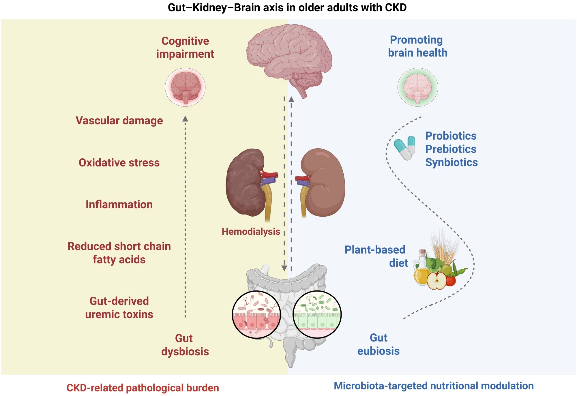
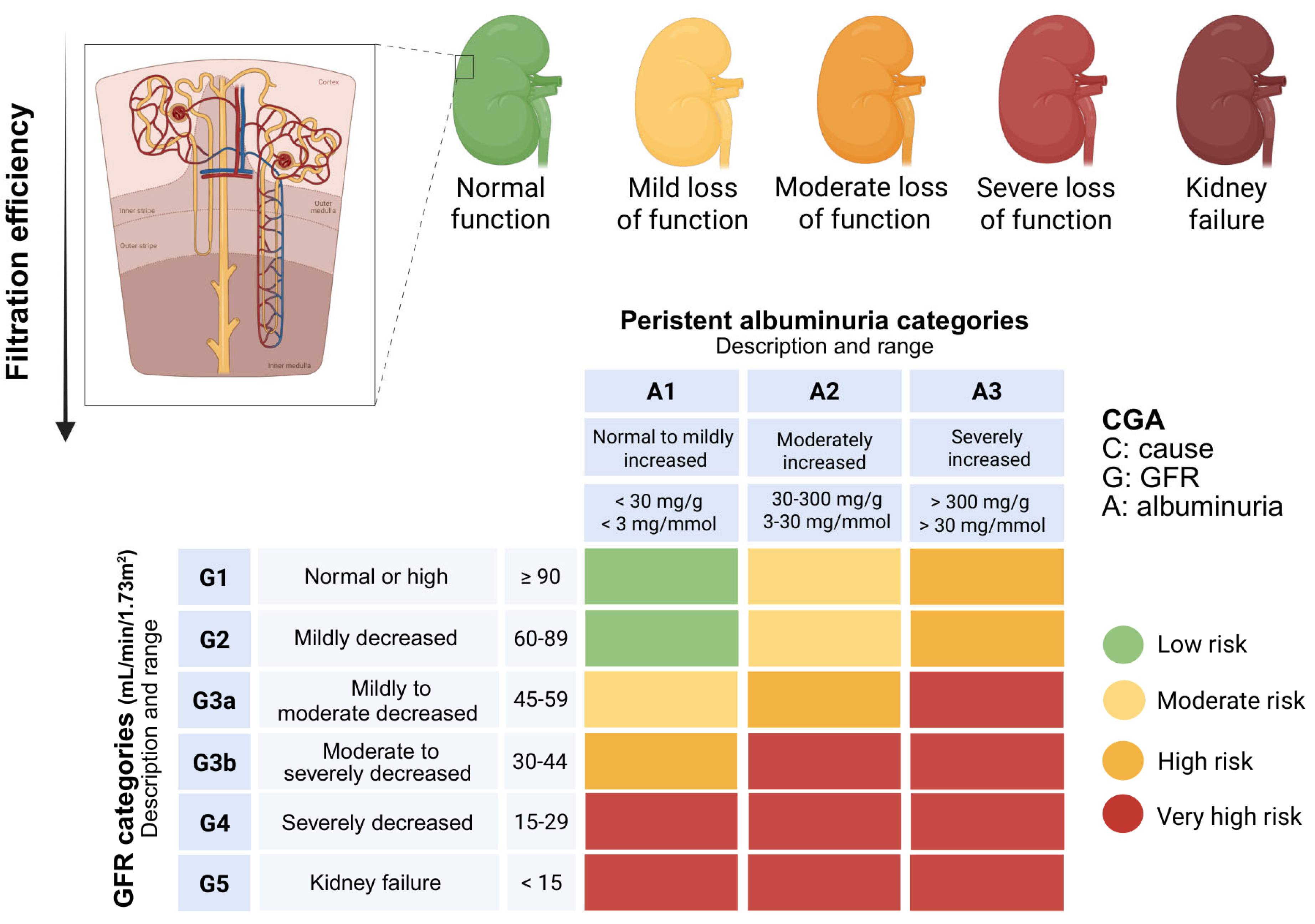
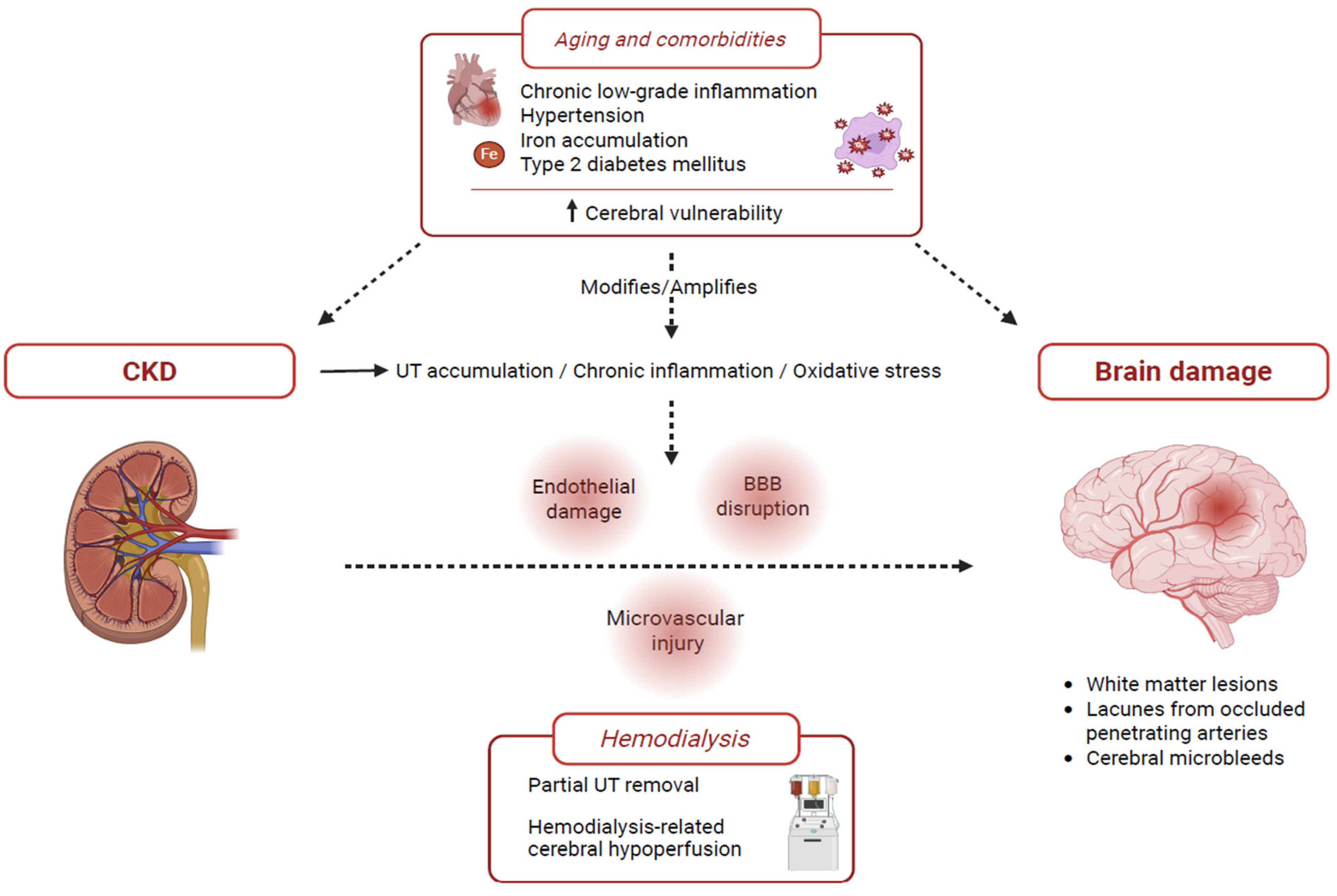
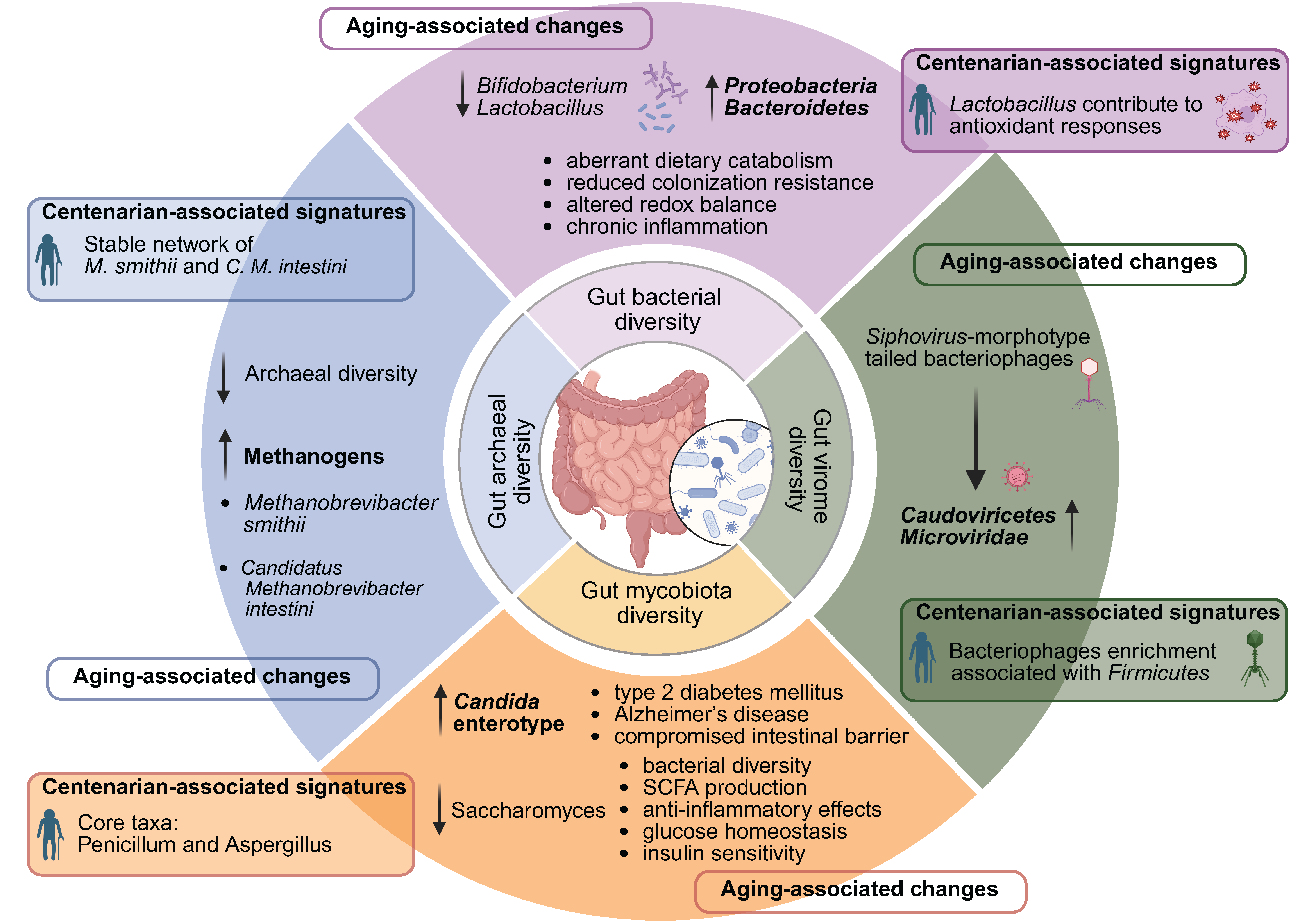
Mind the Gut: Cognitive Decline, Microbiota, and Nutrition-Related Modulators in Older Adults with Chronic Kidney Disease

 , , and
Abstract
1. Introduction
2. Neurological Changes in CKD
3. Epidemiological Evidence Linking Cognitive Decline with CKD and HD
4. Gut Dysbiosis in Aging and in CKD
4.1. Gut Bacterial Community
4.2. Non-Bacterial Components of the Gut Ecosystem: Virome, Mycobiota, and Archaeome
5. Dietary Recommendations in CKD: The Possible Contribution of the Microbiota
5.1. Potassium Balance
5.2. Iron Supplementation
5.3. Phosphate Regulation
5.4. Protein Intake and Protein-Energy Wasting
5.5. Uremic Toxins
5.6. Probiotics, Synbiotics, and Postbiotics
5.7. Clinical Implications and Practical Considerations
6. Conclusions and Future Directions
Author Contributions
Funding
Data Availability Statement
Conflicts of Interest
Abbreviations
| CKD | Chronic Kidney Disease |
| HD | Hemodialysis |
| GFR | Glomerular Filtration Rate |
| KDIGO | Kidney Disease: Improving Global Outcomes |
| UTs | Uremic Toxins |
| BBB | Blood–Brain Barrier |
| ESKD | End-Stage Kidney Disease |
| MMSE | Mini-Mental State Examination |
| RBANS | Repeatable Battery for the Assessment of Neuropsychological Status |
| MoCA | Montreal Cognitive Assessment |
| GDS | Geriatric Depression Scale |
| GAI | Geriatric Anxiety Inventory |
| MAC-Q | Memory Complaint Questionnaire |
| SCFAs | Short-Chain Fatty Acids |
| IL-18 | Interleukin-18 |
| TNF-α | Tumor Necrosis Factor alpha |
| IFN-γ | Interferon gamma |
| IL-2Rα | Interleukin-2 Receptor alpha |
| IL-9 | Interleukin-9 |
| TNF-β | Tumor Necrosis Factor beta |
| PLADO | Plant-Dominant Low-Protein Diet |
| FMT | Fecal Microbiota Transplantation |
| PEW | Protein-Energy Wasting |
| PTH | Parathyroid Hormone |
| MIND | Mediterranean-DASH Intervention for Neurodegenerative Delay |
| MBD | Mineral and Bone Disorder |
References
- Herrington, W.G.; Judge, P.K.; Grams, M.E.; Wanner, C. Chronic kidney disease. Lancet 2025, 407, 90–104. [Google Scholar] [CrossRef] [PubMed]
- Stevens, P.E.; Ahmed, S.B.; Carrero, J.J.; Foster, B.; Francis, A.; Hall, R.K.; Herrington, W.G.; Hill, G.; Inker, L.A.; Kazancıoğlu, R.; et al. KDIGO 2024 Clinical Practice Guideline for the Evaluation and Management of Chronic Kidney Disease. Kidney Int. 2024, 105, S117–S314. [Google Scholar] [CrossRef] [PubMed]
- World Health Organization (WHO). Reducing the Burden of Noncommunicable Diseases Through Promotion of Kidney Health and Strengthening Prevention and Control of Kidney Disease. Available online: https://apps.who.int/gb/ebwha/pdf_files/EB156/B156_CONF6-en.pdf (accessed on 27 April 2026).
- GBD 2023 Chronic Kidney Disease Collaborators. Global, regional, and national burden of chronic kidney disease in adults, 1990–2023, and its attributable risk factors: A systematic analysis for the Global Burden of Disease Study 2023. Lancet 2025, 406, 2461–2482. [Google Scholar] [CrossRef] [PubMed]
- Centers for Disease Control and Prevention (CDC). Chronic Kidney Disease in the United States; U.S. Department of Health and Human Services, Centers for Disease Control and Prevention: Atlanta, GA, USA, 2023. Available online: https://www.cdc.gov/kidney-disease/php/data-research/index.html (accessed on 2 February 2026).
- Nitta, K.; Okada, K.; Yanai, M.; Takahashi, S. Aging and chronic kidney disease. Kidney Blood Press. Res. 2013, 38, 109–120. [Google Scholar] [CrossRef] [PubMed]
- Shahbaz, H.; Rout, P.; Gupta, M. Creatinine clearance. In StatPearls [Internet]; StatPearls Publishing: Treasure Island, FL, USA, 2024. [Google Scholar]
- Dowling, T.C.; Wang, E.S.; Ferrucci, L.; Sorkin, J.D. Glomerular filtration rate equations overestimate creatinine clearance in older individuals enrolled in the Baltimore Longitudinal Study on Aging: Impact on renal drug dosing. Pharmacotherapy 2013, 33, 912–921. [Google Scholar] [CrossRef] [PubMed]
- Wang, Y.; Liu, J.; Fang, Y.; Zhou, S.; Liu, X.; Li, Z. Estimating the global prevalence of secondary hyperparathyroidism in patients with chronic kidney disease. Front. Endocrinol. 2024, 15, 1400891. [Google Scholar] [CrossRef] [PubMed]
- Virzì, G.M.; Clementi, A.; Ronco, C.; Zanella, M. Red cell death in renal disease: The role of eryptosis in CKD and dialysis patients. Cells 2025, 14, 967. [Google Scholar] [CrossRef] [PubMed]
- Kourtidou, C.; Tzionalos, K. Epidemiology and Risk Factors for Stroke in Chronic Kidney Disease: A Narrative Review. Biomedicines 2023, 11, 2398. [Google Scholar] [CrossRef] [PubMed]
- Roy, A.; Roy, R.; Bhattacharya, P.; Borah, A. The vicious consequences of chronic kidney disease on cognitive impairment and Alzheimer’s disease. ACS Chem. Neurosci. 2025, 16, 1847–1859. [Google Scholar] [CrossRef] [PubMed]
- Huang, Z.; Yaffe, K.; Li, C.; Xiao, C.; Pan, Y.; Sun, X.; Anderson, A.H.; He, J.; Jaar, B.G.; Han, H.; et al. Chronic kidney disease severity and risk of cognitive impairment. JAMA Netw. Open 2026, 9, e2559834. [Google Scholar] [CrossRef] [PubMed]
- Chen, Y.; Zhu, R.; Zhu, X.; Zhu, Q.; Luo, Z.; Mu, J.; Zhang, M.; Ma, S. Bibliometric analysis of cognitive impairment secondary to chronic kidney disease. Medicine 2026, 17, e47621. [Google Scholar] [CrossRef] [PubMed]
- Mendoza-Ruvalcaba, N.M.; Vázquez-Núñez, K.P.; Rodríguez-Díaz, M. The role of cognitive reserve on successful aging in community-dwelling older adults. Appl. Neuropsychol. Adult 2026, 1–11. [Google Scholar] [CrossRef] [PubMed]
- Wagner, C.A.; Frey-Wagner, I.; Ortiz, A.; Unwin, R.; Liabeuf, S.; Suzumoto, Y.; Iervolino, A.; Stasi, A.; Di Marzo, V.; Gesualdo, L.; et al. The role of the intestinal microbiome in cognitive decline in patients with kidney disease. Nephrol. Dial. Transplant. 2025, 40, ii4–ii17. [Google Scholar] [CrossRef] [PubMed]
- Coradduzza, D.; Sedda, S.; Cruciani, S.; De Miglio, M.R.; Ventura, C.; Nivoli, A.; Maioli, M. Age-related cognitive decline, focus on microbiome: A systematic review and meta-analysis. Int. J. Mol. Sci. 2023, 24, 13680. [Google Scholar] [CrossRef] [PubMed]
- Zhou, L.; Wang, Z.; Wang, M.; Li, X.; Ren, Q. Gut microbiome mediates the causal relationship between chronic kidney disease and cognitive dysfunction: A Mendelian randomization study. Physiol. Behav. 2025, 301, 115068. [Google Scholar] [CrossRef] [PubMed]
- Raskin, N.H.; Fishman, R.A. Neurologic disorders in renal failure (first of two parts). N. Engl. J. Med. 1976, 294, 143–148. [Google Scholar] [CrossRef] [PubMed]
- Raskin, N.H.; Fishman, R.A. Neurologic disorders in renal failure (second of two parts). N. Engl. J. Med. 1976, 294, 204–210. [Google Scholar] [CrossRef] [PubMed]
- Marini, S.; Georgakis, M.K.; Anderson, C.D. Interactions Between Kidney Function and Cerebrovascular Disease: Vessel Pathology That Fires Together Wires Together. Front. Neurol. 2021, 12, 785273. [Google Scholar] [CrossRef] [PubMed]
- Bobot, M.; Bruyat, A.; Thomas, L.; Fernandez, S.; Brodovitch, A.; Boucraut, J.; Burtey, S.; Nail, V.; Guillet, B.; Hache, G. Blood-brain barrier permeability in CKD: Link with inflammation and cognitive and mood impairment in rats. Behav. Brain Res. 2025, 493, 115693. [Google Scholar] [CrossRef] [PubMed]
- Sen, P.; Sittig, T.; Hamers, J.; d’Ambrosio, L.; Ornek, I.; Zhang, J.; Shashikadze, B.; Stöckl, J.B.; Bachter, M.; Bierschenk, S.; et al. From kidney injury to cardiac dysfunction: The central role of oxidative stress in diabetes and CKD. Basic Res. Cardiol. 2026, 121, 93–112. [Google Scholar] [CrossRef] [PubMed]
- Hernandez, L.; Ward, L.J.; Arefin, S.; Ebert, T.; Laucyte-Cibulskiene, A.; GOING-FWD Collaborators; Heimbürger, O.; Barany, P.; Wennberg, L.; Stenvinkel, P.; et al. Blood-brain barrier and gut barrier dysfunction in chronic kidney disease with a focus on circulating biomarkers and tight junction proteins. Sci. Rep. 2022, 12, 4414. [Google Scholar] [CrossRef] [PubMed]
- Scheppach, J.B.; Wu, A.; Gottesman, R.F.; Mosley, T.H.; Arsiwala-Scheppach, L.T.; Knopman, D.S.; Grams, M.E.; Sharrett, A.R.; Coresh, J.; Koton, S. Association of kidney function measures with signs of neurodegeneration and small vessel disease on brain magnetic resonance imaging: The Atherosclerosis Risk in Communities (ARIC) Study. Am. J. Kidney Dis. 2023, 81, 261–269.e1. [Google Scholar] [CrossRef] [PubMed]
- Martinez-Vea, A.; Salvadó, E.; Bardají, A.; Gutierrez, C.; Ramos, A.; García, C.; Compte, T.; Peralta, C.; Broch, M.; Pastor, R.; et al. Silent cerebral white matter lesions and their relationship with vascular risk factors in middle-aged predialysis patients with CKD. Am. J. Kidney Dis. 2006, 47, 241–250. [Google Scholar] [CrossRef] [PubMed]
- Bi, R.; Wei, Y.; Li, P.; Peng, H.; Alizadeh, M.; Hu, B.; Li, Y. Associations of cerebral small vessel disease and chronic kidney disease in patients with acute ischemic stroke. J. Am. Heart Assoc. 2025, 14, e038711. [Google Scholar] [CrossRef] [PubMed]
- Nash, P.S.; Fandler-Höfler, S.; Ambler, G.; Zhang, W.; Ozkan, H.; Locatelli, M.; Du, Y.; Obergottsberger, L.; Wünsch, G.; Jäger, H.R.; et al. Associations of cerebral small vessel disease and chronic kidney disease in patients with acute intracerebral hemorrhage: A cross-sectional study. Neurology 2024, 103, e209540. [Google Scholar] [CrossRef] [PubMed]
- Hein, Z.M.; Che Mohd Nassir, C.M.N. Perivascular pathology, not macrovascular complexity, governs glymphatic-related dysfunction in preclinical cerebral small vessel disease. Sci. Rep. 2026, 16, 4528. [Google Scholar] [CrossRef] [PubMed]
- Zhang, K.; Kan, C.; Han, F.; Fan, Q.; Wang, Y.; Li, X.; Pan, R.; Guo, Z.; Hou, N.; Sun, X. Global trends and projections in chronic kidney disease burden from diabetes, hypertension, and glomerulonephritis: A population-based study. Kidney Blood Press. Res. 2026, 51, 72–90. [Google Scholar] [CrossRef] [PubMed]
- Kim, K.S.; Park, S.W.; Cho, Y.W.; Kim, S.K. Higher prevalence and progression rate of chronic kidney disease in elderly patients with type 2 diabetes mellitus. Diabetes Metab. J. 2018, 42, 224–232. [Google Scholar] [CrossRef] [PubMed]
- Li, Y.; Jiang, Y.; Gao, B.; Liu, N.; Zhang, Y.; Zhou, H.; Song, Q.; Wang, N.; Miao, Y. Regional high iron deposition on brain quantitative susceptibility mapping correlates with cognitive decline in chronic kidney disease patients. Brain Imaging Behav. 2025, 19, 395–406. [Google Scholar] [CrossRef] [PubMed]
- Ward, R.J.; Zucca, F.A.; Duyn, J.H.; Crichton, R.R.; Zecca, L. The role of iron in brain ageing and neurodegenerative disorders. Lancet Neurol. 2014, 13, 1045–1060. [Google Scholar] [CrossRef] [PubMed]
- Qian, J.; Wang, X.; Cao, J.; Zhang, W.; Lu, C.; Chen, X. Dihydromyricetin attenuates D-galactose-induced brain aging of mice via inhibiting oxidative stress and neuroinflammation. Neurosci. Lett. 2021, 756, 135963. [Google Scholar] [CrossRef] [PubMed]
- Neyra Chauca, J.M.; VanDyck, M.V.; Espinoza Santana, A.; Martínez, G.G.R.; Romero Vega, K.A.; García Quintana, N.; Sánchez, V.F. Microvascular failure in the aging brain: Converging pathways of oxidative stress, inflammation, and endothelial decline. Biomedicines 2026, 14, 130. [Google Scholar] [CrossRef] [PubMed]
- Yu, J.; Li, Y.; Zhu, B.; Shen, J.; Miao, L. Research progress on the kidney-gut-brain axis in brain dysfunction in maintenance hemodialysis patients. Front. Med. 2025, 12, 1538048. [Google Scholar] [CrossRef]
- Andrews, T.D.; Day, G.S.; Irani, S.R.; Kanekiyo, T.; Hickson, L.J. Uremic toxins, CKD, and cognitive dysfunction. J. Am. Soc. Nephrol. 2025, 36, 1208–1226. [Google Scholar] [CrossRef] [PubMed]
- Hsieh, C.-C.; Lu, K.-C.; Huang, C.-L.; Wang, J.-J.; Yeh, T.-Y.; Lin, S.-M.; Chung, Y.-L.; Hou, Y.-C.; Huang, Y.-S. Indoxyl sulfate is associated with cognitive impairment in ESRD patients by activating the extrinsic apoptosis in the neuronal cells during differentiating process. Int. J. Med. Sci. 2025, 22, 1736–1749. [Google Scholar] [CrossRef] [PubMed]
- Bobot, M.; Guedj, E.; Resseguier, N.; Faraut, J.; Garrigue, P.; Nail, V.; Hache, G.; Gonzalez, S.; McKay, N.; Vial, R.; et al. Increased blood-brain barrier permeability and cognitive impairment in patients with ESKD. Kidney Int. Rep. 2024, 9, 2988–2995. [Google Scholar] [CrossRef] [PubMed]
- Sánchez-Ospina, D.; Mas-Fontao, S.; Gracia-Iguacel, C.; Avello, A.; González de Rivera, M.; Mujika-Marticorena, M.; Gonzalez-Parra, E. Displacing the burden: A review of protein-bound uremic toxin clearance strategies in chronic kidney disease. J. Clin. Med. 2024, 13, 1428. [Google Scholar] [CrossRef] [PubMed]
- Yang, J.; Ke, G.; Liao, Y.; Guo, Y.; Gao, X. Efficacy of medium cut-off dialyzers and comparison with high-flux dialyzers in patients on maintenance hemodialysis: A systematic review and meta-analysis. Ther. Apher. Dial. 2022, 26, 756–768. [Google Scholar] [CrossRef] [PubMed]
- Zhu, Y.; Li, J.; Lu, H.; Shi, Z.; Wang, X. Effect of hemodiafiltration and hemodialysis on mortality of patients with end-stage kidney disease: A meta-analysis. BMC Nephrol. 2024, 25, 372. [Google Scholar] [CrossRef] [PubMed]
- Zhao, Y.; Gan, L.; Niu, Q.; Ni, M.; Zuo, L. Efficacy and safety of expanded hemodialysis in hemodialysis patients: A meta-analysis and systematic review. Ren. Fail. 2022, 44, 541–550. [Google Scholar] [CrossRef] [PubMed]
- Evans, A.R.; Zhao, X.; Ernst, G.L.; Ortiz-Garcia, J.; Dunn, I.F.; Burke, J. Dialysis disequilibrium syndrome in neurosurgery: Literature review and illustrative case example. Geroscience 2024, 46, 5431–5437. [Google Scholar] [CrossRef] [PubMed]
- Polinder-Bos, H.A.; Vállez García, D.; Kuipers, J.; Elting, J.W.J.; Aries, M.J.H.; Krijnen, W.P.; Groen, H.; Willemsen, A.T.M.; van Laar, P.J.; Strijkert, F.; et al. Hemodialysis induces an acute decline in cerebral blood flow in elderly patients. J. Am. Soc. Nephrol. 2018, 29, 1317–1325. [Google Scholar] [CrossRef] [PubMed]
- Guo, Y.; Cui, W.; Ye, P.; Luo, Y. Association between cerebral blood flow variation and cognitive decline in older patients undergoing hemodialysis. Front. Aging Neurosci. 2024, 16, 1457675. [Google Scholar] [CrossRef] [PubMed]
- Kanbay, M.; Ertuglu, L.A.; Afsar, B.; Ozdogan, E.; Siriopol, D.; Covic, A.; Basile, C.; Ortiz, A. An update review of intradialytic hypotension: Concept, risk factors, clinical implications and management. Clin. Kidney J. 2020, 13, 981–993. [Google Scholar] [CrossRef] [PubMed]
- Etgen, T.; Chonchol, M.; Förstl, H.; Sander, D. Chronic kidney disease and cognitive impairment: A systematic review and meta-analysis. Am. J. Nephrol. 2012, 35, 474–482. [Google Scholar] [CrossRef] [PubMed]
- O’Lone, E.; Connors, M.; Masson, P.; Wu, S.; Kelly, P.J.; Gillespie, D.; Parker, D.; Whiteley, W.; Strippoli, G.F.M.; Palmer, S.C.; et al. Cognition in people with end-stage kidney disease treated with hemodialysis: A systematic review and meta-analysis. Am. J. Kidney Dis. 2016, 67, 925–935. [Google Scholar] [CrossRef] [PubMed]
- Chan, F.H.F.; Goh, Z.Z.S.; Zhu, X.; Tudor Car, L.; Newman, S.; Khan, B.A.; Griva, K. Subjective cognitive complaints in end-stage renal disease: A systematic review and meta-analysis. Health Psychol. Rev. 2023, 17, 614–640. [Google Scholar] [CrossRef] [PubMed]
- Tian, X.; Guo, X.; Xia, X.; Yu, H.; Li, X.; Jiang, A. The comparison of cognitive function and risk of dementia in CKD patients under peritoneal dialysis and hemodialysis: A PRISMA-compliant systematic review and meta-analysis. Medicine 2019, 98, e14390. [Google Scholar] [CrossRef] [PubMed]
- Oh, H.; Mo, J.; Seo, W. Correlates of cognitive impairment in patients with chronic kidney failure on haemodialysis: Systematic review and meta-analysis. J. Adv. Nurs. 2019, 75, 962–978. [Google Scholar] [CrossRef] [PubMed]
- Odagiri, G.; Sugawara, N.; Kikuchi, A.; Takahashi, I.; Umeda, T.; Saitoh, H.; Yasui-Furukori, N.; Kaneko, S. Cognitive function among hemodialysis patients in Japan. Ann. Gen. Psychiatry 2011, 10, 20. [Google Scholar] [CrossRef] [PubMed]
- Cavanaugh, K.L.; Osborn, C.Y.; Tentori, F.; Rothman, R.L.; Ikizler, T.A.; Wallston, K.A. Performance of a brief survey to assess health literacy in patients receiving hemodialysis. Clin. Kidney J. 2015, 8, 462–468. [Google Scholar] [CrossRef] [PubMed]
- Chan, F.H.F.; Newman, S.; Khan, B.A.; Griva, K. Prevalence and trajectories of subjective cognitive complaints and implications for patient outcomes: A prospective study of haemodialysis patients. Br. J. Health Psychol. 2023, 28, 651–671. [Google Scholar] [CrossRef] [PubMed]
- Bolignano, D.; Simeoni, M.; Hafez, G.; Pepin, M.; Gallo, A.; Altieri, M.; Liabeuf, S.; Giannakou, K.; De, A.; Capasso, G. Cognitive impairment in CKD patients: A guidance document by the CONNECT network. Clin. Kidney J. 2024, 18, sfae294. [Google Scholar] [CrossRef] [PubMed]
- Thancharoen, O.; Waleekhachonloet, O.; Limwattananon, C.; Anutrakulchai, S. Cognitive impairment, quality of life and healthcare utilization in patients with chronic kidney disease stages 3 to 5. Nephrology 2020, 25, 625–633. [Google Scholar] [CrossRef] [PubMed]
- Chan, F.H.F.; Sim, P.; Lim, P.X.H.; Khan, B.A.; Choo, J.C.J.; Griva, K. Screening for cognitive symptoms in dialysis patients with an extended version of Kidney Disease Quality of Life Cognitive Function subscale (KDQOL-CF): A validation study. BMC Nephrol. 2024, 25, 434. [Google Scholar] [CrossRef] [PubMed]
- van Zwieten, A.; Wong, G.; Ruospo, M.; Palmer, S.C.; Barulli, M.R.; Iurillo, A.; Saglimbene, V.; Natale, P.; Gargano, L.; Murgo, M.; et al. Prevalence and patterns of cognitive impairment in adult hemodialysis patients: The COGNITIVE-HD study. Nephrol. Dial. Transplant. 2018, 33, 1197–1206. [Google Scholar] [CrossRef] [PubMed]
- Drew, D.A.; Tighiouart, H.; Rollins, J.; Duncan, S.; Babroudi, S.; Scott, T.; Weiner, D.E.; Sarnak, M.J. Evaluation of screening tests for cognitive impairment in patients receiving maintenance hemodialysis. J. Am. Soc. Nephrol. 2020, 31, 855–864. [Google Scholar] [CrossRef] [PubMed]
- Gautam, S.; Kiran, U.V. Co-occurrence of cognitive dysfunction and depressive disorders in hemodialysis patients: Demographic patterns and unmet diagnostic needs. Cureus 2025, 17, e99003. [Google Scholar] [CrossRef] [PubMed]
- Paré, M.; Zola, N.; Coudert, L.; Phaneuf, J.-C.; Lavallée, R.; Caplette-Gingras, A.; Fortier, C.; Agharazii, M. A mixed-methods protocol to explore psychological distress and psychosocial needs along the continuum of CKD care: A single healthcare network. BMC Nephrol. 2025, 26, 644. [Google Scholar] [CrossRef] [PubMed]
- Badrkhahan, S.Z.; Ala, M.; Fakhrzadeh, H.; Yaghoobi, A.; Mirzamohamadi, S.; Arzaghi, S.M.; Shahabi, S.; Sharifi, F.; Ostovar, A.; Fahimfar, N.; et al. The prevalence and predictors of geriatric giants in community-dwelling older adults: A cross-sectional study from the Middle East. Sci. Rep. 2023, 13, 12401. [Google Scholar] [CrossRef] [PubMed]
- Cassidy, K.-L.; Rector, N.A. The silent geriatric giant: Anxiety disorders in late life. Geriatr. Aging 2008, 11, 150–156. [Google Scholar]
- World Health Organization (WHO). Mental Health of Older Adults. Available online: https://www.who.int/news-room/fact-sheets/detail/mental-health-of-older-adults (accessed on 29 April 2026).
- Yesavage, J.A.; Brink, T.L.; Rose, T.L.; Lum, O.; Huang, V.; Adey, M.; Leirer, V.O. Development and validation of a geriatric depression screening scale: A preliminary report. J. Psychiatr. Res. 1982, 17, 37–49. [Google Scholar] [CrossRef] [PubMed]
- Pachana, N.A.; Byrne, G.J.; Siddle, H.; Koloski, N.; Harley, E.; Arnold, E. Development and validation of the Geriatric Anxiety Inventory. Int. Psychogeriatr. 2007, 19, 103–114. [Google Scholar] [CrossRef] [PubMed]
- Dow, B.; Lin, X.; Pachana, N.A.; Bryant, C.; LoGiudice, D.; Goh, A.M.Y.; Haralambous, B. Reliability, concurrent validity, and cultural adaptation of the Geriatric Depression Scale and the Geriatric Anxiety Inventory for detecting depression and anxiety symptoms among older Chinese immigrants: An Australian study. Int. Psychogeriatr. 2018, 30, 735–748. [Google Scholar] [CrossRef] [PubMed]
- Ganguli, M.; Snitz, B.E.; Lee, C.-W.; Vanderbilt, J.; Saxton, J.A.; Chang, C.-C.H. Age and education effects and norms on a cognitive test battery from a population-based cohort: The Monongahela-Youghiogheny Healthy Aging Team. Aging Ment. Health 2010, 14, 100–107. [Google Scholar] [CrossRef] [PubMed]
- Karstens, A.J.; Christianson, T.J.; Lundt, E.S.; Machulda, M.M.; Mielke, M.M.; Fields, J.A.; Kremers, W.K.; Graff-Radford, J.; Vemuri, P.; Jack, C.R., Jr.; et al. Mayo normative studies: Regression-based normative data for ages 30–91 years with a focus on the Boston Naming Test, Trail Making Test and Category Fluency. J. Int. Neuropsychol. Soc. 2024, 30, 389–401. [Google Scholar] [CrossRef] [PubMed]
- Tiffin-Richards, F.E.; Costa, A.S.; Holschbach, B.; Frank, R.D.; Vassiliadou, A.; Krüger, T.; Kuckuck, K.; Gross, T.; Eitner, F.; Floege, J.; et al. The Montreal Cognitive Assessment (MoCA)—A sensitive screening instrument for detecting cognitive impairment in chronic hemodialysis patients. PLoS ONE 2014, 9, e106700. [Google Scholar] [CrossRef] [PubMed]
- Classon, E.; van den Hurk, W.; Lyth, J.; Johansson, M.M. Montreal Cognitive Assessment: Normative data for cognitively healthy Swedish 80- to 94-year-olds. J. Alzheimer’s Dis. 2022, 87, 1335–1344. [Google Scholar] [CrossRef] [PubMed]
- Scheenen, M.E.; van den Brink, R.H.S.; Konstantinidou, S.; Lugtenburg, A.; Spit, J.; Hendriks, G.-J.; Naarding, P.; de Vent, N.R.; Kessels, R.P.C.; Oude Voshaar, R.C.; et al. Montreal Cognitive Assessment (MoCA) norms for older patients with a depressive disorder. Med. Sci. 2025, 13, 312. [Google Scholar] [CrossRef] [PubMed]
- Ling, T.-C.; Chang, C.-C.; Li, C.-Y.; Sung, J.-M.; Sun, C.-Y.; Tsai, K.-J.; Cheng, Y.-Y.; Wu, J.-L.; Kuo, Y.-T.; Chang, Y.-T. Development and validation of the dialysis dementia risk score: A retrospective, population-based, nested case-control study. Eur. J. Neurol. 2022, 29, 59–68. [Google Scholar] [CrossRef] [PubMed]
- Lai, G.; Bevilacqua, L.; Giuliani, M.E.; Bigossi, G.; Marcozzi, S.; Casoli, T.; Abbrescia, P.; Frigeri, A.; Malavolta, M.; Balietti, M. The aging choroid plexus and its relationship with gut dysbiosis and Klotho decline: Possible intervention strategies. Geroscience 2025, 47, 6869–6883. [Google Scholar] [CrossRef] [PubMed]
- Gregory, A.C.; Zablocki, O.; Zayed, A.A.; Howell, A.; Bolduc, B.; Sullivan, M.B. The gut virome database reveals age-dependent patterns of virome diversity in the human gut. Cell Host Microbe 2020, 28, 724–740.e8. [Google Scholar] [CrossRef] [PubMed]
- Mayneris-Perxachs, J.; Castells-Nobau, A.; Arnoriaga-Rodríguez, M.; Garre-Olmo, J.; Puig, J.; Ramos, R.; Martínez-Hernández, F.; Burokas, A.; Coll, C.; Moreno-Navarrete, J.M.; et al. Caudovirales bacteriophages are associated with improved executive function and memory in flies, mice, and humans. Cell Host Microbe 2022, 30, 340–356.e8. [Google Scholar] [CrossRef] [PubMed]
- Johansen, J.; Atarashi, K.; Arai, Y.; Hirose, N.; Sørensen, S.J.; Vatanen, T.; Knip, M.; Honda, K.; Xavier, R.J.; Rasmussen, S.; et al. Centenarians have a diverse gut virome with the potential to modulate metabolism and promote healthy lifespan. Nat. Microbiol. 2023, 8, 1064–1078. [Google Scholar] [CrossRef] [PubMed]
- Shuai, M.; Fu, Y.; Zhong, H.-L.; Gou, W.; Jiang, Z.; Liang, Y.; Miao, Z.; Xu, J.-J.; Huynh, T.; Wahlqvist, M.L.; et al. Mapping the human gut mycobiome in middle-aged and elderly adults: Multiomics insights and implications for host metabolic health. Gut 2022, 71, 1812–1820. [Google Scholar] [CrossRef] [PubMed]
- Lai, S.; Yan, Y.; Pu, Y.; Lin, S.; Qiu, J.-G.; Jiang, B.-H.; Keller, M.I.; Wang, M.; Bork, P.; Chen, W.-H.; et al. Enterotypes of the human gut mycobiome. Microbiome 2023, 11, 179. [Google Scholar] [CrossRef] [PubMed]
- Mihajlovski, A.; Doré, J.; Levenez, F.; Alric, M.; Brugère, J.-F. Molecular evaluation of the human gut methanogenic archaeal microbiota reveals an age-associated increase of the diversity. Environ. Microbiol. Rep. 2010, 2, 272–280. [Google Scholar] [CrossRef] [PubMed]
- Dridi, B.; Henry, M.; Richet, H.; Raoult, D.; Drancourt, M. Age-related prevalence of Methanomassiliicoccus luminyensis in the human gut microbiome. APMIS 2012, 120, 773–777. [Google Scholar] [CrossRef] [PubMed]
- Wu, L.; Xie, X.; Li, Y.; Liang, T.; Zhong, H.; Yang, L.; Xi, Y.; Zhang, J.; Ding, Y.; Wu, Q. Gut microbiota as an antioxidant system in centenarians associated with high antioxidant activities of gut-resident Lactobacillus. npj Biofilms Microbiomes 2022, 8, 102. [Google Scholar] [CrossRef] [PubMed]
- Pu, L.; Pang, S.; Mu, W.; Chen, X.; Zou, Y.; Wang, Y.; Ding, Y.; Yan, Q.; Huang, Y.; Chen, X.; et al. The gut mycobiome signatures in long-lived populations. iScience 2024, 27, 110412. [Google Scholar] [CrossRef] [PubMed]
- Mohammadzadeh, R.; Mahnert, A.; Shinde, T.; Kumpitsch, C.; Weinberger, V.; Schmidt, H.; Moissl-Eichinger, C. Age-related dynamics of predominant methanogenic archaea in the human gut microbiome. BMC Microbiol. 2025, 25, 193. [Google Scholar] [CrossRef] [PubMed]
- Chen, H.; Wang, J.; Ouyang, Q.; Peng, X.; Yu, Z.; Wang, J.; Huang, J. Alterations of gut microbes and their correlation with clinical features in middle and end-stages chronic kidney disease. Front. Cell. Infect. Microbiol. 2023, 13, 1105366. [Google Scholar] [CrossRef] [PubMed]
- Zhang, P.; Wang, X.; Li, S.; Cao, X.; Zou, J.; Fang, Y.; Shi, Y.; Xiang, F.; Shen, B.; Li, Y.; et al. Metagenome-wide analysis uncovers gut microbial signatures and implicates taxon-specific functions in end-stage renal disease. Genome Biol. 2023, 24, 226. [Google Scholar] [CrossRef] [PubMed]
- Zhao, J.; Ning, X.; Liu, B.; Dong, R.; Bai, M.; Sun, S. Specific alterations in gut microbiota in patients with chronic kidney disease: An updated systematic review. Ren. Fail. 2021, 43, 102–112. [Google Scholar] [CrossRef] [PubMed]
- Stanford, J.; Charlton, K.; Stefoska-Needham, A.; Ibrahim, R.; Lambert, K. The gut microbiota profile of adults with kidney disease and kidney stones: A systematic review of the literature. BMC Nephrol. 2020, 21, 215. [Google Scholar] [CrossRef] [PubMed]
- Candeliere, F.; Simone, M.; Leonardi, A.; Rossi, M.; Amaretti, A.; Raimondi, S. Indole and p-cresol in feces of healthy subjects: Concentration, kinetics, and correlation with microbiome. Front. Mol. Med. 2022, 2, 959189. [Google Scholar] [CrossRef] [PubMed]
- Amini Khiabani, S.; Asgharzadeh, M.; Samadi Kafil, H. Chronic kidney disease and gut microbiota. Heliyon 2023, 9, e18991. [Google Scholar] [CrossRef] [PubMed]
- Laiola, M.; Koppe, L.; Larabi, A.; Thirion, F.; Lange, C.; Quinquis, B.; David, A.; Le Chatelier, E.; Benoit, B.; Sequino, G.; et al. Toxic microbiome and progression of chronic kidney disease: Insights from a longitudinal CKD-Microbiome Study. Gut 2025, 74, 1624–1637. [Google Scholar] [CrossRef] [PubMed]
- Wyczalkowska-Tomasik, A.; Czarkowska-Paczek, B.; Giebultowicz, J.; Wroczynski, P.; Paczek, L. Age-dependent increase in serum levels of indoxyl sulphate and p-cresol sulphate is not related to their precursors: Tryptophan and tyrosine. Geriatr. Gerontol. Int. 2017, 17, 1022–1026. [Google Scholar] [CrossRef] [PubMed]
- Wong, J.; Piceno, Y.M.; DeSantis, T.Z.; Pahl, M.; Andersen, G.L.; Vaziri, N.D. Expansion of urease- and uricase-containing, indole- and p-cresol-forming and contraction of short-chain fatty acid-producing intestinal microbiota in ESRD. Am. J. Nephrol. 2014, 39, 230–237. [Google Scholar] [CrossRef] [PubMed]
- Karbowska, M.; Hermanowicz, J.M.; Tankiewicz-Kwedlo, A.; Kalaska, B.; Kaminski, T.W.; Nosek, K.; Wisniewska, R.J.; Pawlak, D. Neurobehavioral effects of uremic toxin-indoxyl sulfate in the rat model. Sci. Rep. 2020, 10, 9483. [Google Scholar] [CrossRef] [PubMed]
- Iwata, K.; Watanabe, H.; Morisaki, T.; Matsuzaki, T.; Ohmura, T.; Hamada, A.; Saito, H. Involvement of indoxyl sulfate in renal and central nervous system toxicities during cisplatin-induced acute renal failure. Pharm. Res. 2007, 24, 662–671. [Google Scholar] [CrossRef] [PubMed]
- Łukawski, K.; Raszewski, G.; Czuczwar, S.J. Effects of the uremic toxin indoxyl sulfate on seizure activity, learning and brain oxidative stress parameters in mice. Neurosci. Lett. 2024, 820, 137594. [Google Scholar] [CrossRef] [PubMed]
- Adesso, S.; Magnus, T.; Cuzzocrea, S.; Campolo, M.; Rissiek, B.; Paciello, O.; Autore, G.; Pinto, A.; Marzocco, S. Indoxyl sulfate affects glial function increasing oxidative stress and neuroinflammation in chronic kidney disease: Interaction between astrocytes and microglia. Front. Pharmacol. 2017, 8, 370. [Google Scholar] [CrossRef] [PubMed]
- Vaziri, N.D.; Wong, J.; Pahl, M.; Piceno, Y.M.; Yuan, J.; DeSantis, T.Z.; Ni, Z.; Nguyen, T.-H.; Andersen, G.L. Chronic kidney disease alters intestinal microbial flora. Kidney Int. 2013, 83, 308–315. [Google Scholar] [CrossRef] [PubMed]
- Sampaio-Maia, B.; Simões-Silva, L.; Pestana, M.; Araujo, R.; Soares-Silva, I.J. The role of the gut microbiome on chronic kidney disease. Adv. Appl. Microbiol. 2016, 96, 65–94. [Google Scholar] [CrossRef] [PubMed]
- Cao, Q.; Shen, M.; Li, R.; Liu, Y.; Zeng, Z.; Zhou, J.; Niu, D.; Zhang, Q.; Wang, R.; Yao, J.; et al. Elucidating the specific mechanisms of the gut-brain axis: The short-chain fatty acids-microglia pathway. J. Neuroinflamm. 2025, 22, 133. [Google Scholar] [CrossRef] [PubMed]
- Fock, E.; Parnova, R. Mechanisms of blood-brain barrier protection by microbiota-derived short-chain fatty acids. Cells 2023, 12, 657. [Google Scholar] [CrossRef] [PubMed]
- Moțățăianu, A.; Șerban, G.; Andone, S. The role of short-chain fatty acids in microbiota-gut-brain cross-talk with a focus on amyotrophic lateral sclerosis: A systematic review. Int. J. Mol. Sci. 2023, 24, 15094. [Google Scholar] [CrossRef] [PubMed]
- Lu, C.; Wang, X.; Chen, X.; Qin, T.; Ye, P.; Liu, J.; Wang, S.; Luo, W. Causal analysis between gut microbes, aging indicator, and age-related disease, involving the discovery and validation of biomarkers. Aging Cell 2025, 24, e70057. [Google Scholar] [CrossRef] [PubMed]
- Zhang, S.; Easwaran, M.; Elafify, M.; Mahmoud, A.A.; Wang, X.; Ahn, J. Prophages and their interactions with lytic phages in the human gut microbiota and their impact on microbial diversity, gut health, and disease. Appl. Environ. Microbiol. 2025, 91, e0189925. [Google Scholar] [CrossRef] [PubMed]
- Chaudhari, D.S.; Jain, S.; Yata, V.K.; Mishra, S.P.; Kumar, A.; Fraser, A.; Kociolek, J.; Dangiolo, M.; Smith, A.; Golden, A.; et al. Unique trans-kingdom microbiome structural and functional signatures predict cognitive decline in older adults. Geroscience 2023, 45, 2819–2834. [Google Scholar] [CrossRef] [PubMed]
- Ghorbani, M.; Ferreira, D.; Maioli, S. A metagenomic study of gut viral markers in amyloid-positive Alzheimer’s disease patients. Alzheimer’s Res. Ther. 2023, 15, 141. [Google Scholar] [CrossRef] [PubMed]
- Chen, W.; Guo, R.; Zhang, W.; Yan, Q.; Wang, X.; Chen, R.; Hu, X.; Liang, J.; Xing, G.; Xu, D.; et al. Alterations of the gut virome in patients with Parkinson’s disease. J. Gerontol. A Biol. Sci. Med. Sci. 2026, 81, glag001. [Google Scholar] [CrossRef] [PubMed]
- Zhang, P.; Guo, R.; Ma, S.; Jiang, H.; Yan, Q.; Li, S.; Wang, K.; Deng, J.; Zhang, Y.; Zhang, Y.; et al. A metagenome-wide study of the gut virome in chronic kidney disease. Theranostics 2025, 15, 1642–1661. [Google Scholar] [CrossRef] [PubMed]
- Badillo-Pazmay, G.V.; Fortunato, C.; Cianfruglia, L.; Novazzi, F.; Spezia, P.G.; Rosa, L.; Limongi, D.; Prezioso, C.; D’Argenio, V.; Scudiero, O.; et al. The gut and circulating virome: Emerging players in aging and longevity. Front. Aging 2025, 6, 1731621. [Google Scholar] [CrossRef] [PubMed]
- Hallen-Adams, H.E.; Suhr, M.J. Fungi in the healthy human gastrointestinal tract. Virulence 2017, 8, 352–358. [Google Scholar] [CrossRef] [PubMed]
- Hu, J.; Wei, S.; Gu, Y.; Wang, Y.; Feng, Y.; Sheng, J.; Hu, L.; Gu, C.; Jiang, P.; Tian, Y.; et al. Gut mycobiome in patients with chronic kidney disease was altered and associated with immunological profiles. Front. Immunol. 2022, 13, 843695. [Google Scholar] [CrossRef] [PubMed]
- Qiu, J.; Zhao, L.; Cheng, Y.; Chen, Q.; Xu, Y.; Lu, Y.; Gao, J.; Lei, W.; Yan, C.; Ling, Z.; et al. Exploring the gut mycobiome: Differential composition and clinical associations in hypertension, chronic kidney disease, and their comorbidity. Front. Immunol. 2023, 14, 1317809. [Google Scholar] [CrossRef] [PubMed]
- Ren, Y.; Chen, L.; Guo, R.; Ma, S.; Li, S.; Zhang, Y.; Jiang, H.; Shi, H.; Zhang, P. Altered gut mycobiome in patients with end-stage renal disease and its correlations with serum and fecal metabolomes. J. Transl. Med. 2024, 22, 202. [Google Scholar] [CrossRef] [PubMed]
- Nie, T.; Li, J.; You, L.; Wu, Q. Environmental mycotoxins: A potential etiological factor for neurodegenerative diseases? Toxicology 2025, 511, 154056. [Google Scholar] [CrossRef] [PubMed]
- Nagpal, R.; Neth, B.J.; Wang, S.; Mishra, S.P.; Craft, S.; Yadav, H. Gut mycobiome and its interaction with diet, gut bacteria and Alzheimer’s disease markers in subjects with mild cognitive impairment: A pilot study. EBioMedicine 2020, 59, 102950. [Google Scholar] [CrossRef] [PubMed]
- Umamahesan, C.; Pilcicka, A.; Yick, J.; Baker, K.; Smith, M.; Taylor, D.; Ma, Y.; Mullish, B.H.; Marchesi, J.R.; Gilbert, S.; et al. Interplay of constipation, intestinal barrier dysfunction and fungal exposome in aetiopathogenesis of Parkinson’s disease: Hypothesis with supportive data. Biochem. J. 2025, 482, 807–821. [Google Scholar] [CrossRef] [PubMed]
- Mafra, D.; Alvarenga, L.; Cardozo, L.F.M.F.; Schultz, J.; Rosado, A.S.; Borges, N.A. Gut microbiota and NLRP3 inflammasome activation in hemodialysis patients: Exploring the link with systemic inflammation. Mol. Biol. Rep. 2025, 52, 465. [Google Scholar] [CrossRef] [PubMed]
- Kemp, J.A.; Schultz, J.; Modolon, F.; Ribeiro-Alves, M.; Rosado, A.S.; Mafra, D. Is there a correlation between TMAO plasma levels and archaea in the gut of patients undergoing hemodialysis? Int. Urol. Nephrol. 2025, 57, 1269–1275. [Google Scholar] [CrossRef] [PubMed]
- Fumagalli, A.; Castells-Nobau, A.; Trivedi, D.; Garre-Olmo, J.; Puig, J.; Ramos, R.; Ramió-Torrentà, L.; Pérez-Brocal, V.; Moya, A.; Swann, J.; et al. Archaea methanogens are associated with cognitive performance through the shaping of gut microbiota, butyrate and histidine metabolism. Gut Microbes 2025, 17, 2455506. [Google Scholar] [CrossRef] [PubMed]
- Duru, I.C.; Lecomte, A.; Shishido, T.K.; Laine, P.; Suppula, J.; Paulin, L.; Scheperjans, F.; Pereira, P.A.B.; Auvinen, P. Metagenome-assembled microbial genomes from Parkinson’s disease fecal samples. Sci. Rep. 2024, 14, 18906. [Google Scholar] [CrossRef] [PubMed]
- Shoubridge, A.P.; Carpenter, L.; Flynn, E.; Papanicolas, L.E.; Collins, J.; Gordon, D.; Lynn, D.J.; Whitehead, C.; Leong, L.E.X.; Cations, M.; et al. Severe cognitive decline in long-term care is related to gut microbiome production of metabolites involved in neurotransmission, immunomodulation, and autophagy. J. Gerontol. A Biol. Sci. Med. Sci. 2025, 80, glaf053. [Google Scholar] [CrossRef] [PubMed]
- Tanaka, K.; Saito, H.; Iwasaki, T.; Oda, A.; Watanabe, S.; Kanno, M.; Kimura, H.; Shimabukuro, M.; Asahi, K.; Watanabe, T.; et al. Association between serum potassium levels and adverse outcomes in chronic kidney disease: The Fukushima CKD cohort study. Clin. Exp. Nephrol. 2021, 25, 410–417. [Google Scholar] [CrossRef] [PubMed]
- Suzuki, Y.; Misaka, T.; Sato, Y.; Okochi, S.; Ogawara, R.; Ichimura, S.; Yokokawa, T.; Sato, A.; Shimizu, T.; Sato, T.; et al. Association between serum potassium variability during hospitalization and clinical outcomes in patients with heart failure. Eur. J. Intern. Med. 2025, 141, 106408. [Google Scholar] [CrossRef] [PubMed]
- Iqbal, I.M.; Obaid, M.; Haider, A.S.; Asif, A.; Ul Haq, Z.; Salman, M.; Rehman, I. Prevalence of electrolyte imbalances in critically ill medical intensive care unit patients and their association with clinical outcomes. Cureus 2025, 17, e94698. [Google Scholar] [CrossRef] [PubMed]
- Guo, Y.; Qiu, Y.; Xue, T.; Yan, P.; Zhao, W.; Wang, M.; Liu, C.; Zhang, N. Association between admission baseline blood potassium levels and all-cause mortality in patients with acute kidney injury combined with sepsis: A retrospective cohort study. PLoS ONE 2024, 19, e0309764. [Google Scholar] [CrossRef] [PubMed]
- Arnold, R.; Pussell, B.A.; Howells, J.; Grinius, V.; Kiernan, M.C.; Lin, C.S.-Y.; Krishnan, A.V. Evidence for a causal relationship between hyperkalaemia and axonal dysfunction in end-stage kidney disease. Clin. Neurophysiol. 2014, 125, 179–185. [Google Scholar] [CrossRef] [PubMed]
- Kiernan, M.C.; Walters, R.J.L.; Andersen, K.V.; Taube, D.; Murray, N.M.F.; Bostock, H. Nerve excitability changes in chronic renal failure indicate membrane depolarization due to hyperkalaemia. Brain 2002, 125, 1366–1378. [Google Scholar] [CrossRef] [PubMed]
- Babaker, M.A.; Alazabi, N.I.; Yousef, E.M.; Haredy, S.A.; Algohary, A.M.; Mansour, D.F.; Ahmed-Farid, O.A. Taurine mitigates spironolactone-induced hyperkalemia and cognitive dysfunction: A biochemical and histological study in a rat model. Appl. Biochem. Biotechnol. 2026, 198, 1962–1993. [Google Scholar] [CrossRef] [PubMed]
- Melekoglu, E.; Samur, F.G. Dietary strategies for gut-derived protein-bound uremic toxins and cardio-metabolic risk factors in chronic kidney disease: A focus on dietary fibers. Crit. Rev. Food Sci. Nutr. 2023, 63, 3994–4008. [Google Scholar] [CrossRef] [PubMed]
- St-Jules, D.E.; Goldfarb, D.S.; Sevick, M.A. Nutrient non-equivalence: Does restricting high-potassium plant foods help to prevent hyperkalemia in hemodialysis patients? J. Ren. Nutr. 2016, 26, 282–287. [Google Scholar] [CrossRef] [PubMed]
- Joshi, S.; Shah, S.; Kalantar-Zadeh, K. Adequacy of plant-based proteins in chronic kidney disease. J. Ren. Nutr. 2019, 29, 112–117. [Google Scholar] [CrossRef] [PubMed]
- Avesani, C.M.; Heimbürger, O.; Rubin, C.; Sallstrom, T.; Fáxen-Irving, G.; Lindholm, B.; Stenvinkel, P. Plant-based diet in hyperkalemic chronic kidney disease patients receiving sodium zirconium cyclosilicate: A feasibility clinical trial. Am. J. Clin. Nutr. 2024, 120, 719–726. [Google Scholar] [CrossRef] [PubMed]
- Michail, A.; Andreou, E. A plant-dominant low-protein diet in chronic kidney disease management: A narrative review with considerations for Cyprus. Nutrients 2025, 17, 970. [Google Scholar] [CrossRef] [PubMed]
- Zarantonello, D.; Brunori, G. The Role of Plant-Based Diets in Preventing and Mitigating Chronic Kidney Disease: More Light than Shadows. J. Clin. Med. 2023, 12, 6137. [Google Scholar] [CrossRef] [PubMed]
- Shannon, O.M.; Ranson, J.M.; Gregory, S.; Macpherson, H.; Milte, C.; Lentjes, M.; Mulligan, A.; McEvoy, C.; Griffiths, A.; Matu, J.; et al. Mediterranean diet adherence is associated with lower dementia risk, independent of genetic predisposition: Findings from the UK Biobank prospective cohort study. BMC Med. 2023, 21, 81. [Google Scholar] [CrossRef] [PubMed]
- Chen, H.; Dhana, K.; Huang, Y.; Huang, L.; Tao, Y.; Liu, X.; Melo van Lent, D.; Zheng, Y.; Ascherio, A.; Willett, W.; et al. Association of the Mediterranean dietary approaches to stop hypertension intervention for neurodegenerative delay (MIND) diet with the risk of dementia. JAMA Psychiatry 2023, 80, 630–638. [Google Scholar] [CrossRef] [PubMed]
- Babitt, J.L.; Berns, J.S.; Bozkurt, B.; Cheung Khedairy, R.S.; Cuevas, Y.; Effa, E.E.; Eisenga, M.F.; Fishbane, S.; Ginzburg, Y.Z.; Haase, V.H.; et al. Executive summary of the KDIGO 2026 clinical practice guideline for the management of anemia in chronic kidney disease (CKD). Kidney Int. 2026, 109, 44–56. [Google Scholar] [CrossRef] [PubMed]
- Valletta, M.; Vetrano, D.L.; Qiu, C.; Canevelli, M.; Miccoli, E.; Andersson, S.; Fredolini, C.; Bruno, G.; Winblad, B.; Fratiglioni, L.; et al. Anemia and blood biomarkers of Alzheimer disease in dementia development. JAMA Netw. Open 2026, 9, e264029. [Google Scholar] [CrossRef] [PubMed]
- Choi, S.; O’Neil, S.H.; Joshi, A.A.; Li, J.; Bush, A.M.; Coates, T.D.; Leahy, R.M.; Wood, J.C. Anemia predicts lower white matter volume and cognitive performance in sickle and non-sickle cell anemia syndrome. Am. J. Hematol. 2019, 94, 1055–1065. [Google Scholar] [CrossRef] [PubMed]
- Hanna, R.M.; Streja, E.; Kalantar-Zadeh, K. Burden of anemia in chronic kidney disease: Beyond erythropoietin. Adv. Ther. 2021, 38, 52–75. [Google Scholar] [CrossRef] [PubMed]
- Greenwood, S.A.; Beckley-Hoelscher, N.; Asgari, E.; Ayis, S.; Baker, L.A.; Banerjee, D.; Bhandari, S.; Bramham, K.; Chilcot, J.; Burton, J.; et al. The effect of intravenous iron supplementation on exercise capacity in iron-deficient but not anaemic patients with chronic kidney disease: Study design and baseline data for a multicentre prospective double-blind randomised controlled trial. BMC Nephrol. 2022, 23, 268. [Google Scholar] [CrossRef] [PubMed]
- Liu, H.; Wu, W.; Luo, Y. Oral and intravenous iron treatment alter the gut microbiome differentially in dialysis patients. Int. Urol. Nephrol. 2023, 55, 759–767. [Google Scholar] [CrossRef] [PubMed]
- Shearer, J.; Shah, S.; MacInnis, M.J.; Shen-Tu, G.; Mu, C. Dose-responsive effects of iron supplementation on the gut microbiota in middle-aged women. Nutrients 2024, 16, 786. [Google Scholar] [CrossRef] [PubMed]
- Rozen-Zvi, B.; Gafter-Gvili, A.; Paul, M.; Leibovici, L.; Shpilberg, O.; Gafter, U. Intravenous versus oral iron supplementation for the treatment of anemia in CKD: Systematic review and meta-analysis. Am. J. Kidney Dis. 2008, 52, 897–906. [Google Scholar] [CrossRef] [PubMed]
- Li, Y.; Han, M.; Song, J.; Liu, S.; Wang, Y.; Su, X.; Wei, K.; Xu, Z.; Li, H.; Wang, Z. The prebiotic effects of soluble dietary fiber mixture on renal anemia and the gut microbiota in end-stage renal disease patients on maintenance hemodialysis: A prospective, randomized, placebo-controlled study. J. Transl. Med. 2022, 20, 599. [Google Scholar] [CrossRef] [PubMed]
- Patel, P.; Hashmi, M.F. Chronic kidney disease-mineral bone disorder (CKD-MBD). In StatPearls [Internet]; StatPearls Publishing: Treasure Island, FL, USA, 2026. [Google Scholar]
- Li, T.; Xie, Y.; Bowe, B.; Xian, H.; Al-Aly, Z. Serum phosphorus levels and risk of incident dementia. PLoS ONE 2017, 12, e0171377. [Google Scholar] [CrossRef] [PubMed]
- Rodríguez-Ortiz, M.E.; Jurado-Montoya, D.; Valdés-Díaz, K.; García-Sáez, R.M.; Torralbo, A.I.; Obrero, T.; Vidal-Jiménez, V.; Jiménez, M.J.; Carmona, A.; Guerrero, F.; et al. Cognitive impairment related to chronic kidney disease is associated with a decreased abundance of membrane-bound Klotho in the cerebral cortex. Int. J. Mol. Sci. 2024, 25, 4194. [Google Scholar] [CrossRef] [PubMed]
- Lampen, A.; Lachenmeier, D.W.; Diel, P.; Ensenauer, R.; Frommherz, L.; Guth, S.; Humpf, H.-U.; Kulling, S.E.; Villar-Fernández, M.A.; Wätjen, W.; et al. Dietary phosphate exposure-strategies to protect vulnerable population groups. Arch. Toxicol. 2026, 100, 1657–1684. [Google Scholar] [CrossRef] [PubMed]
- Biruete, A.; Chen, N.X.; Metzger, C.E.; Srinivasan, S.; O’Neill, K.; Fallen, P.B.; Fonseca, A.; Wilson, H.E.; de Loor, H.; Evenepoel, P.; et al. The dietary fiber inulin slows progression of chronic kidney disease-mineral bone disorder (CKD-MBD) in a rat model of CKD. JBMR Plus 2023, 7, e10837. [Google Scholar] [CrossRef] [PubMed]
- Lai, S.; Molfino, A.; Testorio, M.; Perrotta, A.M.; Currado, A.; Pintus, G.; Pietrucci, D.; Unida, V.; La Rocca, D.; Biocca, S.; et al. Effect of low-protein diet and inulin on microbiota and clinical parameters in patients with chronic kidney disease. Nutrients 2019, 11, 3006. [Google Scholar] [CrossRef] [PubMed]
- Zhang, Y.; Hu, X.-Y.; Yang, S.-Y.; Hu, Y.-C.; Duan, K. Effects of resistant starch supplementation on renal function and inflammatory markers in patients with chronic kidney disease: A meta-analysis of randomized controlled trials. Ren. Fail. 2024, 46, 2416609. [Google Scholar] [CrossRef] [PubMed]
- Du, X.; Wu, J.; Gao, C.; Tan, Q.; Xu, Y. Effects of resistant starch on patients with chronic kidney disease: A systematic review and meta-analysis. J. Diabetes Res. 2022, 2022, 1861009. [Google Scholar] [CrossRef] [PubMed]
- Hu, B.; Wang, Y.; Yu, L.; Cao, L.; Liu, S.; Zhong, L.; Wang, G.; Qiu, X.; Hou, H. Biomimetic wrinkled prebiotic microspheres with enhanced intestinal retention for hyperphosphatemia and vascular calcification. Sci. Adv. 2025, 11, eads5286. [Google Scholar] [CrossRef] [PubMed]
- Fouque, D.; Kalantar-Zadeh, K.; Kopple, J.; Cano, N.; Chauveau, P.; Cuppari, L.; Franch, H.; Guarnieri, G.; Ikizler, T.A.; Kaysen, G.; et al. A proposed nomenclature and diagnostic criteria for protein-energy wasting in acute and chronic kidney disease. Kidney Int. 2008, 73, 391–398. [Google Scholar] [CrossRef] [PubMed]
- Carrero, J.J.; Thomas, F.; Nagy, K.; Arogundade, F.; Avesani, C.M.; Chan, M.; Chmielewski, M.; Cordeiro, A.C.; Espinosa-Cuevas, A.; Fiaccadori, E.; et al. Global prevalence of protein-energy wasting in kidney disease: A meta-analysis of contemporary observational studies from the International Society of Renal Nutrition and Metabolism. J. Ren. Nutr. 2018, 28, 380–392. [Google Scholar] [CrossRef] [PubMed]
- Hu, J.; Zhong, X.; Liu, Y.; Yan, J.; Zhou, D.; Qin, D.; Xiao, X.; Zheng, Y.; Wen, L.; Tan, R.; et al. Correlation between intestinal flora disruption and protein-energy wasting in patients with end-stage renal disease. BMC Nephrol. 2022, 23, 130. [Google Scholar] [CrossRef] [PubMed]
- Bi, X.; Liu, Y.; Yao, L.; Ling, L.; Lu, J.; Hu, C.; Ding, W. Gut microbiota dysbiosis and protein energy wasting in patients on hemodialysis: An observational longitudinal study. Front. Nutr. 2024, 10, 1270690. [Google Scholar] [CrossRef] [PubMed]
- De Souza da Costa Brum, I.; de Luca Corrêa, H.; Rosa, T.S.; de Souza, J.F.; Fouque, D.; Mafra, D. Low-protein diet adherence and CKD progression during long-term follow-up. Nephrol. Dial. Transplant. 2025, 41, 1093–1104. [Google Scholar] [CrossRef]
- Kim, S.M.; Jung, J.Y. Nutritional management in patients with chronic kidney disease. Korean J. Intern. Med. 2020, 35, 1279–1290. [Google Scholar] [CrossRef] [PubMed]
- Hahn, D.; Hodson, E.M.; Fouque, D. Low protein diets for non-diabetic adults with chronic kidney disease. Cochrane Database Syst. Rev. 2020, 10, CD001892. [Google Scholar] [CrossRef]
- Ikizler, T.A. Optimal nutrition in hemodialysis patients. Adv. Chronic Kidney Dis. 2013, 20, 181–189. [Google Scholar] [CrossRef] [PubMed]
- Ebersolt, M.; Machado, T.S.; Mallmann, C.; Mc-Kay, N.; Dou, L.; Bouchouareb, D.; Brunet, P.; Burtey, S.; Sallée, M. Protein/fiber index modulates uremic toxin concentrations in hemodialysis patients. Toxins 2022, 14, 589. [Google Scholar] [CrossRef] [PubMed]
- Ozcan, B.; Ikizler, T.A. Portein Intake in Hemodialysis Patients Should Be Higher Than 1.2 g/Kg per Day: CON. Kidney360 2026, 7, 414–416. [Google Scholar] [CrossRef] [PubMed]
- De Filippis, F.; Pellegrini, N.; Vannini, L.; Jeffery, I.B.; La Storia, A.; Laghi, L.; Serrazanetti, D.I.; Di Cagno, R.; Ferrocino, I.; Lazzi, C.; et al. High-level adherence to a Mediterranean diet beneficially impacts the gut microbiota and associated metabolome. Gut 2016, 65, 1812–1821. [Google Scholar] [CrossRef] [PubMed]
- David, L.A.; Maurice, C.F.; Carmody, R.N.; Gootenberg, D.B.; Button, J.E.; Wolfe, B.E.; Ling, A.V.; Devlin, A.S.; Varma, Y.; Fischbach, M.A.; et al. Diet rapidly and reproducibly alters the human gut microbiome. Nature 2014, 505, 559–563. [Google Scholar] [CrossRef] [PubMed]
- Hung, K.-Y.; Chiou, T.T.-Y.; Wu, C.-H.; Liao, Y.-C.; Chen, C.-N.; Yang, P.-H.; Wang, H.-J.; Lee, C.-T. Effects of diet intervention on body composition in the elderly with chronic kidney disease. Int. J. Med. Sci. 2017, 14, 735–740. [Google Scholar] [CrossRef] [PubMed]
- Yang, W.-C.; Hsieh, H.-M.; Chen, J.-P.; Liu, L.-C.; Chen, C.-H. Effects of a low-protein nutritional formula with dietary counseling in older adults with chronic kidney disease stages 3–5: A randomized controlled trial. BMC Nephrol. 2023, 24, 372. [Google Scholar] [CrossRef] [PubMed]
- Liu, J.; Wan, J.; Chen, K.; He, Y.; Zhang, W.; Luo, J.; Li, D. The relationship between protein-energy wasting and cognitive impairment in patients receiving maintenance hemodialysis. Front. Neurol. 2025, 16, 1619083. [Google Scholar] [CrossRef] [PubMed]
- Yang, X.; Quan, Y.; Wu, E.; Jiang, Y.; Song, Q.; Li, Y.; Li, Q.; Sun, Z.; Yuan, J.; Zha, Y.; et al. The association of cognition with protein energy wasting and synaptic transmission in chronic kidney disease. Semin. Dial. 2023, 36, 326–336. [Google Scholar] [CrossRef] [PubMed]
- Connell, E.; Sami, S.; Khondoker, M.; Minihane, A.M.; Pontifex, M.G.; Müller, M.; McArthur, S.; Le Gall, G.; Vauzour, D. Circulatory dietary and gut-derived metabolites predict early cognitive decline. Gut Microbes 2026, 18, 2649487. [Google Scholar] [CrossRef] [PubMed]
- Pieniazek, A.; Bernasinska-Slomczewska, J.; Gwozdzinski, L. Uremic toxins and their relation with oxidative stress induced in patients with CKD. Int. J. Mol. Sci. 2021, 22, 6196. [Google Scholar] [CrossRef] [PubMed]
- Ticinesi, A.; Guerra, A.; Nouvenne, A.; Meschi, T.; Maggi, S. Disentangling the complexity of nutrition, frailty and gut microbial pathways during aging: A focus on hippuric acid. Nutrients 2023, 15, 1138. [Google Scholar] [CrossRef] [PubMed]
- Xia, J.; Zhang, Y.; Zhang, S.; Lu, C.; Huan, H.; Guan, X. Oat dietary fiber delays the progression of chronic kidney disease in mice by modulating the gut microbiota and reducing uremic toxin levels. J. Agric. Food Chem. 2024, 72, 13757–13768. [Google Scholar] [CrossRef] [PubMed]
- Rocchetti, M.T.; Di Iorio, B.R.; Vacca, M.; Cosola, C.; Marzocco, S.; di Bari, I.; Calabrese, F.M.; Ciarcia, R.; De Angelis, M.; Gesualdo, L. Ketoanalogs’ effects on intestinal microbiota modulation and uremic toxins serum levels in chronic kidney disease (Medika2 study). J. Clin. Med. 2021, 10, 840. [Google Scholar] [CrossRef] [PubMed]
- Liu, X.; Zhang, M.; Wang, X.; Liu, P.; Wang, L.; Li, Y.; Wang, X.; Ren, F. Fecal microbiota transplantation restores normal fecal composition and delays malignant development of mild chronic kidney disease in rats. Front. Microbiol. 2022, 13, 1037257. [Google Scholar] [CrossRef] [PubMed]
- Barba, C.; Soulage, C.O.; Caggiano, G.; Glorieux, G.; Fouque, D.; Koppe, L. Effects of fecal microbiota transplantation on composition in mice with CKD. Toxins 2020, 12, 741. [Google Scholar] [CrossRef] [PubMed]
- Arteaga-Muller, G.Y.; Flores-Treviño, S.; Bocanegra-Ibarias, P.; Robles-Espino, D.; Garza-González, E.; Fabela-Valdez, G.C.; Camacho-Ortiz, A. Changes in the progression of chronic kidney disease in patients undergoing fecal microbiota transplantation. Nutrients 2024, 16, 1109. [Google Scholar] [CrossRef] [PubMed]
- Ye, J.; Jiang, Y.; Yang, K.; Hu, J.; Lu, J.; Tian, B. Changyanning ameliorates dextran sodium sulfate-induced colitis by modulating gut microbiota inflammation and intestinal barrier in mice. Mol. Nutr. Food Res. 2026, 70, e70461. [Google Scholar] [CrossRef] [PubMed]
- Huang, P.; Hu, J.; Yang, K.; Tian, B. Digestion characteristics of Changyanning formula and its regulatory effect on fecal microbiota and metabolites during in vitro fermentation. Fitoterapia 2025, 187, 106949. [Google Scholar] [CrossRef] [PubMed]
- Pei, T.; Zhu, D.; Yang, S.; Hu, R.; Wang, F.; Zhang, J.; Yan, S.; Ju, L.; He, Z.; Han, Z.; et al. Bacteroides plebeius improves muscle wasting in chronic kidney disease by modulating the gut-renal muscle axis. J. Cell. Mol. Med. 2022, 26, 6066–6078. [Google Scholar] [CrossRef] [PubMed]
- Ni, J.; Yin, Y.; Liang, P.; Zheng, Y.; Li, Y.; Pang, L.; Zhong, X.; Hu, J. Faecalibacterium prausnitzii suppresses mitophagy to alleviate muscle atrophy in chronic renal failure with protein-energy wasting. APMIS 2025, 133, e70014. [Google Scholar] [CrossRef] [PubMed]
- Anegkamol, W.; Bowonsomsarit, W.; Taweevisit, M.; Tumwasorn, S.; Thongsricome, T.; Kaewwongse, M.; Pitchyangkura, R.; Tosukhowong, P.; Chuaypen, N.; Dissayabutra, T. Synbiotics as a novel therapeutic approach for hyperphosphatemia and hyperparathyroidism in chronic kidney disease rats. Sci. Rep. 2025, 15, 7493. [Google Scholar] [CrossRef] [PubMed]
- Iwashita, Y.; Ohya, M.; Yashiro, M.; Sonou, T.; Kawakami, K.; Nakashima, Y.; Yano, T.; Iwashita, Y.; Mima, T.; Negi, S.; et al. Dietary changes involving Bifidobacterium longum and other nutrients delays chronic kidney disease progression. Am. J. Nephrol. 2018, 47, 325–332. [Google Scholar] [CrossRef] [PubMed]
- Haghighat, N.; Mohammadshahi, M.; Shayanpour, S.; Haghighizadeh, M.H.; Rahmdel, S.; Rajaei, M. The effect of synbiotic and probiotic supplementation on mental health parameters in patients undergoing hemodialysis: A double-blind, randomized, placebo-controlled trial. Indian J. Nephrol. 2021, 31, 149–156. [Google Scholar] [CrossRef] [PubMed]
- Kooshki, A.; Akbarzadeh, R.; Amin, B.; Tofighiyan, T.; Foroumandi, E. Synbiotic supplement for treatment of iron deficiency anaemia in haemodialysis patients: A randomized controlled trial. Nephrology 2023, 28, 234–239. [Google Scholar] [CrossRef] [PubMed]
- Viramontes-Hörner, D.; Márquez-Sandoval, F.; Martín-del-Campo, F.; Vizmanos-Lamotte, B.; Sandoval-Rodríguez, A.; Armendáriz-Borunda, J.; García-Bejarano, H.; Renoirte-López, K.; García-García, G. Effect of a symbiotic gel (Lactobacillus acidophilus + Bifidobacterium lactis + inulin) on presence and severity of gastrointestinal symptoms in hemodialysis patients. J. Ren. Nutr. 2015, 25, 284–291. [Google Scholar] [CrossRef] [PubMed]
- Liu, C.; Yang, L.; Wei, W.; Fu, P. Efficacy of probiotics/synbiotics supplementation in patients with chronic kidney disease: A systematic review and meta-analysis of randomized controlled trials. Front. Nutr. 2024, 11, 1434613. [Google Scholar] [CrossRef] [PubMed]
- Mirzaeian, S.; Saraf-Bank, S.; Entezari, M.H.; Hekmatdoost, A.; Feizi, A.; Atapour, A. Effects of synbiotic supplementation on microbiota-derived protein-bound uremic toxins, systemic inflammation, and biochemical parameters in patients on hemodialysis: A double-blind, placebo-controlled, randomized clinical trial. Nutrition 2020, 73, 110713. [Google Scholar] [CrossRef] [PubMed]
- Mitrović, M.; Stanković-Popović, V.; Tolinački, M.; Golić, N.; Soković Bajić, S.; Veljović, K.; Nastasijević, B.; Soldatović, I.; Svorcan, P.; Dimković, N. The impact of synbiotic treatment on the levels of gut-derived uremic toxins, inflammation, and gut microbiome of chronic kidney disease patients—A randomized trial. J. Ren. Nutr. 2023, 33, 278–288. [Google Scholar] [CrossRef] [PubMed]
- Cedillo-Flores, R.; Cuevas-Budhart, M.A.; Cavero-Redondo, I.; Kappes, M.; Ávila-Díaz, M.; Paniagua, R. Impact of gut microbiome modulation on uremic toxin reduction in chronic kidney disease: A systematic review and network meta-analysis. Nutrients 2025, 17, 1247. [Google Scholar] [CrossRef] [PubMed]
- Favero, C.; Giordano, L.; Mihaila, S.M.; Masereeuw, R.; Ortiz, A.; Sanchez-Niño, M.D. Postbiotics and kidney disease. Toxins 2022, 14, 623. [Google Scholar] [CrossRef] [PubMed]
- Ikizler, T.A.; Burrowes, J.D.; Byham-Gray, L.D.; Campbell, K.L.; Carrero, J.J.; Chan, W.; Fouque, D.; Friedman, A.N.; Ghaddar, S.; Goldstein-Fuchs, D.J.; et al. KDOQI clinical practice guideline for nutrition in CKD: 2020 update. Am. J. Kidney Dis. 2020, 76, S1–S107. [Google Scholar] [CrossRef] [PubMed]
- Ertuglu, L.; Ikizler, T.A. Nutrition management in geriatric patients with CKD. Kidney360 2024, 5, 310–319. [Google Scholar] [CrossRef] [PubMed]
- Porcari, S.; Mullish, B.H.; Asnicar, F.; Ng, S.C.; Zhao, L.; Hansen, R.; O’Toole, P.W.; Raes, J.; Hold, G.; Putignani, L.; et al. International consensus statement on microbiome testing in clinical practice. Lancet Gastroenterol. Hepatol. 2025, 10, 154–167. [Google Scholar] [CrossRef] [PubMed]
- McFarlane, C.; Ramos, C.I.; Johnson, D.W.; Campbell, K.L. Prebiotic, probiotic, and synbiotic supplementation in chronic kidney disease: A systematic review and meta-analysis. J. Ren. Nutr. 2019, 29, 209–220. [Google Scholar] [CrossRef] [PubMed]



| Key Domains Assessed | Test | Cognitive Functions/Outcome | Feasibility Considerations |
|---|---|---|---|
| Global cognition | MMSE | Global cognition | Brief and widely used, but limited sensitivity to executive dysfunction and milder cognitive impairment; scores should be interpreted according to age and education |
| Modified MMSE | Orientation, memory, attention, language, visuospatial skills | More informative than the standard MMSE but still limited for executive dysfunction; requires attention to education, culture, sensory impairment, and fatigue | |
| MoCA | Executive function, memory, attention, language, visuospatial skills | Relatively feasible and more sensitive to executive and multidomain impairment; interpretation should consider education, culture, mood symptoms, sensory impairment, and fatigue | |
| Memory/executive function | Mini-Cog Test | Memory and executive function screening | Very brief and easy to administer; useful for rapid screening but limited in domain-specific characterization |
| Attention/executive functions | Trail Making Test Part B | Attention/executive functions | Sensitive to executive dysfunction but influenced by visual ability, motor speed, and education |
| Digit Symbol-Coding Test | Executive function, attention, information processing | Informative for processing speed and attention but requires visual scanning, motor speed, and sustained effort | |
| Long-term verbal memory | Rey Auditory Verbal Learning Test—immediate and delayed recall | Learning and memory | Provides detailed memory assessment but is more time-consuming and may be burdensome in frail or fatigued patients |
| Complex attention | Symbol Digit Modalities Test | Sustained attention and processing speed | Sensitive to processing speed and attention; performance may be affected by visual-motor limitations and fatigue |
| Digit Span Forward | Attentional capacity | Brief and low-burden; useful for attentional capacity but limited as a standalone cognitive assessment | |
| Executive function | Digit Span Backward | Working memory | Brief but cognitively demanding; useful for working memory and executive control, but affected by fatigue and reduced attention |
| F-A-S Phonemic Fluency | Cognitive organization, initiation, execution of search strategies | Useful for executive-language assessment; performance is influenced by education, language, and cultural background | |
| Language | RBANS Semantic Fluency | Semantic fluency screening | Provides domain-specific information but requires trained administration and interpretation; language and education should be considered |
| RBANS Picture Naming | Naming function | Useful for naming assessment but may be affected by visual impairment, cultural familiarity with stimuli, and education | |
| Visuospatial/perceptual-motor functions | RBANS Line Orientation | Visuospatial orientation | Informative for visuospatial function but dependent on visual acuity and patient cooperation |
| RBANS Figure Copy | Organizational and visuoconstructional abilities | Useful for visuoconstructional assessment but influenced by visual ability, motor function, and fatigue | |
| Depression and anxiety | GDS | Depression screening | Feasible in older adults; reduced emphasis on somatic symptoms supports use in medically complex patients |
| GAI | Anxiety screening | Feasible in older adults; simple response format and limited somatic emphasis support use in medically complex patients | |
| Subjective memory complaints | MAC-Q | Self-reported memory complaints | Low-burden tool for subjective complaints; should complement, not replace, objective cognitive assessment |
Disclaimer/Publisher’s Note: The statements, opinions and data contained in all publications are solely those of the individual author(s) and contributor(s) and not of MDPI and/or the editor(s). MDPI and/or the editor(s) disclaim responsibility for any injury to people or property resulting from any ideas, methods, instructions or products referred to in the content. |
© 2026 by the authors. Licensee MDPI, Basel, Switzerland. This article is an open access article distributed under the terms and conditions of the Creative Commons Attribution (CC BY) license.
Share and Cite
Bevilacqua, L.; Lenci, F.; Biscetti, L.; Giorgetti, B.; Giacconi, R.; Balietti, M. Mind the Gut: Cognitive Decline, Microbiota, and Nutrition-Related Modulators in Older Adults with Chronic Kidney Disease. Nutrients 2026, 18, 1978. https://doi.org/10.3390/nu18121978
Bevilacqua L, Lenci F, Biscetti L, Giorgetti B, Giacconi R, Balietti M. Mind the Gut: Cognitive Decline, Microbiota, and Nutrition-Related Modulators in Older Adults with Chronic Kidney Disease. Nutrients. 2026; 18(12):1978. https://doi.org/10.3390/nu18121978
Chicago/Turabian StyleBevilacqua, Lisa, Federica Lenci, Leonardo Biscetti, Belinda Giorgetti, Robertina Giacconi, and Marta Balietti. 2026. "Mind the Gut: Cognitive Decline, Microbiota, and Nutrition-Related Modulators in Older Adults with Chronic Kidney Disease" Nutrients 18, no. 12: 1978. https://doi.org/10.3390/nu18121978
APA StyleBevilacqua, L., Lenci, F., Biscetti, L., Giorgetti, B., Giacconi, R., & Balietti, M. (2026). Mind the Gut: Cognitive Decline, Microbiota, and Nutrition-Related Modulators in Older Adults with Chronic Kidney Disease. Nutrients, 18(12), 1978. https://doi.org/10.3390/nu18121978