Review

Immunometabolic Organ Crosstalk in Heart Failure with Preserved Ejection Fraction: The Role of Dietary Patterns in Obesity-Related Inflammation

Department of Neuroendocrinology, Centre of Postgraduate Medical Education, Marymoncka 99/103, 01-813 Warsaw, Poland
*
Author to whom correspondence should be addressed.
Nutrients 2026, 18(11), 1720; https://doi.org/10.3390/nu18111720
Submission received: 14 April 2026 / Revised: 22 May 2026 / Accepted: 23 May 2026 / Published: 28 May 2026
(This article belongs to the Special Issue Dietary Interventions for Obesity and Obesity-Related Complications)

Abstract

Heart failure with preserved ejection fraction (HFpEF) is a major healthcare problem affecting approximately 1–2% of the adult population in highly developed countries. It is a heterogeneous condition with cardiometabolic disorders such as obesity, insulin resistance, diabetes mellitus, and hypertension. Pathophysiologically, HFpEF is currently recognized as a systemic disease characterized not only by impaired left ventricular relaxation and increased ventricular stiffness but also by chronic inflammation, endothelial dysfunction, myocardial fibrosis, and multi-organ involvement. This study aims to investigate the role of metabolic organ crosstalk and nutritional modulation in different HFpEF phenotypes. This review analyzes the current literature to investigate the interactions between the heart, adipose tissue, liver, and spleen in the pathophysiology of HFpEF. Chronic low-grade inflammation and adipose tissue dysfunction play central roles in the progression of HFpEF by activating the immune system, favoring ectopic lipid accumulation, and exacerbating fibrosis. The identification of specific HFpEF phenotypes (e.g., obesity-related versus aging-related) requires the application of distinct nutritional strategies that target metabolic inflammation and organ crosstalk, which may improve both myocardial and systemic function. HFpEF is a complex systemic disorder that requires phenotype-specific therapeutic approaches. Precision nutrition based on specific biomarkers, together with comprehensive cardiovascular management, may enhance therapeutic efficacy and complement pharmacological treatment in patients with HFpEF.

1. Introduction

In highly developed countries, heart failure (HF) prevalence is approximately 1–2% of the total adult population, and approximately 50% of those are heart failure with preserved ejection fraction (HFpEF) [1]. The global increase in cardiometabolic diseases, which is driven by unhealthy lifestyle factors and the growing prevalence of coexisting metabolic diseases, is therefore a significant factor in the increase in the incidence of HFpEF [2]. HFpEF is not a single condition, but the result of many different pathologies such as obesity, diabetes mellitus (DM), hypertension, and chronic kidney disease.
Initially, it seemed that the pathophysiology of HFpEF is caused only by left ventricular (LV) diastolic dysfunction. However, it is now recognized to encompass systemic abnormalities, including myocardial fibrosis, impaired cardiac contraction and relaxation, chronotropic incompetence, hypertension, skeletal muscle dysfunction, kidney impairment, and systemic inflammation [1]. It has been suggested that a central mechanism in HFpEF pathophysiology may be chronic inflammation, leading to endothelial dysfunction and cardiac abnormalities [3]. This wide range of biological disorders, in their clinical presentation, underscores the importance of integrating multiple metabolic organs to understand cardiac and extracardiac mechanisms and to identify the most suitable therapy for the HFpEF phenotype.
HFpEF is usually associated with obesity and insulin resistance (IR), which leads to adipose tissue (AT) dysfunction, increased inflammation, ectopic lipid deposition, and altered adipokine signaling, all of which adversely affect myocardial structure and function. Chronic inflammation activates the liver and spleen to produce inflammatory cells, which are then mobilized to the heart, leading to chronic inflammation, progressive fibrosis, and potentially decreased cardiac function. In metabolic and inflammatory disorders, nutrition represents a modifiable therapeutic factor. Therefore, nutritional strategies that modulate metabolic organ function represent a promising approach to addressing the complex nature of HFpEF and may complement pharmacologic therapies.

2. Pathophysiology of HFpEF

Despite optimal medical treatment, HFpEF is associated with a 40% mortality rate within 5 years of diagnosis [4,5,6]. The European Society of Cardiology (ESC) definition of HFpEF is as follows: “Symptoms and signs of HF, with evidence of structural and/or functional cardiac abnormalities and/or raised natriuretic peptides (NPs), and with a left ventricular ejection fraction (LVEF) ≥ 50%” [7]. Unlike heart failure with reduced ejection fraction (HFrEF), there is no single abnormal echocardiographic parameter that defines HFpEF, a heterogeneous clinical syndrome. It is also difficult to diagnose HFpEF because approximately 20% of patients, mostly with obesity, have normal NP levels [8]. Importantly, obesity and type 2 diabetes mellitus (T2DM) impair heart function and lead to metabolic disorders that contribute to increased inflammation, tissue fibrosis, and increased myocardial cell stiffness, all of which are key features observed in HF [9,10]. AT, recognized as an organ involved in regulating metabolic, hormonal, and immune responses, promotes lipotoxity, hypertrophy, and cardiac remodeling, ultimately leading to cardiomyocyte damage [11,12].

2.1. Role of Chronic Low-Grade Inflammation and Endothelial Dysfunction

HFpEF is characterized by chronic and low-grade inflammation. It occurs at both the systemic and myocardial levels [13]. It results in endothelial inflammation and coronary microvascular disorders, which limit nitric oxide (NO) bioavailability and the activity of protein kinase G (PKG) in cardiomyocytes. The resulting cardiomyocyte hypertrophy and fibrosis, together with reduced ventricular compliance, contribute to the pathogenesis of HFpEF [14]. Furthermore, inflammation in HFpEF is often attributed to comorbidities.

2.2. Phenotypes of HFpEF

HFpEF is a highly heterogeneous clinical syndrome that may present with various combinations of cardiovascular (CV), metabolic, pulmonary, renal, and geriatric conditions, which are estimated to be present in patients with HFpEF (Table 1) [15]. Identifying the most common HFpEF phenotypes and treatment strategies associated with each clinical profile appears to be the most promising approach to improving clinical outcomes.

2.2.1. Obesity-Related HFpEF

Among all phenotypes, obesity-related HFpEF has received particular attention because of the central role of adipose tissue dysfunction in metabolic and inflammatory regulation. Patients with obesity-related HFpEF exhibit several characteristic CV irregularities, including increased pulmonary vascular resistance, elevated mean pulmonary arterial pressure, and evidence of right ventricular dysfunction, manifested by an increased right atrial-to-pulmonary capillary wedge pressure ratio and interventricular septal flattening [15].

2.2.2. Age-Related HFpEF

However, although HFpEF is generally considered a disease of older adults, up to 40% of cases occur in patients younger than 65 years of age [15]. Younger patients are more often Black and Asian men with obesity and T2DM, lower comorbidity burden, and poorer quality of life, whereas older patients are predominantly white women with hypertension, atrial fibrillation, and chronic kidney disease. Older individuals experience higher overall mortality, driven predominantly by non-CV causes, while younger patients are more likely to experience CV mortality, particularly sudden cardiac death [15].

2.2.3. Sex-Related HFpEF

Women are likely to be affected by HFpEF, not HFrEF, compared to men, due to sex-specific differences in genetics and epigenetics, smaller ventricular chambers and smaller vasculature, estrogen exposure, and signaling pathways for protection against oxidative stress and inflammation. However, after menopause, women typically exhibit greater metabolic risk factors (obesity, hypertension, diabetes, dyslipidemia), iron deficiency, higher prevalence of metabolic and autoimmune comorbidities, preeclampsia, breast cancer, and its treatment. Despite experiencing more severe symptoms and poorer quality of life, women with HFpEF are generally characterized by lower CV and non-CV mortality compared with men [15].

2.2.4. Type 2 Diabetes Mellitus (T2DM)-Related HFpEF

T2DM is one of the most prevalent comorbidities in HFpEF, and its prevalence ranges from approximately 29% in Europe and 44–57% in the Asia-Pacific region [15]. In patients with HFpEF, this disease has been shown to independently increase the risk of hospitalization, all-cause mortality, and CV mortality.

2.2.5. Frailty- and Cachexia-Related HFpEF

HFpEF is associated with older age, multiple comorbidities, reduced physical function, and a higher prevalence of sarcopenia and cachexia. These conditions may identify patients with more advanced or end-stage HFpEF and much higher mortality. However, their impact on treatment response remains poorly understood, although current evidence suggests that frailty does not diminish the benefits of SGLT2 inhibitors. Further research is needed to determine optimal therapeutic strategies and the potential role of advanced treatments in this vulnerable HFpEF phenotype.

3. Adipose Tissue Dysfunction in HFpEF

Importantly, the metabolic and inflammatory activity of AT differs between fat depots, which may influence cardiometabolic risk. Obesity, especially visceral adipose tissue (VAT) accumulation, is a common clinical phenotype of HFpEF [16]. This phenotype significantly worsens the patient’s prognosis. Obesity contributes to the development of HFpEF through increased blood volume, cardiomyocyte hypertrophy and fibrosis, inflammation, and impaired metabolic homeostasis. These mechanisms induce diastolic dysfunction and elevated LV filling pressures, both of which are characteristic symptoms of HFpEF [17].
AT functions as an endocrine organ that produces adipokines—bioactive proteins that mediate a wide range of metabolic, immune, inflammatory, and CV processes [18]. These mediators include polypeptides and proteins (e.g., leptin, adiponectin), steroids (e.g., aldosterone), lipokines (e.g., fatty acids (FAs)), metabokines (by-products of cellular energy metabolism), and adipoexosomes—nanosized extracellular vesicles that may contain proteins and microRNAs capable of regulating gene transcription in distant tissues [19]. Moreover, increased adipoexosome abundance is associated with adipocyte inflammation, which is observed in patients with obesity or HFpEF [20].
Visceral obesity and ectopic fat depots, particularly increased epicardial adipose tissue (EAT), are harmful because they act as metabolically active endocrine organs, increasing inflammation and potentially contributing to endothelial dysfunction, myocardial fibrosis, and ventricular remodeling [21].
AT inflammation induces structural and functional remodeling, such as adipocyte hypertrophy, hypoxia, increased extracellular matrix deposition, and immune cell infiltration. These modifications promote a shift from an anti-inflammatory M2 macrophage phenotype to a pro-inflammatory M1 macrophage phenotype. In obese individuals, hypertrophic adipocytes increase production of pro-inflammatory cytokines such as tumor necrosis factor-alpha (TNF-α), Interleukin-6 (IL-6), and Monocyte chemoattractant protein-1 (MCP-1). The accumulation of macrophages enhances local and systemic inflammation. It activates intracellular signaling pathways such as Nuclear Factor kappa-Light-Chain-Enhancer of Activated B cells (NF-κB) and c-Jun N-terminal kinases (JNK). Moreover, AT inflammation leads to systemic IR by disrupting insulin signaling in adipocytes and peripheral tissues. In addition, inflamed AT releases increased amounts of free fatty acids (FFAs) into the circulation, further exacerbating lipotoxicity and metabolic dysfunction. On the other hand, in people of normal weight, M2 macrophages predominate. Their activation helps prevent inflammation by increasing the secretion of the anti-inflammatory Interleukin-10 (IL-10) and reducing levels of the pro-inflammatory Interleukin-12 (IL-12) and Interleukin-23 (IL-23) [22,23,24].

3.1. Visceral vs. Subcutaneous Adipose Tissue

Subcutaneous adipose tissue (SAT) and VAT are two principal compartments of abdominal adiposity. Some studies suggest a link between elevated VAT and the risk of an unfavorable metabolic profile, regardless of total body fat [24]. VAT has also been shown to increase the risk of cardiometabolic diseases [25,26]. SAT, on the other hand, is considered a neutral fat storage area. There are also suggestions that SAT has a protective effect against T2DM and coronary heart disease (CHD) [24,27,28,29]. A systematic review, in turn, indicated that a higher VAT to SAT ratio in the abdomen may be associated with the development of cardiovascular disease (CVD) [30].

3.2. Obesity, Insulin Resistance, and Myocardial Stiffness

Cardiac IR, often associated with obesity, is defined as the inability of insulin to activate its signaling to effectively regulate cellular processes, including glucose uptake and utilization in the heart [31]. It is known that cardiac IR contributes to cardiac dysfunction [32]. Zhu et al. demonstrated that early-onset systemic IR in non-diabetic patients can disrupt the physiological function of EAT and lead to myocardial fibrosis [33]. Moreover, in patients with metabolic syndrome (MetS) and IR, abnormal EAT metabolism can exacerbate myocardial fibrosis [34]. Insulin receptors are widely expressed in the heart and vasculature, where they regulate cardiac growth and function, substrate uptake and utilization, mitochondrial metabolism, and the suppression of apoptosis and autophagy. Experimental studies suggest that a mouse model of cardiomyocyte insulin receptor deletion (CIRKO) may exhibit accelerated LV remodeling in response to pressure overload [35]. Impaired insulin signaling is also thought to contribute to pathological cardiac remodeling and myocardial stiffness, potentially promoting the development of HFpEF [36].

3.3. Pericardial and Epicardial Adipose Tissue and Cardiac Mechanics

EAT is located around the atrioventricular sulci, interventricular sulcus, coronary arteries, atria, free wall of the right ventricle, and apex of the left ventricle. Importantly, there is no anatomical barrier between EAT and the heart muscle. Furthermore, these structures share a common coronary microcirculation. Together, these features enable EAT to directly modulate myocardial metabolism, including providing FFAs and protecting the heart against excess circulating lipids [37,38]. EAT can contribute to the development and progression of HFpEF; its amount is typically increased and correlates with worse function/outcomes [39]. Quantification of EAT volume or thickness may provide clinically relevant information regarding cardiovascular and metabolic risk. Echocardiography has been the most widely used noninvasive imaging technique in clinical practice. This technique has some limitations; EAT thickness is usually measured in parasternal long-axis and short-axis views at end-systole, when the EAT layer is least compressed. Echocardiography provides limited information regarding total EAT volume and regional fat distribution. Multimodality imaging, compared to echocardiography, such as cardiac computed tomography (CT) and magnetic resonance imaging (MRI), enables more precise quantification of EAT distribution, volume, and thickness. CT using HU-based measurements allows for an objective quantification of EAT. Increased EAT attenuation provides important prognostic information for CVD and cardiac mortality. Novel imaging techniques, such as the use of 18F-fluorodeoxiglucose (18F-FDG) positron emission tomography (PET) is becoming more widespread, not only in the research setting but also for diagnosis of EAT’s inflammation, which may have implications in different cardiovascular processes [40].
Accumulation of EAT over the ventricles is associated with characteristic symptoms of HFpEF such as myocardial fibrosis, ventricular hypertrophy, and increased cardiac filling pressures and other markers of diastolic dysfunction [40]. These associations may be explained by the pro-inflammatory and profibrotic activity of EAT, which can contribute to myocardial remodeling through paracrine and systemic mechanisms.
EAT volume or thickness has gained attention as a potential therapeutic biomarker that may reflect the effectiveness of lifestyle interventions and pharmacological therapies, including weight reduction strategies, sodium-glucose cotransporter-2 (SGLT2) inhibitors, and glucagon-like peptide-1 receptor agonists. Therefore, further investigation will define the pathophysiological cause-and-effect relationship between the transformation and increase in EAT.
Pericardial adipose tissue (PAT), on the other hand, is located between the visceral and parietal pericardium. Unlike EAT, pericardial fat is the outer fat accumulation of the heart [41]. Excessive PAT deposits compress the heart muscle, reducing elasticity and remodeling, which can lead to diastolic dysfunction [42].

3.4. Systemic Inflammation and Microvascular Dysfunction

Microvascular dysfunction of the coronary vessels is recognized as a new element linking EAT and HFpEF [14,43,44,45,46]. It has been shown that HFpEF is associated with reduced microvascular density in coronary vessels, which is linked to myocardial fibrosis [47]. Some studies have shown that EAT surrounding the left ventricle correlates with the mean coronary flow reserve, which affects diastole [48]. Infiltration by activated macrophages thereby causes inflammation in cardiac tissues. Oxidative stress is also considered a key factor in the development of microvascular dysfunction of the coronary vessels [14,49]. When coronary microvascular endothelial cells produce excessive amounts of reactive oxygen species (ROS), the bioavailability of NO in adjacent cardiomyocytes is reduced. This process impairs the cGMP/PKG pathway, which under physiological conditions limits cardiomyocyte hypertrophy. Consequently, progressive ventricular hypertrophy and remodeling develop, leading to impairment of cardiac diastolic function [14,45].

3.5. Lipotoxicity and Altered Myocardial Energetics

In the context of obesity and IR, excessive lipolysis increases FFA concentrations, which accumulate in cardiomyocytes and promote cardiac lipotoxicity [50]. Moreover, FAs oxidation leads to increased ROS production and mitochondrial dysfunction. This is followed by cell apoptosis, driven by increased sphingolipid ceramide synthesis, which induces NO synthase [51]. This mechanism causes cardiac remodeling, including fibrosis. In patients with obesity, cardiomyocyte hypertrophy, extracellular matrix lipid infiltration, and pericardial fat accumulation are observed. These factors accelerate the progression of HFpEF [52,53].

4. The Role of the Spleen in HFpEF

The spleen is a highly vascularized immune organ with a crucial role in CVD [53]. It consists of red pulp (RP), white pulp (WP), and the marginal zone (MZ), which coordinate innate and adaptive immunity [54]. RP contains highly phagocytic macrophages, which remove aged and damaged erythrocytes and platelets [55,56]. WP contains T-cell–rich periarteriolar lymphoid sheaths (PALS) and B-cell follicles with dendritic cells and germinal center macrophages [57,58]. MZ macrophages interface innate and adaptive responses by capturing blood-borne antigens [58,59]. This structure ensures rapid activation of the immune system and systemic regulation of inflammation.

4.1. Splenic Monocyte Reservoir and Inflammatory Activation

The spleen serves as a major reservoir for Ly6C^high monocytes localized in the subcapsular RP [55,60]. Inflammatory or hemodynamic stress, including hypertension or myocardial injury, triggers angiotensin II–AT1R-dependent mobilization of these cells, distinct from CCR2–CCL2-mediated bone marrow egress [61]. Mobilized monocytes migrate to the injured myocardium, differentiating into inflammatory and reparative macrophages that coordinate tissue repair and modulate local inflammation [55]. While acute recruitment is protective, persistent monocyte activation contributes to chronic inflammation and fibrosis.
Splenic monocytes also influence systemic pathology by contributing to tumor-associated macrophage formation, atherosclerotic lesions development, and liver fibrosis [62,63,64], illustrating the spleen’s role as a regulator of systemic immune responses.

4.2. Neuroimmune Interaction in CVD

Autonomic innervation regulates splenic immunity. In detail, sympathetic fibers release norepinephrine, which acts on β2-adrenergic receptors on lymphocytes and macrophages, enhancing splenic contraction, monocyte mobilization, and hematopoiesis during CV stress [60,65]. Parasympathetic modulation occurs via the cholinergic anti-inflammatory pathway, in which acetylcholine binds to the α7 nicotinic acetylcholine receptor, thereby suppressing proinflammatory cytokines [66]. Cytokines such as Interleukin-1β (IL-1β) activate vagal circuits, thereby establishing a bidirectional neuroimmune loop [67]. It is worth noticing that dysregulated autonomic–splenic signaling perpetuates inflammation in HFpEF and HFrEF [68,69].

4.3. Splenic Involvement in Chronic Heart Failure: Evidence from HFpEF and Related Inflammatory Conditions

Systemic metabolic stress in HFpEF activates splenic macrophages and remodels the hematopoietic stem cell (HSC) niche, unlike the primarily myocardial inflammation in HFrEF [70]. Red-pulp macrophages upregulate Vascular Cell Adhesion Molecule-1 (VCAM-1) through FA-driven mitochondrial and epigenetic mechanisms, thereby enhancing HSC retention and proliferation. Elevated endothelial and myeloid VCAM-1 facilitates inflammatory cell trafficking, while myeloid-specific VCAM-1 suppression reduces splenic hematopoiesis [44,71,72,73].
Splenic macrophages in HFpEF adopt a hybrid inflammatory–reparative phenotype, TNF-α, IL-1β, IL-10, Cluster of Differentiation 206, and accumulate lipids that drive IL-23, Macrophage Colony-Stimulating Factor, and Granulocyte Colony-Stimulating Factor secretion, promoting HSC expansion [74,75]. Dysregulated FAs handling is reflected in elevated circulating long-chain acylcarnitines, a process that is mitigated by SGLT2 inhibitors through CD36 modulation and GMP suppression [76].

4.4. Contribution to Systemic Inflammation and Fibrosis

The spleen plays a key role in HFpEF-associated myocardial fibrosis. Ly6C^high splenic monocytes infiltrate the myocardium, differentiating into macrophages that secrete IL-1β, IL-6, Transforming Growth Factor- β, and Matrix Metalloproteinases, which activate fibroblasts, enhance collagen cross-linking, and increase diastolic stiffness [13,77]. The C-X-C Chemokine Receptor type 4 (CXCR4)-dependent mobilization amplifies cardiac macrophage accumulation, while CXCR4 blockade reduces fibrosis and improves diastolic function [78].
In summary, splenic monocyte trafficking and profibrotic reprogramming constitute a major extramyocardial mechanism in HFpEF, linking systemic inflammation, immune dysregulation, and myocardial stiffening.

5. Liver Dysfunction and HFpEF

Liver dysfunction often coexists with HFpEF. Liver impairment can exacerbate the CV mortality and hospitalization risks, while hepatic congestion caused by elevated right-sided filling pressures can further worsen liver function [79,80].
Congestive hepatopathy, associated with cardiocirculatory dysfunction, is characterized by sinusoid dilation, centrilobular fibrosis, and hepatocyte hypoxia. This condition promotes low-grade, inflammation-related activation of hepatocytes and nonparenchymal cells, including hepatic stellate cells, endothelial cells, and macrophages [81]. These cells form a vascular network that regulates blood flow and the filtration efficiency [82]. Injury to hepatocytes and cholangiocytes contributes to inflammation and fibrosis, as well as dysregulation of normal liver metabolic pathways, including glucose, lipid, amino acid, and bile acids (BAs) metabolism. Hepatic IR, impaired FAs utilization, systemic inflammation, altered hepatic urea cycle, and ammonia handling contribute to skeletal muscle dysfunction and exercise intolerance, which are hallmark clinical features of HFpEF.

5.1. Metabolically Associated Fatty Liver Disease

More than 50% of patients with HFpEF may also have non-alcoholic fatty liver disease (NAFLD) [83]. NAFLD and HFpEF share cardiometabolic risk factors, like obesity, DM, hypertension, and aging. It was demonstrated that NAFLD is associated with an increased incidence of CVD, including impaired cardiac structure and function, arterial hypertension, endothelial dysfunction, and early carotid atherosclerosis [83]. Evidence suggests that both conditions are manifestations of whole-body metabolism dysregulation rather than isolated organ dysfunction.
The secretion of adipokines and pro-inflammatory cytokines mediates the crosstalk between HFpEF and NAFLD. High leptin concentrations exhibit profibrotic activity by activating phosphoinositide 3-kinase, thereby enhancing osteopontin synthesis [84]. Additionally, it leads to cardiac hypertrophy and endothelial dysfunction. In AT, macrophages shift from an anti-inflammatory M2 phenotype to a pro-inflammatory M1 phenotype and secrete cytokines such as TNF-α and IL-6, which contribute to hepatocyte injury and NAFLD. Damaged liver cells produce Interleukin-33 (IL-33), which promotes a profibrogenic effect [85]. In the heart, IL-33 is released in response to myocyte stretch and promotes inflammation, myocardial fibrosis, and hypertrophy [86].

5.2. Hepatokines and CV Function

There are approximately 560 bioactive proteins, known as hepatokines, that are produced mainly by hepatocytes. These molecules may influence CV structure and function, including pathogenesis of HF, heart remodeling, microvascular inflammation, renal injury, and skeletal muscle myopathies [87]. Among hepatokines, adropin, fetuin-A, fetuin-B, fibroblast growth factor-21 (FGF-21), selenoprotein P, and α1-microglobulin regulate metabolic homeostasis and cardiac dysfunction in HF [87]. FGF-21 may exert a protective effect against cardiac hypertrophy by maintaining energy balance, increasing autophagy, reducing inflammation, oxidative stress, and apoptosis [88].
The BAs, long recognized for their role in lipid digestion, have recently emerged as signaling molecules that regulate metabolism and CV function [89]. In liver injury and NAFLD, BAs homeostasis is disrupted resulting in altered lipid accumulation and hepatic inflammation. Elevated concentrations of BAs in the heart may be cardiotoxic, leading to cardiomyopathy [90]. There are two main BAs receptors, the farnesoid X receptor (FXR) and Takeda G protein-coupled receptor 5 (TGR5)/GPBAR1, expressed in a wide range of tissues. Animal and clinical studies suggest that FXR agonism improves atherosclerosis [91,92]. In a mouse model of cholestatic liver disease induced by 3,5-diethoxycarbonyl-1,4-dihydrocollidine, cardiac hypertrophy has been observed and was suggested to be associated with overactive TGR5–Akt signaling [93].

6. Inter-Organ Crosstalk: Heart–Adipose Tissue–Spleen–Liver Axis

Metabolic factors, cytokines, adipokines, hepatokines, neurohormones, and immune cells mediate crosstalk among the heart, adipose tissue, spleen, and liver. The spleen-liver axis contributes to obesity-induced immune dysregulation, which is prominent in the comorbidities associated with HFpEF.
Systemic inflammation and excessive lipolysis elevate FFAs, disturbing cardiac function through structural remodeling, inflammation, and metabolic imbalance. Structurally, increased blood volume and myocardial wall stress contribute to LV hypertrophy and impaired diastolic dysfunction. Inflammatory cytokines drive fibrosis of cardiomyocytes via immune cell infiltration and collagen accumulation. Metabolically, elevated FFAs may infiltrate the cardiac extracellular matrix (ECM), which comprises type I and III collagen and fibronectin, thereby significantly influencing ECM stiffness and contributing to myocardial remodeling and diastolic dysfunction [94]. Furthermore, elevated FFAs concentration leads to mitochondrial dysfunction and energy imbalance in cardiomyocytes [95].
The expansion of AT, a metabolically active organ, promotes hypoxia, macrophage infiltration, and production of pro-inflammatory cytokines. Additionally, obesity leads to increased numbers of neutrophils, mast cells, and subsets of dendritic cells, further increasing the influx of pro-inflammatory cells [96]. Moreover, excessive lipolysis in AT contributes to the release of high levels of FFAs, leading to their accumulation in the liver and heart and inducing lipotoxicity, mitochondrial dysfunction, and oxidative stress. Increased myocardial FFAs uptake leads to overexpression of fatty acid transport protein 1 and lipotoxicity-mediated cardiomyopathy [97]. These processes promote structural remodeling of the heart.
High concentration of circulating FFAs ameliorates hepatic steatosis. Mechanistic studies indicate that increased uptake of FAs and de novo lipogenesis contribute to excessive intracellular accumulation of FFAs. These processes are accompanied by dysfunction in FA oxidation and lipid export from the liver [98]. It was confirmed that FFAs are toxic, and liver triglycerides (TG) accumulation may play a protective role. Inhibition of diacylglycerol acyltransferase 2 in the liver of a mouse model of T2DM and obesity on a severe nutritional deficiency diet may help prevent hepatic steatosis. Still, it led to increased ROS, inflammation, and liver fibrosis [99]. The liver, in turn, regulates systemic metabolic and CV homeostasis through the production of hepatokines and the regulation of lipoprotein and glucose metabolism.
The spleen plays an important role in regulating the immune system within this axis. It has been shown that splenic monocytes are involved in cardioprotective mechanisms following ischemic myocardial injury by enhancing their motility, migrating from the spleen, accumulating in injured tissue, and participating in tissue repair [55]. In the liver, during NAFLD, inflammation was associated with increased numbers of splenic myeloid-derived suppressor cells and natural killer T cells. In contrast, hepatic T- and B-cells loss was not reflected in the splenic lymphocyte landscape. Persistent low-grade inflammation, characteristic of obesity, sustains splenic activation, reinforcing systemic immune dysregulation.
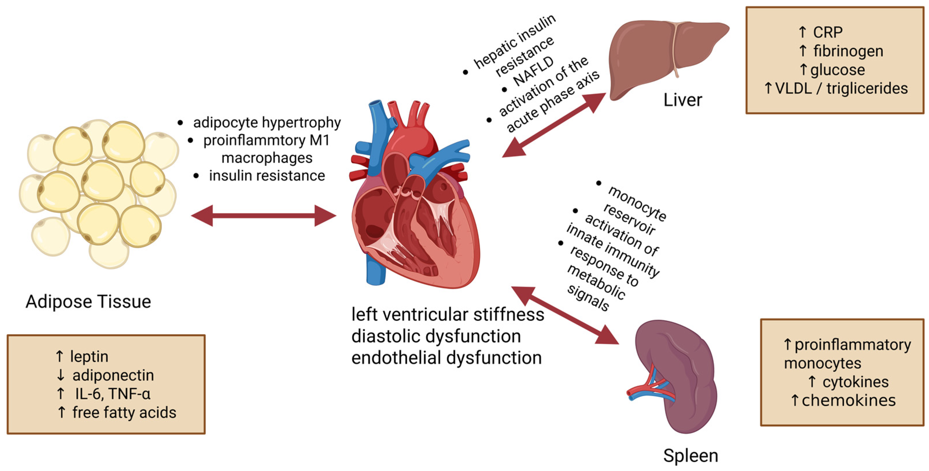
Collectively, the heart–adipose tissue–spleen–liver axis operates through crosstalk involving metabolic regulation and immune activation, IR, oxidative stress, neurohormonal activation, and chronic inflammation as common mechanisms linking these organs (Figure 1).

7. Nutritional Patterns and Their Impact on HFpEF

It is important to understand that in patients with HFpEF, metabolic diseases comprising hypertension, DM, and obesity often coexist. Paradoxically, these patients also frequently suffer from malnutrition, which leads to worsening of their clinical state. In these patients, upregulated catabolic processes combined with reduced appetite contribute to metabolic imbalance [100].

7.1. Western Diet and Ultra-Processed Foods

Ultra-processed foods are characterized by extensive industrial processing and typically contain high levels of sugars, sodium, and trans fats, while being low in fiber. Such foods play an important role in disrupting glycemic control and insulin signaling [101]. Moreover, this type of food is generally a poor source of anti-inflammatory bioactive compounds, such as polyphenols [102]. A pilot study conducted in Australia in 2025 reported that high consumption of ultra-processed food was associated with a 19% higher risk of CVD-related mortality [103]. Notably, ultra-processed foods may also increase ROS production and oxidative stress, thereby activating pro-inflammatory pathways [104].
Overall, high consumption of ultra-processed food has been associated with obesity, hypertension, dyslipidemia, and gastrointestinal disturbances, subsequently leading to CVD, the development of MetS, and higher risks of cancer incidence [105].

7.2. Mediterranean Diet

The Mediterranean diet shares many features with the DASH (Dietary Approaches to Stop Hypertension) diet, such as a low intake of red meat and processed foods and the extensive use of extra-virgin olive oil (EVOO). EVOO is a great source of anti-inflammatory, unsaturated fatty acids. This includes monounsaturated FAs (MUFAs) and polyunsaturated FAs (PUFAs), which positively influence glucose and lipid metabolism [100].
It has been shown that the Mediterranean diet is more effective than a low-fat diet in preventing CVD [106,107]. A recent study showed that omega-3 FAs improved health parameters, including 6 min walking test results and VO2 peak in patients with HFpEF [108]. On the other hand, omega-3 supplementation may increase the risk of atrial fibrillation [100].

7.3. Plant-Based and Vegetarian Diets

Plant-based diets include large amounts of dietary fiber, antioxidants (primarily polyphenols), and healthy fats. Fiber, found in plants, helps slow glucose absorption and improve overall glucose management. Many polyphenols, in addition to their antioxidant potential, exhibit a wide range of therapeutic activities, including anti-inflammatory, anti-cancer, anti-diabetic, antimicrobial, cardioprotective, and neuroprotective effects [109].
Stavitz and colleagues demonstrated that this dietary pattern benefits patients with MetS by increasing insulin sensitivity, suppressing inflammation, and enhancing cardiovascular health through improved lipid metabolic homeostasis [110]. According to the researchers, the best outcomes are achieved with a whole-food plant-based diet, as well as Mediterranean and vegan diets [111].
Plant fat sources, such as nuts and EVOO, which are rich in unsaturated fats, may improve lipid metabolism and lower TG and low-density lipoprotein (LDL) cholesterol levels. Fiber is also improving cholesterol excretion. In consequence, people who put on plant-based diets have a lower risk of developing hypertension, HF, and other CVD [112]. Cholesterol is found only in animal products, whereas plants contain phytosterols that compete with dietary cholesterol for intestinal absorption, thereby lowering blood cholesterol levels [113]. Moreover, dietary fiber has beneficial effects on gut microbiota, helping to maintain the gut barrier and reducing circulating pro-inflammatory agents [114]. Furthermore, fiber stimulates the secretion of YY peptide and glucagon-like peptide-1 (GLP-1), two hormones that signal satiety and help regulate appetite. Evidence suggests that well-balanced plant-based diets may be beneficial for the therapeutic management and prevention of HFpEF.

7.4. Low-Carbohydrate, Ketogenic, and High-Protein Diets

A ketogenic diet is characterized by very low carbohydrate intake (<50 g/day) and a high intake of fats and protein. It may be beneficial for people with T2DM and obesity, but it is not considered optimal for CV and liver health, as demonstrated in animal and human studies. For example, in animal studies, rodents had developed NAFLD and IR while being on a ketogenic diet [115]. The mechanism of this diet is to deplete the body’s glucose reserves, thereby stimulating ketone body production in the liver. Ketone bodies are later used as a backup energy supply for the nervous system [116].
In 2019, a clinical study reported that a two-year ketogenic diet in adults with T2DM improved glycemic control, reduced medication use, lowered body weight and abdominal fat, decreased blood pressure and TG, while elevating liver alanine aminotransferase and HDL and LDL cholesterol [117]. Overall, this diet may benefit patients with diabetes, but caution is advised in those with comorbidities, particularly CVD [117]. The influence of the ketogenic diet on lipid levels depends on its fat composition. In human studies, a ketogenic diet rich in unsaturated fats is associated with a better lipid profile, whereas a high intake of saturated fats is linked with worse blood lipid levels. There is insufficient evidence to determine the effect of the ketogenic diet on blood pressure. Available reports vary and are often contradictory [115].

7.5. Sodium Intake, Micronutrients, Vitamins, and the Genome-Based Nutrition

A low-sodium diet is often used in patients with CVD. Hospitalization commonly occurs after a period of high sodium intake [118]. On the other hand, excessively low salt intake can also reduce renal perfusion [100]. The strategy of salt restriction is associated with high readmission rates after cardiac events and even increased mortality, likely because it worsens existing dietary deficits rather than improving overall diet quality [119]. On the contrary, the SODIUM-HF study results did not show a significant difference in hospitalization and mortality rates between groups with reduced dietary sodium intake and those with normal sodium levels [120]. According to Weinberger, only about 25% of normotensive individuals are salt-sensitive, and in the hypertensive population, this percentage ranges from 30% to 50% [121].
Another approach to help those individuals suffering from hypertension is the DASH diet. It combines a low-sodium supply with increased consumption of fiber, protein, and healthy, unsaturated fats. Two clinical studies confirmed the validity of this dietary model: the GOURMET-HF trial [119] and a study by Hummel and colleagues [122].
It has been shown that a high intake of minerals such as potassium, magnesium, and calcium reduces blood pressure in patients with hypertension. Furthermore, high intake of mineral-rich fruits and vegetables has been shown to improve blood pressure and reduce the risk of CHD and stroke [123].
In a 2021 preclinical study, it was demonstrated that NAD+ precursors, which are derivatives of vitamin B3, were suggested to improve mitochondrial function and reduce inflammation in HFpEF [124]. In animal and cellular models, all-trans retinoic acid (ATRA) has been suggested to mitigate cardiac remodeling and prevent functional decline in heart failure by counteracting fibrosis, hypertrophy, oxidative stress, and impaired calcium handling [125].
Nowadays, the study of lipid transporter genes requires consideration of differential allele distributions. Hyperglyceridemia and hypercholesterolemia are associated with the ApoE e2 and e4 alleles [126]. Chia, pumpkin seeds, and amaranth are abundant in plant-based MUFA and PUFA and exert anti-inflammatory effects [127].
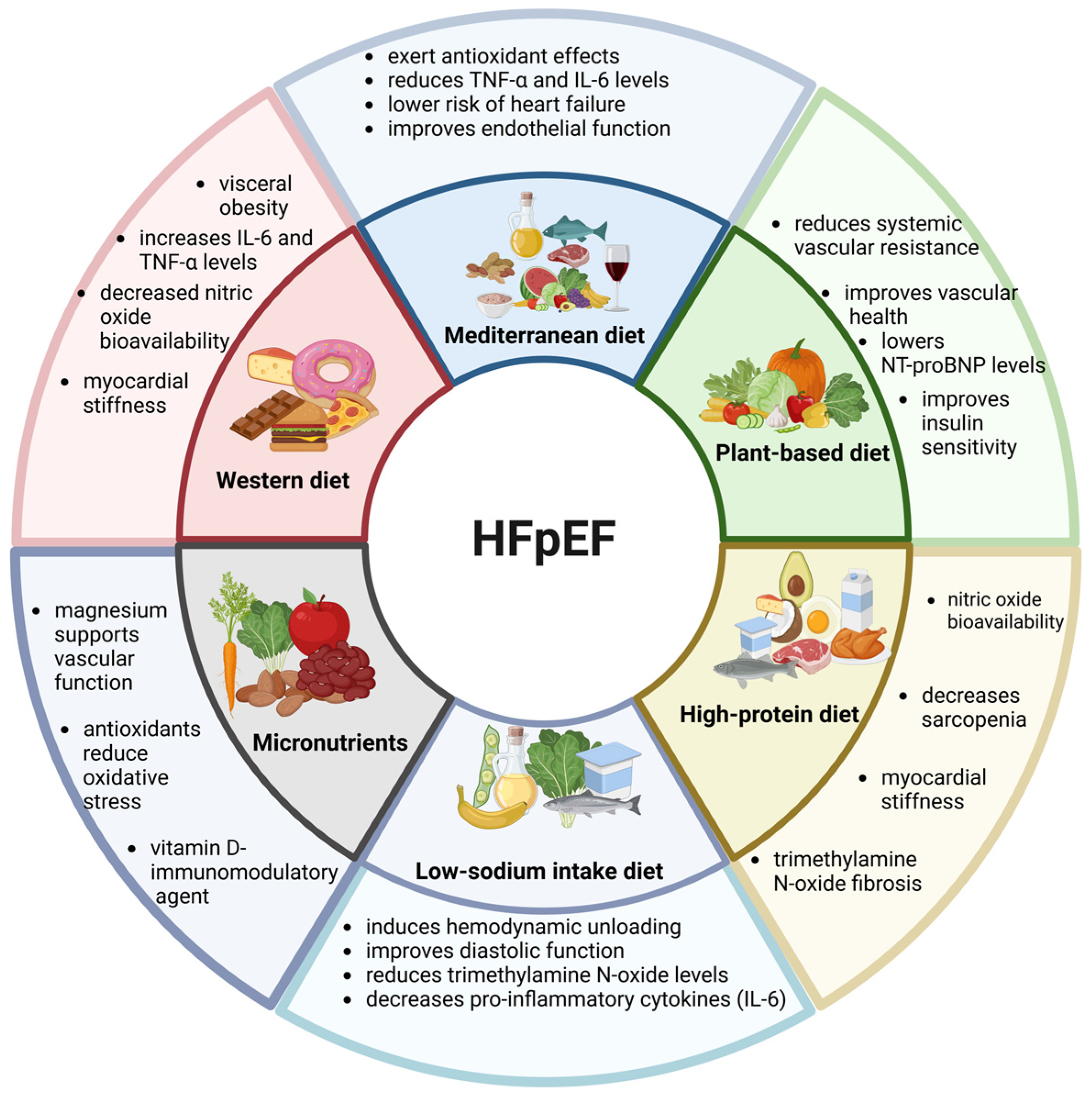
In summary, the Western diet promotes metabolic dysfunction, inflammation, and myocardial hypertrophy. In contrast, the Mediterranean and plant-based diets may improve metabolic control, reduce inflammation, and improve myocardial and endothelial function (Figure 2). In contrast, restrictive diets such as ketogenic regimens may benefit glycemic control but require caution in patients with CVD. Supplementing with minerals reduces blood pressure in patients with hypertension. It is important to individualize nutritional strategies in the management of HFpEF.

8. Dietary Modulation of Adipose Tissue, Liver, and Immune Function

Overweight and obesity contribute to the development of HFpEF and the progression of the illness [100]. The STEP-HFpEF trial showed that semaglutide improved HF-related symptoms, including physical limitations, exercise function, inflammation, and body weight. It reduced C-reactive protein (CRP) and N-terminal pro-B-type natriuretic peptide (NT-proBNP) [128]. Very alarming is the high percentage of patients regaining AT mass after the end of dietary or pharmacological intervention, which leads to the recurrence of the severity of HFpEF. It is even more dangerous, considering that HFpEF patients tend to develop sarcopenia alongside obesity, which further disrupts their metabolism [100].
In patients with obesity phenotype of HFpEF, especially those with excess VAT, sodium retention, and higher aldosterone levels have been observed. Moreover, enhanced leptin receptor signaling may activate the sympathetic nervous system and further mobilize the renin-angiotensin-aldosterone system, thereby additionally upregulating aldosterone secretion. Overactivity of neurohormones leads to increased plasma volume, which in turn causes heart remodeling and fibrosis. All the above mechanisms may promote adipocyte hypertrophy and increase adipogenesis, enhancing the positive feedback loop. The release of leptin and other pro-inflammatory adipokines induces systemic inflammation, thereby promoting fibrosis in the heart and other organs [129].
The role of the gut microbiota should be considered alongside dietary changes. It plays a key role in digestive processes, regulates neurohumoral signaling and gut barrier integrity, and alleviates systemic inflammation. A balanced microbiota prevents diseases, such as T2DM, CVD, obesity, and chronic inflammation [130].
The brain-gut-liver axis communicates via the vagus nerve. It involves the hypothalamus and pituitary gland and plays an important role in the treatment of MetS. Hormones involved in this axis, such as leptin, insulin, GLP-1, ghrelin, and cholecystokinin, regulate glucose homeostasis and hepatic lipid metabolism and reduce appetite. It is important to highlight that insulin is a protein that can partially cross the blood–brain barrier. IR leads to hyperglycemia, which worsens brain health and function, and is involved in glucose neurotoxicity and oxidative stress [130]. Disruption of the gut microbiota results in weakening of the gut barrier, with chemokines and cytokines released to the gut microenvironment that can modulate the function of various immune cells, thereby causing an inflammatory state [130].
The liver is recognized as a center of physiological responses within the gut-brain-liver axis [131]. The liver receives gut-derived signals via the portal veins. An imbalance in the microflora increases gut permeability, allowing toxins to enter the circulation and eading to gastrointestinal conditions such as bloating, constipation, or diarrhea, which further exacerbates systemic inflammation [132].
The Western diet promotes the development of Metabolic-Associated Fatty Liver Disease (MAFLD), with or without fibrosis [133]. In MAFLD, excessive TG accumulation in hepatocytes contributes to hepatic steatosis, chronic inflammation, hepatocyte degeneration, necrosis, and fibrosis [133]. Moreover, the Western diet may disrupt the BAs profile, thereby increasing ROS and further exacerbating inflammation.

9. Clinical Implications and Future Therapeutic Strategies

Newer antidiabetic therapies, particularly SGLT2 inhibitors and GLP-1 receptor agonists, have been shown to reduce EAT volume and inflammation while improving CV outcomes, including HFpEF. On the other hand, therapies such as sulfonylureas, thiazolidinediones, and insulin have been associated with increased adipogenesis, EAT accumulation, and a higher risk of HF. SGLT2 inhibitors reduce the risk of HF hospitalization and death, potentially through modulation of EAT, reduction in myocardial inflammation and fibrosis, and improvement of microvascular and hemodynamic function [134,135]. Reduction in EAT accumulation is likely related to decreased overall adiposity and may be partially mediated by the glucose-lowering effects of SGLT2 inhibitors and GLP-1 receptor agonists. A growing body of evidence suggests that the CV effect of GLP-1 and SGLT2 inhibitors may differ [40]. Unlike GLP-1 receptor agonists, which generally show a neutral effect on HF outcomes, SGLT2 inhibitors have consistently been associated with a reduced risk of HF hospitalization.
A growing body of evidence highlights the impact of personalized nutrition in the treatment of HFpEF phenotypes, emphasizing the combination of dietary interventions with standard pharmacotherapy to improve management. As shown in Table 1, each HFpEF phenotype—from cardiometabolic/obesity T2DM to atrial fibrillation, hypertension, ischemia (Coronary Artery Disease (CAD) with Coronary Microvascular Dysfunction (CMD)), chronotropic incompetence, right ventricular/pulmonary and valvular hypertension—has distinct pathophysiological mechanisms and clinical features. Personalized nutritional strategies, such as diets rich in unsaturated fats (MUFA/PUFA), a Mediterranean diet enriched with omega-3 fatty acids, the DASH diet, and optimization of protein/micronutrient intake, can complement pharmacological therapies [128,136,137]. It is hypothesized that this approach may improve the potential of future therapeutic strategies to enhance functional capacity, mitigate the adverse effects of cardiac remodeling, and improve quality of life in patients with HFpEF.
Anti-inflammatory approaches, including inhibition of pro-inflammatory cytokines (e.g., IL-1 and IL-6 pathways), colchicine therapy, statins, and modulation of oxidative stress and inflammasome activation, may also represent a promising strategy for reducing EAT-associated CV risk [138]. In addition, weight-loss interventions, including caloric restriction, exercise programs, or bariatric procedures, have been associated with increased EAT volume and inflammatory activity, leading to improved metabolic and cardiovascular outcomes. It seems that selective reduction in VAT and EAT may be more important than weight loss.
Collectively, these findings suggest that integrating personalized nutrition and exercise with pharmacological therapies, including SGLT2 inhibitors, GLP-1 receptor agonists, and anti-inflammatory agents, may offer synergistic benefits by reducing obesity, EAT, inflammation, and insulin resistance, thereby potentially improving CV outcomes.

10. Potential Biomarkers Linking Diet and Organ Crosstalk

HFpEF is often described as a systemic syndrome whose pathophysiology involves complex organ crosstalk between the heart, vascular system, kidneys, liver, adipose tissue, gut, and immune system [139]. Biomarkers that reflect these interactions may facilitate personalized nutritional approaches and help select appropriate therapies for specific HFpEF phenotypes. For example, inflammatory biomarkers such as IL-6 and CRP identify patients more likely to benefit from anti-inflammatory dietary patterns, including Mediterranean or plant-based diets [140]. Evidence from weight-loss–exercise interventions in obese HFpEF demonstrates that reductions in CRP correlate with improvements in peak VO2 and functional capacity, supporting the use of inflammatory markers to monitor dietary response [46].
Metabolic biomarkers—including leptin, adiponectin, glycated hemoglobin (HbA1c), ketone concentrations, and indices of IR—aid in stratifying obesity HFpEF phenotypes and predicting benefit from hypocaloric diets, low-glycemic dietary patterns, or unsaturated fatty acid–rich interventions [141,142,143]. In the UFA-Preserved2 trial, increases in circulating MUFA/PUFA biomarkers were associated with trends toward improved peak VO2, linking dietary lipid composition to peripheral oxygen utilization and microvascular energetics [144].
Endothelial biomarkers can be used to measure vessel interactions and diet-induced repair. The Mediterranean diet improves flow-mediated vasodilation (FMD), increases endothelial progenitor cells (EPCs), and reduces endothelial microparticles (EMPs), oxidative stress, and eat senescence—changes that directly correlate with improved arterial stiffness and hemodynamic in HFpEF with hypertension or atrial fibrillation [145]. Markers of the oxygen pathway (NO metabolites, nitrate/nitrite balance) have been used to evaluate nitrate supplementation; however, recent meta-analyses have showed no improvement in peak VO2 despite modest reductions in Systolic blood pressure (SBP), Diastolic blood pressure (DBP), Mean arterial pressure (MAP), and Systemic vascular resistance (SVR) in HFpEF, limiting their utility as predictive biomarkers [146].
Cardiac remodeling biomarkers, particularly NT-proBNP and galectin-3, provide insight into ventricular–vascular coupling and fibrosis [147]. Although NT-proBNP is often suppressed in obesity, omega-3 fatty acid supplementation has been shown to reduce NT-proBNP levels and improve diastolic parameters in HF, suggesting potential relevance for HFpEF [148].
Gut microbiome-derived metabolites, particularly trimethylamine N-oxide (TMAO) and short-chain fatty acids (SCFAs), link dietary quality to endothelial dysfunction and fibrosis, representing targets for dietary modulation or pre-/probiotic therapy [149].
The above biomarkers support integrating dietary interventions with pharmacological management to target phenotype-specific mechanisms in HFpEF.

11. Future Directions and Research Gaps

Despite progression in the understanding of HFpEF pathophysiology, there is no single diagnostic biomarker to definitively identify this disease. Natriuretic peptides, including NT-proBNP, remain the most widely used biomarkers in clinical practice. Consequently, there is a growing need to identify new biomarkers of HFpEF that better reflect the complex metabolic, inflammatory, and microvascular mechanisms. A recent study suggests the potential role of soluble suppression of tumorigenicity-2, galectin-3, growth differentiation factor-15, and adipokines (adiponectin, leptin) as markers of myocardial remodeling, inflammation, and adipose tissue dysfunction [150]. However, most of these biomarkers require validation in large prospective cohorts.
The use of dietary biomolecules in HFpEF therapy is promising, but several gaps remain. Future research should integrate proteomics, metabolomics, and metagenomics to identify precise “nutritypes” corresponding to HFpEF phenotype across different populations. Nevertheless, important knowledge gaps remain regarding the reproducibility, longitudinal stability, and clinical applicability of these biomarkers.
Recently, attention has also been focused on biomarkers reflecting interactions between the gut microbiome and CV system. The latest research has identified several potential biomarkers, including gut-derived metabolites (TMAO, SCFAs) that have been associated with systemic inflammation, endothelial dysfunction, myocardial fibrosis, and adverse cardiovascular outcomes. Similarly, circulating EPCs may provide insight into vascular repair capacity and microvascular dysfunction, both recognized contributors to HFpEF pathogenesis. It seems that validating these in large-scale and longitudinal dietary intervention trials will be important.
Another important area of investigation concerns the impact of dietary fatty acids (specifically MUFA and PUFA) on mitochondrial function and oxygen utilization in CVD; however, their mechanistic role in HFpEF remains incompletely understood.
Overall, the complex interplay between diet, adipose tissue biology, gut microbiota, systemic inflammation, and organ crosstalk highlights the need for a systems-biology approach to HFpEF. Combining biomarker discovery with phenotype-specific nutritional and pharmacological interventions may facilitate the development of precision medicine strategies to the specific clinical phenotypes of patients with HFpEF. At present, however, biomarkers in routine clinical practice remain limited by methodological heterogeneity, lack of standardized thresholds, and insufficient prospective validation.

12. Conclusions

HFpEF is a multifaceted systemic syndrome that necessitates precision nutrition strategies corresponding to specific phenotypes. The complex crosstalk between the heart, adipose tissue, liver, spleen, and the gut microbiome appears to involve multidimensional biomolecules rather than a single indicator. Different categories of biomarkers associated with inflammatory, metabolic, or endothelial disorders can be used to classify HFpEF into specific phenotypes (e.g., obese vs. aging). This will help target specific dietary interventions to the connection of comorbidities. Evidence suggests that biomarkers such as CRP, HbA1c, and circulating FAs profiles may also provide insight into the biological efficacy of nutritional interventions. Ultimately, integrating nutritional science, CV medicine, and research on organ crosstalk will be important for future HFpEF management.

Author Contributions

Conceptualization, M.K.; writing—original draft preparation, M.K., A.L., N.S., and D.S.; writing—review and editing, M.K., A.L., N.S., D.S., and W.B.; visualization, M.K.; supervision, M.K. All authors have read and agreed to the published version of the manuscript.

Funding

This research was funded by the National Science Centre Grant Number 2020/39/D/NZ5/02514, and CMKP grants No. 506-1-031-02-26.

Institutional Review Board Statement

Not applicable.

Informed Consent Statement

Not applicable.

Data Availability Statement

No new data were created or analyzed in this study. Data sharing is not applicable to this article.

Acknowledgments

During the preparation of this manuscript, the author(s) used ChatGPT (versions GPT-4-turbo and GPT-4o) for the purposes of enhancing the readability and language of this work. The authors have reviewed and edited the output and take full responsibility for the content of this publication.

Conflicts of Interest

The authors declare no conflicts of interest.

Abbreviations

The following abbreviations are used in this manuscript:
6MWD6-Minute walk distance
ACEiAngiotensin-converting enzyme inhibitors
ACSAcute coronary syndrome
ARBAngiotensin receptor blockers
ATAdipose tissue
ATRAAll-trans retinoic acid
BAsBile acids
CADCoronary artery disease
CIChronotropic incompetence
CIRKOMouse model of cardiomyocyte deletion of insulin receptors
CHDCoronary heart disease
CMDCoronary microvascular dysfunction
COPDChronic obstructive pulmonary disease
CRPC-reactive protein
CVCardiovascular
CVDCardiovascular disease
CXCR4C-X-C Chemokine receptor type 4
DASH dietDietary approaches to stop hypertension
DBPDiastolic blood pressure
DMDiabetes mellitus
EATEpicardial adipose tissue
ECMExtracellular matrix
EMPsReduces endothelial microparticles
EPCsIncreases endothelial progenitor cells
ESCThe European society of cardiology
EVOOExtra-virgin olive oil
FAsFatty acids
FBF Forearm blood flow
FFAsFree fatty acids
FGF-21Fibroblast growth factor-21
FMDFlow-mediated vasodilation
FMRFunctional mitral regurgitation
FTRFunctional tricuspid regurgitation
FXRFarnesoid X receptor
GLP-1Glucagon-like peptide-1
HbA1cGlycated hemoglobin
HCDHypocaloric diet
HDLHigh-density lipoprotein
HFHeart failure
HFpEFHeart failure with preserved ejection fraction
HFrEFHeart failure with reduced ejection fraction
HOCMHypertrophic obstructive cardiomyopathy
HSCHematopoietic stem cell
HYVETHypertension in the very elderly trial
IL-1βInterleukin-1β
IL-6Interleukins-6
IL-10Interleukin-10
IL-12Interleukins-12
IL-23Interleukin-23
IL-33Interleukin-33
IRInsulin resistance
JNKc-Jun N-terminal kinases
KCCQ-CSSKansas City cardiomyopathy questionnaire—clinical summary score
LDLLow-density lipoprotein
LVLeft ventricle
LVEFLeft ventricular ejection fraction
MAFLDMetabolic-associated fatty liver disease
MAPMean arterial pressure
MCP-1Monocyte chemoattractant protein-1
MetSMetabolic syndrome
MUFA Monounsaturated fatty acids
MZMarginal zone
NAFLDNon-alcoholic fatty liver disease
NF-κBNuclear factor kappa-light-chain-enhancer of activated B cells
NONitric oxide
NPsNatriuretic peptides
NT-proBNPN-terminal pro-B-type natriuretic peptide
PALSPeriarteriolar lymphoid sheaths
PATPericardial adipose tissue
PDE5-IPhosphodiesterase-5 Inhibitors
PHPulmonary hypertension
PKGProtein kinase G
PUFAPolyunsaturated fatty acids
ROSReactive oxygen species
RPRed pulp
QoLQuality of life
SATSubcutaneous adipose tissue
SBPSystolic blood pressure
SCFAsShort-chain fatty acids
SGLT2Sodium-glucose co-transporter 2
STEP-HFpEFAgonists semaglutide treatment effect in people with obesity and HFpEF
STEP-HFpEF-DMSemaglutide treatment effect in people with obesity and HFpEF and diabetes mellitus
SVRSystemic vascular resistance
T2DMType 2 diabetes mellitus
TGTriglycerides
TGR5Takeda G protein-coupled receptor 5
TNF-αTumor necrosis factor-alpha
TMAOTrimethylamine N-oxide
UFAUnsaturated fatty acids
WPWhite pulp
VATVisceral adipose tissue
VCAM-1Vascular cell adhesion molecule-1

References

  1. Campbell, P.; Rutten, F.H.; Lee, M.M.; Hawkins, N.M.; Petrie, M.C. Heart Failure with Preserved Ejection Fraction: Everything the Clinician Needs to Know. Lancet 2024, 403, 1083–1092. [Google Scholar] [CrossRef]
  2. Miranda, J.J.; Barrientos-Gutiérrez, T.; Corvalan, C.; Hyder, A.A.; Lazo-Porras, M.; Oni, T.; Wells, J.C.K. Understanding the Rise of Cardiometabolic Diseases in Low- and Middle-Income Countries. Nat. Med. 2019, 25, 1667–1679. [Google Scholar] [CrossRef]
  3. Lourenço, A.P.; Leite-Moreira, A.F.; Balligand, J.-L.; Bauersachs, J.; Dawson, D.; de Boer, R.A.; de Windt, L.J.; Falcão-Pires, I.; Fontes-Carvalho, R.; Franz, S.; et al. An Integrative Translational Approach to Study Heart Failure with Preserved Ejection Fraction: A Position Paper from the Working Group on Myocardial Function of the European Society of Cardiology. Eur. J. Heart Fail. 2018, 20, 216–227. [Google Scholar] [CrossRef] [PubMed]
  4. Tan, Y.T.; Wenzelburger, F.; Lee, E.; Heatlie, G.; Leyva, F.; Patel, K.; Frenneaux, M.; Sanderson, J.E. The Pathophysiology of Heart Failure with Normal Ejection Fraction: Exercise Echocardiography Reveals Complex Abnormalities of Both Systolic and Diastolic Ventricular Function Involving Torsion, Untwist, and Longitudinal Motion. J. Am. Coll. Cardiol. 2009, 54, 36–46. [Google Scholar] [CrossRef]
  5. Rosch, S.; Kresoja, K.-P.; Besler, C.; Fengler, K.; Schöber, A.R.; von Roeder, M.; Lücke, C.; Gutberlet, M.; Klingel, K.; Thiele, H.; et al. Characteristics of Heart Failure with Preserved Ejection Fraction Across the Range of Left Ventricular Ejection Fraction. Circulation 2022, 146, 506–518. [Google Scholar] [CrossRef]
  6. Pecchia, B.; Samuel, R.; Shah, V.; Newman, E.; Gibson, G.T. Mechanisms of Exercise Intolerance in Heart Failure with Preserved Ejection Fraction (HFpEF). Heart Fail. Rev. 2025, 30, 777–789. [Google Scholar] [CrossRef]
  7. McDonagh, T.A.; Metra, M.; Adamo, M.; Gardner, R.S.; Baumbach, A.; Böhm, M.; Burri, H.; Butler, J.; Čelutkienė, J.; Chioncel, O.; et al. 2021 ESC Guidelines for the Diagnosis and Treatment of Acute and Chronic Heart Failure. Eur. Heart J. 2021, 42, 3599–3726. [Google Scholar] [CrossRef]
  8. Pieske, B.; Tschöpe, C.; de Boer, R.A.; Fraser, A.G.; Anker, S.D.; Donal, E.; Edelmann, F.; Fu, M.; Guazzi, M.; Lam, C.S.P.; et al. How to Diagnose Heart Failure with Preserved Ejection Fraction: The HFA-PEFF Diagnostic Algorithm: A Consensus Recommendation from the Heart Failure Association (HFA) of the European Society of Cardiology (ESC). Eur. Heart J. 2019, 40, 3297–3317. [Google Scholar] [CrossRef] [PubMed]
  9. Prausmüller, S.; Weidenhammer, A.; Heitzinger, G.; Spinka, G.; Goliasch, G.; Arfsten, H.; Abdel Mawgoud, R.; Gabler, C.; Strunk, G.; Hengstenberg, C.; et al. Obesity in Heart Failure with Preserved Ejection Fraction with and without Diabetes: Risk Factor or Innocent Bystander? Eur. J. Prev. Cardiol. 2023, 30, 1247–1254. [Google Scholar] [CrossRef] [PubMed]
  10. Borlaug, B.A.; Sharma, K.; Shah, S.J.; Ho, J.E. Heart Failure with Preserved Ejection Fraction: JACC Scientific Statement. J. Am. Coll. Cardiol. 2023, 81, 1810–1834. [Google Scholar] [CrossRef]
  11. Girona, J.; Soler, O.; Samino, S.; Junza, A.; Martínez-Micaelo, N.; García-Altares, M.; Ràfols, P.; Esteban, Y.; Yanes, O.; Correig, X.; et al. Lipidomics Reveals Myocardial Lipid Composition in a Murine Model of Insulin Resistance Induced by a High-Fat Diet. Int. J. Mol. Sci. 2024, 25, 2702. [Google Scholar] [CrossRef]
  12. Geng, J.; Zhang, X.; Guo, Y.; Wen, H.; Guo, D.; Liang, Q.; Pu, S.; Wang, Y.; Liu, M.; Li, Z.; et al. Moderate-Intensity Interval Exercise Exacerbates Cardiac Lipotoxicity in High-Fat, High-Calories Diet-Fed Mice. Nat. Commun. 2025, 16, 613. [Google Scholar] [CrossRef]
  13. Hulsmans, M.; Sager, H.B.; Roh, J.D.; Valero-Muñoz, M.; Houstis, N.E.; Iwamoto, Y.; Sun, Y.; Wilson, R.M.; Wojtkiewicz, G.; Tricot, B.; et al. Cardiac Macrophages Promote Diastolic Dysfunction. J. Exp. Med. 2018, 215, 423–440. [Google Scholar] [CrossRef]
  14. Paulus, W.J.; Tschöpe, C. A Novel Paradigm for Heart Failure with Preserved Ejection Fraction: Comorbidities Drive Myocardial Dysfunction and Remodeling through Coronary Microvascular Endothelial Inflammation. J. Am. Coll. Cardiol. 2013, 62, 263–271. [Google Scholar] [CrossRef]
  15. Anker, S.D.; Usman, M.S.; Anker, M.S.; Butler, J.; Böhm, M.; Abraham, W.T.; Adamo, M.; Chopra, V.K.; Cicoira, M.; Cosentino, F.; et al. Patient Phenotype Profiling in Heart Failure with Preserved Ejection Fraction to Guide Therapeutic Decision Making. A Scientific Statement of the Heart Failure Association, the European Heart Rhythm Association of the European Society of Cardiology, and the European Society of Hypertension. Eur. J. Heart Fail. 2023, 25, 936–955. [Google Scholar] [CrossRef]
  16. Zheng, W.; Qi, Q.; Li, J.; He, C.; Fan, H. Heart Failure with Preserved Ejection Fraction and Obesity: Emerging Metabolic Therapeutic Strategies. Diabetol. Metab. Syndr. 2025, 17, 336. [Google Scholar] [CrossRef] [PubMed]
  17. El Hadj Othmane, T.; El Hadj Othmane, O.; Nizar, H. Obesity-Related Phenotype of Heart Failure with Preserved Ejection Fraction: A Comprehensive Review. Cureus 2025, 17, e81512. [Google Scholar] [CrossRef] [PubMed]
  18. Clemente-Suárez, V.J.; Redondo-Flórez, L.; Beltrán-Velasco, A.I.; Martín-Rodríguez, A.; Martínez-Guardado, I.; Navarro-Jiménez, E.; Laborde-Cárdenas, C.C.; Tornero-Aguilera, J.F. The Role of Adipokines in Health and Disease. Biomedicines 2023, 11, 1290. [Google Scholar] [CrossRef]
  19. Packer, M. The Adipokine Hypothesis of Heart Failure with a Preserved Ejection Fraction: A Novel Framework to Explain Pathogenesis and Guide Treatment. J. Am. Coll. Cardiol. 2025, 86, 1269–1373. [Google Scholar] [CrossRef] [PubMed]
  20. Matilainen, J.; Berg, V.; Vaittinen, M.; Impola, U.; Mustonen, A.-M.; Männistö, V.; Malinen, M.; Luukkonen, V.; Rosso, N.; Turunen, T.; et al. Increased Secretion of Adipocyte-Derived Extracellular Vesicles Is Associated with Adipose Tissue Inflammation and the Mobilization of Excess Lipid in Human Obesity. J. Transl. Med. 2024, 22, 623. [Google Scholar] [CrossRef]
  21. Packer, M.; Kitzman, D.W. Obesity-Related Heart Failure with a Preserved Ejection Fraction: The Mechanistic Rationale for Combining Inhibitors of Aldosterone, Neprilysin, and Sodium-Glucose Cotransporter-2. JACC Heart Fail. 2018, 6, 633–639. [Google Scholar] [CrossRef]
  22. Góralska, M.; Majewska-Szczepanik, M.; Szczepanik, M. Immunological mechanisms involved in obesity and their role in metabolic syndrome. Postep. Hig. Med. Dosw. 2015, 69, 1384–1404. [Google Scholar]
  23. Kawai, T.; Autieri, M.V.; Scalia, R. Adipose Tissue Inflammation and Metabolic Dysfunction in Obesity. Am. J. Physiol. Cell Physiol. 2021, 320, C375–C391. [Google Scholar] [CrossRef]
  24. Kahn, D.; Macias, E.; Zarini, S.; Garfield, A.; Zemski Berry, K.; MacLean, P.; Gerszten, R.E.; Libby, A.; Solt, C.; Schoen, J.; et al. Exploring Visceral and Subcutaneous Adipose Tissue Secretomes in Human Obesity: Implications for Metabolic Disease. Endocrinology 2022, 163, bqac140. [Google Scholar] [CrossRef]
  25. Sato, F.; Maeda, N.; Yamada, T.; Namazui, H.; Fukuda, S.; Natsukawa, T.; Nagao, H.; Murai, J.; Masuda, S.; Tanaka, Y.; et al. Association of Epicardial, Visceral, and Subcutaneous Fat with Cardiometabolic Diseases. Circ. J. 2018, 82, 502–508. [Google Scholar] [CrossRef] [PubMed]
  26. Karlsson, T.; Rask-Andersen, M.; Pan, G.; Höglund, J.; Wadelius, C.; Ek, W.E.; Johansson, Å. Contribution of Genetics to Visceral Adiposity and Its Relation to Cardiovascular and Metabolic Disease. Nat. Med. 2019, 25, 1390–1395. [Google Scholar] [CrossRef]
  27. Chen, P.; Hou, X.; Hu, G.; Wei, L.; Jiao, L.; Wang, H.; Chen, S.; Wu, J.; Bao, Y.; Jia, W. Abdominal Subcutaneous Adipose Tissue: A Favorable Adipose Depot for Diabetes? Cardiovasc. Diabetol. 2018, 17, 93. [Google Scholar] [CrossRef] [PubMed]
  28. Porter, S.A.; Massaro, J.M.; Hoffmann, U.; Vasan, R.S.; O’Donnel, C.J.; Fox, C.S. Abdominal Subcutaneous Adipose Tissue: A Protective Fat Depot? Diabetes Care 2009, 32, 1068–1075. [Google Scholar] [CrossRef] [PubMed]
  29. Narumi, H.; Yoshida, K.; Hashimoto, N.; Umehara, I.; Funabashi, N.; Yoshida, S.; Komuro, I. Increased Subcutaneous Fat Accumulation Has a Protective Role against Subclinical Atherosclerosis in Asymptomatic Subjects Undergoing General Health Screening. Int. J. Cardiol. 2009, 135, 150–155. [Google Scholar] [CrossRef]
  30. Emamat, H.; Jamshidi, A.; Farhadi, A.; Ghalandari, H.; Ghasemi, M.; Tangestani, H. The Association between the Visceral to Subcutaneous Abdominal Fat Ratio and the Risk of Cardiovascular Diseases: A Systematic Review. BMC Public Health 2024, 24, 1827. [Google Scholar] [CrossRef]
  31. Riehle, C.; Abel, E.D. Insulin Signaling and Heart Failure. Circ. Res. 2016, 118, 1151–1169. [Google Scholar] [CrossRef]
  32. Witteles, R.M.; Fowler, M.B. Insulin-Resistant Cardiomyopathy Clinical Evidence, Mechanisms, and Treatment Options. J. Am. Coll. Cardiol. 2008, 51, 93–102. [Google Scholar] [CrossRef]
  33. Zhu, R.; Wang, W.; Gao, Y.; Wang, J.; Li, B.; Cheng, Z.; Ji, C.; Gu, H.; Yuan, X.; Yang, S.; et al. Insulin Resistance Aggravates Myocardial Fibrosis in Non-Diabetic Hypertensive Patients by Altering the Function of Epicardial Adipose Tissue: A Cardiac Magnetic Resonance Study. Diabetol. Metab. Syndr. 2025, 17, 133. [Google Scholar] [CrossRef]
  34. Lakka, H.-M.; Laaksonen, D.E.; Lakka, T.A.; Niskanen, L.K.; Kumpusalo, E.; Tuomilehto, J.; Salonen, J.T. The Metabolic Syndrome and Total and Cardiovascular Disease Mortality in Middle-Aged Men. JAMA 2002, 288, 2709–2716. [Google Scholar] [CrossRef]
  35. Hu, P.; Zhang, D.; Swenson, L.; Chakrabarti, G.; Abel, E.D.; Litwin, S.E. Minimally Invasive Aortic Banding in Mice: Effects of Altered Cardiomyocyte Insulin Signaling during Pressure Overload. Am. J. Physiol. Heart Circ. Physiol. 2003, 285, H1261–H1269. [Google Scholar] [CrossRef]
  36. Abel, E.D. Insulin Signaling in the Heart. Am. J. Physiol. Endocrinol. Metab. 2021, 321, E130–E145. [Google Scholar] [CrossRef]
  37. Iacobellis, G. Epicardial Adipose Tissue in Contemporary Cardiology. Nat. Rev. Cardiol. 2022, 19, 593–606. [Google Scholar] [CrossRef]
  38. Wong, C.X.; Ganesan, A.N.; Selvanayagam, J.B. Epicardial Fat and Atrial Fibrillation: Current Evidence, Potential Mechanisms, Clinical Implications, and Future Directions. Eur. Heart J. 2017, 38, 1294–1302. [Google Scholar] [CrossRef]
  39. Paterek, A.; Załęska-Kocięcka, M.; Wojdyńska, Z.; Kalisz, M.; Litwiniuk, A.; Leszek, P.; Mączewski, M. Epicardial Fat in Heart Failure-Friend, Foe, or Bystander. Obes. Rev. 2024, 25, e13820. [Google Scholar] [CrossRef]
  40. Goldman, S.A.; Requena-Ibanez, J.A.; Devesa, A.; Santos-Gallego, C.G.; Badimon, J.J.; Fuster, V. Uncovering the Role of Epicardial Adipose Tissue in Heart Failure with Preserved Ejection Fraction. JACC Adv. 2023, 2, 100657. [Google Scholar] [CrossRef]
  41. Iacobellis, G. Epicardial and Pericardial Fat: Close, but Very Different. Obesity 2009, 17, 625-627; author reply 626–627. [Google Scholar] [CrossRef]
  42. Hirata, Y.; Yamada, H.; Sata, M. Epicardial Fat and Pericardial Fat Surrounding the Heart Have Different Characteristics. Circ. J. 2018, 82, 2475–2476. [Google Scholar] [CrossRef]
  43. Del Buono, M.G.; Montone, R.A.; Camilli, M.; Carbone, S.; Narula, J.; Lavie, C.J.; Niccoli, G.; Crea, F. Coronary Microvascular Dysfunction Across the Spectrum of Cardiovascular Diseases: JACC State-of-the-Art Review. J. Am. Coll. Cardiol. 2021, 78, 1352–1371. [Google Scholar] [CrossRef]
  44. Franssen, C.; Chen, S.; Unger, A.; Korkmaz, H.I.; De Keulenaer, G.W.; Tschöpe, C.; Leite-Moreira, A.F.; Musters, R.; Niessen, H.W.M.; Linke, W.A.; et al. Myocardial Microvascular Inflammatory Endothelial Activation in Heart Failure with Preserved Ejection Fraction. JACC Heart Fail. 2016, 4, 312–324. [Google Scholar] [CrossRef]
  45. Shah, S.J.; Lam, C.S.P.; Svedlund, S.; Saraste, A.; Hage, C.; Tan, R.-S.; Beussink-Nelson, L.; Ljung Faxén, U.; Fermer, M.L.; Broberg, M.A.; et al. Prevalence and Correlates of Coronary Microvascular Dysfunction in Heart Failure with Preserved Ejection Fraction: PROMIS-HFpEF. Eur. Heart J. 2018, 39, 3439–3450. [Google Scholar] [CrossRef] [PubMed]
  46. Kitzman, D.W.; Brubaker, P.; Morgan, T.; Haykowsky, M.; Hundley, G.; Kraus, W.E.; Eggebeen, J.; Nicklas, B.J. Effect of Caloric Restriction or Aerobic Exercise Training on Peak Oxygen Consumption and Quality of Life in Obese Older Patients with Heart Failure with Preserved Ejection Fraction: A Randomized Clinical Trial. JAMA 2016, 315, 36–46. [Google Scholar] [CrossRef]
  47. Mohammed, S.F.; Hussain, S.; Mirzoyev, S.A.; Edwards, W.D.; Maleszewski, J.J.; Redfield, M.M. Coronary Microvascular Rarefaction and Myocardial Fibrosis in Heart Failure with Preserved Ejection Fraction. Circulation 2015, 131, 550–559. [Google Scholar] [CrossRef]
  48. Nakanishi, K.; Fukuda, S.; Tanaka, A.; Otsuka, K.; Taguchi, H.; Shimada, K. Relationships Between Periventricular Epicardial Adipose Tissue Accumulation, Coronary Microcirculation, and Left Ventricular Diastolic Dysfunction. Can. J. Cardiol. 2017, 33, 1489–1497. [Google Scholar] [CrossRef] [PubMed]
  49. Masi, S.; Rizzoni, D.; Taddei, S.; Widmer, R.J.; Montezano, A.C.; Lüscher, T.F.; Schiffrin, E.L.; Touyz, R.M.; Paneni, F.; Lerman, A.; et al. Assessment and Pathophysiology of Microvascular Disease: Recent Progress and Clinical Implications. Eur. Heart J. 2021, 42, 2590–2604. [Google Scholar] [CrossRef]
  50. Jin, X.; Qiu, T.; Li, L.; Yu, R.; Chen, X.; Li, C.; Proud, C.G.; Jiang, T. Pathophysiology of Obesity and Its Associated Diseases. Acta Pharm. Sin. B 2023, 13, 2403–2424. [Google Scholar] [CrossRef] [PubMed]
  51. Trivedi, P.S.; Barouch, L.A. Cardiomyocyte Apoptosis in Animal Models of Obesity. Curr. Hypertens. Rep. 2008, 10, 454–460. [Google Scholar] [CrossRef]
  52. Obokata, M.; Reddy, Y.N.V.; Pislaru, S.V.; Melenovsky, V.; Borlaug, B.A. Evidence Supporting the Existence of a Distinct Obese Phenotype of Heart Failure with Preserved Ejection Fraction. Circulation 2017, 136, 6–19. [Google Scholar] [CrossRef]
  53. Kruszewska, J.; Cudnoch-Jedrzejewska, A.; Czarzasta, K. Remodeling and Fibrosis of the Cardiac Muscle in the Course of Obesity-Pathogenesis and Involvement of the Extracellular Matrix. Int. J. Mol. Sci. 2022, 23, 4195. [Google Scholar] [CrossRef]
  54. Wang, Y.; Anesi, J.C.; Panicker, I.S.; Cook, D.; Bista, P.; Fang, Y.; Oqueli, E. Neuroimmune Interactions and Their Role in Immune Cell Trafficking in Cardiovascular Diseases and Cancer. Int. J. Mol. Sci. 2025, 26, 2553. [Google Scholar] [CrossRef]
  55. Swirski, F.K.; Nahrendorf, M.; Etzrodt, M.; Wildgruber, M.; Cortez-Retamozo, V.; Panizzi, P.; Figueiredo, J.-L.; Kohler, R.H.; Chudnovskiy, A.; Waterman, P.; et al. Identification of Splenic Reservoir Monocytes and Their Deployment to Inflammatory Sites. Science 2009, 325, 612–616. [Google Scholar] [CrossRef] [PubMed]
  56. den Haan, J.M.M.; Kraal, G. Innate Immune Functions of Macrophage Subpopulations in the Spleen. J. Innate Immun. 2012, 4, 437–445. [Google Scholar] [CrossRef]
  57. Elmore, S.A. Enhanced Histopathology of the Spleen. Toxicol. Pathol. 2006, 34, 648–655. [Google Scholar] [CrossRef]
  58. Fernández-García, V.; González-Ramos, S.; Martín-Sanz, P.; Castrillo, A.; Boscá, L. Contribution of Extramedullary Hematopoiesis to Atherosclerosis. The Spleen as a Neglected Hub of Inflammatory Cells. Front. Immunol. 2020, 11, 586527. [Google Scholar] [CrossRef]
  59. Miyake, Y.; Asano, K.; Kaise, H.; Uemura, M.; Nakayama, M.; Tanaka, M. Critical Role of Macrophages in the Marginal Zone in the Suppression of Immune Responses to Apoptotic Cell-Associated Antigens. J. Clin. Investig. 2007, 117, 2268–2278. [Google Scholar] [CrossRef] [PubMed]
  60. Perrotta, S.; Carnevale, L.; Perrotta, M.; Pallante, F.; Mikołajczyk, T.P.; Fardella, V.; Migliaccio, A.; Fardella, S.; Nejat, S.; Kapelak, B.; et al. A Heart-Brain-Spleen Axis Controls Cardiac Remodeling to Hypertensive Stress. Immunity 2025, 58, 648–665.e7. [Google Scholar] [CrossRef] [PubMed]
  61. Mellak, S.; Ait-Oufella, H.; Esposito, B.; Loyer, X.; Poirier, M.; Tedder, T.F.; Tedgui, A.; Mallat, Z.; Potteaux, S. Angiotensin II Mobilizes Spleen Monocytes to Promote the Development of Abdominal Aortic Aneurysm in Apoe-/- Mice. Arterioscler. Thromb. Vasc. Biol. 2015, 35, 378–388. [Google Scholar] [CrossRef]
  62. Cortez-Retamozo, V.; Etzrodt, M.; Newton, A.; Ryan, R.; Pucci, F.; Sio, S.W.; Kuswanto, W.; Rauch, P.J.; Chudnovskiy, A.; Iwamoto, Y.; et al. Angiotensin II Drives the Production of Tumor-Promoting Macrophages. Immunity 2013, 38, 296–308. [Google Scholar] [CrossRef]
  63. Potteaux, S.; Ait-Oufella, H.; Mallat, Z. Role of Splenic Monocytes in Atherosclerosis. Curr. Opin. Lipidol. 2015, 26, 457–463. [Google Scholar] [CrossRef]
  64. Han, C.; Zhai, Y.; Wang, Y.; Peng, X.; Zhang, X.; Dai, B.; Leng, Y.; Zhang, Z.; Qi, S. Intravital Imaging of Splenic Classical Monocytes Modifying the Hepatic CX3CR1+ Cells Motility to Exacerbate Liver Fibrosis via Spleen-Liver Axis. Theranostics 2024, 14, 2210–2231. [Google Scholar] [CrossRef]
  65. Carnevale, D.; Perrotta, M.; Pallante, F.; Fardella, V.; Iacobucci, R.; Fardella, S.; Carnevale, L.; Carnevale, R.; De Lucia, M.; Cifelli, G.; et al. A Cholinergic-Sympathetic Pathway Primes Immunity in Hypertension and Mediates Brain-to-Spleen Communication. Nat. Commun. 2016, 7, 13035. [Google Scholar] [CrossRef]
  66. Wang, H.; Yu, M.; Ochani, M.; Amella, C.A.; Tanovic, M.; Susarla, S.; Li, J.H.; Wang, H.; Yang, H.; Ulloa, L.; et al. Nicotinic Acetylcholine Receptor Alpha7 Subunit Is an Essential Regulator of Inflammation. Nature 2003, 421, 384–388. [Google Scholar] [CrossRef]
  67. Sühs, K.-W.; Gudi, V.; Eckermann, N.; Fairless, R.; Pul, R.; Skripuletz, T.; Stangel, M. Cytokine Regulation by Modulation of the NMDA Receptor on Astrocytes. Neurosci. Lett. 2016, 629, 227–233. [Google Scholar] [CrossRef]
  68. Adamo, L.; Rocha-Resende, C.; Prabhu, S.D.; Mann, D.L. Reappraising the Role of Inflammation in Heart Failure. Nat. Rev. Cardiol. 2020, 17, 269–285. [Google Scholar] [CrossRef]
  69. Epelman, S.; Lavine, K.J.; Beaudin, A.E.; Sojka, D.K.; Carrero, J.A.; Calderon, B.; Brija, T.; Gautier, E.L.; Ivanov, S.; Satpathy, A.T.; et al. Embryonic and Adult-Derived Resident Cardiac Macrophages Are Maintained through Distinct Mechanisms at Steady State and during Inflammation. Immunity 2014, 40, 91–104. [Google Scholar] [CrossRef]
  70. Paulus, W.J.; Zile, M.R. From Systemic Inflammation to Myocardial Fibrosis: The Heart Failure with Preserved Ejection Fraction Paradigm Revisited. Circ. Res. 2021, 128, 1451–1467. [Google Scholar] [CrossRef]
  71. Patel, R.B.; Colangelo, L.A.; Reis, J.P.; Lima, J.A.C.; Shah, S.J.; Lloyd-Jones, D.M. Association of Longitudinal Trajectory of Albuminuria in Young Adulthood with Myocardial Structure and Function in Later Life: Coronary Artery Risk Development in Young Adults (CARDIA) Study. JAMA Cardiol. 2020, 5, 184–192. [Google Scholar] [CrossRef]
  72. Ferrell, M.; Wang, Z.; Anderson, J.T.; Li, X.S.; Witkowski, M.; DiDonato, J.A.; Hilser, J.R.; Hartiala, J.A.; Haghikia, A.; Cajka, T.; et al. A Terminal Metabolite of Niacin Promotes Vascular Inflammation and Contributes to Cardiovascular Disease Risk. Nat. Med. 2024, 30, 424–434. [Google Scholar] [CrossRef]
  73. Dutta, P.; Hoyer, F.F.; Grigoryeva, L.S.; Sager, H.B.; Leuschner, F.; Courties, G.; Borodovsky, A.; Novobrantseva, T.; Ruda, V.M.; Fitzgerald, K.; et al. Macrophages Retain Hematopoietic Stem Cells in the Spleen via VCAM-1. J. Exp. Med. 2015, 212, 497–512. [Google Scholar] [CrossRef]
  74. Filipp, M.; Ge, Z.-D.; DeBerge, M.; Lantz, C.; Glinton, K.; Gao, P.; Smolgovsky, S.; Dai, J.; Zhao, Y.-Y.; Yvan-Charvet, L.; et al. Myeloid Fatty Acid Metabolism Activates Neighboring Hematopoietic Stem Cells to Promote Heart Failure with Preserved Ejection Fraction. Circulation 2025, 151, 1451–1466. [Google Scholar] [CrossRef]
  75. Westerterp, M.; Gourion-Arsiquaud, S.; Murphy, A.J.; Shih, A.; Cremers, S.; Levine, R.L.; Tall, A.R.; Yvan-Charvet, L. Regulation of Hematopoietic Stem and Progenitor Cell Mobilization by Cholesterol Efflux Pathways. Cell Stem Cell 2012, 11, 195–206. [Google Scholar] [CrossRef]
  76. Kim, J.-W.; Kim, Y.-K.; Hwang, J.-A.; Yoon, H.-K.; Ko, Y.-H.; Han, C.; Lee, H.-J.; Ham, B.-J.; Lee, H.S. Plasma Levels of IL-23 and IL-17 before and after Antidepressant Treatment in Patients with Major Depressive Disorder. Psychiatry Investig. 2013, 10, 294–299. [Google Scholar] [CrossRef]
  77. Grune, J.; Bajpai, G.; Ocak, P.T.; Kaufmann, E.; Mentkowski, K.; Pabel, S.; Kumowski, N.; Pulous, F.E.; Tran, K.A.; Rohde, D.; et al. Virus-Induced Acute Respiratory Distress Syndrome Causes Cardiomyopathy Through Eliciting Inflammatory Responses in the Heart. Circulation 2024, 150, 49–61. [Google Scholar] [CrossRef]
  78. Zhang, N.; Ma, Q.; You, Y.; Xia, X.; Xie, C.; Huang, Y.; Wang, Z.; Ye, F.; Yu, Z.; Xie, X. CXCR4-Dependent Macrophage-to-Fibroblast Signaling Contributes to Cardiac Diastolic Dysfunction in Heart Failure with Preserved Ejection Fraction. Int. J. Biol. Sci. 2022, 18, 1271–1287. [Google Scholar] [CrossRef]
  79. Liang, W.; He, X.; Wu, D.; Xue, R.; Dong, B.; Owusu-Agyeman, M.; Zhao, J.; Cai, L.; You, Z.; Dong, Y.; et al. Prognostic Implication of Liver Function Tests in Heart Failure with Preserved Ejection Fraction Without Chronic Hepatic Diseases: Insight From TOPCAT Trial. Front. Cardiovasc. Med. 2021, 8, 618816. [Google Scholar] [CrossRef]
  80. Zhang, J.; Xu, M.; Chen, T.; Zhou, Y. Correlation Between Liver Stiffness and Diastolic Function, Left Ventricular Hypertrophy, and Right Cardiac Function in Patients with Ejection Fraction Preserved Heart Failure. Front. Cardiovasc. Med. 2021, 8, 748173. [Google Scholar] [CrossRef]
  81. Park, A.C.; Schilling, J.D. The Cardiohepatic Axis in Heart Failure. JACC Basic. Transl. Sci. 2025, 10, 101312. [Google Scholar] [CrossRef]
  82. Krings, G.; Can, B.; Ferrell, L. Aberrant Centrizonal Features in Chronic Hepatic Venous Outflow Obstruction: Centrilobular Mimicry of Portal-Based Disease. Am. J. Surg. Pathol. 2014, 38, 205–214. [Google Scholar] [CrossRef]
  83. Itier, R.; Guillaume, M.; Ricci, J.-E.; Roubille, F.; Delarche, N.; Picard, F.; Galinier, M.; Roncalli, J. Non-Alcoholic Fatty Liver Disease and Heart Failure with Preserved Ejection Fraction: From Pathophysiology to Practical Issues. ESC. Heart Fail. 2021, 8, 789–798. [Google Scholar] [CrossRef]
  84. Coombes, J.D.; Choi, S.S.; Swiderska-Syn, M.; Manka, P.; Reid, D.T.; Palma, E.; Briones-Orta, M.A.; Xie, G.; Younis, R.; Kitamura, N.; et al. Osteopontin Is a Proximal Effector of Leptin-Mediated Non-Alcoholic Steatohepatitis (NASH) Fibrosis. Biochim. Biophys. Acta 2016, 1862, 135–144. [Google Scholar] [CrossRef]
  85. Pejnovic, N.; Jeftic, I.; Jovicic, N.; Arsenijevic, N.; Lukic, M.L. Galectin-3 and IL-33/ST2 Axis Roles and Interplay in Diet-Induced Steatohepatitis. World J. Gastroenterol. 2016, 22, 9706–9717. [Google Scholar] [CrossRef]
  86. Wettersten, N.; Maisel, A.S. Biomarkers for Heart Failure: An Update for Practitioners of Internal Medicine. Am. J. Med. 2016, 129, 560–567. [Google Scholar] [CrossRef]
  87. Shouman, W.A.; Najmeddine, S.; Sinno, L.; Dib Nehme, R.; Ghawi, A.; Ziade, J.A.; Altara, R.; Amin, G.; Booz, G.W.; Zouein, F.A. Hepatokines and Their Role in Cardiohepatic Interactions in Heart Failure. Eur. J. Pharmacol. 2025, 992, 177356. [Google Scholar] [CrossRef]
  88. Chen, L.; Gao, M.; Ong, S.-B.; Gong, G. Functions of FGF21 and Its Role in Cardiac Hypertrophy. J. Adv. Res. 2026, 79, 475–489. [Google Scholar] [CrossRef]
  89. Xia, Y.; Xu, X.; Guo, Y.; Lin, C.; Xu, X.; Zhang, F.; Fan, M.; Qi, T.; Li, C.; Hu, G.; et al. Mesenchymal Stromal Cells Overexpressing Farnesoid X Receptor Exert Cardioprotective Effects Against Acute Ischemic Heart Injury by Binding Endogenous Bile Acids. Adv. Sci. 2022, 9, e2200431. [Google Scholar] [CrossRef]
  90. Vasavan, T.; Ferraro, E.; Ibrahim, E.; Dixon, P.; Gorelik, J.; Williamson, C. Heart and Bile Acids—Clinical Consequences of Altered Bile Acid Metabolism. Biochim. Biophys. Acta Mol. Basis Dis. 2018, 1864, 1345–1355. [Google Scholar] [CrossRef]
  91. Zhang, Y.; Wang, X.; Vales, C.; Lee, F.Y.; Lee, H.; Lusis, A.J.; Edwards, P.A. FXR Deficiency Causes Reduced Atherosclerosis in Ldlr-/- Mice. Arterioscler. Thromb. Vasc. Biol. 2006, 26, 2316–2321. [Google Scholar] [CrossRef]
  92. Neuschwander-Tetri, B.A.; Loomba, R.; Sanyal, A.J.; Lavine, J.E.; Van Natta, M.L.; Abdelmalek, M.F.; Chalasani, N.; Dasarathy, S.; Diehl, A.M.; Hameed, B.; et al. Farnesoid X Nuclear Receptor Ligand Obeticholic Acid for Non-Cirrhotic, Non-Alcoholic Steatohepatitis (FLINT): A Multicentre, Randomised, Placebo-Controlled Trial. Lancet 2015, 385, 956–965. [Google Scholar] [CrossRef] [PubMed]
  93. Desai, M.S.; Shabier, Z.; Taylor, M.; Lam, F.; Thevananther, S.; Kosters, A.; Karpen, S.J. Hypertrophic Cardiomyopathy and Dysregulation of Cardiac Energetics in a Mouse Model of Biliary Fibrosis. Hepatology 2010, 51, 2097–2107. [Google Scholar] [CrossRef]
  94. Frangogiannis, N.G. The Extracellular Matrix in Ischemic and Nonischemic Heart Failure. Circ. Res. 2019, 125, 117–146. [Google Scholar] [CrossRef] [PubMed]
  95. Zhou, Y.; Yan, H.; Zhu, Y.; Jiang, W.; Zhang, S. Multifactorial Mechanisms of Obesity-Related HFpEF: The Central Role of Epicardial Adipose Tissue and Therapeutic Perspectives. Front. Cardiovasc. Med. 2025, 12, 1701459. [Google Scholar] [CrossRef]
  96. Huh, J.Y.; Park, Y.J.; Ham, M.; Kim, J.B. Crosstalk between Adipocytes and Immune Cells in Adipose Tissue Inflammation and Metabolic Dysregulation in Obesity. Mol. Cells 2014, 37, 365–371. [Google Scholar] [CrossRef]
  97. Chiu, H.-C.; Kovacs, A.; Blanton, R.M.; Han, X.; Courtois, M.; Weinheimer, C.J.; Yamada, K.A.; Brunet, S.; Xu, H.; Nerbonne, J.M.; et al. Transgenic Expression of Fatty Acid Transport Protein 1 in the Heart Causes Lipotoxic Cardiomyopathy. Circ. Res. 2005, 96, 225–233. [Google Scholar] [CrossRef]
  98. Ji, X.; Yin, H.; Gu, T.; Xu, H.; Fang, D.; Wang, K.; Sun, H.; Tian, S.; Wu, T.; Nie, Y.; et al. Excessive Free Fatty Acid Sensing in Pituitary Lactotrophs Elicits Steatotic Liver Disease by Decreasing Prolactin Levels. Cell Rep. 2024, 43, 114465. [Google Scholar] [CrossRef]
  99. Yamaguchi, K.; Yang, L.; McCall, S.; Huang, J.; Yu, X.X.; Pandey, S.K.; Bhanot, S.; Monia, B.P.; Li, Y.-X.; Diehl, A.M. Inhibiting Triglyceride Synthesis Improves Hepatic Steatosis but Exacerbates Liver Damage and Fibrosis in Obese Mice with Nonalcoholic Steatohepatitis. Hepatology 2007, 45, 1366–1374. [Google Scholar] [CrossRef]
  100. Billingsley, H.E.; Carbone, S.; Driggin, E.; Kitzman, D.W.; Hummel, S.L. Dietary Interventions in Heart Failure with Preserved Ejection Fraction: A Scoping Review. JACC Adv. 2025, 4, 101465. [Google Scholar] [CrossRef] [PubMed]
  101. Fardet, A. Minimally Processed Foods Are More Satiating and Less Hyperglycemic than Ultra-Processed Foods: A Preliminary Study with 98 Ready-to-Eat Foods. Food Funct. 2016, 7, 2338–2346. [Google Scholar] [CrossRef]
  102. Coletro, H.N.; Bressan, J.; Diniz, A.P.; Hermsdorff, H.H.M.; Pimenta, A.M.; Meireles, A.L.; Mendonça, R.D.D.; Carraro, J.C.C. Habitual Polyphenol Intake of Foods According to NOVA Classification: Implications of Ultra-Processed Foods Intake (CUME Study). Int. J. Food Sci. Nutr. 2023, 74, 338–349. [Google Scholar] [CrossRef]
  103. Gauci, S.; Lotfaliany, M.; Machado, P.; Hodge, A.; Gamage, E.; Levy, R.B.; da Costa Louzada, M.L.; Redfern, J.; O’Neil, A.; Marx, W.; et al. Exposure to Ultra-Processed Food and Risk of Cardiovascular Mortality: A Prospective Cohort Study. Eur. J. Prev. Cardiol. 2025, 32, 1564–1572. [Google Scholar] [CrossRef] [PubMed]
  104. Elliott, P.S.; Kharaty, S.S.; Phillips, C.M. Plant-Based Diets and Lipid, Lipoprotein, and Inflammatory Biomarkers of Cardiovascular Disease: A Review of Observational and Interventional Studies. Nutrients 2022, 14, 5371. [Google Scholar] [CrossRef]
  105. Monteiro, C.A.; Cannon, G.; Levy, R.B.; Moubarac, J.-C.; Louzada, M.L.; Rauber, F.; Khandpur, N.; Cediel, G.; Neri, D.; Martinez-Steele, E.; et al. Ultra-Processed Foods: What They Are and How to Identify Them. Public Health Nutr. 2019, 22, 936–941. [Google Scholar] [CrossRef]
  106. Estruch, R.; Ros, E.; Salas-Salvadó, J.; Covas, M.-I.; Corella, D.; Arós, F.; Gómez-Gracia, E.; Ruiz-Gutiérrez, V.; Fiol, M.; Lapetra, J.; et al. Primary Prevention of Cardiovascular Disease with a Mediterranean Diet Supplemented with Extra-Virgin Olive Oil or Nuts. N. Engl. J. Med. 2018, 378, e34. [Google Scholar] [CrossRef]
  107. Delgado-Lista, J.; Alcala-Diaz, J.F.; Torres-Peña, J.D.; Quintana-Navarro, G.M.; Fuentes, F.; Garcia-Rios, A.; Ortiz-Morales, A.M.; Gonzalez-Requero, A.I.; Perez-Caballero, A.I.; Yubero-Serrano, E.M.; et al. Long-Term Secondary Prevention of Cardiovascular Disease with a Mediterranean Diet and a Low-Fat Diet (CORDIOPREV): A Randomised Controlled Trial. Lancet 2022, 399, 1876–1885. [Google Scholar] [CrossRef]
  108. Lechner, K.; Scherr, J.; Lorenz, E.; Lechner, B.; Haller, B.; Krannich, A.; Halle, M.; Wachter, R.; Duvinage, A.; Edelmann, F. Omega-3 Fatty Acid Blood Levels Are Inversely Associated with Cardiometabolic Risk Factors in HFpEF Patients: The Aldo-DHF Randomized Controlled Trial. Clin. Res. Cardiol. 2022, 111, 308–321. [Google Scholar] [CrossRef]
  109. Singh, N.; Yadav, S.S. A Review on Health Benefits of Phenolics Derived from Dietary Spices. Curr. Res. Food Sci. 2022, 5, 1508–1523. [Google Scholar] [CrossRef] [PubMed]
  110. Stavitz, J.; Porcelli, R.; Gentile, J. The Role of Plant-Based Nutrition and Exercise in Metabolic Syndrome: A Narrative Review. Nutrients 2025, 17, 1498. [Google Scholar] [CrossRef] [PubMed]
  111. Łuszczki, E.; Boakye, F.; Zielińska, M.; Dereń, K.; Bartosiewicz, A.; Oleksy, Ł.; Stolarczyk, A. Vegan Diet: Nutritional Components, Implementation, and Effects on Adults’ Health. Front. Nutr. 2023, 10, 1294497. [Google Scholar] [CrossRef]
  112. Feingold, K.R. The Effect of Diet on Cardiovascular Disease and Lipid and Lipoprotein Levels. In Endotext; Feingold, K.R., Adler, R.A., Ahmed, S.F., Anawalt, B., Blackman, M.R., Chrousos, G., Corpas, E., de Herder, W.W., Dhatariya, K., Dungan, K., et al., Eds.; MDText.com, Inc.: South Dartmouth, MA, USA, 2000. [Google Scholar]
  113. Soliman, G.A. Dietary Fiber, Atherosclerosis, and Cardiovascular Disease. Nutrients 2019, 11, 1155. [Google Scholar] [CrossRef]
  114. Thomas, M.S.; Calle, M.; Fernandez, M.L. Healthy Plant-Based Diets Improve Dyslipidemias, Insulin Resistance, and Inflammation in Metabolic Syndrome. A Narrative Review. Adv. Nutr. 2023, 14, 44–54. [Google Scholar] [CrossRef]
  115. Kosinski, C.; Jornayvaz, F.R. Effects of Ketogenic Diets on Cardiovascular Risk Factors: Evidence from Animal and Human Studies. Nutrients 2017, 9, 517. [Google Scholar] [CrossRef] [PubMed]
  116. Owen, O.E.; Morgan, A.P.; Kemp, H.G.; Sullivan, J.M.; Herrera, M.G.; Cahill, G.F. Brain Metabolism during Fasting. J. Clin. Investig. 1967, 46, 1589–1595. [Google Scholar] [CrossRef] [PubMed]
  117. Athinarayanan, S.J.; Adams, R.N.; Hallberg, S.J.; McKenzie, A.L.; Bhanpuri, N.H.; Campbell, W.W.; Volek, J.S.; Phinney, S.D.; McCarter, J.P. Long-Term Effects of a Novel Continuous Remote Care Intervention Including Nutritional Ketosis for the Management of Type 2 Diabetes: A 2-Year Non-Randomized Clinical Trial. Front. Endocrinol. 2019, 10, 348. [Google Scholar] [CrossRef]
  118. Arcand, J.; Ivanov, J.; Sasson, A.; Floras, V.; Al-Hesayen, A.; Azevedo, E.R.; Mak, S.; Allard, J.P.; Newton, G.E. A High-Sodium Diet Is Associated with Acute Decompensated Heart Failure in Ambulatory Heart Failure Patients: A Prospective Follow-up Study. Am. J. Clin. Nutr. 2011, 93, 332–337. [Google Scholar] [CrossRef] [PubMed]
  119. Hummel, S.L.; Karmally, W.; Gillespie, B.W.; Helmke, S.; Teruya, S.; Wells, J.; Trumble, E.; Jimenez, O.; Marolt, C.; Wessler, J.D.; et al. Home-Delivered Meals Postdischarge from Heart Failure Hospitalization. Circ. Heart Fail. 2018, 11, e004886. [Google Scholar] [CrossRef]
  120. Ezekowitz, J.A.; Colin-Ramirez, E.; Ross, H.; Escobedo, J.; Macdonald, P.; Troughton, R.; Saldarriaga, C.; Alemayehu, W.; McAlister, F.A.; Arcand, J.; et al. Reduction of Dietary Sodium to Less than 100 Mmol in Heart Failure (SODIUM-HF): An International, Open-Label, Randomised, Controlled Trial. Lancet 2022, 399, 1391–1400. [Google Scholar] [CrossRef]
  121. Weinberger, M.H. Salt Sensitivity of Blood Pressure in Humans. Hypertension 1996, 27, 481–490. [Google Scholar] [CrossRef]
  122. Hummel, S.L.; Seymour, E.M.; Brook, R.D.; Kolias, T.J.; Sheth, S.S.; Rosenblum, H.R.; Wells, J.M.; Weder, A.B. Low-Sodium Dietary Approaches to Stop Hypertension Diet Reduces Blood Pressure, Arterial Stiffness, and Oxidative Stress in Hypertensive Heart Failure with Preserved Ejection Fraction. Hypertension 2012, 60, 1200–1206. [Google Scholar] [CrossRef]
  123. Houston, M.C.; Harper, K.J. Potassium, Magnesium, and Calcium: Their Role in Both the Cause and Treatment of Hypertension. J. Clin. Hypertens. 2008, 10, 3–11. [Google Scholar] [CrossRef] [PubMed]
  124. O’Brien, K.D.; Tian, R. Boosting Mitochondrial Metabolism with Dietary Supplements in Heart Failure. Nat. Rev. Cardiol. 2021, 18, 685–686. [Google Scholar] [CrossRef] [PubMed]
  125. Parker, L.E.; Papanicolaou, K.N.; Zalesak-Kravec, S.; Weinberger, E.M.; Kane, M.A.; Foster, D.B. Retinoic Acid Signaling and Metabolism in Heart Failure. Am. J. Physiol. Heart Circ. Physiol. 2025, 328, H792–H813. [Google Scholar] [CrossRef]
  126. Torres-Valadez, R.; Roman, S.; Ojeda-Granados, C.; Gonzalez-Aldaco, K.; Panduro, A. Differential Distribution of Gene Polymorphisms Associated with Hypercholesterolemia, Hypertriglyceridemia, and Hypoalphalipoproteinemia among Native American and Mestizo Mexicans. World J. Hepatol. 2022, 14, 1408–1420. [Google Scholar] [CrossRef]
  127. Roman, S.; Ojeda-Granados, C.; Ramos-Lopez, O.; Panduro, A. Genome-Based Nutrition: An Intervention Strategy for the Prevention and Treatment of Obesity and Nonalcoholic Steatohepatitis. World J. Gastroenterol. 2015, 21, 3449–3461. [Google Scholar] [CrossRef]
  128. Kosiborod, M.N.; Abildstrøm, S.Z.; Borlaug, B.A.; Butler, J.; Rasmussen, S.; Davies, M.; Hovingh, G.K.; Kitzman, D.W.; Lindegaard, M.L.; Møller, D.V.; et al. Semaglutide in Patients with Heart Failure with Preserved Ejection Fraction and Obesity. N. Engl. J. Med. 2023, 389, 1069–1084. [Google Scholar] [CrossRef]
  129. Packer, M. Leptin-Aldosterone-Neprilysin Axis: Identification of Its Distinctive Role in the Pathogenesis of the Three Phenotypes of Heart Failure in People with Obesity. Circulation 2018, 137, 1614–1631. [Google Scholar] [CrossRef]
  130. De Cól, J.P.; de Lima, E.P.; Pompeu, F.M.; Cressoni Araújo, A.; de Alvares Goulart, R.; Bechara, M.D.; Laurindo, L.F.; Méndez-Sánchez, N.; Barbalho, S.M. Underlying Mechanisms behind the Brain-Gut-Liver Axis and Metabolic-Associated Fatty Liver Disease (MAFLD): An Update. Int. J. Mol. Sci. 2024, 25, 3694. [Google Scholar] [CrossRef] [PubMed]
  131. Medina-Julio, D.; Ramírez-Mejía, M.M.; Cordova-Gallardo, J.; Peniche-Luna, E.; Cantú-Brito, C.; Mendez-Sanchez, N. From Liver to Brain: How MAFLD/MASLD Impacts Cognitive Function. Med. Sci. Monit. 2024, 30, e943417. [Google Scholar] [CrossRef]
  132. Hsu, C.L.; Schnabl, B. The Gut-Liver Axis and Gut Microbiota in Health and Liver Disease. Nat. Rev. Microbiol. 2023, 21, 719–733. [Google Scholar] [CrossRef]
  133. Cobbina, E.; Akhlaghi, F. Non-Alcoholic Fatty Liver Disease (NAFLD)—Pathogenesis, Classification, and Effect on Drug Metabolizing Enzymes and Transporters. Drug Metab. Rev. 2017, 49, 197–211. [Google Scholar] [CrossRef] [PubMed]
  134. Anker, S.D.; Butler, J.; Filippatos, G.; Ferreira, J.P.; Bocchi, E.; Böhm, M.; Brunner-La Rocca, H.P.; Choi, D.J.; Chopra, V.; Chuquiure-Valenzuela, E.; et al. Empagliflozin in heart failure with a preserved ejection fraction. N. Engl. J. Med. 2021, 385, 1451–1461. [Google Scholar] [CrossRef]
  135. Requena-Ibanez, J.A.; Santos-Gallego, C.G.; Zafar, M.U.; Badimon, J.J. SGLT2-inhibitors on HFpEF patients. Role of ejection fration. Cardiovasc. Drugs Ther. 2022, 37, 989–996. [Google Scholar] [CrossRef]
  136. Vacca, A.; Wang, R.; Nambiar, N.; Capone, F.; Farrelly, C.; Mostafa, A.; Sechi, L.A.; Schiattarella, G.G. Lifestyle Interventions in Cardiometabolic HFpEF: Dietary and Exercise Modalities. Heart Fail. Rev. 2025, 30, 637–655. [Google Scholar] [CrossRef] [PubMed]
  137. Kramer, C.M.; Borlaug, B.A.; Zile, M.R.; Ruff, D.; DiMaria, J.M.; Menon, V.; Ou, Y.; Zarante, A.M.; Hurt, K.C.; Murakami, M.; et al. Tirzepatide Reduces LV Mass and Paracardiac Adipose Tissue in Obesity-Related Heart Failure: SUMMIT CMR Substudy. J. Am. Coll. Cardiol. 2025, 85, 699–706. [Google Scholar] [CrossRef]
  138. Papamichail, A.; Kourek, C.; Briasoulis, A.; Xanthopoulos, A.; Tsougos, E.; Farmakis, D.; Paraskevaidis, I. Targeting Key Inflammatory Mechanisms Underlying Heart Failure: A Comprehensive Review. Int. J. Mol. Sci. 2023, 25, 510. [Google Scholar] [CrossRef] [PubMed]
  139. Luo, L.; Zuo, Y.; Dai, L. Metabolic Rewiring and Inter-Organ Crosstalk in Diabetic HFpEF. Cardiovasc. Diabetol. 2025, 24, 155. [Google Scholar] [CrossRef]
  140. Urpi-Sarda, M.; Casas, R.; Sacanella, E.; Corella, D.; Andrés-Lacueva, C.; Llorach, R.; Garrabou, G.; Cardellach, F.; Sala-Vila, A.; Ros, E.; et al. The 3-Year Effect of the Mediterranean Diet Intervention on Inflammatory Biomarkers Related to Cardiovascular Disease. Biomedicines 2021, 9, 862. [Google Scholar] [CrossRef]
  141. Theodorakis, N.; Kreouzi, M.; Hitas, C.; Anagnostou, D.; Nikolaou, M. Adipokines and Cardiometabolic Heart Failure with Preserved Ejection Fraction: A State-of-the-Art Review. Diagnostics 2024, 14, 2677. [Google Scholar] [CrossRef]
  142. Gu, J.; Fan, Y.-Q.; Zhang, J.-F.; Wang, C.-Q. Association of Hemoglobin A1c Variability and the Incidence of Heart Failure with Preserved Ejection Fraction in Patients with Type 2 Diabetes Mellitus and Arterial Hypertension. Hell. J. Cardiol. 2018, 59, 91–97. [Google Scholar] [CrossRef]
  143. Lu, W.; Liu, A.; Liu, M.; Hu, Y.; Yang, K.; Deng, Y.; Li, Q.; Wang, B.; Li, Y.; Jiang, B.; et al. Potential Causes and Significance of Elevated Blood Ketone Levels in Patients with Heart Failure with Preserved Ejection Fraction. Front. Nutr. 2025, 12, 1678905. [Google Scholar] [CrossRef]
  144. Carbone, S.; Canada, J.M.; Buckley, L.F.; Trankle, C.R.; Billingsley, H.E.; Dixon, D.L.; Mauro, A.G.; Dessie, S.; Kadariya, D.; Mezzaroma, E.; et al. Dietary Fat, Sugar Consumption, and Cardiorespiratory Fitness in Patients with Heart Failure with Preserved Ejection Fraction. JACC Basic Transl. Sci. 2017, 2, 513–525. [Google Scholar] [CrossRef]
  145. Fatima, K.; Rashid, A.M.; Memon, U.A.A.; Fatima, S.S.; Javaid, S.S.; Shahid, O.; Zehri, F.; Obaid, M.A.; Ahmad, M.; Almas, T.; et al. Mediterranean Diet and Its Effect on Endothelial Function: A Meta-Analysis and Systematic Review. Ir. J. Med. Sci. 2023, 192, 105–113. [Google Scholar] [CrossRef]
  146. Lv, F.; Zhang, J.; Tao, Y. Efficacy and Safety of Inorganic Nitrate/Nitrite Supplementary Therapy in Heart Failure with Preserved Ejection Fraction. Front. Cardiovasc. Med. 2023, 10, 1054666. [Google Scholar] [CrossRef]
  147. Gondko, D.; Dębiec, P.; Roman, J.; Pietrzak, N.; Kocot, K.; Kusa, J. Galectin-3: Heart Failure Biomarker in Pediatric Heart Defects. Cardiol. J. 2025, 32, 175–188. [Google Scholar] [CrossRef]
  148. Chrysohoou, C.; Metallinos, G.; Georgiopoulos, G.; Mendrinos, D.; Papanikolaou, A.; Magkas, N.; Pitsavos, C.; Vyssoulis, G.; Stefanadis, C.; Tousoulis, D. Short Term Omega-3 Polyunsaturated Fatty Acid Supplementation Induces Favorable Changes in Right Ventricle Function and Diastolic Filling Pressure in Patients with Chronic Heart Failure; A Randomized Clinical Trial. Vasc. Pharmacol. 2016, 79, 43–50. [Google Scholar] [CrossRef] [PubMed]
  149. Yu, J.; Yang, Y.-N.; Chen, W.; Hu, J.; Jin, Z.; Wu, C.; Li, Y. Role of Gut Microbiota and Derived Metabolites in Cardiovascular Diseases. iScience 2025, 28, 113247. [Google Scholar] [CrossRef] [PubMed]
  150. Gil-Millan, P.; Rives, J.; Viladés, D.; García-Osuna, Á.; Genua, I.; Miñambres, I.; Grau-Agramunt, M.; Gich, I.; Camacho, M.; Benitez, S.; et al. Association of Epicardial Adipose Tissue with Novel Inflammation and Heart Failure Biomarkers in Type 2 Diabetes Patients: Effect of Metabolic Control. J. Clin. Med. 2025, 14, 4687. [Google Scholar] [CrossRef] [PubMed]
Figure 1. Heart–adipose tissue–spleen–liver axis. Created in BioRender. Kalisz, M. (2026) https://BioRender.com/4f1seic (accessed on 19 March 2026). Agreement number: GY29HQYHKN. ↑ indicates an increase; ↓ indicates a decrease.
Nutrients 18 01720 g001
Figure 2. Nutritional Patterns and Their Impact on HFpEF. Created in BioRender. Kalisz, M. (2026) https://BioRender.com/6tpzv4m (accessed on 23 March 2026). Agreement number: HM29IBHE6Y.
Nutrients 18 01720 g002
Table 1. Key Comorbidities and Risk Factors influencing individual treatment decisions. A scientific statement from the Heart Failure Association, the European Heart Rhythm Association of the European Society of Cardiology, and the European Society of Hypertension [15].
Key Comorbidities and Risk Factors of HFpEFThe Estimated Prevalence of Important Phenotypes in HFpEF PatientsOutcomes
Arterial Hypertension60–80%Associated with a higher risk of death
Elderly (>65 years)60–70%Higher morbidity burden (women, white)
CAD40–70%Severe hemodynamic impairment is associated with a worse prognosis
Female sex40–50%More severe symptoms, poorer quality of life, and lower mortality
Chronotropic Incompetence30–50%Associated with reduced tolerance to physical exertion
Obesity30–40%More severe symptoms, poorer quality of life, and worse prognosis
Iron Deficiency20–50%Lower quality of life and worse prognosis
Sleep Apnea20–50%The impact on progression and prognosis of HFpEF is not well-defined
T2DM20–40%Poorer quality of life and worse prognosis
Chronic Kidney Disease20–40%Increased mortality and complication rates
Atrial FMR20–40%Excess mortality
Functional Tricuspid Regurgitation20–40%Excess mortality
High Heart Rate (>80 bpm)20–30%Related to increased CV risk
Pulmonary Hypertension20–30%Severe symptoms and excess mortality
Atrial Fibrillation15–30%Higher risk of HF hospitalization due to HF
Cachexia15–20%Worse prognosis; increased risk of adverse drug reaction
COPD15–20%The safe use of long-acting beta-agonists and muscarinic agonists is not well-defined
Ejection fraction (50–55%)10–20%Clinical characteristics and therapeutic response resemble those in HFrEF
Ejection fraction >65%8–10%Secondary causes of HFpEF should be considered: amyloidosis and HOCM
Arterial Hypotension5–10%Limits the initiation of HF therapies
Abbreviations: COPD, chronic obstructive pulmonary disease; CV, cardiovascular; FMR, functional mitral regurgitation; HF, heart failure; HFrEF, heart failure with reduced ejection fraction; HOCM, hypertrophic obstructive cardiomyopathy; T2DM, type 2 diabetes; CAD, coronary artery disease.
Disclaimer/Publisher’s Note: The statements, opinions and data contained in all publications are solely those of the individual author(s) and contributor(s) and not of MDPI and/or the editor(s). MDPI and/or the editor(s) disclaim responsibility for any injury to people or property resulting from any ideas, methods, instructions or products referred to in the content.