
Effect of Chrysin, a Flavonoid Present in Food, on the Skeletal System in Rats with Experimental Type 1 Diabetes
 ,
,  , , , ,
Abstract
1. Introduction
2. Materials and Methods
2.1. Substances Administered to Rats
2.2. Animals
2.3. Induction of Experimental Type 1 Diabetes Mellitus
- Control—healthy control rats; n = 9.
- T1D—control rats with STZ-induced diabetes; n = 8.
- T1D + CH50—diabetic rats receiving chrysin (50 mg/kg); n = 9.
- T1D + CH100—diabetic rats receiving chrysin (100 mg/kg); n = 7.
2.4. Chrysin Administration
2.5. Biological Material Collection
2.6. Bone Macrometric Parameters
2.7. Biochemical Studies
2.8. Bone Composition and Mineralization Studies
2.9. Bone Histomorphometric Studies
2.10. Bone Mechanical Property Studies
2.11. Statistical Analysis
3. Results
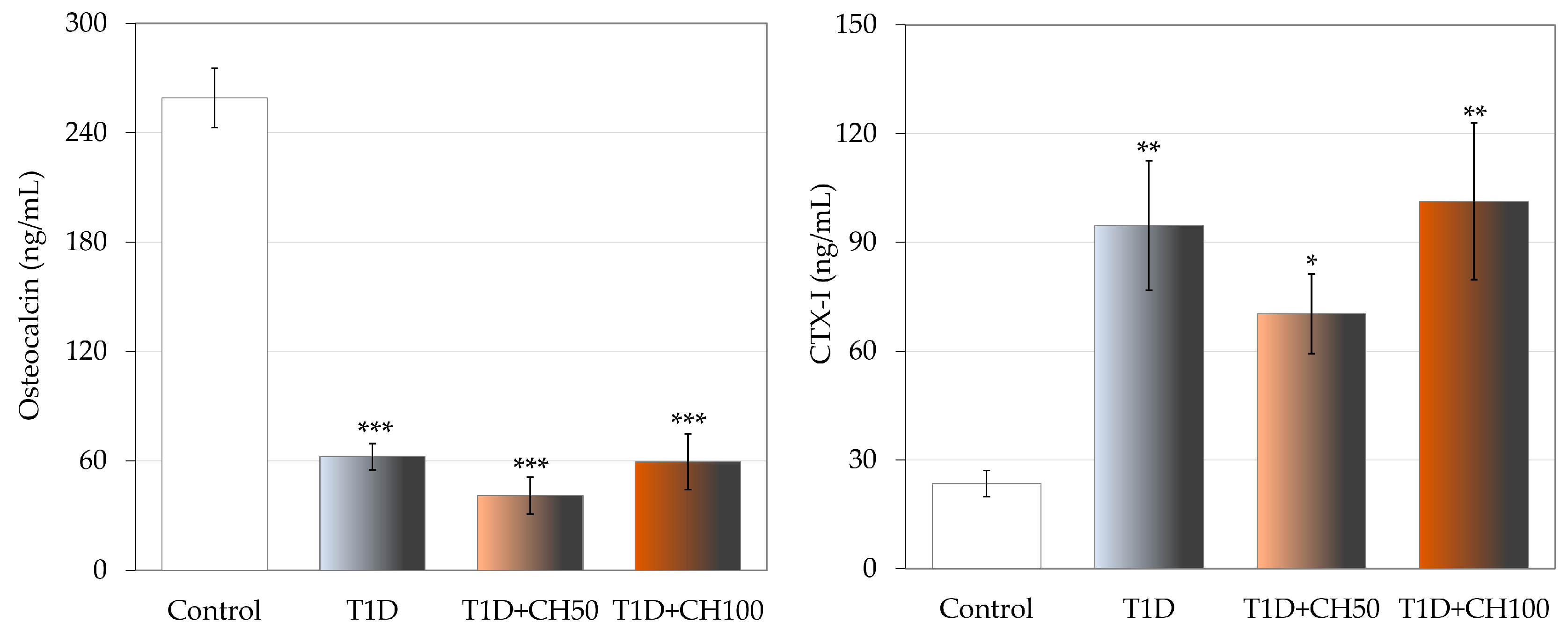
3.1. Effect of Chrysin on Body Mass and Concentration of Biochemical Markers of Carbohydrate Metabolism and Bone Turnover in the Serum in Rats with Experimental T1D
3.2. Effect of Chrysin on the Bone Macrometric Parameters, Mass, Density and Mineralization in Rats with Experimental T1D
3.3. Effect of Chrysin on the Histomorphometric Parameters of the Femur in Rats with Experimental T1D
3.4. Effect of Chrysin on Bone Mechanical Properties of the Tibia and Femur in Rats with Experimental T1D
4. Discussion
5. Conclusions
Author Contributions
Funding
Institutional Review Board Statement
Data Availability Statement
Conflicts of Interest
References
- Moghadam, E.R.; Ang, H.L.; Asnaf, S.E.; Zabolian, A.; Saleki, H.; Yavari, M.; Esmaeili, H.; Zarrabi, A.; Ashrafizadeh, M.; Kumar, A.P. Broad-Spectrum Preclinical Antitumor Activity of Chrysin: Current Trends and Future Perspectives. Biomolecules 2020, 10, 1374. [Google Scholar] [CrossRef]
- Gao, S.; Siddiqui, N.; Etim, I.; Du, T.; Zhang, Y.; Liang, D. Developing Nutritional Component Chrysin as a Therapeutic Agent: Bioavailability and Pharmacokinetics Consideration, and ADME Mechanisms. Biomed. Pharmacother. 2021, 142, 112080. [Google Scholar] [CrossRef]
- Mishra, A.; Shakti Mishra, P.; Bandopadhyay, R.; Khurana, N.; Angelopoulou, E.; Nath Paudel, Y.; Piperi, C. Neuroprotective Potential of Chrysin: Mechanistic Insights and Therapeutic Potential for Neurological Disorders. Molecules 2021, 26, 6456. [Google Scholar] [CrossRef]
- Stompor-Gorący, M.; Bajek-Bil, A.; Machaczka, M. Chrysin: Perspectives on Contemporary Status and Future Possibilities as Pro-Health Agent. Nutrients 2021, 13, 2038. [Google Scholar] [CrossRef]
- Talebi, M.; Talebi, M.; Farkhondeh, T.; Simal-Gandara, J.; Kopustinskiene, D.M.; Bernatoniene, J.; Samarghandian, S. Emerging Cellular and Molecular Mechanisms Underlying Anticancer Indications of Chrysin. Cancer Cell Int. 2021, 21, 214. [Google Scholar] [CrossRef]
- Wojnar, W.; Zych, M.; Borymski, S.; Kaczmarczyk-Sedlak, I. Chrysin Reduces Oxidative Stress but Does Not Affect Polyol Pathway in the Lenses of Type 1 Diabetic Rats. Antioxidants 2020, 9, 160. [Google Scholar] [CrossRef]
- Filho, C.B.; Jesse, C.R.; Donato, F.; Del Fabbro, L.; de Gomes, M.G.; Goes, A.T.R.; Souza, L.C.; Giacomeli, R.; Antunes, M.; Luchese, C.; et al. Neurochemical Factors Associated with the Antidepressant-like Effect of Flavonoid Chrysin in Chronically Stressed Mice. Eur. J. Pharmacol. 2016, 791, 284–296. [Google Scholar] [CrossRef] [PubMed]
- Kim, S.M.; Imm, J.Y. The Effect of Chrysin-Loaded Phytosomes on Insulin Resistance and Blood Sugar Control in Type 2 Diabetic db/db Mice. Molecules 2020, 25, 5503. [Google Scholar] [CrossRef] [PubMed]
- Mani, R.; Natesan, V. Chrysin: Sources, Beneficial Pharmacological Activities, and Molecular Mechanism of Action. Phytochemistry 2018, 145, 187–196. [Google Scholar] [CrossRef] [PubMed]
- Nabavi, S.F.; Braidy, N.; Habtemariam, S.; Orhan, I.E.; Daglia, M.; Manayi, A.; Gortzi, O.; Nabavi, S.M. Neuroprotective Effects of Chrysin: From Chemistry to Medicine. Neurochem. Int. 2015, 90, 224–231. [Google Scholar] [CrossRef]
- Liu, Y.; Song, X.; Li, C.; Hu, H.; Li, W.; Wang, L.; Hu, J.; Liao, C.; Liang, H.; He, Z.; et al. Chrysin Ameliorates Influenza Virus Infection in the Upper Airways by Repressing Virus-Induced Cell Cycle Arrest and Mitochondria-Dependent Apoptosis. Front. Immunol. 2022, 13, 872958. [Google Scholar] [CrossRef] [PubMed]
- Oggero, J.; Gasser, F.B.; Zacarías, S.M.; Burns, P.; Baravalle, M.E.; Renna, M.S.; Ortega, H.H.; Vaillard, S.E.; Vaillard, V.A. PEGylation of Chrysin Improves Its Water Solubility While Preserving the In Vitro Biological Activity. J. Agric. Food Chem. 2023, 71, 19817–19831. [Google Scholar] [CrossRef] [PubMed]
- Cao, L.; Wang, J.; Zhang, Y.; Tian, F.; Wang, C. Osteoprotective Effects of Flavonoids: Evidence from in Vivo and in Vitro Studies (Review). Mol. Med. Rep. 2022, 25, 200. [Google Scholar] [CrossRef]
- El-Marasy, S.A.; AbouSamra, M.M.; El-Mosallamy, A.E.M.K.; Emam, A.N.; Mabrok, H.B.; Galal, A.F.; Ahmed-Farid, O.A.; Abd El-Rahman, S.S.; Moustafa, P.E. Chrysin Loaded Nanovesicles Ameliorated Diabetic Peripheral Neuropathy. Role of NGF/AKT/GSK-3β Pathway. Chem. Biol. Interact. 2023, 375, 110402. [Google Scholar] [CrossRef]
- Ahad, A.; Ganai, A.A.; Mujeeb, M.; Siddiqui, W.A. Chrysin, an Anti-Inflammatory Molecule, Abrogates Renal Dysfunction in Type 2 Diabetic Rats. Toxicol. Appl. Pharmacol. 2014, 279, 1–7. [Google Scholar] [CrossRef] [PubMed]
- Shoieb, S.M.; Esmat, A.; Khalifa, A.E.; Abdel-Naim, A.B. Chrysin Attenuates Testosterone-Induced Benign Prostate Hyperplasia in Rats. Food Chem. Toxicol. 2018, 111, 650–659. [Google Scholar] [CrossRef]
- Ibrahim, S.O.; Mada, S.B.; Abarshi, M.M.; Tanko, M.S.; Babangida, S. Chrysin Alleviates Alteration of Bone-Remodeling Markers in Ovariectomized Rats and Exhibits Estrogen-like Activity in Silico. Hum. Exp. Toxicol. 2021, 40, 125–136. [Google Scholar] [CrossRef]
- Lu, J.J.; Zhou, F.M.; Hu, X.J.; Fang, J.J.; Liu, C.X.; Zhu, B.Q.; Ding, Z.S. Molecular Docking Simulation and in Vitro Studies on Estrogenic Activities of Flavonoids from Leaves of Carya Cathayensis Sarg. Steroids 2020, 163, 108726. [Google Scholar] [CrossRef]
- Han, D.-H.; Denison, M.S.; Tachibana, H.; Yamada, K. Relationship between Estrogen Receptor-Binding and Estrogenic Activities of Environmental Estrogens and Suppression by Flavonoids. Biosci. Biotechnol. Biochem. 2002, 66, 1479–1487. [Google Scholar] [CrossRef]
- Kuiper, G.G.J.M.; Lemmen, J.G.; Carlsson, B.O.; Corton, J.C.; Safe, S.H.; Van Der Saag, P.T.; Van Der Burg, B.; Gustafsson, J.Å. Interaction of Estrogenic Chemicals and Phytoestrogens with Estrogen Receptor. Endocrinology 1998, 139, 4252–4263. [Google Scholar] [CrossRef]
- Balam, F.H.; Ahmadi, Z.S.; Ghorbani, A. Inhibitory Effect of Chrysin on Estrogen Biosynthesis by Suppression of Enzyme Aromatase (CYP19): A Systematic Review. Heliyon 2020, 6, e03557. [Google Scholar] [CrossRef]
- Oršolić, N.; Nemrava, J.; Jeleč, Ž.; Kukolj, M.; Odeh, D.; Jakopović, B.; Jazvinšćak Jembrek, M.; Bagatin, T.; Fureš, R.; Bagatin, D. Antioxidative and Anti-Inflammatory Activities of Chrysin and Naringenin in a Drug-Induced Bone Loss Model in Rats. Int. J. Mol. Sci. 2022, 23, 2872. [Google Scholar] [CrossRef]
- Oršolić, N.; Goluža, E.; Dikić, D.; Lisičić, D.; Sašilo, K.; Rodak, E.; Jeleč, Ž.; Lazarus, M.V.; Orct, T. Role of Flavonoids on Oxidative Stress and Mineral Contents in the Retinoic Acid-Induced Bone Loss Model of Rat. Eur. J. Nutr. 2014, 53, 1217–1227. [Google Scholar] [CrossRef] [PubMed]
- Huo, J.F.; Zhang, M.L.; Wang, X.X.; Zou, D.H. Chrysin Induces Osteogenic Differentiation of Human Dental Pulp Stem Cells. Exp. Cell Res. 2021, 400, 112466. [Google Scholar] [CrossRef] [PubMed]
- Menon, A.H.; Soundarya, S.P.; Sanjay, V.; Chandran, S.V.; Balagangadharan, K.; Selvamurugan, N. Sustained Release of Chrysin from Chitosan-Based Scaffolds Promotes Mesenchymal Stem Cell Proliferation and Osteoblast Differentiation. Carbohydr. Polym. 2018, 195, 356–367. [Google Scholar] [CrossRef]
- Wu, Z.; Li, C.; Chen, Y.; Liu, Q.; Li, N.; He, X.; Li, W.; Shen, R.; Li, L.; Wei, C.; et al. Chrysin Protects Against Titanium Particle-Induced Osteolysis by Attenuating Osteoclast Formation and Function by Inhibiting NF-κB and MAPK Signaling. Front. Pharmacol. 2022, 13, 793087. [Google Scholar] [CrossRef]
- Zeng, W.; Yan, Y.; Zhang, F.; Zhang, C.; Liang, W. Chrysin Promotes Osteogenic Differentiation via ERK/MAPK Activation. Protein Cell 2013, 4, 539–547. [Google Scholar] [CrossRef]
- American Diabetes Association Professional Practice Committee. 2. Diagnosis and Classification of Diabetes: Standards of Care in Diabetes—2024. Diabetes Care 2024, 47 (Suppl. S1), S20–S42. [Google Scholar] [CrossRef]
- Hygum, K.; Starup-Linde, J.; Langdahl, B.L. Diabetes and Bone. Osteoporos. Sarcopenia 2019, 5, 29–37. [Google Scholar] [CrossRef] [PubMed]
- Kalaitzoglou, E.; Popescu, I.; Bunn, R.C.; Fowlkes, J.L.; Thrailkill, K.M. Effects of Type 1 Diabetes on Osteoblasts, Osteocytes and Osteoclasts. Curr. Osteoporos. Rep. 2016, 14, 310–319. [Google Scholar] [CrossRef]
- Khan, T.S.; Fraser, L. Type 1 Diabetes and Osteoporosis: From Molecular Pathways to Bone Phenotype. J. Osteoporos. 2015, 8, 174186. [Google Scholar] [CrossRef]
- Martiniakova, M.; Biro, R.; Penzes, N.; Sarocka, A.; Kovacova, V.; Mondockova, V.; Omelka, R. Links among Obesity, Type 2 Diabetes Mellitus, and Osteoporosis: Bone as a Target. Int. J. Mol. Sci. 2024, 25, 4827. [Google Scholar] [CrossRef] [PubMed]
- Hofbauer, L.C.; Busse, B.; Eastell, R.; Ferrari, S.; Frost, M.; Müller, R. Bone Fragility in Diabetes: Novel Concepts and Clinical Implications. Lancet Diabetes Endocrinol. 2022, 10, 207–220. [Google Scholar] [CrossRef] [PubMed]
- Salama, A.; Asaad, G.F.; Shaheen, A. Chrysin Ameliorates STZ-Induced Diabetes in Rats: Possible Impact of Modulation of TLR4/NF-Κβ Pathway. Res. Pharm. Sci. 2022, 17, 1–11. [Google Scholar] [CrossRef] [PubMed]
- Sayed, H.M.; Awaad, A.S.; El-Zahraa Abdel Rahman, F.S.; Al-Dossari, M.; Abd El-Gawaad, N.S.; Ahmed, O.M. Combinatory Effect and Modes of Action of Chrysin and Bone Marrow-Derived Mesenchymal Stem Cells on Streptozotocin/Nicotinamide-Induced Diabetic Rats. Pharmaceuticals 2023, 16, 34. [Google Scholar] [CrossRef]
- Li, Y.; Wang, X. Chrysin Attenuates High Glucose-Induced BMSC Dysfunction via the Activation of the PI3K/AKT/Nrf2 Signaling Pathway. Drug Des. Dev. Ther. 2022, 16, 165–182. [Google Scholar] [CrossRef]
- Folwarczna, J.; Londzin, P.; Borymska, W.; Zych, M.; Kaczmarczyk-Żebrowska, I. Effect of Diosmin on Changes in the Rat Skeletal System Induced by Experimental Type 1 Diabetes. 21st International Congress of the Polish Pharmacological Society, Katowice, Poland, 28–30 September 2023, Book of Abstracts. p. 33. Available online: https://zjazd-ptf.katowice.pl/static/sites/bn8pg_inter_congr_polis_ph_2023/PTF_book_of_abstracts_2023.pdf (accessed on 12 January 2025).
- Londzin, P.; Siudak, S.; Cegieła, U.; Pytlik, M.; Janas, A.; Waligóra, A.; Folwarczna, J. Phloridzin, an Apple Polyphenol, Exerted Unfavorable Effects on Bone and Muscle in an Experimental Model of Type 2 Diabetes in Rats. Nutrients 2018, 10, 1701. [Google Scholar] [CrossRef] [PubMed]
- Janas, A.; Kruczek, E.; Londzin, P.; Borymski, S.; Czuba, Z.P.; Folwarczna, J. Negligible Effect of Estrogen Deficiency on Development of Skeletal Changes Induced by Type 1 Diabetes in Experimental Rat Models. Mediat. Inflamm. 2020, 21, 2793804. [Google Scholar] [CrossRef] [PubMed]
- Dempster, D.W.; Compston, J.E.; Drezner, M.K.; Glorieux, F.H.; Kanis, J.A.; Malluche, H.; Meunier, P.J.; Ott, S.M.; Recker, R.R.; Parfitt, A.M. Standardized Nomenclature, Symbols, and Units for Bone Histomorphometry: A 2012 Update of the Report of the ASBMR Histomorphometry Nomenclature Committee. J. Bone Miner. Res. 2013, 28, 2–17. [Google Scholar] [CrossRef]
- Folwarczna, J.; Pytlik, M.; Zych, M.; Cegiela, U.; Kaczmarczyk-Sedlak, I.; Nowińska, B.; Śliwiński, L. Favorable Effect of Moderate Dose Caffeine on the Skeletal System in Ovariectomized Rats. Mol. Nutr. Food Res. 2013, 57, 1772–1784. [Google Scholar] [CrossRef]
- Turner, C.H.; Burr, D.B. Basic Biomechanical Measurements of Bone: A Tutorial. Bone 1993, 14, 595–608. [Google Scholar] [CrossRef] [PubMed]
- Stürmer, E.K.; Seidlová-Wuttke, D.; Sehmisch, S.; Rack, T.; Wille, J.; Frosch, K.H.; Wuttke, W.; Stürmer, K.M. Standardized Bending and Breaking Test for the Normal and Osteoporotic Metaphyseal Tibias of the Rat: Effect of Estradiol, Testosterone, and Raloxifene. J. Bone Miner. Res. 2006, 21, 89–96. [Google Scholar] [CrossRef]
- Sheu, A.; White, C.P.; Center, J.R. Bone Metabolism in Diabetes: A Clinician’s Guide to Understanding the Bone–Glucose Interplay. Diabetologia 2024, 67, 1493–1506. [Google Scholar] [CrossRef] [PubMed]
- Shanbhogue, V.V.; Hansen, S.; Frost, M.; Brixen, K.; Hermann, A.P. Bone Disease in Diabetes: Another Manifestation of Microvascular Disease? Lancet Diabetes Endocrinol. 2017, 10, 827–838. [Google Scholar] [CrossRef]
- Napoli, N.; Chandran, M.; Pierroz, D.D.; Abrahamsen, B.; Schwartz, A.V.; Ferrari, S.L. Mechanisms of Diabetes Mellitus-Induced Bone Fragility. Nat. Rev. Endocrinol. 2017, 13, 208–219. [Google Scholar] [CrossRef]
- Szkudelski, T. The Mechanism of Alloxan and Streptozotocin Action in B Cells of the Rat Pancreas. Physiol. Res. 2001, 50, 536–546. [Google Scholar] [CrossRef]
- Lenzen, S. The Mechanisms of Alloxan- and Streptozotocin-Induced Diabetes. Diabetologia 2008, 51, 216–226. [Google Scholar] [CrossRef]
- Folwarczna, J.; Janas, A.; Pytlik, M.; Cegieła, U.; Śliwinśki, L.; Krivošíková, Z.; Štefíková, K.; Gajdoš, M. Effects of Trigonelline, an Alkaloid Present in Coffee, on Diabetes-Induced Disorders in the Rat Skeletal System. Nutrients 2016, 8, 133. [Google Scholar] [CrossRef] [PubMed]
- Londzin, P.; Kisiel-Nawrot, E.; Kocik, S.; Janas, A.; Trawczyński, M.; Cegieła, U.; Folwarczna, J. Effects of Diosgenin on the Skeletal System in Rats with Experimental Type 1 Diabetes. Biomed. Pharmacother. 2020, 129, 110342. [Google Scholar] [CrossRef]
- Anitha, T.A.; Rajadurai, M. Antioxidative Potential of Chrysin, a Flavone in Streptozotocin–Nicotinamide-Induced Diabetic Rats. Biomed. Prev. Nutr. 2014, 4, 511–517. [Google Scholar] [CrossRef]
- Sengupta, P. The Laboratory Rat: Relating Its Age with Human’s. Int. J. Prev. Med. 2013, 4, 624–630. [Google Scholar] [PubMed]
- Singh, K.B.; Dixit, M.; Dev, K.; Maurya, R.; Singh, D. Formononetin, a Methoxy Isoflavone, Enhances Bone Regeneration in a Mouse Model of Cortical Bone Defect. Br. J. Nutr. 2017, 117, 1511–1522. [Google Scholar] [CrossRef] [PubMed]
- Jin, X.; Wang, H.; Li, F.; Liang, X.; Deng, X.; Gao, S.; Ru, K.; Qiu, W.; Huai, Y.; Zhang, J.; et al. Formononetin Ameliorates Simulated Microgravity-Induced Bone Loss by Suppressing Bone Turnover in Rats. Acta Astronaut. 2022, 200, 77–85. [Google Scholar] [CrossRef]
- Zakłos-Szyda, M.; Budryn, G.; Grzelczyk, J.; Pérez-Sánchez, H. Evaluation of Isoflavones as Bone Resorption Inhibitors upon Interactions with Receptor Activator of Nuclear Factor-κB Ligand (RANKL). Molecules 2020, 25, 206. [Google Scholar] [CrossRef]
- Qiu, Y.; Zhao, Y.; Long, Z.; Song, A.; Huang, P.; Wang, K.; Xu, L.; Molloy, D.P.; He, G. Liquiritigenin Promotes Osteogenic Differentiation and Prevents Bone Loss via Inducing Auto-Lysosomal Degradation and Inhibiting Apoptosis. Genes Dis. 2021, 10, 284–300. [Google Scholar] [CrossRef] [PubMed]
- Lu, R.; Zheng, Z.; Yin, Y.; Jiang, Z. Genistein Prevents Bone Loss in Type 2 Diabetic Rats Induced by Streptozotocin. Food Nutr. Res. 2020, 64, 3666. [Google Scholar] [CrossRef]
- Laurent, M.; Antonio, L.; Sinnesael, M.; Dubois, V.; Gielen, E.; Classens, F.; Vanderschueren, D. Androgens and Estrogens in Skeletal Sexual Dimorphism. Asian J. Androl. 2014, 16, 213–222. [Google Scholar] [CrossRef] [PubMed]
- Joseph, J.S.; Lam, V.; Patel, M.I. Preventing Osteoporosis in Men Taking Androgen Deprivation Therapy for Prostate Cancer: A Systematic Review and Meta-Analysis. Eur. Urol. Oncol. 2019, 2, 551–561. [Google Scholar] [CrossRef]


| Parameter/Group | Control | T1D | T1D + CH50 | T1D + CH100 |
|---|---|---|---|---|
| Glucose (mg/dL) | 141.4 ± 11.0 | 641.8 ± 28.6 *** | 687.9 ± 21.5 *** | 588.8 ± 53.8 *** |
| Fructosamine (µmol/L) | 281.9 ± 9.8 | 495.6 ± 23.1 *** | 483.3 ± 32.5 *** | 444.4 ± 23.7 *** |
| Insulin (µg/L) | 0.434 ± 0.096 | 0.089 ± 0.019 *** | 0.171 ± 0.045 ** | 0.136 ± 0.057 ** |
| Calcium (mg/dL) | 8.88 ± 0.18 | 10.16 ± 0.29 ** | 9.85 ± 0.21 ** | 10.18 ± 0.38 ** |
| Phosphorus (mg/dL) | 6.75 ± 0.19 | 8.19 ± 1.13 | 7.33 ± 0.51 | 7.85 ± 1.09 |
| Parameter/Group | Control | T1D | T1D + CH50 | T1D + CH100 |
|---|---|---|---|---|
| Bone length (mm) | 36.40 ± 0.34 | 34.28 ± 0.31 *** | 34.26 ± 0.23 *** | 34.59 ± 0.22 *** |
| Bone diameter (mm) | 3.60 ± 0.05 | 3.33 ± 0.09 * | 3.39 ± 0.06 * | 3.37 ± 0.10 * |
| Bone mass (g) | 0.855 ± 0.022 | 0.702 ± 0.024 *** | 0.693 ± 0.015 *** | 0.717 ± 0.027 *** |
| Bone density (g/cm3) | 1.626 ± 0.005 | 1.551 ± 0.012 *** | 1.560 ± 0.014 *** | 1.552 ± 0.013 *** |
| Bone mineral density (g/cm3) | 0.753 ± 0.006 | 0.695 ± 0.010 *** | 0.714 ± 0.011 ** | 0.709 ± 0.007 ** |
| Bone mineral mass (g) | 0.396 ± 0.009 | 0.314 ± 0.010 *** | 0.318 ± 0.008 *** | 0.327 ± 0.011 *** |
| Bone water mass (g) | 0.259 ± 0.007 | 0.218 ± 0.010 ** | 0.212 ± 0.006 *** | 0.220 ± 0.011 ** |
| Bone organic substances mass (g) | 0.201 ± 0.006 | 0.169 ± 0.007 *** | 0.163 ± 0.004 *** | 0.170 ± 0.006 *** |
| Bone mineral mass/bone mass ratio (g/g) | 0.463 ± 0.003 | 0.448 ± 0.005 | 0.458 ± 0.006 | 0.457 ± 0.005 |
| Bone water mass/bone mass ratio (g/g) | 0.302 ± 0.003 | 0.310 ± 0.006 | 0.306 ± 0.006 | 0.306 ± 0.007 |
| Bone organic substances mass/bone mass ratio | 0.234 ± 0.002 | 0.241 ± 0.006 | 0.236 ± 0.002 | 0.237 ± 0.003 |
| Calcium content (g/g bone mineral) | 0.422 ± 0.003 | 0.419 ± 0.003 | 0.420 ± 0.003 | 0.424 ± 0.004 |
| Phosphorus content (g/g bone mineral) | 0.171 ± 0.001 | 0.167 ± 0.001 * | 0.167 ± 0.001 * | 0.167 ± 0.001 * |
| Parameter/Group | Control | T1D | T1D + CH50 | T1D + CH100 |
|---|---|---|---|---|
| Bone length & (mm) | 40.17 ± 0.34 | 38.46 ± 0.37 *** | 38.24 ± 0.27 *** | 38.58 ± 0.22 ** |
| Bone diameter & (mm) | 2.88 ± 0.05 | 2.74 ± 0.05 * | 2.69 ± 0.03 ** | 2.71 ± 0.07 * |
| Bone mass (g) | 0.535 ± 0.012 | 0.438 ± 0.017 *** | 0.425 ± 0.008 *** | 0.440 ± 0.019 *** |
| Bone density (g/cm3) | 1.636 ± 0.007 | 1.586 ± 0.009 *** | 1.602 ± 0.009 ** | 1.593 ± 0.010 ** |
| Bone mineral density (g/cm3) | 0.776 ± 0.008 | 0.723 ± 0.008 *** | 0.752 ± 0.009 *# | 0.739 ± 0.007 ** |
| Bone mineral mass (g) | 0.253 ± 0.006 | 0.200 ± 0.007 *** | 0.200 ± 0.004 *** | 0.204 ± 0.008 *** |
| Bone water mass (g) | 0.148 ± 0.004 | 0.129 ± 0.008 * | 0.122 ± 0.004 ** | 0.132 ± 0.007 |
| Bone organic substances mass (g) | 0.133 ± 0.004 | 0.109 ± 0.005 *** | 0.104 ± 0.002 *** | 0.104 ± 0.004 *** |
| Bone mineral mass/ bone mass ratio | 0.474 ± 0.004 | 0.456 ± 0.003 ** | 0.470 ± 0.005 # | 0.464 ± 0.003 |
| Bone water mass/ bone mass ratio | 0.276 ± 0.005 | 0.294 ± 0.009 | 0.286 ± 0.007 | 0.300 ± 0.006 |
| Bone organic substances mass/bone mass ratio | 0.249 ± 0.003 | 0.250 ± 0.008 | 0.245 ± 0.003 | 0.236 ± 0.004 |
| Calcium content (g/g bone mineral) | 0.423 ± 0.003 | 0.429 ± 0.003 | 0.439 ± 0.007 | 0.431 ± 0.002 |
| Phosphorus content (g/g bone mineral) | 0.172 ± 0.002 | 0.170 ± 0.002 | 0.170 ± 0.002 | 0.169 ± 0.001 |
| Parameter/Group | Control | T1D | T1D + CH50 | T1D + CH100 |
|---|---|---|---|---|
| Bone mass (g) | 0.208 ± 0.010 | 0.155 ± 0.005 *** | 0.150 ± 0.004 *** | 0.148 ± 0.012 *** |
| Bone density (g/cm3) | 1.559 ± 0.013 | 1.472 ± 0.015 * | 1.474 ± 0.016 * | 1.547 ± 0.046 # |
| Bone mineral density (g/cm3) | 0.703 ± 0.009 | 0.651 ± 0.019 * | 0.672 ± 0.013 | 0.707 ± 0.019 # |
| Bone mineral mass (g) | 0.094 ± 0.004 | 0.069 ± 0.003 *** | 0.068 ± 0.002 *** | 0.068 ± 0.006 *** |
| Bone water mass (g) | 0.062 ± 0.004 | 0.048 ± 0.002 *** | 0.045 ± 0.002 *** | 0.044 ± 0.003 *** |
| Bone organic substances mass (g) | 0.052 ± 0.002 | 0.038 ± 0.002 *** | 0.037 ± 0.001 *** | 0.036 ± 0.003 *** |
| Bone mineral mass/ bone mass ratio | 0.451 ± 0.005 | 0.442 ± 0.010 | 0.456 ± 0.006 | 0.458 ± 0.006 |
| Bone water mass/ bone mass ratio | 0.300 ± 0.006 | 0.310 ± 0.013 | 0.298 ± 0.008 | 0.296 ± 0.009 |
| Bone organic substances mass/bone mass ratio | 0.249 ± 0.002 | 0.248 ± 0.004 | 0.246 ± 0.005 | 0.246 ± 0.003 |
| Calcium content (g/g bone mineral) | 0.436 ± 0.010 | 0.444 ± 0.008 | 0.447 ± 0.009 | 0.441 ± 0.008 |
| Phosphorus content (g/g bone mineral) | 0.171 ± 0.001 | 0.169 ± 0.002 | 0.171 ± 0.001 | 0.166 ± 0.001 |
| Parameter/Group | Control | T1D | T1D + CH50 | T1D + CH100 | |
|---|---|---|---|---|---|
| Femoral diaphysis | Tt.Ar (mm2) | 9.543 ± 0.235 | 8.617 ± 0.272 * | 8.465 ± 0.132 ** | 8.537 ± 0.340 ** |
| Ct.Ar (mm2) | 5.927 ± 0.193 | 5.224 ± 0.116 ** | 5.228 ± 0.085 ** | 5.162 ± 0.191 ** | |
| Ma.Ar (mm2) | 3.616 ± 0.199 | 3.392 ± 0.175 | 3.237 ± 0.079 | 3.375 ± 0.215 | |
| Ma.Ar/Tt.Ar (%) | 37.8 ± 1.6 | 39.2 ± 0.9 | 38.2 ± 0.6 | 39.4 ± 1.4 |
| Parameter/Group | Control | T1D | T1D + CH50 | T1D + CH100 | |
|---|---|---|---|---|---|
| Femoral metaphysis | BV/TV (%) | 33.05 ± 2.21 | 27.89 ± 4.55 | 27.80 ± 1.67 | 27.60 ± 1.12 |
| Tb.Th (μm) | 47.34 ± 2.85 | 38.70 ± 5.40 | 41.04 ± 1.89 | 36.77 ± 2.01 | |
| Tb.Sp (μm) | 96.27 ± 4.58 | 102.02 ± 9.80 | 107.90 ± 5.94 | 96.54 ± 4.46 | |
| Tb.N (1/mm) | 6.98 ± 0.17 | 7.13 ± 0.25 | 6.76 ± 0.24 | 7.55 ± 0.32 |
| Parameter/Group | Control | T1D | T1D + CH50 | T1D + CH100 |
|---|---|---|---|---|
| Displacement for yield point load (mm) | 0.285 ± 0.062 | 0.217 ± 0.025 | 0.172 ± 0.032 | 0.155 ± 0.015 |
| Maximum load (N) | 65.0 ± 3.2 | 31.9 ± 2.3 *** | 36.4 ± 3.5 *** | 34.6 ± 5.3 *** |
| Displacement for maximum load (mm) | 0.903 ± 0.060 | 0.531 ± 0.078 ** | 0.591 ± 0.088 ** | 0.694 ± 0.093 |
| Energy for maximum load (mJ) | 37.2 ± 3.0 | 10.7 ± 1.9 *** | 13.8 ± 1.8 *** | 16.3 ± 3.1 *** |
| Stress for maximum load (MPa) | 46.0 ± 2.7 | 25.6 ± 2.1 *** | 29.7 ± 1.9 *** | 26.5 ± 2.6 *** |
| Fracture load (N) | 48.7 ± 3.3 | 24.1 ± 2.5 *** | 25.4 ± 2.4 *** | 28.2 ± 5.3 *** |
| Displacement for fracture load (mm) | 1.396 ± 0.065 | 0.911 ± 0.070 *** | 1.050 ± 0.083 ** | 0.970 ± 0.082 *** |
| Energy for fracture load (mJ) | 65.0 ± 3.7 | 21.4 ± 2.4 *** | 28.0 ± 3.4 *** | 24.8 ± 3.7 *** |
| Stress for fracture load (MPa) | 34.8 ± 3.2 | 19.3 ± 2.1 *** | 20.7 ± 1.5 *** | 21.3 ± 2.7 *** |
| Parameter/Group | Control | T1D | T1D + CH50 | T1D + CH100 | |
|---|---|---|---|---|---|
| Femoral diaphysis | Displacement for yield point load (mm) | 0.275 ± 0.004 | 0.247 ± 0.017 | 0.260 ± 0.014 | 0.292 ± 0.014 |
| Maximum load (N) | 139.2 ± 5.8 | 113.7 ± 6.4 ** | 121.1 ± 6.9 * | 115.3 ± 4.3 ** | |
| Displacement for maximum load (mm) | 0.543 ± 0.034 | 0.497 ± 0.041 | 0.563 ± 0.033 | 0.533 ± 0.030 | |
| Energy for maximum load (mJ) | 45.2 ± 4.9 | 34.0 ± 5.6 | 40.2 ± 4.9 | 34.9 ± 3.6 | |
| Stress for maximum load (MPa) | 153.3 ± 4.4 | 150.0 ± 7.9 | 164.8 ± 5.9 | 155.0 ± 5.2 | |
| Fracture load (N) | 139.1 ± 5.7 | 113.1 ± 6.1 ** | 120.8 ± 6.9 * | 113.0 ± 3.3 ** | |
| Displacement for fracture load (mm) | 0.548 ± 0.035 | 0.504 ± 0.045 | 0.569 ± 0.035 | 0.557 ± 0.045 | |
| Energy for fracture load (mJ) | 45.8 ± 5.0 | 35.0 ± 6.2 | 40.9 ± 5.1 | 37.8 ± 5.8 | |
| Stress for fracture load (MPa) | 153.1 ± 4.3 | 149.3 ± 7.7 | 164.4 ± 5.9 | 152.6 ± 6.6 | |
| Maximum load in the femoral neck (N) | 96.8 ± 4.2 | 84.8 ± 5.9 | 91.2 ± 3.8 | 88.2 ± 7.2 |
Disclaimer/Publisher’s Note: The statements, opinions and data contained in all publications are solely those of the individual author(s) and contributor(s) and not of MDPI and/or the editor(s). MDPI and/or the editor(s) disclaim responsibility for any injury to people or property resulting from any ideas, methods, instructions or products referred to in the content. |
© 2025 by the authors. Licensee MDPI, Basel, Switzerland. This article is an open access article distributed under the terms and conditions of the Creative Commons Attribution (CC BY) license (https://creativecommons.org/licenses/by/4.0/).
Share and Cite
Klasik-Ciszewska, S.; Londzin, P.; Grzywnowicz, K.; Borymska, W.; Zych, M.; Kaczmarczyk-Żebrowska, I.; Folwarczna, J. Effect of Chrysin, a Flavonoid Present in Food, on the Skeletal System in Rats with Experimental Type 1 Diabetes. Nutrients 2025, 17, 316. https://doi.org/10.3390/nu17020316
Klasik-Ciszewska S, Londzin P, Grzywnowicz K, Borymska W, Zych M, Kaczmarczyk-Żebrowska I, Folwarczna J. Effect of Chrysin, a Flavonoid Present in Food, on the Skeletal System in Rats with Experimental Type 1 Diabetes. Nutrients. 2025; 17(2):316. https://doi.org/10.3390/nu17020316
Chicago/Turabian StyleKlasik-Ciszewska, Sylwia, Piotr Londzin, Kacper Grzywnowicz, Weronika Borymska, Maria Zych, Ilona Kaczmarczyk-Żebrowska, and Joanna Folwarczna. 2025. "Effect of Chrysin, a Flavonoid Present in Food, on the Skeletal System in Rats with Experimental Type 1 Diabetes" Nutrients 17, no. 2: 316. https://doi.org/10.3390/nu17020316
APA StyleKlasik-Ciszewska, S., Londzin, P., Grzywnowicz, K., Borymska, W., Zych, M., Kaczmarczyk-Żebrowska, I., & Folwarczna, J. (2025). Effect of Chrysin, a Flavonoid Present in Food, on the Skeletal System in Rats with Experimental Type 1 Diabetes. Nutrients, 17(2), 316. https://doi.org/10.3390/nu17020316