Review

Hydration Meets Regulation: Insights into Bicarbonate Mineral Water and Acid–Base Balance

1
Institute of Food and One Health, Leibniz University Hanover, 30159 Hanover, Germany
2
Independent Researcher, 75016 Paris, France
*
Author to whom correspondence should be addressed.
Nutrients 2025, 17(14), 2291; https://doi.org/10.3390/nu17142291
Submission received: 11 June 2025 / Revised: 4 July 2025 / Accepted: 8 July 2025 / Published: 10 July 2025
(This article belongs to the Section Nutrition and Public Health)

Abstract

Acid–base balance is critical to human health and can be significantly influenced by dietary choices. The Western diet, characterized by high meat and cheese consumption, induces excess acidity, highlighting the need for strategies to mitigate this. Recent studies have focused on bicarbonate-rich mineral water as a viable solution. In this context, the present narrative review synthesizes the findings from recent scientific studies on bicarbonate-rich mineral water, specifically those with bicarbonate levels over 1300 mg/L and medium or low PRAL values. This water has been shown to exert beneficial effects on both urinary and blood parameters. The key effects include an increase in the urine pH and a profound reduction in net acid excretion as a sign for a reduced acid load. Additionally, bicarbonate mineral water has been shown to decrease the excretion of nephrolithiasis-related constituents, including calcium and oxalates, as well as inhibitory substances such as magnesium and citrates. In blood, bicarbonate-rich water has been demonstrated to stabilize pH and increase bicarbonate levels, thereby enhancing systemic buffering capacity. Clinically, these changes have been associated with a lowered risk of calcium oxalate stone formation and improved kidney health. Furthermore, bicarbonate-rich water has been shown to support bone health by reducing bone resorption markers. Consequently, the integration of bicarbonate-rich mineral water into the diet has the potential to enhance urinary and blood parameters, mitigate the risk of kidney stones, and strengthen skeletal integrity, thereby serving as a promising strategy for health promotion and disease prevention. While promising, these findings underscore the need for further research to establish long-term recommendations. Future interventional studies should be designed with rigorous randomization, larger sample sizes, cross-over methodologies, and comprehensive dietary assessments to address the methodological limitations of previous research.

1. Introduction

The maintenance of the acid–base balance in the body represents a fundamental aspect of physiological homeostasis, essential for life. Blood pH, typically ranging between 7.35 and 7.45 (arterial) [1], is carefully regulated to maintain cellular functions, enzymatic activity, and overall metabolic integrity [2]. Any deviation from this narrow pH range results in significant health consequences [3], manifesting as either acidosis or alkalosis. The lungs and kidneys serve as the primary organs that are crucial for the regulation of acid–base homeostasis [1]. Specifically, volatile acids are excreted via the lungs as carbon dioxide, whereas non-volatile acids must be excreted via the kidneys [4].
Diet composition significantly affects acid–base balance [1]. Different dietary components can either contribute to endogenous acid production or provide alkaline precursors [5]. Food rich in protein, particularly those with sulfur-containing amino acids and phosphorus, such as meat, cheese and eggs, tend to increase acid load upon metabolic processing [6]. By contrast, fruits and vegetables, particularly those rich in potassium and magnesium, provide alkaline precursors, and metabolization promotes a more alkaline environment [1,6,7]. Although grain products contain an average amount of sulfur-containing proteins [4], they still contribute to an increase in acid load. Therefore, grains should be considered as acidifying foods because of their comparatively high intake. Due to a high protein consumption and the lack of fruits and vegetables, the modern Western diet is considered to cause an excess of acid equivalents [6,7,8]. Consequently, such dietary patterns are suggested to cause imbalances in the acid–base balance in the long-term [4] resulting in a low-grade metabolic acidosis.
This narrative review aims to give a short overview of the acid–base balance and the association between high acid load and health consequences in general. However, the main part deals with the effects of bicarbonate-rich mineral water consumption on acid–base balance, as this water deliver higher amounts of bicarbonate potentially modulating the parameters of acid–base homeostasis.

2. Literature Search Strategy

To identify the relevant literature for this narrative review, a comprehensive search of the electronic database PubMed was conducted. Reference lists of selected articles were also screened to identify additional relevant publications. The focus of this review is on the literature published in English, with a few exceptions. Therefore, the search was limited to articles published in English. However, relevant references in French and Italian were identified in the reference list of the selected publications. Therefore, these studies were also included. The following keywords and keyword combinations were employed: “mineral water”, “water”, “bicarbonate”, “acid-base”, “blood gas”, and “pH”. In addition, the Boolean operators “AND” and “OR” should be used. Only full-text versions of the literature that reported on the relationship between bicarbonate-rich mineral water and acid–base balance were included. This encompasses studies on classical acid–base parameters in urine (acid excretion) and blood (blood gas) as well as related studies on kidney stones and bones.

3. Intake of Acid Precursors and Acid Excretion

The acidizing or alkalizing potential of foods and beverages is usually estimated using equations developed for this purpose. In general, these estimations show a good correlation to the directly measured renal net acid excretion [4,9]. However, their accuracy can be questioned at the individual level, due to limitations in the precise measurement of dietary intake, the individual absorption of nutrients in the gastrointestinal tract, and the nutrient composition of foods [4]. Nevertheless, three equations or measurements are frequently used in nutritional studies to assess the effect of foods and beverages on the acid–base balance: the Potential Renal Acid Load (PRAL) score, net endogenous acid production (NEAP), and net acid excretion (NAE). These parameters offer valuable insights into the complex interplay of dietary intake, endogenous metabolism, and renal function in maintaining acid–base balance.
Foods and beverages usually contain acid precursors, with fruits and vegetables additionally containing base precursors [10]. The PRAL calculation of foods and beverages is a commonly used method developed to estimate the acid or base forming potential of the foods [11], expressed in milliequivalents per 100 g (mEq/100 g). This value reflects the amount of acid excreted by the kidneys when 100 g of a food is consumed [1,12]. The PRAL values of different foods can vary between negative and positive values. Negative values indicate a more alkaline potential and positive values indicate a more acidic potential [1,13]. In general, the more negative the PRAL value, the less acid needs to be excreted via urine [12]. The formula for the calculation of PRAL was developed by Remer and Manz in the 1990s. It takes into account the average intestinal absorption rate of the various nutrients that indicate an acidifying or alkalizing effect of food. In addition to the amount of the predominant anions (phosphorus, protein, and chloride), the amount of the predominant cations (magnesium, calcium, potassium, and sodium) is integrated in the original formula (PRALR) [11]. Although six minerals were initially considered, it has been demonstrated that the PRAL estimation using only four minerals (phosphorus, magnesium, calcium, and potassium) allows for a simpler assessment of dietary acidity. Both methods showed a reasonable agreement with the measured NAE. In this formula chloride and sodium are omitted, due to large inaccuracies in estimating sodium and chloride intake via processed foods in food tables [14]. A second, slightly modified equation was developed by Sebastian et al. (PRALS) [15], taking into account the sulfate content of the diet instead of the protein content.
However, the PRAL value does not completely reflect the renal net acid excretion, as it excludes endogenously synthesized organic acids (OAs) in the equation. In order to enhance the precision of quantifying the total acid excreted in urine, the NEAP (net endogenous acid production) estimation was developed by Remer and Manz [11] and modified by Lemann [16] and Sebastian et al. [15]. However, all equations include estimated OAs: NEAP = PRAL + OA. The estimated organic acids thereby reflect an anthropometry-based estimation using individual body surface (OAanthro) [11] or the dietary intake of the main anions and cations (OAdiet) [16] for the calculation (Table 1).
In addition, a simplified algorithm has been developed by Frassetto et al. [17], focusing on the two primary determinants of endogenous acid production, protein intake for acid production and potassium intake for base production, as they are the major variable components and independent predictors for NEAP. While protein is an indicator for the rate of sulfuric acid production, the potassium salts of organic acids are an indicator of bicarbonate generation upon metabolism [18]. There are two equations: NEAPF = protein—potassium or protein/potassium. Both equations from Frassetto et al. offer the possibility to estimate NEAP under conditions of restricted information on dietary nutrient and mineral content. Nonetheless, an apparent discrepancy exists among the calculation models in terms of their validity in relation to the measured NAE. The calculation of NEAP using the equations by Remer (NEAPR) and Lemann (NEAPL) showed a good agreement with NAE, although both equations were imprecise. However, the equations by Sebastian (NEAPS) and Frassetto (NEAPF) did not provide reasonable correlations [19].
In general, the NEAP values vary widely between individuals, typically ranging from 20 mEq to 120 mEq per day [20]. However, contemporary Western diets usually lead to a total acid load of about 50–100 mEq/d [11,13,21,22]. By contrast, the ingestion of a vegan diet has been shown to result in a reduced acid excretion, as demonstrated by a lower mean NEAP value of −0.72 ± 29.8 mEq/day [23].
Table 1. Equations for the estimation of net endogenous acid production (NEAP).
Name of the EquationEquation/FormulaReference
NEAPR (mEq/d)PRALR (mEq/d) * + OAanthro (mEq/d)[14]
               OAantho (mEq/d)BSA ** (m2) × 41 (mEq/d)/1.73 (m2)[11]
NEAPL (mEq/d)
               OAdiet (mEq/d)
PRALR (mEq/d) * + OAdiet (mEq/d)
32.9 + (0.15 × [{potassium} + {calcium × 2} + {magnesium × 2} − {phosphorus × 1.8}]) (all in mmol/d)
[16]
NEAPS (mEq/d)
               OAdiet (mEq/d)
PRALS (mEq/d) * + OAdiet (mEq/d)
32.9 + (0.15 × [{potassium} + {calcium × 2} + {magnesium × 2} − {phosphorus × 1.8}]) (all in mmol/d)
[15]
NEAPF (mEq/d)Equation (1): [54.4 × protein (g/d)/potassium (mEq/d)] − 10.2
Equation (2): [0.91 × protein (g/d)] − [0.57 × potassium (mEq/d)] + 21
[17]
* using the simplified formula of potential renal acid load from Remer et al., omitting Cl and Na [14]; OA = endogenously produced organic acids (derived from anthropometric measurements or diet); ** BSA = individual body surface area, for example calculated by the formula of Du Bois and Du Bois [24]: BSA = 0.007184 × height (cm)0.725 × weight (kg)0.425.
In a “steady state”, NEAP is equivalent to renal net acid excretion (NAE) [2,6,17], encompassing both dietary acid load and endogenous acid production. Therefore, NEAP is reflected in the net result of the acid–base regulatory processes. Unlike PRAL and NEAP, which estimate acid production resulting from food intake, NAE can be measured directly in 24-h urine samples to quantify the amount of acid excreted via urine. Its determination is based on the measurement of titratable acids, ammonium, and bicarbonate in the urine: NAE = TA + NH4+ − HCO3− [17].
In general, the renal acid excretion capacity surpasses the usual acid load derived from dietary intake. Consequently, significant diet-induced disturbances of the acid–base balance in the blood are uncommon in healthy individuals [10]. Excess acids are excreted via urine, which is reflected by a decrease in the urine pH [2]. Nonetheless, there are physiological limits; a decrease in the pH value to below 4.5 results in a reduction of H+ excretion due to impaired protein function in the proximal tubule. Furthermore, a urinary pH below 5.4 indicates a maximally stimulated acid excretion [2], which is associated with serious metabolic consequences [25]. Unlike healthy individuals, individuals with impaired renal function may experience clinically evident metabolic acidosis even with a lower dietary acid load [2,25]. This particularly affects individuals with chronic kidney disease and the elderly, whose ability to excrete acids decreases with increasing age [10].
Nevertheless, prolonged excessive intake of acidic precursors, as commonly observed in the typical Western diet [26], can result in chronic depletion of the body’s buffering systems. Maintaining a constant systemic pH to sustain essential biochemical processes can lead to alterations in the acid–base balance, even among healthy people. If the daily dietary acid load exceeds 1 mmol H+ per kilogram of body weight, the risk of a positive acid balance increases [16]. This physiological state is known by various terms, including “low-grade metabolic acidosis” [12], “chronic sub-clinical systemic metabolic acidosis (CSSMA)” [27], “subclinical acidosis” [28], “preclinical acidosis” [29], or “eubicarbonatemic acidosis” [30]. At this stage, there is a noticeable shift towards acidity, even though the pH of the blood is still within the normal range. Additionally, this condition is often accompanied by a slight decrease in the systemic bicarbonate pool and a notable increase in renal acid excretion [12]. If this condition persists over a long period of time, it may contribute to the development and aggravation of several diseases [1,12,27]. Small adaptations occur throughout the body; all aimed at eliminating excess acid equivalents. However, these adaptations do not occur without consequences for the organism in the long term [2].

4. Associations Between Dietary Acid Load and Metabolic Alterations or Diseases

Numerous epidemiological studies have demonstrated an association between high dietary acid load and the occurrence of various metabolic alterations and diseases. On the one hand, increased cortisol levels are shown in individuals with high dietary acid load [31,32]. In addition, serum uric acid levels are increased [33,34] and the risk of hyperuricemia is elevated [35]. On the other hand, increases in the urinary excretion of calcium [36] and ammonium [37] are demonstrated, while citrate excretion decreases [38,39], accompanied by a decrease in the urinary pH [40,41]. Furthermore, the synthesis of endothelin-1, angiotensin II, and aldosterone in the kidney is stimulated, thereby activating several pro-fibrotic factors [1]. These alterations are accompanied by systemic changes affecting the kidneys, bones, muscles, liver, and the endocrine system (Figure 1).

5. Mineral Water

Mineral water is a calorie-free beverage that can contribute significantly to hydration. Beyond its role in maintaining fluid balance, mineral water provides essential minerals, such as calcium and magnesium. Consequently, its consumption offers a range of potential health benefits that extend beyond mere caloric considerations. Several intervention studies have shown the beneficial effects of mineral water consumption in supporting the treatment of kidney stones [42], osteoporosis [43], metabolic syndrome [44], cardiovascular diseases [45], and digestive disorders [46]. In addition, mineral water consumption has been associated with improved exercise performance and rehydration [47,48]. These benefits are attributed to several underlying mechanisms, including its effect on the acid–base balance [43,44], its stimulation of intestinal activity through osmotically active compounds [46,49], and its contribution to the supply status of various minerals [50]. However, it is important to note that the health benefits of mineral water vary significantly depending on the amount of dissolved ions. Mineral water used to relieve digestive disorders, such as constipation, may not necessarily provide benefits in the prevention of kidney stones [51]. In addition, mineral water that is effective against calcium oxalate stones have proven to be counterproductive against magnesium ammonium phosphate and calcium phosphate stones [52]. Therefore, the specific composition of each mineral water explains its supportive or counterproductive effects in terms of treatment support and symptom alleviation

6. Bicarbonate-Rich Mineral Water

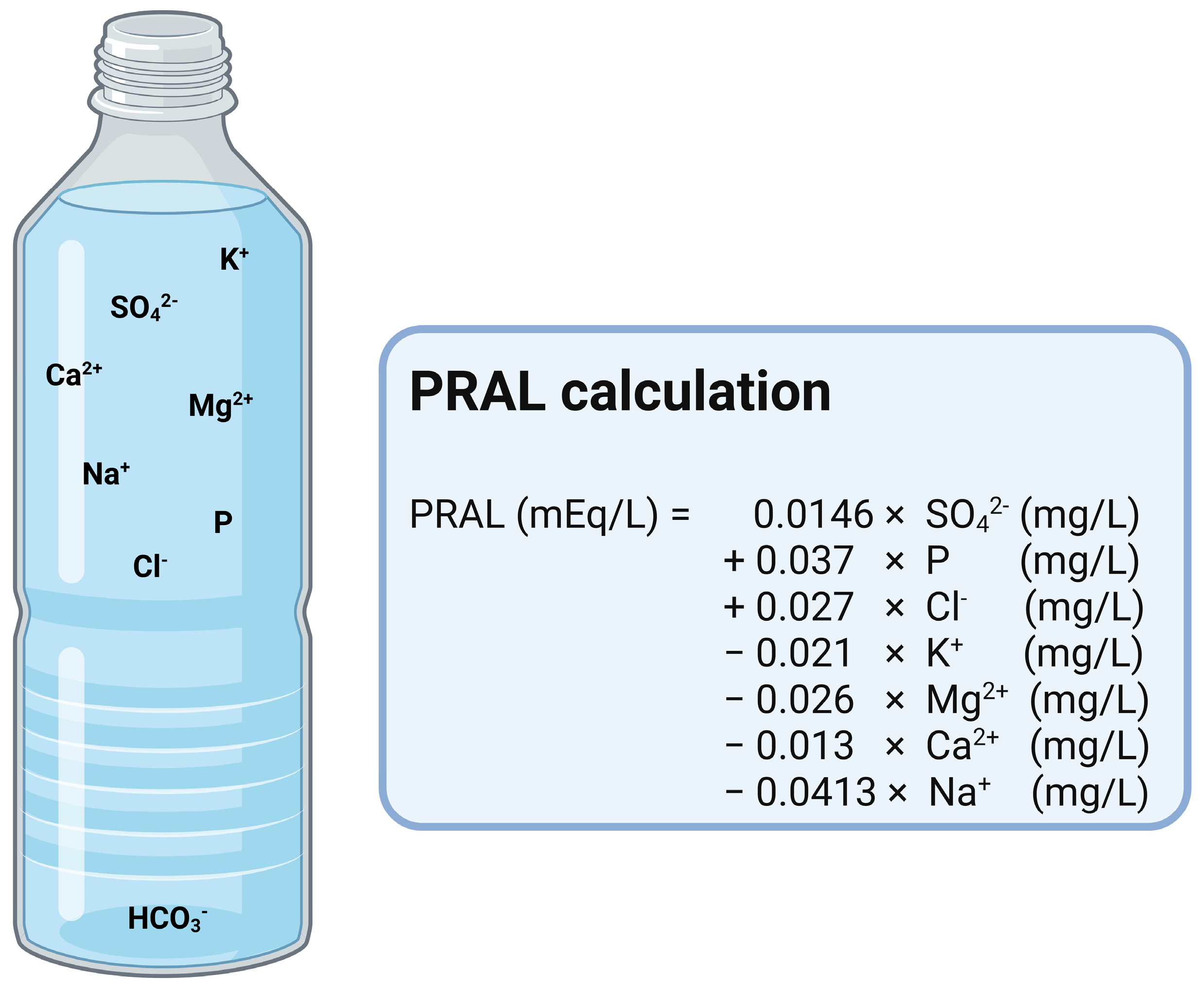
Most mineral water contains lower amounts of bicarbonate (50–100 mg/L). However, there is mineral water with much higher bicarbonate levels, reaching several grams per liter [53]. In a recent study examining the composition of several commercial mineral water brands available in 10 European countries, the mineral water with the lowest bicarbonate levels were sold in Italy and United Kingdom, while those with the highest bicarbonate levels were sold in France and Spain. Poland had the highest calculated average level of bicarbonate in the mineral water on the market [54,55], indicating several mineral water brands with a high bicarbonate content in this European country. However, the mineral water with the highest bicarbonate level included in this study was sold in France, showing a bicarbonate content of 4368 mg/L [55]. In this context, mineral water with a high concentration of bicarbonate contains a minimum amount of at least 600 milligrams per liter. However, studies on its effects regarding health issues have been conducted utilizing mineral water with a bicarbonate content of approximately 1000 mg/L and above. The relationships between the components of mineral water differ fundamentally between alkalizing and acidifying water [51]. Bicarbonate-rich mineral water is known to have an alkalizing effect on human urine [43], similar to medication [52]. In order to estimate the alkalinizing or acidifying potential of mineral water (PRAL), modifications have been made to the original calculation model by Remer and colleagues in the literature [11]. Wynn et al. [56] substituted the amount of protein by the sulfate content of the mineral water. The original equation for the PRAL calculation used the protein content of food to estimate the acidifying effect of sulfate. In food, sulfate originates from cysteine and methionine, which are ingested along with dietary protein. However, in mineral water where sulfate is in solution, the molecular weight and absorption rate of the sulfate must be considered, not those of the amino acids from the dietary proteins [57]. This requires the substitution of protein and sulfate in the calculation [56] (Figure 2).
Given that bicarbonate and sulfate typically do not coexist in the same geological layer, bicarbonate-rich water typically contains minimal amounts of highly acidifying sulfate [51]. This explains the strong correlation between the PRAL value of a mineral water and its bicarbonate content [43]. In a study evaluating the relationship between dissolved ions in mineral water and its PRAL value, bicarbonate showed the strongest negative correlation with the calculated PRAL value [51]. This finding suggests that, despite the omission of bicarbonate content from the PRAL calculations, it may nevertheless exert a significant influence on the acid–base balance, in addition to the contributions of alkalizing and acidifying ions present in mineral water. It has been established that the substance is absorbed by the body in certain quantities, and subsequently excreted via the urine [53].
Moreover, alkalizing mineral water often contains high amounts of calcium. However, alkalizing water is not necessarily rich in calcium. Rather, some bicarbonate-rich water is also characterized by a high sodium content [42], which is shown in Table 2.

7. Bicarbonate-Rich Mineral Water and Human Health

7.1. Urinary Parameters

7.1.1. Urine pH and Net Acid Excretion (NAE)

Several authors have stated that there is a beneficial effect of consuming bicarbonate-rich mineral water on the acid–base balance, both in terms of the urine [13,56,58] and the blood parameters [13,48,58,59].
Under normal physiological conditions, human urine is slightly acidic [52], a characteristic influenced by the intake of food and beverages. Nevertheless, there are conditions requiring a more alkaline pH to prevent unfavorable conditions or diseases, such as the occurrence of calcium oxalate and uric acid stones. Against this background, several studies have been conducted to evaluate the effects of bicarbonate supplements on urinary composition. Supplement intake has been demonstrated to lead to an increase in the urinary pH [60,61,62,63] and a reduction in renal net acid excretion [63,64,65,66,67,68,69]. However, the use of supplements containing bicarbonate is not quite common, whereas mineral water with a high content in bicarbonate can contribute to maintain the acid–base balance as well as to achieve hydration.

7.1.2. Effects of Bicarbonate-Rich Mineral Water on Urine pH and NAE

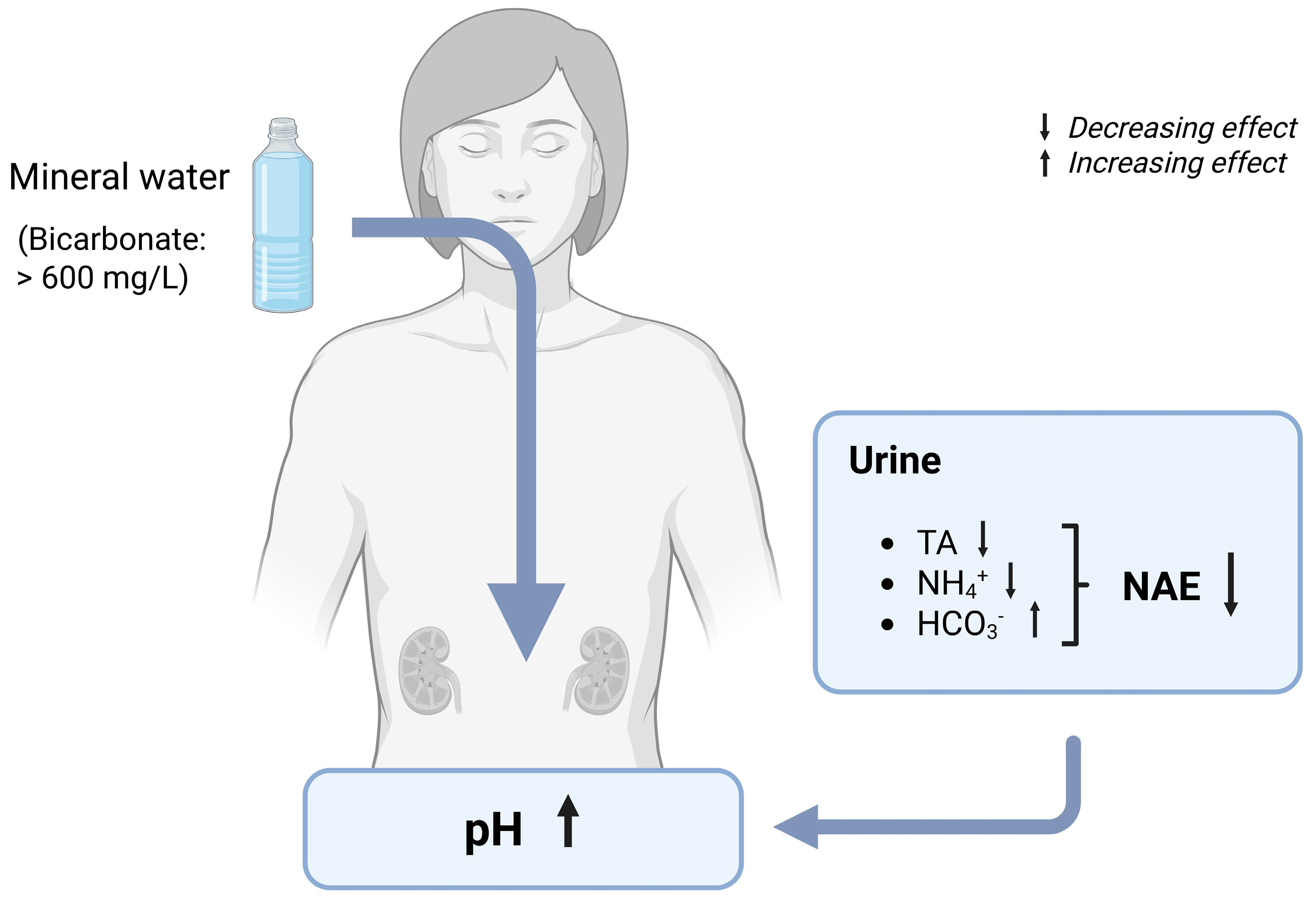
In recent decades, several studies have examined the impact of bicarbonate-rich mineral water on various aspects of urine composition (Table 3). These investigations have consistently reported favorable outcomes concerning the urinary pH and renal net acid excretion (NAE) (Figure 3). However, it should be noted that the majority of the studies only included a small number of participants (<50 subjects: [48,56,59,70,71,72,73,74,75,76,77,78,79,80], which limits the generalizability of the results. Moreover, only a very small part of the studies were conducted in a double-blind manner [59,70,77,79]. This might have affected the results, as the absence of blinding introduces a significant risk of bias, including the potential for falsification and exaggeration.
Bicarbonate-rich mineral water has been consistently demonstrated to have an alkalizing effect on urine. This effect has been demonstrated in numerous studies analyzing 24-h urine samples, as well as spontaneous or 2-h fasting urine [13,48,56,58,59,71,73,75,76,77,78,79,82,85,86]. Only two studies did not observe a significant change in the urinary pH following mineral water intake [72,74]. Remarkably, in both studies, the pH was measured in specimens differently from 24-h urine. Instead, measurements were carried out in morning fasting urine and spontaneous urine, potentially contributing to the absence of the observed changes in pH. An increase in the urinary pH has been observed following sub-chronic consumption of bicarbonate-rich mineral water, with intake ranging from 1000 mL to 2000 mL per day over periods spanning three days to twelve weeks. For example, one study demonstrated a rapid and sustained rise in the urinary pH throughout the day after mineral water intake. This observation was derived from the collection of urine on multiple occasions over the course of a 24-h cycle (with three-hour collections during the day and nine-hour collections at night) with the objective of evaluating the circadian rhythm [75]. This finding is supported by research showing that the consumption of bicarbonate-rich mineral water resulted in a significant increase in the urinary pH after just three days, whereas control water had no such effect. Similar outcomes were observed after a four-week consumption period in the same study. This indicates a rapid response with a long-lasting effect [58]. Furthermore, the alkalizing effect of such type of mineral water has been found to be comparable to that of potassium citrate supplementation [73].
There are two contributors to the effect of mineral water on the urinary pH. On the one hand, the amount of bicarbonate in the mineral water determines the level of effect on the urinary pH [52]. On the other hand, the alkalinity of the mineral water (PRAL value) also affects the urinary pH. Therefore, the interplay between bicarbonate content and mineralization seems to affect the urinary pH. To evaluate the possible differences, Wasserfurth et al. [13] conducted a study in which they examined the impact of three mineral water brands with varying bicarbonate content and the PRAL values on healthy, omnivorous adults. The urinary pH was affected by all three types of bicarbonate-rich mineral water. However, differences occurred. The water with low PRAL values led to significant pH increases regardless of the bicarbonate content (2451 mg/L and 1846 mg/L), whereas the water with a medium PRAL value produced only a marginally non-significant change. The authors concluded that PRAL may be more important than bicarbonate concentration in determining urinary alkalization.
In the same study, the consumption of different bicarbonate-rich mineral water brands resulted in a notable decrease in net acid excretion (NAE), attributed to a reduction in urinary titratable acids and ammonium. Interestingly, the advantageous impact on NAE was particularly pronounced among the groups consuming the bicarbonate-rich mineral water, characterized by a low potential renal acid load (PRAL) value. This is in line with a recent study, showing an even larger reduction in NAE after the consumption of a mineral water with a very low PRAL value and a very high bicarbonate content. As a result of daily mineral water intake over a period of 28 days, titratable acids were reduced to almost zero and ammonium excretion decreased by half. In addition, urinary bicarbonate excretion increased significantly [58]. There are additional studies demonstrating the beneficial effects on parts of the NAE equation, namely ammonium [75] and bicarbonate excretion [56]. In a study conducted with healthy male subjects, a significant decrease in urinary ammonium excretion was observed following a 5-day consumption of bicarbonate-rich mineral water while adhering to a standardized diet. The same effect was shown in a subsequent phase of this study, evaluating a 4-week mineral water consumption under the conditions of usual dietary and beverage consumption [75]. In an additional study, the consumption of bicarbonate-rich mineral water for a duration of four weeks resulted in a significant increase of urinary bicarbonate levels in healthy women [56]. However, both studies assessed only the components of the net acid excretion equation, focusing solely on either urinary ammonium or bicarbonate excretion. Hence, it is plausible that mineral water consumption may have influenced the unmeasured component, yet alterations in these parameters of the acid–base balance were not quantified.
The observed beneficial effects on NAE may potentially influence other urinary constituents, such as calcium excretion. A meta-analysis comprising five studies unveiled a statistically significant linear relationship between renal net acid excretion and urinary calcium excretion. Specifically, higher NAE levels were associated with elevated renal calcium excretion. For each 10-mEq increase in NAE, the urinary calcium increased by 0.3 mmol/d [88]. On the other side, a lower NAE value was accompanied by lower calcium excretion, which could be demonstrated for the intake of base supplements [36]. In line with these findings, the consumption of bicarbonate-rich mineral water has been demonstrated to reduce the urinary calcium excretion [70,76,87]. Nonetheless, mineral water with a calcium content between 170 and 548 mg/L has been shown to increase calcium excretion, presumably due to an overload in calcium intake [13,56,71,74,75,77,86]. Further information on this topic can be found in Section 7.2.2 on “stone risk”.

7.2. Mechanisms of Changed Urinary Composition by Bicarbonate-Rich Mineral Water

Besides the known effects of minerals on the acid–base balance, the consumption of bicarbonate via mineral water leads to an increase in the urine pH [42]. The increase in pH can be attributed to an increase in alkali reserve, which is caused by the intake of bicarbonate via mineral water [77]. Oral bicarbonate intake affects the acid–base equilibrium primarily due to its role as a buffer in the body [42]. When bicarbonate is administered, it increases the buffering capacity of the body; bicarbonate contributes to the primary human systemic buffer system—the plasma carbonic acid/bicarbonate buffer system— by direct intestinal absorption [2,89]. With an adequate buffer system in place, the protons generated during metabolism can be efficiently buffered and subsequently excreted via urine [2]. This also affects renal ammonia excretion. Ammoniagenesis potentially decreases as less acids need to be neutralized by the ammonia/ammonium buffer system in the kidneys. In addition, excess bicarbonate itself can be excreted directly via urine by upregulation of the switching cells (type B) in the collecting duct, contributing to urine alkalinization [90,91].

7.2.1. Renal Stones

Among other things, dietary habits and the intake of certain medications significantly modulate the risk of kidney and urinary tract stones [92,93]. Most of them are related to acid–base balance. For example, the intake of magnesium and potassium is associated with a reduced risk of stone formation, while a higher intake of animal protein, sodium, and sucrose is associated with an increased risk of stone formation [38]. In addition, the daily consumption of mineral water has been shown to be associated with a reduced risk of kidney and urinary stones [93]. It is noteworthy that there are different types of urinary stones and the dietary causes of stone formation are as multifactorial as the different stone compositions. However, calcium oxalate (CaOx) stones are identified as the predominant causes of stones in over 80% of individuals with stone-related issues, often displaying a recurrent pattern [92]. Calcium phosphate stones (CaP), uric acid stones (UA), struvite stones, and cystine stones are less common in stone disease [94]. Since CaOx are the most common stones in the urinary tract, the focus of this section is on CaOx stones.

7.2.2. Effects of Bicarbonate-Rich Mineral Water on Stone Risk

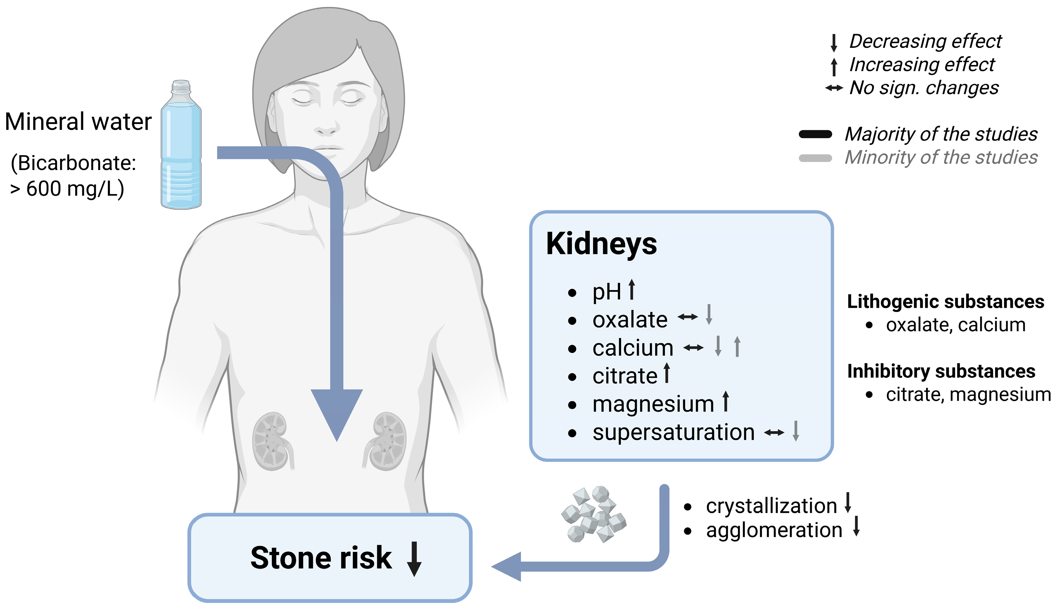
Among the various applications of bicarbonate-rich mineral water in the context of the acid–base balance, the prevention or recurrence of CaOx stone formation is by far the most extensively studied (Table 4) [71,72,74,75,77,81,86]. In this context, all studies on mineral water have focused on altering the composition of urine to establish a favorable balance between lithogenic and crystallization-inhibiting substances. In the past decades, the effect of bicarbonate-rich mineral water on stone risk was evaluated in several randomized controlled trials, showing mainly positive results (Figure 4). Most studies were conducted over a rather short or sub-chronical consumption period, ranging from three days to twelve weeks. In these studies, several urinary markers reflecting stone risk were tested, such as oxalates, calcium, citrates, and magnesium. Moreover, changes in the urinary pH were examined.
However, it is important to note that the majority of these studies had a limited sample size (<50 subjects: [56,70,71,72,73,74,75,76,77,95]), which reduces the extent to which their findings can be generalized. Moreover, only two studies were conducted in a double-blind manner [70,77]. This may have exerted an influence on the results, as the absence of blinding engenders a considerable risk of bias, including the potential for falsification and exaggeration.
The impact of bicarbonate-rich mineral water on raising the urinary pH is well-established and has been discussed in Section 7.1.1 on “urine pH”.
Besides urinary pH, one of the most important markers for the formation of CaOx stones is urinary oxalate. Its presence in increased concentrations has been associated with an elevated risk of stone formation [52]. The results of randomized controlled trials regarding the influence of bicarbonate-rich mineral water on urinary oxalate excretion are inconsistent. However, only a single study has demonstrated an increasing effect after a three-day consumption of 1500 mL mineral water in subjects with known urolithiasis [77], indicating a negative effect on stone risk. On the contrary, there are several studies showing a significant reduction in oxalate excretion after the daily consumption of 2000 mL bicarbonate-rich mineral water [71,72,73]. For instance, in one study conducted with healthy younger men, bicarbonate-rich mineral water demonstrated the same effectivity as a base supplement in reducing urinary oxalate concentrations under a standardized diet. While oxalate levels decreased by 10% following the consumption of K citrate, they were even lowered by 25% after the consumption of bicarbonate-rich mineral water. However, the authors claimed only a non-significant tendency towards a reduction under a usual diet [81]. Thus, dietary differences might be the main influencing factor regarding oxalate excretion. It is therefore not surprising that there are also studies showing no significant effect on the modulation of oxalate excretion in urine when food intake is not standardized [74,75,86]. It is worth mentioning that the effect of bicarbonate-rich mineral water on oxalate excretion could be influenced by both overall dietary intake and the calcium content of the ingested water. Furthermore, the timing of water consumption may be significant, particularly if the water is high in calcium. A high calcium intake during meals can alter oxalate excretion through the intestinal complexation of oxalates [86]. The higher the calcium intake during meals the lower the absorption rate of oxalates in the gut, thus a lesser amount of oxalates must be excreted via urine [96].
Besides urinary oxalates, urinary calcium excretion is also one of the important factors negatively affecting stone risk. Therefore, it could be assumed that a diet of mineral water low in calcium would be preferable for stone formers, as they do not involve an excessive intake of calcium that may need to be excreted via urine. However, there are studies indicating a decreased risk of stone formation associated with increased calcium intake [97,98]. This phenomenon may arise from the already mentioned effect of reduced oxalate absorption. Therefore, about half of the studies examining the impact on stone risk and bone health rely on the utilization of calcium-rich mineral water to augment dietary calcium intake. In some of these studies, the higher calcium intake resulted in an increase in urinary calcium excretion, possibly due to an excess in calcium intake [13,56,71,74,75,86]. However, the evaluation of group differences between bicarbonate-rich mineral water and the corresponding control water showed conflicting results. Three of the studies reported a higher calcium excretion in the bicarbonate group than the control group [71,74,75]. On the contrary, one study demonstrated lower levels after the consumption of bicarbonate-rich mineral water compared to a calcium- and sulfate-rich mineral water [55]. A further study underlined the influence of the calcium content of the studied mineral water brands. Groups receiving mineral water with a high calcium content showed a significant increase in calcium excretion, while the group consuming calcium-poor mineral water showed no significant changes in calcium excretion [13]. Nevertheless, there are also studies that have demonstrated no significant different effects on urinary calcium excretion, despite the use of calcium-rich mineral water in the bicarbonate-group [72,74,86]. In subjects with idiopathic calcium nephrolithiasis, the studied mineral water led to a non-significant rise in urinary calcium excretion after a 20-day consumption period [72]. In addition, one study reported a significant time × group interaction, showing the opposite effects in both intervention groups. While there was a non-significant rise in calcium excretion in the bicarbonate-group, the control group showed a decrease in calcium excretion [74]. In addition to the use of calcium-rich mineral water, there are also studies investigating the effect of bicarbonate-rich mineral water that was simultaneously low in calcium [58,70,76,77,81,85,95]. Surprisingly, one of these studies reported a significant increase in urinary calcium excretion in the bicarbonate group [77]. By contrast, no significant effects were demonstrated, although a tendency towards a reduction occurred [81]. In addition, there are several studies that have reported a significant reduction in calcium excretion [58,70,76,85,95]. However, only three studies on calcium-poor mineral water have reported a significantly lower calcium excretion in the bicarbonate group compared to the control group [76,85,95]. One could speculate that the discrepancies observed in the low-calcium mineral water could be due to differences in the dietary calcium intake. However, only seven out of fifteen studies have provided precise information on the dietary calcium intake, which makes it difficult to assess the influence of dietary calcium. The dietary interventions implemented in these studies were characterized by the provision of a standardized diet, which generally encompassed balanced calcium (approximately 1000 mg/d) and protein intake (75 to 96 g/d). Only two studies were conducted wherein the subjects followed a low-calcium diet (400–600 mg/d). Notwithstanding the implementation of a standardized diet, the outcomes pertaining to renal calcium excretion exhibited a great variability across these studies. Two studies have demonstrated no significant group differences [72,73], while others have reported a significant higher [71,74,75] or a significant lower calcium excretion in the bicarbonate group [56,95]. Taking into account the whole calcium intake (diet and mineral water), did not clarify the differences. The higher total calcium intake in the bicarbonate groups compared to the respective control groups [71,72,73,74,75] did not automatically result in a higher calcium excretion. Moreover, under both high [56] and low [95] calcium intake, consumption of bicarbonate-rich mineral water resulted in significantly lower calcium excretion compared to the control group that consumed the same amount of calcium. Therefore, the underlying causes seem to be independent from calcium intake.
In addition, urinary citrate and magnesium excretion play a pivotal role in stone inhibition [52]. A daily consumption of 1250–2000 mL of bicarbonate-rich mineral water has been shown to increase urinary citrate excretion in healthy subjects and stone formers, regardless the duration of the study [71,72,73,74,75,77,86]. Some of these studies have demonstrated the superiority of bicarbonate-rich mineral water over the control [71,75,77], while others have reported no significant group differences [72,73,74]. In addition, one study showed an opposite reaction in the two intervention groups, with an increase in the bicarbonate group and a decrease in the group consuming a low mineralized mineral water. However, no p-values are reported for time effects within each intervention group. Nevertheless, the differences in change over time were not significant (p = 0.084) [86].
Regarding magnesium excretion, several studies using bicarbonate-rich mineral water have demonstrated a beneficial effect. Participants who consumed 1250–2000 mL of this type of water showed a significant increase in urinary magnesium [71,72,75,77,86]. Changes occurred both in individuals without health issues [75] and those with a history of urinary stones [71,72,75,86]. Moreover, most of the studies have reported significant group differences for the used mineral water with a higher urinary magnesium excretion in the bicarbonate group compared to the control group. Only one study demonstrated a significant rise in magnesium excretion, which was lower in the bicarbonate group than the control group [72]. On the contrary, a minority of studies show no significant changes in urinary magnesium excretion [73,74], although magnesium excretion tended to increase in one of them (p = 0.058) [74]. Interestingly, these studies were conducted with healthy subjects, suggesting differences between stone formers and healthy participants.
Stone risk is usually assessed by evaluating the supersaturation of various urinary constituents, mainly CaOx and CaP. Although bicarbonate-rich mineral water positively affects the levels of various urinary constituents, its effect on stone risk appears to be inconsistent in terms of relative supersaturation. Despite the increased calcium excretion observed in some studies, the risk of stone formation was not adversely affected. On the contrary, the risk of CaOx stone formation was shown to decrease with the consumption of both calcium-rich and calcium-poor mineral water. Thus, beneficial effects were demonstrated for stone risk indices, especially for relative supersaturation and the Tiselius index [71,73,75,77], both methods for assessing the risk of recurrent stones [99]. For instance, in a study where the participants followed a standardized diet, the relative supersaturation of CaOx (RS CaOx) decreased by 43% after the consumption of bicarbonate-rich mineral water. This reduction was higher than the reduction in the control phase, where the participants received an alkaline supplement (K citrate). The control phase showed only a decrease of 25%. Moreover, the relative supersaturation of uric acid (RS UA) decreased by 73% in both study groups [73]. However, there are also studies that have showed positive changes in urinary composition, indicating a lower risk, despite the lack of significant effects on supersaturation [72,74,86]. One explanation for the absence of risk reduction in these studies could be the lack of significant reduction in urinary oxalates [74,86].

7.3. Mechanisms of Stone Risk Reduction by Bicarbonate-Rich Mineral Water

As a non-caloric beverage, bicarbonate-rich mineral water is one of the recommended beverages for subjects suffering from urolithiasis [42,52]. Increasing urine volume through higher fluid intake stands as the primary principle of stone metaphylaxis, with the aim of achieving a diluting effect on the urinary substances [42,52,100,101]. It is essential to augment fluid consumption systematically across diurnal and nocturnal periods to optimize urinary tract flushing, thereby reducing the retention time of urine in the urinary tract. This makes crystallization more difficult over the whole course of the day [52]. Indeed, a meta-analysis has showed that raising the fluid intake to achieve a urinary output ≥2500 mL/d reduced stone recurrence rates in patients with a history of nephrolithiasis [102].
As already stated, urinary pH is also an important factor contributing to stone risk. It influences the solubility of lithogenic substances, hence influencing crystallization and agglomeration. While CaOx stones and uric stones need a more acidic pH for crystallization, CaP stones and struvite stones are formed in a more alkaline pH [52]. Therefore, it is crucial to analyze the composition of stones in subjects suffering from urolithiasis to recommend the suitable mineral water. Gundermann et al. [52] stated that a bicarbonate amount of at least 1500 mg/L is necessary to raise the urinary pH in order to prevent CaOx or UA stones. Depending on the concentration of bicarbonate in mineral water and the consumed amount of mineral water, urinary pH increases differently. In most of the mineral water studies an increase up to pH 5.9–6.9 was demonstrated. However, urinary pH can even increase beyond the neutral point after the consumption of bicarbonate-rich mineral water [58,103].
All the above-mentioned urinary constituents (oxalates, calcium, citrates, magnesium) can be positively influenced by the consumption of bicarbonate-rich mineral water contributing to a risk reduction. Urinary lithogenic substances (calcium, oxalates) are partly demonstrated to be lowered by the consumption of bicarbonate-rich mineral water, resulting in a decrease in relative supersaturation. As both substances contribute to stone formation, the relative importance of oxalate excretion compared to calcium excretion is crucial in determining the risk of stone formation. This is due to the fact that calcium is a weaker crystallization promoter compared to oxalates [52,72]. Moreover, the excretion of inhibitory substances (magnesium, citrate) has been shown to increase after the consumption of bicarbonate-rich mineral water. Regarding citrate excretion, a pH-dependent mechanism for citrate re-uptake has been identified, resulting in an increased citrate excretion under more alkaline tubular pH conditions [104]. This in turn affects the crystallization process [52]. The formation of complexes between citrate and calcium leads to a reduction in the concentration of CaOx crystals [105]. In addition, magnesium forms soluble complexes with oxalates, thereby reducing supersaturation of the oxalate [106].
In terms of calcium excretion there are several possible mechanisms postulated. On the one hand, the reduction in calcium excretion may be due to a direct renal mechanism [85], mainly an increased calcium reabsorption in the distal tubules [76]. On the other hand, it may be due to the reduction of bone resorption as a consequence of a more balanced acid–base equilibrium. For a detailed description of this postulated mechanism, please refer to Section 7.5.2 on “bicarbonate-rich mineral water and bone health”. In addition, it is also postulated that the reduced calcium excretion after the consumption of bicarbonate-rich mineral water may be due to a compensatory mechanism for a reduced calcium absorption in the gut. Due to the higher pH in the digestive tract after the consumption of bicarbonate-rich mineral water, a lower solubility of calcium may lead to a lower calcium absorption, thus there is no need to excrete excess calcium [76,85].

Blood Gas Parameters

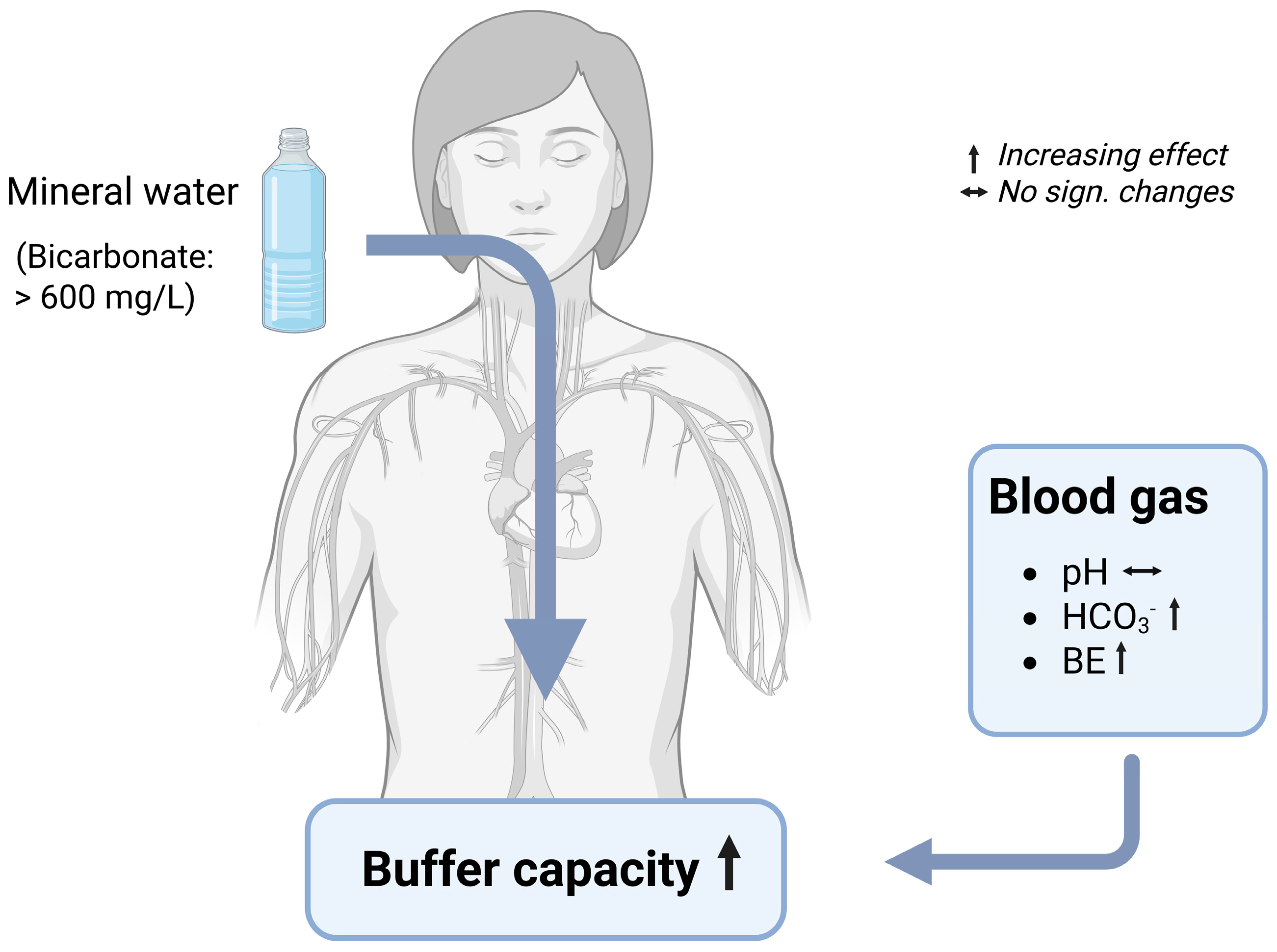
Studies on the effects of mineral water with a high content in bicarbonate on blood gas parameters are rare. The effect on bicarbonate, pH, and base excess in capillary or venous blood have mainly been assessed in athletes, evaluating the potential to improve performance; whereas non-sport studies are scarce (Table 5, Figure 5).
In consideration of the sample size, it is important to note that only two studies have evaluated the effects in a larger study collective (>90 subjects: [13,58]), while the others were conducted with a maximum of 39 subjects [107]. Additionally, the absence of blinding in the larger ones [13,58] should be regarded as a limitation, which may have had an impact on the results.
The influence of bicarbonate-rich mineral water on the blood pH depends on the study conditions, as the results differ in some respects. On the one hand, there are beneficial effects on the blood pH after anaerobic exercise. Studies with recreationally active subjects were able to demonstrate a significantly higher blood pH immediately post exercise [59,107], 3 min and 5 min post exercise [59], and even 10 min post exercise [107] after a 7-day consumption of 1500 to 2000 mL bicarbonate-rich mineral water daily, in comparison to low mineralized mineral water. In another study, a single ingestion of 3000 mL bicarbonate-rich mineral water per day resulted in a significantly higher blood pH after anaerobic cycling and an isokinetic endurance test compared to the consumption of low mineralized mineral water [108]. This short-term consumption suggests a rapid effect in the context of anaerobic sports. In contrast to the findings observed after anaerobic exercise, results concerning resting pH demonstrate significant variation. A recent study demonstrated a significantly higher resting blood pH in highly trained athletes who consumed an alkalizing diet in combination with mineral water high in bicarbonate, in comparison to the control group who consumed mineral water low in bicarbonate [79]. However, on the other hand, the resting blood pH did not differ between the two intervention groups receiving bicarbonate-rich and control water for 21 days [48]. Consistent with the latter findings, studies outside the exercise-related context have shown no effects on the blood pH. One study conducted with 129 healthy subjects demonstrated significant group differences after the consumption of four different mineral water brands (1500–2000 mL/day) for four weeks. However, none of the mineral water showed significant increases in the blood pH [13]. In line with this, a recent study with 94 healthy subjects was not able to demonstrate significant changes in the blood pH after the consumption of 1500 to 2000 mL daily of bicarbonate-rich mineral water for four weeks, although the pH tended to increase over the first three days of mineral water consumption (p = 0.068) [58].
Besides blood pH, bicarbonate levels and base excess are valuable parameters to determine the acid–base balance in blood. Regarding bicarbonate levels in blood gas samples, there are conflicting results. In some of the studies, the consumption of bicarbonate-rich mineral water did not influence blood bicarbonate levels compared to low mineralized water [13,59,107], regardless of whether the parameter was measured in the context of exercise or not. However, there are also studies where the consumption of bicarbonate-rich mineral water led to significantly higher bicarbonate levels in the blood compared to the consumption of low mineralized mineral water [48,58,59,79,108]. The discrepancy may have occurred because of the different study designs. A study design with a higher bicarbonate intake via mineral water [48,79] and intake of mineral water during exercise [108] may be the reason for the positive findings.
The data on base excess (BE), a parameter that reflects the metabolic component of the acid–base balance by quantifying the amount of the excess or deficient base in the blood, is limited. Very few studies have evaluated the effect of bicarbonate-rich mineral water on the BE, with inconsistent results. An intake of about 3000 mg per day for 4 weeks did not change the base excess in healthy adults significantly, while a slightly higher bicarbonate intake (3677 mg/d) led to a significant increase in the base excess in the same study. However, there was no significant difference in the BE between the different study groups at the end of the study [13]. In addition, during anerobic cycling, the BE decreased equally in the bicarbonate group and the control group due to exercise [107]. On the contrary, a recent study was able to demonstrate a positive effect on the BE. In this study, conducted with healthy adults, the consumption of almost 7000 mg of bicarbonate per day from mineral water over a 4-week period led to a significant increase in the BE. The increase was observed after only 3 days of mineral water consumption and was maintained until the end of the study [58]. Similar results were obtained in a study on highly trained athletes. One hour after a 400 m run, the level of BE was found to be significantly higher in the group consuming an alkalizing diet in combination with mineral water high in bicarbonate, in comparison to the control group, who consumed mineral water low in bicarbonate in addition to an alkalizing diet [79]. Thus, the amount of bicarbonate ingested may contribute to positive or neutral effects in terms of the base excess.

7.4. Mechanisms of Changes in Blood Gas Parameters by Bicarbonate-Rich Mineral Water

Several mechanisms have been suggested to explain the beneficial effects of drinking bicarbonate-rich mineral water on blood gas parameters, in particular the increasing blood bicarbonate levels. When consumed, bicarbonate from mineral water interacts with hydrochloric acid in the stomach lumen, resulting in the formation of carbonic acid. This carbonic acid subsequently decomposes into water and carbon dioxide, with the latter being exhaled directly [109]. This mechanism likely explains the potential gastrointestinal side effects associated with the consumption of bicarbonate supplements, such as flatulence and belching [89]. Furthermore, it is hypothesized that some of the bicarbonate is absorbed into the bloodstream via stimulation of the basolateral Cl/HCO3 antiporter in the stomach, thereby increasing systemic bicarbonate levels [89]. At sufficiently high intake levels, bicarbonate also appears to pass through the stomach. This bicarbonate influx continues into the small intestine, where it is absorbed in the jejunum [110]. Once absorbed in the bloodstream bicarbonate acts as part of the bicarbonate–carbon dioxide buffer system, the physiologically most important buffer system in the human body [111].

7.4.1. Bone Health

Diet and lifestyle factors are known to influence bone health, with several nutrients playing a key role in protecting human bones [8]. Among these nutrients, calcium is recognized as a bone-protective nutrient present in various foods and beverages [112,113,114]. Studies have shown that the bioavailability of calcium from mineral water is comparable to that from milk and dairy products [115,116]. Consequently, research using calcium-rich mineral water has been able to demonstrate a bone-protective effect in humans. However, there is evidence that this effect is not solely due to the calcium content; rather, the bicarbonate content of the mineral water also seems to play an important role [56,57]. As mentioned above, calcium-rich mineral water is either rich in bicarbonate or sulfate [56], resulting in either an alkalinizing or an acidifying effect on the body [43]. Therefore, the consumption of calcium- and bicarbonate-rich mineral water has the potential to counteract the acidogenic effects of an acidic diet, whereas a mineral water rich in calcium and sulfate can aggravate its adverse effects on the acid–base balance.

7.4.2. Effects of Bicarbonate-Rich Mineral Water on Bone Turnover

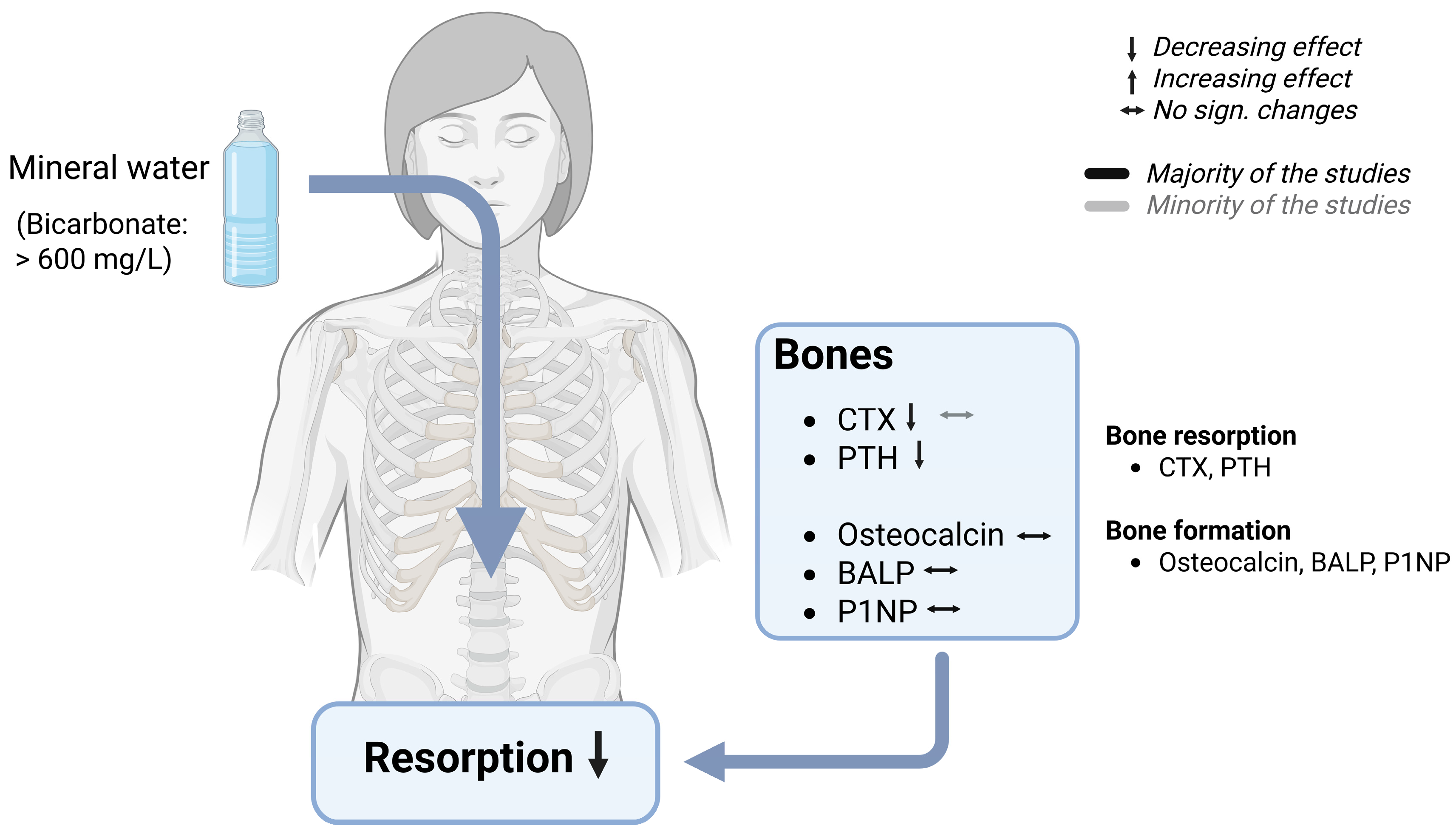
Several randomized controlled trials have assessed the effects of bicarbonate-rich mineral water on bone metabolism, predominantly reporting favorable outcomes (Table 6, Figure 6). However, as already stated, the existing body of evidence suggests that these effects might result from the interplay between bicarbonate and calcium [53,56,117]. Studies examining the effects on bone metabolism have typically used mineral water rich in both calcium and bicarbonate, encompassing both calcium-deficient and calcium-sufficient participants. Moreover, these studies have evaluated the effects over a longer period of time, ranging from four days to eight weeks. In these studies, several markers reflecting both bone resorption and bone formation were assessed following a daily consumption of 1000–2000 mL of mineral water. Notably, none of the trials reported adverse effects, while several demonstrated beneficial effects [56,71,78]. Only one study failed to demonstrate positive effects [76].
However, it is imperative to acknowledge the limitations inherent in these studies. A single study was conducted in a larger cohort, encompassing 60 postmenopausal women [82]. Furthermore, it is to be assumed that none of the studies employed a blinding method, as no such information was reported in the papers.
Short-term studies ranging from 4 days to 4 weeks have shown that calcium- and bicarbonate-rich mineral water can reduce the biomarkers of bone resorption in humans [56,71,82]. While calcium-rich mineral water alone did not affect bone markers, mineral water rich in bicarbonate and calcium demonstrated beneficial effects on bone resorption, highlighting the beneficial influence of bicarbonate [56]. In this study, the bone resorption marker carboxyterminal type I collagen telopeptide (CTX) in blood samples decreased by 15% following the consumption of mineral water, showing significant group differences. In addition, urinary CTX showed a nonsignificant slight reduction, resulting in no significant group differences. However, in an earlier study, urinary CTX was shown to be reduced after bicarbonate-rich mineral water consumption with significantly lower levels after the bicarbonate-rich mineral phase and the control phases [71]. On the contrary, one study reported no significant differences in serum CTX after drinking a bicarbonate- and sodium-rich, but calcium-poor mineral water and a low mineralized mineral water [76]. On the other hand, there are indications that levels in the parathyroid hormone (PTH), a hormone responsible for bone resorption, can also be affected by mineral water consumption [57]. Three different randomized controlled trials have demonstrated a reduction in the parathyroid hormone (PTH), a hormone involved in bone demineralization and degradation of bone substance [43]. In postmenopausal women, ionized PTH levels in plasma decreased by 11% [82], while in young women, plasma PTH decreased by 16% [56] after the consumption of either 1000 or 1500 mL of bicarbonate and calcium-rich mineral water. However, one of these studies showed no significant group differences, indicating no different water effects [82]. On the contrary, the other study showed the superiority of bicarbonate-rich mineral water over a low mineralized mineral water [56]. In addition, in middle-aged men and women, a slightly higher intake of a bicarbonate- and calcium-rich mineral water (2000 mL/d) also led to a significant reduction of PTH in comparison to a mineral water poor in bicarbonate and calcium [71].
However, bone health is a constant interplay between bone resorption and bone formation. Hence, the effect on several bone-forming substances was also examined. In contrast to the biomarkers of bone resorption, bone-forming biomarkers, namely osteocalcin, bone-specific alkaline phosphatase (BALP), and procollagen type 1 N-terminal propeptide (P1NP), remained unchanged following the consumption of calcium- and bicarbonate-rich or bicarbonate- and sodium-rich mineral water [56,71,76,82].

7.5. Mechanism of Bone Protection by Bicarbonate-Rich Mineral Water

One possible mechanism underlying the bone-protective effects of bicarbonate-rich mineral water may be due to its pH-increasing properties. Evidence is derived from in vitro studies. In these studies, it has been shown that both osteoclasts, bone cells responsible for bone dissolution, and osteoblasts, bone cells responsible for bone formation, respond to changes in pH. Osteoclasts work best at a lower pH (around 7.0), and become progressively less active with increasing pH. Above pH 7.4, resorption is switched off. On the contrary, osteoblasts work best at a pH of 7.4, and osteoblast function is decreased by metabolic acidosis due to a decreased expression of extracellular matrix genes [118,119,120,121]. In brief, hydrogen ions generated from the digestion of foods rich in acid-forming precursors bind to osteoblasts and trigger the release of the receptor activator of the nuclear factor kappa-B ligand (RANK-L). This, in turn, promotes an increase in the number and activation of osteoclasts, thereby activating bone resorption [8]. This association could potentially be reversed or positively influenced by an alkaline diet or adequate consumption of bicarbonate-rich mineral water. Thus, the pH-increasing effects may modulate osteoclast activity and reduce bone resorption. Furthermore, the bone-protective effects may be partly mediated by the PTH-lowering properties of calcium- and bicarbonate-rich mineral water, thereby protecting against bone resorption.
Moreover, it has been postulated that decreasing the system pH increases the release of calcium from bones [122]. However, opponents of this acid–ash bone theory question the extent of this interrelationship. They argue that the higher urinary calcium, as it occurs under acidic diets, may also stem from increased intestinal calcium absorption, and not necessarily from the increased calcium loss that is caused by increased bone resorption [18,36].

7.5.1. Effects of Bicarbonate-Rich Mineral Water on Bone Density and Fracture Risk

Despite the observed short-term effects on the markers of bone resorption, the long-term effects of consuming bicarbonate- and calcium-rich mineral water on bone density and fracture risk remain unclear [18,43]. There is a lack of observational and interventional studies examining this aspect specifically in the context of mineral water consumption over a longer period of time. Nevertheless, its bone-protective effect is conceivable, as indicated by a meta-analysis suggesting the potential benefits of alkaline supplements. This analysis revealed that interventions with alkaline supplements significantly increased bone mineral density at the femoral neck, lumbar spine, and total hip [36]. Similar results were observed in a longitudinal analysis that pooled data from two Mediterranean populations of middle-aged and older individuals. An analysis of the data revealed an increase in bone mass at different sites (total femur, femoral neck, femoral diaphysis, trochanter) associated with an acid-neutral diet (PRAL = 0) [123]. Nevertheless, the evidence of the relationship between dietary acid load and fracture risk remains inconclusive. The same longitudinal analysis reported a reduced risk of osteoporotic fractures with an acid-neutral diet. However, a low dietary PRAL score, which indicates an alkaline diet, was also associated with an increased risk of fractures [123]. By contrast, another meta-analysis found no association between fracture risk and adherence to an acid-producing diet. In addition, one study observed a weak association between NEAP and bone mineral density, while another study found no association between PRAL and bone mineral density [3].

7.5.2. Bicarbonate-Rich Mineral Water and the Complexity with Other Nutrients Regarding Bone Health

In the context of mineral water and its potential effect on bone health, several components may contribute to positive effects. One such element is the calcium content, as mentioned above. In addition, the high potassium content of mineral water has been suggested to contribute to bone health [53]. However, research using low-potassium and high bicarbonate water has also demonstrated a successful reduction in bone resorption [82]. In addition, the potassium content of most water is generally considered to be insufficient to significantly affect bone metabolism [43].
Conversely, the relationship between fluoride-rich water and bone mineral density and fracture risk has also been studied in epidemiological studies. Some of the results suggest a potential positive association between fluoride intake and bone mineral density/fracture risk [124,125]. However, other studies could not confirm these associations [126,127,128]. In fact, there was even an increased fracture risk observed in postmenopausal women [125]. However, it is important to note that these findings specifically relate to fluoridated drinking water, not mineral water [43].

8. Summary Bicarbonate-Rich Mineral Water

Acid–base balance, a fundamental aspect of human health, can be influenced by the choice of consumed foods and beverages. A dietary pattern characterized by a high intake of fruits and vegetables has been shown to contribute to a balanced acid–base status by reducing the acid load contrast, while the frequent consumption of meat, cheese and cereals is associated with an increased dietary acid load. Given the propensity of the modern Western diet to induce excess acidity, there is a need to explore strategies aimed at mitigating this acidity. Consequently, considerable attention has been devoted in recent decades to evaluate the effects of bicarbonate-rich mineral water on the acid–base balance and related diseases.
Taken together, the findings from various scientific reports have indicated that mineral water with a bicarbonate level of more than 1300 milligrams per liter and a medium or low PRAL value exerts a beneficial and multifaceted effect on the urinary and blood parameters. One of the primary effects is the increase in the urine pH, caused by the enhancement of the body’s alkali reserves due to bicarbonate and mineral intake. This alkalization lowers renal NAE by reducing the excretion of TA and NH4+, while simultaneously increasing HCO3 excretion. These changes in the urinary composition contribute to decreased urinary concentrations of stone-forming constituents, such as calcium and oxalates, with a more pronounced effect seen on oxalates. Conversely, the excretion of inhibitory substances, such as magnesium and citrate, are promoted. Moreover, it has been demonstrated that the alkalinization leads to a reduction in the bone resorption marker CTX, potentially by a reduction in osteoclast activation and a decrease in PTH levels. In terms of blood parameters, the consumption of bicarbonate-rich water stabilizes the blood pH while increasing the blood bicarbonate levels and the base excess, indicating a strengthened bicarbonate buffer system. This is achieved through bicarbonate absorption in the gastrointestinal tract, which enhances systemic buffering capacity and modulates pH, supporting general acid–base homeostasis.
From a clinical perspective, these changes lead to a lower risk of CaOx stone formation and promote kidney health due to an increase in the urine pH and favorable alterations in the urine composition. By preventing excessive crystallization and agglomeration of calcium and oxalates within the urinary tract, bicarbonate-rich mineral water is particularly beneficial for individuals with a history of CaOx urolithiasis. Additionally, by promoting an alkaline environment that favors osteoblastic activity and impedes osteoclastic activity, bicarbonate-rich mineral water can support bone health, although further research is required to fully assess its impact on bone density and fracture risk over time.
The studies included in this review exhibit several methodological limitations that must be considered when interpreting the overall findings. Most notably, the majority of the studies had a relatively small sample sizes, typically around 20 participants, which limits statistical power and generalizability. Only a few studies included more than 50 participants, reducing confidence in the robustness of the reported effects. Another important limitation concerns the study design quality. Randomization was not consistently applied across the studies, and information regarding blinding was often missing. Specifically, only five studies reported using a double-blind design, another five were single-blinded, five studies explicitly stated that no blinding was used, and, in eight studies, blinding procedures were not described at all. The frequent lack of blinding introduces a substantial risk of performance and detection bias. Furthermore, nearly half of the studies employed a parallel-group design rather than a crossover design (14 crossover vs. 9 parallel-group studies). While both designs have their merits, crossover trials generally offer increased statistical efficiency in within-subject comparisons, which is particularly valuable in studies with small sample sizes. The use of a crossover design may therefore represent a missed opportunity to enhance internal validity and reduce inter-individual variability. Collectively, these limitations highlight the need for more rigorously designed, adequately powered, and transparently reported trials in this field. Future studies should be conducted as randomized, double-blind, controlled studies in a crossover design. Additionally, a sufficient high sample size is imperative to enhance the statistical significance and the study’s generalizability, making the findings more representative of the broader population, and improving the external validity of the results. As the extant literature has predominantly focused on the effects of very high drinking volumes, it is imperative that future studies also examine the consequences of lower volumes. Finally, it is essential to record the diet using food frequency questionnaires or 3-day dietary protocols. However, the implementation of a controlled diet would be a superior approach.
In conclusion, bicarbonate-rich mineral water offers considerable benefits for acid–base regulation. In this context it appears to play a significant role in optimizing the urinary and blood parameters, offering protective effects against certain health conditions. Its incorporation into the diet as a strategy to counteract dietary-induced acidity, lowering kidney stone risk, and enhancing skeletal integrity holds promise. However, further methodologically improved studies are necessary to fully elucidate these benefits and to optimize recommendations for long-term health promotion.

Author Contributions

Conceptualization: K.M. and A.H.; formal Analysis: K.M. and A.H.; data Curation: K.M.; writing—Original Draft Preparation: K.M.; writing—Review and Editing: K.M., A.H. and T.H.; visualization: K.M.; supervision: A.H.; project administration: A.H. All authors have read and agreed to the published version of the manuscript.

Funding

This research received no external funding.

Conflicts of Interest

The authors declare no conflicts of interest.

Abbreviations

PRAL Potential Renal Acid Load
NEAP Net Endogenous Acid Production
NAE Net Acid Excretion
OA Organic acids
TA Titrable acids
NH4+ Ammonium
HCO3- Bicarbonate
Ca Calcium
Mg Magnesium
Na Sodium
K Potassium
Cl Chloride
SO4 Sulfate
P Phosphorus
BE Base Excess
CaOx Calcium oxalate
CaP Calcium phosphate
RS Relative supersaturation
UA Uric acid
Cr Creatinine
CTX C-terminal fragment of the type I collagen (CTX)
iCa Ionized calcium
P1NP Procollagen type 1 N-terminal propeptide
PTH Parathyroid hormone

References

  1. Osuna-Padilla, I.A.; Leal-Escobar, G.; Garza-García, C.A.; Rodríguez-Castellanos, F.E. Dietary acid load: Mechanisms and evidence of its health repercussions. Nefrol. Engl. Ed. 2019, 39, 343–354. [Google Scholar] [CrossRef]
  2. Ströhle, A.; Remer, T. Ernährung und Säure-Basen-Haushalt. Physiologie und Prävention. Ernähr. Im Fokus 2014, 14, 314–324. [Google Scholar]
  3. Gholami, F.; Naghshi, S.; Samadi, M.; Rasaei, N.; Mirzaei, K. Dietary Acid Load and Bone Health: A Systematic Review and Meta-Analysis of Observational Studies. Front. Nutr. 2022, 9, 869132. [Google Scholar] [CrossRef]
  4. Wieërs, M.L.A.J.; Beynon-Cobb, B.; Visser, W.J.; Attaye, I. Dietary acid load in health and disease. Pflüg. Arch.-Eur. J. Physiol. 2024, 476, 427–443. [Google Scholar] [CrossRef] [PubMed]
  5. Remer, T. Influence of nutrition on acid-base balance—Metabolic aspects. Eur. J. Nutr. 2001, 40, 214–220. [Google Scholar] [CrossRef]
  6. Adeva, M.M.; Souto, G. Diet-induced metabolic acidosis. Clin. Nutr. 2011, 30, 416–421. [Google Scholar] [CrossRef]
  7. Betz, M.V.; Penniston, K.L. Primary Contributors to Dietary Acid Load in Patients With Urolithiasis. J. Ren. Nutr. 2023, 33, 53–58. [Google Scholar] [CrossRef]
  8. Dawson-Hughes, B. Acid–base balance of the diet—Implications for bone and muscle. Eur. J. Clin. Nutr. 2020, 74, 7–13. [Google Scholar] [CrossRef] [PubMed]
  9. Storz, M.A.; Müller, A.; Ronco, A.L. Nutrient Intake and Dietary Acid Load of Special Diets in the NHANES: A Descriptive Analysis (2009–2018). Int. J. Environ. Res. Public. Health 2022, 19, 5748. [Google Scholar] [CrossRef]
  10. Frassetto, L.; Banerjee, T.; Powe, N.; Sebastian, A. Acid Balance, Dietary Acid Load, and Bone Effects—A Controversial Subject. Nutrients 2018, 10, 517. [Google Scholar] [CrossRef]
  11. Remer, T.; Manz, F. Estimation of the renal net acid excretion by adults consuming diets containing variable amounts of protein. Am. J. Clin. Nutr. 1994, 59, 1356–1361. [Google Scholar] [CrossRef] [PubMed]
  12. Carnauba, R.; Baptistella, A.; Paschoal, V.; Hübscher, G. Diet-Induced Low-Grade Metabolic Acidosis and Clinical Outcomes: A Review. Nutrients 2017, 9, 538. [Google Scholar] [CrossRef] [PubMed]
  13. Wasserfurth, P.; Schneider, I.; Ströhle, A.; Nebl, J.; Bitterlich, N.; Hahn, A. Effects of mineral waters on acid-base status in healthy adults: Results of a randomized trial. Food Nutr. Res. 2019, 63, 3515. [Google Scholar] [CrossRef]
  14. Remer, T.; Dimitriou, T.; Manz, F. Dietary potential renal acid load and renal net acid excretion in healthy, free-living children and adolescents. Am. J. Clin. Nutr. 2003, 77, 1255–1260. [Google Scholar] [CrossRef]
  15. Sebastian, A.; Frassetto, L.A.; Sellmeyer, D.E.; Merriam, R.L.; Morris, R.C. Estimation of the net acid load of the diet of ancestral preagricultural Homo sapiens and their hominid ancestors. Am. J. Clin. Nutr. 2002, 76, 1308–1316. [Google Scholar] [CrossRef]
  16. Frassetto, L.; Shi, L.; Schloetter, M.; Sebastian, A.; Remer, T. Established dietary estimates of net acid production do not predict measured net acid excretion in patients with Type 2 diabetes on Paleolithic–Hunter–Gatherer-type diets. Eur. J. Clin. Nutr. 2013, 67, 899–903. [Google Scholar] [CrossRef] [PubMed]
  17. Frassetto, L.A.; Lanham-New, S.A.; Macdonald, H.M.; Remer, T.; Sebastian, A.; Tucker, K.L.; Tylavsky, F.A. Standardizing Terminology for Estimating the Diet-Dependent Net Acid Load to the Metabolic System. J. Nutr. 2007, 137, 1491–1492. [Google Scholar] [CrossRef]
  18. Frassetto, L.A.; Todd, K.M.; Morris, R.C.; Sebastian, A. Estimation of net endogenous noncarbonic acid production in humans from diet potassium and protein contents123. Am. J. Clin. Nutr. 1998, 68, 576–583. [Google Scholar] [CrossRef]
  19. Parmenter, B.H.; Dymock, M.; Banerjee, T.; Sebastian, A.; Slater, G.J.; Frassetto, L.A. Performance of Predictive Equations and Biochemical Measures Quantifying Net Endogenous Acid Production and the Potential Renal Acid Load. Kidney Int. Rep. 2020, 5, 1738–1745. [Google Scholar] [CrossRef]
  20. Nagami, G.T.; Kraut, J.A. The Role of the Endocrine System in the Regulation of Acid–Base Balance by the Kidney and the Progression of Chronic Kidney Disease. Int. J. Mol. Sci. 2024, 25, 2420. [Google Scholar] [CrossRef]
  21. Rylander, R.; Remer, T.; Berkemeyer, S.; Vormann, J. Acid-Base Status Affects Renal Magnesium Losses in Healthy, Elderly Persons. J. Nutr. 2006, 136, 2374–2377. [Google Scholar] [CrossRef] [PubMed]
  22. Gannon, R.H.T.; Millward, D.J.; Brown, J.E.; Macdonald, H.M.; Lovell, D.P.; Frassetto, L.A.; Remer, T.; Lanham-New, S.A. Estimates of daily net endogenous acid production in the elderly UK population: Analysis of the National Diet and Nutrition Survey (NDNS) of British adults aged 65 years and over. Br. J. Nutr. 2008, 100, 615–623. [Google Scholar] [CrossRef]
  23. Ströhle, A.; Waldmann, A.; Koschizke, J.; Leitzmann, C.; Hahn, A. Diet-Dependent Net Endogenous Acid Load of Vegan Diets in Relation to Food Groups and Bone Health-Related Nutrients: Results from the German Vegan Study. Ann. Nutr. Metab. 2011, 59, 117–126. [Google Scholar] [CrossRef] [PubMed]
  24. Wang, Y.; Moss, J.; Thisted, R. Predictors of body surface area. J. Clin. Anesth. 1992, 4, 4–10. [Google Scholar] [CrossRef] [PubMed]
  25. Manz, F. History of nutrition and acid-base physiology. Eur. J. Nutr. 2001, 40, 189–199. [Google Scholar] [CrossRef]
  26. Jehle, S.; Zanetti, A.; Muser, J.; Hulter, H.N.; Krapf, R. Partial Neutralization of the Acidogenic Western Diet with Potassium Citrate Increases Bone Mass in Postmenopausal Women with Osteopenia. J. Am. Soc. Nephrol. 2006, 17, 3213–3222. [Google Scholar] [CrossRef]
  27. Naude, D.F. Chronic Sub-Clinical Systemic Metabolic Acidosis—A Review with Implications for Clinical Practice. J. Evid.-Based Integr. Med. 2022, 27, 2515690X221142352. [Google Scholar] [CrossRef]
  28. Raphael, K.L. Metabolic Acidosis and Subclinical Metabolic Acidosis in CKD. J. Am. Soc. Nephrol. JASN 2018, 29, 376–382. [Google Scholar] [CrossRef]
  29. DuBose, T.D. Urine Ammonium and Preclinical Acidosis in CKD. J. Am. Soc. Nephrol. JASN 2017, 28, 2258–2260. [Google Scholar] [CrossRef]
  30. Alpern, R.J.; Sakhaee, K. The clinical spectrum of chronic metabolic acidosis: Homeostatic mechanisms produce significant morbidity. Am. J. Kidney Dis. Off. J. Natl. Kidney Found. 1997, 29, 291–302. [Google Scholar] [CrossRef]
  31. Maurer, M.; Riesen, W.; Muser, J.; Hulter, H.N.; Krapf, R. Neutralization of Western diet inhibits bone resorption independently of K intake and reduces cortisol secretion in humans. Am. J. Physiol.-Ren. Physiol. 2003, 284, F32–F40. [Google Scholar] [CrossRef] [PubMed]
  32. Esche, J. Higher diet-dependent renal acid load associates with higher glucocorticoid secretion and potentially bioactive free glucocorticoids in healthy children. Kidney Int. 2016, 90, 325–333. [Google Scholar] [CrossRef] [PubMed]
  33. Esche, J.; Krupp, D.; Mensink, G.B.; Remer, T. Dietary Potential Renal Acid Load Is Positively Associated with Serum Uric Acid and Odds of Hyperuricemia in the German Adult Population. J. Nutr. 2018, 148, 49–55. [Google Scholar] [CrossRef] [PubMed]
  34. Dobrzyńska, M.; Przysławski, J. The relationship between serum uric acid concentration and cardiovascular risk factors in normotensivepostmenopausal women with dyslipidemia. Acta Sci. Pol. Technol. Aliment. 2020, 19, 123–131. [Google Scholar] [CrossRef] [PubMed]
  35. Zhang, M.; Ye, C.; Wang, R.; Zhang, Z.; Huang, X.; Halimulati, M.; Sun, M.; Ma, Y.; Zhang, Z. Association between Dietary Acid Load and Hyperuricemia in Chinese Adults: Analysis of the China Health and Nutrition Survey (2009). Nutrients 2023, 15, 1806. [Google Scholar] [CrossRef]
  36. Han, Y.; An, M.; Yang, L.; Li, L.; Rao, S.; Cheng, Y. Effect of Acid or Base Interventions on Bone Health: A Systematic Review, Meta-Analysis, and Meta-Regression. Adv. Nutr. 2021, 12, 1540–1557. [Google Scholar] [CrossRef]
  37. Passey, C. Reducing the Dietary Acid Load: How a More Alkaline Diet Benefits Patients With Chronic Kidney Disease. J. Ren. Nutr. 2017, 27, 151–160. [Google Scholar] [CrossRef]
  38. Trinchieri, A.; Lizzano, R.; Marchesotti, F.; Zanetti, G. Effect of potential renal acid load of foods on urinary citrate excretion in calcium renal stone formers. Urol. Res. 2006, 34, 1–7. [Google Scholar] [CrossRef]
  39. Vezzoli, G.; Dogliotti, E.; Terranegra, A.; Arcidiacono, T.; Macrina, L.; Tavecchia, M.; Pivari, F.; Mingione, A.; Brasacchio, C.; Nouvenne, A.; et al. Dietary style and acid load in an Italian population of calcium kidney stone formers. Nutr. Metab. Cardiovasc. Dis. 2015, 25, 588–593. [Google Scholar] [CrossRef]
  40. Miki, A.; Hashimoto, Y.; Tanaka, M.; Kobayashi, Y.; Wada, S.; Kuwahata, M.; Kido, Y.; Yamazaki, M.; Fukui, M. Urinary pH reflects dietary acid load in patients with type 2 diabetes. J. Clin. Biochem. Nutr. 2017, 61, 74–77. [Google Scholar] [CrossRef]
  41. Mofrad, M.D.; Daneshzad, E.; Azadbakht, L. Dietary acid load, kidney function and risk of chronic kidney disease: A systematic review and meta-analysis of observational studies. Int. J. Vitam. Nutr. Res. 2021, 91, 343–355. [Google Scholar] [CrossRef] [PubMed]
  42. Siener, R. Nutrition and Kidney Stone Disease. Nutrients 2021, 13, 1917. [Google Scholar] [CrossRef]
  43. Ströhle, A.; Hahn, A. Ossäre Bedeutung von Mineral- und Heilwässern: Physiologie und epidemiologische Evidenz. Osteologie 2023, 32, 278–294. [Google Scholar] [CrossRef]
  44. Costa-Vieira, D.; Monteiro, R.; Martins, M.J. Metabolic Syndrome Features: Is There a Modulation Role by Mineral Water Consumption? A Review. Nutrients 2019, 11, 1141. [Google Scholar] [CrossRef]
  45. Dolati, S.; Razmjouei, S.; Alizadeh, M.; Faghfouri, A.H.; Moridpour, A.H. A high dietary acid load can potentially exacerbate cardiometabolic risk factors: An updated systematic review and meta-analysis of observational studies. Nutr. Metab. Cardiovasc. Dis. 2024, 34, 569–580. [Google Scholar] [CrossRef]
  46. Dupont, C.; Hébert, G. Magnesium Sulfate-Rich Natural Mineral Waters in the Treatment of Functional Constipation—A Review. Nutrients 2020, 12, 2052. [Google Scholar] [CrossRef]
  47. Harris, P.R.; Keen, D.A.; Constantopoulos, E.; Weninger, S.N.; Hines, E.; Koppinger, M.P.; Khalpey, Z.I.; Konhilas, J.P. Fluid type influences acute hydration and muscle performance recovery in human subjects. J. Int. Soc. Sports Nutr. 2019, 16, 15. [Google Scholar] [CrossRef] [PubMed]
  48. Chycki, J.; Kostrzewa, M.; Maszczyk, A.; Zajac, A. Chronic Ingestion of Bicarbonate-Rich Water Improves Anaerobic Performance in Hypohydrated Elite Judo Athletes: A Pilot Study. Int. J. Environ. Res. Public. Health 2021, 18, 4948. [Google Scholar] [CrossRef] [PubMed]
  49. Bellini, M.; Tonarelli, S.; Barracca, F.; Rettura, F.; Pancetti, A.; Ceccarelli, L.; Ricchiuti, A.; Costa, F.; De Bortoli, N.; Marchi, S.; et al. Chronic Constipation: Is a Nutritional Approach Reasonable? Nutrients 2021, 13, 3386. [Google Scholar] [CrossRef]
  50. Pampaloni, B.; Brandi, M.L. Mineral water as food for bone: An overview. Int. J. Bone Fragility 2022, 2, 48–55. [Google Scholar] [CrossRef]
  51. Wynn, E.; Raetz, E.; Burckhardt, P. The composition of mineral waters sourced from Europe and North America in respect to bone health: Composition of mineral water optimal for bone. Br. J. Nutr. 2009, 101, 1195–1199. [Google Scholar] [CrossRef]
  52. Gundermann, G.; Hoffmann, H.; Gutenbrunner, C. Drinking—But what?—Medicinal waters in urinary stone metaphylaxis. Ernähr. Med. 2004, 19, 178–183. [Google Scholar] [CrossRef]
  53. Burckhardt, P. Der Einfluss von Mineralwasser auf die Knochengesundheit. Schweiz. Z. Für ERnährungsmedizin 2011, 2, 15–19. [Google Scholar]
  54. Stoots, S.J.M.; Geraghty, R.; Kamphuis, G.M.; Jamnadass, E.; Henderickx, M.M.E.L.; Ventimiglia, E.; Traxer, O.; Keller, E.X.; DeConinck, V.; Talso, M.; et al. Variations in the Mineral Content of Bottled “Still” Water Across Europe: Comparison of 182 Brands Across 10 Countries. J. Endourol. 2021, 35, 206–214. [Google Scholar] [CrossRef] [PubMed]
  55. Stoots, S.J.M.; Geraghty, R.; Kamphuis, G.M.; Jamnadass, E.; Henderickx, M.M.E.L.; Ventimiglia, E.; Traxer, O.; Keller, E.X.; De Coninck, V.; Talso, M.; et al. Variations in the mineral content of bottled ‘carbonated or sparkling’ water across Europe: A comparison of 126 brands across 10 countries. Cent. Eur. J. Urol. 2021, 74, 71–75. [Google Scholar] [CrossRef]
  56. Wynn, E.; Krieg, M.-A.; Aeschlimann, J.-M.; Burckhardt, P. Alkaline mineral water lowers bone resorption even in calcium sufficiency. Bone 2009, 44, 120–124. [Google Scholar] [CrossRef]
  57. Burckhardt, P. The Effect of the Alkali Load of Mineral Water on Bone Metabolism: Interventional Studies. J. Nutr. 2008, 138, 435S–437S. [Google Scholar] [CrossRef]
  58. Mansouri, K.; Greupner, T.; van de Flierdt, E.; Schneider, I.; Hahn, A. Acid-Base Balance in Healthy Adults: Beneficial Effects of Bicarbonate and Sodium-Rich Mineral Water in a Randomized Controlled Trial: The BicarboWater Study. J. Nutr. Metab. 2024, 2024, 3905500. [Google Scholar] [CrossRef]
  59. Chiron, F.; Thomas, C.; Bardin, J.; Mullie, F.; Bennett, S.; Chéradame, J.; Caliz, L.; Hanon, C.; Tiollier, E. Influence of Ingestion of Bicarbonate-Rich Water Combined with an Alkalizing or Acidizing Diet on Acid-Base Balance and Anaerobic Performance. J. Hum. Kinet. 2024, 93, 105–117. [Google Scholar] [CrossRef]
  60. He, F.J.; Marciniak, M.; Carney, C.; Markandu, N.D.; Anand, V.; Fraser, W.D.; Dalton, R.N.; Kaski, J.C.; MacGregor, G.A. Effects of Potassium Chloride and Potassium Bicarbonate on Endothelial Function, Cardiovascular Risk Factors, and Bone Turnover in Mild Hypertensives. Hypertension 2010, 55, 681–688. [Google Scholar] [CrossRef]
  61. Limmer, M.; De Marées, M.; Platen, P. Effects of daily ingestion of sodium bicarbonate on acid-base status and anaerobic performance during an altitude sojourn at high altitude: A randomized controlled trial. J. Int. Soc. Sports Nutr. 2020, 17, 22. [Google Scholar] [CrossRef] [PubMed]
  62. Kendrick, J.; You, Z.; Andrews, E.; Farmer-Bailey, H.; Moreau, K.; Chonchol, M.; Steele, C.; Wang, W.; Nowak, K.L.; Patel, N. Sodium Bicarbonate Treatment and Vascular Function in CKD: A Randomized, Double-Blind, Placebo-Controlled Trial. J. Am. Soc. Nephrol. 2023, 34, 1433. [Google Scholar] [CrossRef]
  63. Tyson, C.C.; Luciano, A.; Modliszewski, J.L.; Corcoran, D.L.; Bain, J.R.; Muehlbauer, M.; Ilkayeva, O.; Pourafshar, S.; Allen, J.; Bowman, C.; et al. Effect of Bicarbonate on Net Acid Excretion, Blood Pressure, and Metabolism in Patients With and Without CKD: The Acid Base Compensation in CKD Study. Am. J. Kidney Dis. Off. J. Natl. Kidney Found. 2021, 78, 38–47. [Google Scholar] [CrossRef]
  64. Lemann, J.; Gray, R.W.; Pleuss, J.A. Potassium bicarbonate, but not sodium bicarbonate, reduces urinary calcium excretion and improves calcium balance in healthy men. Kidney Int. 1989, 35, 688–695. [Google Scholar] [CrossRef]
  65. Sebastian, A.; Harris, S.T.; Ottaway, J.H.; Todd, K.M.; Morris, R.C. Improved Mineral Balance and Skeletal Metabolism in Postmenopausal Women Treated with Potassium Bicarbonate. N. Engl. J. Med. 1994, 330, 1776–1781. [Google Scholar] [CrossRef]
  66. Ceglia, L.; Harris, S.S.; Abrams, S.A.; Rasmussen, H.M.; Dallal, G.E.; Dawson-Hughes, B. Potassium Bicarbonate Attenuates the Urinary Nitrogen Excretion That Accompanies an Increase in Dietary Protein and May Promote Calcium Absorption. J. Clin. Endocrinol. Metab. 2009, 94, 645–653. [Google Scholar] [CrossRef]
  67. Dawson-Hughes, B.; Harris, S.S.; Palermo, N.J.; Castaneda-Sceppa, C.; Rasmussen, H.M.; Dallal, G.E. Treatment with Potassium Bicarbonate Lowers Calcium Excretion and Bone Resorption in Older Men and Women. J. Clin. Endocrinol. Metab. 2009, 94, 96–102. [Google Scholar] [CrossRef] [PubMed]
  68. Margolis, L.; Ceglia, L.; Rivas, D.; Dawson-Hughes, B.; Fielding, R. Pilot Study Examining the Influence of Potassium Bicarbonate Supplementation on Nitrogen Balance and Whole-Body Ammonia and Urea Turnover Following Short-Term Energy Restriction in Older Men. Nutrients 2018, 10, 624. [Google Scholar] [CrossRef] [PubMed]
  69. Frassetto, L.A.; Nash, E.; Morris, R.C.; Sebastian, A. Comparative effects of potassium chloride and bicarbonate on thiazide-induced reduction in urinary calcium excretion. Kidney Int. 2000, 58, 748–752. [Google Scholar] [CrossRef]
  70. Schorr, U.; Distler, A.; Sharma, A.M. Effect of sodium chloride- and sodium bicarbonate-rich mineral water on blood pressure and metabolic parameters in elderly normotensive individuals: A randomized double-blind crossover trial. J. Hypertens. 1996, 14, 131–135. [Google Scholar]
  71. Marangella, M.; Vitale, C.; Petrarulo, M.; Rovera, L.; Dutto, F. Effects of Mineral Composition of Drinking Water on Risk for Stone Formation and Bone Metabolism in Idiopathic Calcium Nephrolithiasis. Clin. Sci. 1996, 91, 313–318. [Google Scholar] [CrossRef]
  72. Caudarella, R.; Rizzoli, E.; Buffa, A. Comparative study of the influence of 3 types of mineral water in patients with idiopathic calcium lithiasis. J. Urol. 1998, 159, 658–663. [Google Scholar] [CrossRef] [PubMed]
  73. Keßler, T.; Hesse, A. Cross-over study of the influence of bicarbonate-rich mineral water on urinary composition in comparison with sodium potassium citrate in healthy male subjects. Br. J. Nutr. 2000, 84, 865–871. [Google Scholar] [CrossRef] [PubMed]
  74. Coen, G.; Sardella, D.; Barbera, G.; Ferrannini, M.; Comegna, C.; Ferazzoli, F.; Dinnella, A.; D’Anello, E.; Simeoni, P. Urinary Composition and Lithogenic Risk in Normal Subjects following Oligomineral versus Bicarbonate-Alkaline High Calcium Mineral Water Intake. Urol. Int. 2001, 67, 49–53. [Google Scholar] [CrossRef]
  75. Siener, R.; Jahnen, A.; Hesse, A. Influence of a mineral water rich in calcium, magnesium and bicarbonate on urine composition and the risk of calcium oxalate crystallization. Eur. J. Clin. Nutr. 2004, 58, 270–276. [Google Scholar] [CrossRef] [PubMed]
  76. Schoppen, S.; Pérez-Granados, A.M.; Carbajal, Á.; Piedra, C.D.L.; Vaquero, M.P. Bone remodelling is not affected by consumption of a sodium-rich carbonated mineral water in healthy postmenopausal women. Br. J. Nutr. 2005, 93, 339–344. [Google Scholar] [CrossRef] [PubMed]
  77. Karagülle, O.; Smorag, U.; Candir, F.; Gundermann, G.; Jonas, U.; Becker, A.J.; Gehrke, A.; Gutenbrunner, C. Clinical study on the effect of mineral waters containing bicarbonate on the risk of urinary stone formation in patients with multiple episodes of CaOx-urolithiasis. World J. Urol. 2007, 25, 315–323. [Google Scholar] [CrossRef]
  78. Pérez-Granados, A.M.; Navas-Carretero, S.; Schoppen, S.; Vaquero, M.P. Reduction in cardiovascular risk by sodium-bicarbonated mineral water in moderately hypercholesterolemic young adults. J. Nutr. Biochem. 2010, 21, 948–953. [Google Scholar] [CrossRef]
  79. Chiron, F.; Erblang, M.; Gulören, B.; Bredariol, F.; Hamri, I.; Leger, D.; Hanon, C.; Tiollier, E.; Thomas, C. Exploring the Influence of Acid-Base Status on Athletic Performance during Simulated Three-Day 400 m Race. Nutrients 2024, 16, 1987. [Google Scholar] [CrossRef] [PubMed]
  80. Schoppen, S.; Pérez-Granados, A.M.; Carbajal, Á.; Sarriá, B.; Navas-Carretero, S.; Pilar Vaquero, M. Sodium-bicarbonated mineral water decreases aldosterone levels without affecting urinary excretion of bone minerals. Int. J. Food Sci. Nutr. 2008, 59, 347–355. [Google Scholar] [CrossRef] [PubMed]
  81. Keßler, T.; Hesse, A. Harnsäure- und Kalzium-oxalatsteinmetaphylaxe mit natriumbikarbonathaltigem Heilwasser. Urol. B 1998, 38, 363–369. [Google Scholar] [CrossRef]
  82. Roux, S.; Baudoin, C.; Boute, D.; Brazier, M.; De La Guéronniere, V.; De Vernejoul, M.C. Biological effects of drinking-water mineral composition on calcium balance and bone remodeling markers. J. Nutr. Health Aging 2004, 8, 380–384. [Google Scholar]
  83. Brancaccio, P. Influence of Acqua Lete® (Bicarbonate Calcic Natural Mineral Water) Hydration on Blood Lactate After Exercise. Open Sports Med. J. 2012, 5, 1–7. [Google Scholar] [CrossRef]
  84. Brancaccio, P.; Limongelli, F.M.; Paolillo, I.; D’Aponte, A.; Donnarumma, V.; Rastrelli, L. Supplementation of Acqua Lete® (Bicarbonate Calcic Mineral Water) improves hydration status in athletes after short term anaerobic exercise. J. Int. Soc. Sports Nutr. 2012, 9, 35. [Google Scholar] [CrossRef]
  85. Toxqui, L.; Vaquero, M.P. An Intervention with Mineral Water Decreases Cardiometabolic Risk Biomarkers. A Crossover, Randomised, Controlled Trial with Two Mineral Waters in Moderately Hypercholesterolaemic Adults. Nutrients 2016, 8, 400. [Google Scholar] [CrossRef] [PubMed]
  86. Lu, Y.; Sundaram, P.; Li, H.; Chong, T.W. The effects of drinking bicarbonate-rich mineral water in calcium oxalate stone formers: An open label prospective randomized controlled study in an Asian cohort. Int. Urol. Nephrol. 2022, 54, 2133–2140. [Google Scholar] [CrossRef] [PubMed]
  87. Mansouri, K.; Greupner, T.; Hahn, A. Blood Pressure Stability and Plasma Aldosterone Reduction: The Effects of a Sodium and Bicarbonate-Rich Water—A Randomized Controlled Intervention Study. Blood Press. 2023, 33, 2291411. [Google Scholar] [CrossRef]
  88. Fenton, T.R.; Lyon, A.W.; Eliasziw, M.; Tough, S.C.; Hanley, D.A. Meta-analysis of the effect of the acid-ash hypothesis of osteoporosis on calcium balance. J. Bone Miner. Res. Off. J. Am. Soc. Bone Miner. Res. 2009, 24, 1835–1840. [Google Scholar] [CrossRef]
  89. Grgic, J.; Pedisic, Z.; Saunders, B.; Artioli, G.G.; Schoenfeld, B.J.; McKenna, M.J.; Bishop, D.J.; Kreider, R.B.; Stout, J.R.; Kalman, D.S.; et al. International Society of Sports Nutrition position stand: Sodium bicarbonate and exercise performance. J. Int. Soc. Sports Nutr. 2021, 18, 61. [Google Scholar] [CrossRef]
  90. Kavanagh, O.N. Alkalising agents in urinary tract infections: Theoretical contraindications, interactions and synergy. Ther. Adv. Drug Saf. 2022, 13, 204209862210807. [Google Scholar] [CrossRef]
  91. Horn, F. Biochemie des Menschen: Das Lehrbuch für das Medizinstudium, 6th ed.; Thieme: Stuttgart, Germany, 2015; ISBN 978-3-13-130886-3. [Google Scholar]
  92. Meyers, A.M.; Naicker, S. Nephrolithiasis (part 1): Epidemiology, causes and pathogenesis of recurrent nephrolithiasis. S. Afr. Med. J. 2021, 111, 930–933. [Google Scholar] [CrossRef]
  93. Shabani, E.; Khorshidi, A.; Sayehmiri, K.; Moradi, K.; Abdolyousefi, E.N. The effect of nutritional factors on urolithiasis: A case-control study. J. Med. Life 2023, 16, 1062–1069. [Google Scholar] [CrossRef]
  94. Lieske, J.C.; Rule, A.D.; Krambeck, A.E.; Williams, J.C.; Bergstralh, E.J.; Mehta, R.A.; Moyer, T.P. Stone composition as a function of age and sex. Clin. J. Am. Soc. Nephrol. CJASN 2014, 9, 2141–2146. [Google Scholar] [CrossRef]
  95. Luft, F.C.; Zemel, M.B.; Sowers, J.A.; Fineberg, N.S.; Weinberger, M.H. Sodium bicarbonate and sodium chloride: Effects on blood pressure and electrolyte homeostasis in normal and hypertensive man. J. Hypertens. 1990, 8, 663–670. [Google Scholar] [CrossRef] [PubMed]
  96. Von Unruh, G.E.; Voss, S.; Sauerbruch, T.; Hesse, A. Dependence of Oxalate Absorption on the Daily Calcium Intake. J. Am. Soc. Nephrol. 2004, 15, 1567–1573. [Google Scholar] [CrossRef]
  97. Rodgers, A.L. Effect of Mineral Water Containing Calcium and Magnesium on Calcium Oxalate Urolithiasis Risk Factors. Urol. Int. 1997, 58, 93–99. [Google Scholar] [CrossRef]
  98. Curhan, G.C.; Willett, W.C.; Rimm, E.B.; Stampfer, M.J. A prospective study of dietary calcium and other nutrients and the risk of symptomatic kidney stones. N. Engl. J. Med. 1993, 328, 833–838. [Google Scholar] [CrossRef] [PubMed]
  99. Robertson, W.G. Methods for diagnosing the risk factors of stone formation. Arab. J. Urol. 2012, 10, 250–257. [Google Scholar] [CrossRef]
  100. Sulaiman, S.K.; Enakshee, J.; Traxer, O.; Somani, B.K. Which Type of Water Is Recommended for Patients with Stone Disease (Hard or Soft Water, Tap or Bottled Water): Evidence from a Systematic Review over the Last 3 Decades. Curr. Urol. Rep. 2020, 21, 6. [Google Scholar] [CrossRef]
  101. Peerapen, P.; Thongboonkerd, V. Kidney Stone Prevention. Adv. Nutr. 2023, 14, 555–569. [Google Scholar] [CrossRef]
  102. Fink, H.A.; Akornor, J.W.; Garimella, P.S.; MacDonald, R.; Cutting, A.; Rutks, I.R.; Monga, M.; Wilt, T.J. Diet, Fluid, or Supplements for Secondary Prevention of Nephrolithiasis: A Systematic Review and Meta-Analysis of Randomized Trials. Eur. Urol. 2009, 56, 72–80. [Google Scholar] [CrossRef] [PubMed]
  103. Gutenbrunner, C. Kontrollierte Studie über die Wirkung einer Haustrinkkur mit einem Natrium-Hydrogencarbonat-Säuerling auf die Blutzuckerregulation bei gesunden Versuchspersonen. Phys. Med. Rehabil. Kurortmed. 1993, 03, 108–110. [Google Scholar] [CrossRef]
  104. Hamm, L.L. Renal handling of citrate. Kidney Int. 1990, 38, 728–735. [Google Scholar] [CrossRef]
  105. Kok, D.J.; Papapoulos, S.E.; Bijvoet, O.L.M. Excessive Crystal Agglomeration With Low Citrate Excretion in Recurrent Stone Formers. Lancet 1986, 327, 1056–1058. [Google Scholar] [CrossRef] [PubMed]
  106. Liebman, M.; Costa, G. Effects of calcium and magnesium on urinary oxalate excretion after oxalate loads. J. Urol. 2000, 163, 1565–1569. [Google Scholar] [CrossRef]
  107. Hagele, A.M.; Boring, J.L.; Moon, J.M.; Sunderland, K.L.; Mumford, P.W.; Kerksick, C.M. Naturally Bicarbonated Water Supplementation Does Not Improve Anaerobic Cycling Performance or Blood Gas Parameters in Active Men and Women. Nutrients 2023, 15, 5052. [Google Scholar] [CrossRef] [PubMed]
  108. Richard, R.; Jimenez, L.; Duvallet, A.; Rieu, M. Effet d’une boisson bicarbonatée sodée sur les adaptations physiologiques à l’effort. Sci. Sports 2000, 15, 18–25. [Google Scholar] [CrossRef]
  109. Fordtran, J.S.; Morawski, S.G.; Santa Ana, C.A.; Rector, F.C. Gas production after reaction of sodium bicarbonate and hydrochloric acid. Gastroenterology 1984, 87, 1014–1021. [Google Scholar] [CrossRef]
  110. Turnberg, L.A.; Fordtran, J.S.; Carter, N.W.; Rector, F.C. Mechanism of bicarbonate absorption and its relationship to sodium transport in the human jejunum. J. Clin. Investig. 1970, 49, 548–556. [Google Scholar] [CrossRef]
  111. Hamm, L.L.; Nakhoul, N.; Hering-Smith, K.S. Acid-Base Homeostasis. Clin. J. Am. Soc. Nephrol. CJASN 2015, 10, 2232–2242. [Google Scholar] [CrossRef]
  112. Cormick, G.; Betran, A.; Romero, I.; Cormick, M.; Belizán, J.; Bardach, A.; Ciapponi, A. Effect of Calcium Fortified Foods on Health Outcomes: A Systematic Review and Meta-Analysis. Nutrients 2021, 13, 316. [Google Scholar] [CrossRef] [PubMed]
  113. Liu, C.; Kuang, X.; Li, K.; Guo, X.; Deng, Q.; Li, D. Effects of combined calcium and vitamin D supplementation on osteoporosis in postmenopausal women: A systematic review and meta-analysis of randomized controlled trials. Food Funct. 2020, 11, 10817–10827. [Google Scholar] [CrossRef]
  114. Hidayat, K.; Zhang, L.-L.; Rizzoli, R.; Guo, Y.-X.; Zhou, Y.; Shi, Y.-J.; Su, H.-W.; Liu, B.; Qin, L.-Q. The Effects of Dairy Product Supplementation on Bone Health Indices in Children Aged 3 to 18 Years: A Meta-Analysis of Randomized Controlled Trials. Adv. Nutr. 2023, 14, 1187–1196. [Google Scholar] [CrossRef] [PubMed]
  115. Böhmer, H.; Müller, H.; Resch, K.-L. Calcium Supplementation with Calcium-Rich Mineral Waters: A Systematic Review and Meta-analysis of its Bioavailability. Osteoporos. Int. 2000, 11, 938–943. [Google Scholar] [CrossRef]
  116. Greupner, T.; Schneider, I.; Hahn, A. Calcium Bioavailability from Mineral Waters with Different Mineralization in Comparison to Milk and a Supplement. J. Am. Coll. Nutr. 2017, 36, 386–390. [Google Scholar] [CrossRef] [PubMed]
  117. Naumann, J.; Sadaghiani, C.; Alt, F.; Huber, R. Effects of Sulfate-Rich Mineral Water on Functional Constipation: A Double-Blind, Randomized, Placebo-Controlled Study. Complement. Med. Res. 2016, 23, 356–363. [Google Scholar] [CrossRef]
  118. Arnett, T.R.; Dempster, D.W. Effect of pH on Bone Resorption by Rat Osteoclasts in Vitro. Endocrinology 1986, 119, 119–124. [Google Scholar] [CrossRef]
  119. Frick, K.K.; Bushinsky, D.A. Chronic metabolic acidosis reversibly inhibits extracellular matrix gene expression in mouse osteoblasts. Am. J. Physiol. 1998, 275, F840–F847. [Google Scholar] [CrossRef]
  120. Brandao-Burch, A.; Utting, J.C.; Orriss, I.R.; Arnett, T.R. Acidosis inhibits bone formation by osteoblasts in vitro by preventing mineralization. Calcif. Tissue Int. 2005, 77, 167–174. [Google Scholar] [CrossRef]
  121. Arnett, T.R. Extracellular pH regulates bone cell function. J. Nutr. 2008, 138, 415S–418S. [Google Scholar] [CrossRef]
  122. Bushinsky, D.A.; Frick, K.K. The effects of acid on bone. Curr. Opin. Nephrol. Hypertens. 2000, 9, 369–379. [Google Scholar] [CrossRef]
  123. García-Gavilán, J.F.; Martínez, A.; Konieczna, J.; Mico-Perez, R.; García-Arellano, A.; Basora, J.; Barrubés, L.; Goday, A.; Canudas, S.; Salas-Salvadó, J.; et al. U-Shaped Association between Dietary Acid Load and Risk of Osteoporotic Fractures in 2 Populations at High Cardiovascular Risk. J. Nutr. 2021, 151, 152–161. [Google Scholar] [CrossRef] [PubMed]
  124. Cauley, J.A.; Buhari, A.M.; Murphy, P.A.; Riley, T.J. Effects of fluoridated drinking water on bone mass and fractures: The study of osteoporotic fractures. J. Bone Miner. Res. 1995, 10, 1076–1086. [Google Scholar] [CrossRef]
  125. Helte, E.; Donat Vargas, C.; Kippler, M.; Wolk, A.; Michaëlsson, K.; Åkesson, A. Fluoride in Drinking Water, Diet, and Urine in Relation to Bone Mineral Density and Fracture Incidence in Postmenopausal Women. Environ. Health Perspect. 2021, 129, 047005. [Google Scholar] [CrossRef] [PubMed]
  126. Hillier, S.; Cooper, C.; Kellingray, S.; Russell, G.; Hughes, H.; Coggon, D. Fluoride in drinking water and risk of hip fracture in the UK: A case-control study. Lancet 2000, 355, 265–269. [Google Scholar] [CrossRef] [PubMed]
  127. Näsman, P.; Ekstrand, J.; Granath, F.; Ekbom, A.; Fored, C.M. Estimated drinking water fluoride exposure and risk of hip fracture: A cohort study. J. Dent. Res. 2013, 92, 1029–1034. [Google Scholar] [CrossRef]
  128. Yin, X.-H.; Huang, G.-L.; Lin, D.-R.; Wan, C.-C.; Wang, Y.-D.; Song, J.-K.; Xu, P. Exposure to Fluoride in Drinking Water and Hip Fracture Risk: A Meta-Analysis of Observational Studies. PLoS ONE 2015, 10, e0126488. [Google Scholar] [CrossRef]
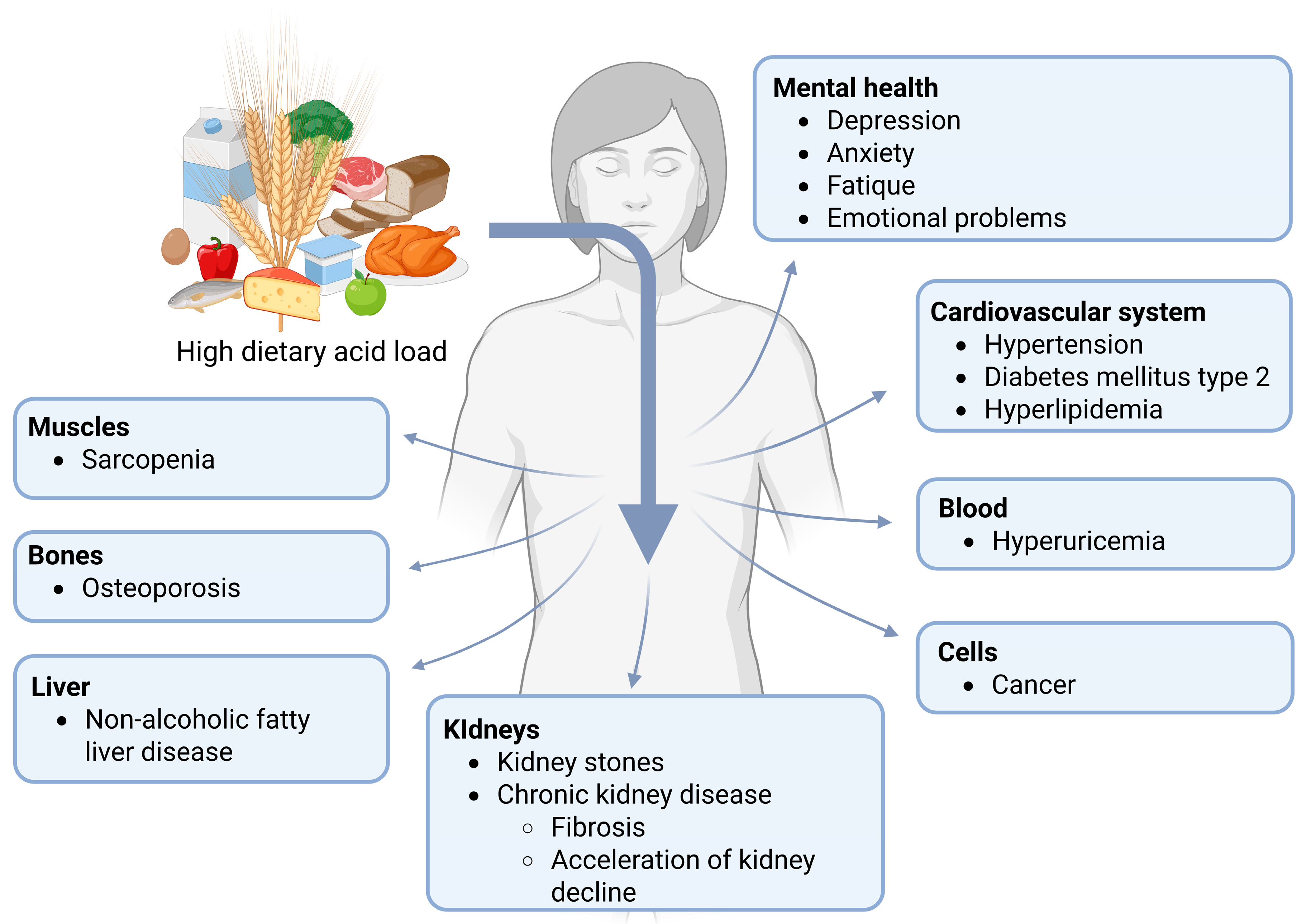
Figure 1. Discussed consequences of a high dietary acid load.
Nutrients 17 02291 g001
Figure 2. PRAL calculation for mineral water [56].
Nutrients 17 02291 g002
Figure 3. Effects of bicarbonate-rich mineral water on the urinary parameters. TA = titrable acids, NH4+ = ammonium, HCO3 = bicarbonate, NAE = net acid excretion.
Nutrients 17 02291 g003
Figure 4. Effects of bicarbonate-rich mineral water on stone risk (CaOx/UA stones). Main effects are reported with black arrows. Grey arrows indicate effects in the minority of the studies. CaOx = calcium oxalate, UA = uric acid.
Nutrients 17 02291 g004
Figure 5. Effects of bicarbonate-rich mineral water on blood gas parameters. BE = Base excess, HCO3 = bicarbonate.
Nutrients 17 02291 g005
Figure 6. Effects of bicarbonate-rich mineral water on bone turnover. The main effects are reported with black arrows. Grey arrows indicate effects in the minority of the studies. CTX = C-terminal fragment of the type I collagen; BALP = bone specific alkaline phosphatase; P1NP = Procollagen type 1 N-terminal propeptide; PTH = parathyroid hormone.
Nutrients 17 02291 g006
Table 2. Composition of selected bicarbonate-rich mineral water and calculation of the PRAL values. The mineral water brands are arranged according to the increasing PRAL value. The PRAL was calculated by the authors using the following equation [56]: PRAL (mEq/L) = 0.0146 × SO42− (mg/L) + 0.027 × Cl (mg/L) − 0.021 × K+ (mg/L) − 0.026 × Mg2+ (mg/L) − 0.0413 × Na+ (mg/L) − 0.013 × Ca2+ (mg/L).
BrandCountryNaKCaMgClSO42−PHCO3−PRAL
Rogaska DonatSlovenia1500133801030592400---7700−57.3
St-YorreFrance1708110901132217404368−63.1
BorjomiGeorgia147830858539350 3965−53.6
Adelheidquelle, AdelholzenerGermany95047.215210211231702999−37.2
Vichy CelestinsFrance11726610310235138---2989−43.0
Heppinger Extra Medicinal WaterGermany48127.1150199118600.062495−23.5
JamnicaCroatia9213211534252109---2247−32.7
Ardesy (Arvie)France6501301709238731---2195−23.3
RadenskaSlovenia40070220954472---2000−21.1
RozanaFrance49352301160649230---1837−8.6
KryniczankaPoland43543668198---1818−8.7
GerolsteinerGermany118113481084038---1816−10.8
ApollinarisGermany470309012013010001800−19.4
RhodiusGermany137331431512237---1562−11.0
FerrarelleItaly505039222204---1433−8.2
KalnickaCroatia650862233500 1410−19.0
BadoitFrance1651019085443801300−10.0
RhäzünserSwitzerland14972244618131---1120−8.0
VerniereFrance1104018017314140 1100−9.8
QuezacFrance11001706900---1100−8.5
SangeminiItaly19.63.932316.518.5610996−4.1
Lete Acqua mineraleItaly4.92.131315.18.26.60981−4.4
Table 3. Effects of bicarbonate-rich mineral water on urine pH and NAE.
AuthorDesign
Target Group
InterventionCharacteristics of Mineral
Water/Treatment (Rich in)
Bicarbonate/Day
Main Results
Time Effects *
(Bicarbonate Group)
Group Differences
Time × Group Interaction
Sub-chronic studies
Schorr et al., 1996
[70]
Cross-over, randomized, double-blind
21 healthy older (60–72 years) subjects
NaCl reduction (<100 mmol/d) + 4 weeks (each)
3 different mineral water brands
1.5 L/d
Water A: HCO3, Na, Mg
Water B: HCO3, Na, Cl
Water C: low mineralized
HCO3
Water A: 2975 mg/d
Water B: 1318 mg/d
Water C: 18 mg/d
24-h urine
NAE: (water A) ↔
(water B)
Week 4
NAE: n.s. group differences
Marangella et al., 1996
[71]
Cross-over, randomized
21 subjects with idiopathic calcium nephrolithiasis
1 month (each)
3 different mineral water brands
Standardized diet
2 L/d
Water A: HCO3, Ca
Water B: SO4
Water C: low mineralized
HCO3
Water A: 3051 mg/d
Water B: 610 mg/d
Water C: 31 mg/d
24-h urine
pH: ↑
NAE: ↓
pH: water A > water B
NAE: water A < water B and C
Caudarella et al., 1998
[72]
Cross-over
22 subjects with idiopathic calcium nephrolithiasis
20 days (each)
3 different mineral water brands
Standardized diet
2 L/d
Water A: HCO3, Ca
Water B: SO4−rich
Water C: low mineralized
HCO3
Water A: 2794 mg/d
Water B: 610 mg/d
Water C: 100 mg/d
Fasting morning urine
pH: ↔ pH: n.s. group differences
Keßler and Hesse, 1998, 2000
[73,81]
Cross-over, randomized
24 kidney-healthy younger (23–38 years) men
Run-In (standardized diet) + 2 days treatment (standardized diet)
4 weeks follow-up, same as cross-over week 2 (usual diet)
Mineral water (”Staatl. Fachingen”) vs. supplement
2 L/d
Water: HCO3, Na, Mg
Supplement: K citrate
HCO3
Water 3430 mg/d
K citrate: not reported
24-h urine
Standardized diet
pH: ↑
Usual diet
pH: ↑
Not reported
Coen et al., 2001
[74]
Parallel-group
21 healthy subjects
2 weeks
2 different mineral water brands
Standardized diet
2 L/d
Water A: HCO3, Ca
Water B: low mineralized
HCO3
Water A: 2780 mg/d
Water B: 121 mg/d
Spot urine
pH: ↔pH: n.s. time × group interaction
Siener et al., 2004
[75]
Cross-over (first control, later mineral water) + single-arm
12 young healthy male subjects
2 weeks baseline (usual diet/beverages)
5 days cross-over (standardized diet); Mineral water vs. fruit tea (control)
4 weeks follow-up with mineral water (usual diet/beverages)
1.4 L/d
Water: HCO3, Ca, Mg, Na
HCO3
Water: 4743 mg/d
24-h urine
Standardized diet
pH: ↑
NH4: ↓
Usual diet
pH:
NH4: ↓
Standardized diet
pH: water > control
NH4: water < control
----
Roux et al., 2004
[82]
Cross-over, randomized
60 postmenopausal women
4 weeks (each)
2 different mineral water brands
1 L/d
Water A: HCO3, Ca
Water B: Ca, Mg, SO4
HCO3
Water A: 2179 mg/d
Water B: 292 mg/d
2-h fasting urine
pH: ↑
TA- HCO3: ↓
NH4: ↓
pH: water A > B
TA- HCO3: water A < water B
NH4: water A < water B
Schoppen et al., 2005
[76]
Cross-over
18 postmenopausal women
8 weeks (each)
2 different mineral water brands
1 L/d
Water A: HCO3, Na, Cl
Water B: low mineralized
HCO3
Water A: 2094 mg/d
Water B: 71 mg/d
24-h urine
Not reportedpH: water A > water B
Karagülle et al., 2007
[77]
Cross-over, double-blind
34 subjects with multiepisodic CaOx stone formation
3 days (each)
2 different mineral water brands (water A: “Heppinger”, water B: “Bad Harzburger Urquell”)
1.5 L/d
Water A: HCO3, Mg, Na, Cl
Water B: low mineralized
HCO3
Water A: 4010 mg/d
Water B: 149 mg/d
24-h urine
pH: ↑ pH: water A > water B
Wynn et al., 2009
[56]
Parallel-group,
randomized
30 young (18–45 years) women
4 weeks
2 different mineral water brands (water A: “Adelbodner”; water B: “Kryniczanka”)
Standardized diet
1.5 L/d
Water A: HCO3, Ca, Mg
Water B: Ca, SO4
HCO3
Water A: 3258 mg/d
Water B: 437 mg/d
24-h urine
pH: ↑
HCO3: ↑
pH: water A > water B
HCO3: water A > water B
Perez-Granados et al., 2010
[78]
Cross-over (first water B, later water A), single-blind
18 young (>18–<40 years) hypercholesterolemic subjects
8 weeks (each)
2 different mineral water brands
1 L/d
Water A: HCO3, Na, Cl
Water B: low mineralized
HCO3
Water A: 2120 mg/d
Water B: 104 mg/d
24-h urine
Not reportedpH: water A > water B
Brancaccio et al., 2012/12
[83,84]
Parallel-group
88 amateur athletes
7 days
2 different mineral water brands (water A “Aqua Lete”, water B)
Repeated Wingate Tests (cycling)
1.5 L/d + 750 mL 1 h before exercise + 250 mL after exercise
Water A: HCO3, Ca
Water B: low mineralized
HCO3 (1.5 L)
Water A: 1472 mg/d
Water B: 5 mg/d
Urine (mixture of several time points)
Not reported
During the day at the end of the intervention
pH: ↑
pH: no changes over time with water B
Toxqui and Vaquero, 2016
[85]
Cross-over,
randomized, single-blind
64 moderately hypercholesterolemic men and women
8 weeks (each)
2 different mineral water brands
1 L/d
Water A: HCO3, Na, Cl
Water B: low mineralized
HCO3
Water A: 2050 mg/d
Water B: 75 mg/d
Fasting morning urine
pH: ↑ pH: sign. time × group interaction
Wasserfurth et al., 2019
[13]
Parallel-group, randomized
129 healthy subjects
4 weeks
4 different mineral water brands
1.5–2 L/d
Water A: HCO3, Ca, Mg
Water B: HCO3, Ca, Mg, Na
Water C: HCO3, Mg, Na
Water D: Ca, Mg, SO4
HCO3 (1.5 L)
Water A: 2724 mg/d
Water B: 3677 mg/d
Water C: 2769 mg/d
Water D: 605 mg/d
24-h urine
pH: ↔ (water A) (p = 0.068)
↑ (water B and C)
TA: ↓ (water A-C)
HCO3: ↑ (water A and B)
↔ (water C)
NH4: ↓ (water A-C)
NAE: ↓ (water A-C)
Week 4
pH: sign. group differences
TA: n.s. group differences
HCO3: sign. group differences
NH4: n.s. group differences (p = 0.052)
NAE: sign. group differences
Spontaneous urine
pH: ↑ (water A and C) pH: sign. group differences
Chycki et al., 2021
[48]
Cross-over (first table water, then HCO3 water), single-blind
8 elite judo athletes
3 weeks (each)
Mineral water (water A) vs. table water (water B)
Standardized meals
Tests under hydrated conditions, treadmill to induce hypohydration, anaerobic Wingate tests under dehydrated conditions, later rehydration
Amount individualized, approx. 3.2–3.4 L/d
Water A: HCO3, Na, Cl
Water B: low mineralized
HCO3 (3.3 L)
Water A: 13,207 mg/d
Water B: 12 mg/d
24-h urine
Not reportedPost-supplementation time point
Hydrated + dehydrated condition
pH: n.s. group differences (trend: water A > water B)
Lu et al., 2022
[86]
Parallel-group, randomized
58 subjects with Ca stones
12 weeks
Mineral water (water A: “Ardesy”) vs. tap water (water B)
1.25 L/d
Water A: HCO3, Ca, Mg, Na, Cl
Water B: low mineralized
HCO3
Water A: 2744 mg/d
Water B: not reported
24-h urine
(no p-values reported)
pH: ↑ (trend)
Week 12 §
pH: n.s. group differences (p = 0.071)
Chiron et al., 2024
[59]
Parallel group (diet)
Cross-over (water, within a diet group), randomized, double-blind
24 recreationally active men
7 days
2 different mineral water brands (water A: “St-Yorre”, water B)
Dietary restrictions (alkalizing diet vs. acidifying diet)
1-min supra-maximal rowing Wingate Test
1.5–2 L/d
Water A: HCO3, Na, Cl
Water B: low mineralized
HCO3 (2 L)
Water A: 8736 mg/d
Water B: 612 mg/d
24-h urine
Not reportedPost-supplementation time point
Water effects (whole group) #
pH: water A > water B
Water effects (alkalizing diet)
pH: water A > water B
Water effects (acidifying diet)
pH: water A > water B
Mansouri et al., 2023, 2024
[58,87]
Parallel-group, randomized
94 healthy subjects
4 weeks
2 different mineral water brands
1.5–2 L/d
Water A: HCO3, Na, Cl
Water B: low mineralized
HCO3 (1.5 L)
Water A: 6552 mg/d
Water B: 342 mg/d
24-h urine
pH: ↑
TA: ↓
NH4: ↓
HCO3: ↑
NAE: ↓
pH: sign. time × water interaction (water A ↑, water B ↔)
TA: sign. time × water interaction (water A < water B)
NH4: sign. time × water interaction (water A < water B)
HCO3: sign. time × water interaction (water A ↑; water B ↓)
NAE: sign. time × water interaction (water A < water B)
Chiron et al., 2024
[79]
Parallel-group, randomized, double-blind
22 highly trained athletes
6 days
2 different mineral water brands (water A: “St-Yorre”, water B)
Dietary restrictions (alkalizing diet)
Last 3 days: 400 m race + handgrip strength + squat jumps (each day)
4 × 500 mL /d
Water A: HCO3, Na, Cl
Water B: low mineralized
HCO3
Water A: 8736 mg/d
Water B: 612 mg/d
24-h urine
pH: ↑pH: water A > water B
Acute studies
Schoppen et al., 2008
[80]
Cross-over, randomized
18 postmenopausal women
---
3 different mineral water brands
500 mL
Water A: HCO3, Na (higher than water B), Cl
Water B: HCO3, Na, Cl
Water C: low mineralized
HCO3
Water A: 1047 mg
Water B: 1007 mg
Water C: 36 mg
Postprandial urine
 Not reportedpH: n.s. group differences
Minerals: HCO3 = bicarbonate; Ca = calcium; Mg = magnesium; Na = sodium; Cl = chloride; SO4 = sulfate. Urinary parameters: Ca = calcium; TA = titratable acid; NH4 = ammonium; HCO3 = bicarbonate; NAE = net acid excretion. Notes: ↑ = significant increase (p < 0.05); ↔ = no significant change (p > 0.05); ↓ = significant decrease (p < 0.05); * between begin and end of each intervention period; categorization of the authors; no p-values reported for group differences; § time effects extracted from tables reporting changes between baseline and different weeks (no p-values reported) + text, group differences are calculated between changes at one time point; # adjusted for diet group.
Table 4. Effects of bicarbonate-rich mineral water on stone risk.
AuthorDesign
Target Group
InterventionCharacteristics of Mineral Water/ Treatment
Bicarbonate/Day
Main Results
Time Effects *
(Bicarbonate Group)
Group Differences
Time × Water Interaction
Sub-chronic studies
Luft et al., 1990
[95]
Cross-over, randomized, single-blind
10 subjects (hypertensive + normotensive)
4 days Run-In + 7 days (each)
Mineral water (“Staatl. Fachingen”) vs. control solution (NaCl)
Standardized diet (low sodium, low calcium)
3 L/d
Water: HCO3, Na, Mg
Control solution: Na, Cl, Mg
HCO3
Water A: 6046 mg/d
Water B: 0 mg/d
Urine (not specified)
Not reportedCa: sign. group differences
water < NaCl
normotensives < hypertensives
blacks < whites
Schorr et al., 1996
[70]
Cross-over, randomized, double-blind
21 healthy older (60–72 years) subjects
NaCl reduction (<100 mmol/d) + 4 weeks (each)
3 different mineral water brands
1.5 L/d
Water A: HCO3, Na, Mg
Water B: HCO3, Na, Cl
Water C: low mineralized
HCO3
Water A: 2975 mg/d
Water B: 1318 mg/d
Water C: 18 mg/d
24-h urine
NAE: (water A) ↔
(water B) ↑
Ca: (water A) ↓
(water B) ↔
Week 4
NAE: (water A): n.s. group differences
(water B): n.s. group differences
Ca: (water A): n.s. group differences
(water B): n.s. group differences
Marangella et al., 1996
[71]
Cross-over, randomized
21 subjects with idiopathic calcium nephrolithiasis
1 month (each)
3 different mineral water brands
Standardized diet
2 L/d
Water A: HCO3, Ca
Water B: SO4
Water C: low mineralized
HCO3
Water A: 3051 mg/d
Water B: 610 mg/d
Water C: 31 mg/d
24-h urine
pH: ↑
Ca: ↑
Oxalate: ↓
Citrate: ↑
Mg: ↑
RS CaOx: ↓
pH: water A > water B
Ca: water A > water B
Oxalate: water A < water B and C
Citrate: water A > water B and C
Mg: water A > water B and C
RS CaOx: n.s. group differences
Caudarella et al., 1998
[72]
Cross-over
22 subjects with idiopathic calcium nephrolithiasis
20 days (each)
3 different mineral water brands
Standardized diet
2 L/d
Water A: HCO3, Ca
Water B: SO4-rich
Water C: low mineralized
HCO3
Water A: 2794 mg/d
Water B: 610 mg/d
Water C: 100 mg/d
Fasting morning urine
pH: ↔
RS CaOx: ↔
pH: n.s. group differences
RS CaOx: n.s. group differences
24-h urine
Ca: ↔ (tendency ↑)
Oxalate: ↓
Mg: ↑
Ca: n.s. group differences
Oxalate: n.s. group differences
Mg: water A > water B
Urine (not specified)
Citrate: ↑ Citrate: n.s. group differences
Keßler and Hesse, 1998, 2000
[73,81]
Cross-over, randomized
24 kidney-healthy younger (23–38 years) men
Run-In (standardized diet) + 2 days treatment (standardized diet)
4 weeks follow-up, same as cross-over week 2 (usual diet)
Mineral water (”Staatl. Fachingen”) vs. supplement
2 L/d
Water: HCO3, Na, Mg
Supplement: K Citrate
HCO3
Water 3430 mg/d
K Citrate: not reported
24-h urine

Standardized diet
pH: ↑
Ca: ↔ (tendency ↓)
Oxalate: ↓
Citrate: ↑
Mg: ↔
RS CaOx: ↓
RS UA: ↓
Usual diet
pH: ↑
Ca: ↔ (tendency ↓)
Oxalate: ↔ (tendency ↓)
Citrate: ↑
Mg: ↔ (tendency ↑)
RS CaOx: ↓
RS UA: ↓
Day 5
Standardized diet
n.s. group differences
Week 4
Usual diet
n.s. group differences
Coen et al., 2001
[74]
Parallel-group
21 healthy subjects
2 weeks
2 different mineral water brands
Standardized diet
2 L/d
Water A: HCO3, Ca
Water B: low mineralized
HCO3
Water A: 2780 mg/d
Water B: 121 mg/d
Spot urine
pH: ↔
Ca: ↔
RS CaOx (Tiselius Index): ↔
pH: n.s. time × group interaction
Ca: sign. time × group interaction (water A > water B)
RS CaOx (Tiselius Index): n.s. time × group interaction
24-h urine
Oxalate: ↔ (tendency: ↑ p = 0.068)
Citrate: ↑
Mg: ↔ (tendency: ↑ p = 0.058)
Oxalate: n.s. time × group interaction
Citrate: n.s. time × group interaction
Mg: n.s. time × group interaction
Siener et al., 2004
[75]
Cross-over (first control, later mineral water) + single-arm
12 young healthy male subjects
2 weeks baseline (usual diet/beverages)
5 days cross-over (standardized diet); Mineral water vs. fruit tea (control)
4 weeks follow-up with mineral water (usual diet/beverages)
1.4 L/d
Water: HCO3, Ca, Mg, Na
HCO3
Water: 4743 mg/d
24-h urine
Standardized diet
pH: ↑
Ca: ↑
Oxalate: ↔
Citrate: ↑
Mg: ↑
RS CaOx: ↔
Usual diet
pH: ↑
Ca: ↔
Oxalate: ↔
Citrate: ↑
Mg: ↑
RS CaOx: ↓
Standardized diet
pH: water A > control
Ca: water A > control
Oxalate: n.s. group differences
Citrate: water A > control
Mg: water A > control
RS CaOx: n.s. group differences
----
Schoppen et al., 2005
[76]
Cross-over
18 postmenopausal women
8 weeks (each)
2 different mineral water brands
1 L/d
Water A: HCO3, Na, Cl
Water B: low mineralized
HCO3
Water A: 2094 mg/d
Water B: 71 mg/d
24-h urine
Not reportedpH: water A > water B
Ca: water A < water B
Karagülle et al., 2007
[77]
Cross-over, double-blind
34 subjects with multiepisodic CaOx stone formation
3 days (each)
2 different mineral water brands (water A: “Heppinger”, water B: “Bad Harzburger Urquell”)
1.5 L/d
Water A: HCO3, Mg, Na, Cl
Water B: low mineralized
HCO3
Water A: 4010 mg/d
Water B: 149 mg/d
24-h urine
pH: ↑
Ca:
Oxalate: ↑
Citrate: ↑
Mg: ↑
RS CaOx: ↓
pH: water A > water B
Ca: n.s. group differences
Oxalate: n.s. group differences
Citrate: water A > water B
Mg: water A > water B
RS CaOx: n.s. group differences
Wynn et al., 2009
[56]
Parallel-group, randomized
30 young (18–45 years) women
4 weeks
2 different mineral water brands (water A: “Adelbodner”, water B: “Kryniczanka”)
Standardized diet
1.5 L/d
Water A: HCO3, Ca, Mg
Water B: Ca, SO4
HCO3
Water A: 3258 mg/d
Water B: 437 mg/d
24-h urine
pH: ↑
Ca: ↑
pH: water A > water B
Ca: water A < water B
Toxqui and Vaquero, 2016
[85]
Cross-over, randomized, single-blind
64 moderately hypercholesteremic men and women
8 weeks (each)
2 different mineral water brands
1 L/d
Water A: HCO3, Na, Cl
Water B: low mineralized
HCO3
Water A: 2050 mg/d
Water B: 75 mg/d
Fasting morning urine
pH: ↑
Ca/creatinine: ↓
pH: sign. time × group interaction
Ca/creatinine: sign. time × group interaction
Wasserfurth et al., 2019
[13]
Parallel-group, randomized
129 healthy subjects
4 weeks
4 different mineral water brands
1.5–2 L/d
Water A: HCO3, Ca, Mg
Water B: HCO3, Ca, Mg, Na
Water C: HCO3, Mg, Na
Water D: Ca, Mg, SO4
HCO3 (1.5 L)
Water A: 2724 mg/d
Water B: 3677 mg/d
Water C: 2769 mg/d
Water D: 605 mg/d
24-h urine
pH: ↔ (water A) (p = 0.068)
↑ (water B and C)
Ca: ↑ (water A, B and D)
↔ (water C)
pH: sign. group differences
Ca: sign group differences
Lu et al., 2022
[86]
Parallel-group, randomized
58 subjects with Ca stones
12 weeks
Mineral water (water A: “Ardesy”) vs. tap water (water B)
1.25 L/d
Water A: HCO3, Ca, Mg, Na, Cl
Water B: low mineralized
HCO3
Water A: 2744 mg/d
Water B: not reported
24-h urine
pH: ↑ (trend)
Ca: ↑
Oxalate: ↓
Citrate: ↑ (trend)
Mg: ↑ (trend)
RS CaOx (Tiselius Index): ↑
(No p-values reported)
Week 12 
pH: n.s. group differences (p = 0.071)
Ca: n.s. group differences
Oxalate: n.s. group differences
Citrate: n.s. group differences (p = 0.084)
Mg: water A > water B
RS CaOx (Tiselius Index): n.s. group differences (p = 0.060)
Mansouri et al., 2023, 2024
[58,87]
Parallel-group, randomized
94 healthy subjects
4 weeks
2 different mineral water brands
1.5–2 L/d
Water A: HCO3, Na, Cl
Water B: low mineralized
HCO3 (1.5 L)
Water A: 6552 mg/d
Water B: 342 mg/d
24-h urine
pH: ↑
Ca: ↓
pH: sign. time × water interaction (water A ↑, water B ↔)
Ca: n.s. time × water interaction (p = 0.060)
Acute studies
Schoppen et al., 2008
[80]
Cross-over, randomized
18 postmenopausal women
---
3 different mineral water brands
500 mL
Water A: HCO3, Na (higher than water B), Cl
Water B: HCO3, Na, Cl
Water C: low mineralized
HCO3
Water A: 1047 mg
Water B: 1007 mg
Water C: 36 mg
Postprandial urine
Not reportedpH: n.s. group differences
Ca: n.s. group differences
Minerals: HCO3 = bicarbonate; Ca = calcium; Mg = magnesium; Na = sodium; Cl = chloride; SO4 = sulfate. Urinary parameters: Ca = calcium; Mg = magnesium; RS CaOx = calcium oxalate supersaturation. Notes: ↑ = significant increase (p < 0.05); ↔ = no significant change (p > 0.05); ↓ = significant decrease (p < 0.05); * between begin and end of each intervention period; categorization of the authors; time effects extracted from tables reporting changes between baseline and different weeks (no p-values reported) + text, group differences are calculated between changes at one time point.
Table 5. Effects of bicarbonate-rich mineral water on blood gas parameters.
AuthorDesign
Target Group
InterventionCharacteristics of Mineral Water/Treatment
Bicarbonate/day
Main Results
Time Effects *
(Bicarbonate Group)
Group Differences
Time × Water Interaction **
Sub-chronic studies
Wasserfurth et al., 2019
[13]
Parallel-group, randomized
129 healthy subjects
4 weeks
4 different mineral water brands
1.5–2 L/d
Water A: HCO3, Ca, Mg
Water B: HCO3, Ca, Mg, Na
Water C: HCO3, Mg, Na
Water D: Ca, Mg, SO4
HCO3 (1.5 L)
Water A: 2724 mg/d
Water B: 3677 mg/d
Water C: 2769 mg/d
Water D: 605 mg/d
12-h fasting blood (venous)
pH: ↔ (water B and C)
↓ (water A)
HCO3: ↑ (water C)
↔ (water A and B)
(water B p = 0.057)
BE: ↑ (water C)
↔ (water A and B)
pH: sign. group differences
HCO3: n.s. group differences
BE: n.s. group differences
Chycki et al., 2021
[48]
Cross-over (first table water, later HCO3 water), single-blind
8 elite judo athletes
21 days (each)
Mineral water (water A) vs. table water (water B)
Standardized diet
Anaerobic Wingate tests (high intensity) under hydrated + dehydrated conditions, treadmill to induce hypohydration, later rehydration
Amount individualized, approx. 3.2–3.4 L/d
Water A: HCO3, Na, Cl
Water B: low mineralized
HCO3 (3.3 L)
Water A: 13,207 mg/d
Water B: 12 mg/d
Blood (capillary: fingertip)
Not reported
During post-supplementation time point (no p-values reported)
pH (resting):
HCO3 (resting):
Post-supplementation time point
Hydrated and dehydrated condition
pH (resting): n.s. group differences
HCO3 (resting): water A > water B
Hagele et al., 2023
[107]
Parallel-group, randomized, double-blind
39 recreationally active men and women
7 days
Mineral water (water A: “Borjomi”) vs. spring water (water B)
Same diet before each visit
Anaerobic cycling
10 mL/kg, 40–60 min prior to exercise tests
Water A: HCO3, Na, Cl
Water B: low mineralized
HCO3
Water A: approx. 3000 mg/d
Water B: not reported
8-h fasting blood (venous)
Not reported
During post-supplementation time point
pH: ↓
HCO3: ↓
BE: ↓

pH: n.s. time × group interaction (water A > water B at immediate post + 10 min post exercise)
HCO3: n.s. time × group interaction
BE: n.s. time × group interaction
Chiron et al., 2024
[59]
Parallel-group (diet)
Cross-over (water, within a diet group), randomized, double-blind
24 recreationally active men
7 days
2 different mineral water brands (water A: “St-Yorre”, water B)
Dietary restrictions (alkalizing diet vs. acidifying diet)
1-min supra-maximal rowing Wingate Test
1.5–2 L/d
Water A: HCO3, Na, Cl
Water B: low mineralized
HCO3 (2 L)
Water A: 8736 mg/d
Water B: 612 mg/d
Blood (capillary: earlobe)
Not reported
During post-supplementation time point (no p-values reported)
Water effect
pH: ↓
HCO3: ↓
Water effect (alkalizing diet)
Not reported
Water effects (acidifying diet)
Not reported
Water effects (whole group) 
pH: water A > water B (immediately after + 3 min + 5 min post exercise)
HCO3: n.s. group difference
Water effects (alkalizing diet)
pH: water A > water B (immediately after + 5 min post exercise)
HCO3: water A > water B (warm-up + immediately post exercise)
HCO3 (peak): water A > water B
Water effects (acidifying diet)
pH: n.s. group differences
HCO3: n.s. group differences
Mansouri et al., 2024
[58]
Parallel-group, randomized
94 healthy subjects
4 weeks
2 different mineral water brands
1.5–2 L/d
Water A: HCO3, Na, Cl
Water B: low mineralized
HCO3 (1.5 L)
Water A: 6552 mg/d
Water B: 342 mg/d
12-h fasting blood (venous)
pH: ↔
HCO3: ↑
BE: ↑
pH: n.s. time × group interaction
HCO3: sign. time × group interaction (water A ↑, water B ↔)
BE: sign. time × group interaction (water A ↑, water B ↔)
Chiron et al., 2024
[79]
Parallel-group, randomized, double-blind
22 highly trained athletes
6 days
2 different mineral water brands (water A: “St-Yorre”, water B)
Dietary restrictions (alkalizing diet)
Last 3 days: 400 m run + handgrip strength + squat jumps (each day)
4 × 500 mL/d
Water A: HCO3, Na, Cl
Water B: low mineralized
HCO3
Water A: 8736 mg/d
Water B: 612 mg/d
Blood (capillary: fingertip)
Not reported
During post-supplementation time point (no p-values reported)
pH: ↓
HCO3: ↓
BE:
pH: water A > water B (pre 400 m run)
HCO3: water A > water B (HCO3 max)
BE: water A > water B (1 h post 400 m run)
Acute Studies
Richard et al., 2000
[108]
Cross-over, randomized, single-blind
12 regularly trained athletes
---
3 different mineral water brands (water A: “St-Yorre”, water B, water C)
Standardized meal
Anaerobic cycling + isokinetic endurance test (after recovery)
3 L (1.5 l before exercise + 0.5 L during exercise + 1 L during recovery)
Water A: HCO3, Na
Water B: Na
water C: low mineralized
HCO3
Water A: 13,104 mg/d
Water B: not reported
Water C: not reported
Blood (capillary)
(no p-values reported)
pH: ↑ (pre—post cycling)
↓ (until end of isokinetic test)
HCO3: ↓ (pre- post cycling)
↑ (recovery)
↓ (slightly, until end of
isokinetic test)
pH: water A > water C (immediately after exercise + after isokinetic test)
HCO3: water A > water C
Minerals: HCO3 = bicarbonate; Ca = calcium; Mg = magnesium; Na = sodium; Cl = chloride; SO4 = sulfate; Blood parameters: BE = base excess; HCO3 = bicarbonate. Notes: ↑ = significant increase (p < 0.05); ↔ = no significant change (p > 0.05); ↓ = significant decrease (p < 0.05); * between begin and end of each intervention period; ** at end of each intervention period; adjusted for diet group.
Table 6. Effects of bicarbonate-rich mineral water on bone turnover.
AuthorDesign
Target Group
InterventionCharacteristics of Mineral
Water/Treatment
Bicarbonate/Day
Main Results
Time Effects *
(Bicarbonate Group)
Group Differences
Time × Water Interaction
Sub-chronic studies
Marangella et al., 1996
[71]
Cross-over, randomized
21 subjects with idiopathic calcium nephrolithiasis
1 month (each)
3 different mineral water brands
Standardized diet
2 L/d
Water A: HCO3, Ca
Water B: SO4
Water C: low mineralized
HCO3
Water A: 3051 mg/d
Water B: 610 mg/d
Water C: 31 mg/d
Fasting urine
Not reportedHydroxyproline: water A < water C
Cross-linked N-telopeptide type I: water A < water B and C
Blood (not specified)
Not reportedPTH: water A and B < water C
Osteocalcin: n.s. group differences
Roux et al., 2004
[82]
Cross-over, randomized
60 postmenopausal women
4 weeks (each)
2 different mineral water brands
1 L/d
Water A: HCO3, Ca
Water B: Ca, Mg, SO4
HCO3
Water A: 2179 mg/d
Water B: 292 mg/d
2-h fasting urine
pH: ↑
CTX/Cr: ↓
Pyr/Cr: ↓
pH: water A > water B
CTX/Cr: n.s. group differences
Pyr/Cr: n.s. group differences
Fasting blood
iCa (serum): ↑
total Ca (serum): ↔
P (serum): ↔
iPTH (plasma): ↓
Osteocalcin (serum): ↔
BALP (serum): ↔
No sign. group differences
Schoppen et al., 2005
[76]
Cross-over
18 postmenopausal women
8 weeks (each)
2 different mineral water brands
1 L/d
Water A: HCO3, Na, Cl
Water B: low mineralized
HCO3
Water A: 2094 mg/d
Water B: 71 mg/d
24-h urine
Not reportedpH: water A > water B
Ca: water A < water B
12-h fasting blood
Not reportedCTX (serum): n.s. group differences
P1NP (serum): n.s. group differences
Wynn et al., 2009
[56]
Parallel-group, randomized
30 young (18–45 years) women
4 weeks
2 different mineral water brands (water A: “Adelbodner”, water B: “Kryniczanka”)
Standardized diet
1.5 L/d
Water A: HCO3, Ca, Mg
Water B: Ca, SO4
HCO3
Water A: 3258 mg/d
Water B: 437 mg/d
24-h urine
pH: ↑
Ca: ↑
CTX: ↔ (slightly ↓)
pH: water A > water B
Ca: water A < water B
CTX: n.s. group differences
Blood (not specified)
iCa: ↔
total Ca: ↔
P: ↔
CTX (serum): ↓
PTH (plasma): ↓
BALP: ↔
iCa: n.s. group differences
total Ca: n.s. group differences
P: n.s. group differences
CTX (serum): changes water A > water B
PTH (plasma): changes water A > water B
BALP: n.s. group differences
Minerals: HCO3 = bicarbonate; Ca = calcium; Mg = magnesium; Na = sodium; Cl = chloride; SO4 = sulfate. Urinary parameters: Ca = calcium; Cr = creatinine; CTX = C-terminal fragment of the type I collagen; NTX = cross-linked N-telopeptide type I; Pyr = pyridinoline. Blood parameters: BALP = bone specific alkaline phosphatase; CTX = C-terminal fragment of the type I collagen; iCa = ionized calcium; P = phosphate; P1NP = procollagen type 1 N-terminal propeptide; PTH = parathyroid hormone. Notes: ↑ = significant increase (p < 0.05); ↔ = no significant change (p > 0.05); ↓ = significant decrease (p < 0.05); * between begin and end of each intervention period; categorization of the authors (no p-value reported).
Disclaimer/Publisher’s Note: The statements, opinions and data contained in all publications are solely those of the individual author(s) and contributor(s) and not of MDPI and/or the editor(s). MDPI and/or the editor(s) disclaim responsibility for any injury to people or property resulting from any ideas, methods, instructions or products referred to in the content.