Review

Hyperhomocysteinemia: Underlying Links to Stroke and Hydrocephalus, with a Focus on Polyphenol-Based Therapeutic Approaches

by
Carmen Ortiz-Salguero
1,
Marina Romero-Bernal
1,
Ángela González-Díaz
1,
Elaheh Sobh Doush
1,
Carmen del Río
1,*,
Miriam Echevarría
1 and
Joan Montaner
2,3,*
1
Instituto de Biomedicina de Sevilla, IBiS, Hospital Universitario Virgen del Rocío, CSIC, Universidad de Sevilla, 41013 Sevilla, Spain
2
Instituto de Biomedicina de Sevilla, IBiS, Hospital Universitario Virgen Macarena, CSIC, Universidad de Sevilla, 41004 Sevilla, Spain
3
Department of Neurology, Hospital Universitario Virgen Macarena, 41004 Sevilla, Spain
*
Authors to whom correspondence should be addressed.
Nutrients 2025, 17(1), 40; https://doi.org/10.3390/nu17010040
Submission received: 22 November 2024 / Revised: 19 December 2024 / Accepted: 23 December 2024 / Published: 26 December 2024
(This article belongs to the Section Proteins and Amino Acids)

Highlights

  • Mutations (e.g., MTHFR) and B-vitamin deficiencies elevate homocysteine (HCys) levels.
  • Elevated HCys levels induce oxidative stress, inflammation and neurodegeneration.
  • HCys modulates endothelial dysfunction and fluid dynamics, with implications for stroke and hydrocephalus pathogenesis.
  • Polyphenols can reduce HCys levels by enhancing key metabolic enzymes and counteracting oxidative and inflammatory damage, offering neurovascular protection.
  • Polyphenol-rich diets and supplementation show potential in mitigating neurovascular damage.

Abstract

:
Hyperhomocysteinemia (HHcy), characterized by elevated homocysteine (HCys) levels, is associated with increased risks of neurovascular diseases such as stroke or hydrocephalus. HHcy promotes oxidative stress, neuroinflammation, and endothelial dysfunction, disrupting the blood–brain barrier and accelerating neurodegeneration. These processes highlight HCys as both a biomarker and a potential therapeutic target in vascular-related neurological disorders. Current research suggests that polyphenols, known for their antioxidant and anti-inflammatory properties, may reduce HCys levels and offer neuroprotection. Polyphenols have demonstrated effectiveness in modulating oxidative stress and inflammatory pathways triggered by HHcy. These compounds may also upregulate enzymatic functions involved in HCys metabolism, thus reducing neurotoxicity. Furthermore, polyphenol-rich diets, like the Mediterranean diet, have been linked to lower HCys levels and a reduced incidence of neurovascular disorders. This review provides an overview of HHcy’s role in neurovascular pathologies and examines the therapeutic potential of polyphenols in managing HCys levels and preventing HCys-induced neurovascular damage.

1. Introduction

Homocysteine (HCys) is a non-essential amino acid with a crucial role in cellular metabolism. Elevated HCys levels are a known risk factor for cardiovascular and neurovascular diseases as they promote oxidative stress, neuroinflammation, and endothelial dysfunction. High HCys concentrations have been associated with increased blood–brain barrier (BBB) permeability, heightened platelet aggregation, and the activation of proinflammatory pathways, all of which can accelerate neurodegeneration and vascular injury. These associations position HCys as a potential therapeutic target, especially in neurological conditions where vascular health is critical, such as stroke and hydrocephalus.
Dietary habits, particularly those rich in B-vitamins and polyphenols, play a critical role in regulating homocysteine metabolism, providing an opportunity for dietary strategies to mitigate its associated risks. The effects of B6, B9, and B12 on HCys levels in neurovascular conditions suggest that these vitamins are essential for HCys metabolism, underscoring how dietary interventions may help reduce elevated HCys plasma levels. Recent research also indicates that polyphenols may lower HCys concentrations due to their antioxidative and anti-inflammatory properties. This review aims to summarize current evidence on HCys’s role in disease development and to examine dietary strategies, particularly the use of polyphenols, as preventive measures against HCys-related neurovascular damage.

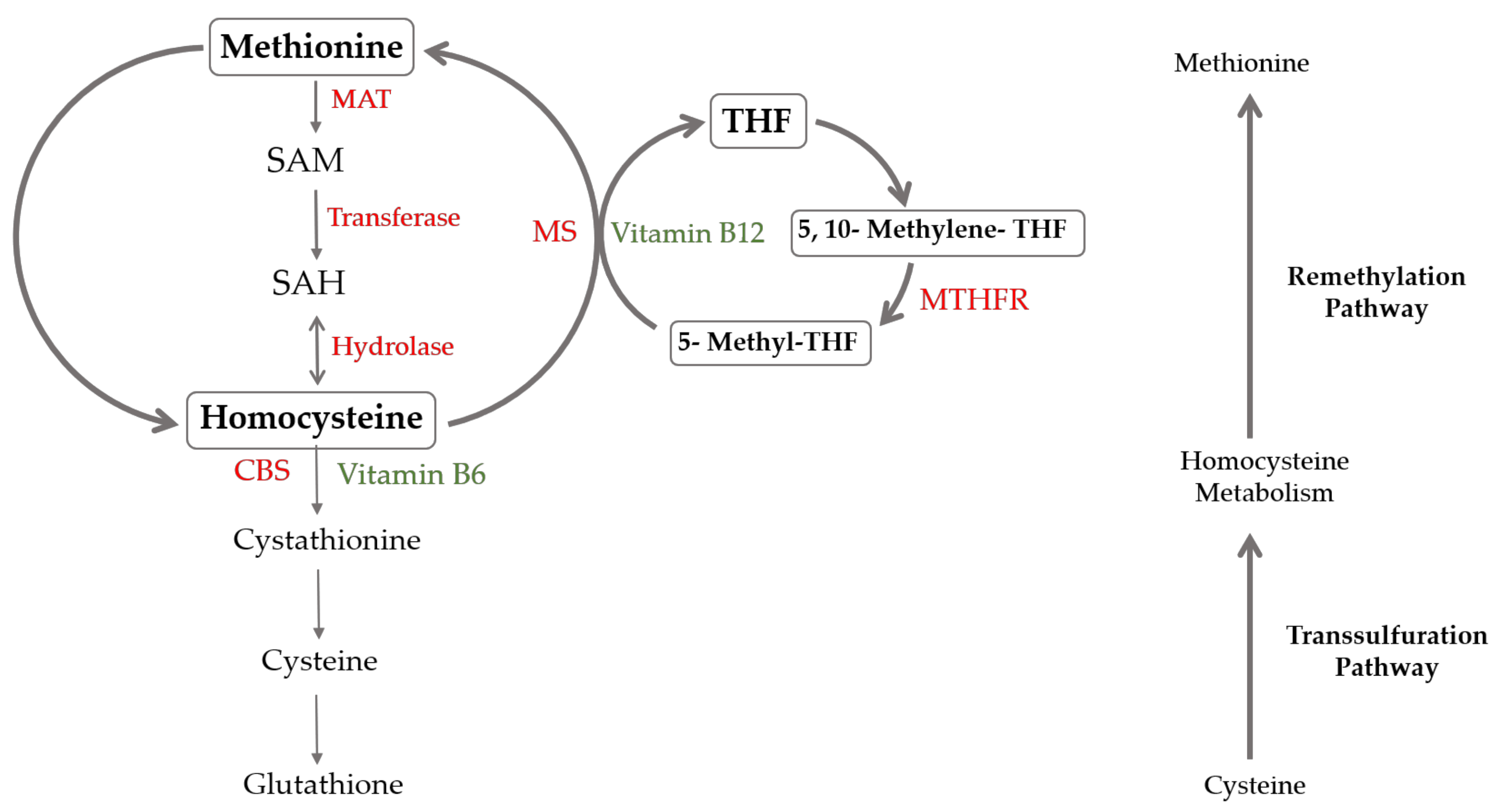
2. Hyperhomocysteinemia

The elevated blood level of HCys, a condition known as hyperhomocysteinemia, is a risk factor for different cardiovascular and neurodegenerative diseases [1,2]. HCys can be detected in both plasma and cerebrospinal fluid (CSF). While the measurement in CSF is not very common in routine clinical practice, it can be used in research or to evaluate specific neurological diseases [3]. A variety of techniques are available to measure HCys levels, but the most used ones are immunoassay methods or high-pressure liquid chromatography (HLPC) [4]. Immunoassays are favored for their speed, automation, and ease of use, despite limitations in accuracy and the potential for overestimation. In contrast, HPLC, while offering superior precision and reliability, is more labor-intensive, requires specialized equipment, and is less suitable for high-throughput analysis. Despite these challenges, HPLC remains the preferred method for accurate HCys measurement in clinical practice. [5]. The choice of measurement technique can influence the clinical interpretation of HCys levels, impacting patient management and the understanding of the role of HCys in various diseases.
The physiological HCys level is considered normal between 5 and 15 μmol/L, with an optimal upper limit of 10 μmol/L [6]. Hyperhomocysteinemia is diagnosed when blood levels exceed 16 μmol/L. It is classified as mild when levels range between 16 and 30 μmol/L, moderate when levels are between 30 and 100 μmol/L, and severe when values exceed 100 μmol/L [7]. In recent years, HCys has shifted from being considered solely as a risk factor to a key biomarker. HCys is a component cause in multifactorial diseases and may reflect an unhealthy lifestyle [8].
Multiple factors can contribute to hyperhomocysteinemia, including nutritional deficiencies, genetic alterations, and certain lifestyle habits. The most severe forms are typically associated with genetic mutations in the enzymes involved in the metabolism of HCys. The importance of these enzymes is their role at the branching point of two critical pathways in the methionine cycle: remethylation, regulated by 5,10-methylene tetrahydrofolate reductase (MTHFR); and transsulfuration, carried out by cystathionine-β-synthase (CBS) (Figure 1). The most prevalent genetic polymorphism worldwide is a mutation in the MTHFR gene, specifically a single nucleotide substitution (C to T) at position 677. This mutation produces a thermolabile enzyme variant with approximately half the normal activity, commonly associated with mild to moderate hyperhomocysteinemia. The prevalence of the 677C>T mutation varies by population; approximately 10–15% of Caucasians and Asians are homozygous for this mutation, while the heterozygous prevalence reaches 30–40% in some populations [9]. Another frequent mutation, the A1298C variant of MTHFR, also impacts HCys metabolism, though its effect on HCys levels is generally milder [10].
The most severe forms of hyperhomocysteinemia are associated with homozygous mutations in the CBS gene, which significantly elevate HCys levels while reducing cysteine synthesis. Although homozygous mutations in MTHFR and methionine synthase can also cause severe hyperhomocysteinemia, they are less common [11,12]. The prevalence of CBS deficiency-related homocystinuria is rare, estimated at 1 in 200,000 to 335,000 individuals worldwide [13].
On the other hand, hyperhomocysteinemia can also be due to deficiencies in the cofactors required for the metabolism of HCys, such as vitamins B6, B9, and B12. In the absence of these vitamins, HCys cannot be metabolized via either remethylation or transsulfuration and accumulates in plasma. Therefore, the deficiencies of these vitamins in the diet are significant contributors to hyperhomocysteinemia [14]. Dietary habits, particularly those rich in B-vitamins and polyphenols, play a critical role in regulating homocysteine metabolism, providing an opportunity for dietary strategies to mitigate its associated risks. Excessive alcohol consumption elevates hepatic S-adenosylmethionine levels, which in turn lead to an increase in HCys concentration. This association has been observed in patients with early-stage alcoholic liver disease and hyperhomocysteinemia [15]. Smoking has also been described to contribute to hyperhomocysteinemia by decreasing folate levels, thereby impairing the HCys remethylation pathway [16]. Other factors associated with increased plasma HCys include male sex, advanced age, and unhealthy diet. In contrast, a good physical condition and adherence to the Mediterranean diet, which is naturally abundant with these vitamins and antioxidant-rich foods, have been shown to lower HCys levels and reduce neurovascular risks [17].

3. Vascular Effects of Homocysteine

The role of HCys in contributing to endothelial dysfunction has been extensively documented. The endothelium consists of a single layer of endothelial cells that line the blood vessels, playing a crucial role in regulating vascular tone by controlling vasodilation and vasoconstriction. This regulation of blood vessel diameter is mainly mediated by nitric oxide (NO) [18]. In a typical physiological state, NO is generated in response to vasodilatory stimuli, such as shear stress, by endothelial nitric oxide synthase (eNOS) from L-arginin, requiring several cofactors like BH4y [19]. High levels of HCys have been shown to reduce BH4 availability in vitro, through both enhanced degradation and diminished synthesis [20]. Additionally, in vitro studies have shown that HCys leads to a reduction in the expression of the L-arginine transporter (CAT-1), limiting the substrate availability for NO production [21]. As a result of these processes, eNOS becomes uncoupled, leading to a reduced production of NO and ultimately disturbing the balance between vasodilation and vasoconstriction in the vascular system [22]. The uncoupling of eNOS contributes to heightened oxidative stress by producing reactive oxygen species (ROS) [23]. In fact, in vitro studies in endothelial cells have shown that the increase in ROS induced by HCys stimulates the secretion of proinflammatory cytokines like IL-6 and adhesion molecules such as ICAM-1 [24]. The proinflammatory environment generated by HCys leads to the activation of caspases 3 and 9, resulting in apoptosis-mediated cell death [25]. HCys is also implicated in other forms of cell death, such as pyroptosis [26] and ferroptosis [27]. In addition, a role for HCys in accelerating endothelial cell senescence has been reported [28].
The disruption of endothelial function caused by HCys accumulation is strongly linked to the progression of vascular diseases [29]. Research indicates that a hyperhomocysteinemia-inducing diet can greatly accelerate the development of atherosclerosis in vivo, which causes considerable plaque buildup in the arteries [30]. In addition, individuals with the homozygous form (C677T) of the MTHFR gene are more prone to hypertension [31]. Moreover, HCys has been highly correlated with increased prothrombotic activity in endothelial cells. HCys treatment leads to the externalization of phosphatidylserine, providing a procoagulant surface for platelet aggregation. Moreover, HCys promotes the formation of procoagulant microparticles that can enhance platelet activation and thrombin generation [32]. Research on a hyperhomocysteinemia rat model has also suggested a prothrombotic effect of HCys in vivo, observed through changes in blood composition, including decreased fibrinogen and increased platelet counts [33]. Moreover, this effect has also been confirmed for hyperhomocysteinemia on the activation of platelets in human plasma [33]. As part of the vascular system, HCys can also affect the BBB, a specialized network of blood vessels that tightly regulates what enters the brain. BBB dysfunction is a critical factor in the development of several conditions, such as AD [34], stroke [35], and hydrocephalus [36]. In addition to HCys’s known effects on the individual cells that form the BBB, such as endothelial cells and astrocytes, elevated HCys levels can disrupt the integrity of this barrier in vitro by reducing adherent proteins, which results in increased permeability [37].
In conclusion, elevated HCys levels contribute significantly to vascular dysfunction by impairing nitric oxide production, promoting oxidative stress, and inducing proinflammatory responses. Dietary patterns rich in processed foods and low in essential nutrients can exacerbate vascular damage induced by HCys [38]. These effects accelerate atherosclerosis, promote hypertension, and increase clotting risks, while also compromising the BBB’s integrity, which highlight a link between HCys and both cardiovascular and neurological diseases. Preventive strategies, including the adoption of balanced diets rich in antioxidants and B-vitamins, can mitigate endothelial dysfunction and reduce the risk of related vascular diseases [39].

4. Hyperhomocysteinemia and Stroke

Stroke is a neurovascular disorder produced via a significant reduction in the cerebral blood flow due to either an obstruction in the vasculature (ischemic stroke) or as a result of a bleeding caused by the rupture of brain vasculature (haemorrhagic stroke) [40]. Stroke represents the second most common cause of death and is a leading cause of disability globally [41,42]. Due to the reduced cerebral blood flow, there is a depletion of oxygen and glucose. This process leads to cell death within minutes in the ischemic core and the disruption of normal cell function in the surrounding area (penumbra) [43,44]. Cell death is followed by inflammation and ROS production, causing disruption in the BBB and brain edema [43,45].
The association between high levels of HCys and the risk of stroke has been extensively studied. Systematic reviews of observational studies have shown a strong, positive, and dose-related association between the serum concentration of total HCys and the risk of thrombosis and stroke, which is independent of other vascular risk factors [46,47,48]. In fact, it has been found that individuals with the MTHFR-TT genotype present higher levels of HCys and also have a greater risk of stroke than those with the CC/CT genotype [46,49,50].
Stroke is often associated with cerebral small vessel disease (CSVD), a group of clinical imaging syndromes often associated with stroke, underlying vascular cognitive impairment that is caused by cerebral vascular anomalies affecting small arteries, arterioles, capillaries, and venules [51,52]. CSVD is characterized by lacunar infarcts, white matter hyperintensities, cerebral microbleeds, enlarged perivascular spaces, and brain atrophy. It is considered an important cause of lacunar stroke and intracerebral hemorrhage [51]. Multiple studies have demonstrated that HCys levels are directly correlated with the presence of lacunar infarcts and inversely correlated with brain parenchymal fraction [52]. Hyperhomocysteinemia has also been reported to independently accelerate the progression of CSVD by elevating CSVD burden scores and, consequently, increase the risk of cognitive impairment [53]. Moreover, increases in HCys are associated with the development of different imaging markers of CSVD, such as cerebral microbleed, enlarged perivascular space, the presence of white matter hyperintensity, or lacunar infarction [53,54,55,56].
Although the specific mechanisms underlying the association between HCys and CSVD remain unclear, endothelial dysfunction is reported to play a crucial role. Hyperhomocysteinemia may compromise blood vessel architecture and induce endothelial dysfunction through different processes [48,57]. Recent evidence indicates that increased levels of total HCys are associated with histopathological features in vascular lesions (intimal thickening, elastic lamina rupture, smooth muscle hypertrophy, or platelet accumulation) through oxidative stress or inflammation processes, contributing to the association between serum HCys and CSVD [48,57]. Some factors such as smoking, renal impairment, atherogenic diet, cystine or folate deficiency, hypertension, diabetes, or hypercholesterolemia could also be contributing to the increased risk of stroke by affecting the HCys serum concentration [58,59]. This systematic review demonstrated that the strength of the association between homocysteine and risk of vascular disease varies according to study design. The association was strongest in cross-sectional and case–control studies, whereas prospective studies indicated less or, in some cases, no predictive ability for plasma homocysteine in cardiovascular disease [60]. Different hypotheses have proposed hyperhomocysteinemia as a general poor lifestyle biomarker [8]. Evidence has shown that a 5 µmol/L increase in HCys elevates the relative risk of ischemic stroke by 1.5 times, while a reduction of 3 µmol/L total HCys is associated with a 24% decrease in ischemic stroke [61]. Further studies are needed to establish HCys as an appropriate biomarker of stroke development and progression. Given the strong association between hyperhomocysteinemia and stroke, the modulation of the diet emerge as a promising preventive strategy due to the ability to lower homocysteine levels and counteract vascular damage.

5. Hyperhomocysteinemia and Hydrocephalus

Hydrocephalus is a condition characterized by CSF accumulation in the brain, resulting in elevated intraventricular pressure due to imbalances between CSF formation and absorption [62]. A disease commonly observed in older adults is the idiopathic normal pressure hydrocephalus (iNPH). It is characterized by a triad consisting of dementia, gait disturbance, and urinary incontinence, combined with ventriculomegaly, collectively referred to as Hakim’s triad [63]. The incidence of iNPH varies significantly by region. In hospital-based studies from the Netherlands, Germany, Sweden, Norway, and the USA, iNPH had an incidence of 0.5–1.2 patients per 100,000 inhabitants. In Japan, however, the incidence is notably higher, reaching 1.2/1000 inhabitants/year in people aged 70 years or older. A Norwegian study of iNPH in a defined population reported an incidence of at least 5.5 per 100,000 inhabitants/year, indicating that iNPH may be an underdiagnosed disease [64]. In iNPH, the origin of ventricles enlargement is still controversial, and mechanical, vascular, inflammatory, and metabolic factors have been indicated [65]. This ventriculomegaly exerts pressure on the surrounding structures and compresses periventricular arterioles and venules, which finally leads to poor tissue perfusion. The combined effects of hypoperfusion and mechanical compression trigger a number of pathophysiological changes in brain tissue, especially lesions in the periventricular white matter, but also in the gray matter. Other lesions include the following: alterations in metabolism, especially in glucose metabolism, which affect cellular energy balance; astrogliosis and neuroinflammation that worsen parenchymal stiffness and decrease brain distensibility; and alterations in the BBB, further complicating nutrient transport and waste clearance [66,67].
Another underlying mechanism of iNPH involves high concentrations of HCys in the CSF of patients. It has been suggested that high levels of this amino acid may reduce the distensibility of intracranial arteries, impacting the CSF dynamics. Typically, CSF diffuses into the subarachnoid space via pulsations of the intracranial arteries. Each arterial pulsation is accompanied by CSF absorption. As the distensibility of the arterial wall is reduced by the toxic effects of HCys, pulsations weaken, and CSF accumulates in the ventricles. Additionally, alterations of the BBB are also associated with elevated levels of HCys. Through mechanisms such as redox imbalance and oxidative stress, HCys leads to endothelial dysfunction and, ultimately, to the rupture of the BBB, which allows substances transported in the blood to enter the brain parenchyma. This imbalance between the vascular and ventricular system results in an abnormal osmotic gradient, driving water molecules into the ventricular system and leading to hydrocephalus [68,69,70].
The most common therapeutic approach to iNPH is the insertion of a shunt, which connects the cerebral ventricles to an alternative drainage site, usually the abdominal cavity. Lumbar drainage tests are used to decide whether shunt insertion is appropriate for a patient. To assess the patient’s progress after the shunt, biomarkers such as HCys can be used. Post-shunt, HCys levels in both plasma and CSF decrease progressively as excess CSF is removed, often improving gait, memory, and urinary symptoms as CSF HCys values normalize [71]. However, from CSF shunting until 2 years, plasma HCys levels may rise again, potentially re-triggering cognitive decline, reflecting neurodegenerative progression. Thus, plasma HCys monitoring serves as a valuable marker for tracking disease progression in shunt-treated patients [72]. For congenital hydrocephalus, although direct studies on HCys levels are limited, research suggests that genetic defects in this condition indirectly affect the folate pathway in which HCys is involved. Enzyme imbalances in folate metabolism observed in congenital hydrocephalus may predispose individuals to this condition, though HCys levels may not reach the elevated levels seen in iNPH due to differential folate utilization [73].

6. Homocysteine and Neurological Diseases

HCys is well known for its neurotoxicity, affecting both the viability of neurons and their ability to transmit signals. It affects the formation of functional neuronal networks, indicating that the effects of hyperhomocysteinemia are not only limited to the survival of neurons but also interfere with their proper functioning and connectivity [74]. Furthermore, HCys has been reported to impair hippocampal plasticity and synaptic transmission, resulting in learning and memory deficits, as observed in Alzheimer’s disease (AD) [75].
The mechanisms by which HCys contributes to neurodegeneration are multifactorial and complex and include excitotoxicity, oxidative stress, and mitochondrial dysfunction. HCys acts as an agonist of several glutamate receptors such as AMPA and NMDA. The overstimulation of these receptors causes an increase in intracellular calcium, inducing the production of ROS and the activation of caspases, finally leading to cell apoptosis [76]. HCys contributes to oxidative stress through multiple mechanisms that disrupt cellular redox balance and promote the generation of ROS. Elevated HCys levels inhibit key antioxidant enzymes [77]. HCys has been shown to interfere with the methionine cycle, reducing glutathione synthesis [78]. Furthermore, HCys upregulates NADPH oxidase, increasing superoxide production, and disrupts mitochondrial function, leading to further ROS accumulation [79].
HCys affects not only neuronal cells but also glial cells. Studies have shown that astrocytes exposed to elevated homocysteine levels exhibit cytoskeletal alterations, particularly in glial fibrillary acidic protein, leading to significant morphological changes. As a result of these structural alterations, astrocytes lose their ability to support essential functions, such as neurogenesis [80], prompting them to secrete proinflammatory factors, cytokines, and neurotoxic compounds that are damaging to the surrounding neurons [81]. In conclusion, hyperhomocysteinemia creates a hostile environment for CNS cells, promoting neurodegeneration and potentially contributing to BBB dysfunction. By modifying the integrity of the blood–brain barrier, HCys increases its permeability, which facilitates the infiltration of neurotoxic substances and immune cells into the brain parenchyma. This not only accelerates neuronal loss but also impairs the clearance of pathological proteins, such as beta-amyloid in Alzheimer’s disease, further amplifying disease progression [82].
Elevated levels of HCys have been considered a risk factor for developing neurological conditions such as AD or Parkinson’s disease [14]. Moreover, in AD, HCys levels are being investigated as a marker to assess disease progression. Higher and prolonged elevations in HCys are associated with increased neuronal damage, potentially accelerating the rate of cognitive decline [83]. In addition to its neurotoxic effect, HCys can also aggravate the symptoms that are already present in other neurodegenerative disorders. In pathologies such as Parkinson, where motor and behavioral symptoms predominate, elevated levels of HCys have been shown to worsen these deficits, exacerbating rigidity, tremors, and cognitive alterations [84].
HCyS has an important impact on vascular functionality, a factor increasingly recognized as a common denominator in various neurodegenerative diseases. A substantial body of evidence underscores the significant vascular component in conditions such as AD and Parkinson’s disease (PD). Notably, vascular alterations often accompany, and in some cases precede, the development of neurodegenerative pathology, suggesting a contributory role in the development of age-related dementia [85,86].
Levodopa, the most effective symptomatic treatment for PD, has been consistently linked to elevated HCys levels due to its metabolism via catechol-O-methyltransferase (COMT). Elevated HCys levels are associated with cognitive decline and structural brain changes in PD patients, such as increased ventricular volume as well as microscopic and macroscopic damage. Furthermore, research indicates that even before the initiation of levodopa therapy, PD patients with elevated HCys levels face a higher risk of cognitive impairment. Supplementation with vitamins B6, B12, and folate has demonstrated potential in countering levodopa-induced hyperhomocysteinemia and reducing its associated complications. However, despite these promising results, the integration of preventative supplementation into clinical practice remains limited, highlighting the need for further research and consensus on the therapeutic role of HCys management in PD [87].
Although there is ongoing debate about whether HCys functions as a biomarker or acts as a direct pathogenic agent, its involvement in driving the progression and development of neurovascular and neurodegenerative changes is evident. This warrants further investigation to inform on the development of novel preventive and therapeutic strategies.

7. Therapeutic Perspectives on Homocysteine Modulation

The metabolism of HCys is highly dependent on vitamin B6, vitamin B12, and folate; therefore, deficiencies in these nutrients can increase its serum concentration [88]. Elevated HCys is associated with a higher risk of vascular disease [89], as supported by evidence linking healthy diets, like the Mediterranean diet, with high dietary intake of vitamins and antioxidants, improper Hcy metabolism, and reduced cardiovascular risk [39,90]. Several studies have demonstrated that food fortification with folic acid, B6, or B12 vitamins is effective in reducing HCys levels [91,92,93,94,95], potentially reducing the prevalence of vascular diseases.
In the USA, a folate fortification policy introduced in 1998 led to reduced Hcy levels [96], and it was suggested to have contributed to the decline in stroke-related mortality [97]. Other studies have also suggested that folate and B vitamins supplementation may reduce the risk and improve prognosis of stroke by targeting elevated HCys levels. However, findings are mixed, as some studies report modest effects while others find no substantial impact on recurrent stroke or cardiovascular events. The Vitamin Intervention for Stroke Prevention trial assessed the effects of high-dose folic acid, B6, and B12 on patients with ischemic stroke, showing limited benefit in reducing recurrent stroke and myocardial infarction [98,99]. Additionally, a study in patients with type 2 diabetes highlighted that higher serum folate and B12 levels were associated with lower cardiovascular mortality, suggesting potential protective effects specific to populations with metabolic or vascular conditions [100]. Similarly, a systematic review and meta-analysis indicated that folic acid supplementation may be more effective in populations with low baseline folate levels, though results remain inconclusive across varied demographics and health backgrounds [101]. Dietary interventions with vitamins B6, B12, and folate have also been proposed to reduce the deleterious effects of HCys accumulation in hydrocephalus [102] and may potentially reduce the risk of congenital hydrocephalus [103]. However, the impact is modest, and further research is needed to clarify optimal intervention protocols.
Evidence has recently emerged showing that polyphenols may also reduce plasma HCys [104] and counteract the damage induced by high HCys levels [105] (Figure 2). In this sense, polyphenol-rich diets, such as the Mediterranean diet, are associated with a direct reduction in HCys but also with an increased risk of cardiovascular disease [106]. Polyphenols are secondary metabolites found in certain plants, known for their antioxidant, anti-inflammatory, and anti-apoptotic properties, as well as their antithrombotic effects [107]. Polyphenol-rich foods encompass a variety of fruits and vegetables, each characterized by distinct bioactive compounds. Some of the most prominent examples are apples (containing quercetin) [108,109], green tea (rich in catechins) [110,111], grapes and red wine (containing resveratrol) [112,113], turmeric (which has curcumin) [114], blueberries (rich in anthocyanins) [115], olive oil (containing hydroxytyrosol and tyrosol) [116,117], dark chocolate (high in flavanols) [118], spinach (with kaempferol and luteolin) [109], cinnamon (containing cinnamaldehyde and procyanidins) [119], and salicornia (rich in dicaffeoylquinic acids) [120].
Luteolin, a flavonoid polyphenol, has been shown to reduce homocysteine levels by promoting folate transport from the choroid plexus and increasing the activity of the proton-coupled folate transporter (PCFT). By maintaining an active folate pathway, luteolin helps prevent homocysteine accumulation, which is intricately linked to folate metabolism [121]. Other polyphenols, such as quercetin, work by increasing the mRNA expression of methionine synthase, cystathionine β-synthase, and cystathionine γ-lyase, enzymes involved in the remethylation and transmethylation of homocysteine, respectively, causing homocysteine to be reduced in the blood [122].
The antioxidant effects of polyphenols have been observed to work through the direct scavenging of ROS radicals [123] along with activating antioxidant systems such as the expression of antioxidant enzymes like superoxide dismutase (SOD) [124], thereby enhancing cellular resilience to oxidative stress. These effects are often accompanied by reductions in oxidative stress markers like malondialdehyde (MDA) levels [125]. Antioxidants, such as folate, and polyphenols, such as quercetin and resveratrol, have been shown to reduce hyperhomocysteinemia-induced oxidative stress by enhancing plasma antioxidant capacity, normalizing antioxidant enzyme levels, and increasing intraerythrocyte-reduced glutathione, a key molecule in oxidative stress defense [126,127]. HCys is also linked to lipid peroxidation and apoptosis. For example, resveratrol was able to decrease LDL oxidation, decrease DNA fragmentation, and reduce p53 mRNA expression, inhibiting HCys-induced cell apoptosis [128].
Polyphenols exert their anti-inflammatory effects through multiple mechanisms. For instance, quercetin promoted the anti-inflammatory M2 microglial phenotype, primarily through PI3K/Akt/NF-κB pathway regulation [129]. Similarly, resveratrol suppressed neuroinflammation by inhibiting NF-κB signaling, which reduces hippocampal inflammation and cognitive impairment in neuroinflammatory models [130]. Resveratrol suppressed immune activation cascades and mitogen-driven T-cell proliferation, thereby reducing HCys production and preventing its accumulation in the blood [131]. Similarly, olive oil phenolics inhibited HCys-induced ICAM-1 expression, a process associated with vascular damage. This effect was observed independent of the antioxidant activity of these phenolic compounds, suggesting that their protective role against HCys-induced endothelial dysfunction involves mechanisms beyond antioxidation [132]. Caffeic acid also demonstrated anti-inflammatory effects, particularly against hyperhomocysteinemia-induced inflammation in venular endothelial cells. This polyphenol inhibited HCys-induced leukocyte adhesion and rolling by reducing the expression of adhesion molecules, such as E-selectin and ICAM-1, in the cerebrovascular endothelium. Moreover, caffeic acid decreased ROS production and plasma levels of key chemokines involved in immune response and inflammation, including KC, MIP-2, and MCP-1 [133].
Other beneficial effects of polyphenols point directly to their action on kidney damage, with hypertension and hyperhomocysteinemia being the most common causes. Resveratrol reversed the effect of hyperhomocysteinemia on the kidney, increasing superoxide dismutase and glomerular filtration and decreasing the serum levels of MDA and urinary creatinine [134]. Recently, a polyphenol-rich extract was found to reduce HCys levels after three months of administration. This extract, which main polyphenols were quercetin, isorhamnetin, and dicaffeoylquinic acid derivatives, induced biochemical and proteomic changes related to cardiovascular health, such as lowered LDL, and increased GFR [135].
In addition, several polyphenols have been shown to modulate brain fluid dynamics by regulating the expression of aquaporins (AQPs). AQPs are a family of integral membrane proteins that function as selective channels to facilitate the transport of water molecules across cell membranes and are essential for brain fluid balance. The flavanone pinocembrin crosses the BBB, where it helps to mitigate brain edema, neuronal apoptosis, and endothelial cell damage by reducing inflammatory signals and decreasing the expression of AQP4. Curcumin has also been shown to dose-dependently reduce both gene expression and abundance of AQP4 and AQP9 proteins in a rat model of hypoxic–ischemic brain injury [136]. Epigallocatechin-3-gallate (EGCG) is also considered an anti-vasogenic edema agent that protects the integrity of the BBB. This compound regulates AQP4 expression in endothelial cells and astrocytes as well as inhibits the p38 MAPK-PI3K/AKT-eNOS signaling pathway in endothelial cells [137]. Similarly, tea polyphenols have also shown efficacy in reducing brain edema and protect the BBB through the regulation of the PI3K/Akt pathway and AQP4 levels. In particular, this type of polyphenol increased the levels of the anti-apoptotic protein Bcl-2 and tight junction proteins such us Zona Occludens 1 (ZO-1), claudin-5, and occludin, after subarachnoid hemorrhage [138,139].
These properties make polyphenols a promising therapeutic approach for conditions that are frequently associated with elevated plasma HCys levels. Stroke and hydrocephalus share risk factors such as aging and hyperhomocysteinemia, with overlapping symptoms including cognitive impairment and motor dysfunction. Both conditions involve disruptions in cerebral or cerebrospinal fluid flow, emphasizing the importance of early intervention for improved outcomes.
Polyphenols have shown neuroprotective effects through multiple pathways in both hydrocephalus and ischemic stroke models, potentially due to their antioxidative and anti-inflammatory properties. In hydrocephalus-induced Wistar rats, a polyphenol extract from C. sinensis was administered intraperitoneally for 9 days and was found to reduce reactive astrocyte activity, as well as increase corpus callosum myelination, compared to untreated animals [140]. Similarly, treatment with EGCG, a polyphenolic compound found in green tea, has been shown to reduce periventricular oxidative damage in young rats with induced hydrocephalus through its capacity to eliminate ROS [141]. Additionally, studies have shown that curcumin can modulate the expression of AQP4, a protein critical for water transport in the brain and closely linked to the development of cerebral edema and hydrocephalus. By reducing AQP4 expression, curcumin helps limit cerebral edema, controlling water flow in the brain and decreasing fluid accumulation [142]. On the other hand, growing research highlights the preventive potential of dietary supplementation with polyphenols against neurovascular diseases, which have been extensively reviewed [102,120]. In fact, some of the beforementioned polyphenols have been shown to not only reduce the infarcted area in the brain in stroke models but also to improve cognitive functions by enhancing neurological score. For instance, quercetin reduced hippocampal neuron loss and brain edema as well as improved neurological function in rats when administered before the bilateral occlusion of the common carotid arteries [143]. In addition, vanillic acid [144] or kaempferol [145] demonstrated efficacy when administered as prophylactic treatments, while ferulic acid [146] or chlorogenic acid [147] showed benefits when administered after ischemia. These compounds share a common mechanism of action, which involves reducing inflammatory markers by neutralizing excessive free radicals and modulating inflammatory pathways such as NF-κB and TLR4, as well as inhibiting apoptosis. Moreover, a systematic review assessing the role of polyphenols on health outcomes in post-stroke patients highlighted that most of the studies found improvements in at least one of the study outcomes which included vascular function, blood pressure, or glycaemia [148]. Antioxidant supplementation with vitamin E, beta-carotene, and apple polyphenol extract over a 12-month period did not lead to significant improvements in functional recovery among stroke survivors. However, due to the controlled dietetic treatment, HCyS levels were reduced, highlighting the importance of monitoring the nutritional status in stroke patients [149]. Recently, a polyphenol-rich extract containing chlorogenic acid derivatives and flavonoid derivatives of luteolin, quercetin, and isorhamnetin was found to reduce HCyS levels and impact blood pressure in patients after transient ischemic attack, a well- established risk factor for future ischemic stokes [150]. However, studies with longer duration and larger sample sizes are needed to clarify how polyphenol-based therapies could mitigate vascular risk. Therefore, polyphenols, through their different mechanisms of action, are a promising therapeutic option in the treatment of neurovascular diseases. Their ability to influence multiple molecular pathways opens new possibilities for the development of effective treatments that can prevent or mitigate the effects of these diseases.

8. Conclusions

The reviewed evidence supports the therapeutic potential of lowering homocysteine levels in the prevention and management of neurovascular disorders. Elevated HCys levels correlate with increased vascular damage and neurological dysfunction, making it a promising biomarker and intervention target. While B-vitamin supplementation has shown some efficacy in reducing HCys, its effects on clinical outcomes for stroke and hydrocephalus are still under investigation. Polyphenols, known for their antioxidant, anti-inflammatory, and neuroprotective properties, offer an additional dietary approach to reduce HCys-related risks. More clinical studies are needed to establish optimal dosages, effective intervention protocols, and the long-term impacts of HCys-lowering strategies in different populations. Overall, integrating HCys-lowering interventions may contribute significantly to neuroprotection and improved outcomes in patients with stroke or hydrocephalus.

Author Contributions

Conceptualization: C.d.R., M.E. and J.M.; Investigation: C.O.-S., M.R.-B., Á.G.-D., E.S.D. and C.d.R.; Visualization: C.O.-S. and C.d.R.; Funding acquisition: M.E. and J.M.; Writing—original draft: C.O.-S., M.R.-B., Á.G.-D., E.S.D. and C.d.R.; Writing—review and editing: C.d.R., M.E. and J.M. All authors have read and agreed to the published version of the manuscript.

Funding

This work was funded by ISCIII by the FORTALECE program of the Spanish Ministry of Science and Innovation, aimed at accredited Health Research Institutes as part of the Strategic Action in Health 2021–2023, project Code: FORT23/0000. This work was also supported by project RD21/0006/0015, funded by the National Institute of Health Carlos III (ISCIII) with public funding from the Cooperative Research Networks for Health Results (RICORS) RICORS-ICTUS. Thematic Area: cerebrovascular diseases. “Funded by the European Union—NextGenerationEU. European Union Recovery and Resilience Facility, measures integrated into the PRTR”. Additionally, Grant PI19/01096 from the Spanish Ministry of Economy and Competitiveness, co-financed by the Carlos III Health Institute (ISCIII) and European Regional Development Fund (FEDER), and grant P20_00646 from the Regional Department of Economy, Knowledge, Business and University, Government of Andalucía, Andalusian Plan for Research (PAIDI-2020), awarded to M.E, also support this work. C.R. received financial support from the Sara Borrell program funded by ISCIII, grant number CD21/00148. M.R. received financial support from the PFIS program funded by ISCIII grant number FI22/00202.

Conflicts of Interest

The authors declare no conflicts of interest.

References

  1. Guieu, R.; Ruf, J.; Mottola, G. Hyperhomocysteinemia and cardiovascular diseases. Ann. Biol. Clin. 2022, 80, 7–14. [Google Scholar] [CrossRef] [PubMed]
  2. Sharma, M.; Tiwari, M.; Tiwari, R.K. Hyperhomocysteinemia: Impact on Neurodegenerative Diseases. Basic. Clin. Pharmacol. Toxicol. 2015, 117, 287–296. [Google Scholar] [CrossRef] [PubMed]
  3. Valentino, F.; Bivona, G.; Butera, D.; Paladino, P.; Fazzari, M.; Piccoli, T.; La Bella, V. Elevated cerebrospinal fluid and plasma homocysteine levels in ALS. Eur. J. Neurol. 2010, 17, 84–89. [Google Scholar] [CrossRef] [PubMed]
  4. Alam, S.F.; Kumar, S.; Ganguly, P. Measurement of homocysteine: A historical perspective. J. Clin. Biochem. Nutr. 2019, 65, 171–177. [Google Scholar] [CrossRef] [PubMed]
  5. Paprotny, Ł.; Wianowska, D.; Izdebska, M.; Celejewska, A.; Szewczak, D.; Solski, J. Analysis of serum homocysteine in the laboratory practice—comparison of the direct chemiluminescence immunoassay and high performance liquid chromatography coupled with fluorescent detection. Biochem. Med. 2020, 30, 030703. [Google Scholar] [CrossRef]
  6. Rasmussen, K.; Moller, J. Total homocysteine measurement in clinical practice. Ann. Clin. Biochem. 2000, 37, 627–648. [Google Scholar] [CrossRef]
  7. Stanger, O.; Herrmann, W.; Pietrzik, K.; Fowler, B.; Geisel, J.; Dierkes, J.; Weger, M. DACH-LIGA homocystein (german, austrian and swiss homocysteine society): Consensus paper on the rational clinical use of homocysteine, folic acid and B-vitamins in cardiovascular and thrombotic diseases: Guidelines and recommendations. Clin. Chem. Lab. Med. 2003, 41, 1392–1403. [Google Scholar] [CrossRef]
  8. Smith, A.D.; Refsum, H. Homocysteine—from disease biomarker to disease prevention. J. Intern. Med. 2021, 290, 826–854. [Google Scholar] [CrossRef]
  9. Wilcken, B.; Bamforth, F.; Li, Z.; Zhu, H.; Ritvanen, A.; Redlund, M.; Botto, L.D. Geographical and ethnic variation of the 677C>T allele of 5,10 methylenetetrahydrofolate reductase (MTHFR): Findings from over 7000 newborns from 16 areas world wide. J. Med. Genet. 2003, 40, 619–625. [Google Scholar] [CrossRef]
  10. Weisberg, I.; Tran, P.; Christensen, B.; Sibani, S.; Rozen, R. A second genetic polymorphism in methylenetetrahydrofolate reductase (MTHFR) associated with decreased enzyme activity. Mol. Genet. Metab. 1998, 64, 169–172. [Google Scholar] [CrossRef]
  11. Gupta, S.; Wang, L.; Hua, X.; Krijt, J.; Kozich, V.; Kruger, W.D. Cystathionine beta-synthase p.S466L mutation causes hyperhomocysteinemia in mice. Hum. Mutat. 2008, 29, 1048–1054. [Google Scholar] [CrossRef] [PubMed]
  12. Ibrahim, S.; Maqbool, S.; Azam, M.; Iqbal, M.P.; Qamar, R. CBS mutations and MTFHR SNPs causative of hyperhomocysteinemia in Pakistani children. Mol. Biol. Rep. 2018, 45, 353–360. [Google Scholar] [CrossRef] [PubMed]
  13. Mudd, S.H.; Skovby, F.; Levy, H.L.; Pettigrew, K.D.; Wilcken, B.; Pyeritz, R.E.; Andria, G.; Boers, G.H.; Bromberg, I.L.; Cerone, R.; et al. The natural history of homocystinuria due to cystathionine beta-synthase deficiency. Am. J. Hum. Genet. 1985, 37, 1–31. [Google Scholar] [PubMed]
  14. Obeid, R.; McCaddon, A.; Herrmann, W. The role of hyperhomocysteinemia and B-vitamin deficiency in neurological and psychiatric diseases. Clin. Chem. Lab. Med. 2007, 45, 1590–1606. [Google Scholar] [CrossRef]
  15. Vatsalya, V.; Gala, K.S.; Hassan, A.Z.; Frimodig, J.; Kong, M.; Sinha, N.; Schwandt, M.L. Characterization of Early-Stage Alcoholic Liver Disease with Hyperhomocysteinemia and Gut Dysfunction and Associated Immune Response in Alcohol Use Disorder Patients. Biomedicines 2020, 9, 7. [Google Scholar] [CrossRef]
  16. Okumura, K.; Tsukamoto, H. Folate in smokers. Clin. Chim. Acta 2011, 412, 521–526. [Google Scholar] [CrossRef]
  17. Shenoy, V.; Mehendale, V.; Prabhu, K.; Shetty, R.; Rao, P. Correlation of serum homocysteine levels with the severity of coronary artery disease. Indian J. Clin. Biochem. 2014, 29, 339–344. [Google Scholar] [CrossRef]
  18. Feletou, M. The Endothelium: Part 1: Multiple Functions of the Endothelial Cells-Focus on Endothelium-Derived Vasoactive Mediators. In Colloquium Series on Integrated Systems Physiology; Morgan & Claypool Life Sciences: Williston, VT, USA, 2011. [Google Scholar]
  19. Deanfield, J.; Donald, A.; Ferri, C.; Giannattasio, C.; Halcox, J.; Halligan, S.; Lerman, A.; Mancia, G.; Oliver, J.J.; Pessina, A.C.; et al. Endothelial function and dysfunction. Part I: Methodological issues for assessment in the different vascular beds: A statement by the Working Group on Endothelin and Endothelial Factors of the European Society of Hypertension. J. Hypertens. 2005, 23, 7–17. [Google Scholar] [CrossRef]
  20. Topal, G.; Brunet, A.; Millanvoye, E.; Boucher, J.L.; Rendu, F.; Devynck, M.A.; David-Dufilho, M. Homocysteine induces oxidative stress by uncoupling of NO synthase activity through reduction of tetrahydrobiopterin. Free Radic. Biol. Med. 2004, 36, 1532–1541. [Google Scholar] [CrossRef]
  21. Jin, L.; Caldwell, R.B.; Li-Masters, T.; Caldwell, R.W. Homocysteine induces endothelial dysfunction via inhibition of arginine transport. J. Physiol. Pharmacol. 2007, 58, 191–206. [Google Scholar]
  22. Lai, W.K.; Kan, M.Y. Homocysteine-Induced Endothelial Dysfunction. Ann. Nutr. Metab. 2015, 67, 1–12. [Google Scholar] [CrossRef]
  23. Shin, W.S.; Berkowitz, D.E.; Ryoo, S.W. Increased arginase II activity contributes to endothelial dysfunction through endothelial nitric oxide synthase uncoupling in aged mice. Exp. Mol. Med. 2012, 44, 594–602. [Google Scholar] [CrossRef] [PubMed]
  24. Han, S.; Wu, H.; Li, W.; Gao, P. Protective effects of genistein in homocysteine-induced endothelial cell inflammatory injury. Mol. Cell. Biochem. 2015, 403, 43–49. [Google Scholar] [CrossRef] [PubMed]
  25. Wang, X.; Wang, Y.; Zhang, L.; Zhang, D.; Bai, L.; Kong, W.; Huang, Y.; Tang, C.; Du, J.; Jin, H. L-Cystathionine Protects against Homocysteine-Induced Mitochondria-Dependent Apoptosis of Vascular Endothelial Cells. Oxid. Med. Cell. Longev. 2019, 2019, 1253289. [Google Scholar] [CrossRef] [PubMed]
  26. Xi, H.; Zhang, Y.; Xu, Y.; Yang, W.Y.; Jiang, X.; Sha, X.; Cheng, X.; Wang, J.; Qin, X.; Yu, J.; et al. Caspase-1 Inflammasome Activation Mediates Homocysteine-Induced Pyrop-Apoptosis in Endothelial Cells. Circ. Res. 2016, 118, 1525–1539. [Google Scholar] [CrossRef]
  27. Shi, J.; Chen, D.; Wang, Z.; Li, S.; Zhang, S. Homocysteine induces ferroptosis in endothelial cells through the systemXc(-)/GPX4 signaling pathway. BMC Cardiovasc. Disord. 2023, 23, 316. [Google Scholar] [CrossRef]
  28. Zhang, D.; Sun, X.; Liu, J.; Xie, X.; Cui, W.; Zhu, Y. Homocysteine accelerates senescence of endothelial cells via DNA hypomethylation of human telomerase reverse transcriptase. Arter. Thromb. Vasc. Biol. 2015, 35, 71–78. [Google Scholar] [CrossRef]
  29. Fu, Y.; Wang, X.; Kong, W. Hyperhomocysteinaemia and vascular injury: Advances in mechanisms and drug targets. Br. J. Pharmacol. 2018, 175, 1173–1189. [Google Scholar] [CrossRef]
  30. Andrews, S.G.; Koehle, A.M.; Paudel, D.; Neuberger, T.; Ross, A.C.; Singh, V.; Bottiglieri, T.; Castro, R. Diet-Induced Severe Hyperhomocysteinemia Promotes Atherosclerosis Progression and Dysregulates the Plasma Metabolome in Apolipoprotein-E-Deficient Mice. Nutrients 2024, 16, 330. [Google Scholar] [CrossRef]
  31. Al Hageh, C.; Alefishat, E.; Ghassibe-Sabbagh, M.; Platt, D.E.; Hamdan, H.; Tcheroyan, R.; Chammas, E.; O’Sullivan, S.; Abchee, A.; Wang, B.; et al. Homocysteine levels, H-Hypertension, and the MTHFR C677T genotypes: A complex interaction. Heliyon 2023, 9, e16444. [Google Scholar] [CrossRef]
  32. Zhu, J.; Xie, R.; Piao, X.; Hou, Y.; Zhao, C.; Qiao, G.; Yang, B.; Shi, J.; Lu, Y. Homocysteine enhances clot-promoting activity of endothelial cells via phosphatidylserine externalization and microparticles formation. Amino Acids 2012, 43, 1243–1250. [Google Scholar] [CrossRef] [PubMed]
  33. Litvinov, R.I.; Peshkova, A.D.; Le Minh, G.; Khaertdinov, N.N.; Evtugina, N.G.; Sitdikova, G.F.; Weisel, J.W. Effects of Hyperhomocysteinemia on the Platelet-Driven Contraction of Blood Clots. Metabolites 2021, 11, 354. [Google Scholar] [CrossRef] [PubMed]
  34. Zipser, B.D.; Johanson, C.E.; Gonzalez, L.; Berzin, T.M.; Tavares, R.; Hulette, C.M.; Vitek, M.P.; Hovanesian, V.; Stopa, E.G. Microvascular injury and blood-brain barrier leakage in Alzheimer’s disease. Neurobiol. Aging 2007, 28, 977–986. [Google Scholar] [CrossRef] [PubMed]
  35. Candelario-Jalil, E.; Dijkhuizen, R.M.; Magnus, T. Neuroinflammation, Stroke, Blood-Brain Barrier Dysfunction, and Imaging Modalities. Stroke 2022, 53, 1473–1486. [Google Scholar] [CrossRef]
  36. Filippidis, A.S.; Kalani, M.Y.; Rekate, H.L. Hydrocephalus and aquaporins: Lessons learned from the bench. Childs Nerv. Syst. 2011, 27, 27–33. [Google Scholar] [CrossRef]
  37. Beard, R.S., Jr.; Reynolds, J.J.; Bearden, S.E. Hyperhomocysteinemia increases permeability of the blood-brain barrier by NMDA receptor-dependent regulation of adherens and tight junctions. Blood 2011, 118, 2007–2014. [Google Scholar] [CrossRef]
  38. Schenkelaars, N.; van Rossem, L.; Willemsen, S.P.; Faas, M.M.; Schoenmakers, S.; Steegers-Theunissen, R.P.M. The intake of ultra-processed foods and homocysteine levels in women with(out) overweight and obesity: The Rotterdam Periconceptional Cohort. Eur. J. Nutr. 2024, 63, 1257–1269. [Google Scholar] [CrossRef]
  39. Foscolou, A.; Rallidis, L.S.; Tsirebolos, G.; Critselis, E.; Katsimardos, A.; Drosatos, A.; Chrysohoou, C.; Tousoulis, D.; Pitsavos, C.; Panagiotakos, D.B. The association between homocysteine levels, Mediterranean diet and cardiovascular disease: A case-control study. Int. J. Food Sci. Nutr. 2019, 70, 603–611. [Google Scholar] [CrossRef]
  40. Mehndiratta, P.; Chapman Smith, S.; Worrall, B.B. Etiologic stroke subtypes: Updated definition and efficient workup strategies. Curr. Treat Options Cardiovasc. Med. 2015, 17, 357. [Google Scholar] [CrossRef]
  41. Soto, A.; Guillen-Grima, F.; Morales, G.; Munoz, S.; Aguinaga-Ontoso, I.; Fuentes-Aspe, R. Prevalence and incidence of ictus in Europe: Systematic review and meta-analysis. An. Sist. Sanit. Navar. 2022, 45, e0979. [Google Scholar] [CrossRef]
  42. Collaborators, G.B.D.S. Global, regional, and national burden of stroke and its risk factors, 1990–2019: A systematic analysis for the Global Burden of Disease Study 2019. Lancet Neurol. 2021, 20, 795–820. [Google Scholar]
  43. Campbell, B.C.V.; De Silva, D.A.; Macleod, M.R.; Coutts, S.B.; Schwamm, L.H.; Davis, S.M.; Donnan, G.A. Ischaemic stroke. Nat. Rev. Dis. Primers 2019, 5, 70. [Google Scholar] [CrossRef] [PubMed]
  44. Datta, A.; Sarmah, D.; Mounica, L.; Kaur, H.; Kesharwani, R.; Verma, G.; Veeresh, P.; Kotian, V.; Kalia, K.; Borah, A.; et al. Cell Death Pathways in Ischemic Stroke and Targeted Pharmacotherapy. Transl. Stroke Res. 2020, 11, 1185–1202. [Google Scholar] [CrossRef]
  45. Rajashekar, D.; Liang, J.W. Intracerebral Hemorrhage. In StatPerls; StatPearls Publishing: Treasure Island, FL, USA, 2023. [Google Scholar]
  46. Hankey, G.J.; Eikelboom, J.W. Homocysteine and stroke. Lancet 2005, 365, 194–196. [Google Scholar] [CrossRef]
  47. Perry, I.J. Homocysteine and risk of stroke. J. Cardiovasc. Risk 1999, 6, 235–240. [Google Scholar] [CrossRef]
  48. Gonzalez-Lamuno, D.; Arrieta-Blanco, F.J.; Fuentes, E.D.; Forga-Visa, M.T.; Morales-Conejo, M.; Pena-Quintana, L.; Vitoria-Minana, I. Hyperhomocysteinemia in Adult Patients: A Treatable Metabolic Condition. Nutrients 2023, 16, 135. [Google Scholar] [CrossRef]
  49. Zhao, M.; Wang, X.; He, M.; Qin, X.; Tang, G.; Huo, Y.; Li, J.; Fu, J.; Huang, X.; Cheng, X.; et al. Homocysteine and Stroke Risk: Modifying Effect of Methylenetetrahydrofolate Reductase C677T Polymorphism and Folic Acid Intervention. Stroke 2017, 48, 1183–1190. [Google Scholar] [CrossRef]
  50. Holmes, M.V.; Newcombe, P.; Hubacek, J.A.; Sofat, R.; Ricketts, S.L.; Cooper, J.; Breteler, M.M.; Bautista, L.E.; Sharma, P.; Whittaker, J.C.; et al. Effect modification by population dietary folate on the association between MTHFR genotype, homocysteine, and stroke risk: A meta-analysis of genetic studies and randomised trials. Lancet 2011, 378, 584–594. [Google Scholar] [CrossRef]
  51. Markus, H.S.; de Leeuw, F.E. Cerebral small vessel disease: Recent advances and future directions. Int. J. Stroke 2023, 18, 4–14. [Google Scholar] [CrossRef]
  52. Cao, Y.; Su, N.; Zhang, D.; Zhou, L.; Yao, M.; Zhang, S.; Cui, L.; Zhu, Y.; Ni, J. Correlation between total homocysteine and cerebral small vessel disease: A Mendelian randomization study. Eur. J. Neurol. 2021, 28, 1931–1938. [Google Scholar] [CrossRef]
  53. Teng, Z.; Feng, J.; Liu, R.; Ji, Y.; Xu, J.; Jiang, X.; Chen, H.; Dong, Y.; Meng, N.; Xiao, Y.; et al. Cerebral small vessel disease mediates the association between homocysteine and cognitive function. Front Aging Neurosci. 2022, 14, 868777. [Google Scholar] [CrossRef] [PubMed]
  54. Kloppenborg, R.P.; Nederkoorn, P.J.; van der Graaf, Y.; Geerlings, M.I. Homocysteine and cerebral small vessel disease in patients with symptomatic atherosclerotic disease. The SMART-MR study. Atherosclerosis 2011, 216, 461–466. [Google Scholar] [CrossRef] [PubMed]
  55. Miwa, K.; Tanaka, M.; Okazaki, S.; Yagita, Y.; Sakaguchi, M.; Mochizuki, H.; Kitagawa, K. Increased Total Homocysteine Levels Predict the Risk of Incident Dementia Independent of Cerebral Small-Vessel Diseases and Vascular Risk Factors. J. Alzheimers Dis. 2016, 49, 503–513. [Google Scholar] [CrossRef] [PubMed]
  56. Nam, K.W.; Kwon, H.M.; Jeong, H.Y.; Park, J.H.; Kwon, H.; Jeong, S.M. Serum homocysteine level is related to cerebral small vessel disease in a healthy population. Neurology 2019, 92, e317–e325. [Google Scholar] [CrossRef]
  57. Zhang, P.; Xie, X.; Zhang, Y. Associations between homocysteine, vitamin B12, and folate and the risk of all-cause mortality in American adults with stroke. Front. Nutr. 2023, 10, 1279207. [Google Scholar] [CrossRef]
  58. Hankey, G.J.; Eikelboom, J.W. Homocysteine and vascular disease. Lancet 1999, 354, 407–413. [Google Scholar] [CrossRef]
  59. Christen, W.G.; Ajani, U.A.; Glynn, R.J.; Hennekens, C.H. Blood levels of homocysteine and increased risks of cardiovascular disease: Causal or casual? Arch. Intern Med. 2000, 160, 422–434. [Google Scholar] [CrossRef]
  60. Meleady, R.; Graham, I. Plasma homocysteine as a cardiovascular risk factor: Causal, consequential, or of no consequence? Nutr. Rev. 1999, 57, 299–305. [Google Scholar] [CrossRef]
  61. Ntaios, G. Homocysteine, B Vitamins, and Cardiovascular Risk. In Foods and Dietary Supplements in the Prevention and Treatment of Disease in Older Adults; Academic Press: Cambridge, MA, USA, 2015; pp. 309–318. [Google Scholar]
  62. Davson, H.; Hollingsworth, G.; Segal, M.B. The mechanism of drainage of the cerebrospinal fluid. Brain 1970, 93, 665–678. [Google Scholar] [CrossRef]
  63. Wallenstein, M.B.; McKhann, G.M. 2nd. Salomon Hakim and the discovery of normal-pressure hydrocephalus. Neurosurgery 2010, 67, 155–159. [Google Scholar] [CrossRef]
  64. Pyykko, O.T.; Nerg, O.; Niskasaari, H.M.; Niskasaari, T.; Koivisto, A.M.; Hiltunen, M.; Pihlajamaki, J.; Rauramaa, T.; Kojoukhova, M.; Alafuzoff, I.; et al. Incidence, Comorbidities, and Mortality in Idiopathic Normal Pressure Hydrocephalus. World Neurosurg. 2018, 112, e624–e631. [Google Scholar] [CrossRef] [PubMed]
  65. Skalicky, P.; Mladek, A.; Vlasak, A.; De Lacy, P.; Benes, V.; Bradac, O. Normal pressure hydrocephalus-an overview of pathophysiological mechanisms and diagnostic procedures. Neurosurg. Rev. 2020, 43, 1451–1464. [Google Scholar] [CrossRef] [PubMed]
  66. Vanneste, J.A. Diagnosis and management of normal-pressure hydrocephalus. J. Neurol. 2000, 247, 5–14. [Google Scholar] [CrossRef]
  67. Wang, Z.; Zhang, Y.; Hu, F.; Ding, J.; Wang, X. Pathogenesis and pathophysiology of idiopathic normal pressure hydrocephalus. CNS Neurosci. Ther. 2020, 26, 1230–1240. [Google Scholar] [CrossRef]
  68. Ye, S.; Feng, K.; Li, Y.; Liu, S.; Wu, Q.; Feng, J.; Liao, X.; Jiang, C.; Liang, B.; Yuan, L.; et al. High homocysteine is associated with idiopathic normal pressure hydrocephalus in deep perforating arteriopathy: A cross-sectional study. BMC Geriatr. 2023, 23, 382. [Google Scholar]
  69. Bateman, G.A. Vascular compliance in normal pressure hydrocephalus. AJNR Am. J. Neuroradiol. 2000, 21, 1574–1585. [Google Scholar]
  70. Guillotin, S.; Vallet, A.; Lorthois, S.; Cestac, P.; Schmidt, E.; Delcourt, N. Association Between Homocysteine, Frailty and Biomechanical Response of the CNS in NPH-Suspected Patients. J. Gerontol. 2022, 77, 1335–1343. [Google Scholar] [CrossRef]
  71. Sosvorova, L.; Bestak, J.; Bicikova, M.; Mohapl, M.; Hill, M.; Kubatova, J.; Hampl, R. Determination of homocysteine in cerebrospinal fluid as an indicator for surgery treatment in patients with hydrocefalus. Physiol. Res. 2014, 63, 521–527. [Google Scholar] [CrossRef]
  72. Sosvorova, L.; Mohapl, M.; Hill, M.; Starka, L.; Bicikova, M.; Vitku, J.; Kanceva, R.; Bestak, J.; Hampl, R. Steroid hormones and homocysteine in the outcome of patients with normal pressure hydrocephalus. Physiol. Res. 2015, 64 (Suppl. 2), S227–S236. [Google Scholar] [CrossRef]
  73. Cains, S.; Shepherd, A.; Nabiuni, M.; Owen-Lynch, P.J.; Miyan, J. Addressing a folate imbalance in fetal cerebrospinal fluid can decrease the incidence of congenital hydrocephalus. J. Neuropathol. Exp. Neurol. 2009, 68, 404–416. [Google Scholar] [CrossRef]
  74. Wyse, A.T.S.; Bobermin, L.D.; Dos Santos, T.M.; Quincozes-Santos, A. Homocysteine and Gliotoxicity. Neurotox. Res. 2021, 39, 966–974. [Google Scholar] [CrossRef] [PubMed]
  75. Kamat, P.K.; Kalani, A.; Givvimani, S.; Sathnur, P.B.; Tyagi, S.C.; Tyagi, N. Hydrogen sulfide attenuates neurodegeneration and neurovascular dysfunction induced by intracerebral-administered homocysteine in mice. Neuroscience 2013, 252, 302–319. [Google Scholar] [CrossRef] [PubMed]
  76. Bhatia, P.; Singh, N. Homocysteine excess: Delineating the possible mechanism of neurotoxicity and depression. Fundam. Clin. Pharmacol. 2015, 29, 522–528. [Google Scholar] [CrossRef] [PubMed]
  77. Wilcken, D.E.; Wang, X.L.; Adachi, T.; Hara, H.; Duarte, N.; Green, K.; Wilcken, B. Relationship between homocysteine and superoxide dismutase in homocystinuria: Possible relevance to cardiovascular risk. Arterioscler. Thromb. Vasc. Biol. 2000, 20, 1199–1202. [Google Scholar] [CrossRef]
  78. Vitvitsky, V.; Thomas, M.; Ghorpade, A.; Gendelman, H.E.; Banerjee, R. A functional transsulfuration pathway in the brain links to glutathione homeostasis. J. Biol. Chem. 2006, 281, 35785–35793. [Google Scholar] [CrossRef]
  79. Siow, Y.L.; Au-Yeung, K.K.; Woo, C.W.; O, K. Homocysteine stimulates phosphorylation of NADPH oxidase p47phox and p67phox subunits in monocytes via protein kinase Cbeta activation. Biochem. J. 2006, 398, 73–82. [Google Scholar] [CrossRef]
  80. Lehotsky, J.; Tothova, B.; Kovalska, M.; Dobrota, D.; Benova, A.; Kalenska, D.; Kaplan, P. Role of Homocysteine in the Ischemic Stroke and Development of Ischemic Tolerance. Front. Neurosci. 2016, 10, 538. [Google Scholar] [CrossRef]
  81. Chen, S.; Dong, Z.; Cheng, M.; Zhao, Y.; Wang, M.; Sai, N.; Wang, X.; Liu, H.; Huang, G.; Zhang, X. Homocysteine exaggerates microglia activation and neuroinflammation through microglia localized STAT3 overactivation following ischemic stroke. J. Neuroinflammation 2017, 14, 187. [Google Scholar] [CrossRef]
  82. Cheng, Z.; Yang, X.; Wang, H. Hyperhomocysteinemia and Endothelial Dysfunction. Curr. Hypertens Rev. 2009, 5, 158–165. [Google Scholar] [CrossRef]
  83. Sachdev, P. Homocysteine and neuropsychiatric disorders. Braz. J. Psychiatry 2004, 26, 50–56. [Google Scholar] [CrossRef]
  84. Paul, R.; Dutta, A.; Phukan, B.C.; Mazumder, M.K.; Justin-Thenmozhi, A.; Manivasagam, T.; Bhattacharya, P.; Borah, A. Accumulation of Cholesterol and Homocysteine in the Nigrostriatal Pathway of Brain Contributes to the Dopaminergic Neurodegeneration in Mice. Neuroscience 2018, 388, 347–356. [Google Scholar] [CrossRef] [PubMed]
  85. Cortes-Canteli, M.; Iadecola, C. Alzheimer’s Disease and Vascular Aging: JACC Focus Seminar. J. Am. Coll. Cardiol. 2020, 75, 942–951. [Google Scholar] [CrossRef] [PubMed]
  86. Paul, G.; Elabi, O.F. Microvascular Changes in Parkinson’s Disease- Focus on the Neurovascular Unit. Front Aging Neurosci. 2022, 14, 853372. [Google Scholar] [CrossRef] [PubMed]
  87. Ahlskog, J.E. Levodopa, homocysteine and Parkinson’s disease: What’s the problem? Park. Relat. Disord. 2023, 109, 105357. [Google Scholar] [CrossRef] [PubMed]
  88. Stanger, O.; Fowler, B.; Piertzik, K.; Huemer, M.; Haschke-Becher, E.; Semmler, A.; Lorenzl, S.; Linnebank, M. Homocysteine, folate and vitamin B12 in neuropsychiatric diseases: Review and treatment recommendations. Expert Rev. Neurother. 2009, 9, 1393–1412. [Google Scholar] [CrossRef]
  89. Lentz, S.R. Homocysteine and vascular dysfunction. Life Sci. 1997, 61, 1205–1215. [Google Scholar] [CrossRef]
  90. Chrysohoou, C.; Panagiotakos, D.B.; Pitsavos, C.; Das, U.N.; Stefanadis, C. Adherence to the Mediterranean diet attenuates inflammation and coagulation process in healthy adults: The ATTICA Study. J. Am. Coll. Cardiol. 2004, 44, 152–158. [Google Scholar] [CrossRef]
  91. Kataria, N.; Yadav, P.; Kumar, R.; Kumar, N.; Singh, M.; Kant, R.; Kalyani, V. Effect of Vitamin B6, B9, and B12 Supplementation on Homocysteine Level and Cardiovascular Outcomes in Stroke Patients: A Meta-Analysis of Randomized Controlled Trials. Cureus 2021, 13, e14958. [Google Scholar] [CrossRef]
  92. Liu, S.; West, R.; Randell, E.; Longerich, L.; O’Connor, K.S.; Scott, H.; Crowley, M.; Lam, A.; Prabhakaran, V.; McCourt, C. A comprehensive evaluation of food fortification with folic acid for the primary prevention of neural tube defects. BMC Pregnancy Childbirth 2004, 4, 20. [Google Scholar] [CrossRef]
  93. Collaboration, H.L.T. Lowering blood homocysteine with folic acid based supplements: Meta-analysis of randomised trials. Homocysteine Lowering Trialists’ Collaboration. BMJ 1998, 316, 894–898. [Google Scholar] [CrossRef]
  94. Katsiki, N.; Perez-Martinez, P.; Mikhailidis, D.P. Homocysteine and Non-Cardiac Vascular Disease. Curr. Pharm. Des. 2017, 23, 3224–3232. [Google Scholar] [CrossRef] [PubMed]
  95. Ford, T.C.; Downey, L.A.; Simpson, T.; McPhee, G.; Oliver, C.; Stough, C. The Effect of a High-Dose Vitamin B Multivitamin Supplement on the Relationship between Brain Metabolism and Blood Biomarkers of Oxidative Stress: A Randomized Control Trial. Nutrients 2018, 10, 1860. [Google Scholar] [CrossRef] [PubMed]
  96. Jacques, P.F.; Selhub, J.; Bostom, A.G.; Wilson, P.W.; Rosenberg, I.H. The effect of folic acid fortification on plasma folate and total homocysteine concentrations. N. Engl. J. Med. 1999, 340, 1449–1454. [Google Scholar] [CrossRef] [PubMed]
  97. Yang, Q.; Botto, L.D.; Erickson, J.D.; Berry, R.J.; Sambell, C.; Johansen, H.; Friedman, J.M. Improvement in stroke mortality in Canada and the United States, 1990 to 2002. Circulation 2006, 113, 1335–1343. [Google Scholar] [CrossRef]
  98. Toole, J.F.; Malinow, M.R.; Chambless, L.E.; Spence, J.D.; Pettigrew, L.C.; Howard, V.J.; Stampfer, M. Lowering homocysteine in patients with ischemic stroke to prevent recurrent stroke, myocardial infarction, and death: The Vitamin Intervention for Stroke Prevention (VISP) randomized controlled trial. JAMA 2004, 291, 565–575. [Google Scholar] [CrossRef]
  99. Wang, Y.; Jin, Y.; Wang, Y.; Li, L.; Liao, Y.; Zhang, Y.; Yu, D. The effect of folic acid in patients with cardiovascular disease: A systematic review and meta-analysis. Medicine 2019, 98, e17095. [Google Scholar] [CrossRef]
  100. Heinz, J.; Kropf, S.; Luley, C.; Dierkes, J. Homocysteine as a risk factor for cardiovascular disease in patients treated by dialysis: A meta-analysis. Am. J. Kidney Dis. 2009, 54, 478–489. [Google Scholar] [CrossRef]
  101. Liu, Y.; Geng, T.; Wan, Z.; Lu, Q.; Zhang, X.; Qiu, Z.; Li, L.; Zhu, K.; Liu, L.; Pan, A.; et al. Associations of Serum Folate and Vitamin B12 Levels With Cardiovascular Disease Mortality Among Patients With Type 2 Diabetes. JAMA Netw. Open 2022, 5, e2146124. [Google Scholar] [CrossRef]
  102. Ding, Y.; Wang, Q.; Gong, C.X. Hydrocephalus presented as the prominent symptom of severe 5,10-methylenetetrahydrofolate reductase deficiency in an infant: A case report. Med. Int. 2022, 2, 12. [Google Scholar] [CrossRef]
  103. Ingrid Goh, Y.; Bollano, E.; Einarson, T.R.; Koren, G. Prenatal multivitamin supplementation and rates of congenital anomalies: A meta-analysis. J Obstet. Gynaecol. Can. 2006, 28, 680–689. [Google Scholar] [CrossRef]
  104. Deakova, Z.; Orszaghova, Z.; Andrezalova, L.; Slezak, P.; Lehotay, J.; Muchova, J.; Burki, C.; Durackova, Z. Influence of oak wood polyphenols on cysteine, homocysteine and glutathione total levels and PON1 activities in human adult volunteers—A pilot study. Gen. Physiol. Biophys. 2015, 34, 73–80. [Google Scholar] [CrossRef] [PubMed]
  105. Jiang, Q.; Wang, L.; Si, X.; Tian, J.L.; Zhang, Y.; Gui, H.L.; Li, B.; Tan, D.H. Current progress on the mechanisms of hyperhomocysteinemia-induced vascular injury and use of natural polyphenol compounds. Eur. J. Pharmacol. 2021, 905, 174168. [Google Scholar] [CrossRef] [PubMed]
  106. Delgado-Lista, J.; Alcala-Diaz, J.F.; Torres-Pena, J.D.; Quintana-Navarro, G.M.; Fuentes, F.; Garcia-Rios, A.; Ortiz-Morales, A.M.; Gonzalez-Requero, A.I.; Perez-Caballero, A.I.; Yubero-Serrano, E.M.; et al. Long-term secondary prevention of cardiovascular disease with a Mediterranean diet and a low-fat diet (CORDIOPREV): A randomised controlled trial. Lancet 2022, 399, 1876–1885. [Google Scholar] [CrossRef]
  107. Tresserra-Rimbau, A.; Lamuela-Raventos, R.M.; Moreno, J.J. Polyphenols, food and pharma. Current knowledge and directions for future research. Biochem. Pharmacol. 2018, 156, 186–195. [Google Scholar] [CrossRef]
  108. Lee, J.; Mitchell, A.E. Pharmacokinetics of quercetin absorption from apples and onions in healthy humans. J. Agric. Food Chem. 2012, 60, 3874–3881. [Google Scholar] [CrossRef]
  109. Somerset, S.M.; Johannot, L. Dietary flavonoid sources in Australian adults. Nutr. Cancer 2008, 60, 442–449. [Google Scholar] [CrossRef]
  110. Farhan, M. Green Tea Catechins: Nature’s Way of Preventing and Treating Cancer. Int. J. Mol. Sci. 2022, 23, 10713. [Google Scholar] [CrossRef]
  111. Musial, C.; Kuban-Jankowska, A.; Gorska-Ponikowska, M. Beneficial Properties of Green Tea Catechins. Int. J. Mol. Sci. 2020, 21, 1744. [Google Scholar] [CrossRef]
  112. Castaldo, L.; Narvaez, A.; Izzo, L.; Graziani, G.; Gaspari, A.; Minno, G.D.; Ritieni, A. Red Wine Consumption and Cardiovascular Health. Molecules 2019, 24, 3626. [Google Scholar] [CrossRef]
  113. Tabeshpour, J.; Mehri, S.; Shaebani Behbahani, F.; Hosseinzadeh, H. Protective effects of Vitis vinifera (grapes) and one of its biologically active constituents, resveratrol, against natural and chemical toxicities: A comprehensive review. Phytother. Res. 2018, 32, 2164–2190. [Google Scholar] [CrossRef]
  114. Kocaadam, B.; Sanlier, N. Curcumin, an active component of turmeric (Curcuma longa), and its effects on health. Crit. Rev. Food Sci. Nutr. 2017, 57, 2889–2895. [Google Scholar] [CrossRef] [PubMed]
  115. Miller, K.; Feucht, W.; Schmid, M. Bioactive Compounds of Strawberry and Blueberry and Their Potential Health Effects Based on Human Intervention Studies: A Brief Overview. Nutrients 2019, 11, 1510. [Google Scholar] [CrossRef] [PubMed]
  116. Visioli, F.; Poli, A.; Gall, C. Antioxidant and other biological activities of phenols from olives and olive oil. Med. Res. Rev. 2002, 22, 65–75. [Google Scholar] [CrossRef] [PubMed]
  117. Salis, C.; Papageorgiou, L.; Papakonstantinou, E.; Hagidimitriou, M.; Vlachakis, D. Olive Oil Polyphenols in Neurodegenerative Pathologies. Adv. Exp. Med. Biol. 2020, 1195, 77–91. [Google Scholar]
  118. Magrone, T.; Russo, M.A.; Jirillo, E. Cocoa and Dark Chocolate Polyphenols: From Biology to Clinical Applications. Front Immunol. 2017, 8, 677. [Google Scholar] [CrossRef]
  119. Ciaramelli, C.; Palmioli, A.; Angotti, I.; Colombo, L.; De Luigi, A.; Sala, G.; Salmona, M.; Airoldi, C. NMR-Driven Identification of Cinnamon Bud and Bark Components With Anti-Abeta Activity. Front. Chem. 2022, 10, 896253. [Google Scholar] [CrossRef]
  120. Najar, A.M.; Romero-Bernal, M.; Del Rio, C.; Montaner, J. A Review on Polyphenols in Salicornia ramosissima with Special Emphasis on Their Beneficial Effects on Brain Ischemia. Nutrients 2023, 15, 793. [Google Scholar] [CrossRef]
  121. Li, H.Z.; Liu, K.G.; Zeng, N.X.; Wu, X.F.; Lu, W.J.; Xu, H.F.; Yan, C.; Wu, L.L. Luteolin Enhances Choroid Plexus 5-MTHF Brain Transport to Promote Hippocampal Neurogenesis in LOD Rats. Front. Pharmacol. 2022, 13, 826568. [Google Scholar] [CrossRef]
  122. Meng, B.; Gao, W.; Wei, J.; Pu, L.; Tang, Z.; Guo, C. Quercetin Increases Hepatic Homocysteine Remethylation and Transsulfuration in Rats Fed a Methionine-Enriched Diet. Biomed. Res. Int. 2015, 2015, 815210. [Google Scholar] [CrossRef]
  123. Turovskaya, M.V.; Gaidin, S.G.; Mal’tseva, V.N.; Zinchenko, V.P.; Turovsky, E.A. Taxifolin protects neurons against ischemic injury in vitro via the activation of antioxidant systems and signal transduction pathways of GABAergic neurons. Mol. Cell. Neurosci. 2019, 96, 10–24. [Google Scholar] [CrossRef]
  124. Liang, G.; Shi, B.; Luo, W.; Yang, J. The protective effect of caffeic acid on global cerebral ischemia-reperfusion injury in rats. Behav. Brain Funct. 2015, 11, 18. [Google Scholar] [CrossRef] [PubMed]
  125. Sun, J.; Li, Y.Z.; Ding, Y.H.; Wang, J.; Geng, J.; Yang, H.; Ren, J.; Tang, J.Y.; Gao, J. Neuroprotective effects of gallic acid against hypoxia/reoxygenation-induced mitochondrial dysfunctions in vitro and cerebral ischemia/reperfusion injury in vivo. Brain Res. 2014, 1589, 126–139. [Google Scholar] [CrossRef] [PubMed]
  126. Racek, J.; Rusnakova, H.; Trefil, L.; Siala, K.K. The influence of folate and antioxidants on homocysteine levels and oxidative stress in patients with hyperlipidemia and hyperhomocysteinemia. Physiol. Res. 2005, 54, 87–95. [Google Scholar] [CrossRef] [PubMed]
  127. Celik, N.; Vurmaz, A.; Kahraman, A. Protective effect of quercetin on homocysteine-induced oxidative stress. Nutrition 2017, 33, 291–296. [Google Scholar] [CrossRef]
  128. Koz, S.T.; Etem, E.O.; Baydas, G.; Yuce, H.; Ozercan, H.I.; Kuloglu, T.; Koz, S.; Etem, A.; Demir, N. Effects of resveratrol on blood homocysteine level, on homocysteine induced oxidative stress, apoptosis and cognitive dysfunctions in rats. Brain Res. 2012, 1484, 29–38. [Google Scholar] [CrossRef]
  129. Li, L.; Jiang, W.; Yu, B.; Liang, H.; Mao, S.; Hu, X.; Feng, Y.; Xu, J.; Chu, L. Quercetin improves cerebral ischemia/reperfusion injury by promoting microglia/macrophages M2 polarization via regulating PI3K/Akt/NF-kappaB signaling pathway. Biomed. Pharmacother. 2023, 168, 115653. [Google Scholar] [CrossRef]
  130. Zeini, S.; Davoodian, N.; Kazemi, H.; Shareghi Brojeni, M.; Ghani, E.; Arab Firouzjaei, M.; Atashabparvar, A. Resveratrol prevents cognitive impairment and hippocampal inflammatory response induced by lipopolysaccharide in a mouse model of chronic neuroinflammation. Physiol. Behav. 2024, 278, 114508. [Google Scholar] [CrossRef]
  131. Schroecksnadel, K.; Winkler, C.; Wirleitner, B.; Schennach, H.; Weiss, G.; Fuchs, D. Anti-inflammatory compound resveratrol suppresses homocysteine formation in stimulated human peripheral blood mononuclear cells in vitro. Clin. Chem. Lab. Med. 2005, 43, 1084–1088. [Google Scholar] [CrossRef]
  132. Manna, C.; Napoli, D.; Cacciapuoti, G.; Porcelli, M.; Zappia, V. Olive oil phenolic compounds inhibit homocysteine-induced endothelial cell adhesion regardless of their different antioxidant activity. J. Agric. Food Chem. 2009, 57, 3478–3482. [Google Scholar] [CrossRef]
  133. Zhao, H.P.; Feng, J.; Sun, K.; Liu, Y.Y.; Wei, X.H.; Fan, J.Y.; Huang, P.; Mao, X.W.; Zhou, Z.; Wang, C.S.; et al. Caffeic acid inhibits acute hyperhomocysteinemia-induced leukocyte rolling and adhesion in mouse cerebral venules. Microcirculation 2012, 19, 233–244. [Google Scholar] [CrossRef]
  134. Zhao, X.; Hui, Q.C.; Xu, R.; Gao, N.; Cao, P. Resveratrol: A new approach to ameliorate hyperhomocysteinaemia-induced renal dysfunction. Exp. Ther. Med. 2022, 24, 510. [Google Scholar] [CrossRef] [PubMed]
  135. Nájar, A.M.; Pérez-Sánchez, S.; del Río, C.; Domínguez, C.; Azcárate, C.L.; de Torres, R.; Lamana-Vallverdú, M.; Romero-Bernal, M.; González-Díaz, A.; Cádiz-Gurrea, M.d.l.L.; et al. Dietary supplementation with polyphenol-rich Salicornia ramosissima extracts: Assessing safety, efficacy, and impact on cardiovascular health biomarkers in healthy volunteers. J. Funct. Foods. 2024, 122, 106539. [Google Scholar] [CrossRef]
  136. Portincasa, P.; Calamita, G. Phytocompounds modulating Aquaporins: Clinical benefits are anticipated. Food Chem. 2019, 274, 642–650. [Google Scholar] [CrossRef] [PubMed]
  137. Kim, J.E.; Park, H.; Jeong, M.J.; Kang, T.C. Epigallocatechin-3-Gallate and PEDF 335 Peptide, 67LR Activators, Attenuate Vasogenic Edema, and Astroglial Degeneration Following Status Epilepticus. Antioxidants 2020, 9, 854. [Google Scholar] [CrossRef]
  138. Wu, J.; Zhang, S. Protective mechanisms of tea polyphenols regulating the PI3K/Akt pathway on early brain injury after subarachnoid hemorrhage in rats. Cell. Mol. Biol. 2023, 69, 76–82. [Google Scholar]
  139. Liu, X.; Wang, Z.; Wang, P.; Yu, B.; Liu, Y.; Xue, Y. Green tea polyphenols alleviate early BBB damage during experimental focal cerebral ischemia through regulating tight junctions and PKCalpha signaling. BMC Complement. Altern. Med. 2013, 13, 187. [Google Scholar] [CrossRef]
  140. Catalao, C.H.; Correa, D.A.; Saito, S.T.; Lopes Lda, S. Camellia sinensis neuroprotective role in experimentally induced hydrocephalus in Wistar rats. Childs Nerv. Syst. 2014, 30, 591–597. [Google Scholar] [CrossRef]
  141. Etus, V.; Altug, T.; Belce, A.; Ceylan, S. Green tea polyphenol (-)-epigallocatechin gallate prevents oxidative damage on periventricular white matter of infantile rats with hydrocephalus. Tohoku J. Exp. Med. 2003, 200, 203–209. [Google Scholar] [CrossRef]
  142. Laird, M.D.; Sukumari-Ramesh, S.; Swift, A.E.; Meiler, S.E.; Vender, J.R.; Dhandapani, K.M. Curcumin attenuates cerebral edema following traumatic brain injury in mice: A possible role for aquaporin-4? J. Neurochem. 2010, 113, 637–648. [Google Scholar] [CrossRef]
  143. Jin, Z.; Ke, J.; Guo, P.; Wang, Y.; Wu, H. Quercetin improves blood-brain barrier dysfunction in rats with cerebral ischemia reperfusion via Wnt signaling pathway. Am. J. Transl. Res. 2019, 11, 4683–4695. [Google Scholar]
  144. Jin, W.; Ying, G.; ShaoYue, Z. Vanillic Acid Improve Neural Function after Focal Cerebral Ischemia-reperfusion Rats. Int. J. Pharmacol. 2018, 14, 488–494. [Google Scholar]
  145. Wang, J.; Mao, J.; Wang, R.; Li, S.; Wu, B.; Yuan, Y. Kaempferol Protects Against Cerebral Ischemia Reperfusion Injury Through Intervening Oxidative and Inflammatory Stress Induced Apoptosis. Front Pharmacol. 2020, 11, 424. [Google Scholar] [CrossRef] [PubMed]
  146. Ren, Z.; Zhang, R.; Li, Y.; Li, Y.; Yang, Z.; Yang, H. Ferulic acid exerts neuroprotective effects against cerebral ischemia/reperfusion-induced injury via antioxidant and anti-apoptotic mechanisms in vitro and in vivo. Int. J. Mol. Med. 2017, 40, 1444–1456. [Google Scholar] [CrossRef]
  147. Shah, M.A.; Kang, J.B.; Park, D.J.; Kim, M.O.; Koh, P.O. Chlorogenic acid alleviates neurobehavioral disorders and brain damage in focal ischemia animal models. Neurosci. Lett. 2021, 760, 136085. [Google Scholar] [CrossRef]
  148. Bayes, J.; Bedaso, A.; Peng, W.; Adams, J.; Sibbritt, D. The effect of polyphenols in post stroke adults: A systematic review of randomised controlled trials. Clin. Nutr. ESPEN 2023, 54, 113–121. [Google Scholar] [CrossRef]
  149. Garbagnati, F.; Cairella, G.; De Martino, A.; Multari, M.; Scognamiglio, U.; Venturiero, V.; Paolucci, S. Is antioxidant and n-3 supplementation able to improve functional status in poststroke patients? Results from the Nutristroke Trial. Cerebrovasc. Dis. 2009, 27, 375–383. [Google Scholar] [CrossRef]
  150. Nájar, A.M.; Lopez-Azcarate, C.; Domínguez Ruiz, C.; Núñez-Jurado, D.; de Torres, R.; López, R.; Camino-Moya, M.; Magni, E.; Montero-Ramirez, E.; Bocero, A.; et al. Evaluating the Clinical Impact of a Polyphenol-Rich Extract from Salicornia ramosissima on Patients with Transient Ischemic Attack and Minor Stroke. Nutrients 2024, 16, 4307. [Google Scholar] [CrossRef]
Figure 1. Schematic diagram of homocysteine metabolic pathways. Homocysteine is converted to methionine by MS enzyme, which utilizes vitamin B12 as a cofactor and accepts a methyl group from 5-methyl-THF, which is converted into THF. Then, methionine is converted to SAM through the activity of MAT. SAM is converted to SAH via a methyltransferase. SAH is finally hydrolyzed in homocysteine. Homocysteine is either remethylated back to methionine or transsulfurated to cystathionine by the vitamin B6-dependent enzyme, CBS. MTHFR is involved in the remethylation pathway, in which homocysteine is converted back to methionine. Abbreviations are as follows: CBS—cystathionine β-synthase; MAT—methionine adenosyl transferase; MS—methionine synthase; MTHFR—methylenetetrahydrofolate reductase; SAM—s-adenosyl-methionine; SAH—S-adenosylhomocysteine.
Nutrients 17 00040 g001
Figure 2. Role of polyphenols in hyperhomocysteinemia. Polyphenols mitigate HCyS-induced damage by regulating homocysteine levels and exerting antioxidant and anti-inflammatory effects. Abbreviations are as follows: CBS—cystathionine β-synthase; CyL—cystathionine γ-lyase; MDA—malondialdehyde; MS—methionine synthase; ROS—reactive oxygen species; SOD—superoxide dismutase.
Nutrients 17 00040 g002
Disclaimer/Publisher’s Note: The statements, opinions and data contained in all publications are solely those of the individual author(s) and contributor(s) and not of MDPI and/or the editor(s). MDPI and/or the editor(s) disclaim responsibility for any injury to people or property resulting from any ideas, methods, instructions or products referred to in the content.

