Review

An Overview of Different Vitamin D Compounds in the Setting of Adiposity

by
Eva E. Spyksma
1,2,
Anastasia Alexandridou
2,
Knut Mai
3,4,5,6,
Dietrich A. Volmer
2 and
Caroline S. Stokes
1,7,*
1
Food and Health Research Group, Faculty of Life Sciences, Humboldt University Berlin, 14195 Berlin, Germany
2
Bioanalytical Chemistry, Department of Chemistry, Humboldt University Berlin, 12489 Berlin, Germany
3
Department of Endocrinology & Metabolism, Charité—Universitätsmedizin Berlin, Corporate Member of Freie Universität Berlin and Humboldt-Universität zu Berlin, 10117 Berlin, Germany
4
German Centre for Cardiovascular Research (DZHK), Partner Site Berlin, 10785 Berlin, Germany
5
German Center for Diabetes Research, 90451 Nuremberg, Germany
6
Department of Human Nutrition, German Institute of Human Nutrition, 14558 Nuthetal, Germany
7
Department of Molecular Toxicology, German Institute of Human Nutrition, 14558 Nuthetal, Germany
*
Author to whom correspondence should be addressed.
Nutrients 2024, 16(2), 231; https://doi.org/10.3390/nu16020231
Submission received: 19 December 2023 / Revised: 5 January 2024 / Accepted: 6 January 2024 / Published: 11 January 2024
(This article belongs to the Special Issue Fat-Soluble Vitamins for Disease Prevention and Management)

Abstract

:
A large body of research shows an association between higher body weight and low vitamin D status, as assessed using serum 25-hydroxyvitamin D concentrations. Vitamin D can be metabolised in adipose tissue and has been reported to influence gene expression and modulate inflammation and adipose tissue metabolism in vitro. However, the exact metabolism of vitamin D in adipose tissue is currently unknown. White adipose tissue expresses the vitamin D receptor and hydroxylase enzymes, substantially involved in vitamin D metabolism and efficacy. The distribution and concentrations of the generated vitamin D compounds in adipose tissue, however, are largely unknown. Closing this knowledge gap could help to understand whether the different vitamin D compounds have specific health effects in the setting of adiposity. This review summarises the current evidence for a role of vitamin D in adipose tissue and discusses options to accurately measure vitamin D compounds in adipose tissue using liquid chromatography tandem mass spectrometry (LC/MS-MS).

1. Introduction

Vitamin D is a fat-soluble vitamin that has been reported to have many health effects. The common and most well known are the beneficial effects of vitamin D on bone density and skeletal health [1,2]. Moreover, vitamin D supplementation has been reported to decrease mortality in cancer patients and ameliorate type 2 diabetes in vitamin D-deficient individuals [3]. There are several risk factors for vitamin D deficiency, such as age [4], decreased sun exposure and skin pigmentation [5], the use of certain medications and increased adiposity [6]. Recently, vitamin D has come under scrutiny in relation to COVID-19, with studies showing a relation between low vitamin D status and higher susceptibility to infection [7] and a possible beneficial effect of vitamin D supplementation in individuals with low vitamin D levels [8]. Furthermore, Karampela et al. posed that vitamin D might be the missing link between adipose tissue and chronic inflammation [9], which is supported by in vitro evidence [10,11,12,13,14]. In addition, a positive effect of vitamin D supplementation on glucose metabolism has been reported [15,16]. The latter two effects are relevant in the setting of adiposity, a state in which white adipose tissue (WAT) is increased, which can lead to low-grade chronic inflammation and support the development of (cardio)metabolic complications, such as type 2 diabetes, hypertension and dyslipidaemia [17,18]. These findings suggest a possible synergistic role for vitamin D with other nutrients and parameters related to inflammatory and immune processes and underline its importance in contributing to an overall optimal nutrient status.
Although vitamin D has been the subject of research for decades, the full picture of vitamin D’s action in the setting of adiposity is still unclear. An unexplored area is the focus on the levels of other vitamin D compounds, in addition to the current status marker 25-hydroxyvitamin D (25(OH)D). Several of these vitamin D compounds have already been under scrutiny [19,20,21], but many questions remain unanswered. The aim here is to provide a broad overview of the current evidence surrounding the different effects of vitamin D compounds, with a special focus on people with higher body weight. Even though not all people with higher body weight are metabolically unhealthy [22], a growing part of the global population is diagnosed with metabolic syndrome [23]. Interestingly, a recent study has shown the relevance of focusing on metabolic health status instead of body weight only. Soll et al. demonstrated that the beneficial effects of a weight loss intervention were stronger and longer-term in metabolically unhealthy participants than in metabolically healthy participants with higher body weight [24]. It is of great interest to understand the relation between adiposity, metabolic status and vitamin D metabolism, which might be better understood by considering all the vitamin D compounds. Ultimately, a deeper understanding of the presence and effects of different vitamin D compounds in adipose tissue could contribute to adapting vitamin D supplementation regimens for people with higher body weight and to improving metabolic health for metabolically unhealthy people with higher body weight.
A note regarding the terminology that is used throughout this review: several studies on preferred terminology emphasise that the term “obese” is generally less favoured and that “higher weight” is a preferred and more neutral term [25]. Meadows et al. and Puhl et al. acknowledge that the use of person-first terminology is not universally appreciated and comes with its own problems [26]. This review has nevertheless chosen to use “people with higher body weight” and adiposity instead of obesity. Furthermore, to clarify the weight range being discussed, the BMI of participants is mentioned when discussing findings in human studies. In addition, adequate vitamin D levels have been stated as a serum concentration of 25(OH)D > 20 ng/mL by the US Institute of Medicine [27] and as 25(OH)D > 30 ng/mL by the Endocrine Society [6]. However, there is an ongoing debate about the sufficient serum levels of 25(OH)D, and even higher levels have also been suggested for optimal health effects [28,29]. To avoid confusion, we therefore specify 25(OH)D serum levels in ng/mL, which is the current status marker [30], when discussing studies on vitamin D status or supplementation.

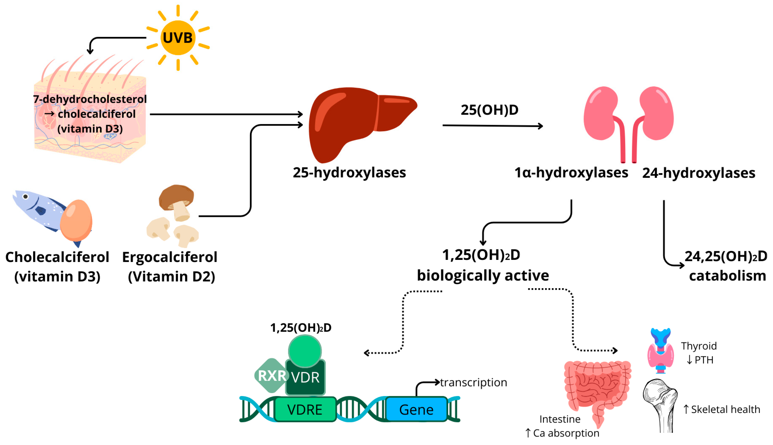
1.1. Vitamin D Metabolism

The precursors of 25(OH)D include vitamin D3 (cholecalciferol), which is found in animal products, and vitamin D2 (ergocalciferol), which is mainly present in mushrooms [31,32]. In humans, vitamin D3 can also be generated in the skin by photosynthesis after UVB exposure of the precursor 7-dehydrocholesterol. Vitamin D2 and vitamin D3 follow the same metabolic pathway. In this review, the term vitamin D is used when vitamin D2 or D3 could be used interchangeably. The conversion of vitamin D to its different compounds is achieved through the action of cytochrome P450 enzymes, for example, vitamin D 25-hydroxylases (CYP2R1, CYP27A1 and CYP2J2), 1α-hydroxylase (CYP27B1) and the aforementioned CYP24A1. The activity of these enzymes is partially dependent on magnesium as a cofactor [33,34]. Hydroxylase enzymes are mainly expressed in the liver and kidney, as well as in other tissues and cell types, such as testes, blood cells, immune cells and adipose tissue [30]. Vitamin D is converted in the liver (25 hydroxylases) and kidneys (1α hydroxylases) into bioactive 1,25-dihydroxyvitamin D (1,25(OH)2D). Vitamin D is catabolised by 1,25-dihydroxyvitamin D hydroxylase (CYP24A1) to 24,25-dihydroxyvitamin D (24,25(OH)2D) or 1,24,25-trihydroxyvitamin D (1,24,25(OH)3D) [30]. In circulation, vitamin D compounds are bound to the vitamin D binding protein (DBP) and albumin.
A schematic overview of vitamin D metabolism is provided in Figure 1. Vitamin D binds to the vitamin D receptor (VDR), which is expressed in almost all human tissues, including adipose tissue [35]. After binding, the VDR forms a complex with the retinoid X receptor (RXR). This complex is transferred to the nucleus, where it acts as a transcription factor. Many genes have a vitamin D response element (VDRE) in their promotor regions. Target genes are related to inflammatory processes and cell differentiation, in addition to bone and calcium metabolism [36]. The transcription regulation by vitamin D is reported to be highly tissue-specific.
Vitamin D metabolism is tightly regulated by parathyroid hormone (PTH) concentrations and is linked to calcium levels [37]. Hypocalcaemia triggers the release of PTH, which, in turn, stimulates the activation of 25(OH)D to 1,25(OH)2D in the kidneys by CYP27B1. The compound 1,25(OH)2D increases intestinal calcium absorption, contributing to the normalisation of serum calcium levels and skeletal health, which is a major common health effect of vitamin D [1].
In addition to the metabolic pathways described above, a C-3 epimerisation pathway occurs in the liver by 3-epimerases as described by Al-Zohily et al. [38]. This results in the C-3α vitamin D compound, which has lower biological activity and less calcaemic effects. Epimers and their possible clinical relevance are discussed in more detail in the section on the specific health effects of C-3α vitamin D compounds.

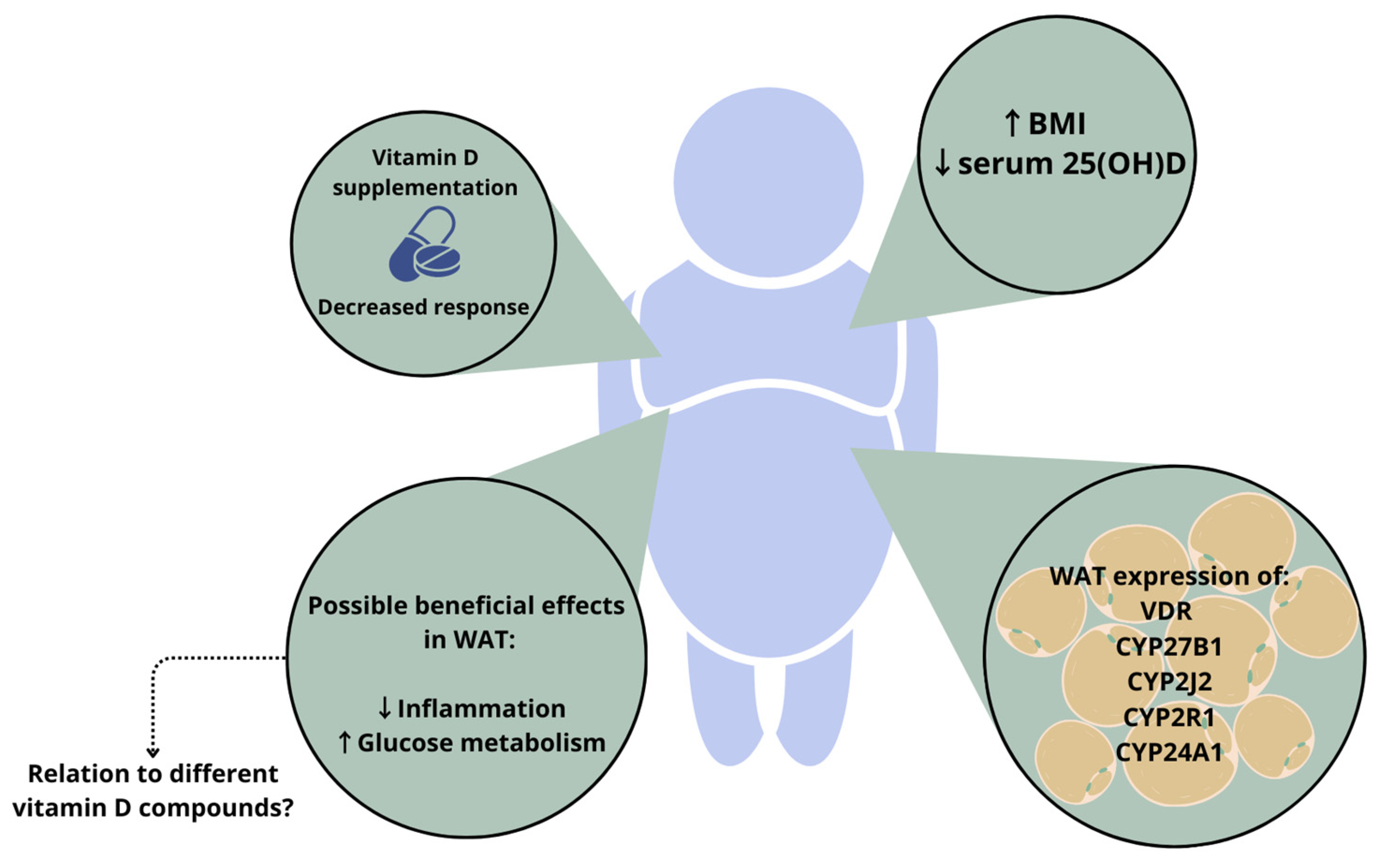
1.2. Vitamin D in Adiposity

Two meta-analyses demonstrated a significantly higher prevalence of low vitamin D levels (serum 25(OH)D < 30 ng/mL) in adults and children with higher body weight [39,40]. Currently, the major hypotheses for this association are the sequestration of fat-soluble vitamin D in an increased amount of adipose tissue in people with elevated body fat mass [41] and the volumetric distribution of vitamin D over a larger amount of body mass [42]. Logically, this leads to the hypothesis that weight loss could contribute to achieving vitamin D sufficiency in people with higher body weight. However, the findings are currently inconclusive. Blum et al. showed a correlation between serum vitamin D3 and WAT vitamin D3 levels [43]. They measured a mean vitamin D3 content in WAT of 102.8 ± 42 nmol/kg in people with a mean body mass index (BMI) of 50.6 kg/m2. This means that serum vitamin D could significantly improve upon weight loss if all vitamin D is released from adipose tissue. However, Himbert et al. reviewed the effect of surgery and diet-induced weight loss on serum 25(OH)D concentrations, and they concluded that weight loss did increase serum 25(OH)D levels but that this is often not enough to reach sufficient vitamin D concentrations [44]. Patients often need to be supplemented with additional vitamin D [45,46], meaning that the release of vitamin D from adipose tissue is not enough to replete individuals. This might be partially ascribed to decreased vitamin D reabsorption in the case of gastrectomy; nevertheless, a more detailed understanding of the metabolic fate of vitamin D in adipose tissue is warranted.
The expression and protein secretion of the hydroxylase enzymes involved in the conversion of vitamin D have been reported in many tissues, including WAT [47]. Human adipocytes were shown to express CYP27B1, as well as CYP24A1 [48]. The expression of the enzymes CYP2R1 and CYP2J2 was also shown in human adipose tissue [49]. Jonas et al. demonstrated an increased CYP27B1 expression in WAT of people with higher body weight (BMI > 40 kg/m2) [50]. In another study, however, the expression of CYP2J2 and CYP27B1 was decreased in women with higher body weight (BMI > 35 kg/m2) [49]. These contradicting results illustrate that more research is necessary to understand the link between vitamin D metabolism and adipose tissue. However, hypotheses do exist; for example, the expression of CYP24A1 in adipose tissue could lead to an increased degradation of 1,25(OH)2D in people with higher body weight. This effect could be aggravated by the concomitant downregulation of CYP2R1, another 25-hydroxylase, which was seen in a mouse model of obesity [51]. Such changes in hydroxylase enzyme expression could be a partial explanation for the decreased vitamin D status in people with higher body weight and underline the link between adipose tissue and vitamin D metabolism.
In addition to hydroxylase enzymes, adipocytes express the VDR [48,49], meaning that vitamin D compounds binding the receptor can have a direct local effect on gene transcription in adipose tissue. Adipose VDR is reported to be increased in obesity [50]. A genomic study showed that the rs3782905 single-nucleotide polymorphism (SNP) in the VDR gene is associated with differences in adiposity [52], which indicates a relation between vitamin D signalling and the development of adipose tissue. However, the impact of different VDR genotypes on the development of adiposity is not supported by all data [53]. Together, these findings on hydroxylase enzymes and VDR expression indicate once again that vitamin D metabolism is linked to adipose tissue mass. The relations between WAT and vitamin D metabolism in people with higher body weight are depicted in Figure 2.
Body weight has also been reported to influence the response to vitamin D supplementation [54]. A recent meta-analysis reported that vitamin D3 supplementation is more effective than vitamin D2 at raising serum 25(OH)D concentrations. However, the authors reported that baseline status needs to be considered and that this seems to be less relevant for people with higher body weight, since having a BMI > 25 k/m2 reduced the difference in response to vitamin D3 and vitamin D2 supplementation [55]. Moreover, a large vitamin D supplementation trial demonstrated that BMI modified the effect of vitamin D supplementation (2000 IU/day) on serum 25(OH)D levels. In a post hoc analysis, the participants were stratified in BMI categories, and the increase in serum 25(OH)D was observed to be blunted in higher BMI classes. Furthermore, altered vitamin D metabolism in people with higher body weight (BMI > 30 kg/m2) was reported in a randomised controlled trial (RCT) in which the effectiveness of supplementation with vitamin D3 was compared to that of 25(OH)D3 [56]. Compared to 25(OH)D3, an extra 30 µg/week (1200 IU) of vitamin D3 was required to achieve comparable serum concentrations of 25(OH)D in people with higher body weight. This effect was not seen in those with a lower BMI. Several reviews have described a relation between vitamin D status, body weight and health outcomes [57,58,59], and they have concluded that vitamin D might influence adipose tissue metabolism via so far unknown pathways and that vitamin D supplementation could be beneficial for people with higher body weight. However, the results described above show that vitamin D supplementation needs to be tailored to each individual.
In this context, it is also important to consider the differences between the different types of adipose tissue. Visceral adipose tissue (VAT) expansion seems to have a more unfavourable effect on metabolic health than an increase in subcutaneous adipose tissue (SAT) [60,61]. Cordeiro et al. [62] summarised the studies that assessed the relation between vitamin D status and SAT and VAT. Due to the lack of sufficient data, no final conclusions could be drawn regarding tissue-specific differences in vitamin D metabolism and VDR expression. However, a small body of data is currently available. Jonas et al. did not show any differences in VDR and CYP27B1 expression in SAT and VAT [50]. Wamberg et al. reported differences in decreased CYP27A1 expression in SAT compared to VAT [49]. Biopsy studies revealed higher vitamin D3 concentrations in VAT than in SAT [63]. Accordingly, the increase in serum 25(OH)D in response to vitamin D supplementation has been correlated with VAT but not with SAT mass [64]. In summary, these data indicate that there are differences between vitamin D metabolism in SAT and VAT; however, more research on the association between vitamin D compound levels and vitamin D metabolism is needed to fully understand the specific effects of vitamin D on different adipose tissue depots.
Of note, the rapid expansion of WAT in people with higher body weight can lead to an inflammatory response in adipocytes and adipose tissue macrophages [17]. These changes in adipose tissue function induce an increase in insulin resistance, which is a marker of a metabolically unhealthy phenotype in people with higher body weight [23,65]. Considering the reported association of vitamin D with inflammation [66,67] and glucose metabolism [15,16], differences in vitamin D availability or action in adipose tissue could play a crucial role in this context. Vitamin D can locally affect adipose tissue via the adipose VDR and is metabolised within adipose tissue by hydroxylase enzymes. Therefore, it is of importance to know more about the levels of vitamin D compounds and to determine whether they have specific health effects within adipose tissue.

2. Relevance of Different Vitamin D Compounds

2.1. Free Vitamin D Compound

The majority of vitamin D is bound to the vitamin D binding protein (DBP). Megalin/cubulin receptors enable the entry of the vitamin D-DBP complex into cells [68]. The proportion of free vitamin D is a very small fraction in comparison to that of protein-bound vitamin D, but it can move freely over cell membranes [69]. According to the free hormone hypothesis, this makes free vitamin D the most biologically active vitamin D compound [69,70,71], and, thus, this would make it very relevant in relation to the health effects of vitamin D.
Schwartz et al. performed a large cross-sectional study, in which they measured total serum 25(OH)D and free 25(OH)D levels in people with liver cirrhosis, pregnant women and healthy controls [72]. They observed that the levels of free 25(OH)D were significantly higher in people with liver disease, most likely due to a decreased capacity to produce DBP. A study by Shieh et al. offered in vivo indicators suggesting that free vitamin D plays a physiologically relevant role in humans [73]. They showed that, during the early stages of vitamin D repletion in deficient participants, the association between free vitamin D and 24,25(OH)2D and PTH in serum was stronger than the association between these compounds and total serum 25(OH)D. This indicates that free vitamin D was converted to 1,25(OH)2D and then inactivated in the kidneys. Thus, the free form was taken up into the parathyroid and kidney cells, regardless of the megalin/cubilin expression in these cell types, where it was converted to other compounds and exerted biological effects.
A bariatric surgery study showed that weight loss led to differential changes in the serum levels of free 25(OH)D and total 25(OH)D [74]. The serum levels of total 25(OH)D did not significantly increase when assessed one-year post-surgery, whereas a rise in the serum levels of free 25(OH)D could be observed. This could indicate that free vitamin D is the main storage compound of vitamin D in adipose tissue. However, it should be noted that there are several other mechanisms that could explain this differential effect, such as changes in serum protein levels or the expression of the vitamin D binding protein after weight loss. In a study comparing lean women to women with higher body weight (BMI = 39.1 ± 4.6 kg/m2), DBP levels were higher and free vitamin D levels were lower in the latter group [75]. The increased DBP levels could potentially have an effect on the availability of free vitamin D in tissues. Of note, the free vitamin D levels in this study were calculated rather than quantified with immunoassays or mass spectrometry. However, Rivera-Paredez et al. observed no differences between free vitamin D and total serum 25(OH)D when comparing associations with markers of metabolic health in Mexican adults [76]. However, the participants of this study had a BMI between 24 and 30 kg/m2, which is considerably less than in the previously discussed studies. This could be an indication that adiposity significantly alters the role and presence of different vitamin D compounds. In conclusion, there are some findings that indicate the differential effects of adiposity and weight loss on free and bound or total vitamin D levels; however, more research is needed to clearly define the physiological relevance of free vitamin D in the context of adiposity.

2.2. 1,25-Dihydroxyvitamin D (1,25(OH)2D)

The 1,25(OH)2D compound is generally considered the active form due to its high affinity for the VDR [77]. This section highlights two health effects of 1,25(OH)2D that are highly relevant in the setting of adiposity: firstly, the relation to inflammation and, secondly, the effect on adipokine secretion.
With regard to the anti-inflammatory effects of 1,25(OH)2D, NFkB signalling has been implicated as a potential mechanism, since vitamin D has been reported to decrease NFkB expression [12] and to block NFkB translocation to the nucleus [13,78,79], thus decreasing the transcription of pro-inflammatory cytokines. Even though these findings directly link 1,25(OH)2D to decreased inflammation in vitro, there are contradicting reports of the effects of vitamin D supplementation on inflammatory markers in vivo. Wamberg et al. [80] observed a diminishing effect on inflammation in an adipocyte cell line treated with 1,25(OH)2D3. However, this effect was not reproduced in primary human adipocytes, and it was not reflected in circulating inflammatory markers in vivo [80]. A cross-sectional study did show a negative correlation between serum 25(OH)D levels and the serum levels of IL-6 and TNF-α in women with obesity (mean BMI of 43.6 ± 4.3 kg/m2) [81]. This is confirmed in other cross-sectional studies that report a relation between vitamin D status and the levels of inflammatory markers in blood [82]. These studies reported the serum levels of 25(OH)D but not those of 1,25(OH)2D. Nevertheless, 25(OH)D is a precursor of 1,25(OH)2D, and it would be of great interest to further investigate, for example, the effect of vitamin D supplementation on inflammatory markers within adipose tissue because this is a major storage organ of vitamin D [83].
Adipokines are messenger molecules produced by adipocytes. They affect energy metabolism, insulin resistance, inflammation and blood pressure and are strongly related to metabolic health [84,85]. Two well-known examples are leptin and adiponectin. Leptin is known as the satiety hormone, which, in healthy conditions, supresses food intake, regulates energy metabolism and acts as a negative feedback loop upon adipose tissue accumulation [86,87]. However, in people with higher body weight, leptin resistance frequently occurs [88,89,90]. Adiponectin is an adipose-tissue-derived hormone with health-promoting effects. It has been reported to increase insulin sensitivity and have anti-inflammatory properties [91]. Adiponectin levels are often decreased in people with increased body fat [92]. In two in vitro studies, 1,25(OH)2D3 was reported to decrease leptin production in primary human adipocytes [12,93]. This is supported by several human studies, although these assessed 25(OH)D instead of 1,25(OH)2D. Cross-sectional studies showed a negative association between low serum 25(OH)D and high leptin levels [94,95,96]. In a study in patients with prediabetes, the association was present after adjustment for BMI, supporting the notion that vitamin D could have direct effects on leptin [97]. However, negative associations between leptin and 25(OH)D have also been reported [95,98].
The effects of 1,25(OH)2D exposure on adiponectin have also been reported, e.g., decreased adiponectin production in human primary adipocytes [99]. Accordingly, a negative association between adiponectin levels and serum 25(OH)D was observed in humans [97]. However, this effect was only reported in a subgroup of participants with a BMI > 25 kg/m2. In another study, no association between serum 25(OH)D and adiponectin was reported [100]. And yet another study found a positive correlation between 25(OH)D and adiponectin levels [96]. Two trials did not show an effect of vitamin D on adipokine levels [101,102]. O’Sullivan et al. found no effect of 600 IU/day supplementation during a four-week period, but the sample consisted mostly of participants with baseline vitamin D sufficiency. Upon further analysis, it was found that a subsample of participants who were vitamin D-deficient had higher serum adiponectin levels at baseline and were more responsive to vitamin D supplementation.
Together, these findings illustrate that 1,25(OH)2D can regulate important processes in human adipose tissue. This makes it of interest to assess the levels of this vitamin D compound in the adipose tissue or serum of people with higher body weight.

2.3. 24,25-Dihydroxyvitamin D (24,25(OH)2D)

The 24,25(OH)2D compound is a clearance product in the vitamin D metabolic cascade. The ratio of 24,25(OH)2D to 25(OH)D has been suggested to be an alternative marker of vitamin D status [103]. This ratio takes vitamin D metabolism and feedback loops into account and is not dependent on the level of DBP [104,105]. When assessing other vitamin D compounds, Binkley et al. described the predictive value of 24,25(OH)2D for the response to vitamin D supplementation [20]. In this study, the ratio of 24,25(OH)2D/25(OH)D did not predict the response to vitamin D supplementation, contradicting earlier suggestions of 24,25(OH)2D/25(OH)D being an improved status marker [103,106]. However, the sample size was relatively small (n = 62) and consisted of postmenopausal women only. These data indicate that the measurement of 24,25(OH)2D could have an additional clinical relevance to measuring only serum 25(OH)D [106]. Furthermore, 24,25(OH)2D has its own receptor, the membrane receptor family with sequence similarity 57 (FAM57B2) [107], and it has been reported to have biological effects, e.g., in relation to bone fracture healing and decreased tumorigenesis [108]. However, the role in inflammation, insulin resistance and adipose tissue function, as well as the distribution in different adipose tissue compartments, is currently not fully understood.

2.4. C-3 Epimers of Vitamin D Compounds

In vitamin D metabolism, a parallel C-3 epimerisation pathway introduces additional vitamin D compounds. The most important of these is the C-3α isomer of 25(OH)D, which results from the reversal of the stereochemical configuration of the –OH group at C-3 (3β3α). It is important to measure this compound for two reasons. Firstly, the 3α epimer has been reported to lead to the overestimation of vitamin D status with current detection methods, such as immunoassays and MS assays, if it is not properly separated [109]. The extent of this additional contribution depends on physiological and pathological conditions and age; e.g., 3α levels are naturally higher in newborns, especially in those born prematurely [110,111]. Secondly, the 3α epimer has demonstrated biological activity of its own, e.g., anti-proliferative effects, increased phospholipid production [38,111] and cardiovascular function [112]. The latter was concluded in a cohort study that showed an association of aerobic capacity with the 3α epimer of vitamin D in patients with chronic kidney disease.
With respect to body composition, a negative correlation was reported for serum 25(OH)D concentrations and fat mass in infants at 12 months in a vitamin D supplementation trial [113]. The authors reported that serum 25(OH)D was a predictor of lean mass and fat mass in regression models. This correlation was only present at the age of three months for 3α-25(OH)D, even though 3β-25(OH)D was associated with all time points. In a Thai national health survey, the relative amount of 3α-25(OH)D3 was associated with age, sex and living conditions [114]. Males and people living in rural areas had higher relative concentrations of serum 3α-25(OH)D3. The survey observed a negative correlation between BMI and 3β-25(OH)D, but not 3α-25(OH)D. Another study that included women with polycystic ovary syndrome (PCOS) and healthy controls did not observe differences between the two groups in 3β and 3α-25(OH)D3 [115]. A recent clinical study investigated the levels of 1,25(OH)2D3 and 3α-25(OH)D3 in participants with normal versus higher body weight [116]. No association differences between serum vitamin D compounds and body weight were found. Furthermore, none of the compounds were associated with inflammatory or metabolic health markers. Overall, the biological actions of the 3α epimer require more research.

3. Measuring Vitamin D Compounds in Human Adipose Tissue with LC-MS/MS

Vitamin D compounds are usually measured using either immunoassays or mass spectrometry (MS). While immunoassays are automated and fast and the cost of analysis is low in comparison to other instrumental techniques [117], they sometimes lack selectivity and do not provide the possibility of analysing multiple vitamin D compounds at the same time [118,119]. MS assays are currently the “gold standard” method [21] because they provide measurements of multiple vitamin D compounds simultaneously, with high selectivity and very low limits of quantification (LOQ). Sample preparation, however, is more laborious, especially when analysing adipose tissue. For readers interested in a detailed description of mass spectrometric methods for vitamin D compounds, not limited to adipose tissue samples, we refer to other reviews on this topic [120,121]. The benefits of MS assays compared to established immunoassays have also been described previously [122,123,124]. Table 1 summarises several important studies on adipose tissue analysis using MS, along with the required sample quantity, steps of sample preparation, measured compounds, LOQ or limit of detection (LOD) and recovery of the sample preparation protocol. The protocols mentioned in the table are described in brief in this chapter.
Generally, sample preparation protocols for adipose tissue samples for subsequent mass spectrometry analysis are more complicated, time-consuming and expensive than those for conventional blood sample analysis, which usually consist of some or all of the following steps: protein precipitation, which separates analytes from binding proteins and removes proteins from the matrix, and saponification, which removes lipids. This can be achieved by adding potassium hydroxide to the sample and leaving it overnight [125]. Shorter saponification reaction times can be achieved at higher temperatures, but a careful balance has to be found for the thermally instable vitamin D [121]. Usually, these steps are followed by liquid–liquid extraction (LLE) and/or solid-phase extraction (SPE). This is sometimes also followed by a chemical derivatisation step; this enhances ionisation sensitivity for vitamin D compounds, which exhibit low ionisation efficiency [122,126].
Table 1. Overview of sample preparation protocols used to measure vitamin D compounds in adipose tissue.
Author, YearMS,
Ionisation
Sample Weight (g)Sample PreparationMeasured CompoundsLOD
LOQ
Recovery (R%)
Blum et al., 2008 [43]LC-MS
APCI
0.2–0.25
  • Homogenisation
  • Saponification
  • LLE
  • SPE
D3-72%
Höller et al., 2010 [127]LC-MS
APCI
0.5
  • Homogenisation
  • Protein precipitation
  • SPE C-18
25(OH)D3LOD
5 ng/g
-
Beckman et al., 2013 [63]LC
DAD
-
  • Saponification
  • LLE
  • SPE
  • Preparative HPLC
D2
D3
-98.5 ± 5%
Piccolo et al., 2013 [128]LC-MS/MS
APCI
0.1–0.5
  • Homogenisation
  • SPE
  • Filtration
25(OH)D3
Lipkie et al., 2013 [129]LC-MS/MS
ESI
-
  • Homogenisation
  • LLE
  • SPE
  • PTAD derivatisation
25(OH)D2
25(OH)D3
D2
D3
Malmberg et al., 2014 [130]SIMS-TOF D3
25(OH)D3
1,25(OH)2D3
--
Burild et al., 2014 [131]
Didriksen et al., 2015 [132]
Martinaityte et al., 2017 [133]
LC-MS/MS
ESI
0.2–1.0
  • Saponification
  • LLE
  • SPE
  • PTAD derivatisation
D3
25(OH)D3
LOQ < 0.1 ng/g-
Bonnet et al., 2019 [134]
Bonnet et al., 2021 [135]
LC-MS/MS
ESI
0.05
  • Homogenisation
  • LLE
  • SPE
  • Amplifex derivatisation
D3
25(OH)D3
1,25(OH)2D3
--
Best et al., 2021 [136]LC-MS/MS0.01
  • Saponification
  • LLE
  • PTAD derivatisation
D3
25(OH)D3
--
As mentioned above, adipose tissue is a challenging matrix because of its complex lipid composition, from which vitamin D compounds, which are themselves lipids, have to be extracted. As a result, the protocols in Table 1 are very laborious in comparison to other matrices [121] and still do not always guarantee the successful detection of vitamin D compounds. This was shown by Höller et al., who measured 25(OH)D3 in skin, kidney, liver and muscle tissue, and found that the levels of the compounds were lower than the lower limits of quantification (LLOQ) of the method in adipose tissue [127]. Derivatisation can improve LOQ, as demonstrated by Lipkie et al., who measured 25(OH)D2, 25(OH)D3, vitamin D2 and D3 in the liver, muscle and epididymal adipose tissue of rats receiving a vitamin D-enriched diet [129]. The authors used LLE, followed by SPE and derivatisation with 4-phenyl-1,2,4-triazole-3,5-dione (PTAD). LOD was 0.1 ng/mL for all investigated vitamin D compounds. The reported accuracies of quantification were based on National Institute of Standards and Technology (NIST) reference materials, giving 78% and 129% for 25(OH)D2 and 25(OH)D3, respectively, which did not meet the standards outlined by the US Food and Drug Administration (FDA) [137]. Bonnet et al. developed an LC-MS/MS method using high-resolution orbitrap MS and measured vitamin D3, 25(OH)D3 and 1,25(OH)2D3 in the adipose tissue of mice [134,135] using Lipkie et al.’s [129] sample preparation protocol. The authors implemented Amplifex instead of PTAD for lower LOQs of 0.78, 0.19 and 0.02 ng/mL for cholecalciferol, 25(OH)D3 and 1,25(OH)2D3. Precision (RE%) and accuracy (CV%) were <15%, thus complying with the FDA guidelines [137]. Of note, there are other derivatisation options for vitamin D compounds, developed for serum or plasma-based assays equally applied to extracts from tissue samples [126].
The chromatographic separation of vitamin D compounds is usually performed using reversed-phase C-18 stationary phases. Two exceptions were developed by Blum et al. [43] and Picollo et al. [128], who used C-30 stationary phase chemistry, which can exhibit increased selectivity and retention over C-18 columns for some compounds [138]. Ionisation is almost always achieved using either atmospheric pressure chemical ionisation (APCI) or electrospray ionisation (ESI) in positive ion mode prior to MS detection. An exception was reported by Beckman et al., who used diode-array detection (HPLC-DAD) [63]. The authors reported the average recovery of vitamin D2 and D3 to be 98.5% ± 5%. Vitamin D2 was not detectable in most samples. However, vitamin D3 was measured in both subcutaneous and visceral adipose tissue, with VAT showing greater affinity for vitamin D3 storage. No LOD or LOQ was reported. Burild et al. utilised overnight saponification at room temperature, two steps of LLE and PTAD derivatisation. LOQ was reported to be <0.1 ng/g, and accuracies were 84–96% and 113–114% for vitamin D3 and 25(OH)D3, respectively; AOAC validation criteria were met [131]. Didriksen et al. and Martinaityte et al. studied human adipose tissue samples following the same protocol [132,133]. Best et al. described a protocol with a saponification step performed at 95 °C [136]. Unfortunately, no recovery or LOQ or LOD values were reported for the determination of vitamin D compounds in adipose tissue.
Finally, Malmberg et al. reported an imaging mass spectrometry protocol for adipose tissue using secondary ion mass spectrometry (SIMS) [130], which required an entirely different sample preparation protocol, as the spatial integrity of vitamin D3 in the tissue had to be maintained. The authors investigated the spatial distribution of vitamin D3, 25(OH)D3 and 1,25(OH)2D3 in adipose tissue [130]. The protocol consisted of freezing the adipose tissue samples under high pressure, as well as freeze-fracturing them. Vitamin D3, 25(OH)D3, and 1,25(OH)2D3 were found in the adipocyte lipid droplet. The observed signals for 25(OH)D3 and 1,25(OH)2D3 were very low, making a comparison of the distributions for the different compounds challenging.

4. Conclusions

This review explores the evidence base for the associations of different vitamin D compounds with health markers and specific biological effects of vitamin D compounds, specifically in relation to increased body weight and different adipose tissue components. Furthermore, to the best of our knowledge, this is the first review to include an overview of LC-MS/MS sample preparation methods to accurately and simultaneously measure different vitamin D compounds in human adipose tissue.
Vitamin D compounds, such as 1,25(OH)2D, free vitamin D and the 3α epimer of 25(OH)D, in addition to the status marker 25(OH)D, have been reported to have various extra-skeletal health effects that are highly relevant in the setting of increased adiposity. Investigating vitamin D concentrations in human adipose tissue might shed further light on the role of vitamin D with regard to adipose tissue inflammation. Vitamin D has also been shown to affect adipokine levels, which could be an important approach to modulate metabolic health. Vitamin D, though not the only factor influencing the development of inflammation in adipose tissue, likely plays an important role, including in adipokine synthesis and adipocyte biology in general. Thus, a comprehensive approach to these processes is needed in order to understand the impact of individual factors, such as vitamin D.
While the expression of hydroxylase enzymes and the VDR in WAT has been demonstrated [49,50], it is not clear how these enzymes affect the concentrations of different vitamin D compounds in adipose tissue and how this affects health in people with higher body weight. More studies focusing on multiple vitamin D compounds and their concentrations in SAT and VAT are required to shed further light on this topic. Preliminary studies indicate that vitamin D compounds (e.g., serum free 25(OH)D3 and serum total 25(OH)D) show differential responses to weight loss and that vitamin D compounds demonstrate different associations with sex or health conditions. No differential associations have been observed with inflammatory markers, and, to date, no predictive effects of compound ratios on vitamin D supplementation have been demonstrated. However, further research in this area is needed.

Funding

EES is a recipient of the Elsa-Neumann-Scholarship of the federal state of Berlin for her doctoral studies; DAV acknowledges research funding by the German Research Foundation (DFG VO 1355/5-2).

Conflicts of Interest

The authors declare no conflict of interest.

References

  1. Holick, M.F. Vitamin D and bone health. J. Nutr. 1996, 126, 1159S–1164S. [Google Scholar] [CrossRef] [PubMed]
  2. Holick, M.F. The role of vitamin D for bone health and fracture prevention. Curr. Osteoporos. Rep. 2006, 4, 96–102. [Google Scholar] [CrossRef] [PubMed]
  3. Bouillon, R.; Manousaki, D.; Rosen, C.; Trajanoska, K.; Rivadeneira, F.; Richards, J.B. The health effects of vitamin D supplementation: Evidence from human studies. Nat. Rev. Endocrinol. 2022, 18, 96–110. [Google Scholar] [CrossRef] [PubMed]
  4. Holick, M.F. Deficiency of sunlight and vitamin D. BMJ 2008, 336, 1318–1319. [Google Scholar] [CrossRef] [PubMed]
  5. Cashman, K.D. Vitamin D deficiency: Defining, prevalence, causes, and strategies of addressing. Calcif. Tissue Int. 2020, 106, 14–29. [Google Scholar] [CrossRef] [PubMed]
  6. Holick, M.F.; Binkley, N.C.; Bischoff-Ferrari, H.A.; Gordon, C.M.; Hanley, D.A.; Heaney, R.P.; Murad, M.H.; Weaver, C.M. Evaluation, treatment, and prevention of vitamin D deficiency: An Endocrine Society clinical practice guideline. J. Clin. Endocrinol. Metab. 2011, 96, 1911–1930. [Google Scholar] [CrossRef] [PubMed]
  7. Dissanayake, H.A.; de Silva, N.L.; Sumanatilleke, M.; de Silva, S.D.N.; Gamage, K.K.K.; Dematapitiya, C.; Kuruppu, D.C.; Ranasinghe, P.; Pathmanathan, S.; Katulanda, P. Prognostic and therapeutic role of vitamin D in COVID-19: Systematic review and meta-analysis. J. Clin. Endocrinol. Metab. 2022, 107, 1484–1502. [Google Scholar] [CrossRef]
  8. Shah, K.; Saxena, D.; Mavalankar, D. Vitamin D supplementation, COVID-19 and disease severity: A meta-analysis. QJM Int. J. Med. 2021, 114, 175–181. [Google Scholar] [CrossRef]
  9. Karampela, I.; Sakelliou, A.; Vallianou, N.; Christodoulatos, G.-S.; Magkos, F.; Dalamaga, M. Vitamin D and obesity: Current evidence and controversies. Curr. Obes. Rep. 2021, 10, 162–180. [Google Scholar] [CrossRef]
  10. Calton, E.K.; Keane, K.N.; Newsholme, P.; Soares, M.J. The impact of vitamin D levels on inflammatory status: A systematic review of immune cell studies. PLoS ONE 2015, 10, e0141770. [Google Scholar] [CrossRef]
  11. Park, C.Y.; Kim, T.Y.; Yoo, J.S.; Seo, Y.; Pae, M.; Han, S.N. Effects of 1, 25-dihydroxyvitamin D3 on the inflammatory responses of stromal vascular cells and adipocytes from lean and obese mice. Nutrients 2020, 12, 364. [Google Scholar] [CrossRef]
  12. Nimitphong, H.; Guo, W.; Holick, M.F.; Fried, S.K.; Lee, M.J. Vitamin D inhibits adipokine production and inflammatory signaling through the vitamin D receptor in human adipocytes. Obesity 2021, 29, 562–568. [Google Scholar] [CrossRef] [PubMed]
  13. Karkeni, E.; Bonnet, L.; Marcotorchino, J.; Tourniaire, F.; Astier, J.; Ye, J.; Landrier, J.-F. Vitamin D limits inflammation-linked microRNA expression in adipocytes in vitro and in vivo: A new mechanism for the regulation of inflammation by vitamin D. Epigenetics 2018, 13, 156–162. [Google Scholar] [CrossRef] [PubMed]
  14. Marcotorchino, J.; Tourniaire, F.; Astier, J.; Karkeni, E.; Canault, M.; Amiot, M.-J.; Bendahan, D.; Bernard, M.; Martin, J.-C.; Giannesini, B. Vitamin D protects against diet-induced obesity by enhancing fatty acid oxidation. J. Nutr. Biochem. 2014, 25, 1077–1083. [Google Scholar] [CrossRef] [PubMed]
  15. Lei, X.; Zhou, Q.; Wang, Y.; Fu, S.; Li, Z.; Chen, Q. Serum and supplemental vitamin D levels and insulin resistance in T2DM populations: A meta-analysis and systematic review. Sci. Rep. 2023, 13, 12343. [Google Scholar] [CrossRef] [PubMed]
  16. Farahmand, M.A.; Daneshzad, E.; Fung, T.T.; Zahidi, F.; Muhammadi, M.; Bellissimo, N.; Azadbakht, L. What is the impact of vitamin D supplementation on glycemic control in people with type-2 diabetes: A systematic review and meta-analysis of randomized controlled trails. BMC Endocr. Disord. 2023, 23, 15. [Google Scholar] [CrossRef] [PubMed]
  17. Cancello, R.; Clement, K. Is obesity an inflammatory illness? Role of low-grade inflammation and macrophage infiltration in human white adipose tissue. BJOG Int. J. Obstet. Gynaecol. 2006, 113, 1141–1147. [Google Scholar] [CrossRef] [PubMed]
  18. Nguyen, N.T.; Magno, C.P.; Lane, K.T.; Hinojosa, M.W.; Lane, J.S. Association of hypertension, diabetes, dyslipidemia, and metabolic syndrome with obesity: Findings from the National Health and Nutrition Examination Survey, 1999 to 2004. J. Am. Coll. Surg. 2008, 207, 928–934. [Google Scholar] [CrossRef]
  19. Bikle, D.; Bouillon, R.; Thadhani, R.; Schoenmakers, I. Vitamin D metabolites in captivity? Should we measure free or total 25 (OH) D to assess vitamin D status? J. Steroid Biochem. Mol. Biol. 2017, 173, 105–116. [Google Scholar] [CrossRef]
  20. Binkley, N.; Borchardt, G.; Siglinsky, E.; Krueger, D. Does vitamin D metabolite measurement help predict 25 (OH) D change following vitamin D supplementation? Endocr. Pract. 2017, 23, 432–441. [Google Scholar] [CrossRef]
  21. Makris, K.; Sempos, C.; Cavalier, E. The measurement of vitamin D metabolites part II—The measurement of the various vitamin D metabolites. Hormones 2020, 19, 97–107. [Google Scholar] [CrossRef] [PubMed]
  22. Wildman, R.P.; Muntner, P.; Reynolds, K.; McGinn, A.P.; Rajpathak, S.; Wylie-Rosett, J.; Sowers, M.R. The obese without cardiometabolic risk factor clustering and the normal weight with cardiometabolic risk factor clustering: Prevalence and correlates of 2 phenotypes among the US population (NHANES 1999–2004). Arch. Intern. Med. 2008, 168, 1617–1624. [Google Scholar] [CrossRef]
  23. Noubiap, J.J.; Nansseu, J.R.; Lontchi-Yimagou, E.; Nkeck, J.R.; Nyaga, U.F.; Ngouo, A.T.; Tounouga, D.N.; Tianyi, F.L.; Foka, A.J.; Ndoadoumgue, A.L. Geographic distribution of metabolic syndrome and its components in the general adult population: A meta-analysis of global data from 28 million individuals. Diabetes Res. Clin. Pract. 2022, 188, 109924. [Google Scholar] [CrossRef] [PubMed]
  24. Soll, D.; Gawron, J.; Pletsch-Borba, L.; Spranger, J.; Mai, K. Long-term impact of the metabolic status on weight loss-induced health benefits. Nutr. Metab. 2022, 19, 25. [Google Scholar] [CrossRef]
  25. Meadows, A.; Daníelsdóttir, S. What’s in a word? On weight stigma and terminology. Front. Psychol. 2016, 7, 1527. [Google Scholar] [CrossRef]
  26. Puhl, R.M. What words should we use to talk about weight? A systematic review of quantitative and qualitative studies examining preferences for weight-related terminology. Obes. Rev. 2020, 21, e13008. [Google Scholar] [CrossRef] [PubMed]
  27. Institute of Medicine. Dietary Reference Intakes for Calcium and Vitamin D; National Academies Press (US): Washington, DC, USA, 2011. [Google Scholar]
  28. Stokes, C.S.; Lammert, F. Vitamin D supplementation: Less controversy, more guidance needed. F1000Research 2016, 5. [Google Scholar] [CrossRef]
  29. Rosen, C.J.; Abrams, S.A.; Aloia, J.F.; Brannon, P.M.; Clinton, S.K.; Durazo-Arvizu, R.A.; Gallagher, J.C.; Gallo, R.L.; Jones, G.; Kovacs, C.S. IOM committee members respond to Endocrine Society vitamin D guideline. J. Clin. Endocrinol. Metab. 2012, 97, 1146–1152. [Google Scholar] [CrossRef]
  30. Saponaro, F.; Saba, A.; Zucchi, R. An update on vitamin D metabolism. Int. J. Mol. Sci. 2020, 21, 6573. [Google Scholar] [CrossRef]
  31. Rao, D.S.; Raghuramulu, N. Food chain as origin of vitamin D in fish. Comp. Biochem. Physiol. Part A Physiol. 1996, 114, 15–19. [Google Scholar]
  32. Atsuko, T.; Toshio, O.; Makoto, T.; Tadashi, K. Possible origin of extremely high contents of vitamin D3 in some kinds of fish liver. Comp. Biochem. Physiol. Part A Physiol. 1991, 100, 483–487. [Google Scholar] [CrossRef]
  33. Uwitonze, A.M.; Razzaque, M.S. Role of magnesium in vitamin D activation and function. J. Osteopath. Med. 2018, 118, 181–189. [Google Scholar] [CrossRef] [PubMed]
  34. Risco, F.; Traba, M. Influence of magnesium on the in vitro synthesis of 24, 25-dihydroxyvitamin D3 and 1 alpha, 25-dihydroxyvitamin D3. Magnes. Res. 1992, 5, 5–14. [Google Scholar] [PubMed]
  35. Plum, L.A.; DeLuca, H.F. Vitamin D, disease and therapeutic opportunities. Nat. Rev. Drug Discov. 2010, 9, 941–955. [Google Scholar] [CrossRef]
  36. Carlberg, C. Vitamin D and Its Target Genes. Nutrients 2022, 14, 1354. [Google Scholar] [CrossRef]
  37. Goltzman, D.; Mannstadt, M.; Marcocci, C. Physiology of the calcium-parathyroid hormone-vitamin D axis. Vitam. D Clin. Med. 2018, 50, 1–13. [Google Scholar]
  38. Al-Zohily, B.; Al-Menhali, A.; Gariballa, S.; Haq, A.; Shah, I. Epimers of vitamin D: A review. Int. J. Mol. Sci. 2020, 21, 470. [Google Scholar] [CrossRef]
  39. Pereira-Santos, M.; Costa, P.d.F.; Assis, A.d.; Santos, C.d.S.; Santos, D.d. Obesity and vitamin D deficiency: A systematic review and meta-analysis. Obes. Rev. 2015, 16, 341–349. [Google Scholar] [CrossRef]
  40. Zakharova, I.; Klimov, L.; Kuryaninova, V.; Nikitina, I.; Malyavskaya, S.; Dolbnya, S.; Kasyanova, A.; Atanesyan, R.; Stoyan, M.; Todieva, A. Vitamin D insufficiency in overweight and obese children and adolescents. Front. Endocrinol. 2019, 10, 103. [Google Scholar] [CrossRef]
  41. Abbas, M.A. Physiological functions of Vitamin D in adipose tissue. J. Steroid Biochem. Mol. Biol. 2017, 165, 369–381. [Google Scholar] [CrossRef]
  42. Drincic, A.T.; Armas, L.A.; Van Diest, E.E.; Heaney, R.P. Volumetric dilution, rather than sequestration best explains the low vitamin D status of obesity. Obesity 2012, 20, 1444–1448. [Google Scholar] [CrossRef] [PubMed]
  43. Blum, M.; Dolnikowski, G.; Seyoum, E.; Harris, S.S.; Booth, S.L.; Peterson, J.; Saltzman, E.; Dawson-Hughes, B. Vitamin D3 in fat tissue. Endocrine 2008, 33, 90–94. [Google Scholar] [CrossRef]
  44. Himbert, C.; Ose, J.; Delphan, M.; Ulrich, C.M. A systematic review of the interrelation between diet-and surgery-induced weight loss and vitamin D status. Nutr. Res. 2017, 38, 13–26. [Google Scholar] [CrossRef] [PubMed]
  45. Lespessailles, E.; Toumi, H. Vitamin D alteration associated with obesity and bariatric surgery. Exp. Biol. Med. 2017, 242, 1086–1094. [Google Scholar] [CrossRef]
  46. Chakhtoura, M.; Rahme, M.; Fuleihan, G.E.-H. Vitamin D metabolism in bariatric surgery. Endocrinol. Metab. Clin. 2017, 46, 947–982. [Google Scholar] [CrossRef]
  47. Li, J.; Byrne, M.E.; Chang, E.; Jiang, Y.; Donkin, S.S.; Buhman, K.K.; Burgess, J.R.; Teegarden, D. 1α, 25-Dihydroxyvitamin D hydroxylase in adipocytes. J. Steroid Biochem. Mol. Biol. 2008, 112, 122–126. [Google Scholar] [CrossRef]
  48. Nimitphong, H.; Holick, M.F.; Fried, S.K.; Lee, M.-J. 25-hydroxyvitamin D3 and 1, 25-dihydroxyvitamin D3 promote the differentiation of human subcutaneous preadipocytes. PLoS ONE 2012, 7, e52171. [Google Scholar] [CrossRef]
  49. Wamberg, L.; Christiansen, T.; Paulsen, S.; Fisker, S.; Rask, P.; Rejnmark, L.; Richelsen, B.; Pedersen, S. Expression of vitamin D-metabolizing enzymes in human adipose tissue—The effect of obesity and diet-induced weight loss. Int. J. Obes. 2013, 37, 651–657. [Google Scholar] [CrossRef] [PubMed]
  50. Jonas, M.I.; Kuryłowicz, A.; Bartoszewicz, Z.; Lisik, W.; Jonas, M.; Kozniewski, K.; Puzianowska-Kuznicka, M. Vitamin D receptor gene expression in adipose tissue of obese individuals is regulated by miRNA and correlates with the pro-inflammatory cytokine level. Int. J. Mol. Sci. 2019, 20, 5272. [Google Scholar] [CrossRef]
  51. Elkhwanky, M.S.; Kummu, O.; Piltonen, T.T.; Laru, J.; Morin-Papunen, L.; Mutikainen, M.; Tavi, P.; Hakkola, J. Obesity represses CYP2R1, the vitamin D 25-hydroxylase, in the liver and extrahepatic tissues. JBMR Plus 2020, 4, e10397. [Google Scholar] [CrossRef]
  52. Ochs-Balcom, H.M.; Chennamaneni, R.; Millen, A.E.; Shields, P.G.; Marian, C.; Trevisan, M.; Freudenheim, J.L. Vitamin D receptor gene polymorphisms are associated with adiposity phenotypes. Am. J. Clin. Nutr. 2011, 93, 5–10. [Google Scholar] [CrossRef]
  53. Ruiz-Ojeda, F.J.; Anguita-Ruiz, A.; Leis, R.; Aguilera, C.M. Genetic factors and molecular mechanisms of vitamin D and obesity relationship. Ann. Nutr. Metab. 2018, 73, 89–99. [Google Scholar] [CrossRef]
  54. Tobias, D.K.; Luttmann-Gibson, H.; Mora, S.; Danik, J.; Bubes, V.; Copeland, T.; LeBoff, M.S.; Cook, N.R.; Lee, I.-M.; Buring, J.E. Association of Body Weight With Response to Vitamin D Supplementation and Metabolism. JAMA Netw. Open 2023, 6, e2250681. [Google Scholar] [CrossRef] [PubMed]
  55. van den Heuvel, E.G.; Lips, P.; Schoonmade, L.J.; Lanham-New, S.A.; van Schoor, N.M. Comparison of the effect of daily vitamin D2 and vitamin D3 supplementation on serum 25-hydroxyvitamin D concentration (total 25 (OH) D, 25 (OH) D2 and 25 (OHD3) and importance of body mass index: A systematic review and meta-analysis. Adv. Nutr. 2023. [Google Scholar] [CrossRef]
  56. Di Nisio, A.; De Toni, L.; Sabovic, I.; Rocca, M.S.; De Filippis, V.; Opocher, G.; Azzena, B.; Vettor, R.; Plebani, M.; Foresta, C. Impaired release of vitamin D in dysfunctional adipose tissue: New cues on vitamin D supplementation in obesity. J. Clin. Endocrinol. Metab. 2017, 102, 2564–2574. [Google Scholar] [CrossRef]
  57. Vranić, L.; Mikolašević, I.; Milić, S. Vitamin D deficiency: Consequence or cause of obesity? Medicina 2019, 55, 541. [Google Scholar] [CrossRef] [PubMed]
  58. Migliaccio, S.; Di Nisio, A.; Mele, C.; Scappaticcio, L.; Savastano, S.; Colao, A.; Obesity Programs of nutrition, Education, Research and Assessment (OPERA) Group. Obesity and hypovitaminosis D: Causality or casualty? Int. J. Obes. Suppl. 2019, 9, 20–31. [Google Scholar] [CrossRef] [PubMed]
  59. Earthman, C.; Beckman, L.; Masodkar, K.; Sibley, S. The link between obesity and low circulating 25-hydroxyvitamin D concentrations: Considerations and implications. Int. J. Obes. 2012, 36, 387–396. [Google Scholar] [CrossRef] [PubMed]
  60. Liu, J.; Fox, C.S.; Hickson, D.A.; May, W.D.; Hairston, K.G.; Carr, J.J.; Taylor, H.A. Impact of abdominal visceral and subcutaneous adipose tissue on cardiometabolic risk factors: The Jackson Heart Study. J. Clin. Endocrinol. Metab. 2010, 95, 5419–5426. [Google Scholar] [CrossRef]
  61. Fox, C.S.; Massaro, J.M.; Hoffmann, U.; Pou, K.M.; Maurovich-Horvat, P.; Liu, C.-Y.; Vasan, R.S.; Murabito, J.M.; Meigs, J.B.; Cupples, L.A. Abdominal visceral and subcutaneous adipose tissue compartments: Association with metabolic risk factors in the Framingham Heart Study. Circulation 2007, 116, 39–48. [Google Scholar] [CrossRef]
  62. Cordeiro, A.; Santos, A.; Bernardes, M.; Ramalho, A.; Martins, M.J. Vitamin D metabolism in human adipose tissue: Could it explain low vitamin D status in obesity? Horm. Mol. Biol. Clin. Investig. 2018, 33, 20170003. [Google Scholar] [CrossRef] [PubMed]
  63. Beckman, L.M.; Earthman, C.P.; Thomas, W.; Compher, C.W.; Muniz, J.; Horst, R.L.; Ikramuddin, S.; Kellogg, T.A.; Sibley, S.D. Serum 25 (OH) vitamin D concentration changes after Roux-en-Y gastric bypass surgery. Obesity 2013, 21, E599–E606. [Google Scholar] [CrossRef]
  64. Cominacini, M.; Fumaneri, A.; Ballerini, L.; Braggio, M.; Valenti, M.T.; Dalle Carbonare, L. Unraveling the Connection: Visceral Adipose Tissue and Vitamin D Levels in Obesity. Nutrients 2023, 15, 4259. [Google Scholar] [CrossRef] [PubMed]
  65. Eckel, R.H.; Alberti, K.G.; Grundy, S.M.; Zimmet, P.Z. The metabolic syndrome. Lancet 2010, 375, 181–183. [Google Scholar] [CrossRef] [PubMed]
  66. van Etten, E.; Mathieu, C. Immunoregulation by 1, 25-dihydroxyvitamin D3: Basic concepts. J. Steroid Biochem. Mol. Biol. 2005, 97, 93–101. [Google Scholar] [CrossRef] [PubMed]
  67. Charoenngam, N.; Holick, M.F. Immunologic effects of vitamin D on human health and disease. Nutrients 2020, 12, 2097. [Google Scholar] [CrossRef] [PubMed]
  68. Nykjaer, A.; Dragun, D.; Walther, D.; Vorum, H.; Jacobsen, C.; Herz, J.; Melsen, F.; Christensen, E.I.; Willnow, T.E. An endocytic pathway essential for renal uptake and activation of the steroid 25-(OH) vitamin D3. Cell 1999, 96, 507–515. [Google Scholar] [CrossRef] [PubMed]
  69. Bikle, D.D. The free hormone hypothesis: When, why, and how to measure the free hormone levels to assess vitamin D, thyroid, sex hormone, and cortisol status. JBMR Plus 2021, 5, e10418. [Google Scholar] [CrossRef]
  70. Mendel, C.M. The free hormone hypothesis: A physiologically based mathematical model. Endocr. Rev. 1989, 10, 232–274. [Google Scholar] [CrossRef]
  71. Bikle, D.D.; Malmstroem, S.; Schwartz, J. Current controversies: Are free vitamin metabolite levels a more accurate assessment of vitamin D status than total levels? Endocrinol. Metab. Clin. 2017, 46, 901–918. [Google Scholar] [CrossRef]
  72. Schwartz, J.; Lai, J.; Lizaola, B.; Kane, L.; Weyland, P.; Terrault, N.; Stotland, N.; Bikle, D. Variability in free 25 (OH) vitamin D levels in clinical populations. J. Steroid Biochem. Mol. Biol. 2014, 144, 156–158. [Google Scholar] [CrossRef] [PubMed]
  73. Shieh, A.; Ma, C.; Chun, R.F.; Wittwer-Schegg, J.; Swinkels, L.; Huijs, T.; Wang, J.; Donangelo, I.; Hewison, M.; Adams, J.S. Associations between change in total and free 25-hydroxyvitamin D with 24, 25-dihydroxyvitamin D and parathyroid hormone. J. Clin. Endocrinol. Metab. 2018, 103, 3368–3375. [Google Scholar] [CrossRef] [PubMed]
  74. Marques-Pamies, M.; López-Molina, M.; Pellitero, S.; Santillan, C.S.; Martínez, E.; Moreno, P.; Tarascó, J.; Granada, M.L.; Puig-Domingo, M. Differential behavior of 25 (OH) D and f25 (OH) D3 in patients with morbid obesity after bariatric surgery. Obes. Surg. 2021, 31, 3990–3995. [Google Scholar] [CrossRef] [PubMed]
  75. Karlsson, T.; Osmancevic, A.; Jansson, N.; Hulthén, L.; Holmäng, A.; Larsson, I. Increased vitamin D-binding protein and decreased free 25 (OH) D in obese women of reproductive age. Eur. J. Nutr. 2014, 53, 259–267. [Google Scholar] [CrossRef] [PubMed]
  76. Rivera-Paredez, B.; Hidalgo-Bravo, A.; León-Reyes, G.; León-Maldonado, L.S.; Aquino-Gálvez, A.; Castillejos-López, M.; Denova-Gutiérrez, E.; Flores, Y.N.; Salmerón, J.; Velázquez-Cruz, R. Total, bioavailable, and free 25-hydroxyvitamin D equally associate with adiposity markers and metabolic traits in mexican adults. Nutrients 2021, 13, 3320. [Google Scholar] [CrossRef]
  77. Brommage, R.; Deluca, H.F. Evidence that 1, 25-dihydroxyvitamin D3 is the physiologically active metabolite of vitamin D3. Endocr. Rev. 1985, 6, 491–511. [Google Scholar] [CrossRef] [PubMed]
  78. Marcotorchino, J.; Tourniaire, F.; Landrier, J.-F. Vitamin D, adipose tissue, and obesity. Horm. Mol. Biol. Clin. Investig. 2013, 15, 123–128. [Google Scholar] [CrossRef] [PubMed]
  79. Mutt, S.J.; Karhu, T.; Lehtonen, S.; Lehenkari, P.; Carlberg, C.; Saarnio, J.; Sebert, S.; Hyppönen, E.; Järvelin, M.R.; Herzig, K.H. Inhibition of cytokine secretion from adipocytes by 1, 25-dihydroxyvitamin D3 via the NF-κB pathway. FASEB J. 2012, 26, 4400–4407. [Google Scholar] [CrossRef]
  80. Wamberg, L.; Cullberg, K.; Rejnmark, L.; Richelsen, B.; Pedersen, S. Investigations of the anti-inflammatory effects of vitamin D in adipose tissue: Results from an in vitro study and a randomized controlled trial. Horm. Metab. Res. 2013, 45, 456–462. [Google Scholar] [CrossRef]
  81. Bellia, A.; Garcovich, C.; D’Adamo, M.; Lombardo, M.; Tesauro, M.; Donadel, G.; Gentileschi, P.; Lauro, D.; Federici, M.; Lauro, R. Serum 25-hydroxyvitamin D levels are inversely associated with systemic inflammation in severe obese subjects. Intern. Emerg. Med. 2013, 8, 33–40. [Google Scholar] [CrossRef]
  82. Sha, S.; Gwenzi, T.; Chen, L.-J.; Brenner, H.; Schöttker, B. About the associations of vitamin D deficiency and biomarkers of systemic inflammatory response with all-cause and cause-specific mortality in a general population sample of almost 400,000 UK Biobank participants. Eur. J. Epidemiol. 2023, 38, 957–971. [Google Scholar] [CrossRef] [PubMed]
  83. Mawer, E.B.; Backhouse, J.; Holman, C.A.; Lumb, G.; Stanbury, S. The distribution and storage of vitamin D and its metabolites in human tissues. Clin. Sci. 1972, 43, 413–431. [Google Scholar] [CrossRef] [PubMed]
  84. Trayhurn, P.; Bing, C.; Wood, I.S. Adipose tissue and adipokines—Energy regulation from the human perspective. J. Nutr. 2006, 136, 1935S–1939S. [Google Scholar] [CrossRef] [PubMed]
  85. Sahu, B.; Bal, N.C. Adipokines from white adipose tissue in regulation of whole body energy homeostasis. Biochimie 2023, 204, 92–107. [Google Scholar] [CrossRef] [PubMed]
  86. Gruzdeva, O.; Borodkina, D.; Uchasova, E.; Dyleva, Y.; Barbarash, O. Leptin resistance: Underlying mechanisms and diagnosis. Diabetes Metab. Syndr. Obes. Targets Ther. 2019, 12, 191–198. [Google Scholar] [CrossRef] [PubMed]
  87. Izquierdo, A.G.; Crujeiras, A.B.; Casanueva, F.F.; Carreira, M.C. Leptin, obesity, and leptin resistance: Where are we 25 years later? Nutrients 2019, 11, 2704. [Google Scholar] [CrossRef] [PubMed]
  88. Prolo, P.; Wong, M.-L.; Licinio, J. Leptin. Int. J. Biochem. Cell Biol. 1998, 30, 1285–1290. [Google Scholar] [CrossRef]
  89. Paul, R.F.; Hassan, M.; Nazar, H.S.; Gillani, S.; Afzal, N.; Qayyum, I. Effect of body mass index on serum leptin levels. J. Ayub Med. Coll. Abbottabad 2011, 23, 40–43. [Google Scholar] [PubMed]
  90. Kumar, R.; Mal, K.; Razaq, M.K.; Magsi, M.; Memon, M.K.; Memon, S.; Afroz, M.N.; Siddiqui, H.F.; Rizwan, A. Association of leptin with obesity and insulin resistance. Cureus 2020, 12, e12178. [Google Scholar] [CrossRef]
  91. Wang, Z.V.; Scherer, P.E. Adiponectin, the past two decades. J. Mol. Cell Biol. 2016, 8, 93–100. [Google Scholar] [CrossRef]
  92. Fang, H.; Judd, R.L. Adiponectin regulation and function. Compr. Physiol. 2011, 8, 1031–1063. [Google Scholar]
  93. Menendez, C.; Lage, M.; Peino, R.; Baldelli, R.; Concheiro, P.; Dieguez, C.; Casanueva, F. Retinoic acid and vitamin D3 powerfully inhibit in vitro leptin secretion by human adipose tissue. J. Endocrinol. 2001, 170, 425–432. [Google Scholar] [CrossRef]
  94. Hajimohammadi, M.; Shab-Bidar, S.; Neyestani, T. Vitamin D and serum leptin: A systematic review and meta-analysis of observational studies and randomized controlled trials. Eur. J. Clin. Nutr. 2017, 71, 1144–1153. [Google Scholar] [CrossRef] [PubMed]
  95. Vilarrasa, N.; Vendrell, J.; Maravall, J.; Elío, I.; Solano, E.; San José, P.; García, I.; Virgili, N.; Soler, J.; Gómez, J.M. Is plasma 25 (OH) D related to adipokines, inflammatory cytokines and insulin resistance in both a healthy and morbidly obese population? Endocrine 2010, 38, 235–242. [Google Scholar] [CrossRef] [PubMed]
  96. Lwow, F.; Bohdanowicz-Pawlak, A. Vitamin D and selected cytokine concentrations in postmenopausal women in relation to metabolic disorders and physical activity. Exp. Gerontol. 2020, 141, 111107. [Google Scholar] [CrossRef]
  97. de Souza, W.N.; Norde, M.M.; Oki, É.; Rogero, M.M.; Marchioni, D.M.; Fisberg, R.M.; Martini, L.A. Association between 25-hydroxyvitamin D and inflammatory biomarker levels in a cross-sectional population-based study, São Paulo, Brazil. Nutr. Res. 2016, 36, 1–8. [Google Scholar] [CrossRef] [PubMed]
  98. Maetani, M.; Maskarinec, G.; Franke, A.A.; Cooney, R.V. Association of leptin, 25-hydroxyvitamin D, and parathyroid hormone in women. Nutr. Cancer 2009, 61, 225–231. [Google Scholar] [CrossRef]
  99. Lorente-Cebrián, S.; Eriksson, A.; Dunlop, T.; Mejhert, N.; Dahlman, I.; Åström, G.; Sjölin, E.; Wåhlén, K.; Carlberg, C.; Laurencikiene, J. Differential effects of 1α, 25-dihydroxycholecalciferol on MCP-1 and adiponectin production in human white adipocytes. Eur. J. Nutr. 2012, 51, 335–342. [Google Scholar] [CrossRef]
  100. Zhang, M.; Gao, Y.; Tian, L.; Zheng, L.; Wang, X.; Liu, W.; Zhang, Y.; Huang, G. Association of serum 25-hydroxyvitamin D3 with adipokines and inflammatory marker in persons with prediabetes mellitus. Clin. Chim. Acta 2017, 468, 152–158. [Google Scholar] [CrossRef]
  101. O’Sullivan, A.; Gibney, M.J.; Connor, A.O.; Mion, B.; Kaluskar, S.; Cashman, K.D.; Flynn, A.; Shanahan, F.; Brennan, L. Biochemical and metabolomic phenotyping in the identification of a vitamin D responsive metabotype for markers of the metabolic syndrome. Mol. Nutr. Food Res. 2011, 55, 679–690. [Google Scholar] [CrossRef]
  102. Al-Sofiani, M.E.; Jammah, A.; Racz, M.; Khawaja, R.A.; Hasanato, R.; El-Fawal, H.A.; Mousa, S.A.; Mason, D.L. Effect of vitamin D supplementation on glucose control and inflammatory response in type II diabetes: A double blind, randomized clinical trial. Int. J. Endocrinol. Metab. 2015, 13, e22604. [Google Scholar] [CrossRef] [PubMed]
  103. Wagner, D.; Hanwell, H.E.; Schnabl, K.; Yazdanpanah, M.; Kimball, S.; Fu, L.; Sidhom, G.; Rousseau, D.; Cole, D.E.; Vieth, R. The ratio of serum 24, 25-dihydroxyvitamin D3 to 25-hydroxyvitamin D3 is predictive of 25-hydroxyvitamin D3 response to vitamin D3 supplementation. J. Steroid Biochem. Mol. Biol. 2011, 126, 72–77. [Google Scholar] [CrossRef] [PubMed]
  104. Dugar, A.; Hoofnagle, A.N.; Sanchez, A.P.; Ward, D.M.; Corey-Bloom, J.; Cheng, J.H.; Ix, J.H.; Ginsberg, C. The Vitamin D Metabolite Ratio (VMR) is a Biomarker of Vitamin D Status That is Not Affected by Acute Changes in Vitamin D Binding Protein. Clin. Chem. 2023, 69, 718–723. [Google Scholar] [CrossRef] [PubMed]
  105. Ginsberg, C.; Hoofnagle, A.N.; Katz, R.; Becker, J.O.; Kritchevsky, S.B.; Shlipak, M.G.; Sarnak, M.J.; Ix, J.H. The vitamin D metabolite ratio is independent of vitamin D binding protein concentration. Clin. Chem. 2021, 67, 385–393. [Google Scholar] [CrossRef]
  106. Herrmann, M.; Farrell, C.-J.L.; Pusceddu, I.; Fabregat-Cabello, N.; Cavalier, E. Assessment of vitamin D status–a changing landscape. Clin. Chem. Lab. Med. (CCLM) 2017, 55, 3–26. [Google Scholar] [CrossRef]
  107. Martineau, C.; Naja, R.P.; Husseini, A.; Hamade, B.; Kaufmann, M.; Akhouayri, O.; Arabian, A.; Jones, G.; St-Arnaud, R. Optimal bone fracture repair requires 24R, 25-dihydroxyvitamin D 3 and its effector molecule FAM57B2. J. Clin. Investig. 2018, 128, 3546–3557. [Google Scholar] [CrossRef]
  108. Verma, A.; Schwartz, Z.; Boyan, B.D. 24R, 25-dihydroxyvitamin D3 modulates tumorigenicity in breast cancer in an estrogen receptor-dependent manner. Steroids 2019, 150, 108447. [Google Scholar] [CrossRef] [PubMed]
  109. Schorr, P.; Kovacevic, B.; Volmer, D.A. Overestimation of 3α-over 3β-25-Hydroxyvitamin D3 Levels in Serum: A Mechanistic Rationale for the Different Mass Spectral Properties of the Vitamin D Epimers. J. Am. Soc. Mass Spectrom. 2021, 32, 1116–1125. [Google Scholar] [CrossRef]
  110. Chen, Y.-C.; He, Y.-Y.; Li, Y.-M.; Wu, B.-T.; Yang, Y.-W.; Feng, J.-F. The importance of analyzing the serum C3-epimer level for evaluating vitamin D storage in some special populations. Eur. Rev. Med. Pharmacol. Sci. 2022, 26, 5334–5343. [Google Scholar]
  111. Bailey, D.; Veljkovic, K.; Yazdanpanah, M.; Adeli, K. Analytical measurement and clinical relevance of vitamin D3 C3-epimer. Clin. Biochem. 2013, 46, 190–196. [Google Scholar] [CrossRef]
  112. Arroyo, E.; Leber, C.A.; Burney, H.N.; Li, Y.; Li, X.; Lu, T.-s.; Jones, G.; Kaufmann, M.; Ting, S.M.; Hiemstra, T.F. Epimeric vitamin D and cardiovascular structure and function in advanced CKD and after kidney transplantation. Nephrol. Dial. Transplant. 2023, gfad168. [Google Scholar] [CrossRef]
  113. Hazell, T.J.; Gallo, S.; Berzina, l.; Vanstone, C.A.; Rodd, C.; Weiler, H.A. Plasma 25-hydroxyvitamin D, more so than its epimer, has a linear relationship to leaner body composition across infancy in healthy term infants. Appl. Physiol. Nutr. Metab. 2014, 39, 1137–1143. [Google Scholar] [CrossRef] [PubMed]
  114. Chailurkit, L.; Aekplakorn, W.; Ongphiphadhanakul, B. Serum C3 epimer of 25-hydroxyvitamin D and its determinants in adults: A national health examination survey in Thais. Osteoporos. Int. 2015, 26, 2339–2344. [Google Scholar] [CrossRef] [PubMed]
  115. Moin, A.S.M.; Sathyapalan, T.; Atkin, S.L.; Butler, A.E. Inflammatory markers in non-obese women with polycystic ovary syndrome are not elevated and show no correlation with vitamin D metabolites. Nutrients 2022, 14, 3540. [Google Scholar] [CrossRef] [PubMed]
  116. Gariballa, S.; Shah, I.; Yasin, J.; Alessa, A. Vitamin D [25 (OH) D] metabolites and epimers in obese subject: Interaction and correlations with adverse metabolic health risk factors. J. Steroid Biochem. Mol. Biol. 2022, 215, 106023. [Google Scholar] [CrossRef] [PubMed]
  117. Altieri, B.; Cavalier, E.; Bhattoa, H.P.; Perez-Lopez, F.R.; Lopez-Baena, M.T.; Perez-Roncero, G.R.; Chedraui, P.; Annweiler, C.; Della Casa, S.; Zelzer, S. Vitamin D testing: Advantages and limits of the current assays. Eur. J. Clin. Nutr. 2020, 74, 231–247. [Google Scholar] [CrossRef]
  118. van den Ouweland, J.M. Analysis of vitamin D metabolites by liquid chromatography-tandem mass spectrometry. TrAC Trends Anal. Chem. 2016, 84, 117–130. [Google Scholar] [CrossRef]
  119. Binkley, N.; Dawson-Hughes, B.; Durazo-Arvizu, R.; Thamm, M.; Tian, L.; Merkel, J.; Jones, J.; Carter, G.; Sempos, C. Vitamin D measurement standardization: The way out of the chaos. J. Steroid Biochem. Mol. Biol. 2017, 173, 117–121. [Google Scholar] [CrossRef]
  120. Müller, M.J.; Volmer, D.A. Mass spectrometric profiling of vitamin D metabolites beyond 25-hydroxyvitamin D. Clin. Chem. 2015, 61, 1033–1048. [Google Scholar] [CrossRef]
  121. Alexandridou, A.; Volmer, D.A. Sample preparation techniques for extraction of vitamin D metabolites from non-conventional biological sample matrices prior to LC–MS/MS analysis. Anal. Bioanal. Chem. 2022, 414, 4613–4632. [Google Scholar] [CrossRef]
  122. Alexandridou, A.; Schorr, P.; Stokes, C.S.; Volmer, D.A. Analysis of vitamin D metabolic markers by mass spectrometry: Recent progress regarding the “gold standard” method and integration into clinical practice. Mass Spectrom. Rev. 2021, 42, 1647–1687. [Google Scholar] [CrossRef] [PubMed]
  123. Stokes, C.S.; Lammert, F.; Volmer, D.A. Analytical methods for quantification of vitamin D and implications for research and clinical practice. Anticancer Res. 2018, 38, 1137–1144. [Google Scholar] [PubMed]
  124. Qi, Y.; Müller, M.; Stokes, C.S.; Volmer, D.A. Rapid quantification of 25-hydroxyvitamin D3 in human serum by matrix-assisted laser desorption/ionization mass spectrometry. J. Am. Soc. Mass Spectrom. 2018, 29, 1456–1462. [Google Scholar] [CrossRef]
  125. Abu Kassim, N.S.; Gomes, F.P.; Shaw, P.N.; Hewavitharana, A.K. Simultaneous quantitative analysis of nine vitamin D compounds in human blood using LC–MS/MS. Bioanalysis 2016, 8, 397–411. [Google Scholar] [CrossRef]
  126. Alexandridou, A.; Schorr, P.; Volmer, D.A. Comparing derivatization reagents for quantitative LC–MS/MS analysis of a variety of vitamin D metabolites. Anal. Bioanal. Chem. 2023, 415, 4689–4701. [Google Scholar] [CrossRef]
  127. Höller, U.; Quintana, A.P.; Gössl, R.; Olszewski, K.; Riss, G.; Schattner, A.; Nunes, C.S. Rapid determination of 25-hydroxy vitamin D3 in swine tissue using an isotope dilution HPLC-MS assay. J. Chromatogr. B 2010, 878, 963–968. [Google Scholar] [CrossRef] [PubMed]
  128. Piccolo, B.D.; Dolnikowski, G.; Seyoum, E.; Thomas, A.P.; Gertz, E.R.; Souza, E.C.; Woodhouse, L.R.; Newman, J.W.; Keim, N.L.; Adams, S.H. Association between subcutaneous white adipose tissue and serum 25-hydroxyvitamin D in overweight and obese adults. Nutrients 2013, 5, 3352–3366. [Google Scholar] [CrossRef]
  129. Lipkie, T.E.; Janasch, A.; Cooper, B.R.; Hohman, E.E.; Weaver, C.M.; Ferruzzi, M.G. Quantification of vitamin D and 25-hydroxyvitamin D in soft tissues by liquid chromatography–tandem mass spectrometry. J. Chromatogr. B 2013, 932, 6–11. [Google Scholar] [CrossRef]
  130. Malmberg, P.; Karlsson, T.; Svensson, H.; Lönn, M.; Carlsson, N.-G.; Sandberg, A.-S.; Jennische, E.; Osmancevic, A.; Holmäng, A. A new approach to measuring vitamin D in human adipose tissue using time-of-flight secondary ion mass spectrometry: A pilot study. J. Photochem. Photobiol. B Biol. 2014, 138, 295–301. [Google Scholar] [CrossRef]
  131. Burild, A.; Frandsen, H.L.; Poulsen, M.; Jakobsen, J. Quantification of physiological levels of vitamin D3 and 25-hydroxyvitamin D3 in porcine fat and liver in subgram sample sizes. J. Sep. Sci. 2014, 37, 2659–2663. [Google Scholar] [CrossRef]
  132. Didriksen, A.; Burild, A.; Jakobsen, J.; Fuskevåg, O.M.; Jorde, R. Vitamin D3 increases in abdominal subcutaneous fat tissue after supplementation with vitamin D3. Eur. J. Endocrinol. 2015, 172, 235–241. [Google Scholar] [CrossRef] [PubMed]
  133. Martinaityte, I.; Kamycheva, E.; Didriksen, A.; Jakobsen, J.; Jorde, R. Vitamin D stored in fat tissue during a 5-year intervention affects serum 25-hydroxyvitamin D levels the following year. J. Clin. Endocrinol. Metab. 2017, 102, 3731–3738. [Google Scholar] [CrossRef] [PubMed]
  134. Bonnet, L.; Margier, M.; Svilar, L.; Couturier, C.; Reboul, E.; Martin, J.-C.; Landrier, J.-F.; Defoort, C. Simple fast quantification of cholecalciferol, 25-hydroxyvitamin D and 1, 25-dihydroxyvitamin D in adipose tissue using LC-HRMS/MS. Nutrients 2019, 11, 1977. [Google Scholar] [CrossRef] [PubMed]
  135. Bonnet, L.; Karkeni, E.; Couturier, C.; Astier, J.; Defoort, C.; Svilar, L.; Tourniaire, F.; Mounien, L.; Landrier, J.-F. Four days high fat diet modulates vitamin D metabolite levels and enzymes in mice. J. Endocrinol. 2021, 248, 87–93. [Google Scholar] [CrossRef]
  136. Best, C.M.; Riley, D.V.; Laha, T.J.; Pflaum, H.; Zelnick, L.R.; Hsu, S.; Thummel, K.E.; Foster-Schubert, K.E.; Kuzma, J.N.; Cromer, G. Vitamin D in human serum and adipose tissue after supplementation. Am. J. Clin. Nutr. 2021, 113, 83–91. [Google Scholar] [CrossRef]
  137. Fda, U. Bioanalytical Method Validation Guidance for Industry; US Department of Health and Human Services Food and Drug Administration Center for Drug Evaluation and Research and Center for Veterinary Medicine: 2018. Available online: https://www.fda.gov/files/drugs/published/Bioanalytical-Method-Validation-Guidance-for-Industry.pdf (accessed on 15 November 2023).
  138. Hymøller, L.; Jensen, S.K. Vitamin D analysis in plasma by high performance liquid chromatography (HPLC) with C30 reversed phase column and UV detection–easy and acetonitrile-free. J. Chromatogr. A 2011, 1218, 1835–1841. [Google Scholar] [CrossRef]
Figure 1. Schematic overview of vitamin D metabolism in humans. ↑ indicates an increase, ↓ indicates a decrease. Abbreviations: ultraviolet light short wavelength (UVB); calcidiol (25(OH)D); calcitriol (1,25(OH)2D); retinoid X receptor (RXR); vitamin D receptor (VDR); vitamin D response element (VDRE); parathyroid hormone (PTH); calcium (Ca).
Nutrients 16 00231 g001
Figure 2. Depiction of the relation between vitamin D status and white adipose tissue (WAT) mass in people with higher body weight. The response to vitamin D supplementation is altered in people with higher body weight, and an increased BMI is associated with lower serum 25(OH)D levels. In addition, white adipocytes express the VDR and hydroxylase enzymes. Vitamin D can have a beneficial effect on processes in WAT; however, it is unknown whether these effects are mediated by specific vitamin D compounds. ↑ indicates an increase, ↓ indicates a decrease. Abbreviations: BMI, body mass index; VDR, vitamin D receptor; WAT, white adipose tissue; 25(OH)D, 25-hydroxyvitamin D.
Nutrients 16 00231 g002
Disclaimer/Publisher’s Note: The statements, opinions and data contained in all publications are solely those of the individual author(s) and contributor(s) and not of MDPI and/or the editor(s). MDPI and/or the editor(s) disclaim responsibility for any injury to people or property resulting from any ideas, methods, instructions or products referred to in the content.

