Abstract
Obesity and high abdominal fat mass are risk factors for developing the chronic inflammatory skin disease psoriasis. They are associated with increased incidence, prevalence and severity of the disease. A positive effect of weight loss on psoriasis activity has been shown in several studies. Obesity-related factors such as the dysregulation of glucose and lipid metabolism, the activation of adipose tissue and resultant persistent low-grade inflammation have been discussed as links of obesity and inflammatory diseases. Recently, we demonstrated a critical role of free fatty acids (FFAs) in obesity-mediated exacerbation of psoriatic skin inflammation in both mice and humans. In the present study, we translated these findings into a therapeutic intervention. An open-label study focusing on the dietary reduction of FFAs was conducted in patients with mild-to-moderate plaque psoriasis, and disease severity and serum markers of inflammation were analyzed. Here, we show that such a dietary intervention improves psoriatic disease activity independently of weight loss. Diet-related metabolic changes, such as a reduction in saturated free fatty acids (SFAs), may thus be more important than weight loss itself. Moreover, dietary intervention inhibited the overall pro-inflammatory activation status in patients, as shown by analysis of serum inflammatory parameters using the Olink platform. From our pilot study, we conclude that dietary intervention focusing on SFA reduction has the capacity to reduce disease activity and general inflammatory status in psoriasis patients.
1. Introduction
Obesity has reached epidemic proportions, affecting more than 650 million people worldwide [1]. It is associated with a reduced disease-free life and premature death and predisposes individuals to various diseases, including type 2 diabetes, cardiovascular diseases, chronic kidney disease, cancer and infections [1]. In addition, obesity increases the incidence and severity of many inflammatory/autoimmune disorders including hepatic steatosis, inflammatory bowel disease, colitis, atherosclerosis and psoriasis [2,3,4,5,6,7]. Consistent with human data, obesity exacerbates inflammation in several animal models of chronic inflammatory diseases such as psoriatic dermatitis, inflammatory bowel disease and colitis [4,5,6,8,9,10,11,12].
Epidemiological studies demonstrate a substantial impact of obesity on the incidence and severity of psoriasis. For example, obesity and high abdominal fat mass double the risk for developing psoriasis [13,14]. Obesity-related factors such as disturbed glucose and lipid metabolism, the activation of adipose tissue due to the excessive fat deposition and the resulting increased levels of pro-inflammatory adipokines, cytokines and chemokines have been discussed as links between obesity and inflammation [15,16]. Recently, we demonstrated a critical involvement of free fatty acids (FFAs) in obesity-mediated exacerbation of psoriatic skin inflammation [17,18]. Among the obesity-related factors, only serum FFA levels correlated with the severity of skin inflammation in psoriasis patients. In a mouse model, we proved the central role of FFAs in exacerbating psoriasis-like skin inflammation. Non-obese/non-diabetic mice with elevated FFA levels developed a significantly more severe psoriasis-like skin inflammation, which could be prevented by the dietary reduction of FFA [18]. Mechanistically, we showed that increased concentrations of saturated free fatty acids (SFAs), together with the danger molecule S100A9, stimulate inflammasome activation in macrophages, leading to an increased IL-1b secretion, which propagates S100A9 production in keratinocytes. S100A9, in turn, attenuates the differentiation of anti-inflammatory M2-like macrophages, which develop a pro-inflammatory activation profile, thereby initiating a vicious cycle of skin inflammation in obesity. Breaking the vicious cycle of S100A9 overexpression by a dietary reduction in FFAs restored M2-like macrophage activation and ameliorated skin inflammation [19]. Taken together, dietary FFAs seem to play an essential role in the obesity-mediated amplification of psoriatic skin inflammation.
In the present study, we translated these findings into a therapeutic approach. In an open-label study, we conducted a dietary intervention in patients with mild-to-moderate plaque psoriasis and analyzed disease severity and serum inflammatory markers. Indeed, a dietary intervention focused on reducing SFAs improved disease severity and decreased a wide range of serum inflammatory biomarkers detected by the Olink Proteomics Platform.
2. Methods
2.1. Human Study
We included patients with mainly mild to moderate psoriasis vulgaris aged ≥18 who were treated for psoriasis at our medical center (Supplementary Table S1). Three patients had severe psoriasis but declined any systemic treatment. All systemic and topical treatments were permitted but could not be changed at least 4 weeks before and during dietary intervention; otherwise, patients were excluded. The study was approved by the local ethics committee (#163/18-ek), and all patients gave written informed consent. Patients were randomized into two groups. Group 1 (n = 16) started directly with a 12-week dietary intervention. Group 2 (n = 17) kept their usual diet for three months before they started with the same 12-week dietary intervention as patients of group 1. Dietary intervention was designed with the aim of reducing the uptake of SFA (Supplementary Table S2). In detail, the meals were initially replaced by a formula diet according to the manufacturer’s instructions and subsequently based on a modified Mediterranean diet. Specifically, in the first week, all meals were replaced by one portion of the formula diet. A total of 100 g formula diet powder were dissolved in up to 300 mL of still water (or 200 mL of low-fat milk or 200 mL of buttermilk or sugar-free tea), and 2 teaspoons (=6 g) vegetable oil (canola oil, soybean oil, linseed oil, walnut oil) were added. If needed, sugar-free additives (cinnamon, turmeric, baking cocoa) could be added. Patients were asked to take the formula diet in the correct dosage according to the plan, even on vacation, holidays or birthdays. In the second and third week, only two meals were replaced by the formula diet. The third meal consisted of components of the Mediterranean diet, for example, 1–2 slices of whole-meal bread, fat-reduced margarine and a choice of fat-reduced cheese or fat-reduced sausage or ham. In the fourth week, one meal was replaced by the formula diet. The other two meals could be freely chosen by the patients according to Mediterranean referral. The Mediterranean diet was modified according to the recommendations for a wholesome diet of the German Nutrition Society (DGE, https://www.dge.de/ernaehrungspraxis/vollwertige-ernaehrung/10-regeln-der-dge/en/ (accessed on 22 January 2023)) with the additional recommendation of a low SFA intake (Supplementary Table S2). Throughout the study, patients were accompanied by professional nutrition specialists. In the course of the intervention period, a total of 8 nutrition consultations took place. Clinical data including Psoriasis Area and Severity Index (PASI) and body mass index (BMI) were recorded 12, 10, 8 and 4 weeks before start of the intervention, at the start of the intervention and 2, 4, 8 and 12 weeks after the intervention. Serum was collected at each visit.
2.2. Serum Biomarkers
Inflammatory serum biomarkers were detected by the Olink Proteomics Platform (Olink Proteomics, Uppsala, Sweden) using the human Olink target 96 inflammation panel. Analysis was performed by Olink.
2.3. Lipid Extraction and Analysis of Free Fatty Acids
Serum aliquots (60 µL) were mixed with 1 mL methanol and centrifuged at 13,000× g for 2 min to precipitate proteins. Lipids were extracted from the supernatant by the addition of 1 mL chloroform and water according to Bligh and Dyer [20]. After vigorous shaking, samples were centrifuged for phase separation at 3000 rpm for 5 min. The lower (organic) phase was withdrawn, and lipid extraction was repeated by the addition of chloroform, vigorous shaking and centrifugation. Both organic phases of a sample were combined, evaporated to dryness and re-dissolved in 200 µL chloroform. Ten µL of the organic extracts were separated by high-performance thin-layer chromatography (HPTLC). The lipids in the free fatty acid (FFA) spot were automatically eluted by a Plate Express™ TLC plate reader (Advion, Ithaca, NY, USA) with methanol as a solvent and directly analyzed by electrospray ionization mass spectrometry (ESI MS) as described [21]. Total intensities of FFA and, from these, ratios for each FFA were calculated. Total FFA concentration was detected in serum samples by the Free Fatty Acid Quantitation Kit (Sigma-Aldrich; without lipid extraction) according to the manufacturer’s protocol and detected via UV-spectrometer measurement (Hitachi U-2000, Hitachi Medical Systems America, Inc., Twinsburg, OH, USA) or Synergy HT (BioTek). Specific FFA concentrations were calculated from the percentage of specific FFA and total FFA.
2.4. Statistics
For statistical correlation analyses, IBM SPSS Statistics software version 27.0 (IBM, Armonk, NY, USA) was employed. The non-parametric Spearman’s rank correlation test was used to assess univariate relationships between clinical and biochemical markers. A multivariate regression analysis was performed for specific mediators adjusted for BMI, waist–hip ratio (WHR) and serum triacylglycerol concentration (TAG). Non-normally distributed variables as assessed by the Shapiro–Wilk test were logarithmically transformed prior to multivariate testing. Values of p < 0.05 were considered to be significant. The different degrees of significance were indicated by asterisks (* p < 0.05; ** p < 0.01; *** p < 0.001). Data presentation was performed using GraphPad 9.5.
3. Results
3.1. Dietary Modulation of FFA Reduces Disease Severity in Patients with Psoriasis
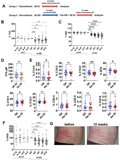
Recent data proved an essential role of elevated FFAs in the obesity-mediated amplification of skin inflammation in a mouse model of psoriasis-like inflammation as well as in patients with psoriasis [18]. In the present study, we investigated whether these findings could be translated into a therapeutic approach. Therefore, 30 patients with mild-to-moderate psoriasis and three patients with severe psoriasis were included in a dietary intervention program. All patients kept their individual pharmacological psoriasis treatment (Supplementary Table S1) for a period of at least 4 weeks before and during the intervention period. Patients in group 1 started directly with a 12-week dietary intervention (Supplementary Table S2; Figure 1A). Patients in group 2 maintained their normal diet for 3 months before they crossed over to the dietary intervention (Figure 1A). The primary endpoint of the study was the impact of dietary intervention on the severity of psoriasis as measured by PASI.

Figure 1.
Dietary intervention reduces TAG, BMI and PASI. (A) Patients with mild-to-moderate psoriasis were randomized into two groups: group 1 with dietary intervention (DI) for 12 weeks (w); group 2 with normal diet (ND) for 12 weeks followed by a change to DI for 12 weeks. (B,C) Percent reduction in serum triglycerides (TAG) and body mass index (BMI) before and after dietary intervention compared to w0 at indicated time points. Number of patients is indicated. (D) Total serum free fatty acid (FFA) before (d0 DI) and 4 weeks of DI (4w DI). (E) Percentage of specific FFA before and 4 weeks after DI. SFA is the sum of C14:0, C16:0 and C18:0. (F) Percent reduction in Psoriasis Area and Severity Index (PASI) before and after DI compared to w0 at indicated time point. Number of patients is indicated. (G) Representative image of skin from patients before and 12 weeks after DI. (B,C,F) ANOVA with multiple comparisons and non-parametric data were used (Friedman test); ** p < 0.01, *** p < 0.001, **** p < 0.0001, compared to w0. (D,E) Paired t-test. * p < 0.05, ** p < 0.01, ns—not significant.
First, the dietary intervention in group 1 significantly reduced both TAG and BMI, demonstrating the compliance of the patients to the diet (Figure 1B,C). In contrast, neither the amount of TAG nor the BMI had changed at the end of the 3-month period of normal diet in group 2 (Figure 1B,C). During dietary intervention, the total amount of FFA did not change (Figure 1D), but the relative amounts of SFAs such as myristic acid (C14:0) and stearic acid (C18:0) were reduced (Figure 1E). The relative amount of the monounsaturated FFA oleic acid (C18:1) increased, and the polyunsaturated FFAs such as linoleic acid (C18:2) and docosahexaenoic acid (C22:6, DHA) were not affected by the dietary intervention. Eicosapentaenoic acid (C20:5, EPA) was not detected. Consistently and most importantly, psoriasis improved significantly during the dietary intervention (Figure 1F,G). Underlining the importance of the dietary intervention, psoriasis was not improved in group 2 at the end of the 3 months of normal diet. Only after the crossover of group 2 to the dietary intervention, psoriasis also improved in these patients, excluding any bias due to the particular settings of the clinical study.
3.2. Impact of Dietary Intervention on Inflammatory Serum Cytokine Pattern
To elucidate the mechanisms by which the dietary intervention reduced disease activity in patients, we analyzed the serum cytokine expression pattern by the Olink Proteomics Platform. The focus was on 92 human serum mediators of inflammation, of which 72 markers were detectable in the patients’ sera. To analyze the impact of the dietary intervention on these biomarkers, we calculated the percentage of PASI reduction and the reduction in biomarkers after 4 and 8 weeks. Correlation analysis revealed that about one-third of the mediators were dependent on disease severity, BMI or TAG (Table 1).

Table 1.
Univariate correlations of inflammatory serum markers and disease activity, BMI and serum TAG.
Improvement of psoriasis (determined as a decrease in the disease severity index PASI) correlated with the change in 13 biomarkers during the observation period. Univariate correlation analysis showed a positive correlation of serum CD6, CST5, IL-18, IL-18R, ENRAGE and CASP8 reduction at week 4 and IL-7, TGFb, IL-6, IL-18, CCL19, IL-10RB, IL-18R1 and INFγ reduction at week 8 (Table 1). Multivariate linear regression analysis indicated that CD6, ENRAGE, CASP8, IL-7, IL-6, IL-18 and INFγ reduction were positively correlated to PASI reduction independent of WHR, BMI and TAG (Table 2). IL-10RB, IL18R and CST5 showed a slight association, and the correlations to CCL19 and LAP-TGFβ were lost.

Table 2.
Multivariate linear regression analysis of inflammatory serum markers and disease activity.
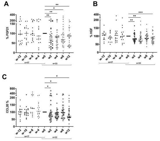
In addition, correlation analysis revealed an association between 14 serum inflammatory markers and TAG reduction and between seven markers and BMI (Table 1). Moreover, CCL20, FGF21 and HGF were significantly reduced during the dietary intervention, although we did not observe any correlation with PASI, BMI or TAG (Figure 2).

Figure 2.
Dietary intervention reduces CCL20, FGF21 and HGF. Percent reduction in FGF21 (A), HGF (B) and CCL20 (C) before and after dietary intervention compared to w0 at indicated time point. Number of patients is indicated. ANOVA with multiple comparisons and non-parametric data were used (Friedman test). * p < 0.05, ** p < 0.01, *** p < 0.001 compared to w0.
Taken together, the dietary intervention affected a wide range of inflammatory markers, depending on the disease activity, BMI or TAG.
4. Discussion
Obesity is an independent risk factor for the incidence, prevalence and severity of psoriasis. Meta-analyses revealed that a higher BMI increases the risk of psoriasis onset. Moreover, obesity reduces treatment efficacy and may predict discontinuation of biological therapy [22]. Therefore, the Medical Board of the National Psoriasis Foundation recommends dietary weight reduction with a hypocaloric diet in overweight and obese patients with psoriasis. This recommendation is based on a meta-analysis that included 77,557 participants, 4534 of whom had psoriasis [23]. However, the underlying mechanisms are only poorly understood. Pro-inflammatory activation of adipose tissue resulting in increased adipokine, cytokine and chemokine secretions may link obesity and inflammation [15,16]. Insulin resistance and high glucose levels are known to promote inflammation. Recently, we have demonstrated a prominent role of FFA in obesity-mediated exacerbation of psoriatic skin inflammation [17,18]. We have shown that serum FFA levels correlate with the severity of skin inflammation in psoriasis patients. Moreover, a dietary reduction in SFAs improved psoriasis-like skin inflammation in the imiquimod-induced mouse model of psoriasis [17,18]. In the present study, this knowledge was translated into a dietary intervention approach in psoriatic patients. Patients with mainly mild to moderate plaque-type psoriasis were enrolled in a dietary intervention program focused on reducing dietary SFAs. The combination of a formula diet and a modified Mediterranean diet in our regime resulted in a significant reduction in serum TAG, proving the compliance of the patients and the success of the chosen diet with a focus on SFA reduction. In fact, our diet led to a significant decrease in serum SFA. In parallel, the patients’ BMI decreased significantly. However, total FFAs did not change during the dietary intervention. Serum FFA levels depend on many daily lifestyle factors that cannot be fully controlled in an outpatient setting. For example, chronic exercise reduces total FFA, whereas acute exercise increases it by inducing adipose tissue lipolysis. Chronic caloric restriction reduces FFAs, whereas acute fasting induces them. Stress, including beta-adrenergic stimulation, induces lipolysis and a subsequent increase in serum FFA. Smoking and sleeping habits, insulin resistance, obesity and dietary habits are other regulators of FFA release into the circulation [24].
Several studies have shown a positive effect of weight loss on psoriasis severity and treatment response. Diet and physical exercise may improve pre-existing psoriasis and prevent the de novo development of psoriasis [22]. Our dietary regime focusing on SFA reduction improved PASI by about 25% in patients who had stable disease activity under anti-psoriatic treatment. In our study, BMI reduction did not correlate with PASI reduction, suggesting that metabolic changes such as the SFA reduction might be more important than weight loss per se. Other metabolic factors such as short fatty acids, cholesterol or uric acid may also contribute to the beneficial effect of our diet on psoriasis severity.
We can exclude that study-related intensified medical care had an effect on the study outcome, since group 2, which did not receive any dietary intervention for the first three months, showed no change in TAG and BMI or PASI. Only after the crossover to the dietary intervention did metabolic parameters and disease severity improve in group 2. Thus, the dietary intervention alone is responsible for the observed effects.
In parallel, serum inflammatory biomarkers are down-regulated by dietary intervention. All of the regulated mediators, both the psoriasis-dependent and BMI/TAG-dependent markers, have been described previously to be related to psoriasis. Among the down-regulated markers, there are molecules known to mediate the interaction of antigen-presenting cells (APCs) and T cells (CD6, CD8a, CDCP1, SLAMF1), resulting in the activation of T cells and supporting a Th1 or Th17 response [25,26,27]. CD6 is expressed by lymphocytes and facilitates the interaction between T cells and APCs, resulting in the secretion of inflammatory mediators such as TNF-α, IFN-α and IL-6. CD6-deficient mice develop an attenuated psoriasis-like skin inflammation [25,28]. Based on this knowledge, inhibiting anti-CD6 antibodies were successfully used for the treatment of psoriasis. Dietary intervention reduces a wide variety of chemokines, which might attenuate the recruitment of inflammatory cells and break the vicious cycle of inflammation in psoriasis. For example, dietary intervention reduces CCL20, a major attractant of CCR6+ Th17 cells, the drivers of an IL-17A-rich cutaneous milieu. Signaling via the CCL20/CCR6 axis was shown to be more pronounced in the lesional skin of human psoriasis and in mouse models of psoriasis-like skin inflammation. Inhibition of the CCL20/CCR6 axis attenuated experimentally induced psoriasiform dermatitis. Thus, the CCL20/CCR6 axis could be a novel promising target for treating psoriasis [29]. Recently, a disulfide-linked CCL20 dimer was developed. This engineered dimer was able to bind and to activate CCR6 but inhibited T cell-mediated chemotaxis, resulting in a reduction in inflammation in an IL-23-induced mouse model of psoriasis [30]. In addition, pro-inflammatory and immune-modulatory cytokines or their receptors such as IL-18/IL-18R, IL-6, IL-7, IL-10/IL-10RB, TNF α and IFN-γ are decreased by dietary intervention. This is in accordance with a former study that showed a correlation between serum levels of IFN-γ, TNFα, IL-18 and IL-12 and psoriasis severity [31]. Additionally, IFN-γ was shown to inhibit the apoptosis of keratinocytes, promoting the hyper-proliferative state in psoriasis [31]. IL-7 is constitutively expressed by keratinocytes and dermal lymph vessels. Several studies demonstrated an increase in IL-7 in psoriasis. Moreover, blocking of IL-7Rα ameliorated imiquimod-induced inflammation in mice [32]. Another study showed that activated keratinocytes express IL-18 [33]. This chemokine is able to stimulate IFN-γ production by Th1 cells and IL-17 expression by Th17 lymphocytes, promoting a Th1/Th17 response [33]. Deletion of IL-18 reduces psoriasis-like skin inflammation in mice [34]. Many S100 proteins including S100A7/A8/A9/A12 are upregulated in psoriasis. Wilsman-Theis and colleagues reported that S100A12 (ENRAGE) shows the closest association with disease activity and therapeutic response [35]. For HGF, which was also reduced by dietary intervention, mitogenic, morphogenic and motogenic activities toward many epithelial cells were described [36]. In addition, HGF was shown to be an important angiogenic factor in the skin, acting indirectly by inducing VEGF expression in keratinocytes and directly by stimulating endothelial cell growth migration [37].
In summary, this study investigated the effect of an easy-to-use dietary intervention program focused on reducing saturated FFA on disease activity in psoriasis. It shows that such a dietary intervention alone has the capacity to reduce disease activity in treated patients by an additional 25%. This is independent of weight loss, suggesting that diet-related metabolic changes such as SFA reduction may be more important than weight loss itself. We emphasize that dietary intervention should not replace an anti-psoriatic treatment but rather represents a valuable add-on approach. It also affects a range of biomarkers, both psoriasis-specific cytokines and more general pro-inflammatory signals also found in other chronic (auto)immune diseases. Thus, dietary intervention with a reduced amount of SFA may be also helpful in other inflammatory autoimmune diseases in which an association of disease activity and obesity has been described.
Supplementary Materials
The following supporting information can be downloaded at: https://www.mdpi.com/article/10.3390/nu15071698/s1. Reference [38] is cited in the Supplementary Materials.
Author Contributions
A.S., A.-T.S., J.K., L.S. and J.C.S. designed the study. A.S. performed the experiments, evaluated and analyzed the data and wrote the manuscript. K.M.E. performed FFA analysis. L.K., A.-T.S., J.K. and L.V. performed patient characterization and collected samples. L.S. performed the nutrition guidance. All authors discussed the data and the study design. They read and edited the manuscript. All authors have read and agreed to the published version of the manuscript.
Funding
This work was supported by a grant from Hautnetz/PsoNet Leipzig/Westsachsen and DFG (EN 1279/3-1).
Institutional Review Board Statement
The study was conducted in accordance with the Declaration of Helsinki, and approved by the Institutional Review Board (or Ethics Committee) of the University of Leipzig (#163/18-ek, 27 April 2018).
Informed Consent Statement
Informed consent was obtained from all subjects involved in the study.
Data Availability Statement
Not applicable.
Acknowledgments
We thank Danny Gutknecht for excellent technical assistance. We thank Samira Zeynalova for supporting the statistical analysis. We acknowledge the support of Leipzig University for open-access publishing.
Conflicts of Interest
The authors declare no conflict of interest.
References
- Kivimäki, M.; Strandberg, T.; Pentti, J.; Nyberg, S.T.; Frank, P.; Jokela, M.; Ervasti, J.; Suominen, S.B.; Vahtera, J.; Sipilä, P.N.; et al. Body-Mass Index and Risk of Obesity-Related Complex Multimorbidity: An Observational Multicohort Study. Lancet Diabetes Endocrinol. 2022, 10, 253–263. [Google Scholar] [CrossRef] [PubMed]
- Brunner, P.M. Weighing in on Asthma. Sci. Transl. Med. 2020, 12, eaba2907. [Google Scholar] [CrossRef]
- Duan, L.; Wu, R.; Zhang, X.; Wang, D.; You, Y.; Zhang, Y.; Zhou, L.; Chen, W. HBx-Induced S100A9 in NF-ΚB Dependent Manner Promotes Growth and Metastasis of Hepatocellular Carcinoma Cells. Cell Death Dis. 2018, 9, 629. [Google Scholar] [CrossRef] [PubMed]
- Endo, Y.; Yokote, K.; Nakayama, T. The Obesity-Related Pathology and Th17 Cells. Cell Mol. Life Sci. 2017, 74, 1231–1245. [Google Scholar] [CrossRef] [PubMed]
- Gremese, E.; Tolusso, B.; Gigante, M.R.; Ferraccioli, G. Obesity as a Risk and Severity Factor in Rheumatic Diseases (Autoimmune Chronic Inflammatory Diseases). Front. Immunol. 2014, 5, 576. [Google Scholar] [CrossRef]
- Schiopu, A.; Cotoi, O.S. S100A8 and S100A9: DAMPs at the Crossroads between Innate Immunity, Traditional Risk Factors, and Cardiovascular Disease. Mediat. Inflamm. 2013, 2013, 828354. [Google Scholar] [CrossRef]
- Zeki, A.A. Twin Towers, Twin Epidemics: Obesity and Asthma. Sci. Transl. Med. 2012, 4, 163ec223. [Google Scholar] [CrossRef]
- Nakamizo, S.; Honda, T.; Adachi, A.; Nagatake, T.; Kunisawa, J.; Kitoh, A.; Otsuka, A.; Dainichi, T.; Nomura, T.; Ginhoux, F.; et al. High Fat Diet Exacerbates Murine Psoriatic Dermatitis by Increasing the Number of IL-17-Producing Γδ T Cells. Sci. Rep. 2017, 7, 14076. [Google Scholar] [CrossRef]
- Paik, J.; Fierce, Y.; Treuting, P.M.; Brabb, T.; Maggio-Price, L. High-Fat Diet-Induced Obesity Exacerbates Inflammatory Bowel Disease in Genetically Susceptible Mdr1a-/- Male Mice. J. Nutr. 2013, 143, 1240–1247. [Google Scholar] [CrossRef]
- Petronilho, F.; Giustina, A.D.; Nascimento, D.Z.; Zarbato, G.F.; Vieira, A.A.; Florentino, D.; Danielski, L.G.; Goldim, M.P.; Rezin, G.T.; Barichello, T. Obesity Exacerbates Sepsis-Induced Oxidativedamage in Organs. Inflammation 2016, 39, 2062–2071. [Google Scholar] [CrossRef]
- Shah, D.; Romero, F.; Duong, M.; Wang, N.; Paudyal, B.; Suratt, B.T.; Kallen, C.B.; Sun, J.; Zhu, Y.; Walsh, K.; et al. Obesity-Induced Adipokine Imbalance Impairs Mouse Pulmonary Vascular Endothelial Function and Primes the Lung for Injury. Sci. Rep. 2015, 5, 11362. [Google Scholar] [CrossRef] [PubMed]
- Higashi, Y.; Yamakuchi, M.; Fukushige, T.; Ibusuki, A.; Hashiguchi, T.; Kanekura, T. High-Fat Diet Exacerbates Imiquimod-Induced Psoriasis-like Dermatitis in Mice. Exp. Dermatol. 2018, 27, 178–184. [Google Scholar] [CrossRef] [PubMed]
- Setty, A.R.; Curhan, G.; Choi, H.K. Obesity, Waist Circumference, Weight Change, and the Risk of Psoriasis in Women: Nurses’ Health Study II. Arch. Intern. Med. 2007, 167, 1670–1675. [Google Scholar] [CrossRef]
- Snekvik, I.; Nilsen, T.I.L.; Romundstad, P.R.; Saunes, M. Metabolic Syndrome and Risk of Incident Psoriasis: Prospective Data from the HUNT Study, Norway. Br. J. Dermatol. 2019, 180, 94–99. [Google Scholar] [CrossRef] [PubMed]
- Zhou, H.; Urso, C.J.; Jadeja, V. Saturated Fatty Acids in Obesity-Associated Inflammation. J. Inflamm. Res. 2020, 13, S229691. [Google Scholar] [CrossRef]
- Shoelson, S.E.; Herrero, L.; Naaz, A. Obesity, Inflammation, and Insulin Resistance. Gastroenterology 2007, 132, 2169–2180. [Google Scholar] [CrossRef]
- Kunz, M.; Simon, J.C.; Saalbach, A. Psoriasis: Obesity and Fatty Acids. Front. Immunol. 2019, 10, 1807. [Google Scholar] [CrossRef]
- Herbert, D.; Franz, S.; Popkova, Y.; Anderegg, U.; Schiller, J.; Schwede, K.; Lorz, A.; Simon, J.C.; Saalbach, A. High-Fat Diet Exacerbates Early Psoriatic Skin Inflammation Independent of Obesity: Saturated Fatty Acids as Key Players. J. Investig. Dermatol. 2018, 138, 1999–2009. [Google Scholar] [CrossRef]
- Franz, S.; Ertel, A.; Engel, K.M.; Simon, J.C.; Saalbach, A. Overexpression of S100A9 in Obesity Impairs Macrophage Differentiation via TLR4-NFkB-Signaling Worsening Inflammation and Wound Healing. Theranostics 2022, 12, 1659–1682. [Google Scholar] [CrossRef]
- Bligh, E.G.; Dyer, W.J. A Rapid method of total lipid extraction and purification. Can. J. Biochem. Physiol. 1959, 37, 911–917. [Google Scholar] [CrossRef]
- Engel, K.M.; Sampels, S.; Dzyuba, B.; Podhorec, P.; Policar, T.; Dannenberger, D.; Schiller, J. Swimming at Different Temperatures: The Lipid Composition of Sperm from Three Freshwater Fish Species Determined by Mass Spectrometry and Nuclear Magnetic Resonance Spectroscopy. Chem. Phys. Lipids. 2019, 221, 65–72. [Google Scholar] [CrossRef]
- Paroutoglou, K.; Papadavid, E.; Christodoulatos, G.S.; Dalamaga, M. Deciphering the Association between Psoriasis and Obesity: Current Evidence and Treatment Considerations. Curr. Obes. Rep. 2020, 9, 165–178. [Google Scholar] [CrossRef]
- Ford, A.R.; Siegel, M.; Bagel, J.; Cordoro, K.M.; Garg, A.; Gottlieb, A.; Green, L.J.; Gudjonsson, J.E.; Koo, J.; Lebwohl, M.; et al. Dietary Recommendations for Adults With Psoriasis or Psoriatic Arthritis From the Medical Board of the National Psoriasis Foundation. JAMA Dermatol. 2018, 154, 934. [Google Scholar] [CrossRef]
- Henderson, G.C. Plasma Free Fatty Acid Concentration as a Modifiable Risk Factor for Metabolic Disease. Nutrients 2021, 13, 2590. [Google Scholar] [CrossRef]
- Consuegra-Fernández, M.; Julià, M.; Martínez-Florensa, M.; Aranda, F.; Català, C.; Armiger-Borràs, N.; Arias, M.-T.; Santiago, F.; Guilabert, A.; Esteve, A.; et al. Genetic and Experimental Evidence for the Involvement of the CD6 Lymphocyte Receptor in Psoriasis. Cell Mol. Immunol. 2018, 15, 898–906. [Google Scholar] [CrossRef]
- De la Calle-Martin, O.; Hernandez, M.; Ordi, J.; Casamitjana, N.; Arostegui, J.I.; Caragol, I.; Ferrando, M.; Labrador, M.; Rodriguez-Sanchez, J.L.; Espanol, T. Familial CD8 Deficiency Due to a Mutation in the CD8α Gene. J. Clin. Investig. 2001, 108, 117–123. [Google Scholar] [CrossRef]
- Howie, D.; Simarro, M.; Sayos, J.; Guirado, M.; Sancho, J.; Terhorst, C. Molecular Dissection of the Signaling and Costimulatory Functions of CD150 (SLAM): CD150/SAP Binding and CD150-Mediated Costimulation. Blood 2002, 99, 957–965. [Google Scholar] [CrossRef]
- Dogra, S.; Shabeer, D.; Rajagopalan, M. Anti-CD6 MAbs for the Treatment of Psoriasis. Expert Opin. Biol. Ther. 2020, 20, 1215–1222. [Google Scholar] [CrossRef]
- Furue, K.; Ito, T.; Tsuji, G.; Nakahara, T.; Furue, M. The CCL20 and CCR6 Axis in Psoriasis. Scand. J. Immunol. 2020, 91, 12846. [Google Scholar] [CrossRef]
- Getschman, A.E.; Imai, Y.; Larsen, O.; Peterson, F.C.; Wu, X.; Rosenkilde, M.M.; Hwang, S.T.; Volkman, B.F. Protein Engineering of the Chemokine CCL20 Prevents Psoriasiform Dermatitis in an IL-23–Dependent Murine Model. Proc. Natl. Acad. Sci. USA 2017, 114, 12460–12465. [Google Scholar] [CrossRef]
- Lanna, C.; Mancini, M.; Gaziano, R.; Cannizzaro, M.V.; Galluzzo, M.; Talamonti, M.; Rovella, V.; Annicchiarico-Petruzzelli, M.; Melino, G.; Wang, Y.; et al. Skin Immunity and Its Dysregulation in Psoriasis. Cell Cycle 2019, 18, 2581–2589. [Google Scholar] [CrossRef] [PubMed]
- Vranova, M.; Friess, M.C.; Haghayegh Jahromi, N.; Collado-Diaz, V.; Vallone, A.; Hagedorn, O.; Jadhav, M.; Willrodt, A.-H.; Polomska, A.; Leroux, J.-C.; et al. Opposing Roles of Endothelial and Leukocyte-Expressed IL-7Rα in the Regulation of Psoriasis-like Skin Inflammation. Sci. Rep. 2019, 9, 11714. [Google Scholar] [CrossRef] [PubMed]
- Yasuda, K.; Nakanishi, K.; Tsutsui, H. Interleukin-18 in Health and Disease. Int. J. Mol. Sci. 2019, 20, 649. [Google Scholar] [CrossRef] [PubMed]
- Niu, X.L.; Huang, Y.; Gao, Y.L.; Sun, Y.Z.; Han, Y.; Chen, H.D.; Gao, X.H.; Qi, R.Q. Interleukin-18 Exacerbates Skin Inflammation and Affects Microabscesses and Scale Formation in a Mouse Model of Imiquimod-Induced Psoriasis. Chin. Med. J. 2019, 132, 690–698. [Google Scholar] [CrossRef]
- Wilsmann-Theis, D.; Wagenpfeil, J.; Holzinger, D.; Roth, J.; Koch, S.; Schnautz, S.; Bieber, T.; Wenzel, J. Among the S100 Proteins, S100A12 Is the Most Significant Marker for Psoriasis Disease Activity. J. Eur. Acad. Dermatol. Venereol. 2016, 30, 1165–1170. [Google Scholar] [CrossRef]
- Delehedde, M.; Vidyasagar, R.; Fernig, D.G.; Lyon, M.; McDonnell, T.J. Hepatocyte Growth Factor/Scatter Factor Binds to Small Heparin-Derived Oligosaccharides and Stimulates the Proliferation of Human HaCaT Keratinocytes. J. Biol. Chem. 2002, 277, 12456–12462. [Google Scholar] [CrossRef]
- Hisadome, M.; Ohnishi, T.; Kakimoto, K.; Kusuyama, J.; Bandow, K.; Kanekura, T.; Matsuguchi, T. Hepatocyte Growth Factor Reduces CXCL10 Expression in Keratinocytes. FEBS Lett. 2016, 590, 3595–3605. [Google Scholar] [CrossRef]
- Rondanelli, M.; Faliva, M.A.; Miccono, A.; Naso, M.; Nichetti, M.; Riva, A.; Guerriero, F.; De Gregori, M.; Peroni, G.; Perna, S. Food Pyramid for Subjects with Chronic Pain: Foods and Dietary Constituents as Anti-Inflammatory and Antioxidant Agents. Nutr. Res. Rev. 2018, 31, 131–151. [Google Scholar] [CrossRef]
Disclaimer/Publisher’s Note: The statements, opinions and data contained in all publications are solely those of the individual author(s) and contributor(s) and not of MDPI and/or the editor(s). MDPI and/or the editor(s) disclaim responsibility for any injury to people or property resulting from any ideas, methods, instructions or products referred to in the content. |
© 2023 by the authors. Licensee MDPI, Basel, Switzerland. This article is an open access article distributed under the terms and conditions of the Creative Commons Attribution (CC BY) license (https://creativecommons.org/licenses/by/4.0/).