Article

Maternal Dietary Deficiencies in Folic Acid and Choline Result in Larger Damage Volume, Reduced Neuro-Degeneration and -Inflammation and Changes in Choline Metabolites after Ischemic Stroke in Middle-Aged Offspring

1
Biomedical Sciences Program, Midwestern University, Glendale, AZ 85308, USA
2
College of Veterinary Medicine, Midwestern University, Glendale, AZ 85308, USA
3
College of Dental Medicine of Arizona, Midwestern University, Glendale, AZ 85308, USA
4
Division of Nutritional Sciences, Cornell University, Ithaca, NY 14853, USA
5
College of Osteopathic Medicine, Midwestern University, Glendale, AZ 85308, USA
6
Department of Child Health, University of Arizona, Phoenix, AZ 85721, USA
7
Department of Neuroscience, Carleton University, Ottawa, ON K1S 5B6, Canada
*
Author to whom correspondence should be addressed.
These authors contributed equally to this work.
Nutrients 2023, 15(7), 1556; https://doi.org/10.3390/nu15071556
Submission received: 14 February 2023 / Revised: 17 March 2023 / Accepted: 20 March 2023 / Published: 23 March 2023
(This article belongs to the Special Issue The Impact of Nutrition on Brain Metabolism and Disease)

Abstract

:
Maternal dietary levels of one-carbon (1C) metabolites (folic acid and choline) during pregnancy play a vital role in neurodevelopment. However, the impact of maternal dietary deficiencies on offspring stroke outcomes later in life remains undefined. The aim of this study was to investigate the role of maternal dietary deficiencies in folic acid and choline on ischemic stroke outcomes in middle-aged offspring. Female mice were maintained on either a control or deficient diet prior to and during pregnancy and lactation. At 10 months of age ischemic stroke was induced in male and female offspring. Stroke outcome was assessed by measuring motor function and brain tissue. There was no difference in offspring motor function; however, sex differences were present. In brain tissue, maternal dietary deficiency increased ischemic damage volume and offspring from deficient mothers had reduced neurodegeneration and neuroinflammation within the ischemic region. Furthermore, there were changes in plasma 1C metabolites as a result of maternal diet and sex. Our data indicate that maternal dietary deficiencies do not impact offspring behavior after ischemic stroke but do play a role in brain histology and one-carbon metabolite levels in plasma. Additionally, this study demonstrates that the sex of mice plays an important role in stroke outcomes.

1. Introduction

Stroke prevalence in young adults and the underlying causes are of increasing concern [1,2,3,4,5,6]. In Brazil, ischemic stroke incidence was found to increase in adults less than 45 years old from 2005 to 2011 by 88% and by 62% from 2005 to 2015 [2]. Rates of hospitalizations for ischemic stroke in young adults have been increasing in European countries since 1987 including Denmark [6], France [1,7], and Sweden [4]. In the Greater Cincinnati/Northern Kentucky region of the United States, the proportion of stroke for people under 55 years old increased from 12.9% in 1993 to 18.6% in 2005 [8] and the U.S. as a whole saw an increase in age-specific hospitalization rates for acute ischemic stroke in patients aged 25 to 44 years and 45 to 64 years old [9].
Nutrition is a modifiable risk factor for stroke [10,11,12]. B vitamins, such as folic acid, or folate, as well as choline, are known for their important role in the closure of the neural tube [13,14,15]. Both folic acid and choline play a major role in one-carbon (1C) metabolism, which is responsible for methylation metabolism, maintenance of cellular redox status, nucleotide metabolism, and lipid biosynthesis [16,17]. These vitamins have also been implicated in the onset and progression of stroke [18,19,20]. Insufficient folic acid and choline levels increase the risk of cardiovascular diseases such as stroke [21].
During pregnancy, there is an increased demand for dietary nutrients, such as folic acid and choline, especially for the closure of the neural tube [22]. There is both clinical and pre-clinical evidence that maternal dietary deficiencies in folic acid impact offspring after birth too [23]. We have previously shown in a mouse model that maternal dietary deficiencies in folic acid and choline hinders hippocampal development, and impacts short-term memory function in young male offspring [24]. However, the long-term impact of maternal dietary deficiencies on offspring's neurological function, especially after ischemic stroke is not known. The aim of this study was to investigate the role of maternal dietary deficiencies in folic acid or choline on ischemic stroke outcomes in middle-aged male and female offspring. This study adds to the literature investigating the long-term impact of maternal dietary deficiencies on offspring's neurological function.

2. Materials and Methods

All experiments involving the use of animals have been approved by the Midwestern University IACUC (2983). Experimental manipulations are summarized in Figure 1. Maternal females were born and raised on a control diet until two months of age, then were placed on either a folic acid (FADD) or choline-deficient diet (ChDD) or control diet (CD). Both male and female parent mice were placed on these diets for 4 weeks prior to mating. Female mice were maintained on either a control diet or deficient diet during pregnancy and lactation. Female and male offspring were weaned and placed on a control diet until ten months of age then had ischemic stroke-induced via photothrombosis targeting the sensorimotor complex. Four weeks after ischemic damage, the offspring underwent behavioral testing to assess motor function and after completion were euthanized with blood and brain tissue samples collected for analysis.

2.1. Dietary Composition

The maternal diets in this study consisted of a control diet (CD), folic acid-deficient diet (FADD), and choline-deficient diet (ChDD). The CD represented normal levels of both folic acid and choline that would be found in a conventional diet, which were determined from the previous literature and experimentation [24,25,26], both mothers and offspring were fed these diets. The deficient diets consisted of lower levels of folic acid or choline compared to the control diet. The CD contained 2 mg/kg of folic acid and 1150 mg/kg of choline bitrate, whereas the FADD contained only 0.3 mg/kg and the ChDD only had 300 mg/kg of choline bitrate.

2.2. Photothrombosis Model

The photothrombosis model is a reproducible model that induces ischemic damage by targeting the sensorimotor cortex [27]. The mice were anesthetized in an isoflurane chamber and the surgical site was prepared by shaving and disinfecting the head. Anesthesia was continued via face mask and body temperature was maintained at 37 °C with a heating pad throughout the entire procedure. To create ischemic damage and simulate stroke, Rose Bengal, a photoactive dye, was injected intraperitoneally at a dose of 10 mg/kg five minutes prior to laser exposure. Activation of the dye with a 532 nm laser beam centered 3 cm over the sensorimotor cortex for 15 min caused formation of free radicals to induce endothelial injury, platelet activation, and aggregation resulting in ischemic damage.

2.3. Behavioral Testing

Behavioral testing in the offspring was conducted four weeks after ischemic damage was induced via photothrombosis to assess motor function. This was performed using accelerating rotarod, forepaw placement, and ladder beam walking tasks.

2.3.1. Accelerating Rotarod

Coordination and balance were tested by placing mice on an accelerating rotarod and measuring latency to fall and rotations per minute. This was completed for three trials per mouse with five-minute lapse periods between trials.

2.3.2. Forepaw Placement

The forepaw placement task assesses forepaw usage by video recording mice placed in a glass cylinder (19 cm high, 14 cm diameter) for ten minutes and analyzing contact between the walls of the cylinder and forepaw of the mouse [27]. The first 20 rears were analyzed during this time.

2.3.3. Ladder Beam Walking

Skilled motor function of impaired or non-impaired fore- and hindlimbs was measured by ladder beam walking. Mice were placed on a horizontal ladder composed of two plexiglass walls with metal bars inserted into the holes at the bottom edge of the walls. The mice were assessed in their ability to travel from one end towards the other, without being permitted to regress. After being familiarized with the ladder, the remaining trials were video-recorded and performance was analyzed in each frame for quality of limb placement as previously described [28].

2.4. Brain Tissue Collection and Sectioning

After completing behavioral analysis and euthanasia, brain tissue was collected and sectioned using a cryostat. Tissue sections were 30 μm thick and mounted onto positively charged microscope slides. Serial sections were collected for each brain tissue sample over six slides total. A minimum of 8 sections within the damaged region of the brain were collected. Slides were stored at −80 °C until tissue analysis was conducted. For damage volume analysis, each animal had a minimum of three sections of damaged area per slide and two slides per animal were stained using cresyl violet (Sigma, St. Louis, MO, USA). Images were taken using an Olympus microscope. ImageJ (NIH) software (version 2.90) was used to quantify volume of damaged tissue by measuring the area of damage in each serial section [29] and multiplying the area between sections (0.03 mm) by the number of sections from the damaged site.

2.5. Immunofluorescence Staining

To investigate neurodegeneration and neuroinflammation, immunofluorescence analysis of ischemic-damaged brain tissue was performed. To assess neurodegeneration by measuring apoptosis in ischemic-damaged brain tissue, primary antibodies to active caspase-3 (1:100, Cell Signaling Technologies, Danvers, MA, USA) were used. All brain sections were stained with a marker for neuronal nuclei, NeuN (1:200, AbCam, Waltham, MA, USA). Primary antibodies were diluted in 0.5% Triton X and incubated on the brain tissue overnight at 4 °C. Brain sections were incubated with secondary antibodies Alexa Fluor 488 or 555 (1:200, Cell Signaling Technologies) the following day at room temperature for 2 h then stained with 4′, 6-diamidino-2phenylindole (DAPI, 1:10,000).
To assess neuroinflammation by measuring active microglial cells, primary antibodies to Ionized calcium-binding adaptor molecule 1 (Iba1, 1:100, AbCam) and CD68 (1:500, Bio-Rad, Hercules, CA, USA) were used. Primary antibodies were diluted in the same manner and incubated on the brain tissue overnight at 4 °C. Anti-rat (594) and anti-rabbit (488, Cell Signaling Technologies) secondary antibodies were added the following day and incubated at room temperature for 2 h then stained with DAPI (1:10,000).
Microscope slides were coverslipped with fluromount and stored at 4 °C until analysis. The staining was visualized using an ECHO microscope and all images were collected at the magnification of 200×. To assess neurodegeneration within the ischemic core region of the brain tissue, co-localization of active caspase-3 with NeuN and DAPI-labeled neurons was counted and averaged per animal. To assess neuroinflammation, co-localization of Iba1 and CD68-labeled cells was counted. Cells were distinguished from debris by identifying a clear cell shape and intact nuclei (indicated by DAPI and NeuN) under the microscope. All cell counts were conducted by two individuals blinded to treatment groups. Using ImageJ, the number of positive cells was counted in three brain sections per animal, and a total of 5 animals were analyzed per group. For each section, three fields were analyzed. The number of positive cells was averaged for each animal.

2.6. Plasma One-Carbon Metabolite Measurements

Plasma tissue from offspring was measured for betaine, choline, dimethylglycine, methionine, phosphocholine, phosphatidylcholine, lysophosphatidylcholine, and, trimethylamine N-oxide (TAMO), sphingomyelin levels using the LC-MS/MS (ThermoElectron Corp, Waltham, MA, USA) method as previously reported [30,31,32].

2.7. Data Analysis and Statistics

GraphPad Prism 6.0 was used to analyze behavioral testing, plasma tHcy measurements, lesion volume, immunofluorescence staining, and plasma choline measurements. In GraphPad Prism 6.0, D’Agostino-Pearson normality test was performed prior to two-way ANOVA analysis when comparing the mean measurement of both sex and dietary group for behavioral testing, one-carbon metabolites, lesion volume, and immunofluorescence staining. Significant main effects of two-way ANOVAs were followed up with Tukey’s post hoc test to adjust for multiple comparisons. All data are presented as mean ± standard error of the mean (SEM). Statistical tests were performed using a significance level of 0.05. Behavioral and brain tissue microscope data were analyzed by two individuals that were blinded to experimental treatment groups. There is a summary of data, see Supplementary Dataset S1.

3. Results

3.1. Accelerating Rotarod

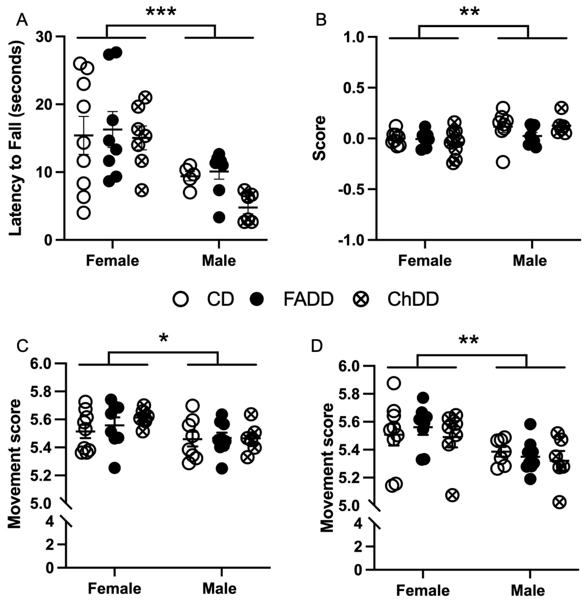
The accelerating rotarod was used to assess coordination and balance by measuring the length of time spent on the rotarod, termed latency to fall. No significant difference was noted between maternal diet groups (Figure 2A; F(2,37) = 1.25, p = 0.30) and no interaction was observed between maternal diet and sex main effects (Figure 2A; F(2,37) = 0.60, p = 0.56). Significant sex differences between male and female offspring were noted with performance (Figure 2; F(1,37) = 18.04, p = 0.0001). No significant difference was observed between maximum speed (rotations per minute, RPM) achieved between groups (F(2,37) = 1.78, p = 0.18), however, female mice reached a higher RPM compared to males (F(2,37) = 21.92, p < 0.0001). No significant interaction between maternal diet and sex was seen (F(2,37) = 0.49, p = 0.62).

3.2. Forepaw Placement

Impaired forepaw usage during the forepaw placement task was not significantly impacted by maternal diet (F(2,41) = 0.33, p = 0.72) or sex (F(1,41) = 0.83, p = 0.37) and no significant interaction was observed between sex and maternal diet for impaired (F(2,41) = 0.44, p = 0.64) or non-impaired forepaw usage (F(2,41) = 0.05, p = 0.95). Non-impaired forepaw usage also did not have a significant main effect on maternal diet (F(2,41) = 0.05, p = 0.95) or sex (F(1,41) = 3.01, p = 0.09), or interaction between sex and maternal diet (F(2,41) = 0.77, p = 0.47). No main effect of maternal diet (F(2,40) = 0.78, p = 0.46) was seen with forepaw placement score, which is the ratio of impaired to non-impaired forepaw usage, however, a significant main effect of sex was observed (Figure 2B; F(1,40) = 9.98, p = 0.003).

3.3. Ladder Beam

The ladder beam test was used to measure skilled motor function. The test consisted of a movement score based on the usage of impaired and non-impaired fore- and hindlimb stepping patterns, as well as the number of errors made and time to cross the ladder. For impaired movement score, there was no difference in maternal dietary groups (Figure 2C; F(2,41) = 0.61, p = 0.55), but a sex difference was present (Figure 2C; F(1,41) = 6.61, p = 0.01) and no interaction between sex and maternal diet (F(2,41) = 0.48, p = 0.62). The movement score for non-impaired fore and hindlimbs showed no difference in maternal dietary groups (Figure 2D; F(2,40) = 0.35, p = 0.71), but a sex difference was present (Figure 2D; F(1,41) = 11.04, p = 0.01) and no interaction between sex and maternal diet was present (F(2,40) = 0.28, p = 0.75).
Percent error, number of errors made while crossing the ladder beam, showed no differences between maternal diet for impaired (F(2,41) = 1.31, p = 0.28) and non-impaired limbs (F(2,42) = 0.03, p = 0.97). There was also no sex difference in the number of errors made for impaired (F(1,41) = 3.6, p = 0.06) and non-impaired limbs (F(1,41) = 1.04, p = 0.31). No interaction between sex or maternal diet with impaired percent error (F(2,41) = 0.23, p = 0.80) or non-impaired percent error (F(2,42) = 0.75, p = 0.48) was observed. Time to cross the ladder beam was not significantly impacted by maternal diet (F(2,42) = 1.22, p = 0.31) or sex (F(1,42) = 0.14, p = 0.11), there was also no interaction between sex and maternal diet (F(2, 42) = 0.15, p = 0.86).

3.4. Ischemic Damage Volume Increased in Offspring as a Result of Maternal Dietary Deficiencies

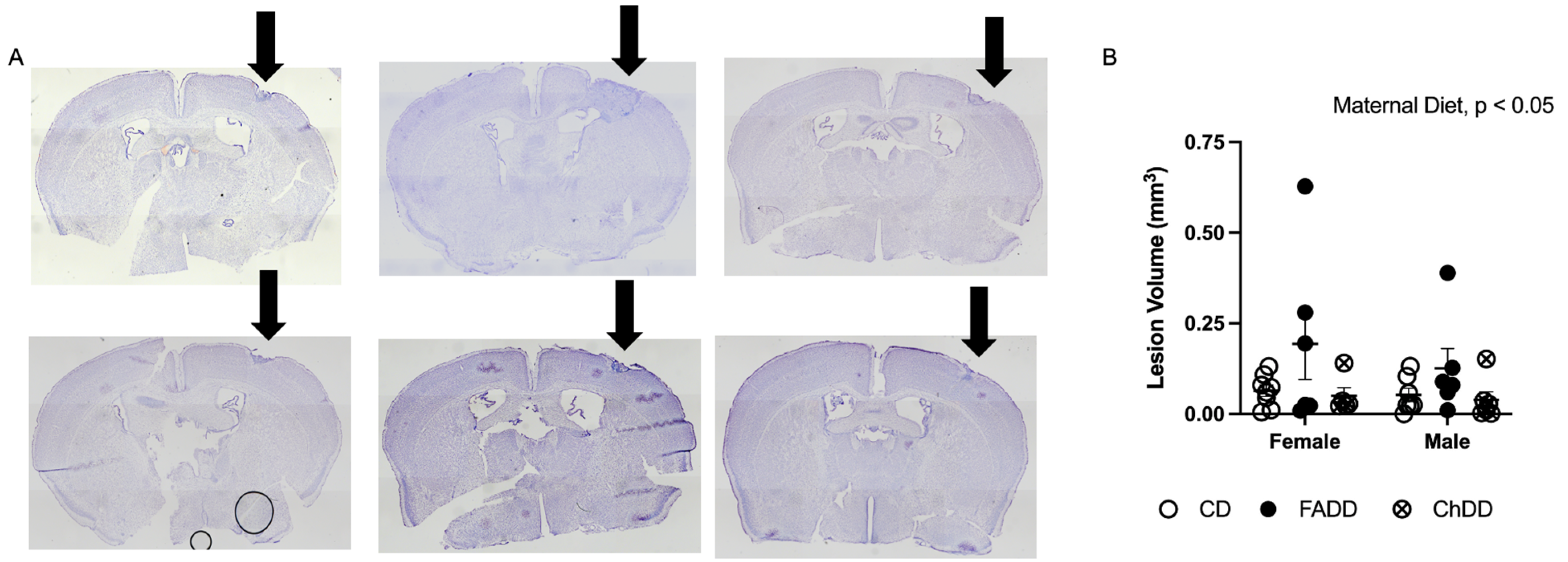
Brain tissue sections were stained with cresyl violet (Sigma) and imaged using an Olympus microscope (Figure 3A). Images were processed using ImageJ software to measure ischemic damage volume. Maternal diet had a significant effect on damage volume (Figure 3B; F(2,32) = 3.53, p = 0.041), but no main effect of sex (Figure 3B; F(1,32) = 0.63, p = 0.43) or significant interaction between maternal diet and sex for ischemic damage volume was observed (Figure 3B; F(2,32) = 0.24, p = 0.79).

3.5. Decreased Neurodegeneration and Neuroinflammation in Male and Female Offspring from Deficient Mothers

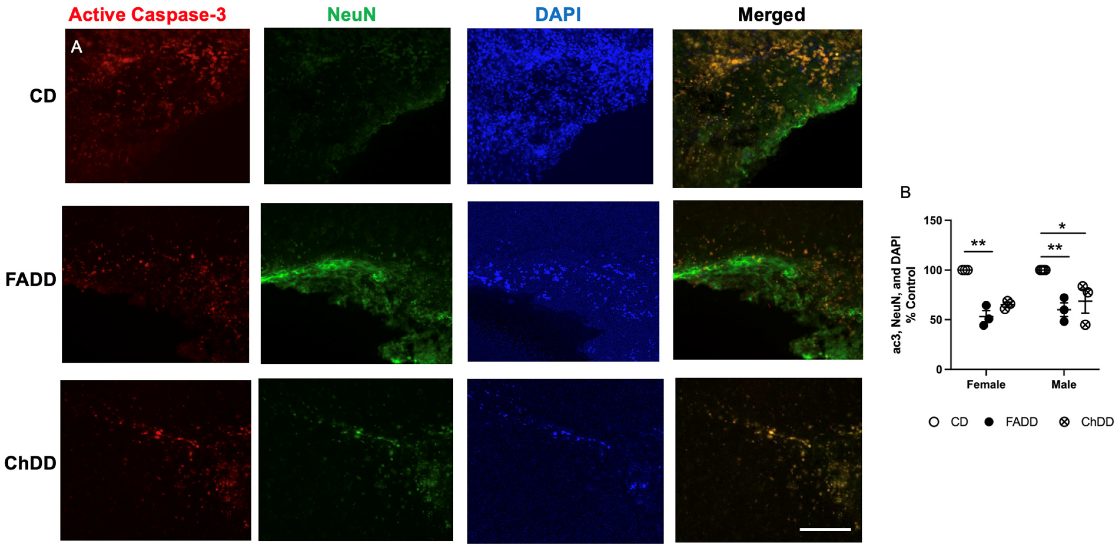
Within the ischemic damaged region of the brain tissue, we measured neurodegeneration and neuroinflammation. Neurodegeneration was assessed using active caspase-3 levels, representative images are shown in Figure 4A. Maternal diet had an impact on the number of positive active caspase 3 neurons (Figure 4B; F(2,13) = 31.22, p < 0.001). There was no effect of sex (F(1,13) = 0.53, p = 0.48) and no interaction between sex and maternal diet (F(2,13) = 0.18, p = 0.84).
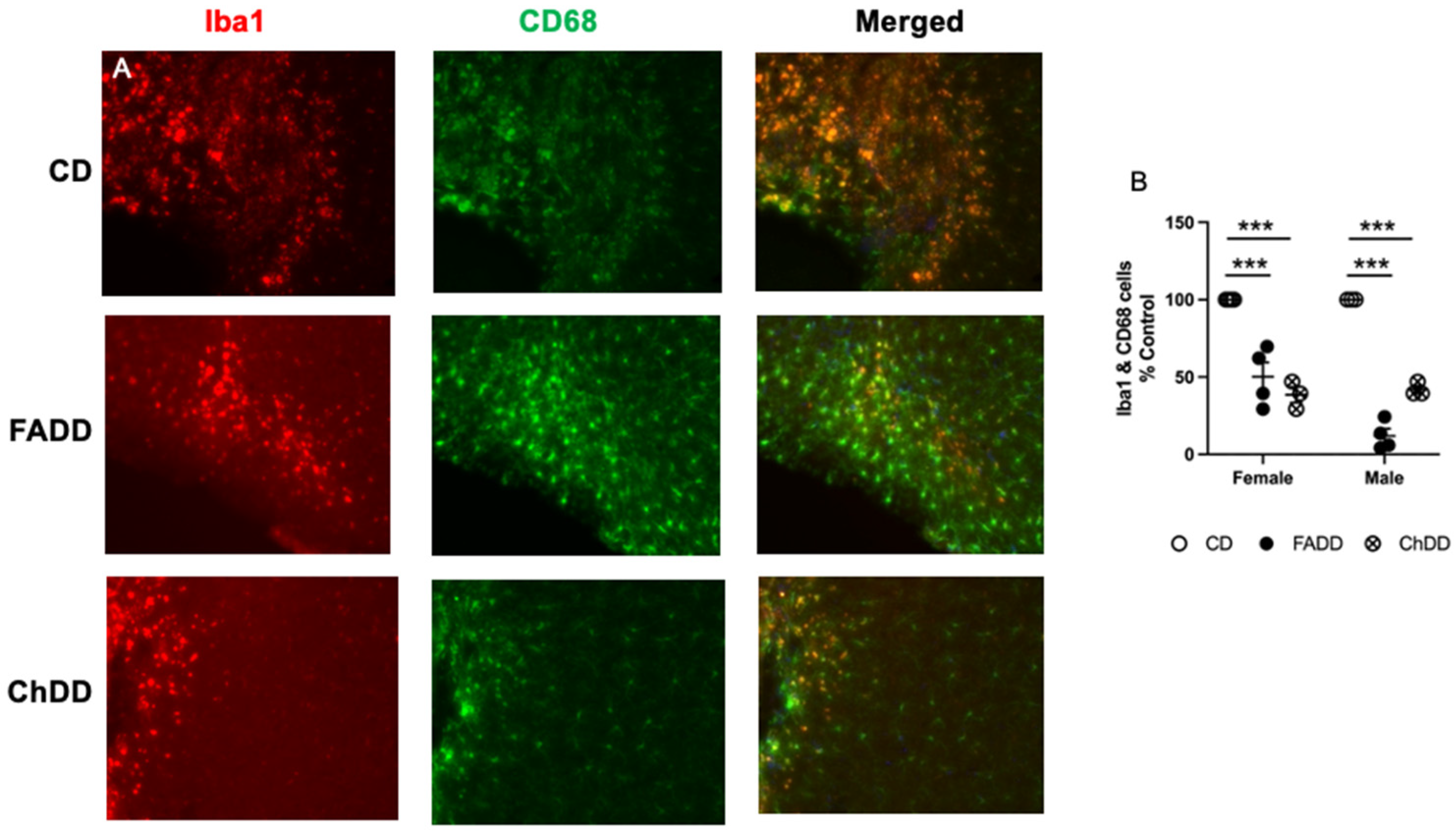
We measured neuroinflammation within the ischemic damage region using Iba1 and CD68 colocalization [33], representative images are shown in Figure 5A. Maternal diet had an impact on the number of positive active caspase 3 neurons (Figure 5B; F(2,22) = 5.51, p = 0.01). There was no effect of sex (F(2,22) = 2.82, p = 0.11) and no interaction between sex and maternal diet (F(2,22) = 2.49, p = 0.11).

3.6. Plasma One-Carbon Metabolites

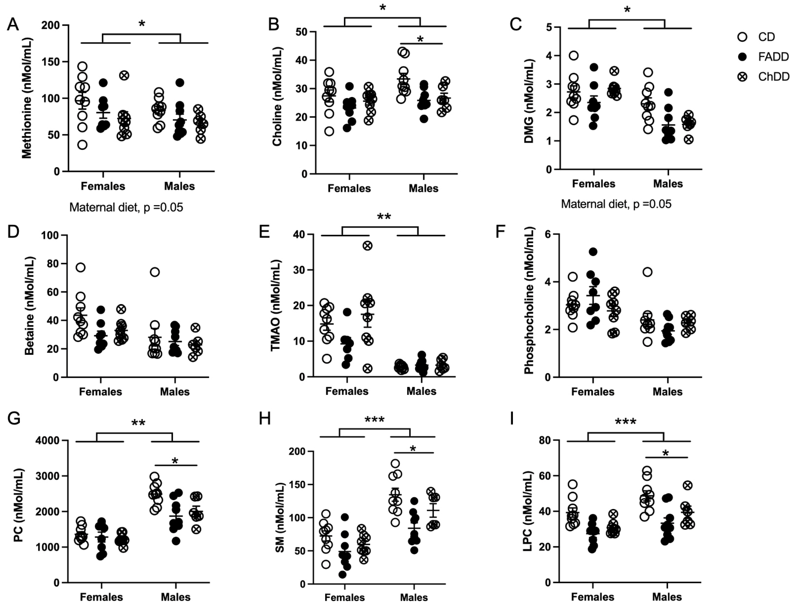
We measured one-carbon metabolites in the plasma of offspring 1.5 months after ischemic stroke and found that maternal diet impacted methionine (Figure 6: F(2,43) = 3.24, p = 0.049), choline (F(2,43) = 6.65, p = 0.003), DMG (F(2,43) = 4.60, p = 0.0001), PC (F(2,43) = 5.86, p = 0.006), SM (F(2,43) = 9.74, p = 0.003), and LPC (F(2,43) = 14.91, p < 0.0001). There were pairwise differences between males from CD and FADD mothers for choline (p = 0.026), PC (p = 0.0032), and SM (p = 0.0011). There was also a difference between females and male animals for the following metabolites: choline (F(1,43) = 5.15, p = 0.03), PC (F(1,43) = 77.66, p < 0.001), SM (F(1,43) = 48.08 p < 0.001), and LPC (F(1,43) = 13.7, p = 0.0006). There was no sex difference in methionine (F(1,43) = 1.96, p = 0.17) and DMG (F(2,43) = 2.4, p = 0.11). A significant interaction between maternal diet and sex was found for PC (F(2,43) = 3.02, p = 0.06), no other metabolites had an interaction (methionine, F(2,43) = 0.071, p = 0.93; choline, F(2,43) = 1.04. p = 0.36; DMG, interaction: F(2,43) = 2.35, p = 0.11; SM, F(2,43) = 1.33, p = 0.28 and LPC, interaction: F(2,43) = 0.26, p = 0.77).

4. Discussion

Maternal nutrition and diet play an important role in the neurodevelopment of offspring. Alterations in this early life nutritional programming can have long-term impacts on offspring health [34]. A maternal diet rich in required vitamins and nutrients has a large impact on the prevention of various diseases including stroke [12]. What remains unclear is the impact of maternal dietary one-carbon deficiencies during pregnancy and lactation, and their impact on offspring stroke outcomes later in life. The aim of this study was to determine the impact of maternal deficiencies in folic acid or choline on middle-aged offspring stroke outcomes. After ischemic stroke, no differences were seen between maternal dietary groups in the accelerating rotarod, forepaw placement, and ladder beam tasks. However, sex differences were observed in behavioral outcomes measured, indicating males had worse outcomes. Analysis of brain tissue indicated that there was a significant main effect of maternal diet on ischemic damage volume, neurodegeneration, and neuroinflammation. Changes in plasma choline metabolites were observed as a result of maternal diet and sex after ischemic stroke in offspring. This study adds to the literature investigating the long-term impact of maternal dietary deficiencies in folic acid or choline on middle-aged offspring stroke outcomes. This is novel data and adds to the literature on maternal dietary deficiencies and the lifelong impact that they have on offspring brain health.
Sex has also been correlated with stroke incidence in young adults, however, which sex has the highest incident rate is still being debated. In a Brazilian population, ischemic stroke incidence was found to be higher in men less than 45 years old and higher in women less than 55 years old [2]. According to the Danish National Patient Register, when reviewing trends in young adults aged 15 to 30 years old, incidence rates for hospitalization for ischemic stroke and transient ischemic attack (TIA) were high in women compared to men [6]. In France, ischemic stroke incidence significantly increased in men aged 25 to 74 years old and in women aged 35 to 64 years old from 2008 to 2014 [7]. In the same study, women with ischemic stroke were also found to have higher mortality when between the ages of 45 and 64 years old and men had a significantly younger mean age in hospitalization [7]. Eastern France had a higher increase in incidence rates for ischemic stroke in men after 2003 as compared to before 2003 [1] and the U.S. had similar trends with age-adjusted acute ischemic stroke hospitalization rates being higher in men from 2000 to 2010 [9]. China, however, saw the onset of first-ever stroke in significantly younger women compared to men, with the average age being 36.9 years old compared to 38.7 years old in men from 2007–2018 [5]. The high variation between regions could be due to the prevalence of risk factors associated with ischemic stroke in each sex.
In the present study, there is no impact of maternal diet on motor function after ischemic stroke. We do describe a significant sex difference in the behavioral outcome, females were observed to perform better on the motor function behavioral tests. These findings may be due to a variety of mechanisms, however, the most plausible appears to be due to the neuroprotective effects of estrogen in female mice post-ischemic stroke [35]. The neuroprotective effects of estrogen on stroke are extremely well documented throughout the literature, with various studies demonstrating significantly greater damage post-ischemia in male mice and rats when compared to females [36,37]. This was further confirmed through the removal of ovaries in female mice and rats, who subsequently appeared to have lost this protective effect after ischemia [38,39]. Therefore, the evidence for estrogen-mediated neuroprotection after ischemic damage is quite clear and is likely the mechanism through which we observed the sex difference in this study. The estrous cycle of our female mice was not monitored, which could also provide an explanation for the variability seen within the females. As circulating concentrations of estrogen vary depending on the point at which a female is in the estrous cycle, it is highly likely that females with high circulating estrogen during and after ischemic stroke induction had greater recovery when compared to those with lower estrogen concentrations during those times [39].
The maternal folic acid deficient diet appears to be driving the main effect of diet seen in the ischemic damage volume quantification and plasma one-carbon metabolites. This variation seen in the folic acid deficient groups may be due to a variety of reasons. The complexity of the 1C metabolic pathway and the various compensatory mechanisms in place in this network may have impacted the ability of the tissue to recover [17]. Furthermore, the amount of diet consumed, as the mice were given food ad libitum, may have varied and impacted the amount of damaged tissue. Overall, a variety of potential influences may have resulted in the differences seen in damage volume. When compared to behavioral tests, the lesion volume quantification is likely more sensitive to alterations in 1C [40]. There is more compensation at a behavioral level due to the complexity of brain pathways and often times damage and injury can be masked in behavior [41]. We have previously reported that a deficient diet during pregnancy and lactation impairs motor function in 3-month-old offspring [26], however, in this study, we show that these effects are only present in brain tissue of middle-aged (11-month-old) offspring and that there is behavioral compensation that occurs after ischemic stroke in both male and female animals. There are still gaps in the literature, and future studies could examine stroke outcomes in aged animals (>18-month-old mice), as well as supplementation with either folic acid or choline after birth. Furthermore, using this study as a basis for further research, the possible neurological mechanisms that allow for behavioral compensation to occur could be explored.

5. Conclusions

The long-term impact of maternal dietary deficiencies on offspring stroke outcomes is not well defined. Our study contributes to the literature demonstrating that these deficiencies can impact offspring brain function, especially under stress, later in life. Furthermore, this study adds to the large amount of current research that investigates the impact of sex differences on ischemic stroke outcomes. The impact of maternal dietary deficiencies is not limited to the closure of the neural tube. In the present study, we have demonstrated these early life nutritional deficiencies in 1C continue to impact offspring’s neurological function in middle age using a mouse model.

Supplementary Materials

The following supporting information can be downloaded at: https://www.mdpi.com/article/10.3390/nu15071556/s1, Dataset S1: Summary of data.

Author Contributions

Conceptualization, N.M.J.; methodology, O.V.M. and N.M.J.; formal analysis, L.H., J.J., S.I., K.P., O.V.M. and N.M.J.; investigation, L.H., J.J., S.I., O.V.M. and N.M.J.; resources, N.M.J.; data curation, L.H., J.J., S.I., O.V.M. and N.M.J.; writing—original draft preparation, J.J., L.H., O.V.M. and N.M.J.; writing—review and editing, L.H., J.J., O.V.M. and N.M.J.; visualization, L.H. and J.J.; supervision, N.M.J.; project administration, N.M.J.; funding acquisition, N.M.J. All authors have read and agreed to the published version of the manuscript.

Funding

This research was funded by the American Heart Association, grant number 20AIREA35050015.

Institutional Review Board Statement

The animal study protocol was approved by the Institutional Review Board of MIDWESTERN UNIVERSITY (2983, 24 February 2020).

Informed Consent Statement

Not applicable.

Data Availability Statement

Not applicable.

Conflicts of Interest

The authors declare no conflict of interest.

Correction Statement

This article has been republished with a minor correction to the supplemental data. This change does not affect the scientific content of the article.

References

  1. Bejot, Y.; Daubail, B.; Jacquin, A.; Durier, J.; Osseby, G.-V.; Rouaud, O.; Giroud, M. Trends in the Incidence of Ischaemic Stroke in Young Adults between 1985 and 2011: The Dijon Stroke Registry. J. Neurol. Neurosurg. Psychiatry 2014, 85, 509–513. [Google Scholar] [CrossRef] [PubMed] [Green Version]
  2. Cabral, N.L.; Freire, A.T.; Conforto, A.B.; Dos Santos, N.; Reis, F.I.; Nagel, V.; Guesser, V.V.; Safanelli, J.; Longo, A.L. Increase of Stroke Incidence in Young Adults in a Middle-Income Country: A 10-Year Population-Based Study. Stroke 2017, 48, 2925–2930. [Google Scholar] [CrossRef] [PubMed]
  3. Li, F.; Yang, L.; Yang, R.; Xu, W.; Chen, F.-P.; Li, N.; Zhang, J.-B. Ischemic Stroke in Young Adults of Northern China: Characteristics and Risk Factors for Recurrence. Eur. Neurol. 2017, 77, 115–122. [Google Scholar] [CrossRef]
  4. Rosengren, A.; Giang, K.W.; Lappas, G.; Jern, C.; Torén, K.; Björck, L. Twenty-Four-Year Trends in the Incidence of Ischemic Stroke in Sweden from 1987 to 2010. Stroke 2013, 44, 2388–2393. [Google Scholar] [CrossRef] [Green Version]
  5. Tang, M.; Yao, M.; Zhu, Y.; Peng, B.; Zhou, L.; Ni, J. Sex Differences of Ischemic Stroke in Young Adults—A Single-Center Chinese Cohort Study. J. Stroke Cerebrovasc. Dis. 2020, 29, 105087. [Google Scholar] [CrossRef] [PubMed]
  6. Tibæk, M.; Dehlendorff, C.; Jørgensen, H.S.; Forchhammer, H.B.; Johnsen, S.P.; Kammersgaard, L.P. Increasing Incidence of Hospitalization for Stroke and Transient Ischemic Attack in Young Adults: A Registry-Based Study. J. Am. Heart Assoc. 2016, 5, e003158. [Google Scholar] [CrossRef] [PubMed] [Green Version]
  7. Lecoffre, C.; de Peretti, C.; Gabet, A.; Grimaud, O.; Woimant, F.; Giroud, M.; Béjot, Y.; Olié, V. National Trends in Patients Hospitalized for Stroke and Stroke Mortality in France, 2008 to 2014. Stroke 2017, 48, 2939–2945. [Google Scholar] [CrossRef]
  8. Kissela, B.M.; Khoury, J.C.; Alwell, K.; Moomaw, C.J.; Woo, D.; Adeoye, O.; Flaherty, M.L.; Khatri, P.; Ferioli, S.; De Los Rios La Rosa, F.; et al. Age at Stroke: Temporal Trends in Stroke Incidence in a Large, Biracial Population. Neurology 2012, 79, 1781–1787. [Google Scholar] [CrossRef] [Green Version]
  9. Ramirez, L.; Kim-Tenser, M.A.; Sanossian, N.; Cen, S.; Wen, G.; He, S.; Mack, W.J.; Towfighi, A. Trends in Acute Ischemic Stroke Hospitalizations in the United States. J. Am. Heart Assoc. 2016, 5, e003233. [Google Scholar] [CrossRef] [Green Version]
  10. Kumar, A.; Aakriti; Gupta, V. A Review on Animal Models of Stroke: An Update. Brain Res. Bull. 2016, 122, 35–44. [Google Scholar] [CrossRef]
  11. Mozaffarian, D.; Benjamin, E.J.; Go, A.S.; Arnett, D.K.; Blaha, M.J.; Cushman, M.; de Ferranti, S.; Després, J.-P.; Fullerton, H.J.; Howard, V.J.; et al. Heart Disease and Stroke Statistics—2015 Update. Circulation 2015, 131, e29–e322. [Google Scholar] [CrossRef] [PubMed] [Green Version]
  12. Sabbouh, T.; Torbey, M.T. Malnutrition in Stroke Patients: Risk Factors, Assessment, and Management. Neurocrit. Care 2018, 29, 374–384. [Google Scholar] [CrossRef]
  13. Ali, S.A.; Economides, D.L. Folic Acid Supplementation. Curr. Opin. Obstet. Gynecol. 2000, 12, 507–512. [Google Scholar] [CrossRef] [PubMed]
  14. Bahous, R.H.; Jadavji, N.M.; Deng, L.; Cosín-Tomás, M.; Lu, J.; Malysheva, O.; Leung, K.-Y.; Ho, M.-K.; Pallàs, M.; Kaliman, P.; et al. High Dietary Folate in Pregnant Mice Leads to Pseudo-MTHFR Deficiency and Altered Methyl Metabolism, with Embryonic Growth Delay and Short-Term Memory Impairment in Offspring. Hum. Mol. Genet. 2017, 26, 888–900. [Google Scholar] [CrossRef] [PubMed] [Green Version]
  15. Zeisel, S.H.; Niculescu, M.D. Perinatal Choline Influences Brain Structure and Function. Nutr. Rev. 2006, 64, 197–203. [Google Scholar] [CrossRef]
  16. Locasale, J.W. Serine, Glycine and One-Carbon Units: Cancer Metabolism in Full Circle. Nat. Rev. Cancer 2013, 13, 572–583. [Google Scholar] [CrossRef] [Green Version]
  17. Mentch, S.J.; Locasale, J.W. One-Carbon Metabolism and Epigenetics: Understanding the Specificity. Ann. N. Y. Acad. Sci. 2016, 1363, 91–98. [Google Scholar] [CrossRef] [Green Version]
  18. Hankey, G.J. B Vitamins for Stroke Prevention. Stroke Vasc. Neurol. 2018, 3, 51–58. [Google Scholar] [CrossRef] [Green Version]
  19. Spence, J.D. B Vitamin Therapy for Homocysteine: Renal Function and Vitamin B12 Determine Cardiovascular Outcomes. Clin. Chem. Lab. Med. 2013, 51, 633–637. [Google Scholar] [CrossRef]
  20. Spence, J.D.; Yi, Q.; Hankey, G.J. B Vitamins in Stroke Prevention: Time to Reconsider. Lancet Neurol. 2017, 16, 750–760. [Google Scholar] [CrossRef]
  21. Li, Y.; Huang, T.; Zheng, Y.; Muka, T.; Troup, J.; Hu, F.B. Folic Acid Supplementation and the Risk of Cardiovascular Diseases: A Meta-Analysis of Randomized Controlled Trials. J. Am. Heart Assoc. 2016, 5, e003768. [Google Scholar] [CrossRef] [Green Version]
  22. Blusztajn, J.K.; Slack, B.E.; Mellott, T.J. Neuroprotective Actions of Dietary Choline. Nutrients 2017, 9, 815. [Google Scholar] [CrossRef] [PubMed] [Green Version]
  23. Virdi, S.; Jadavji, N.M. The Impact of Maternal Folates on Brain Development and Function after Birth. Metabolites 2022, 12, 876. [Google Scholar] [CrossRef] [PubMed]
  24. Jadavji, N.M.; Deng, L.; Malysheva, O.; Caudill, M.A.; Rozen, R. MTHFR Deficiency or Reduced Intake of Folate or Choline in Pregnant Mice Results in Impaired Short-Term Memory and Increased Apoptosis in the Hippocampus of Wild-Type Offspring. Neuroscience 2015, 300, 1–9. [Google Scholar] [CrossRef] [PubMed]
  25. Jadavji, N.M.; Farr, T.; Khalil, A.; Boehm-Sturm, P.; Foddis, M.; Harms, C.; Füchtemeier, M.; Dirnagl, U. Elevated Levels of Plasma Homocysteine, Deficiencies in Dietary Folic Acid and Uracil-DNA Glycosylase Impair Learning in a Mouse Model of Vascular Cognitive Impairment. Behav. Brain Res. 2015, 283, 215–226. [Google Scholar] [CrossRef] [Green Version]
  26. Clementson, M.; Hurley, L.; Coonrod, S.; Bennett, C.; Marella, P.; Pascual, A.S.; Pull, K.; Wasek, B.; Bottiglieri, T.; Malysheva, O.; et al. Maternal Dietary Deficiencies in Folates or Choline during Pregnancy and Lactation Worsen Stroke Outcome in 3-Month-Old Male and Female Mouse Offspring. biorXiv 2022. [Google Scholar] [CrossRef]
  27. Jadavji, N.M.; Emmerson, J.T.; MacFarlane, A.J.; Willmore, W.G.; Smith, P.D. B-Vitamin and Choline Supplementation Increases Neuroplasticity and Recovery after Stroke. Neurobiol. Dis. 2017, 103, 89–100. [Google Scholar] [CrossRef]
  28. Farr, T.D.; Liu, L.; Colwell, K.L.; Whishaw, I.Q.; Metz, G.A. Bilateral Alteration in Stepping Pattern after Unilateral Motor Cortex Injury: A New Test Strategy for Analysis of Skilled Limb Movements in Neurological Mouse Models. J. Neurosci. Methods 2006, 153, 104–113. [Google Scholar] [CrossRef]
  29. Jensen, E.C. Quantitative Analysis of Histological Staining and Fluorescence Using Image. J. Anat. Rec. 2013, 296, 378–381. [Google Scholar] [CrossRef]
  30. Holm, P.; Ueland, P.M.; Kvalheim, G.; Lien, E.A. Determination of Choline, Betaine, and Dimethylglycine in Plasma by a High-Throughput Method Based on Normal-Phase Chromatography—Tandem Mass Spectrometry. Clin. Chem. 2003, 49, 286–294. [Google Scholar] [CrossRef]
  31. Koc, H.; Mar, M.H.; Ranasinghe, A.; Swenberg, J.A.; Zeisel, S.H. Quantitation of Choline and Its Metabolites in Tissues and Foods by Liquid Chromatography/Electrospray Ionization-Isotope Dilution Mass Spectrometry. Anal. Chem. 2002, 74, 4734–4740. [Google Scholar] [CrossRef] [PubMed]
  32. Taesuwan, S.; McDougall, M.Q.; Malysheva, O.V.; Bender, E.; Nevins, J.E.H.; Devapatla, S.; Vidavalur, R.; Caudill, M.A.; Klatt, K.C. Choline Metabolome Response to Prenatal Choline Supplementation across Pregnancy: A Randomized Controlled Trial. FASEB J. Off. Publ. Fed. Am. Soc. Exp. Biol. 2021, 35, e22063. [Google Scholar] [CrossRef] [PubMed]
  33. Jadavji, N.M.; Murray, L.K.; Emmerson, J.T.; Rudyk, C.A.; Hayley, S.; Smith, P.D. Paraquat Exposure Increases Oxidative Stress Within the Dorsal Striatum of Male Mice With a Genetic Deficiency in One-Carbon Metabolism. Toxicol. Sci. 2019, 169, 25–33. [Google Scholar] [CrossRef] [PubMed]
  34. Tulchinsky, T.H. Micronutrient Deficiency Conditions: Global Health Issues. Public Health Rev. 2010, 32, 243–255. [Google Scholar] [CrossRef] [Green Version]
  35. Brann, D.W.; Dhandapani, K.; Wakade, C.; Mahesh, V.B.; Khan, M.M. Neurotrophic and Neuroprotective Actions of Estrogen: Basic Mechanisms and Clinical Implications. Steroids 2007, 72, 381. [Google Scholar] [CrossRef] [PubMed] [Green Version]
  36. Alkayed, N.J.; Harukuni, I.; Kimes, A.S.; London, E.D.; Traystman, R.J.; Hurn, P.D. Gender-Linked Brain Injury in Experimental Stroke. Stroke 1998, 29, 159–166. [Google Scholar] [CrossRef] [PubMed] [Green Version]
  37. Park, E.-M.; Cho, S.; Frys, K.A.; Glickstein, S.B.; Zhou, P.; Anrather, J.; Ross, M.E.; Iadecola, C. Inducible Nitric Oxide Synthase Contributes to Gender Differences in Ischemic Brain Injury. J. Cereb. Blood Flow Metab. 2005, 26, 392–401. [Google Scholar] [CrossRef]
  38. Toung, T.J.K.; Traystman, R.J.; Hurn, P.D. Estrogen-Mediated Neuroprotection After Experimental Stroke in Male Rats. Stroke 1998, 29, 1666–1670. [Google Scholar] [CrossRef]
  39. Miller, N.R.; Jover, T.; Cohen, H.W.; Zukin, R.S.; Etgen, A.M. Estrogen Can Act via Estrogen Receptor α and β to Protect Hippocampal Neurons against Global Ischemia-Induced Cell Death. Endocrinology 2005, 146, 3070–3079. [Google Scholar] [CrossRef] [Green Version]
  40. Ferguson, S.A.; Berry, K.J.; Hansen, D.K.; Wall, K.S.; White, G.; Antony, A.C. Behavioral Effects of Prenatal Folate Deficiency in Mice. Birth Defects Res. Part—Clin. Mol. Teratol. 2005, 73, 249–252. [Google Scholar] [CrossRef]
  41. Labat-gest, V.; Tomasi, S. Photothrombotic Ischemia: A Minimally Invasive and Reproducible Photochemical Cortical Lesion Model for Mouse Stroke Studies. J. Vis. Exp. 2013, 76, e50370. [Google Scholar] [CrossRef] [Green Version]
Figure 1. Summary of experimental manipulations. Maternal mice are born and raised with a conventional diet until two months of age, after which they are assigned experimental diets. Females are mated with males of the same corresponding diet at three months and offspring are born one month after mating. After offspring are weaned, they are placed on a control diet that is continued until ten months of age, at which point the mice underwent photothrombosis. After one month of recovery, the offspring undergo behavioral testing and then are euthanized, and tissue samples collected. Maternal months consist of the time from which the female maternal mice are born until the birth of offspring. Offspring months consist of the time from which offspring are weaned until they were euthanized, and tissue samples were collected. Abbreviations, behavior (B), euthanization (E), and photothrombosis (PT).
Nutrients 15 01556 g001
Figure 2. Impact of maternal diet on skilled motor function in 11-month-old male and female mice one month after induction of ischemic stroke. (A) Latency to fall on accelerating rotarod. (B) Score on forepaw placement task. Movement score on ladder beam walking task (C) impaired and (D) non-impaired forepaw. Depicted are means of ±SEM of 10 to 11 mice per group. Two-way ANOVA statistical analysis was performed on data. * p < 0.05, ** p < 0.01, *** p < 0.001 main sex difference, 2-way ANOVA.
Nutrients 15 01556 g002
Figure 3. Cresyl violet-stained brain tissue sections and ischemic damage volume in offspring. Impact of maternal diet on ischemic damage volume after stroke in 10-month-old male and female mice. (A) Images depicting ischemic-damaged region of brain tissue after stroke in male and female mice of each maternal dietary group; damage indicated by black arrow. (B) Quantification of lesion volume (mm3). Depicted are means ± SEM of 6–9 mice per sex and diet group.
Nutrients 15 01556 g003
Figure 4. Impact of maternal dietary deficiencies on offspring neurodegeneration (neuronal apoptosis) in an ischemic brain region 1.5 months after photothrombosis surgery. (A) Representative images for active caspase-3, neuronal nuclei (NeuN), and 4′,6-diamidino-2-phenylindole (DAPI) staining and quantification of active caspase-3, NeuN, and DAPI cell counts. Scale bars: 50 μm. (B) Depicted are means of ±SEM of 3 to 4 mice per group. * p < 0.05, ** p < 0.01, maternal diet main effect. Abbreviations: CD, control diet; ChDD, choline-deficient diet, and FADD, folic acid-deficient diet.
Nutrients 15 01556 g004
Figure 5. Impact of maternal dietary deficiencies on offspring neuroinflammation in ischemic brain region 1.5 months after photothrombosis surgery. (A) Representative images for Iba1 and CD68 staining. (B) Quantification of Iba1 and CD68 cell counts. Scale bars: 50 μm. Depicted are means of ±SEM of 3 to 4 mice per group. *** p < 0.001, maternal diet main effect. Abbreviations: CD, control diet; ChDD, choline-deficient diet, and FADD, folic acid-deficient diet.
Nutrients 15 01556 g005
Figure 6. Impact of maternal dietary deficiencies on plasma choline metabolite levels 1.5 months after photothrombosis surgery. Levels of methoine (A), choline (B), dimethylglycine (DMG) (C), betaine (D), trimethylamine N-oxide (TAMO) (E) phosphocholine (F), phosphatidylcholine (PC) (G), sphingomyelin (SM) (H) and lysophosphatidylcholine (LPC) (I). Depicted are means of ±SEM of 8 to 10 mice per group. * p < 0.05, ** p < 0.01 *** p < 0.001, sex difference main effect. * p < 0.05 Tukey’s pairwise comparison. Abbreviations: CD, control diet; ChDD, choline-deficient diet, and FADD, folic acid-deficient diet.
Nutrients 15 01556 g006
Disclaimer/Publisher’s Note: The statements, opinions and data contained in all publications are solely those of the individual author(s) and contributor(s) and not of MDPI and/or the editor(s). MDPI and/or the editor(s) disclaim responsibility for any injury to people or property resulting from any ideas, methods, instructions or products referred to in the content.