Review

Roles of Omega-3 Polyunsaturated Fatty Acids in Managing Cognitive Impairment in Chronic Obstructive Pulmonary Disease: A Review

by
Halliru Zailani
1,2,3,
Senthil Kumaran Satyanarayanan
1,
Wei-Chih Liao
4,
Yi-Ting Hsu
5,
Shih-Yi Huang
6,7,
Piotr Gałecki
8,
Kuan-Pin Su
1,9,10,11,* and
Jane Pei-Chen Chang
1,9,*
1
Mind-Body Interface Laboratory (MBI-Lab), Department of Psychiatry, China Medical University Hospital, Taichung 404327, Taiwan
2
Graduate Institute of Nutrition, China Medical University, Taichung 404, Taiwan
3
Department of Biochemistry, Ahmadu Bello University, Zaria 810106, Nigeria
4
Division of Pulmonary and Critical Medicine, Department of Internal Medicine, China Medical University Hospital, Taichung 404327, Taiwan
5
Department of Neurology, China Medical University Hospital, Taichung 404327, Taiwan
6
School of Nutrition and Health Sciences, Taipei Medical University, Taipei 11031, Taiwan
7
Nutrition Research Centre, Taipei Medical University Hospital, Taipei 110, Taiwan
8
Department of Adult Psychiatry, Medical University of Lodz, 91-229 Lodz, Poland
9
College of Medicine, China Medical University, Taichung 404, Taiwan
10
Graduate Institute of Biomedical Sciences, China Medical University, Taichung 404, Taiwan
11
An-Nan Hospital, China Medical University, Tainan 717, Taiwan
*
Authors to whom correspondence should be addressed.
Nutrients 2023, 15(20), 4363; https://doi.org/10.3390/nu15204363
Submission received: 15 September 2023 / Revised: 8 October 2023 / Accepted: 11 October 2023 / Published: 13 October 2023

Abstract

:
Chronic obstructive pulmonary disease (COPD) contributes significantly to the death of people worldwide, especially the elderly. An essential feature of COPD is pulmonary inflammation, which results from long-term exposure to noxious substances from cigarette smoking and other environmental pollutants. Pulmonary inflammatory mediators spill over to the blood, leading to systemic inflammation, which is believed to play a significant role in the onset of a host of comorbidities associated with COPD. A substantial comorbidity of concern in COPD patients that is often overlooked in COPD management is cognitive impairment. The exact pathophysiology of cognitive impairment in COPD patients remains a mystery; however, hypoxia, oxidative stress, systemic inflammation, and cerebral manifestations of these conditions are believed to play crucial roles. Furthermore, the use of medications to treat cognitive impairment symptomatology in COPD patients has been reported to be associated with life-threatening adverse effects, hence the need for alternative medications with reduced side effects. In this Review, we aim to discuss the impact of cognitive impairment in COPD management and the potential mechanisms associated with increased risk of cognitive impairment in COPD patients. The promising roles of omega-3 polyunsaturated fatty acids (ω-3 PUFAs) in improving cognitive deficits in COPD patients are also discussed. Interestingly, ω-3 PUFAs can potentially enhance the cognitive impairment symptomatology associated with COPD because they can modulate inflammatory processes, activate the antioxidant defence system, and promote amyloid-beta clearance from the brain. Thus, clinical studies are crucial to assess the efficacy of ω-3 PUFAs in managing cognitive impairment in COPD patients.

1. Introduction

1.1. Chronic Obstructive Pulmonary Disease

Chronic obstructive pulmonary disease (COPD) is a lung-related inflammatory condition primarily caused by prolonged exposure to harmful substances in the environment, particularly cigarette smoke and other toxic gases [1]. COPD affects over 380 million people globally and is linked to increased healthcare utilisation, decreased quality of life, and higher mortality rates [2,3,4]. In 2019, COPD was responsible for over 3 million deaths, with the majority occurring in low- and middle-income countries [4]. The critical symptoms of COPD include persistent coughing and irreversible airflow restriction, resulting in breathlessness [5]. Additionally, COPD is associated with recurrent periods of worsened lung function, known as exacerbation, primarily triggered by exposure to harmful microbes, like bacteria and viruses, in the environment [6]. Exacerbation in COPD is a common cause of morbidity and mortality related to the condition [6]. In addition to its primary effect on the lungs, COPD is associated with a host of extrapulmonary manifestations.
Inflammation is a crucial clinical aspect of COPD. Exposure to harmful substances from cigarette smoke and other environmental pollutants may lead to pulmonary inflammation. Immune cells become activated when exposed to oxidants from cigarette smoke, producing reactive oxygen and nitrogen species, which lead to oxidative stress. This oxidative stress activates proinflammatory factors, like nuclear kappa beta (NF-κβ), leading to local inflammation. Notably, higher levels of inflammatory cells, such as alveolar macrophages, neutrophils, and T lymphocytes, have been observed in individuals with COPD [7]. These cells release inflammatory mediators, including proinflammatory cytokines, contributing to pulmonary inflammation. The accumulated inflammation markers in the lungs of COPD patients may seep into peripheral tissues, leading to systemic inflammation. Indeed, substantial elevations in inflammatory markers, like C-reactive protein (CRP), leukocytes, interleukin (IL)-6, IL-8, fibrinogen, and tumour necrosis factor (TNF)-α, have been reported in COPD patients when compared to healthy individuals [8,9].
Systemic inflammation has been widely theorised to play a pivotal role in the development of comorbidities beyond the lungs, including mental health issues, like depression [10] and cognitive impairment (CI) [11], in COPD patients. Specifically, inflammatory mediators, including IL-6, TNF-α, and IL-1β, have been shown to breach the central nervous system (CNS) by disrupting the blood–brain barrier (BBB), leading to neuroinflammation [12]. Neuroinflammation, in turn, can damage neurons and impair neuronal functions, which may contribute to CI [13]. Hypoxia, characterised by inadequate oxygen supply in tissues, is also believed to increase the susceptibility of COPD patients to CI. COPD is marked by insufficient airflow in the lungs; thus, hypoxia is common. Hypoxia hinders the production of critical neurotransmitters, such as dopamine and serotonin, in the brain [14] because the essential enzymes responsible for the production of these neurotransmitters rely on oxygen for optimal function [14,15].
CI in COPD patients has far-reaching adverse effects on both COPD clinical management and the quality of life of affected individuals [16,17]. Despite its debilitating impact on COPD management, CI is often overlooked or undiagnosed, receiving limited attention in COPD care. Moreover, pharmacological drugs used to address CI symptoms in COPD patients are associated with various health complications, underscoring the need for alternative therapies with minimal side effects. Increasing evidence suggests that diet, mainly one that is rich in omega-3 polyunsaturated fatty acids (ω-3 PUFAs), plays a significant role in brain health [18,19] and may be beneficial in managing CI in COPD patients [19].

1.2. ω-3 PUFAs

ω-3 PUFAs are essential fatty-acid types characterised by multiple double bonds in their carbon chain. They are deemed essential because the human body lacks the enzymes to produce them endogenously, making obtaining them from external sources crucial. Alpha-linolenic acid (ALA) is the simplest form of ω-3 PUFA and is commonly found in flaxseed and, to a lesser extent, in soybean and canola oils [20]. When consumed, ALA is converted into more physiologically active ω-3 PUFAs, known as eicosapentaenoic acid (EPA) and docosahexaenoic acid (DHA), through a series of enzyme-catalysed reactions involving elongases and desaturases [21]. However, this conversion rate is very limited in humans, with a 7–21% conversion rate for EPA and a 0.01–1% conversion rate for DHA [21]. As the EPA and DHA endogenous synthesis from ALA is insufficient to meet the body’s physiological needs, these important ω-3 PUFAs can be chiefly obtained from external sources, like fish and fish oils. Indeed, a high intake of ω-3 PUFAs has been linked with a decreased likelihood of developing conditions, such as cardiovascular disorders [22], major depressive and anxiety disorders [23,24], and neurodegenerative disorders [25,26]. Additionally, there has been extensive discussion about the potential of ω-3 PUFAs in managing comorbid depression in COPD patients [10].
However, despite the recognised health benefits of ω-3 PUFAs for brain health and cognition, there is a general lack of studies examining their effects on managing CI in COPD patients. Therefore, this Review will discuss the impacts of comorbid CI on COPD management, the potential mechanisms behind the heightened CI associated with COPD, and the potential of ω-3 PUFAs in managing comorbid CI in COPD patients.

2. Comorbid Cognitive Impairment in COPD Patients

CI is a significant comorbidity often seen in COPD patients [27]. A growing body of evidence indicates a notable occurrence of CI in individuals with COPD [28,29,30]. CI can vary in severity, ranging from mild to severe. Among the severe forms of CI, Alzheimer’s disease and Parkinson’s disease are the two most encountered in the COPD population.

2.1. Mild Cognitive Impairment and Alzheimer’s Disease

The relationship between CI and COPD has been extensively studied. Indeed, studies have reported mild CI and Alzheimer’s disease as significant comorbid conditions in COPD patients. For instance, a study showed that approximately one-third (36%) of COPD patients experience mild CI, a higher prevalence when compared to that found among healthy individuals (12%) [31]. Similarly, poorer performances in various neuropsychological tests, such as Raven’s Coloured Progressive Matrices, Trail-Making Parts A and B, visual search, story recall, and phonological and semantic fluency, have been reported among COPD patients (n = 22) when compared to age- and gender-matched healthy controls (n = 22) [32]. Furthermore, COPD patients are 1.74-fold more likely to develop Alzheimer’s disease when compared to individuals without COPD, according to a large retrospective study involving 8640 COPD patients and 17,280 controls [33]. Another study reported a 1.27-fold increased risk of Alzheimer’s disease in COPD patients after accounting for other coexisting conditions [34]. Similarly, a large national cohort study, including COPD and asthma patients (10,260 participants) and healthy controls (20,513 participants), reported a 1.43-times increased risk of Alzheimer’s disease in individuals with COPD when compared to those in the control group [35]. Furthermore, the onset of COPD in midlife has been linked to a 1.85-fold increased risk of CI and dementia later in life [36]. CI in COPD patients has been shown to correlate with COPD acute exacerbations. Specifically, patients with acute exacerbations of COPD exhibited more severe CI than those with stable COPD [27]. Additionally, a stronger association with Alzheimer’s disease was observed in COPD patients who experienced frequent acute exacerbations [34].
The presence of coexisting mild CI and Alzheimer’s disease in COPD patients negatively impacts disease management and outcomes [27]. COPD patients with CI reported lower health-related qualities of life (HRQoLs), increased mortality rates, and higher rates of rehospitalisation when compared to those without CI [37]. Additionally, COPD patients with CI may face challenges in adhering to therapy and self-managing their condition, as CI has been associated with the inability to carry out memory-intensive tasks [38]. Alzheimer’s disease can worsen the severity of COPD, leading to poorer clinical outcomes and higher mortality rates among COPD patients. COPD patients with Alzheimer’s disease may find it challenging to adhere to treatment guidelines and perform memory-intensive tasks, such as using inhalers [39] and performing self-care activities [17], which may predispose these patients to an increased risk of frequent exacerbations. Moreover, COPD patients with Alzheimer’s disease face a significantly higher risk of developing acute respiratory dysfunction, severe sepsis, and hospital mortality when compared to those without Alzheimer’s disease [40]. Therefore, the management of CI in COPD patients is crucial for improving hospital outcomes and the quality of life of COPD patients. The symptomatic treatment of CI and Alzheimer’s disease often involves antipsychotic drugs when necessary. However, antipsychotic drug use in COPD patients has been associated with a 1.66-fold increased risk of acute respiratory failure, according to a recent study [41]. Acetylcholinesterase inhibitors are frequently prescribed to Alzheimer’s disease patients to enhance their acetylcholine levels. Nonetheless, the use of these drugs to treat dementia in COPD patients has been linked to a higher frequency of exacerbations in the initial three months of use [41]. In summary, these side effects associated with the use of antipsychotic drugs and acetylcholinesterase inhibitors highlight the need for alternative medications with more manageable side effects for use in CI in COPD patients.

2.2. Parkinson’s Disease

Parkinson’s disease is a neurological condition marked by motor symptoms and is the second most common neurodegenerative disorder after Alzheimer’s disease [42]. The connection between Parkinson’s disease and COPD has not been extensively studied. A large national cohort study found that COPD patients (n = 20,728) had a 1.73-times higher likelihood of developing Parkinson’s disease when compared with healthy controls (n = 41,147), even after accounting for gender, age, and other existing health conditions [43]. Additionally, a higher occurrence of Parkinson’s disease was observed in COPD patients who had other health issues, like coronary artery disease, stroke, hyperlipidaemia, hypertension, and head injury, in the study [43]. Parkinson’s disease in addition to COPD may worsen the patient’s condition, especially by heightening anxiety and depressive symptoms. This is significant, as elevated rates of anxiety and depression are also reported in individuals with Parkinson’s disease [44,45]. In COPD patients, heightened anxiety and depression may be associated with poorer disease management, higher mortality rates, reduced quality of life, increased rehospitalisation, and prolonged hospital stays [46,47,48]. Moreover, the memory impairment linked with Parkinson’s disease might make it challenging for COPD patients to adhere to their prescribed COPD management plans [49]. Even though pulmonary rehabilitation (PR) involving exercise training is vital for enhancing respiratory health and airflow in COPD patients, comorbid Parkinson’s disease might hinder patients from actively participating in PR owing to the motor difficulties associated with Parkinson’s disease.
Currently, no pharmacological drugs can provide a definitive cure for Parkinson’s disease. Many potential treatments that initially showed promise in pre-clinical studies failed to demonstrate effectiveness in clinical trials [50]. Consequently, present therapies are focused on alleviating the symptoms connected with Parkinson’s disease. Levodopa (L-DOPA) is currently the most efficacious drug in managing Parkinson’s disease [51]. Indeed, a study has shown that L-DOPA enhances pulmonary functions in PD patients [51]. However, there is a lack of studies on the effectiveness of L-DOPA in COPD patients, so its efficacy in this condition is yet to be established. It is worth noting that the use of L-DOPA has been associated with respiratory difficulties in a Parkinson’s disease case with COPD [52].

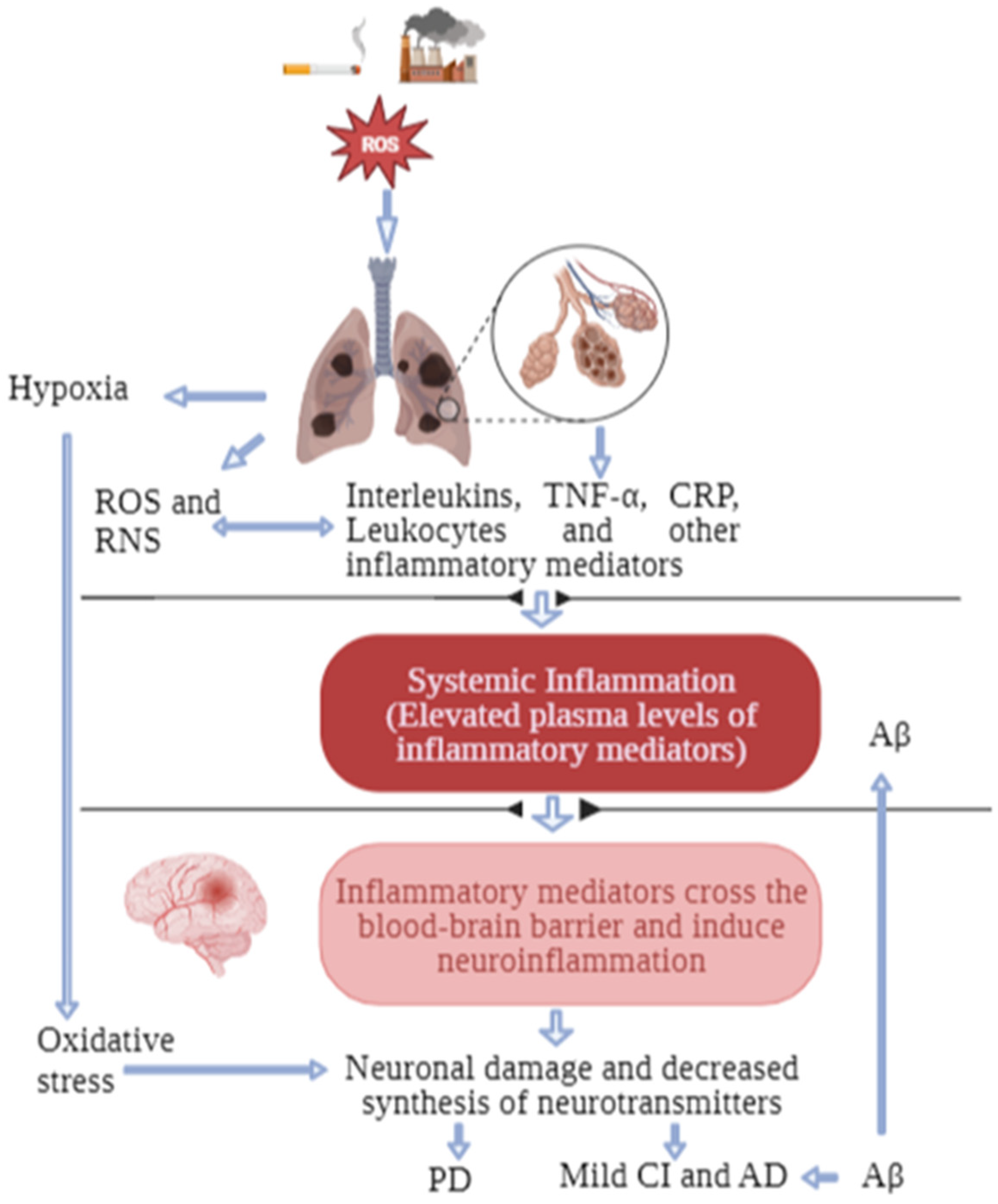
3. Possible Biological Links between COPD and Cognitive Impairment

The exact link between COPD and CI remains elusive. However, it has been theorised that the manifestation of CI in COPD patients may result from some shared mechanisms or risk factors between COPD and CI. These mechanisms may be related to inflammation, oxidative stress, amyloid-β (Aβ) accumulation, cigarette smoking, hypoxia, and gut dysbiosis.
COPD is characterised by systemic inflammation [7] (Figure 1), which is widely believed to originate from the leakage of inflammatory mediators from the lungs into the circulatory system [10]. Previous studies have shown that patients with COPD have higher levels of serum inflammatory cytokines when compared to healthy controls [8,53]. Furthermore, inflammatory cytokines, such as TNF-α, IL-1β, and IL-6, have been demonstrated to be associated with COPD severity [54,55]. On the other hand, systemic inflammation is also a critical pathophysiological phenomenon in CI [56]. Elevations in circulating levels of proinflammatory cytokines have been reported in patients with CI. A meta-analysis showed higher peripheral levels of IL-6, TNF-α, IL-1β, IL1-2, IL-18, and transforming growth factor (TGF)-β in patients with Alzheimer’s disease [57]. Studies have also shown a positive association between the concentration of high-sensitivity (hs)-CRP and mild CI [58] and between both CRP and IL-6 and a decline in cognitive abilities and executive functions [59]. Inflammatory cytokines associated with COPD can cross the BBB and induce neuroinflammation, triggering neuronal damage and a decline in neuronal functions [60]. Additionally, in the peripheral tissues, inflammatory cytokines, such as interferons, have been reported to increase the degradation of tryptophan via the kynurenine pathway, which may hinder the adequate transport of tryptophan to the brain for synthesising neurotransmitters that are crucial for cognition [10]. The link between inflammation and brain-related comorbidities in COPD patients has been well discussed [10,11]. However, studies on the relationship between inflammation and CI in COPD patients are generally scarce. A study reported a negative correlation between serum CRP levels and CI in COPD patients [61]. In another study, serum monocyte chemoattractant protein-1 was reported to negatively correlate with cognitive functions in patients with COPD and comorbid obstructive sleep apnoea-hypopnea syndrome [62]. Similarly, a recent study reported an increased serum Aβ level associated with disease severity in cognitively normal COPD patients compared to normal controls [63]. Furthermore, the Aβ levels were significantly higher in COPD patients with more highly elevated CRP and IL-6 levels [63]. This finding suggests that inflammation in COPD patients may trigger Alzheimer’s disease-related pathogenesis, which may further lead to cognitive decline in this population.
Cigarette smoking, the leading risk factor for COPD, could further increase the risk of CI in COPD patients via its influence on the BBB, leading to neuroinflammation. The BBB plays a crucial role in controlling the movement of molecules into and out of the brain. An impaired BBB will allow the entry of neurotoxic blood-derived debris, cells, and microbial pathogens to the brain, which are associated with several immune and inflammatory responses, leading to neurodegeneration [64]. In the case of COPD, inflammatory mediators can disrupt the integrity of the BBB, leading to cognitive dysfunction. Indeed, it has been demonstrated that the BBB is compromised in CI and Alzheimer’s disease [64,65] and may be strongly related to the severity of these diseases. In vitro studies have shown that the treatment of human BBB endothelial cells with cigarette-smoke extract disrupts the endothelial cells and decreases the expression of tight junction proteins, such as claudin-5, occludin, and ZO-1 [66,67]. Additionally, mice that were exposed to cigarette smoke and lipopolysaccharide (LPS) to induce COPD-like features were found to have reduced expression of claudin-5 and occludin in brain micro-vessels and increased microglial activation in the hippocampal region of the brain [68]. These findings suggest that cigarette smoke and LPS induce neuroinflammation by increasing the BBB permeability, partially owing to the oxidative stress caused by the free radicals in cigarette smoke [69]. Moreover, oxidants from cigarette-smoke exposure trigger an immune response, resulting in elevated levels of proinflammatory cytokines, e.g., TNF-α and IL-6 [70,71,72], which can directly impair the integrity and functions of the BBB. Furthermore, the weakened tight junctions allow the entry of innate immune cells from the systemic circulation through the BBB to the brain tissue, thus triggering inflammatory reactions and, ultimately, neuroinflammation [73]. Aβ deposition in the brain is a central mechanism in the pathogenesis of Alzheimer’s disease [74]. A recent case-control study discovered a correlation between cigarette smoking and elevated levels of Alzheimer’s disease risk indicators; elevated Aβ42 levels, increased oxidative-stress markers, neuroinflammation, and reduced neuroprotection in the cerebrospinal fluid (CSF) of participants who were active smokers [75].
Neuroinflammation pertains to localised inflammatory reactions occurring within the CNS, primarily driven by the release of cytokines, chemokines, and reactive oxygen species (ROS) from critical immune cells in the CNS, known as microglia and astrocytes [76,77]. Microglia, the resident immune cells in the white and grey matter of the brain, constitute roughly 10% of the CNS population [78]. Under ideal conditions, microglial cells are considered to be in a surveillance state, actively scanning the CNS for foreign materials, such as pathogens [79]. Upon encountering a stimulus or injury, microglia extend their processes toward the site of damage and eliminate foreign materials through phagocytosis [80]. Although acute microglial activation is beneficial under pathological conditions, prolonged activation of these cells can lead to detrimental effects, including neuronal damage [81] and impaired cognition. A meta-analysis of 28 studies consisting of participants with mild CI (n = 168), Alzheimer’s disease (n = 269), and healthy controls (n = 318) showed a significant increase in the levels of translocator protein (TSPO), indicating microglial activation [82], in the brains of subjects with mild CI and Alzheimer’s disease when compared with the brains of the healthy controls [83]. Moreover, elevated levels of TGF-β, chitinase-3-like 1, and monocyte chemoattractant protein-1 were reported in the CSF of patients with Alzheimer’s disease when compared to healthy controls [84]. Furthermore, increased immune activation, indicated by high levels of inflammatory mediators and activated microglia, was reported in the substantia nigra and striatum of patients with Parkinson’s disease [85,86]. To the best of our knowledge, whether neuroinflammation is associated with CI in COPD patients has not previously been studied. However, a preclinical study demonstrated significant microglial activation after the administration of the tobacco-specific procarcinogen, 4-N-methyl-N-nitrosamino-1-(3-pyridyl)-1-butanone, in BALB/c mice [87]. This finding corroborates a wealth of existing animal-model research indicating that exposure to cigarette smoke and e-cigarettes tends to provoke a proinflammatory response in the brain, often linked with microglial activation [87,88,89]. Furthermore, sustained nicotine administration in rodents was shown to lead to increased microglial activation in the nucleus accumbens, which diminished after acute nicotine withdrawal [90].
Hypoxia occurs when there is insufficient oxygen at the tissue level to maintain tissue homeostasis. One of the symptoms of COPD is difficulty in breathing, which is associated with inflammation in pulmonary airways and results in insufficient oxygen content in the blood (hypoxaemia) and, ultimately, hypoxia. Hypoxia has been shown to impair the synthesis of neurotransmitters in the brain, leading to changes in neuronal functioning and, eventually, CI [10]. Hypoxia may also induce CI via its stimulatory effect on oxidative stress and inflammation. Indeed, hypoxia has been shown to promote ROS production [91], which may trigger inflammation via the activation of NF-kβ [92]. One of the body’s responses to hypoxia is the upregulation of hypoxia-inducible factor (HIF)-1. Under hypoxic conditions, HIF-1 directs the limited oxygen supply in the brain to synthesise neurotransmitters [93]. HIF-1 stimulates dopamine production and the development of dopaminergic neurons [93] and protects dopaminergic neurons by regulating iron homeostasis and enhancing the resilience to oxidative stress and mitochondrial disruption [94,95]. However, individuals with COPD showed a diminished response to hypoxia, linked to lower histone deacetylase 7 (HDAC-7) and HIF-1α levels [96], which may trigger hypoxia-induced CI in COPD patients.
The relationship between hypoxia and CI in COPD patients has been widely studied; however, the findings have been inconsistent. Several studies have found a significant correlation between hypoxaemia and CI in COPD patients. Notably, low oxygen saturation in COPD patients was positively associated with an increased risk of CI (OR 5.45) [97]. Furthermore, the frequent use of oxygen therapy has significantly reduced the risk of CI in COPD patients [97]. In another study, Karamanli et al. found that long-term oxygen therapy-dependent (LTOTD) COPD patients demonstrated a significantly higher cognitive status when compared with non-LTOTOD COPD patients [98]. On the other hand, CI was reported in COPD patients with and without hypoxaemia [99,100]. Neuroimaging studies have revealed that hypoxemic COPD patients exhibit decreased hippocampal volume [101] and signs of cerebral perfusion [102,103] when compared to normal and non-hypoxemic COPD patients. Conversely, a study reported hippocampal shrinkage and significant decreases in white matter integrity and grey matter functional activation in stable COPD patients with no signs of hypoxaemia [27]. These mixed findings suggest that cognitive dysfunction in COPD patients is not solely attributable to hypoxaemia but a confounding influence of multiple biological mechanisms.
There is emerging evidence that the gut microbiota may also play a role in COPD-induced CI. The gut microbiota is the population of beneficial, non-pathogenic microbes that inhabit the digestive tracts of humans. In physiologically healthy individuals, the gut microbiota provides various advantages, including safeguarding and maintaining the gut and aiding in the absorption of nutrients [104]. It also offers protection against viral diseases [105]. Substantial evidence has shown that a well-diversified gut microbiota is crucial for maintaining good health [106]. The gut microbiota plays an essential role in the connection between the gut and the brain, as it releases metabolic byproducts and produces molecules that trigger physiological changes in the CNS [107]. The two-way communication between the gut microbiota and the brain is highly sensitive to alterations, and external stressors can shift the microbiota’s composition toward an unfavourable microbial community, a condition known as dysbiosis. Dysbiosis has been shown to increase the production of proinflammatory cytokines in the peripheral tissues and CNS [108]. Furthermore, dysbiosis has been indicated to regulate tryptophan availability via the kynurenine pathway, which may impair the optimal transport of tryptophan for synthesising serotonin and may lead to CI [109]. Indeed, gut dysbiosis has been linked to impaired brain functions in many brain diseases, such as Alzheimer’s disease [110], Parkinson’s disease [111], bipolar disorder [112], and major depressive disorder [113]. Recent studies have suggested that cigarette smoke leads to changes in gut dysbiosis in both humans and rodents [114], potentially offering a mechanism for cognitive decline in individuals with COPD. Additionally, Li et al. reported that the gut microbiome of COPD patients significantly varied from that of healthy controls and was characterised by a distinct overall microbial diversity and composition and reduced levels of short-chain fatty acids [115]. It was further demonstrated that compared with healthy controls, COPD patients exhibited 146 different bacterial species in their faecal samples, which were correlated with decreased lung function [116]. Of note, there is a lack of studies on the relationship between gut dysbiosis and CI in COPD patients. Therefore, well-designed epidemiological studies are needed to establish the association between gut dysbiosis and CI in COPD patients.

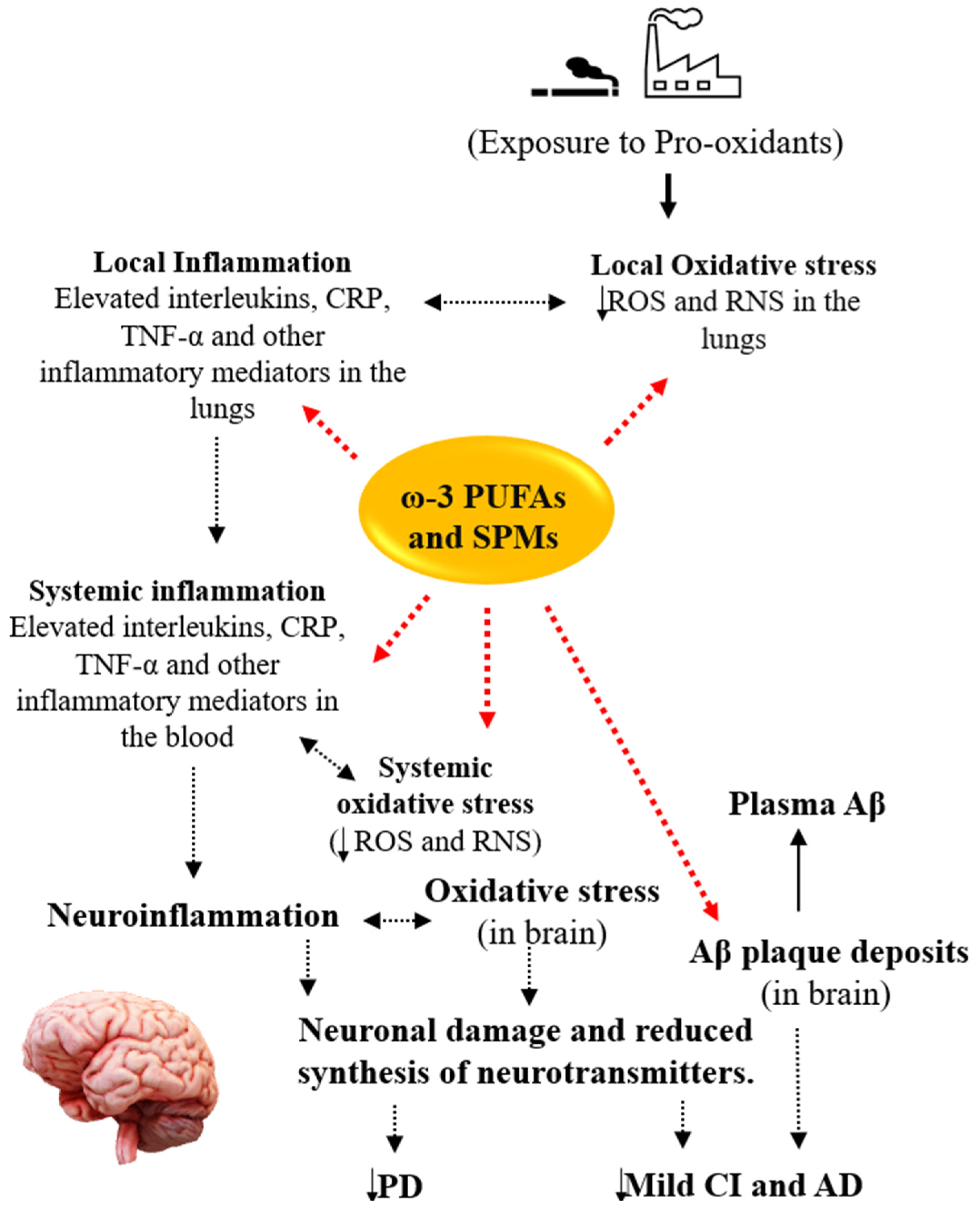
4. Potential Roles of ω-3 PUFAs in COPD and Comorbid Cognitive Impairment

4.1. Mild Cognitive Impairment and Alzheimer’s Disease

The evidence suggests that ω-3 PUFAs play a significant role in mental health. Notably, substantial evidence indicates that low ω-3 PUFA levels are associated with many psychiatric disorders, such as depression [117], attention-deficit hyperactivity disorder [118], bipolar disorder [119], and CI [120,121]. Indeed, elderly patients with mild CI and Alzheimer’s disease have notably lower levels of total ω-3 PUFAs and a lower ratio of ω-3 to ω-6 PUFAs when compared to healthy control groups [120]. Additionally, lower plasma levels of ω-3 PUFAs have been linked to poorer cognitive functioning in older adults with CI and Alzheimer’s disease [122]. Furthermore, higher serum levels of EPA have been associated with a reduced incidence of all-cause dementia (HR 0.76) and Alzheimer’s disease (HR 0.66) in the oldest adults [123]. Similarly, individuals with Alzheimer’s disease who had lower baseline levels of DHA were found to have a higher risk of cognitive decline when compared to individuals with higher baseline levels of DHA (OR 1.131) [124]. Furthermore, a longitudinal study of 899 individuals without dementia, at baseline, found that individuals in the highest quartile of plasma phosphatidylcholine DHA levels had a 47% lower likelihood of developing all-cause dementia over ten years [125]. These findings were further supported by a meta-analytic study of 10 studies, where lower plasma levels of ω-3 PUFAs were reported in patients with CI and Alzheimer’s disease when compared to healthy controls [121]. Indeed, ω-3 PUFA deficiency in patients with mild CI and Alzheimer’s disease may be attributable to the suboptimal dietary intake of these crucial nutrients, as studies have indicated that low consumption of ω-3 PUFAs is associated with an increased risk of mild CI and Alzheimer’s disease. For instance, a cohort study by Barberger-Gateau and colleagues involving 80,085 non-demented individuals above age 65 reported a decreased risk of Alzheimer’s disease with frequent intakes of fish and ω-3 PUFAs [126]. Similarly, high consumption of ω-3 PUFAs was linked to a decreased odds ratio of mild CI in a cohort of individuals without dementia [127]. Furthermore, a prospective study conducted between 1993 and 2000 found that the dietary intake of ω-3 PUFAs and weekly fish consumption were associated with a decreased incidence of Alzheimer’s disease [128]. Moreover, regular consumption of ω-3 PUFAs and seafood was reported to offer protection against cognitive decline in a longitudinal study [129]. A meta-analysis of 21 cohort studies further reported negative associations between the intake of fish products and the risks of CI and Alzheimer’s disease [130]. Whether ω-3 PUFA deficiency is associated with CI in COPD patients is currently unknown, as studies in this area are generally lacking, even though COPD is also characterised by a marked deficiency in ω-3 PUFAs [131]. Thus, studies are needed to assess the relationship between ω-3 PUFAs and CI in COPD patients.
The beneficial effects of ω-3 PUFAs on improving cognition have been widely reported in interventional studies. In a clinical trial, ω-3 PUFA supplementation attenuated cognitive decline in individuals with mild Alzheimer’s disease [132]. Additionally, ω-3 PUFAs were associated with significant improvements in short-term working memory, immediate verbal memory, and delayed recall in subjects with mild CI [133]. Bo and colleagues also found that supplementation with ω-3 PUFAs improved the cognitive functions of older adults with mild CI [134]. In another trial, supplementation with fish oil improved cognitive symptoms in older adults with subjective CI [135]. Meta-analytic studies have further supported the beneficial effects of ω-3 PUFAs on CI. Alex and colleagues reported mild but positive impacts of ω-3 PUFA supplementation on memory functions in older adults without dementia [136]. Another meta-analysis noted improved cognitive function in veterans with mild CI who were supplemented with long-chain ω-3 PUFAs when compared to those who received a placebo [137]. Furthermore, in another meta-analysis, DHA monotherapy or combined with EPA contributed to memory functions in older people with mild memory complaints [138].
Indeed, ω-3 PUFAs may benefit COPD individuals with CI through the ability of ω-3 PUFAs to assist in Aβ clearance, modulate inflammation, and boost the body’s antioxidant capacity (Figure 2). The potential of ω-3 PUFAs for clearing Aβ has been studied. In a study, ω-3 PUFAs were shown to significantly enhance the clearance of interstitial Aβ from the brain and protect against Aβ-induced injury [139]. Additionally, supplementation with ω-3 PUFAs has been found to promote Aβ clearance from the brain to the systemic circulation, as evidenced by reduced Aβ levels and fewer senile plaques in the brain parenchyma, along with a simultaneous increase in Aβ levels in the plasma of mouse models of Alzheimer’s disease [140]. Diets with a higher ratio of ω-6 to ω-3 PUFAs have been associated with increased Aβ levels in the brains of male transgenic mouse models of Alzheimer’s disease; however, this increase in Aβ levels was reversed by diets with a higher ratio of ω-3 to ω-6 PUFAs [141]. Moreover, DHA-enriched diets have been shown to significantly decrease the total Aβ and overall plaque burdens in the hippocampus and parietal cortex of the brains in transgenic mice [142]. Both DHA and EPA have been found to enhance the removal of Aβ in human microglial cells [143]. Maresin (MaR), a pro-resolving mediator derived from DHA, has been reported to inhibit the Aβ-induced increase in cytokine secretion and stimulate the uptake of Aβ in both monocyte-derived microglia and differentiated human monocyte cell lines [144]. Indeed, supplementation with ω-3 PUFAs enhanced Aβ phagocytosis by monocytes and increased resolvin D1 (RvD1) levels in patients with mild CI [145]. Moreover, ω-3 PUFAs improved Aβ macrophage-mediated phagocytosis in patients with mild CI [146].
ω-3 PUFAs could help to improve the cognitive functions of COPD patients via their modulatory effect on inflammation. Indeed, inflammation is one of the suggested links between COPD and CI. Notably, a study found that higher intakes of ALA were linked to lower levels of serum TNF-α in 250 stable COPD patients [147]. Conversely, increased intakes of proinflammatory arachidonic acid were associated with higher levels of serum IL-6 and CRP [147]. Another study revealed that supplementation with ω-3 PUFAs, along with lycopene and rosuvastatin, decreased plasma IL-6 levels and restored leukotriene B4 receptor gene expression to its initial levels in COPD patients [148]. Furthermore, COPD patients with cachexia showed lower IL-6, IL-8, and TNF-α levels after receiving high-doses of ω-3 PUFAs, vitamin D, and high-quality protein [149]. Sugawara and colleagues similarly observed lower serum levels of hs-CRP, IL-6, IL-8, and TNF-α in COPD patients after supplementation with a nutritional drink containing ω-3 PUFAs and vitamin A, in addition to engaging in low-intensity exercise [150]. Moreover, a recent meta-analysis reported a reduction in IL-6 levels among COPD patients who were supplemented with ω-3 PUFAs when compared to those who received a placebo [151]. Indeed, ω-3 PUFAs significantly inhibit the activity of NF-kβ, the master regulator of proinflammatory genes, which leads to the downregulation of IL1β and TNF-α, thus suppressing glial activation in APP/PS1 mice [140]. Similarly, MaR1 decreased the activity of Nf-kβ and chemokine secretion in human monocyte-derived microglia and human monocyte cell lines exposed to Aβ [144]. Furthermore, ω-3 PUFAs have been indicated to increase the production of brain-derived neurotrophic factors, decrease the production of proinflammatory cytokines, and induce anti-inflammatory microglial differentiation in human microglial cells [143]. Similarly, ω-3 PUFAs may help to resolve inflammation via their metabolites, called specialised pro-resolving mediators [152]. This evidence suggests that ω-3 PUFAs may improve the cognitive functions of COPD patients through the inhibition and resolution of inflammation.
In addition to their ability to promote Aβ clearance and modulate inflammatory processes, ω-3 PUFAs have demonstrated antioxidant properties. Indeed, a study has shown that treatment with ω-3 PUFAs remarkably attenuated increases in hippocampal malondialdehyde and 8-hydroxy-2′-deoxyguanosine levels as well as decreases in reduced glutathione (GSH) levels and the GSH-peroxidase activity induced by pentylenetetrazol kindling in young rat models [153]. Similarly, dose-dependent reductions in LPS-induced nitric oxide and ROS generation and inducible nitric oxide synthase expression have been reported in mice following treatment with krill-oil-derived ω-3 PUFAs [154]. Notably, the beneficial effect of ω-3 PUFAs on oxidative stress is related to their ability to enhance the expression of nuclear factor erythroid 2-related factor (Nrf-2), which is the master regulator of the antioxidant enzyme genes. Indeed, ω-3 PUFAs have been shown to improve rats’ antioxidant defence in astrocytes via the Nrf2-dependent mechanism, and this effect depends on the ratio of DHA/EPA that is incorporated into membrane phospholipids [155]. The activation of Nrf2 promotes the expression of key antioxidant enzymes, such as catalase, glutathione peroxidase, and superoxide dismutase, which increases the body’s resilience to oxidative stress [156]. A recent study has demonstrated that DHA directly activates Nrf2-signalling pathways, reducing the degree of oxidative damage caused by Aβ25–35 in PC12 cells [157]. In summary, owing to their antioxidant properties, ω-3 PUFAs may help to manage CI in COPD patients, thus mitigating the oxidative stress associated with COPD.
Despite the compelling evidence suggesting the promising potentials of ω-3 PUFAs in managing COPD comorbid with CI, interventional studies are warranted to test the efficacy of ω-3 PUFAs in this patient population. Meanwhile, ω-3 PUFA supplementation of up to 3.5 g per day in COPD patients is generally safe and well tolerated [149,158], and no severe adverse events associated with ω-3 PUFA supplementation have been reported [159,160]. On the other hand, the use of ω-3 PUFAs in managing CI in COPD patients may be limited by delayed therapeutic responses and suboptimal compliance. In particular, ω-3 PUFAs may take relatively longer than standard medications to bring about their intended effects. COPD is often linked with various additional health conditions. As a result, individuals with COPD are typically prescribed multiple medications to address these accompanying issues, leading to a high incidence of non-adherence to prescribed medications [161,162]. Furthermore, mood disorders, like anxiety and depression, are quite common in individuals with COPD and have been reported to impede adherence to medication regimens [163]. Therefore, we suggest conducting well-designed trials with sufficient follow-up periods (from several weeks to months) and appropriate measures to ensure compliance to assess the effectiveness of ω-3 PUFAs in managing COPD-related CI.

4.2. Parkinson’s Disease

Numerous studies have explored the relationship between ω-3 PUFAs and Parkinson’s disease, and the findings have been promising. A case-control study with a meta-analytic component reported a reverse correlation between Parkinson’s disease and the consumption of PUFAs, particularly ω-3 PUFAs or their precursor (ALA) [164]. Additionally, higher intakes of ω-3 PUFAs were associated with a reduced risk of Parkinson’s disease in a three-decade prospective cohort study [165]. Moreover, a 6-year study involving 5289 individuals found a significant link between the consumption of ω-3 PUFAs and a lower incidence of Parkinson’s disease [166]. A 16-year follow-up study with 131,368 participants attributed a lower risk of Parkinson’s disease to high intakes of fish, poultry, fruits, and vegetables [167]. Because all the above evidence stems from observational studies, a direct cause-and-effect relationship between the consumption of ω-3 PUFAs and the risk of Parkinson’s disease should be approached cautiously. Nevertheless, the evidence suggesting a lower incidence of Parkinson’s disease associated with ω-3 PUFA consumption is intriguing.
Currently, no clinical trials are specifically focused on ω-3 PUFA monotherapy for Parkinson’s disease, and those involving combination therapy are limited. However, the results from these studies are encouraging. In one clinical trial, Parkinson’s disease patients randomly assigned to the supplement group (containing ω-3 PUFAs, ω-6 PUFAs, and antioxidants) exhibited a delayed progression of Parkinson’s disease, as assessed by the Unified Parkinson’s Disease Rating Scale (UPRDS), after a 30-month follow-up [168]. Furthermore, Parkinson’s disease patients treated with ω-3 PUFAs (1000 mg/day) and vitamin E (400 IU) for 12 weeks reported improved UPDRS scores and lower levels of hs-CRP when compared to the placebo group [169]. Additionally, a 3-month daily supplementation with ω-3 PUFAs (1000 mg) and vitamin E (400 IU) led to an upregulation of the expression of the peroxisome proliferator-activated receptor (PPAR)-gamma gene and a downregulation of TNF-α gene expression in peripheral blood mononuclear cells of Parkinson’s disease patients [170]. In summary, the beneficial effects of ω-3 PUFAs on the progression of Parkinson’s disease, as indicated by low UPRDS scores and hs-CRP levels, have been partly attributed to the ability of ω-3 PUFAs to modulate inflammatory processes (see Figure 2). Indeed, ω-3 PUFAs have been shown to regulate inflammatory pathways by modifying the composition of cell membrane phospholipids, disrupting lipid rafts, suppressing the synthesis of eicosanoids from arachidonic acid, inhibiting the activation of NF-kβ, and activating PPAR-γ [171,172,173,174]. Despite the proven benefits of ω-3 PUFAs in managing Parkinson’s disease, studies on the effects of ω-3 PUFAs on comorbid Parkinson’s disease in COPD patients do not currently exist, to the best of our knowledge. Thus, studies are needed in this regard.

5. Conclusions and Future Prospects

COPD is associated with CI, leading to poor clinical outcomes, reduced compliance with treatment protocols, decreased quality of life, and increased mortality among the COPD populations. Despite its devastating effects on COPD patients, CI receives little or no attention in COPD management. Important mechanisms that could predispose COPD patients to CI include hypoxia, oxidative stress, inflammation, cigarette smoking, gut dysbiosis, and Aβ deposition in the brain. Interestingly, ω-3 PUFAs and their metabolites have been proven to modulate inflammatory pathways, activate antioxidant enzymes, and promote Aβ clearance from the brain. However, no evidence indicates a connection between CI in COPD patients and a deficiency in ω-3 PUFAs. Additionally, there is a lack of studies regarding the potential therapeutic benefits of ω-3 PUFAs in managing CI in COPD patients. This gap in research is partly because CI is a neglected issue in managing COPD, with only a small portion of COPD patients receiving treatment for CI. Therefore, studies are needed to investigate the roles of ω-3 PUFAs in CI associated with COPD. The outcomes of these studies will aid in designing interventional studies to assess the impact of these promising nutritional supplements on improving CI in the COPD population. These trials should also determine the appropriate dosage and formulation of ω-3 PUFAs necessary to enhance the cognitive functions of COPD patients. We hypothesise that ω-3 PUFAs will improve the executive functions of patients with COPD. This enhancement will enable COPD patients to effectively adhere to the protocols for managing their condition, resulting in improved clinical outcomes. However, as COPD is associated with several comorbidities, such as cardiovascular disorders, diabetes, hypertension, and mood disorders, managing these comorbidities along with COPD will result in a better quality of life for COPD patients.

Author Contributions

Conceptualisation, H.Z., J.P.-C.C. and W.-C.L.; writing—original draft preparation, H.Z.; writing—review and editing, J.P.-C.C., S.K.S., W.-C.L., S.-Y.H., P.G., Y.-T.H. and K.-P.S. All authors have read and agreed to the published version of the manuscript.

Funding

The authors of this work were supported by the following grants: NSTC 109-2320-B-038-057-MY3, 110-2321-B-006-004, 110-2811-B-039-507, 110-2320-B-039-048-MY2, 110-2320-B-039-047-MY3, 110-2813-C-039-327-B, 110-2314-B-039-029-MY3, 111-2321-B-006-008, 111-2314-B-039-041-MY3, and 113-2923-B-039-001-MY3 from the National Science and Technology Council (NSTC), Taiwan; ANHRF 109-31, 109-40, 110-13, 110-26, 110-44, 110-45, 111-27, 111-28, 111-47, 111-48, and 111-52 from An-Nan Hospital, China Medical University, Tainan, Taiwan; CMRC-CMA-2 from Higher Education Sprout Project by the Ministry of Education (MOE), Taiwan; CMU 110-AWARD-02, 110-N-17, 1110-SR-73 from the China Medical University, Taichung, Taiwan; and DMR-105-053, 106-101, 106-227, 109-102, 109-244, 110-124, 111-245, 112-097, 112-086, 112-109, 112-232 and DMR-HHC-109-11, HHC-109-12, HHC-110-10, and HHC-111-8 from the China Medical University Hospital, Taichung, Taiwan.

Data Availability Statement

Data sharing is not applicable to this article as no new data were created or analyzed in this study.

Conflicts of Interest

The authors declare no conflict of interest.

Abbreviations

Amyloid-beta
ALAAlpha-linolenic acid
BBBBlood–brain barrier
CICognitive impairment
COPDChronic obstructive pulmonary disease
CRPC-reactive protein
CSFCerebrospinal fluid
DHADocosahexaenoic acid
EPAEicosapentaenoic acid
GSHReduced glutathione
HIF-1Hypoxia-inducible factor-1
HRQoLHealth-related quality of life
hs-CRPHigh-sensitivity C-reactive protein
IFN-γInterferon-gamma
ILInterleukin
L-DOPALevodopa
LPSLipopolysaccharide
MaRMaresin
NF-kβNuclear factor kappa-beta
Nrf-2Nuclear factor erythroid 2-related factor
PPAR-γPeroxisome proliferator-activated receptor-gamma
PRPulmonary rehabilitation
ROSReactive oxygen species
RvDResolvin D
TGF-βTransforming growth factor-beta
TNF-αTumour necrosis factor-alpha
TSPOTranslocator protein
UPDRSUnified Parkinson Disease Rating Scale
ω-3 PUFAOmega-3 polyunsaturated fatty acid

References

  1. Christenson, S.A.; Smith, B.M.; Bafadhel, M.; Putcha, N. Chronic obstructive pulmonary disease. Lancet 2022, 399, 2227–2242. [Google Scholar] [CrossRef]
  2. Adeloye, D.; Chua, S.; Lee, C.; Basquill, C.; Papana, A.; Theodoratou, E.; Nair, H.; Gasevic, D.; Sridhar, D.; Campbell, H.; et al. Global and regional estimates of COPD prevalence: Systematic review and meta-analysis. J. Glob. Health 2015, 5, 020415. [Google Scholar] [CrossRef]
  3. Iheanacho, I.; Zhang, S.; King, D.; Rizzo, M.; Ismaila, A.S. Economic Burden of Chronic Obstructive Pulmonary Disease (COPD): A Systematic Literature Review. Int. J. Chron. Obs. Pulmon Dis. 2020, 15, 439–460. [Google Scholar] [CrossRef]
  4. Adeloye, D.; Song, P.; Zhu, Y.; Campbell, H.; Sheikh, A.; Rudan, I. Global, regional, and national prevalence of, and risk factors for, chronic obstructive pulmonary disease (COPD) in 2019: A systematic review and modelling analysis. Lancet Respir. Med. 2022, 10, 447–458. [Google Scholar] [CrossRef]
  5. Hattab, Y.; Alhassan, S.; Balaan, M.; Lega, M.; Singh, A.C. Chronic Obstructive Pulmonary Disease. Crit. Care Nurs. Q. 2016, 39, 124–130. [Google Scholar] [CrossRef]
  6. Wedzicha, J.A.; Seemungal, T.A. COPD exacerbations: Defining their cause and prevention. Lancet 2007, 370, 786–796. [Google Scholar] [CrossRef]
  7. Barnes, P.J. Inflammatory mechanisms in patients with chronic obstructive pulmonary disease. J. Allergy Clin. Immunol. 2016, 138, 16–27. [Google Scholar] [CrossRef]
  8. Su, B.; Liu, T.; Fan, H.; Chen, F.; Ding, H.; Wu, Z.; Wang, H.; Hou, S. Inflammatory Markers and the Risk of Chronic Obstructive Pulmonary Disease: A Systematic Review and Meta-Analysis. PLoS ONE 2016, 11, e0150586. [Google Scholar] [CrossRef]
  9. Gan, W.Q.; Man, S.F.; Senthilselvan, A.; Sin, D.D. Association between chronic obstructive pulmonary disease and systemic inflammation: A systematic review and a meta-analysis. Thorax 2004, 59, 574–580. [Google Scholar] [CrossRef]
  10. Zailani, H.; Satyanarayanan, S.K.; Liao, W.-C.; Liao, H.-F.; Huang, S.-Y.; Gałecki, P.; Su, K.-P.; Chang, J.P.-C. Omega-3 Polyunsaturated Fatty Acids in Managing Comorbid Mood Disorders in Chronic Obstructive Pulmonary Disease (COPD): A Review. J. Clin. Med. 2023, 12, 2653. [Google Scholar]
  11. Pelgrim, C.E.; Peterson, J.D.; Gosker, H.R.; Schols, A.; van Helvoort, A.; Garssen, J.; Folkerts, G.; Kraneveld, A.D. Psychological co-morbidities in COPD: Targeting systemic inflammation, a benefit for both? Eur. J. Pharmacol. 2019, 842, 99–110. [Google Scholar] [CrossRef]
  12. Sun, Y.; Koyama, Y.; Shimada, S. Inflammation From Peripheral Organs to the Brain: How Does Systemic Inflammation Cause Neuroinflammation? Front. Aging Neurosci. 2022, 14, 903455. [Google Scholar] [CrossRef]
  13. Allison, D.J.; Ditor, D.S. The common inflammatory etiology of depression and cognitive impairment: A therapeutic target. J. Neuroinflammation 2014, 11, 151. [Google Scholar] [CrossRef]
  14. Kumar, G.K. Hypoxia. 3. Hypoxia and neurotransmitter synthesis. Am. J. Physiol. Cell Physiol. 2011, 300, C743–C751. [Google Scholar] [CrossRef]
  15. Gonçalves, S.; Nunes-Costa, D.; Cardoso, S.M.; Empadinhas, N.; Marugg, J.D. Enzyme Promiscuity in Serotonin Biosynthesis, From Bacteria to Plants and Humans. Front. Microbiol. 2022, 13, 873555. [Google Scholar] [CrossRef]
  16. Chang, S.S.; Chen, S.; McAvay, G.J.; Tinetti, M.E. Effect of coexisting chronic obstructive pulmonary disease and cognitive impairment on health outcomes in older adults. J. Am. Geriatr. Soc. 2012, 60, 1839–1846. [Google Scholar] [CrossRef]
  17. Baird, C.; Lovell, J.; Johnson, M.; Shiell, K.; Ibrahim, J.E. The impact of cognitive impairment on self-management in chronic obstructive pulmonary disease: A systematic review. Respir. Med. 2017, 129, 130–139. [Google Scholar] [CrossRef]
  18. Marx, W.; Lane, M.; Hockey, M.; Aslam, H.; Berk, M.; Walder, K.; Borsini, A.; Firth, J.; Pariante, C.M.; Berding, K.; et al. Diet and depression: Exploring the biological mechanisms of action. Mol. Psychiatry 2021, 26, 134–150. [Google Scholar] [CrossRef]
  19. Chang, J.P.; Su, K.P. Nutrition and immunology in mental health: Precision medicine and integrative approaches to address unmet clinical needs in psychiatric treatments. Brain Behav. Immun. 2020, 85, 1–3. [Google Scholar] [CrossRef]
  20. Kaur, N.; Chugh, V.; Gupta, A.K. Essential fatty acids as functional components of foods- a review. J. Food Sci. Technol. 2014, 51, 2289–2303. [Google Scholar] [CrossRef]
  21. Saini, R.K.; Prasad, P.; Sreedhar, R.V.; Akhilender Naidu, K.; Shang, X.; Keum, Y.S. Omega-3 Polyunsaturated Fatty Acids (PUFAs): Emerging Plant and Microbial Sources, Oxidative Stability, Bioavailability, and Health Benefits—A Review. Antioxidants 2021, 10, 1627. [Google Scholar] [CrossRef]
  22. Mozaffarian, D.; Wu, J.H. Omega-3 fatty acids and cardiovascular disease: Effects on risk factors, molecular pathways, and clinical events. J. Am. Coll. Cardiol. 2011, 58, 2047–2067. [Google Scholar] [CrossRef]
  23. Hibbeln, J.R. Fish consumption and major depression. Lancet 1998, 351, 1213. [Google Scholar] [CrossRef]
  24. Su, K.P.; Matsuoka, Y.; Pae, C.U. Omega-3 Polyunsaturated Fatty Acids in Prevention of Mood and Anxiety Disorders. Clin. Psychopharmacol. Neurosci. 2015, 13, 129–137. [Google Scholar] [CrossRef]
  25. Avallone, R.; Vitale, G.; Bertolotti, M. Omega-3 Fatty Acids and Neurodegenerative Diseases: New Evidence in Clinical Trials. Int. J. Mol. Sci. 2019, 20, 4256. [Google Scholar] [CrossRef]
  26. Wei, B.Z.; Li, L.; Dong, C.W.; Tan, C.C.; Xu, W. The Relationship of Omega-3 Fatty Acids with Dementia and Cognitive Decline: Evidence from Prospective Cohort Studies of Supplementation, Dietary Intake, and Blood Markers. Am. J. Clin. Nutr. 2023, 117, 1096–1109. [Google Scholar] [CrossRef]
  27. Dodd, J.W.; Chung, A.W.; van den Broek, M.D.; Barrick, T.R.; Charlton, R.A.; Jones, P.W. Brain structure and function in chronic obstructive pulmonary disease: A multimodal cranial magnetic resonance imaging study. Am. J. Respir. Crit. Care Med. 2012, 186, 240–245. [Google Scholar]
  28. Yohannes, A.M.; Chen, W.; Moga, A.M.; Leroi, I.; Connolly, M.J. Cognitive Impairment in Chronic Obstructive Pulmonary Disease and Chronic Heart Failure: A Systematic Review and Meta-analysis of Observational Studies. J. Am. Med. Dir. Assoc. 2017, 18, e451.e1–e451.e11. [Google Scholar] [CrossRef]
  29. Zhang, X.; Cai, X.; Shi, X.; Zheng, Z.; Zhang, A.; Guo, J.; Fang, Y. Chronic Obstructive Pulmonary Disease as a Risk Factor for Cognitive Dysfunction: A Meta-Analysis of Current Studies. J. Alzheimers Dis. 2016, 52, 101–111. [Google Scholar] [CrossRef]
  30. Siraj, R.A.; McKeever, T.M.; Gibson, J.E.; Gordon, A.L.; Bolton, C.E. Risk of incident dementia and cognitive impairment in patients with chronic obstructive pulmonary disease (COPD): A large UK population-based study. Respir. Med. 2021, 177, 106288. [Google Scholar] [CrossRef]
  31. Villeneuve, S.; Pepin, V.; Rahayel, S.; Bertrand, J.A.; de Lorimier, M.; Rizk, A.; Desjardins, C.; Parenteau, S.; Beaucage, F.; Joncas, S.; et al. Mild cognitive impairment in moderate to severe COPD: A preliminary study. Chest 2012, 142, 1516–1523. [Google Scholar] [CrossRef]
  32. De Carolis, A.; Giubilei, F.; Caselli, G.; Casolla, B.; Cavallari, M.; Vanacore, N.; Leonori, R.; Scrocchia, I.; Fersini, A.; Quercia, A.; et al. Chronic obstructive pulmonary disease is associated with altered neuropsychological performance in young adults. Dement. Geriatr. Cogn. Dis. Extra 2011, 1, 402–408. [Google Scholar] [CrossRef]
  33. Liao, K.M.; Ho, C.H.; Ko, S.C.; Li, C.Y. Increased Risk of Dementia in Patients With Chronic Obstructive Pulmonary Disease. Medicine 2015, 94, e930. [Google Scholar] [CrossRef]
  34. Liao, W.C.; Lin, C.L.; Chang, S.N.; Tu, C.Y.; Kao, C.H. The association between chronic obstructive pulmonary disease and dementia: A population-based retrospective cohort study. Eur. J. Neurol. 2015, 22, 334–340. [Google Scholar] [CrossRef]
  35. Yeh, J.J.; Wei, Y.F.; Lin, C.L.; Hsu, W.H. Effect of the asthma-chronic obstructive pulmonary disease syndrome on the stroke, Parkinson’s disease, and dementia: A national cohort study. Oncotarget 2018, 9, 12418–12431. [Google Scholar] [CrossRef]
  36. Rusanen, M.; Ngandu, T.; Laatikainen, T.; Tuomilehto, J.; Soininen, H.; Kivipelto, M. Chronic obstructive pulmonary disease and asthma and the risk of mild cognitive impairment and dementia: A population based CAIDE study. Curr. Alzheimer Res. 2013, 10, 549–555. [Google Scholar] [CrossRef]
  37. Cleutjens, F.; Spruit, M.A.; Ponds, R.; Vanfleteren, L.; Franssen, F.M.E.; Dijkstra, J.B.; Gijsen, C.; Wouters, E.F.M.; Janssen, D.J.A. The Impact of Cognitive Impairment on Efficacy of Pulmonary Rehabilitation in Patients With COPD. J. Am. Med. Dir. Assoc. 2017, 18, 420–426. [Google Scholar] [CrossRef]
  38. Lindbergh, C.A.; Dishman, R.K.; Miller, L.S. Functional Disability in Mild Cognitive Impairment: A Systematic Review and Meta-Analysis. Neuropsychol. Rev. 2016, 26, 129–159. [Google Scholar] [CrossRef]
  39. Turan, O.; Turan, P.A.; Mirici, A. Parameters affecting inhalation therapy adherence in elderly patients with chronic obstructive lung disease and asthma. Geriatr. Gerontol. Int. 2017, 17, 999–1005. [Google Scholar] [CrossRef]
  40. Liao, K.M.; Lin, T.C.; Li, C.Y.; Yang, Y.K. Dementia Increases Severe Sepsis and Mortality in Hospitalized Patients With Chronic Obstructive Pulmonary Disease. Medicine 2015, 94, e967. [Google Scholar] [CrossRef]
  41. Wang, M.-T.; Tsai, C.-L.; Lin, C.W.; Yeh, C.-B.; Wang, Y.-H.; Lin, H.-L. Association Between Antipsychotic Agents and Risk of Acute Respiratory Failure in Patients With Chronic Obstructive Pulmonary Disease. JAMA Psychiatry 2017, 74, 252–260. [Google Scholar] [CrossRef]
  42. Bloem, B.R.; Okun, M.S.; Klein, C. Parkinson’s disease. Lancet 2021, 397, 2284–2303. [Google Scholar] [CrossRef]
  43. Li, C.H.; Chen, W.C.; Liao, W.C.; Tu, C.Y.; Lin, C.L.; Sung, F.C.; Chen, C.H.; Hsu, W.H. The association between chronic obstructive pulmonary disease and Parkinson’s disease: A nationwide population-based retrospective cohort study. QJM Int. J. Med. 2015, 108, 39–45. [Google Scholar] [CrossRef]
  44. Gallagher, D.A.; Schrag, A. Psychosis, apathy, depression and anxiety in Parkinson’s disease. Neurobiol. Dis. 2012, 46, 581–589. [Google Scholar] [CrossRef]
  45. Rihmer, Z.; Gonda, X.; Döme, P. Depression in Parkinson’s disease. Ideggyogy. Sz. 2014, 67, 229–236. [Google Scholar]
  46. Yohannes, A.M.; Alexopoulos, G.S. Pharmacological treatment of depression in older patients with chronic obstructive pulmonary disease: Impact on the course of the disease and health outcomes. Drugs Aging 2014, 31, 483–492. [Google Scholar] [CrossRef]
  47. Coventry, P.A.; Gemmell, I.; Todd, C.J. Psychosocial risk factors for hospital readmission in COPD patients on early discharge services: A cohort study. BMC Pulm. Med. 2011, 11, 49. [Google Scholar] [CrossRef]
  48. Ng, T.P.; Niti, M.; Tan, W.C.; Cao, Z.; Ong, K.C.; Eng, P. Depressive symptoms and chronic obstructive pulmonary disease: Effect on mortality, hospital readmission, symptom burden, functional status, and quality of life. Arch. Intern. Med. 2007, 167, 60–67. [Google Scholar] [CrossRef]
  49. Recio Iglesias, J.; Díez-Manglano, J.; López García, F.; Díaz Peromingo, J.A.; Almagro, P.; Varela Aguilar, J.M. Management of the COPD Patient with Comorbidities: An Experts Recommendation Document. Int. J. Chron. Obs. Pulmon Dis. 2020, 15, 1015–1037. [Google Scholar] [CrossRef]
  50. Stoker, T.B.; Barker, R.A. Recent developments in the treatment of Parkinson’s Disease. F1000Res 2020, 9, 862. [Google Scholar] [CrossRef]
  51. Monteiro, L.; Souza-Machado, A.; Valderramas, S.; Melo, A. The Effect of Levodopa on Pulmonary Function in Parkinson’s Disease: A Systematic Review and Meta-Analysis. Clin. Ther. 2012, 34, 1049–1055. [Google Scholar] [CrossRef]
  52. Ko, P.W.; Kang, K.; Lee, H.W. Levodopa-induced respiratory dysfunction confirmed by levodopa challenge test: A case report. Medicine 2018, 97, e12488. [Google Scholar] [CrossRef]
  53. Chen, J.; Li, X.; Huang, C.; Lin, Y.; Dai, Q. Change of Serum Inflammatory Cytokines Levels in Patients With Chronic Obstructive Pulmonary Disease, Pneumonia and Lung Cancer. Technol. Cancer Res. Treat. 2020, 19, 1533033820951807. [Google Scholar] [CrossRef]
  54. MacNee, W. Systemic inflammatory biomarkers and co-morbidities of chronic obstructive pulmonary disease. Ann. Med. 2013, 45, 291–300. [Google Scholar] [CrossRef]
  55. Verma, S.K.; Singh, S.; Kumar, S.; Ahmad, M.; Waseem, M.; Singh, S.; Nischal, A.; Dixit, R. Association between serum cytokine levels and severity of chronic obstructive pulmonary disease in Northern India. Int. J. Innov. Sci. Res. 2015, 18, 357–361. [Google Scholar]
  56. Gorelick, P.B. Role of inflammation in cognitive impairment: Results of observational epidemiological studies and clinical trials. Ann. N. Y. Acad. Sci. 2010, 1207, 155–162. [Google Scholar] [CrossRef]
  57. Swardfager, W.; Lanctôt, K.; Rothenburg, L.; Wong, A.; Cappell, J.; Herrmann, N. A meta-analysis of cytokines in Alzheimer’s disease. Biol. Psychiatry 2010, 68, 930–941. [Google Scholar] [CrossRef]
  58. Pan, H.; Huang, X.; Li, F.; Ren, M.; Zhang, J.; Xu, M.; Wu, M. Association among plasma lactate, systemic inflammation, and mild cognitive impairment: A community-based study. Neurol. Sci. 2019, 40, 1667–1673. [Google Scholar] [CrossRef]
  59. Schram, M.T.; Euser, S.M.; De Craen, A.J.; Witteman, J.C.; Frölich, M.; Hofman, A.; Jolles, J.; Breteler, M.M.; Westendorp, R.G. Systemic markers of inflammation and cognitive decline in old age. J. Am. Geriatr. Soc. 2007, 55, 708–716. [Google Scholar]
  60. Kempuraj, D.; Thangavel, R.; Natteru, P.A.; Selvakumar, G.P.; Saeed, D.; Zahoor, H.; Zaheer, S.; Iyer, S.S.; Zaheer, A. Neuroinflammation Induces Neurodegeneration. J. Neurol. Neurosurg. Spine 2016, 1, 1003. [Google Scholar]
  61. Crişan, A.F.; Oancea, C.; Timar, B.; Fira-Mladinescu, O.; Crişan, A.; Tudorache, V. Cognitive impairment in chronic obstructive pulmonary disease. PLoS ONE 2014, 9, e102468. [Google Scholar]
  62. Ge, Y.L.; Liu, C.H.; Fu, A.S.; Wang, H.Y. Correlation study between the levels of serum MCP-1,SAA and cognitive function in patients with COPD-OSAHS. J. Clin. Otorhinolaryngol. Head Neck Surg. 2018, 32, 485–488. [Google Scholar] [CrossRef]
  63. Bu, X.L.; Cao, G.Q.; Shen, L.L.; Xiang, Y.; Jiao, S.S.; Liu, Y.H.; Zhu, C.; Zeng, F.; Wang, Q.H.; Wang, Y.R.; et al. Serum Amyloid-Beta Levels are Increased in Patients with Chronic Obstructive Pulmonary Disease. Neurotox. Res. 2015, 28, 346–351. [Google Scholar] [CrossRef]
  64. Sweeney, M.D.; Sagare, A.P.; Zlokovic, B.V. Blood–brain barrier breakdown in Alzheimer disease and other neurodegenerative disorders. Nat. Rev. Neurol. 2018, 14, 133–150. [Google Scholar] [CrossRef]
  65. van de Haar, H.J.; Burgmans, S.; Jansen, J.F.; van Osch, M.J.; van Buchem, M.A.; Muller, M.; Hofman, P.A.; Verhey, F.R.; Backes, W.H. Blood-Brain Barrier Leakage in Patients with Early Alzheimer Disease. Radiology 2016, 281, 527–535. [Google Scholar] [CrossRef]
  66. Prasad, S.; Sajja, R.K.; Park, J.H.; Naik, P.; Kaisar, M.A.; Cucullo, L. Impact of cigarette smoke extract and hyperglycemic conditions on blood–brain barrier endothelial cells. Fluids Barriers CNS 2015, 12, 18. [Google Scholar] [CrossRef]
  67. Hossain, M.; Sathe, T.; Fazio, V.; Mazzone, P.; Weksler, B.; Janigro, D.; Rapp, E.; Cucullo, L. Tobacco smoke: A critical etiological factor for vascular impairment at the blood–brain barrier. Brain Res. 2009, 1287, 192–205. [Google Scholar] [CrossRef]
  68. Pelgrim, C.E.; Wang, L.; Peralta Marzal, L.N.; Korver, S.; van Ark, I.; Leusink-Muis, T.; Braber, S.; Folkerts, G.; Garssen, J.; van Helvoort, A.; et al. Increased exploration and hyperlocomotion in a cigarette smoke and LPS-induced murine model of COPD: Linking pulmonary and systemic inflammation with the brain. Am. J. Physiol. Lung Cell Mol. Physiol. 2022, 323, L251–L265. [Google Scholar] [CrossRef]
  69. Dobric, A.; De Luca, S.N.; Spencer, S.J.; Bozinovski, S.; Saling, M.M.; McDonald, C.F.; Vlahos, R. Novel pharmacological strategies to treat cognitive dysfunction in chronic obstructive pulmonary disease. Pharmacol. Ther. 2022, 233, 108017. [Google Scholar] [CrossRef]
  70. Oostwoud, L.; Gunasinghe, P.; Seow, H.J.; Ye, J.; Selemidis, S.; Bozinovski, S.; Vlahos, R. Apocynin and ebselen reduce influenza A virus-induced lung inflammation in cigarette smoke-exposed mice. Sci. Rep. 2016, 6, 20983. [Google Scholar]
  71. Strzelak, A.; Ratajczak, A.; Adamiec, A.; Feleszko, W. Tobacco Smoke Induces and Alters Immune Responses in the Lung Triggering Inflammation, Allergy, Asthma and Other Lung Diseases: A Mechanistic Review. Int. J. Environ. Res. Public. Health 2018, 15, 1033. [Google Scholar] [CrossRef]
  72. Geng, J.; Wang, L.; Zhang, L.; Qin, C.; Song, Y.; Ma, Y.; Chen, Y.; Chen, S.; Wang, Y.; Zhang, Z. Blood-brain barrier disruption induced cognitive impairment is associated with increase of inflammatory cytokine. Front. Aging Neurosci. 2018, 10, 129. [Google Scholar]
  73. Huang, X.; Hussain, B.; Chang, J. Peripheral inflammation and blood-brain barrier disruption: Effects and mechanisms. CNS Neurosci. Ther. 2021, 27, 36–47. [Google Scholar] [CrossRef]
  74. Sadigh-Eteghad, S.; Sabermarouf, B.; Majdi, A.; Talebi, M.; Farhoudi, M.; Mahmoudi, J. Amyloid-beta: A crucial factor in Alzheimer’s disease. Med. Princ. Pract. 2015, 24, 1–10. [Google Scholar] [CrossRef]
  75. Liu, Y.; Li, H.; Wang, J.; Xue, Q.; Yang, X.; Kang, Y.; Li, M.; Xu, J.; Li, G.; Li, C.; et al. Association of Cigarette Smoking With Cerebrospinal Fluid Biomarkers of Neurodegeneration, Neuroinflammation, and Oxidation. JAMA Netw. Open 2020, 3, e2018777. [Google Scholar] [CrossRef]
  76. Simpson, D.S.A.; Oliver, P.L. ROS Generation in Microglia: Understanding Oxidative Stress and Inflammation in Neurodegenerative Disease. Antioxidants 2020, 9, 743. [Google Scholar]
  77. Shao, F.; Wang, X.; Wu, H.; Wu, Q.; Zhang, J. Microglia and Neuroinflammation: Crucial Pathological Mechanisms in Traumatic Brain Injury-Induced Neurodegeneration. Front. Aging Neurosci. 2022, 14, 825086. [Google Scholar] [CrossRef]
  78. Ochocka, N.; Kaminska, B. Microglia Diversity in Healthy and Diseased Brain: Insights from Single-Cell Omics. Int. J. Mol. Sci. 2021, 22, 3027. [Google Scholar] [CrossRef]
  79. Arcuri, C.; Mecca, C.; Bianchi, R.; Giambanco, I.; Donato, R. The Pathophysiological Role of Microglia in Dynamic Surveillance, Phagocytosis and Structural Remodeling of the Developing CNS. Front. Mol. Neurosci. 2017, 10, 191. [Google Scholar] [CrossRef]
  80. Yu, F.; Wang, Y.; Stetler, A.R.; Leak, R.K.; Hu, X.; Chen, J. Phagocytic microglia and macrophages in brain injury and repair. CNS Neurosci. Ther. 2022, 28, 1279–1293. [Google Scholar] [CrossRef]
  81. Muzio, L.; Viotti, A.; Martino, G. Microglia in Neuroinflammation and Neurodegeneration: From Understanding to Therapy. Front. Neurosci. 2021, 15, 742065. [Google Scholar] [CrossRef]
  82. Yao, R.; Pan, R.; Shang, C.; Li, X.; Cheng, J.; Xu, J.; Li, Y. Translocator Protein 18 kDa (TSPO) Deficiency Inhibits Microglial Activation and Impairs Mitochondrial Function. Front. Pharmacol. 2020, 11, 986. [Google Scholar] [CrossRef]
  83. Bradburn, S.; Murgatroyd, C.; Ray, N. Neuroinflammation in mild cognitive impairment and Alzheimer’s disease: A meta-analysis. Ageing Res. Rev. 2019, 50, 1–8. [Google Scholar] [CrossRef]
  84. Chen, X.; Hu, Y.; Cao, Z.; Liu, Q.; Cheng, Y. Cerebrospinal Fluid Inflammatory Cytokine Aberrations in Alzheimer’s Disease, Parkinson’s Disease and Amyotrophic Lateral Sclerosis: A Systematic Review and Meta-Analysis. Front. Immunol. 2018, 9, 2122. [Google Scholar] [CrossRef]
  85. Liu, B.; Gao, H.M.; Hong, J.S. Parkinson’s disease and exposure to infectious agents and pesticides and the occurrence of brain injuries: Role of neuroinflammation. Environ. Health Perspect. 2003, 111, 1065–1073. [Google Scholar] [CrossRef]
  86. McGeer, P.L.; McGeer, E.G. Inflammation and neurodegeneration in Parkinson’s disease. Park. Relat. Disord. 2004, 10, S3–S7. [Google Scholar] [CrossRef]
  87. Ghosh, D.; Mishra, M.K.; Das, S.; Kaushik, D.K.; Basu, A. Tobacco carcinogen induces microglial activation and subsequent neuronal damage. J. Neurochem. 2009, 110, 1070–1081. [Google Scholar]
  88. Prasedya, E.; Ambana, Y.; Martyasari, N.; Aprizal, Y.M.; Nurrijawati; Sunarpi. Short-term E-cigarette toxicity effects on brain cognitive memory functions and inflammatory responses in mice. Toxicol. Res. 2020, 36, 267–273. [Google Scholar]
  89. Sivandzade, F.; Alqahtani, F.; Sifat, A.; Cucullo, L. The cerebrovascular and neurological impact of chronic smoking on post-traumatic brain injury outcome and recovery: An in vivo study. J. Neuroinflammation 2020, 17, 133. [Google Scholar]
  90. Adeluyi, A.; Guerin, L.; Fisher, M.L.; Galloway, A.; Cole, R.D.; Chan, S.S.; Wyatt, M.D.; Davis, S.W.; Freeman, L.R.; Ortinski, P.I. Microglia morphology and proinflammatory signaling in the nucleus accumbens during nicotine withdrawal. Sci. Adv. 2019, 5, eaax7031. [Google Scholar]
  91. McGarry, T.; Biniecka, M.; Veale, D.J.; Fearon, U. Hypoxia, oxidative stress and inflammation. Free Radic. Biol. Med. 2018, 125, 15–24. [Google Scholar] [CrossRef]
  92. Lingappan, K. NF-κB in Oxidative Stress. Curr. Opin. Toxicol. 2018, 7, 81–86. [Google Scholar] [CrossRef]
  93. Wang, Y.; Yang, J.; Li, H.; Wang, X.; Zhu, L.; Fan, M.; Wang, X. Hypoxia promotes dopaminergic differentiation of mesenchymal stem cells and shows benefits for transplantation in a rat model of Parkinson’s disease. PLoS ONE 2013, 8, e54296. [Google Scholar] [CrossRef]
  94. Lee, D.W.; Rajagopalan, S.; Siddiq, A.; Gwiazda, R.; Yang, L.; Beal, M.F.; Ratan, R.R.; Andersen, J.K. Inhibition of prolyl hydroxylase protects against 1-methyl-4-phenyl-1,2,3,6-tetrahydropyridine-induced neurotoxicity: Model for the potential involvement of the hypoxia-inducible factor pathway in Parkinson disease. J. Biol. Chem. 2009, 284, 29065–29076. [Google Scholar] [CrossRef]
  95. Ben-Shachar, D.; Eshel, G.; Finberg, J.P.; Youdim, M.B. The iron chelator desferrioxamine (Desferal) retards 6-hydroxydopamine-induced degeneration of nigrostriatal dopamine neurons. J. Neurochem. 1991, 56, 1441–1444. [Google Scholar] [CrossRef]
  96. To, M.; Yamamura, S.; Akashi, K.; Charron, C.E.; Haruki, K.; Barnes, P.J.; Ito, K. Defect of adaptation to hypoxia in patients with COPD due to reduction of histone deacetylase 7. Chest 2012, 141, 1233–1242. [Google Scholar] [CrossRef]
  97. Thakur, N.; Blanc, P.D.; Julian, L.J.; Yelin, E.H.; Katz, P.P.; Sidney, S.; Iribarren, C.; Eisner, M.D. COPD and cognitive impairment: The role of hypoxemia and oxygen therapy. Int. J. Chronic Obstr. Pulm. Dis. 2010, 5, 263–269. [Google Scholar]
  98. Karamanli, H.; Ilik, F.; Kayhan, F.; Pazarli, A.C. Assessment of cognitive impairment in long-term oxygen therapy-dependent COPD patients. Int. J. Chronic Obstr. Pulm. Dis. 2015, 10, 2087–2094. [Google Scholar]
  99. Gupta, P.P.; Sood, S.; Atreja, A.; Agarwal, D. A comparison of cognitive functions in non-hypoxemic chronic obstructive pulmonary disease (COPD) patients and age-matched healthy volunteers using mini-mental state examination questionnaire and event-related potential, P300 analysis. Lung India 2013, 30, 5–11. [Google Scholar] [CrossRef]
  100. Dal Negro, R.W.; Bonadiman, L.; Bricolo, F.P.; Tognella, S.; Turco, P. Cognitive dysfunction in severe chronic obstructive pulmonary disease (COPD) with or without Long-Term Oxygen Therapy (LTOT). Multidiscip. Respir. Med. 2015, 10, 17. [Google Scholar]
  101. Li, J.; Fei, G.-H. The unique alterations of hippocampus and cognitive impairment in chronic obstructive pulmonary disease. Respir. Res. 2013, 14, 140. [Google Scholar]
  102. Giordano, A.I.R.M.C. A Cognitive impairment in chronic obstructive pulmonary disease–a neuropsychological and spect study. J. Neurol. 2003, 2503, 325–332. [Google Scholar]
  103. Ortapamuk, H.; Naldoken, S. Brain perfusion abnormalities in chronic obstructive pulmonary disease: Comparison with cognitive impairment. Ann. Nucl. Med. 2006, 20, 99–106. [Google Scholar]
  104. Jandhyala, S.M.; Talukdar, R.; Subramanyam, C.; Vuyyuru, H.; Sasikala, M.; Nageshwar Reddy, D. Role of the normal gut microbiota. World J. Gastroenterol. 2015, 21, 8787–8803. [Google Scholar] [CrossRef]
  105. Domínguez-Díaz, C.; García-Orozco, A.; Riera-Leal, A.; Padilla-Arellano, J.R.; Fafutis-Morris, M. Microbiota and Its Role on Viral Evasion: Is It With Us or Against Us? Front. Cell Infect. Microbiol. 2019, 9, 256. [Google Scholar] [CrossRef]
  106. Zhang, P. Influence of Foods and Nutrition on the Gut Microbiome and Implications for Intestinal Health. Int. J. Mol. Sci. 2022, 23, 9588. [Google Scholar]
  107. Carabotti, M.; Scirocco, A.; Maselli, M.A.; Severi, C. The gut-brain axis: Interactions between enteric microbiota, central and enteric nervous systems. Ann. Gastroenterol. 2015, 28, 203–209. [Google Scholar]
  108. Chidambaram, S.B.; Essa, M.M.; Rathipriya, A.G.; Bishir, M.; Ray, B.; Mahalakshmi, A.M.; Tousif, A.H.; Sakharkar, M.K.; Kashyap, R.S.; Friedland, R.P.; et al. Gut dysbiosis, defective autophagy and altered immune responses in neurodegenerative diseases: Tales of a vicious cycle. Pharmacol. Ther. 2022, 231, 107988. [Google Scholar] [CrossRef]
  109. Kennedy, P.J.; Cryan, J.F.; Dinan, T.G.; Clarke, G. Kynurenine pathway metabolism and the microbiota-gut-brain axis. Neuropharmacology 2017, 112, 399–412. [Google Scholar]
  110. Liu, S.; Gao, J.; Zhu, M.; Liu, K.; Zhang, H.L. Gut Microbiota and Dysbiosis in Alzheimer’s Disease: Implications for Pathogenesis and Treatment. Mol. Neurobiol. 2020, 57, 5026–5043. [Google Scholar] [CrossRef]
  111. Wang, Q.; Luo, Y.; Ray Chaudhuri, K.; Reynolds, R.; Tan, E.K.; Pettersson, S. The role of gut dysbiosis in Parkinson’s disease: Mechanistic insights and therapeutic options. Brain 2021, 144, 2571–2593. [Google Scholar] [CrossRef]
  112. Dai, W.; Liu, J.; Qiu, Y.; Teng, Z.; Li, S.; Yuan, H.; Huang, J.; Xiang, H.; Tang, H.; Wang, B.; et al. Gut Microbial Dysbiosis and Cognitive Impairment in Bipolar Disorder: Current Evidence. Front. Pharmacol. 2022, 13, 893567. [Google Scholar] [CrossRef]
  113. Lin, P.; Li, D.; Shi, Y.; Li, Q.; Guo, X.; Dong, K.; Chen, Q.; Lou, X.; Li, Z.; Li, P.; et al. Dysbiosis of the Gut Microbiota and Kynurenine (Kyn) Pathway Activity as Potential Biomarkers in Patients with Major Depressive Disorder. Nutrients 2023, 15. [Google Scholar] [CrossRef]
  114. Savin, Z.; Kivity, S.; Yonath, H.; Yehuda, S. Smoking and the intestinal microbiome. Arch. Microbiol. 2018, 200, 677–684. [Google Scholar]
  115. Li, N.; Dai, Z.; Wang, Z.; Deng, Z.; Zhang, J.; Pu, J.; Cao, W.; Pan, T.; Zhou, Y.; Yang, Z.; et al. Gut microbiota dysbiosis contributes to the development of chronic obstructive pulmonary disease. Respir. Res. 2021, 22, 274. [Google Scholar] [CrossRef]
  116. Bowerman, K.L.; Rehman, S.F.; Vaughan, A.; Lachner, N.; Budden, K.F.; Kim, R.Y.; Wood, D.L.A.; Gellatly, S.L.; Shukla, S.D.; Wood, L.G.; et al. Disease-associated gut microbiome and metabolome changes in patients with chronic obstructive pulmonary disease. Nat. Commun. 2020, 11, 5886. [Google Scholar] [CrossRef]
  117. Su, K.P. Mind-body interface: The role of n-3 fatty acids in psychoneuroimmunology, somatic presentation, and medical illness comorbidity of depression. Asia Pac. J. Clin. Nutr. 2008, 17 (Suppl. S1), 151–157. [Google Scholar]
  118. Pei-Chen Chang, J. Personalised medicine in child and Adolescent Psychiatry: Focus on omega-3 polyunsaturated fatty acids and ADHD. Brain Behav. Immun. Health 2021, 16, 100310. [Google Scholar] [CrossRef]
  119. Chiu, C.C.; Huang, S.Y.; Su, K.P.; Lu, M.L.; Huang, M.C.; Chen, C.C.; Shen, W.W. Polyunsaturated fatty acid deficit in patients with bipolar mania. Eur. Neuropsychopharmacol. 2003, 13, 99–103. [Google Scholar] [CrossRef]
  120. Conquer, J.A.; Tierney, M.C.; Zecevic, J.; Bettger, W.J.; Fisher, R.H. Fatty acid analysis of blood plasma of patients with Alzheimer’s disease, other types of dementia, and cognitive impairment. Lipids 2000, 35, 1305–1312. [Google Scholar] [CrossRef]
  121. Lin, P.Y.; Chiu, C.C.; Huang, S.Y.; Su, K.P. A meta-analytic review of polyunsaturated fatty acid compositions in dementia. J. Clin. Psychiatry 2012, 73, 1245–1254. [Google Scholar] [CrossRef]
  122. Phillips, M.A.; Childs, C.E.; Calder, P.C.; Rogers, P.J. Lower omega-3 fatty acid intake and status are associated with poorer cognitive function in older age: A comparison of individuals with and without cognitive impairment and Alzheimer’s disease. Nutr. Neurosci. 2012, 15, 271–277. [Google Scholar] [CrossRef]
  123. Melo van Lent, D.; Egert, S.; Wolfsgruber, S.; Kleineidam, L.; Weinhold, L.; Wagner-Thelen, H.; Maier, W.; Jessen, F.; Ramirez, A.; Schmid, M.; et al. Eicosapentaenoic Acid Is Associated with Decreased Incidence of Alzheimer’s Dementia in the Oldest Old. Nutrients 2021, 13, 461. [Google Scholar] [CrossRef]
  124. Chu, C.S.; Hung, C.F.; Ponnusamy, V.K.; Chen, K.C.; Chen, N.C. Higher Serum DHA and Slower Cognitive Decline in Patients with Alzheimer’s Disease: Two-Year Follow-Up. Nutrients 2022, 14, 159. [Google Scholar] [CrossRef]
  125. Schaefer, E.J.; Bongard, V.; Beiser, A.S.; Lamon-Fava, S.; Robins, S.J.; Au, R.; Tucker, K.L.; Kyle, D.J.; Wilson, P.W.; Wolf, P.A. Plasma phosphatidylcholine docosahexaenoic acid content and risk of dementia and Alzheimer disease: The Framingham Heart Study. Arch. Neurol. 2006, 63, 1545–1550. [Google Scholar] [CrossRef]
  126. Barberger-Gateau, P.; Raffaitin, C.; Letenneur, L.; Berr, C.; Tzourio, C.; Dartigues, J.F.; Alpérovitch, A. Dietary patterns and risk of dementia: The Three-City cohort study. Neurology 2007, 69, 1921–1930. [Google Scholar] [CrossRef]
  127. Roberts, R.O.; Cerhan, J.R.; Geda, Y.E.; Knopman, D.S.; Cha, R.H.; Christianson, T.J.; Pankratz, V.S.; Ivnik, R.J.; O’Connor, H.M.; Petersen, R.C. Polyunsaturated fatty acids and reduced odds of MCI: The Mayo Clinic Study of Aging. J. Alzheimers Dis. 2010, 21, 853–865. [Google Scholar] [CrossRef]
  128. Morris, M.C.; Evans, D.A.; Bienias, J.L.; Tangney, C.C.; Bennett, D.A.; Wilson, R.S.; Aggarwal, N.; Schneider, J. Consumption of fish and n-3 fatty acids and risk of incident Alzheimer disease. Arch. Neurol. 2003, 60, 940–946. [Google Scholar] [CrossRef]
  129. van de Rest, O.; Wang, Y.; Barnes, L.L.; Tangney, C.; Bennett, D.A.; Morris, M.C. APOE ε4 and the associations of seafood and long-chain omega-3 fatty acids with cognitive decline. Neurology 2016, 86, 2063–2070. [Google Scholar] [CrossRef]
  130. Zhang, Y.; Chen, J.; Qiu, J.; Li, Y.; Wang, J.; Jiao, J. Intakes of fish and polyunsaturated fatty acids and mild-to-severe cognitive impairment risks: A dose-response meta-analysis of 21 cohort studies. Am. J. Clin. Nutr. 2016, 103, 330–340. [Google Scholar] [CrossRef]
  131. Kotlyarov, S.; Kotlyarova, A. Anti-Inflammatory Function of Fatty Acids and Involvement of Their Metabolites in the Resolution of Inflammation in Chronic Obstructive Pulmonary Disease. Int. J. Mol. Sci. 2021, 22, 12803. [Google Scholar] [CrossRef]
  132. Freund-Levi, Y.; Eriksdotter-Jönhagen, M.; Cederholm, T.; Basun, H.; Faxén-Irving, G.; Garlind, A.; Vedin, I.; Vessby, B.; Wahlund, L.O.; Palmblad, J. Omega-3 fatty acid treatment in 174 patients with mild to moderate Alzheimer disease: OmegAD study: A randomized double-blind trial. Arch. Neurol. 2006, 63, 1402–1408. [Google Scholar] [CrossRef]
  133. Lee, L.K.; Shahar, S.; Chin, A.V.; Yusoff, N.A. Docosahexaenoic acid-concentrated fish oil supplementation in subjects with mild cognitive impairment (MCI): A 12-month randomised, double-blind, placebo-controlled trial. Psychopharmacology 2013, 225, 605–612. [Google Scholar] [CrossRef]
  134. Bo, Y.; Zhang, X.; Wang, Y.; You, J.; Cui, H.; Zhu, Y.; Pang, W.; Liu, W.; Jiang, Y.; Lu, Q. The n-3 Polyunsaturated Fatty Acids Supplementation Improved the Cognitive Function in the Chinese Elderly with Mild Cognitive Impairment: A Double-Blind Randomized Controlled Trial. Nutrients 2017, 9, 54. [Google Scholar] [CrossRef]
  135. McNamara, R.K.; Kalt, W.; Shidler, M.D.; McDonald, J.; Summer, S.S.; Stein, A.L.; Stover, A.N.; Krikorian, R. Cognitive response to fish oil, blueberry, and combined supplementation in older adults with subjective cognitive impairment. Neurobiol. Aging 2018, 64, 147–156. [Google Scholar] [CrossRef]
  136. Alex, A.; Abbott, K.A.; McEvoy, M.; Schofield, P.W.; Garg, M.L. Long-chain omega-3 polyunsaturated fatty acids and cognitive decline in non-demented adults: A systematic review and meta-analysis. Nutr. Rev. 2020, 78, 563–578. [Google Scholar] [CrossRef]
  137. Zhang, X.; Han, H.; Ge, X.; Liu, L.; Wang, T.; Yu, H. Effect of n-3 long-chain polyunsaturated fatty acids on mild cognitive impairment: A meta-analysis of randomized clinical trials. Eur. J. Clin. Nutr. 2020, 74, 548–554. [Google Scholar] [CrossRef]
  138. Yurko-Mauro, K.; Alexander, D.D.; Van Elswyk, M.E. Docosahexaenoic acid and adult memory: A systematic review and meta-analysis. PLoS ONE 2015, 10, e0120391. [Google Scholar] [CrossRef]
  139. Ren, H.; Luo, C.; Feng, Y.; Yao, X.; Shi, Z.; Liang, F.; Kang, J.X.; Wan, J.B.; Pei, Z.; Su, H. Omega-3 polyunsaturated fatty acids promote amyloid-β clearance from the brain through mediating the function of the glymphatic system. FASEB J. 2017, 31, 282–293. [Google Scholar] [CrossRef]
  140. Yan, L.; Xie, Y.; Satyanarayanan, S.K.; Zeng, H.; Liu, Q.; Huang, M.; Ma, Y.; Wan, J.B.; Yao, X.; Su, K.P.; et al. Omega-3 polyunsaturated fatty acids promote brain-to-blood clearance of β-Amyloid in a mouse model with Alzheimer’s disease. Brain Behav. Immun. 2020, 85, 35–45. [Google Scholar] [CrossRef]
  141. Ordóñez-Gutiérrez, L.; Fábrias, G.; Casas, J.; Wandosell, F. Diets with Higher ω-6/ω-3 Ratios Show Differences in Ceramides and Fatty Acid Levels Accompanied by Increased Amyloid-Beta in the Brains of Male APP/PS1 Transgenic Mice. Int. J. Mol. Sci. 2021, 22, 10907. [Google Scholar] [CrossRef]
  142. Lim, G.P.; Calon, F.; Morihara, T.; Yang, F.; Teter, B.; Ubeda, O.; Salem, N., Jr.; Frautschy, S.A.; Cole, G.M. A diet enriched with the omega-3 fatty acid docosahexaenoic acid reduces amyloid burden in an aged Alzheimer mouse model. J. Neurosci. 2005, 25, 3032–3040. [Google Scholar] [CrossRef]
  143. Hjorth, E.; Zhu, M.; Toro, V.C.; Vedin, I.; Palmblad, J.; Cederholm, T.; Freund-Levi, Y.; Faxen-Irving, G.; Wahlund, L.O.; Basun, H.; et al. Omega-3 fatty acids enhance phagocytosis of Alzheimer’s disease-related amyloid-β42 by human microglia and decrease inflammatory markers. J. Alzheimers Dis. 2013, 35, 697–713. [Google Scholar] [CrossRef]
  144. Wang, Y.; Leppert, A.; Tan, S.; van der Gaag, B.; Li, N.; Schultzberg, M.; Hjorth, E. Maresin 1 attenuates pro-inflammatory activation induced by β-amyloid and stimulates its uptake. J. Cell Mol. Med. 2021, 25, 434–447. [Google Scholar] [CrossRef]
  145. Fiala, M.; Halder, R.C.; Sagong, B.; Ross, O.; Sayre, J.; Porter, V.; Bredesen, D.E. ω-3 Supplementation increases amyloid-β phagocytosis and resolvin D1 in patients with minor cognitive impairment. FASEB J. 2015, 29, 2681–2689. [Google Scholar] [CrossRef]
  146. Olivera-Perez, H.M.; Lam, L.; Dang, J.; Jiang, W.; Rodriguez, F.; Rigali, E.; Weitzman, S.; Porter, V.; Rubbi, L.; Morselli, M.; et al. Omega-3 fatty acids increase the unfolded protein response and improve amyloid-β phagocytosis by macrophages of patients with mild cognitive impairment. FASEB J. 2017, 31, 4359–4369. [Google Scholar] [CrossRef]
  147. de Batlle, J.; Sauleda, J.; Balcells, E.; Gómez, F.P.; Méndez, M.; Rodriguez, E.; Barreiro, E.; Ferrer, J.J.; Romieu, I.; Gea, J.; et al. Association between Ω3 and Ω6 fatty acid intakes and serum inflammatory markers in COPD. J. Nutr. Biochem. 2012, 23, 817–821. [Google Scholar] [CrossRef]
  148. Williams, E.J.; Baines, K.J.; Smart, J.M.; Gibson, P.G.; Wood, L.G. Rosuvastatin, lycopene and omega-3 fatty acids: A potential treatment for systemic inflammation in COPD; a pilot study. J. Nutr. Intermed. Metab. 2016, 5, 86–95. [Google Scholar]
  149. Calder, P.C.; Laviano, A.; Lonnqvist, F.; Muscaritoli, M.; Öhlander, M.; Schols, A. Targeted medical nutrition for cachexia in chronic obstructive pulmonary disease: A randomized, controlled trial. J. Cachexia Sarcopenia Muscle 2018, 9, 28–40. [Google Scholar]
  150. Sugawara, K.; Takahashi, H.; Kasai, C.; Kiyokawa, N.; Watanabe, T.; Fujii, S.; Kashiwagura, T.; Honma, M.; Satake, M.; Shioya, T. Effects of nutritional supplementation combined with low-intensity exercise in malnourished patients with COPD. Respir. Med. 2010, 104, 1883–1889. [Google Scholar]
  151. Yu, H.; Su, X.; Lei, T.; Zhang, C.; Zhang, M.; Wang, Y.; Zhu, L.; Liu, J. Effect of omega-3 fatty acids on chronic obstructive pulmonary disease: A systematic review and meta-analysis of randomized controlled trials. Int. J. Chronic Obstr. Pulm. Dis. 2021, 16, 2677–2686. [Google Scholar]
  152. Al-Shaer, A.E.; Buddenbaum, N.; Shaikh, S.R. Polyunsaturated fatty acids, specialized pro-resolving mediators, and targeting inflammation resolution in the age of precision nutrition. Biochim. Biophys. Acta Mol. Cell Biol. Lipids 2021, 1866, 158936. [Google Scholar] [CrossRef]
  153. Abdel-Wahab, B.A.; Al-Qahtani, J.M.; El-Safty, S.A. Omega-3 polyunsaturated fatty acids in large doses attenuate seizures, cognitive impairment, and hippocampal oxidative DNA damage in young kindled rats. Neurosci. Lett. 2015, 584, 173–177. [Google Scholar] [CrossRef]
  154. Choi, J.Y.; Jang, J.S.; Son, D.J.; Im, H.S.; Kim, J.Y.; Park, J.E.; Choi, W.R.; Han, S.B.; Hong, J.T. Antarctic Krill Oil Diet Protects against Lipopolysaccharide-Induced Oxidative Stress, Neuroinflammation and Cognitive Impairment. Int. J. Mol. Sci. 2017, 18, 2554. [Google Scholar] [CrossRef]
  155. Zgórzyńska, E.; Dziedzic, B.; Gorzkiewicz, A.; Stulczewski, D.; Bielawska, K.; Su, K.P.; Walczewska, A. Omega-3 polyunsaturated fatty acids improve the antioxidative defense in rat astrocytes via an Nrf2-dependent mechanism. Pharmacol. Rep. 2017, 69, 935–942. [Google Scholar] [CrossRef]
  156. Wang, L.; Zhang, X.; Xiong, X.; Zhu, H.; Chen, R.; Zhang, S.; Chen, G.; Jian, Z. Nrf2 Regulates Oxidative Stress and Its Role in Cerebral Ischemic Stroke. Antioxidants 2022, 11, 2377. [Google Scholar] [CrossRef]
  157. Huang, X.; Zhen, J.; Dong, S.; Zhang, H.; Van Halm-Lutterodt, N.; Yuan, L. DHA and vitamin E antagonized the Aβ(25-35)-mediated neuron oxidative damage through activation of Nrf2 signaling pathways and regulation of CD36, SRB1 and FABP5 expression in PC12 cells. Food Funct. 2019, 10, 1049–1061. [Google Scholar] [CrossRef]
  158. Engelen, M.; Jonker, R.; Sulaiman, H.; Fisk, H.L.; Calder, P.C.; Deutz, N.E.P. ω-3 polyunsaturated fatty acid supplementation improves postabsorptive and prandial protein metabolism in patients with chronic obstructive pulmonary disease: A randomized clinical trial. Am. J. Clin. Nutr. 2022, 116, 686–698. [Google Scholar] [CrossRef]
  159. Chang, J.P.; Tseng, P.T.; Zeng, B.S.; Chang, C.H.; Su, H.; Chou, P.H.; Su, K.P. Safety of Supplementation of Omega-3 Polyunsaturated Fatty Acids: A Systematic Review and Meta-Analysis of Randomized Controlled Trials. Adv. Nutr. 2023; in press. [Google Scholar] [CrossRef]
  160. Chang, C.H.; Tseng, P.T.; Chen, N.Y.; Lin, P.C.; Lin, P.Y.; Chang, J.P.; Kuo, F.Y.; Lin, J.; Wu, M.C.; Su, K.P. Safety and tolerability of prescription omega-3 fatty acids: A systematic review and meta-analysis of randomized controlled trials. Prostaglandins Leukot. Essent. Fat. Acids 2018, 129, 1–12. [Google Scholar] [CrossRef]
  161. Restrepo, R.D.; Alvarez, M.T.; Wittnebel, L.D.; Sorenson, H.; Wettstein, R.; Vines, D.L.; Sikkema-Ortiz, J.; Gardner, D.D.; Wilkins, R.L. Medication adherence issues in patients treated for COPD. Int. J. Chron. Obs. Pulmon Dis. 2008, 3, 371–384. [Google Scholar] [CrossRef]
  162. Jansen, E.M.; van de Hei, S.J.; Dierick, B.J.H.; Kerstjens, H.A.M.; Kocks, J.W.H.; van Boven, J.F.M. Global burden of medication non-adherence in chronic obstructive pulmonary disease (COPD) and asthma: A narrative review of the clinical and economic case for smart inhalers. J. Thorac. Dis. 2021, 13, 3846–3864. [Google Scholar] [CrossRef]
  163. Volpato, E.; Toniolo, S.; Pagnini, F.; Banfi, P. The Relationship Between Anxiety, Depression and Treatment Adherence in Chronic Obstructive Pulmonary Disease: A Systematic Review. Int. J. Chron. Obs. Pulmon Dis. 2021, 16, 2001–2021. [Google Scholar] [CrossRef]
  164. Kamel, F.; Goldman, S.M.; Umbach, D.M.; Chen, H.; Richardson, G.; Barber, M.R.; Meng, C.; Marras, C.; Korell, M.; Kasten, M.; et al. Dietary fat intake, pesticide use, and Parkinson’s disease. Park. Relat. Disord. 2014, 20, 82–87. [Google Scholar] [CrossRef]
  165. Abbott, R.D.; Ross, G.W.; White, L.R.; Sanderson, W.T.; Burchfiel, C.M.; Kashon, M.; Sharp, D.S.; Masaki, K.H.; Curb, J.D.; Petrovitch, H. Environmental, life-style, and physical precursors of clinical Parkinson’s disease: Recent findings from the Honolulu-Asia Aging Study. J. Neurol. 2003, 250 (Suppl. S3), iii30–iii39. [Google Scholar] [CrossRef]
  166. de Lau, L.M.; Bornebroek, M.; Witteman, J.C.; Hofman, A.; Koudstaal, P.J.; Breteler, M.M. Dietary fatty acids and the risk of Parkinson disease: The Rotterdam study. Neurology 2005, 64, 2040–2045. [Google Scholar] [CrossRef]
  167. Gao, X.; Chen, H.; Fung, T.T.; Logroscino, G.; Schwarzschild, M.A.; Hu, F.B.; Ascherio, A. Prospective study of dietary pattern and risk of Parkinson disease. Am. J. Clin. Nutr. 2007, 86, 1486–1494. [Google Scholar] [CrossRef]
  168. Pantzaris, M.; Loukaides, G.; Paraskevis, D.; Kostaki, E.G.; Patrikios, I. Neuroaspis PLP10™, a nutritional formula rich in omega-3 and omega-6 fatty acids with antioxidant vitamins including gamma-tocopherol in early Parkinson’s disease: A randomized, double-blind, placebo-controlled trial. Clin. Neurol. Neurosurg. 2021, 210, 106954. [Google Scholar] [CrossRef]
  169. Taghizadeh, M.; Tamtaji, O.R.; Dadgostar, E.; Daneshvar Kakhaki, R.; Bahmani, F.; Abolhassani, J.; Aarabi, M.H.; Kouchaki, E.; Memarzadeh, M.R.; Asemi, Z. The effects of omega-3 fatty acids and vitamin E co-supplementation on clinical and metabolic status in patients with Parkinson’s disease: A randomized, double-blind, placebo-controlled trial. Neurochem. Int. 2017, 108, 183–189. [Google Scholar] [CrossRef]
  170. Tamtaji, O.R.; Taghizadeh, M.; Aghadavod, E.; Mafi, A.; Dadgostar, E.; Daneshvar Kakhaki, R.; Abolhassani, J.; Asemi, Z. The effects of omega-3 fatty acids and vitamin E co-supplementation on gene expression related to inflammation, insulin and lipid in patients with Parkinson’s disease: A randomized, double-blind, placebo-controlled trial. Clin. Neurol. Neurosurg. 2019, 176, 116–121. [Google Scholar] [CrossRef]
  171. Li, Q.; Tan, L.; Wang, C.; Li, N.; Li, Y.; Xu, G.; Li, J. Polyunsaturated eicosapentaenoic acid changes lipid composition in lipid rafts. Eur. J. Nutr. 2006, 45, 144–151. [Google Scholar] [CrossRef]
  172. Calder, P.C. Omega-3 polyunsaturated fatty acids and inflammatory processes: Nutrition or pharmacology? Br. J. Clin. Pharmacol. 2013, 75, 645–662. [Google Scholar] [CrossRef]
  173. Paterniti, I.; Impellizzeri, D.; Di Paola, R.; Esposito, E.; Gladman, S.; Yip, P.; Priestley, J.V.; Michael-Titus, A.T.; Cuzzocrea, S. Docosahexaenoic acid attenuates the early inflammatory response following spinal cord injury in mice: In-vivo and in-vitro studies. J. Neuroinflammation 2014, 11, 6. [Google Scholar] [CrossRef]
  174. Naeini, Z.; Toupchian, O.; Vatannejad, A.; Sotoudeh, G.; Teimouri, M.; Ghorbani, M.; Nasli-Esfahani, E.; Koohdani, F. Effects of DHA-enriched fish oil on gene expression levels of p53 and NF-κB and PPAR-γ activity in PBMCs of patients with T2DM: A randomized, double-blind, clinical trial. Nutr. Metab. Cardiovasc. Dis. 2020, 30, 441–447. [Google Scholar] [CrossRef]
Disclaimer/Publisher’s Note: The statements, opinions, and data contained in all publications are solely those of the individual author(s) and contributor(s) and not of MDPI and/or the editor(s). MDPI and/or the editor(s) disclaim(s) responsibility for any injury to people or property resulting from any ideas, methods, instructions, or products referred to in the content.
Figure 1. Possible mechanisms for increased risk of CI in patients with COPD Owing to cigarette smoke and other noxious environmental substances, reactive oxygen species accumulate in the lungs and trigger local inflammation. The inflammatory mediators in the lungs spill into the plasma and cause systemic inflammation. Inflammatory mediators cross the blood–brain barrier and cause neurodegeneration and reduced synthesis of neurotransmitters, resulting in PD, CI, and AD. Hypoxia also leads to PD due to reduced synthesis of dopamine in the brain. The accumulation of Aβ in the plasma of COPD patients accelerates its deposition in the brain, thus predisposing them to AD and CI. AD: Alzheimer’s disease; Aβ: amyloid-beta; CI: cognitive impairment; COPD: chronic obstructive pulmonary disease; CRP: C-reactive protein; PD: Parkinson’s disease; TNF-α: tumour necrosis factor-alpha.
Nutrients 15 04363 g001
Figure 2. Summary of the potentials of ω-3 PUFAs in managing COPD comorbid with CI. ω-3 PUFAs inhibit inflammatory pathways, activate antioxidant enzymes, improve Aβ clearance from the brain, and block inflammatory mediators from entering the brain. Aβ: amyloid-beta; AD: Alzheimer’s disease; CI: cognitive impairment; COPD: chronic obstructive pulmonary disease; CRP: C-reactive protein; PD: Parkinson’s disease; ω-3 PUFAs: omega-3 polyunsaturated fatty acids; RNS: reactive nitrogen species; ROS: reactive oxygen species; SPMs: specialised pro-resolvin mediators; TNF-α: tumour necrosis factor-alpha.
Nutrients 15 04363 g002
Disclaimer/Publisher’s Note: The statements, opinions and data contained in all publications are solely those of the individual author(s) and contributor(s) and not of MDPI and/or the editor(s). MDPI and/or the editor(s) disclaim responsibility for any injury to people or property resulting from any ideas, methods, instructions or products referred to in the content.

