Review

Controlling Chronic Diseases and Acute Infections with Vitamin D Sufficiency

by
Sunil J. Wimalawansa
Department of Medicine, CardioMetabolic & Endocrine Institute, North Brunswick, NJ 08902, USA
Nutrients 2023, 15(16), 3623; https://doi.org/10.3390/nu15163623
Submission received: 28 July 2023 / Revised: 11 August 2023 / Accepted: 15 August 2023 / Published: 18 August 2023
(This article belongs to the Special Issue Role of Vitamin D in Chronic Diseases)

Abstract

:
Apart from developmental disabilities, the prevalence of chronic diseases increases with age especially in those with co-morbidities: vitamin D deficiency plays a major role in it. Whether vitamin D deficiency initiates and/or aggravates chronic diseases or vice versa is unclear. It adversely affects all body systems but can be eliminated using proper doses of vitamin D supplementation and/or safe daily sun exposure. Maintaining the population serum 25(OH)D concentration above 40 ng/mL (i.e., sufficiency) ensures a sound immune system, minimizing symptomatic diseases and reducing infections and the prevalence of chronic diseases. This is the most cost-effective way to keep a population healthy and reduce healthcare costs. Vitamin D facilitates physiological functions, overcoming pathologies such as chronic inflammation and oxidative stress and maintaining broader immune functions. These are vital to overcoming chronic diseases and infections. Therefore, in addition to following essential public health and nutritional guidance, maintaining vitamin D sufficiency should be an integral part of better health, preventing acute and chronic diseases and minimize their complications. Those with severe vitamin D deficiency have the highest burdens of co-morbidities and are more vulnerable to developing complications and untimely deaths. Vitamin D adequacy improves innate and adaptive immune systems. It controls excessive inflammation and oxidative stress, generates antimicrobial peptides, and neutralizes antibodies via immune cells. Consequently, vitamin D sufficiency reduces infections and associated complications and deaths. Maintaining vitamin D sufficiency reduces chronic disease burden, illnesses, hospitalizations, and all-cause mortality. Vulnerable communities, such as ethnic minorities living in temperate countries, older people, those with co-morbidities, routine night workers, and institutionalized persons, have the highest prevalence of vitamin D deficiency—they would significantly benefit from vitamin D and targeted micronutrient supplementation. At least now, health departments, authorities, and health insurance companies should start assessing, prioritizing, and encouraging this economical, non-prescription, safe micronutrient to prevent and treat acute and chronic diseases. This approach will significantly reduce morbidity, mortality, and healthcare costs and ensure healthy aging.

1. Introduction

Vitamin D is not a hormone but is essential for human survival. Compared with white people, darker-skinned people need longer skin exposure to sunlight. However, this is impractical for many because of the sun’s intensity, less time available for exposure, and insufficient UVB rays reaching the surface in northern latitudes (and in mornings and evenings, even in the summer). Consequently, darker-skinned people likely have a higher prevalence of hypovitaminosis, lower fertility rates, higher rates of infections and complications, a higher prevalence of chronic diseases (e.g., hypertension and cardiovascular diseases), and a shorter life span, especially during the winter [1,2]. The further north people migrated and lived, the lighter their skin pigment became as a survival mechanism. In northern latitudes, even white-skinned people cannot produce adequate quantities of vitamin D during the winter. However, in Nordic countries, it is customary for people to regularly consume fatty fish like salmon and mackerel, compensating for the lack of sunlight. This gives them essential fatty acids and fat-soluble vitamins, especially vitamin D, to stay healthy. Over the past few centuries, because of lifestyle changes (e.g., predominant indoor work), despite the lighter skin color in the absence of vitamin D supplements, people have increasingly experienced vitamin D deficiency and associated illnesses, which are intensified during winter. However, these were not particularly appreciated then. The mentioned illnesses, such as respiratory viral diseases, were cyclical, and the incidences were highest during the winter.
Significant advances have been made over the past two decades related to vitamin D, especially in the immune system and its effects on chronic diseases. Parent vitamin D (D), 25-hydroxyvitamin D [25(OH)D: calcifediol], and 1,25-dihydroxyvitamin D [1,25(OH)2D: calcitriol] all have specific roles to play in the physiological activities of vitamin D. For example, the parent vitamin D is important to reach into target cells as a precursor generating calcitriol [3]. Vitamin D and 25(OH)D are precursors for the active form, calcitriol; they are crucial for peripheral target cells to generate intracellular calcitriol for their biological and physiological activities [4]. In contrast, calcitriol has hormonal and non-hormonal effects. The genomic effects occur following calcitriol binding with its receptor, vitamin D (calcitriol)receptors (VDR), migrating to the nucleus, and modulating genes [4]. In addition, calcitriol has essential non-genomic functions, including its effects on membranes [5].

2. Vitamin D—Brief History

Vitamin D (calciferol) is a fat-soluble secosteroid, the clinical importance of which was understood in 1920. Then, patients with tuberculosis were cared for in solariums or with daily exposure to direct sunlight [1]. They were provided a diet containing egg yolks, cod liver oil, and fatty fish that sped up their recovery and reduced mortality. In the 1930s, the chemical structure of vitamin D was established. It was understood that exposure to ultraviolet B (UVB) of sunlight generates vitamin D, which leads to the recovery of patients with tuberculosis. Based on this, some physicians speculated about a connection between sun exposure and enhancing immune functions. However, until recently, the vital association of sun exposure (that is associated with vitamin D generation) with immune system activities was not understood.
UVB converts 7-dehydrocholesterol in the skin into pre-vitamin D, which is hydroxylated to form 25(OH)D in the liver, the precursor of 1,25,(OH)2D (calcitriol), which helps patients’ recovery [6,7]. The skin type (thickness of the skin) and the degree of pigmentation are mainly determined genetically [8], but modifications occur from occupations and environmental exposure. The melanin pigment protects the skin from sunburn and DNA damage. However, it reduces the penetration of the skin by UVB wavelengths between 290 and 315 nm of sun rays in [9,10]. Therefore, those with darker skin (Fitzpatrick skin type V (brown) and VI (dark); https://www.ncbi.nlm.nih.gov/books/NBK481857/table/chapter6.t1/: Accessed, 5 July 2023) restrict the penetration of UV rays to the dermis [10,11]; thus, they have a reduced capacity to generate vitamin D following exposure to sunlight. Therefore, they need prolonged sunlight exposure to generate identical amounts of vitamin D. Figure 1 illustrates the steps involved in generating and catabolizing vitamin D, and 25- and 1α-hydroxylase activation steps.
Evolutionarily, having a varying degree of melanin pigment in the skin was a healthful compromise for those living in regions closer to the equator (e.g., central Africa), where the sun’s rays are intense. However, when humans migrated from central Africa to the northern areas to access more food, they had lesser exposure to sunlight. To overcome this natural disadvantage, the density of melanin pigment in the skin gradually reduced over many generations—a natural evolutionary survival mechanism. Those who developed a lighter skin color (white-skinned people) had a significant survival advantage—better protection against infections, fewer chronic diseases [12], and a higher rate of procreation and longevity [13].
The pre-vitamin D generated in the dermis undergoes thermal isomerization to form vitamin D3, binds to vitamin D binding protein (VDBP), and releases it to circulation within 24 h [14]. Because 25-hydroxylation in hepatocytes is a rate-limiting step, on average, irrespective of the amount of vitamin D reaching the liver, it takes about three days to raise the serum 25(OH)D concentration [15]. Circulatory concentrations of vitamin D and 25(OH)D concentrations depend on the duration of sun exposure, the intensity of skin melanin content, and the ability of the skin to generate pre-vitamin D [10]. In the case of oral vitamin D, the serum 25(OH)D concentrations depend on doses and frequency of administration [16].
The internalization rate of vitamin D into cells is higher than that of 25(OH)D in cells that do not express the megalin–cubilin transport system [17], such as the kidney and parathyroid gland. These cells can not only express the CYP27B1 gene (1α-hydroxylase enzyme) but also the CYP2R1 gene (25-hydroxylase enzyme) so that cells can efficiently generate calcifediol and calcitriol [18,19]. Most other cell types depend on a concentration-dependent gradient diffusion of D and 25(OH)D from the blood into them for their genomic actions and autocrine and paracrine signaling mechanisms. Only free components (not bound to VDBP) diffused through cell membranes. The entry (kinetics) is more restricted with 25(OH)D, as it is more tightly bound to the VDBP than D [20,21,22,23].

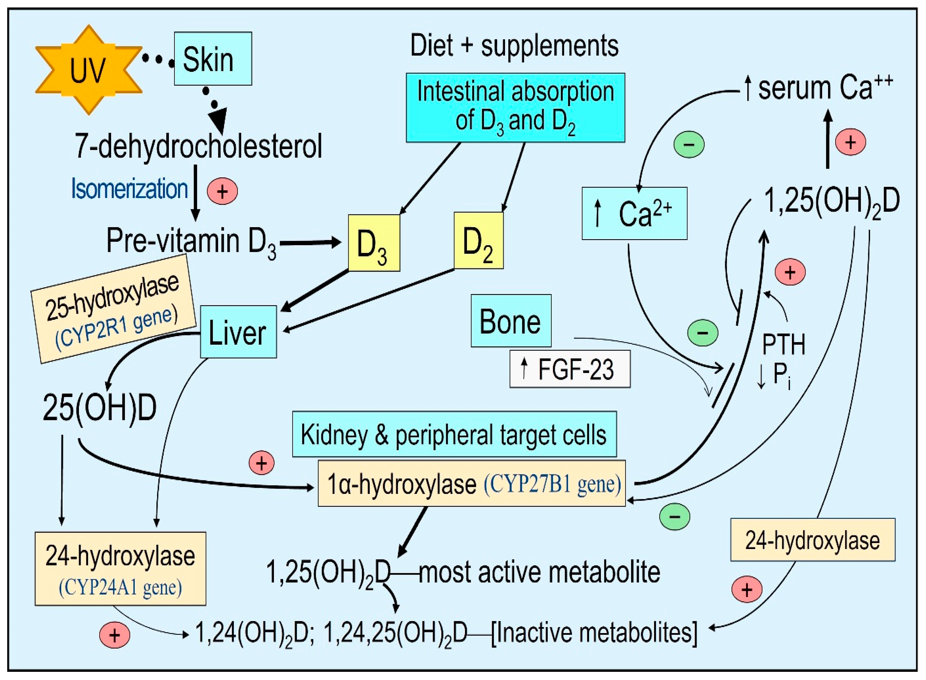
3. Generation/Obtaining Vitamin D and Transportation in Humans

Vitamin D is a secosteroid molecule: fully activated vitamin D, 1,25(OH)2D has broad physiological functions [7,24]. These include immune modulation with anti-inflammatory and antioxidant actions [13], membrane stability [25], metabolic and mitochondrial respiratory functions [13], and reproductive biology. Genomic functions include the favorable transcription of over 1200 genes [26,27].
Vitamin D3 is supposed to be obtained naturally by humans following exposure to UVB rays from sunlight. In the skin, 7-dehydrocholesterol converts to pre-vitamin D3, which isomerizes to form vitamin D3. It binds to VDBP and diffuses via capillaries into the circulation [28]. There is little vitamin D in food: e.g., D2 in sun-exposed mushrooms and D3 n fatty fish. After intestinal absorption as chylomicron, vitamin D is incorporated into VDBP via lipoproteins and reaches the bloodstream through the thoracic duct [29]. These reach hepatocytes, where 25(OH)D is generated and released into the circulation, mostly bound to VDBP [30]. In addition to hepatocytes, 25-hydroxylase (CYP2R1 gene) is present in other target cells/tissues but in minor concentrations [31,32,33] so that D can be converted to 25(OH)D in these cells, as in immune cells [34,35]. However, there is no evidence that peripheral target tissue cell-generated calcifediol contributes to circulatory 25(OH)D.
Vitamin D, 25(OH)D, and 1,25(OH)2D have different dissociation constants in binding to VDBP, which determine the free (unbound to VDBP) D and 25(OH)D concentration in the circulation, which is approximately 1% of the that of the total vitamin D. The dissociation constant of 25(OH)D is about 10−9 m, while for vitamin D and 1,25(OH)2D, it is approximately 10−7 m [20,23]. Consequently, the circulating half-lives of these three compounds are inversely associated with the dissociation constants. For 25(OH)D, its half-live is between two to three weeks (as it is tightly bound to VDBP), depending on the vitamin D status in the body, while for vitamin D, it is one day, and for 1,25(OH)2D, a few hours [36]. Accordingly, the free circulating proportions are highest for 1,25(OH)2D, then D, and lowest for 25(OH)D.
A few thousand International Units (IU) of vitamin D3 could be synthesized in the skin after exposure to sunlight, which takes about 24 h to materialize in the circulation [14]. Ingested vitamin D3 appears in circulation between 12 and 20 h after intestinal absorption and transportation [14,29,37]. The circulating half-life of D2 and D3 is approximately 24 h; that of D2 is less than that of D3 [29]. Because of this short half-life, even higher bolus doses of vitamin D are eliminated from the body in a few days [14,38]. Therefore, the best way to maintain a steady state of vitamin D and 25(OH)D in circulation is through regular daily sun exposure and/or daily supplementation [16].

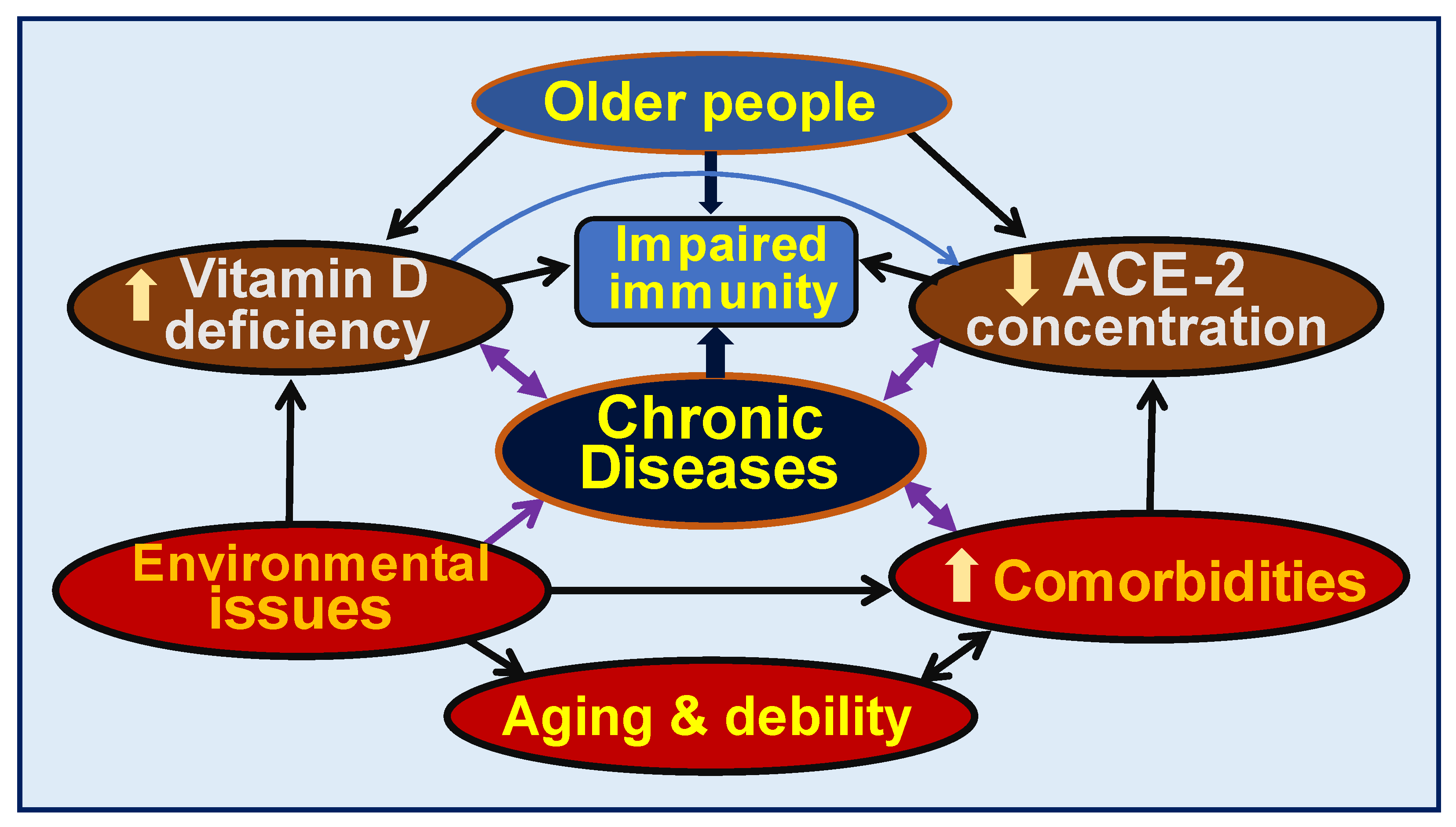
4. Consequences of Vitamin D Deficiency

Vitamin D deficiency universally impairs its intended benefits in all body systems. Its deficiency increases the vulnerability to infections, increases generalized inflammation, increases risks for diseases and infections, and worsens chronic diseases [12,39,40]. Consequently, hypovitaminosis increases the susceptibility to infections and diseases and enhances the severity of illnesses [41], leading to increased complications and premature deaths [42,43,44]. Vitamin D has pleiotropic effects on body systems, especially the immune, musculoskeletal, cardiovascular, pulmonary, neurological, gastrointestinal, and renal systems. Figure 2 illustrates the expected consequences of chronic vitamin D deficiency.
Persons with chronic kidney disease (CKD) have insufficient handling of vitamin D, 25(OH)D, and 1,25(OH)2D. This is due to gastrointestinal malabsorption, increased catabolism, and a significant decrease in renal 1α-hydroxylation by CYP27B1. This results in low circulatory calcitriol that causes hyperphosphatemia and elevated fibroblast growth factor-23 (FGF-23) concentrations [45]; these initiate the CKD of mineral and bone disorder (CKD-MBD) [46,47]. The treatment modality of CKD-MBD has shifted from single biomarkers (measurement of calcitriol) to serial (economical) measurements of calcium, phosphate, and parathyroid hormone (PTH); these provide a broader insight and better control, helping the management of persons with CKD [46].
The abovementioned abnormalities of vitamin D metabolism lead to secondary hyperparathyroidism, which rapidly responds to oral cholecalciferol (D3) [48]. Survival is increased for those with all types of CKD when calcitriol is administered with vitamin D [49]. In contrast, the activation of CYP24A1 catabolizes vitamin D and its active metabolites, increasing serum 24-hydroxyvitamin D, 24,25-dihydroxyvitamin D, and 1,24,25-trihydroxyvitamin D, to the 25(OH)D ratio in the circulation (Figure 1)—known as vitamin D catabolic (metabolic) ratio [50].
The higher catabolic ratios and thus lower 25(OH)D concentrations are associated with modestly increased all-cause mortality [50]. Circulatory concentrations of D3 and 25(OH)D3 are in the micromolar range, while 1,25(OH)2D3 is present in the nanomolar range, with a calcitriol concentration of approximately nine-hundred-fold lower {Wimalawansa, 2023 #17062}. Therefore, calculating this ratio does not include calcitriol or 1,24,25-trihydroxyvitamin D concentrations in circulation as they are minuscule (as other uncommon metabolites and epimers of vitamin D), making it easier to calculate. While controversial, a reverse J shape of all-cause mortality has been reported with total serum 25-hydroxyvitamin D concentration [51], for which explanations and counters are numerous [52,53].

5. Muscular–Skeletal Benefits of Vitamin D

The classical actions of vitamin D involve mineral metabolism—calcium absorption and mineral conservation, skeletal calcification, and musculoskeletal functions [6,7]. These skeletal functions—bone formation/resorption and mineralization—depend on the parathyroid hormone in conjunction with the hormonal form of calcitriol derived from proximal renal tubular cells [13].
The tissue transport mechanism for steroids—megalin–cubilin endocytotic system [17]—is essential for delivering vitamin D and 25(OH)D into proximal renal tubular cells for generating calcitriol [17]. This mechanism is also present in parathyroid cells. This active transportation system is also present in fat and muscle cells—the storage tissues. The musculoskeletal system and parathyroid hormone (PTH)-driven vitamin D activities, like calcium homeostasis, are considered a part of the endocrine functions of vitamin D [54]. In contrast, the intracrine/autocrine and paracrine functions of calcitriol in peripheral target cells, like immune cells, are driven by both genomic and other signaling mechanisms. The generation of calcitriol by 1α-hydroxylase (CYP27B1) within immune cells is dependent on the ability to diffuse enough vitamin D and/or 25(OH)D from the circulation into immune cells [55,56]. This is crucial for all immune cell activities.

6. Hypovitaminosis D and Viral Respiratory Infections

Respiratory tract illnesses, including colds, influenza, and COVID-19, escalate in the winter. There are specific reasons why countries located far north of the equator in the northern (and southern) hemispheres experience winter-associated viral respiratory cycle that increases in colder months with less sunlight [57,58,59]. During the winter, the sunlight does not carry adequate UVB rays. In addition, rays come at a narrow-angle that does not sufficiently penetrate the skin for humans to generate vitamin D. One consequence of insufficient UVB rays is a marked reduction in circulating D and 25(OH)D concentrations. This weakens the immune system. In addition, viruses live longer outside human bodies in cold and dryer climatic conditions, such as in winter time [60,61,62].
Vitamin D deficiency markedly impairs overall immunity and thus increases the risk of illness, including metabolic disorders and infections. This makes individuals vulnerable to microbial infections [63,64], primarily viral respiratory diseases [65,66,67,68,69,70], including coronaviruses [71,72,73]. Vitamin D adequacy—having blood levels greater than 30 ng/mL (older definition) [74] but preferably greater than 50 ng/mL during winter and viral epidemics—significantly reduces the risk of respiratory viral infections [1,67,75].
Children rely primarily on their innate immune systems to counteract pathogenic microbial invasions. Since they have better innate immunity than the elderly, they are less likely to develop symptomatic COVID-19, complications or die from it unless they have severe hypovitaminosis D [76]. Severe vitamin D deficiency (i.e., serum 25(OH)D concentrations of less than 12 ng/mL) increases the risks of developing fatal immunological disorders, like Kawasaki-like disease and multi-system inflammatory syndrome [77,78]. When children with severe vitamin D deficiency are exposed to a high viral load, they could experience severe hyperimmune reactions with the complications mentioned above [77,79,80].

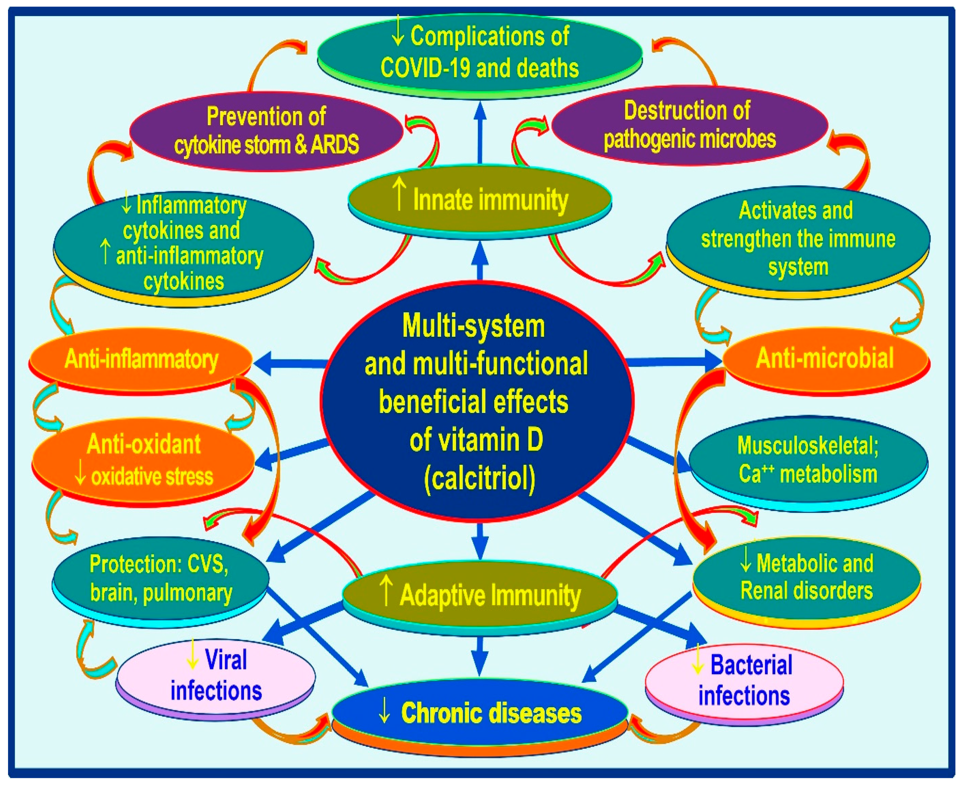
7. Extra-Skeletal Benefits of Vitamin D

Most extra-musculoskeletal biological activities of calcitriol occur following the generation of calcitriol within peripheral target cells (i.e., not via the circulatory, hormonal form), where it acts as a signaling molecule and a local cytokine. The latter functions include controlling cell proliferation and maturity, preventing cancer cell growth, brain development, respiratory and reproductive functions, and mitochondrial energy generation [24,81,82,83]. However, calcitriol’s most prominent and life-saving extra-skeletal role is modulating the immune system [84,85]. Vitamin D maintains a robust immune system, which helps to overcome infections, including COVID-19 [55,86,87], and prevents autoimmunity [88,89].
A large data set and emerging data support multiple physiological functions of vitamin D, via calcitriol. These data suggest vitamin D should be used as a preventative and adjunct therapy in several common disorders, including sepsis and COVID-19 infection [67,70,90,91]. Nevertheless, vitamin D is rarely included in clinical protocols or guidelines, or advised by leading health authorities or by governments to their fellow citizens to keep them healthy [24]. In addition, recommendations from medical and scientific societies are confusing, contradictory, and out of date [41,92]. However, public awareness of vitamin D and its beneficial effects on the immune system has improved since the COVID-19 pandemic. This is primarily due to relentless positive work by small groups of scientists, despite the negative publicity by big pharmaceutical corporations. Examples include the clinical guidelines from the Front-Line COVID-19 Critical Care Alliance (https://COVID19criticalcare.com/treatment-protocols/: Accessed 5 July 2023), affirmative Substack articles, and websites like https://COVID19criticalcare.com (Accessed 1 July 2023) [93].
Sufficient calcitriol synthesis within immune cells prevents autoimmune reactions profoundly and controls inflammation and infections [39,40]. These physiological actions manifest by suppressing the expression of inflammatory cytokines and increasing the expression of anti-inflammatory cytokines and anti-oxidative compounds [70,94]. Most chronic diseases are associated with chronic inflammation that maintains the disease process [39]. In addition, calcitriol enhances the production and release of antimicrobial peptides, cathelicidin, and beta-defensin via its autocrine and paracrine actions (Figure 1).
These antimicrobial peptides stimulate white blood cells, macrophages, and natural killer cells and direct the circulating viruses to macrophages to destroy them [95]. Vitamin D signaling plays a crucial role in intrinsic defense against intracellular microorganisms via generating antimicrobial proteins like cathelicidin [40]. In addition to directly binding to and killing a range of pathogens, cathelicidin acts as a secondary messenger, augmenting vitamin D-mediated reduction in inflammation during infection [96]. In addition, calcitriol stabilizes tight junctions of epithelial cells of the respiratory tract and cardiovascular system, protecting them from fluid leakage and viral dissemination into soft tissues [97,98]. Figure 3 illustrates the generation of calcitriol and the critical difference between the hormonal form and the non-hormonal form of calcitriol.

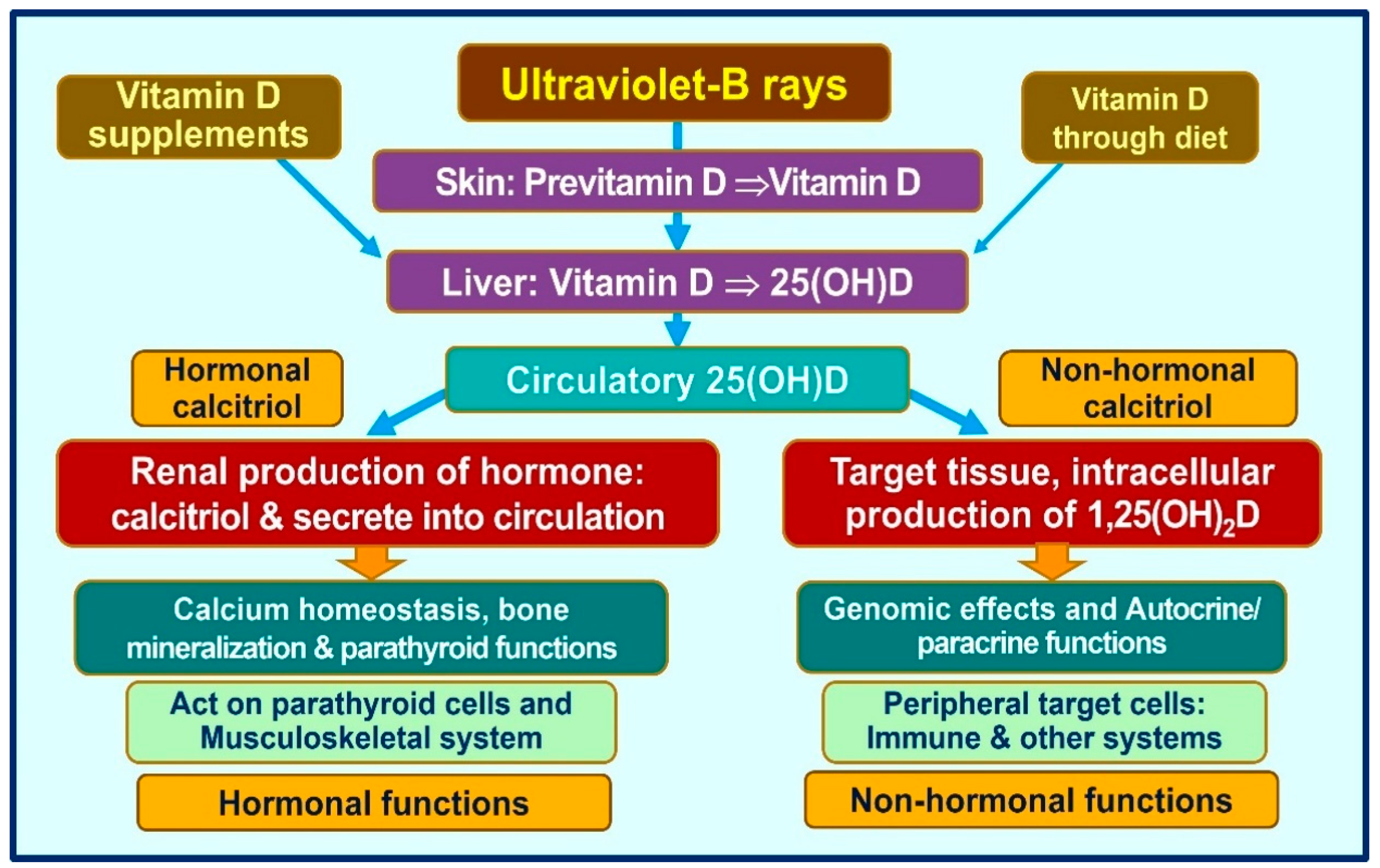
8. Importance of Circulatory Vitamin D and 25(OH)D for Target Cell Generation of Calcitriol

Over the years, the focus has been on cholecalciferol (D3) to prevent musculoskeletal disorders [99]. However, in the past two decades, several fundamental advances have been made by researchers in understanding the biology and physiology of calcifediol and calcitriol and delineating how and when to use them as therapies. Over the past decade, emerging evidence has added more value and highlighted the importance of these vitamin D compounds in human biology and clinical immunology [17]. While the musculoskeletal system functions could be maintained with smaller doses, of between 800 and 2000 IU/day, higher amounts, like 5000 to 10,000 IU per day or 50,000 IU once a week, are necessary for a non-obese 70 kg adult to maintain serum 25(OH)D concentrations above 50 ng/mL, which are needed to overcome infections [55,56].
Those who are obese, taking medications that increase catabolic activity of vitamin D (e.g., anti-epileptic and retroviral agents), or have significant fat malabsorption require several fold-higher doses than those mentioned above. Even with such amounts, a vitamin D-deficient person likely takes several months to increase their serum 25(OH)D to therapeutic levels of over 50 ng/mL [56]. Using the mentioned doses of vitamin D, even in a vitamin D-sufficient person (guidelines for community-dwelling persons) to reach and maintain a serum 25(OH)D concentration of above 40 ng/mL would take a few weeks to raise the serum 25(OH)D concentration above 50 ng/mL [55]. Therefore, such doses could be insufficient (and ineffective) to achieve the desired target serum 25(OH)D concentration in emergencies.
Serum 25(OH)D concentrations are reduced in chronic diseases like metabolic disorders, obesity, cancer, infections, and all-cause mortality [100,101,102,103]. Notably, less frequent administration (i.e., intervals of less than once a month—(i.e., intermittent bolus dosing) and even higher doses, like 300,000 once in six months, do not generate the intended clinical outcomes. This is because the half-life of vitamin D is about one day, and that of 25(OH)D is between two to three weeks (depending on the vitamin D status). No matter how high the doses is, the serum 25(OH)D concentration would not be sufficiently high for more than three months [104,105,106]. In addition, infrequent administrations lead to unphysiological fluctuation of serum and tissue levels of vitamin D metabolites (see below).

9. Clinical Study Outcomes Using Higher Doses of Vitamin D

Meta-analyses of RCTs concerning vitamin D supplementation reported a significant reduction in the incidence and severity of respiratory tract infections [107,108,109]—better clinical outcomes were reported with daily vitamin D than with infrequent administration. In contrast, when vitamin D is administered at longer intervals than once a month, fewer benefits are observed, and the outcomes are not satisfactory [110,111].
Using higher doses of vitamin D consistently has been reported to have better clinical outcomes than the government-recommended doses of 800 IU/day, which have no tangible effect on any disease other than muscular–skeletal disorders [107,112]. For example, adequate supplementation with vitamin D reduces cancer [113], leads to the regression of prostate cancer [114], lowers blood pressure (especially in African Americans) [115], and reduces insulin resistance [116,117], including in obese children [118], and prevents multiple sclerosis [108,119].
However, studies that used minute doses of vitamin D based on outdated recommendations (i.e., using 280 IU/day or less than 1000 IU/day) [109,120], as with the Women’s Health Initiative study of cancer prevention and infrequent administration of 100,000 IU vitamin D3 quarterly [121] failed to prevent cancer and other disorders. Based on vitamin D biology and physiology, this is not surprising. Most clinical studies reported an inverse association between vitamin D status and mortality [103,122], and the relation is curvilinear [41].

10. Entry of D and 25(OH)D into Peripheral Target Cells

Most steroid hormones enter cells via diffusion and endocytosis via the membrane-based megalin–cubilin system as in the kidney and parathyroid gland, muscle, and fat cells [17]. In addition, this mechanism of active cellular entry is essential for generating the hormonal form of calcitriol in renal tubules and parathyroid glands—for vitamin D’s endocrine functions [17,54]. However, unlike the cells mentioned above, other peripheral target cells, like immune cells, do not have an active vitamin D megalin–cubilin transportation system [56]. Thus, in addition to some endocytosis, these cells mainly depend on a concentration-dependent gradient for diffusions of vitamin D and 25(OH)D (mostly bound to VDBP) into them [123].
In addition to diffusion, as illustrated above, VDBP bound D and 25(OH)D enters these cells via endocytosis [22]. Since the affinity of vitamin D to VDBP is less than 25(OH)D, given the same concentration in the blood, more vitamin D could enter immune cells. However, since the half-life of vitamin D is only one day, the total entry of vitamin D is less than 25(OH)D. Figure 4 illustrates the mode of access of vitamin D and 25(OH)D into peripheral target cells, like immune cells [55], from the circulation that leads to the generation of intracellular calcitriol [41], which is crucial not only for the genomic functions but also autocrine and paracrine functions of immune cells and other target cells [87,124,125,126].
When vitamin D is taken daily, the circulatory vitamin D concentrations are likely to be higher than 25(OH)D concentrations [41]. Therefore, more vitamin D could diffuse into peripheral target cells than 25(OH)D because of the higher concentration gradient of D. When this happens, more vitamin D than 25(OH)D would reach into target cells and hydroxylated to form calcitriol. If this is the case, the measurement of serum 25(OH)D alone, as carried out in routine clinical practice, may not provide the correct information about vitamin D adequacy or the replacement requirements for physiological functions, including a robust immune system (Figure 4). The opposite happens when the same dose of vitamin is consumed once a week; a higher concentration of 25(OH)D is present in the circulation than in vitamin D.

11. Vitamin D, Epithelial Barriers, and Gap Junction Stability

D3 enhances epithelial and endothelial stability independently of canonical pathways through calcitriol/CTR-derived genomic outcome [127]. The disruption of endothelial stability and vascular leak enhancement are prevented via D3 supplementation. These rapid membrane-related actions of vitamin D are derived from D3 and its two common metabolites, 25(OH)D and 1,25(OH)2D, at a similar potency.
The deficiency of D3 and its metabolites impairs endothelial barriers, leading to vascular fluid leakage into soft tissues [127]. Similarly, weakening gap junctions and epithelial barriers lead to viral infiltration and the propagation of infections, as seen in sepsis and viral infections like SARS-CoV-2 [128]. These non-transcriptional mechanisms are also essential in controlling inflammation and preventing endothelial and epithelial cell destabilization.

12. What Has Changed over the Years Related to Vitamin D?

A century ago, it was established that sunrays (vitamin D) reverse rickets in children and are effective against tuberculosis. In addition, a large body of scientific evidence demonstrates that vitamin D plays a central role in disease prevention (maintaining a disease-free state) and preventing severe symptoms, diseases, complications, and deaths [44]. A few years ago, exposure to sufficient UVB rays was believed to generate no more than 3000 IU/day. However, recent data confirmed a person with a lighter skin color could generate a few thousand IUs of vitamin D3 after one hour of UVB exposure over a third of the upper body [129,130,131].
Maintaining a steady state of D and 25(OH)D in circulation is helpful for better physiological functions. Marked fluctuating serum 25(OHD concentration due to prolonged interval administration is unphysiological and likely to over-activate 24-hydroxylase enzyme, CYP24A1 concentrations, increasing the catabolism of active metabolites of vitamin D. Based on half-lives in circulation, the frequency of administration of vitamin D must not exceed once in ten days (or once a week), and no more than once a month. Therefore, vitamin D should not be administered at intervals longer than two-week intervals [132]. This will allow for keeping a steady circulatory concentration [110,133].
The importance of the above is highlighted by six positive respiratory tract infection-related RCTs, most conducted in children [42,43,57,58,59]; all of these used daily doses of vitamin D [101,102,134]. Another meta-analysis of RCTs on vitamin D supplementation in respiratory tract infections reported that vitamin D is most effective as a treatment when administered in daily doses than intermittently [135]. Chronic diseases are most common among older people partly due to longer-term vitamin D deficiency [136], and are associated with an increased rate of deaths [44,122]. They also have multiple co-morbidities associated with hypovitaminosis D and low-circulating ACE-2 receptors, increasing the vulnerability to infections and other pathological ailments (Figure 5).

13. Vitamin D Intake Should Depend on Body Weight and Target Serum 25(OH)D Concentration

Different dosing schedules have varied effects on serum vitamin D and 25(OH)D concentrations—daily doses maintain a stable circulating concentration [16]. In contrast, ingesting vitamin D for longer than monthly intervals results in significant circulatory 25(OH)D concentration fluctuations; this is not physiological and may not benefit much [110,132,133]. Schedules used for vitamin D supplementation as prophylactic and treatment or in RCTs will profoundly affect the serum D and 25(OH)D concentrations (primarily due to the short half-life of vitamin D); thus, this needs to be considered for better clinical outcomes.
Vitamin D supplementation and sufficient UV exposure increase maternal circulating 25(OH)D concentration in breast milk [137,138]. It has been known that solely breastfed infants exhibit vitamin D deficiency [139], which is easily correctible with vitamin D drops given to nursing infants [140]. For each 1000 IU/d of vitamin D3-supplemented to a lactating mother, vitamin D concentration in her breast milk increased by about 80 IU/L. The recommended average dose of vitamin D3 for pregnant and lactating mothers is 6000 IU/d; this provides the infants with 400–500 IU of vitamin D per day [120].
The circulating concentration of 25(OH)D in the fetus is approximately 70% of that of the mother; thus, a diffusion of 25(OH)D occurs across the placenta [141]. However, since vitamin D concentrations are slightly below the maternal concentrations, relatively lower amounts are diffused via the placenta [16]. The same phenomenon has been reported in transferring vitamin D and 25(OH)D to breast milk [141,142]. The diffusion gradient can be increased by raising the maternal serum 25(OH)D to 50 ng/mL [143].

14. Vitamin D Is Essential for Activating Immune Cells

Calcitriol is the most active vitamin D metabolite, crucial for combating invading pathogens and preventing autoimmunity, and chronic diseases [1,2]. Through multiple mechanisms, calcitriol modulates the immune system. When secreted into the bloodstream from renal tubular cells, calcitriol functions as a hormone. This alters the behavior of cells involved in calcium-phosphate-bone metabolism, intestinal, bone and parathyroid cells. The average circulatory concentration of calcitriol in the circulation about 0.045 ng/mL, but the concentration of its free, the diffusible form is even less. It is far below the threshold needed to initiate intracellular signaling. Consequently, such the pmolar concentration of hormonal calcitriol, is over 20 times less, thus have no tangible effect on intracellular intracrine signal transduction or genomic functions in immune cells. Besides, vitamin D and 25(OH)D concentrations in the circulation are about 900-fold higher than circulating vitamin D and 25(OH)D concentrations. Consequently, circulating calcitriol has no evident impact outside the muscular-skeletal and fat cells.
Peripheral target cells, like immune cells, depend on vitamin D and 25(OH)D primarily via diffusion from the circulation to generate higher concentrations of non-hormonal calcitriol. When immune cells detect external threats, like circulating microbes or unfamiliar antigens by pattern recognition receptors, like membrane-bound Toll-like receptors TLR (TLR-4) they send signals to increase the expression of 1α of VDR, thus, increasing in the cytoplasm.
Higher nmol range concentrations of calcitriol generated in-tracellularly in response to TLR signaling, provides (hysiological) intracellular autocrine/intracrine signaling that is crucial for immune functions to overcome threats, like infections. Consequently, there is a hold mechanism, increasing serum, i.e., beyond threat like detecting unfamiliar proteins or antigens in the circulation or local tissues. The sporadic increases in the synthesis of calcitriol and VDR in responses to TLR-4 signaling, ensures the formation of sufficient calcitriol-VDR complexes to modulate transcriptions, and intra-cellular autocrine signaling and genomic modulation, as when needed.
Regulates inflammation and oxidative stresses through the abovementioned mechanisms, primarily by suppressing inflammatory cytokines and enhancing the synthesis of anti-inflammatory cytokines. Immunomodulatory effects of vitamin D include activation of immune cells such as T and B cells, macrophage and dendritic cells, and enhanced production of several antimicrobial peptides and neutralizing antibodies [3,4,5]. Figure 6 illustrates broader vitamin D (calcitriol) actions in innate and adaptive immune systems.
Once adequate concentrations are generated within the immune cells, calcitriol activates and bonds to the cytosol’s vitamin D (calcitriol)receptors (VDRs) that translocate into the nucleus for its genomic actions. The interaction of calcitriol with its receptor leads to the translocation of the complex to the nucleus, where it binds to the genome and modulates over 1200 genes [144]. In addition, intracellular calcitriol acts as an autocrine and paracrine signaling. Calcitriol down-regulates inflammation and oxidative stress through multiple mechanisms, primarily by suppressing inflammatory cytokines. Immunomodulatory effects of vitamin D include the activation of immune cells such as T and B cells, macrophage and dendritic cells, and enhanced production of several antimicrobial peptides and neutralizing antibodies [87,126,145]. Figure 6 illustrates broader vitamin D (calcitriol) actions in innate and adaptive immune systems.
Because hypovitaminosis D status does not activate immune cells, it causes relative immune paresis and delayed responses. This increases people’s vulnerability, especially to bacteria (like tuberculosis) [13] and respiratory viruses [147,148], including COVID-19 [149,150]. Recent clinical studies have supported the latter [76]. For example, serum 25(OH)D concentrations are significantly lower in those who are PCR-positive for SARS-CoV-2 (mean concentration of 11.1 ng/mL; p = 0.004) compared with those with negative results (24.6 ng/mL), demonstrating a higher vulnerability [151]. This is striking when using the pre-infection serum 25(OH)D concentration to correlate with infection vulnerability [152].
In addition, there is a strong correlation between severe vitamin D deficiency and cytokine storm—a hyper-inflammatory condition caused by an uncontrolled, overactive immune status [153]. Viral infections lead to symptomatic disease and complications depending on the underlying vulnerability and the viral load. Thus, vitamin D may not prevent a person from contracting COVID-19 but will reduce symptomatic disease, complications, and deaths. While vitamin D has broader beneficial effects, it is not a cure for everything. For example, in bacterial infections, vitamin D should be used as a supportive therapy to boost the immune system naturally, in addition to primary pharmaceuticals like antibiotics.

15. Discussions

A balanced diet with adequate micronutrients, such as vitamins D, B2, K2, and C, and magnesium, trace minerals, and antioxidants, will support a more robust immune system. In most countries, some communities have one or more prevailing micronutrient deficiencies that increase vulnerability to various disorders, such as metabolic, infectious, and non-communicable diseases. In addition to nutrient supplements, fortifying foods with vitamin D and other essential micronutrients will enable them to develop a robust immune system, which prevents them from becoming frequently sick with viral infections.
Nationwide vitamin D supplementation, at least during epidemics and pandemics, markedly reduces acute and chronic diseases, the need for hospitalization, and their complications and deaths, including SARS-CoV-2 infection. It is common to have vitamin D deficiency in those with co-morbidities and chronic diseases, such as hypertension, diabetes mellitus, obesity, and cancer, and to be more vulnerable to developing complications from infections like COVID-19 [154]. Vitamin D sufficiency would reduce the incidence and severity of chronic diseases, such as metabolic disorders (e.g., diabetes, obesity, insulin resistance), cancer, autoimmune disorders, and infections [155,156,157,158]. While the efficacy of vaccines is wading with emerging new mutant Omicron viruses [159,160] and breakthrough SARS-CoV-2 infections [161], the effectiveness of vitamin D will not be affected [55]. Community vitamin D sufficiency is the key to protecting vulnerable populations, especially older people and ethnic minorities with darker skin color, and institutionalized persons [41,122,136,162].
Maintaining serum 25(OH)D concentrations above 40 ng/mL (100 nmol/L) is thought to significantly reduce microbial infections, particularly respiratory viral ones, including COVID-19 [1,163]. Enriching food, such as a targeted food fortification program, is an economical and practical approach for alleviating micronutrient malnutrition in ethnic populations or even for an entire country, as has been carried out with iodine [164]. In the case of COVID-19, those with severe vitamin D deficiency are the most susceptible to complications and deaths, primarily because of weaker immune systems [165]. The addition of other micronutrients, such as zinc and selenium, vitamins A, B2, C, and K2, resveratrol, and magnesium, in combination with essential fatty acids, such as omega-3, would facilitate the maintenance of a robust immune system [153,166,167,168] and keep the communities in good health.
Sustained vitamin D deficiency adversely affects human health, which is cost-effectively prevented with vitamin D supplementation and/or regular safe sun exposure. Maintaining population serum 25(OH)D concentrations above 40 ng/mL ensure a robust immune system. Sustained vitamin D deficiency negatively affects all body systems and increases risks for viral infections, outbreaks, and hospitalization. Thus, government and health administrators should consider nationwide educational campaigns for safe sun exposure, vitamin D supplementation, and targeted food fortification programs to strengthen the population’s immunity and keep them healthy. These acts cost less than 0.01% of one day’s hospitalization and significantly reduce healthcare costs.
Health insurance companies have a financial incentive to take proactive actions to keep their clients healthy by maintaining their vitamin D sufficiency. While the efficacy of vaccines and their boosters is wading with emerging Omicron mutant viruses, the effectiveness of vitamin D sufficiency remains solid and unchanged. The key to protecting the vulnerable is maintaining a higher circulatory vitamin D concentration, especially in ethnic minorities, older adults, and institutionalized persons, so they will maintain a robust innate immune system to fight against infections promptly.
Thus, government and health administrations should consider nationwide educational campaigns for safe sun exposure and vitamin D supplemental programs to strengthen the population’s immunity and keep them healthy. Sun exposure and/or vitamin D supplementation and targeted food fortification can achieve this cost-effectively. This would have marked beneficial effects on reducing symptomatic diseases and preventing complications associated with and deaths caused by COVID-19. The world did not capitalize on this highly cost-effective opportunity during the COVID-19 pandemic. This will also protect vulnerable populations (which have a uniformly high prevalence of vitamin D deficiency), such as ethnic minorities with darker skin color, older people, and institutionalized persons.

16. Conclusions

Maintaining population serum 25(OH)D concentrations above 40 ng/mL ensures a robust immune system in communities, curtailing the spread of infections, minimizing symptomatic diseases, and reducing the prevalence of chronic diseases. Vitamin D sufficiency also minimizes acute viral infections and outbreaks and the need for hospitalization, saving healthcare costs and lives. Thus, governments, health insurance companies, and health administrators should consider nationwide educational campaigns for safe sun exposure, vitamin D supplementation, and targeted food fortification programs to strengthen the population’s immunity and keep them healthy. Implementing these is less than one day’s cost of healthcare.
While the efficacy of vaccines and their boosters has waded with mutants of Omicron viruses, the effectiveness of vitamin D sufficiency remains strong. Vitamin D has multiple beneficial effects on all body systems: however, it is not a panacea for everything. Apart from preventative use, vitamin D should be used as adjunctive therapy with other primary pharmaceuticals and the best/optimal therapies and approaches, such as using antibiotics in bacterial infections. The key to protecting the vulnerable and reducing chronic disease burden in a country is not by expanding hospitals and health centers and recruiting more healthcare professionals but by educating the public on health preservation and maintaining a higher circulatory vitamin D concentration, especially in vulnerable communities—ethnic minorities, older adults, and institutionalized persons—so that they will have robust immune systems to fight against any infection and minimize chronic diseases.

Funding

This research received no funding or aid from funding agencies or commercial or not-for-profit sectors.

Institutional Review Board Statement

Not applicable.

Informed Consent Statement

Not applicable.

Data Availability Statement

Not applicable.

Conflicts of Interest

The author declares no conflict of interest. He did not receive any funding or writing assistance.

References

  1. Zdrenghea, M.T.; Makrinioti, H.; Bagacean, C.; Bush, A.; Johnston, S.L.; Stanciu, L.A. Vitamin D modulation of innate immune responses to respiratory viral infections. Rev. Med. Virol. 2017, 27, e1909. [Google Scholar] [CrossRef]
  2. Gotelli, E.; Soldano, S.; Hysa, E.; Paolino, S.; Campitiello, R.; Pizzorni, C.; Sulli, A.; Smith, V.; Cutolo, M. Vitamin D and COVID-19: Narrative Review after 3 Years of Pandemic. Nutrients 2022, 14, 4907. [Google Scholar] [CrossRef] [PubMed]
  3. Hollis, B.W.; Marshall, D.T.; Savage, S.J.; Garrett-Mayer, E.; Kindy, M.S.S.; Gattoni-Celli, S. Vitamin D3 supplementation, low-risk prostate cancer, and health disparities. J. Steroid Biochem. Mol. Biol. 2013, 136, 233–237. [Google Scholar] [CrossRef] [PubMed]
  4. Xiaoyu, Z.; Payal, B.; Melissa, O.; Zanello, L.P. 1alpha,25(OH)2-vitamin D3 membrane-initiated calcium signaling modulates exocytosis and cell survival. J. Steroid Biochem. Mol. Biol. 2007, 103, 457–461. [Google Scholar] [CrossRef]
  5. Liang, F.; Liu, C.; Li, L.; Guo, Y.; Bai, L. Effects of gastrin on rat intestinal epithelial 1,25(OH)2D3-membrane associated rapid response steroid binding protein. Nan Fang Yi Ke Da Xue Xue Bao 2013, 33, 990–993. [Google Scholar] [PubMed]
  6. Vieth, R. Why the optimal requirement for Vitamin D3 is probably much higher than what is officially recommended for adults. J. Steroid Biochem. Mol. Biol. 2004, 89–90, 575–579. [Google Scholar] [CrossRef]
  7. Wimalawansa, S.J. Vitamin D in the new millennium. Curr. Osteoporos. Rep. 2012, 10, 4–15. [Google Scholar] [CrossRef]
  8. Bouillon, R. Genetic and racial differences in the vitamin D endocrine system. Endocrinol. Metab. Clin. N. Am. 2017, 46, 1119–1135. [Google Scholar] [CrossRef]
  9. Holick, M.F.; Chen, T.C.; Lu, Z.; Sauter, E. Vitamin D and skin physiology: A D-lightful story. J. Bone Miner. Res. 2007, 22 (Suppl. S2), V28–V33. [Google Scholar] [CrossRef]
  10. Engelsen, O. The relationship between ultraviolet radiation exposure and vitamin D status. Nutrients 2010, 2, 482–495. [Google Scholar] [CrossRef]
  11. Webb, A.R.; Alghamdi, R.; Kift, R.; Rhodes, L.E. 100 Years of Vitamin D: Dose-response for change in 25-hydroxyvitamin D after UV exposure: Outcome of a systematic review. Endocr. Connect. 2021, 10, R248–R266. [Google Scholar] [CrossRef] [PubMed]
  12. Wimalawansa, S.J. Public health interventions for chronic diseases: Cost-benefit modelizations for eradicating chronic kidney disease of multifactorial origin (CKDmfo/CKDu) from tropical countries. Heliyon 2019, 5, e02309. [Google Scholar] [CrossRef] [PubMed]
  13. Wimalawansa, S.J. Biology of vitamin D. J. Steroids Horm Sci. 2019, 10, 2. [Google Scholar] [CrossRef]
  14. Adams, J.S.; Clemens, T.L.; Parrish, J.A.; Holick, M.F. Vitamin-D synthesis and metabolism after ultraviolet irradiation of normal and vitamin-D-deficient subjects. N. Engl. J. Med. 1982, 306, 722–725. [Google Scholar] [CrossRef]
  15. Vieth, R. Vitamin D supplementation, 25-hydroxyvitamin D concentrations, and safety. Am. J. Clin. Nutr. 1999, 69, 842–856. [Google Scholar] [CrossRef]
  16. Hollis, B.W.; Johnson, D.; Hulsey, T.C.; Ebeling, M.; Wagner, C.L. Vitamin D supplementation during pregnancy: Double-blind, randomized clinical trial of safety and effectiveness. J. Bone Miner. Res. 2011, 26, 2341–2357. [Google Scholar] [CrossRef]
  17. Nykjaer, A.; Dragun, D.; Walther, D.; Vorum, H.; Jacobsen, C.; Herz, J.; Melsen, F.; Christensen, E.I.; Willnow, T.E. An endocytic pathway essential for renal uptake and activation of the steroid 25-(OH) vitamin D3. Cell 1999, 96, 507–515. [Google Scholar] [CrossRef]
  18. Aatsinki, S.M.; Elkhwanky, M.S.; Kummu, O.; Karpale, M.; Buler, M.; Viitala, P.; Rinne, V.; Mutikainen, M.; Tavi, P.; Franko, A.; et al. Fasting-Induced Transcription Factors Repress Vitamin D Bioactivation, a Mechanism for Vitamin D Deficiency in Diabetes. Diabetes 2019, 68, 918–931. [Google Scholar] [CrossRef]
  19. Elkhwanky, M.S.; Kummu, O.; Piltonen, T.T.; Laru, J.; Morin-Papunen, L.; Mutikainen, M.; Tavi, P.; Hakkola, J. Obesity represses CYP2R1, the vitamin D 25-Hydroxylase, in the liver and extrahepatic tissues. JBMR Plus 2020, 4, e10397. [Google Scholar] [CrossRef]
  20. Haddad, J.G.; Hillman, L.; Rojanasathit, S. Human serum binding capacity and affinity for 25-hydroxyergocalciferol and 25-hydroxycholecalciferol. J. Clin. Endocrinol. Metab. 1976, 43, 86–91. [Google Scholar] [CrossRef]
  21. Keenan, M.J.; Holmes, R.P. The uptake and metabolism of 25-hydroxyvitamin D3 and vitamin D binding protein by cultured porcine kidney cells (LLC-PK1). Int. J. Biochem. 1991, 23, 1225–1230. [Google Scholar] [CrossRef]
  22. Kissmeyer, A.; Mathiasen, I.S.; Latini, S.; Binderup, L. Pharmacokinetic studies of vitamin D analogues: Relationship to vitamin D binding protein (DBP). Endocrine 1995, 3, 263–266. [Google Scholar] [CrossRef] [PubMed]
  23. Vieth, R.; Kessler, M.J.; Pritzker, K.P. Species differences in the binding kinetics of 25-hydroxyvitamin D3 to vitamin D binding protein. Can. J. Physiol. Pharmacol. 1990, 68, 1368–1371. [Google Scholar] [CrossRef] [PubMed]
  24. Wimalawansa, S.J. Non-musculoskeletal benefits of vitamin D. J. Steroid Biochem. Mol. Biol. 2018, 175, 60–81. [Google Scholar] [CrossRef] [PubMed]
  25. Zhang, Q.; Wang, M.; Han, C.; Wen, Z.; Meng, X.; Qi, D.; Wang, N.; Du, H.; Wang, J.; Lu, L.; et al. Intraduodenal Delivery of Exosome-Loaded SARS-CoV-2 RBD mRNA Induces a Neutralizing Antibody Response in Mice. Vaccines 2023, 11, 673. [Google Scholar] [CrossRef]
  26. Jiang, Y.; Chen, L.; Taylor, R.N.; Li, C.; Zhou, X. Physiological and pathological implications of retinoid action in the endometrium. J. Endocrinol. 2018, 236, R169–R188. [Google Scholar] [CrossRef]
  27. Keane, K.N.; Cruzat, V.F.; Calton, E.K.; Hart, P.H.; Soares, M.J.; Newsholme, P.; Yovich, J.L. Molecular actions of vitamin D in reproductive cell biology. Reproduction 2017, 153, R29–R42. [Google Scholar] [CrossRef]
  28. Holick, M.F. The cutaneous photosynthesis of previtamin D3: A unique photoendocrine system. J. Investig. Dermatol. 1981, 77, 51–58. [Google Scholar] [CrossRef]
  29. Haddad, J.G.; Matsuoka, L.Y.; Hollis, B.W.; Hu, Y.Z.; Wortsman, J. Human plasma transport of vitamin D after its endogenous synthesis. J. Clin. Investig. 1993, 91, 2552–2555. [Google Scholar] [CrossRef]
  30. Ponchon, G.; Kennan, A.L.; DeLuca, H.F. “Activation” of vitamin D by the liver. J. Clin. Investig. 1969, 48, 2032–2037. [Google Scholar] [CrossRef]
  31. Hosseinpour, F.; Wikvall, K. Porcine microsomal vitamin D(3) 25-hydroxylase (CYP2D25). Catalytic properties, tissue distribution, and comparison with human CYP2D6. J. Biol. Chem. 2000, 275, 34650–34655. [Google Scholar] [CrossRef] [PubMed]
  32. Flanagan, J.N.; Young, M.V.; Persons, K.S.; Wang, L.; Mathieu, J.S.; Whitlatch, L.W.; Holick, M.F.; Chen, T.C. Vitamin D metabolism in human prostate cells: Implications for prostate cancer chemoprevention by vitamin D. Anticancer Res. 2006, 26, 2567–2572. [Google Scholar] [PubMed]
  33. Zhu, P.; Ren, M.; Yang, C.; Hu, Y.X.; Ran, J.M.; Yan, L. Involvement of RAGE, MAPK and NF-kappaB pathways in AGEs-induced MMP-9 activation in HaCaT keratinocytes. Exp. Dermatol. 2012, 21, 123–129. [Google Scholar] [CrossRef]
  34. McBrearty, N.; Cho, C.; Chen, J.; Zahedi, F.; Peck, A.R.; Radaelli, E.; Assenmacher, C.A.; Pavlak, C.; Devine, A.; Yu, P.; et al. Tumor-Suppressive and Immune-Stimulating Roles of Cholesterol 25-hydroxylase in Pancreatic Cancer Cells. Mol. Cancer Res. 2023, 21, 228–239. [Google Scholar] [CrossRef]
  35. Karlgren, M.; Miura, S.; Ingelman-Sundberg, M. Novel extrahepatic cytochrome P450s. Toxicol. Appl. Pharmacol. 2005, 207, 57–61. [Google Scholar] [CrossRef]
  36. Smith, J.E.; Goodman, D.S. The turnover and transport of vitamin D and of a polar metabolite with the properties of 25-hydroxycholecalciferol in human plasma. J. Clin. Investig. 1971, 50, 2159–2167. [Google Scholar] [CrossRef] [PubMed]
  37. Lo, C.W.; Paris, P.W.; Clemens, T.L.; Nolan, J.; Holick, M.F. Vitamin D absorption in healthy subjects and in patients with intestinal malabsorption syndromes. Am. J. Clin. Nutr. 1985, 42, 644–649. [Google Scholar] [CrossRef]
  38. Heaney, R.P.; Recker, R.R.; Grote, J.; Horst, R.L.; Armas, L.A. Vitamin D(3) is more potent than vitamin D(2) in humans. J. Clin. Endocrinol. Metab. 2011, 96, E447–E452. [Google Scholar] [CrossRef]
  39. Mangin, M.; Sinha, R.; Fincher, K. Inflammation and vitamin D: The infection connection. Inflamm. Res. 2014, 63, 803–819. [Google Scholar] [CrossRef]
  40. Chung, M.K.; Karnik, S.; Saef, J.; Bergmann, C.; Barnard, J.; Lederman, M.M.; Tilton, J.; Cheng, F.; Harding, C.V.; Young, J.B.; et al. SARS-CoV-2 and ACE2: The biology and clinical data settling the ARB and ACEI controversy. EBioMedicine 2020, 58, 102907. [Google Scholar] [CrossRef]
  41. Wimalawansa, S.J. Physiological basis for using vitamin D to improve health. Biomedicines 2023, 11, 1542. [Google Scholar] [CrossRef] [PubMed]
  42. Arai, Y.; Kanda, E.; Iimori, S.; Naito, S.; Noda, Y.; Kawasaki, T.; Sato, H.; Ando, R.; Sasaki, S.; Sohara, E.; et al. The use of vitamin D analogs is independently associated with the favorable renal prognosis in chronic kidney disease stages 4-5: The CKD-ROUTE study. Clin. Exp. Nephrol. 2017, 21, 481–487. [Google Scholar] [CrossRef] [PubMed]
  43. Oh, T.R.; Kim, C.S.; Bae, E.H.; Ma, S.K.K.; Han, S.H.; Sung, S.A.; Lee, K.; Oh, K.H.; Ahn, C.; Kim, S.W.; et al. Association between vitamin D deficiency and health-related quality of life in patients with chronic kidney disease from the KNOW-CKD study. PLoS ONE 2017, 12, e0174282. [Google Scholar] [CrossRef]
  44. Zittermann, A.; Iodice, S.; Pilz, S.; Grant, W.B.; Bagnardi, V.; Gandini, S. Vitamin D deficiency and mortality risk in the general population: A meta-analysis of prospective cohort studies. Am. J. Clin. Nutr. 2012, 95, 91–100. [Google Scholar] [CrossRef] [PubMed]
  45. Jean, G.; Souberbielle, J.C.; Chazot, C. Vitamin D in Chronic Kidney Disease and Dialysis Patients. Nutrients 2017, 9, 328. [Google Scholar] [CrossRef] [PubMed]
  46. Waziri, B.; Duarte, R.; Naicker, S. Chronic Kidney Disease-Mineral and Bone Disorder (CKD-MBD): Current Perspectives. Int. J. Nephrol. Renovasc. Dis. 2019, 12, 263–276. [Google Scholar] [CrossRef] [PubMed]
  47. Urena-Torres, P.; Metzger, M.; Haymann, J.P.; Karras, A.; Boffa, J.J.; Flamant, M.; Vrtovsnik, F.; Gauci, C.; Froissart, M.; Houillier, P.; et al. Association of kidney function, vitamin D deficiency, and circulating markers of mineral and bone disorders in CKD. Am. J. Kidney Dis. 2011, 58, 544–553. [Google Scholar] [CrossRef]
  48. Kanai, G.; Fukagawa, M. CKD-MBD (Chronic Kidney Disease-Mineral and Bone Disorder). Gene therapy for secondary hyperparathyroidism. Clin. Calcium 2010, 20, 1052–1059. [Google Scholar]
  49. Zelnick, L.R.; de Boer, I.H.; Kestenbaum, B.R.; Chonchol, M.; Kendrick, J. Comparative Effects of Cholecalciferol and Calcitriol on Circulating Markers of CKD Mineral Bone Disorder: A Randomized Clinical Trial. Clin. J. Am. Soc. Nephrol. 2018, 13, 927–928. [Google Scholar] [CrossRef]
  50. Bansal, N.; Katz, R.; Appel, L.; Denburg, M.; Feldman, H.; Go, A.S.; He, J.; Hoofnagle, A.; Isakova, T.; Kestenbaum, B.; et al. Vitamin D Metabolic Ratio and Risks of Death and CKD Progression. Kidney Int. Rep. 2019, 4, 1598–1607. [Google Scholar] [CrossRef]
  51. Durazo-Arvizu, R.A.; Dawson-Hughes, B.; Kramer, H.; Cao, G.; Merkel, J.; Coates, P.M.; Sempos, C.T. The reverse J-shaped association between serum total 25-hydroxyvitamin D concentration and all-cause mortality: The impact of assay standardization. Am. J. Epidemiol. 2017, 185, 720–726. [Google Scholar] [CrossRef] [PubMed]
  52. Durup, D.; Jorgensen, H.L.; Christensen, J.; Schwarz, P.; Heegaard, A.M.; Lind, B. A reverse J-shaped association of all-cause mortality with serum 25-hydroxyvitamin D in general practice: The CopD study. J. Clin. Endocrinol. Metab. 2012, 97, 2644–2652. [Google Scholar] [CrossRef] [PubMed]
  53. Grant, W.B. Letter to the Editor: The J-shaped 25-hydroxyvitamin D concentration-cardiovascular disease mortality relation is very likely due to starting vitamin D supplementation late in life. J. Clin. Endocrinol. Metab. 2015, 100, L49–L50. [Google Scholar] [CrossRef] [PubMed]
  54. Marzolo, M.P.; Farfan, P. New insights into the roles of megalin/LRP2 and the regulation of its functional expression. Biol. Res. 2011, 44, 89–105. [Google Scholar] [CrossRef]
  55. Wimalawansa, S. Overcoming infections including COVID-19, by maintaining circulating 25(OH)D concentrations above 50 ng/mL. Pathol. Lab. Med. Int. 2022, 14, 37–60. [Google Scholar] [CrossRef]
  56. Wimalawansa, S.J. Rapidly Increasing Serum 25(OH)D Boosts the Immune System, against Infections-Sepsis and COVID-19. Nutrients 2022, 14, 2997. [Google Scholar] [CrossRef]
  57. Ianevski, A.; Zusinaite, E.; Shtaida, N.; Kallio-Kokko, H.; Valkonen, M.; Kantele, A.; Telling, K.; Lutsar, I.; Letjuka, P.; Metelitsa, N.; et al. Low Temperature and Low UV Indexes Correlated with Peaks of Influenza Virus Activity in Northern Europe during 2010–2018. Viruses 2019, 11, 207. [Google Scholar] [CrossRef]
  58. Imai, C.M.; Halldorsson, T.I.; Eiriksdottir, G.; Cotch, M.F.; Steingrimsdottir, L.; Thorsdottir, I.; Launer, L.J.; Harris, T.; Gudnason, V.; Gunnarsdottir, I. Depression and serum 25-hydroxyvitamin D in older adults living at northern latitudes—AGES-Reykjavik Study. J. Nutr. Sci. 2015, 4, e37. [Google Scholar] [CrossRef]
  59. Devaraj, S.; Jialal, G.; Cook, T.; Siegel, D.; Jialal, I. Low vitamin D levels in Northern American adults with the metabolic syndrome. Horm. Metab. Res. 2011, 43, 72–74. [Google Scholar] [CrossRef]
  60. Reichrath, J.; Saternus, R.; Vogt, T. Challenge and perspective: The relevance of ultraviolet (UV) radiation and the vitamin D endocrine system (VDES) for psoriasis and other inflammatory skin diseases. Photochem. Photobiol. Sci. 2017, 16, 433–444. [Google Scholar] [CrossRef]
  61. Premkumar, M.; Sable, T.; Dhanwal, D.; Dewan, R. Vitamin D homeostasis, bone mineral metabolism, and seasonal affective disorder during 1 year of Antarctic residence. Arch. Osteoporos. 2013, 8, 129. [Google Scholar] [CrossRef] [PubMed]
  62. Saraiva, G.L.; Cendoroglo, M.S.; Ramos, L.R.; Araujo, L.M.; Vieira, J.G.; Kunii, I.; Hayashi, L.F.; Correa, M.P.; Lazaretti-Castro, M. Influence of ultraviolet radiation on the production of 25 hydroxyvitamin D in the elderly population in the city of Sao Paulo (23 degrees 34′S), Brazil. Osteoporos. Int. 2005, 16, 1649–1654. [Google Scholar] [CrossRef] [PubMed]
  63. Pletz, M.W.; Terkamp, C.; Schumacher, U.; Rohde, G.; Schutte, H.; Welte, T.; Bals, R.; Group, C.A.-S. Vitamin D deficiency in community-acquired pneumonia: Low levels of 1,25(OH)2 D are associated with disease severity. Respir. Res. 2014, 15, 53. [Google Scholar] [CrossRef]
  64. Ginde, A.A.; Mansbach, J.M.; Camargo, C.A., Jr. Vitamin D, respiratory infections, and asthma. Curr. Allergy Asthma Rep. 2009, 9, 81–87. [Google Scholar] [CrossRef] [PubMed]
  65. Czaja, A.J. Factoring the intestinal microbiome into the pathogenesis of autoimmune hepatitis. World J. Gastroenterol. 2016, 22, 9257–9278. [Google Scholar] [CrossRef]
  66. Jin, D.; Wu, S.; Zhang, Y.G.; Lu, R.; Xia, Y.; Dong, H.; Sun, J. Lack of Vitamin D Receptor Causes Dysbiosis and Changes the Functions of the Murine Intestinal Microbiome. Clin. Ther. 2015, 37, 996–1009 e1007. [Google Scholar] [CrossRef]
  67. Zhou, Y.F.; Luo, B.A.; Qin, L.L. The association between vitamin D deficiency and community-acquired pneumonia: A meta-analysis of observational studies. Medicine 2019, 98, e17252. [Google Scholar] [CrossRef]
  68. Laplana, M.; Royo, J.L.L.; Fibla, J. Vitamin D Receptor polymorphisms and risk of enveloped virus infection: A meta-analysis. Gene 2018, 678, 384–394. [Google Scholar] [CrossRef]
  69. Platitsyna, N.G.; Bolotnova, T.V. Vitamin D deficiency as a risk factor for chronic non-infectious diseases. Adv. Gerontol. 2017, 30, 873–879. [Google Scholar]
  70. Grant, W.B.; Lahore, H.; McDonnell, S.L.; Baggerly, C.A.; French, C.B.; Aliano, J.L.; Bhattoa, H.P. Evidence that Vitamin D Supplementation Could Reduce Risk of Influenza and COVID-19 Infections and Deaths. Nutrients 2020, 12, 1626. [Google Scholar] [CrossRef]
  71. Eroglu, C.; Demir, F.; Erge, D.; Uysal, P.; Kirdar, S.; Yilmaz, M.; Kurt Omurlu, I. The relation between serum vitamin D levels, viral infections and severity of attacks in children with recurrent wheezing. Allergol. Immunopathol. (Madr.) 2019, 47, 591–597. [Google Scholar] [CrossRef]
  72. Arihiro, S.; Nakashima, A.; Matsuoka, M.; Suto, S.; Uchiyama, K.; Kato, T.; Mitobe, J.; Komoike, N.; Itagaki, M.; Miyakawa, Y.; et al. Randomized Trial of Vitamin D Supplementation to Prevent Seasonal Influenza and Upper Respiratory Infection in Patients with Inflammatory Bowel Disease. Inflamm. Bowel Dis. 2019, 25, 1088–1095. [Google Scholar] [CrossRef]
  73. Jolliffe, D.A.; Greiller, C.L.; Mein, C.A.; Hoti, M.; Bakhsoliani, E.; Telcian, A.G.; Simpson, A.; Barnes, N.C.; Curtin, J.A.; Custovic, A.; et al. Vitamin D receptor genotype influences risk of upper respiratory infection. Br. J. Nutr. 2018, 120, 891–900. [Google Scholar] [CrossRef] [PubMed]
  74. Wagner, C.L.; Hollis, B.W.; Kotsa, K.; Fakhoury, H.; Karras, S.N. Vitamin D administration during pregnancy as prevention for pregnancy, neonatal and postnatal complications. Rev. Endocr. Metab. Disord. 2017, 18, 307–322. [Google Scholar] [CrossRef] [PubMed]
  75. Morris, S.K.; Pell, L.G.; Rahman, M.Z.; Dimitris, M.C.; Mahmud, A.; Islam, M.M.; Ahmed, T.; Pullenayegum, E.; Kashem, T.; Shanta, S.S.; et al. Maternal vitamin D supplementation during pregnancy and lactation to prevent acute respiratory infections in infancy in Dhaka, Bangladesh (MDARI trial): Protocol for a prospective cohort study nested within a randomized controlled trial. BMC Pregnancy Childbirth 2016, 16, 309. [Google Scholar] [CrossRef] [PubMed]
  76. Wimalawansa, S.J.; Polonowita, A. Boosting immunity with vitamin D for preventing complications and deaths from COVID-19. In Proceedings of the COVID 19: Impact, Mitigation, Opportunities and Building Resilience “From Adversity to Serendipity”, Perspectives of Global Relevance Based on Research, Experience and Successes in Combating COVID-19 in Sri Lanka, Colombo, Sri Lanka, 27–28 January 2021; pp. 171–198. [Google Scholar]
  77. Kone-Paut, I.; Cimaz, R. Is it Kawasaki shock syndrome, Kawasaki-like disease or pediatric inflammatory multisystem disease? The importance of semantic in the era of COVID-19 pandemic. RMD Open 2020, 6, e001333. [Google Scholar] [CrossRef]
  78. Pouletty, M.; Borocco, C.; Ouldali, N.; Caseris, M.; Basmaci, R.; Lachaume, N.; Bensaid, P.; Pichard, S.; Kouider, H.; Morelle, G.; et al. Paediatric multisystem inflammatory syndrome temporally associated with SARS-CoV-2 mimicking Kawasaki disease (Kawa-COVID-19): A multicentre cohort. Ann. Rheum. Dis. 2020, 79, 999–1006. [Google Scholar] [CrossRef]
  79. Stagi, S.; Rigante, D.; Lepri, G.; Matucci Cerinic, M.; Falcini, F. Severe vitamin D deficiency in patients with Kawasaki disease: A potential role in the risk to develop heart vascular abnormalities? Clin. Rheumatol. 2016, 35, 1865–1872. [Google Scholar] [CrossRef]
  80. Torpoco Rivera, D.; Misra, A.; Sanil, Y.; Sabzghabaei, N.; Safa, R.; Garcia, R.U. Vitamin D and morbidity in children with Multisystem inflammatory syndrome related to COVID-19. Prog Pediatr Cardiol. 2022, 66, 101507. Available online: https://www.ncbi.nlm.nih.gov/pubmed/35250251 (accessed on 14 August 2023). [CrossRef]
  81. Oliver, S.M. The immune system and new therapies for inflammatory joint disease. Musculoskelet. Care 2003, 1, 44–57. [Google Scholar] [CrossRef]
  82. Wimalawansa, S.J. Global epidemic of coronavirus—COVID-19: What can we do to minimize risks? Eur. J. Biomed. Pharma Sci. 2020, 7, 432–438. [Google Scholar]
  83. Wimalawansa, S.J.; Razzaque, M.S.; Al-Daghri, N.M. Calcium and vitamin D in human health: Hype or real? J. Steroid Biochem. Mol. Biol. 2018, 180, 4–14. [Google Scholar] [CrossRef]
  84. Soltani-Zangbar, M.S.; Mahmoodpoor, A.; Dolati, S.; Shamekh, A.; Valizadeh, S.; Yousefi, M.; Sanaie, S. Serum levels of vitamin D and immune system function in patients with COVID-19 admitted to intensive care unit. Gene Rep. 2022, 26, 101509. [Google Scholar] [CrossRef]
  85. Arora, J.; Wang, J.; Weaver, V.; Zhang, Y.; Cantorna, M.T. Novel insight into the role of the vitamin D receptor in the development and function of the immune system. J. Steroid Biochem. Mol. Biol. 2022, 219, 106084. [Google Scholar] [CrossRef] [PubMed]
  86. Quraishi, S.A.; Bittner, E.A.; Blum, L.; McCarthy, C.M.; Bhan, I.; Camargo, C.A., Jr. Prospective study of vitamin D status at initiation of care in critically ill surgical patients and risk of 90-day mortality. Crit. Care Med. 2014, 42, 1365–1371. [Google Scholar] [CrossRef]
  87. Chauss, D.; Freiwald, T.; McGregor, R.; Yan, B.; Wang, L.; Nova-Lamperti, E.; Kumar, D.; Zhang, Z.; Teague, H.; West, E.E.; et al. Autocrine vitamin D signaling switches off pro-inflammatory programs of T(H)1 cells. Nat. Immunol. 2022, 23, 62–74. [Google Scholar] [CrossRef] [PubMed]
  88. Sun, L.; Arbesman, J.; Piliang, M. Vitamin D, autoimmunity and immune-related adverse events of immune checkpoint inhibitors. Arch. Dermatol. Res. 2021, 313, 1–10. [Google Scholar] [CrossRef] [PubMed]
  89. Johnson, C.R.; Thacher, T.D. Vitamin D: Immune function, inflammation, infections and auto-immunity. Paediatr. Int. Child Health 2023, 1–11. [Google Scholar] [CrossRef] [PubMed]
  90. McCartney, D.M.; Byrne, D.G. Optimisation of vitamin D status for enhanced immuno-protection against COVID-19. Ir. Med. J. 2020, 113, 58. [Google Scholar] [PubMed]
  91. Tsujino, I.; Ushikoshi-Nakayama, R.; Yamazaki, T.; Matsumoto, N.; Saito, I. Pulmonary activation of vitamin D(3) and preventive effect against interstitial pneumonia. J. Clin. Biochem. Nutr. 2019, 65, 245–251. [Google Scholar] [CrossRef]
  92. Holick, M.F.; Binkley, N.C.; Bischoff-Ferrari, H.A.; Gordon, C.M.; Hanley, D.A.; Heaney, R.P.; Murad, M.H.; Weaver, C.M.; Endocrine, S. Evaluation, treatment, and prevention of vitamin D deficiency: An Endocrine Society clinical practice guideline. J. Clin. Endocrinol. Metab. 2011, 96, 1911–1930. [Google Scholar] [CrossRef] [PubMed]
  93. Anonymus. Vitamin D for COVID-19: Real-Time Analysis of All 300 Studies. Available online: https://c19vitamind.com/ (accessed on 25 January 2023).
  94. Adams, J.S.; Modlin, R.L.; Diz, M.M.M.; Barnes, P.F. Potentiation of the macrophage 25-hydroxyvitamin D-1-hydroxylation reaction by human tuberculous pleural effusion fluid. J. Clin. Endocrinol. Metab. 1989, 69, 457–460. [Google Scholar] [CrossRef] [PubMed]
  95. Antal, A.S.; Dombrowski, Y.; Koglin, S.; Ruzicka, T.; Schauber, J. Impact of vitamin D3 on cutaneous immunity and antimicrobial peptide expression. Dermatoendocrinology 2011, 3, 18–22. [Google Scholar] [CrossRef] [PubMed]
  96. Ahorsu, D.K.; Imani, V.; Lin, C.Y.; Timpka, T.; Brostrom, A.; Updegraff, J.A.; Arestedt, K.; Griffiths, M.D.; Pakpour, A.H. Associations between Fear of COVID-19, Mental Health, and Preventive Behaviours Across Pregnant Women and Husbands: An Actor-Partner Interdependence Modelling. Int. J. Ment. Health Addict. 2022, 20, 68–82. [Google Scholar] [CrossRef] [PubMed]
  97. Aloia, J.F.; Li-Ng, M. Re: Epidemic influenza and vitamin D. Epidemiol. Infect. 2007, 135, 1095–1096. [Google Scholar] [CrossRef]
  98. Fleming, D.M.; Elliot, A.J. Epidemic influenza and vitamin D. Epidemiol. Infect. 2007, 135, 1091–1092. [Google Scholar] [CrossRef]
  99. Ali, M.; Uddin, Z. Factors associated with vitamin D deficiency among patients with musculoskeletal disorders seeking physiotherapy intervention: A hospital-based observational study. BMC Musculoskelet. Disord. 2022, 23, 817. [Google Scholar] [CrossRef]
  100. Dawodu, A.; Saadi, H.F.F.; Bekdache, G.; Javed, Y.; Altaye, M.; Hollis, B.W. Randomized controlled trial (RCT) of vitamin D supplementation in pregnancy in a population with endemic vitamin D deficiency. J. Clin. Endocrinol. Metab. 2013, 98, 2337–2346. [Google Scholar] [CrossRef]
  101. Camargo, C.A., Jr.; Ganmaa, D.; Frazier, A.L.; Kirchberg, F.F.; Stuart, J.J.; Kleinman, K.; Sumberzul, N.; Rich-Edwards, J.W. Randomized trial of vitamin D supplementation and risk of acute respiratory infection in Mongolia. Pediatrics 2012, 130, e561–e567. [Google Scholar] [CrossRef]
  102. Bergman, P.; Norlin, A.C.; Hansen, S.; Rekha, R.S.; Agerberth, B.; Bjorkhem-Bergman, L.; Ekstrom, L.; Lindh, J.D.; Andersson, J. Vitamin D3 supplementation in patients with frequent respiratory tract infections: A randomised and double-blind intervention study. BMJ Open 2012, 2, e001663. [Google Scholar] [CrossRef]
  103. Hutchinson, M.S.; Grimnes, G.; Joakimsen, R.M.; Figenschau, Y.; Jorde, R. Low serum 25-hydroxyvitamin D levels are associated with increased all-cause mortality risk in a general population: The Tromso study. Eur. J. Endocrinol. 2010, 162, 935–942. [Google Scholar] [CrossRef]
  104. Murdoch, D.R.; Slow, S.; Chambers, S.T.; Jennings, L.C.; Stewart, A.W.; Priest, P.C.; Florkowski, C.M.; Livesey, J.H.; Camargo, C.A.; Scragg, R. Effect of vitamin D3 supplementation on upper respiratory tract infections in healthy adults: The VIDARIS randomized controlled trial. JAMA 2012, 308, 1333–1339. [Google Scholar] [CrossRef]
  105. Manaseki-Holland, S.; Maroof, Z.; Bruce, J.; Mughal, M.Z.Z.; Masher, M.I.I.; Bhutta, Z.A.; Walraven, G.; Chandramohan, D. Effect on the incidence of pneumonia of vitamin D supplementation by quarterly bolus dose to infants in Kabul: A randomised controlled superiority trial. Lancet 2012, 379, 1419–1427. [Google Scholar] [CrossRef] [PubMed]
  106. Martineau, A.R.; Timms, P.M.; Bothamley, G.H.; Hanifa, Y.; Islam, K.; Claxton, A.P.; Packe, G.E.; Moore-Gillon, J.C.; Darmalingam, M.; Davidson, R.N.; et al. High-dose vitamin D(3) during intensive-phase antimicrobial treatment of pulmonary tuberculosis: A double-blind randomised controlled trial. Lancet 2011, 377, 242–250. [Google Scholar] [CrossRef] [PubMed]
  107. Ross, A.C.; Manson, J.E.; Abrams, S.A.; Aloia, J.F.; Brannon, P.M.; Clinton, S.K.; Durazo-Arvizu, R.A.; Gallagher, J.C.; Gallo, R.L.; Jones, G.; et al. The 2011 report on dietary reference intakes for calcium and vitamin D from the Institute of Medicine: What clinicians need to know. J. Clin. Endocrinol. Metab. 2011, 96, 53–58. [Google Scholar] [CrossRef] [PubMed]
  108. Kimball, S.; Vieth, R.; Dosch, H.M.; Bar-Or, A.; Cheung, R.; Gagne, D.; O’Connor, P.; D’Souza, C.; Ursell, M.; Burton, J.M. Cholecalciferol plus calcium suppresses abnormal PBMC reactivity in patients with multiple sclerosis. J. Clin. Endocrinol. Metab. 2011, 96, 2826–2834. [Google Scholar] [CrossRef]
  109. Heaney, R.P.; Davies, K.M.; Chen, T.C.; Holick, M.F.; Barger-Lux, M.J. Human serum 25-hydroxycholecalciferol response to extended oral dosing with cholecalciferol. Am. J. Clin. Nutr. 2003, 77, 204–210. [Google Scholar] [CrossRef]
  110. Sanders, K.M.; Stuart, A.L.; Williamson, E.J.; Simpson, J.A.; Kotowicz, M.A.; Young, D.; Nicholson, G.C. Annual high-dose oral vitamin D and falls and fractures in older women: A randomized controlled trial. JAMA 2010, 303, 1815–1822. [Google Scholar] [CrossRef]
  111. Nowson, C.A.; McGrath, J.J.; Ebeling, P.R.; Haikerwal, A.; Daly, R.M.; Sanders, K.M.; Seibel, M.J.; Mason, R.S.; Working Group of Australian and New Zealand Bone and Mineral Society, Endocrine Society of Australia and Osteoporosis Australia. Vitamin D and health in adults in Australia and New Zealand: A position statement. Med. J. Aust. 2012, 196, 686–687. [Google Scholar] [CrossRef]
  112. Schwalfenberg, G.K.; Whiting, S.J. A Canadian response to the 2010 Institute of Medicine vitamin D and calcium guidelines. Public Health Nutr. 2011, 14, 746–748. [Google Scholar] [CrossRef]
  113. Lappe, J.M.; Travers-Gustafson, D.; Davies, K.M.; Recker, R.R.; Heaney, R.P. Vitamin D and calcium supplementation reduces cancer risk: Results of a randomized trial. Am. J. Clin. Nutr. 2007, 85, 1586–1591. [Google Scholar] [CrossRef]
  114. Marshall, D.T.; Savage, S.J.; Garrett-Mayer, E.; Keane, T.E.; Hollis, B.W.; Horst, R.L.; Ambrose, L.H.; Kindy, M.S.S.; Gattoni-Celli, S. Vitamin D3 supplementation at 4000 international units per day for one year results in a decrease of positive cores at repeat biopsy in subjects with low-risk prostate cancer under active surveillance. J. Clin. Endocrinol. Metab. 2012, 97, 2315–2324. [Google Scholar] [CrossRef] [PubMed]
  115. Forman, J.P.; Scott, J.B.; Ng, K.; Drake, B.F.; Suarez, E.G.; Hayden, D.L.; Bennett, G.G.; Chandler, P.D.; Hollis, B.W.; Emmons, K.M.; et al. Effect of vitamin D supplementation on blood pressure in blacks. Hypertension 2013, 61, 779–785. [Google Scholar] [CrossRef] [PubMed]
  116. Mitri, J.; Dawson-Hughes, B.; Hu, F.B.; Pittas, A.G. Effects of vitamin D and calcium supplementation on pancreatic beta cell function, insulin sensitivity, and glycemia in adults at high risk of diabetes: The Calcium and Vitamin D for Diabetes Mellitus (CaDDM) randomized controlled trial. Am. J. Clin. Nutr. 2011, 94, 486–494. [Google Scholar] [CrossRef]
  117. von Hurst, P.R.; Stonehouse, W.; Coad, J. Vitamin D supplementation reduces insulin resistance in South Asian women living in New Zealand who are insulin resistant and vitamin D deficient—A randomised, placebo-controlled trial. Br. J. Nutr. 2010, 103, 549–555. [Google Scholar] [CrossRef]
  118. Belenchia, A.M.; Tosh, A.K.; Hillman, L.S.; Peterson, C.A. Correcting vitamin D insufficiency improves insulin sensitivity in obese adolescents: A randomized controlled trial. Am. J. Clin. Nutr. 2013, 97, 774–781. [Google Scholar] [CrossRef] [PubMed]
  119. Derakhshandi, H.; Etemadifar, M.; Feizi, A.; Abtahi, S.H.; Minagar, A.; Abtahi, M.A.; Abtahi, Z.A.; Dehghani, A.; Sajjadi, S.; Tabrizi, N. Preventive effect of vitamin D3 supplementation on conversion of optic neuritis to clinically definite multiple sclerosis: A double blind, randomized, placebo-controlled pilot clinical trial. Acta Neurol. Belg. 2013, 113, 257–263. [Google Scholar] [CrossRef] [PubMed]
  120. Hollis, B.W.; Wagner, C.L. Vitamin D requirements during lactation: High-dose maternal supplementation as therapy to prevent hypovitaminosis D for both the mother and the nursing infant. Am. J. Clin. Nutr. 2004, 80, 1752S–1758S. [Google Scholar] [CrossRef]
  121. Trivedi, D.; Doll, R.; Khaw, K.T. Effect of four monthly oral vitamin D3 (cholecalciferol) supplementation on fractures and mortality in men and women living in the community: Randomised double blind controlled trial. BMJ 2003, 326, 469. [Google Scholar] [CrossRef]
  122. Johansson, H.; Oden, A.; Kanis, J.; McCloskey, E.; Lorentzon, M.; Ljunggren, O.; Karlsson, M.K.; Thorsby, P.M.; Tivesten, A.; Barrett-Connor, E.; et al. Low serum vitamin D is associated with increased mortality in elderly men: MrOS Sweden. Osteoporos. Int. 2012, 23, 991–999. [Google Scholar] [CrossRef]
  123. Hollis, B.W.; Wagner, C.L. Clinical review: The role of the parent compound vitamin D with respect to metabolism and function: Why clinical dose intervals can affect clinical outcomes. J. Clin. Endocrinol. Metab. 2013, 98, 4619–4628. [Google Scholar] [CrossRef]
  124. Morris, H.A.; Anderson, P.H. Autocrine and paracrine actions of vitamin d. Clin. Biochem. Rev. 2010, 31, 129–138. [Google Scholar]
  125. Atkinson, R.L. Viruses as an etiology of obesity. Mayo Clin. Proc. 2007, 82, 1192–1198. [Google Scholar] [CrossRef] [PubMed]
  126. McGregor, R.; Chauss, D.; Freiwald, T.; Yan, B.; Wang, L.; Nova-Lamperti, E.; Zhang, Z.; Teague, H.; West, E.E.E.; Bibby, J.; et al. An autocrine Vitamin D-driven Th1 shutdown program can be exploited for COVID-19. bioRxiv 2020. [Google Scholar] [CrossRef]
  127. Gibson, C.C.; Davis, C.T.; Zhu, W.; Bowman-Kirigin, J.A.; Walker, A.E.; Tai, Z.; Thomas, K.R.; Donato, A.J.; Lesniewski, L.A.; Li, D.Y. Dietary Vitamin D and Its Metabolites Non-Genomically Stabilize the Endothelium. PLoS ONE 2015, 10, e0140370. [Google Scholar] [CrossRef]
  128. Moromizato, T.; Litonjua, A.A.; Braun, A.B.; Gibbons, F.K.; Giovannucci, E.; Christopher, K.B. Association of low serum 25-hydroxyvitamin D levels and sepsis in the critically ill. Crit. Care Med. 2014, 42, 97–107. [Google Scholar] [CrossRef] [PubMed]
  129. Tolppanen, A.M.; Fraser, A.; Fraser, W.D.; Lawlor, D.A. Risk factors for variation in 25-hydroxyvitamin D(3) and D(2) concentrations and vitamin D deficiency in children. J. Clin. Endocrinol. Metab. 2012, 97, 1202–1210. [Google Scholar] [CrossRef]
  130. Bae, J.H.; Choe, H.J.; Holick, M.F.; Lim, S. Association of vitamin D status with COVID-19 and its severity: Vitamin D and COVID-19: A narrative review. Rev. Endocr. Metab. Disord. 2022, 23, 579–599. [Google Scholar] [CrossRef]
  131. Holick, M.F. Sunlight, UV Radiation, Vitamin D, and Skin Cancer: How Much Sunlight Do We Need? Adv. Exp. Med. Biol. 2020, 1268, 19–36. [Google Scholar] [CrossRef]
  132. Heaney, R.; Armas, L.; Shary, J.; Bell, N.; Binkley, N.; Hollis, B. 25-Hydroxylation of vitamin D3: Relation to circulating vitamin D3 under various input conditions. Am. J. Clin. Nutr. 2008, 87, 1738–1742. [Google Scholar] [CrossRef]
  133. Hollis, B.W. Short-term and long-term consequences and concerns regarding valid assessment of vitamin D deficiency: Comparison of recent food supplementation and clinical guidance reports. Curr. Opin. Clin. Nutr. Metab. Care 2011, 14, 598–604. [Google Scholar] [CrossRef] [PubMed]
  134. Urashima, M.; Segawa, T.; Okazaki, M.; Kurihara, M.; Wada, Y.; Ida, H. Randomized trial of vitamin D supplementation to prevent seasonal influenza A in schoolchildren. Am. J. Clin. Nutr. 2010, 91, 1255–1260. [Google Scholar] [CrossRef] [PubMed]
  135. Bergman, P.; Lindh, A.U.; Bjorkhem-Bergman, L.; Lindh, J.D. Vitamin D and Respiratory Tract Infections: A Systematic Review and Meta-Analysis of Randomized Controlled Trials. PLoS ONE 2013, 8, e65835. [Google Scholar] [CrossRef] [PubMed]
  136. Borel, P.; Caillaud, D.; Cano, N.J. Vitamin D bioavailability: State of the art. Crit. Rev. Food Sci. Nutr. 2015, 55, 1193–1205. [Google Scholar] [CrossRef] [PubMed]
  137. Greer, F.R.; Hollis, B.W.; Napoli, J.L. High concentrations of vitamin D2 in human milk associated with pharmacologic doses of vitamin D2. J. Pediatr. 1984, 105, 61–64. [Google Scholar] [CrossRef]
  138. Greer, F.R.; Hollis, B.W.; Cripps, D.J.; Tsang, R.C. Effects of maternal ultraviolet B irradiation on vitamin D content of human milk. J. Pediatr. 1984, 105, 431–433. [Google Scholar] [CrossRef]
  139. Ziegler, E.E.; Hollis, B.W.; Nelson, S.E.; Jeter, J.M. Vitamin D deficiency in breastfed infants in Iowa. Pediatrics 2006, 118, 603–610. [Google Scholar] [CrossRef]
  140. Wagner, C.L.; Greer, F.R.; American Academy of Pediatrics Section on Breastfeeding; American Academy of Pediatrics Committee on Nutrition. Prevention of rickets and vitamin D deficiency in infants, children, and adolescents. Pediatrics 2008, 122, 1142–1152. [Google Scholar] [CrossRef]
  141. Hollis, B.W.; Pittard, W.B., 3rd. Evaluation of the total fetomaternal vitamin D relationships at term: Evidence for racial differences. J. Clin. Endocrinol. Metab. 1984, 59, 652–657. [Google Scholar] [CrossRef]
  142. Hillman, L.S.; Haddad, J.G. Human perinatal vitamin D metabolism. I. 25-Hydroxyvitamin D in maternal and cord blood. J. Pediatr. 1974, 84, 742–749. [Google Scholar] [CrossRef]
  143. Heyden, E.L.; Wimalawansa, S.J. Vitamin D: Effects on human reproduction, pregnancy, and fetal well-being. J. Steroid Biochem. Mol. Biol. 2018, 180, 41–50. [Google Scholar] [CrossRef] [PubMed]
  144. Hanel, A.; Bendik, I.; Carlberg, C. Transcriptome-Wide Profile of 25-Hydroxyvitamin D(3) in Primary Immune Cells from Human Peripheral Blood. Nutrients 2021, 13, 4100. [Google Scholar] [CrossRef] [PubMed]
  145. Aygun, H. Vitamin D can prevent COVID-19 infection-induced multiple organ damage. Naunyn Schmiedebergs Arch. Pharmacol. 2020, 393, 1157–1160. [Google Scholar] [CrossRef] [PubMed]
  146. Wimalawansa, S. Commonsense approaches to minimizing risks from COVID-19. J. Pulmonol. Resp. Med. 2020, 2, 28–37. [Google Scholar] [CrossRef]
  147. Grant, W.B. Variations in vitamin D production could possibly explain the seasonality of childhood respiratory infections in Hawaii. Pediatr. Infect. Dis. J. 2008, 27, 853. [Google Scholar] [CrossRef] [PubMed]
  148. Martineau, A.R.; Jolliffe, D.A.; Hooper, R.L.; Greenberg, L.; Aloia, J.F.; Bergman, P.; Dubnov-Raz, G.; Esposito, S.; Ganmaa, D.; Ginde, A.A.; et al. Vitamin D supplementation to prevent acute respiratory tract infections: Systematic review and meta-analysis of individual participant data. BMJ 2017, 356, i6583. [Google Scholar] [CrossRef] [PubMed]
  149. Wimalawansa, S.J. Fighting against COVID-19: Boosting the immunity with micronutrients, stress reduction, physical activity, and vitamin D. Nutr. Food Sci. J. (Sci. Lit.) 2020, 3, 126. [Google Scholar]
  150. Chetty, V.; Chetty, M. Potential benefit of vitamin D supplementation in people with respiratory illnesses, during the COVID-19 pandemic. Clin. Transl. Sci. 2021, 14, 2111–2116. [Google Scholar] [CrossRef]
  151. D’Avolio, A.; Avataneo, V.; Manca, A.; Cusato, J.; De Nicolo, A.; Lucchini, R.; Keller, F.; Cantu, M. 25-Hydroxyvitamin D Concentrations Are Lower in Patients with Positive PCR for SARS-CoV-2. Nutrients 2020, 12, 1359. [Google Scholar] [CrossRef]
  152. Kaufman, H.W.; Niles, J.K.; Kroll, M.H.; Bi, C.; Holick, M.F. SARS-CoV-2 positivity rates associated with circulating 25-hydroxyvitamin D levels. PLoS ONE 2020, 15, e0239252. [Google Scholar] [CrossRef]
  153. DiNicolantonio, J.; O’Keefe, J. Magnesium and vitamin D deficiency as a potential cause of immune dysfunction, cytokine storm and disseminated intravascular coagulation in covid-19 patients. Mol. Med. 2021, 118, 68–73. [Google Scholar]
  154. Richardson, S.; Hirsch, J.S.; Narasimhan, M.; Crawford, J.M.; McGinn, T.; Davidson, K.W.; the Northwell, C.-R.C.; Barnaby, D.P.; Becker, L.B.; Chelico, J.D.; et al. Presenting Characteristics, Co-morbidities, and Outcomes among 5700 Patients Hospitalized with COVID-19 in the New York City Area. JAMA 2020, 323, 2052–2059. [Google Scholar] [CrossRef]
  155. Kurylowicz, A.; Bednarczuk, T.; Nauman, J. The influence of vitamin D deficiency on cancers and autoimmune diseases development. Endokrynol. Pol. 2007, 58, 140–152. [Google Scholar]
  156. Quraishi, S.A.; Bittner, E.A.; Blum, L.; Hutter, M.M.; Camargo, C.A., Jr. Association between preoperative 25-hydroxyvitamin D level and hospital-acquired infections following Roux-en-Y gastric bypass surgery. JAMA Surg. 2014, 149, 112–118. [Google Scholar] [CrossRef] [PubMed]
  157. Quraishi, S.A.; De Pascale, G.; Needleman, J.S.; Nakazawa, H.; Kaneki, M.; Bajwa, E.K.; Camargo, C.A., Jr.; Bhan, I. Effect of Cholecalciferol Supplementation on Vitamin D Status and Cathelicidin Levels in Sepsis: A Randomized, Placebo-Controlled Trial. Crit. Care Med. 2015, 43, 1928–1937. [Google Scholar] [CrossRef] [PubMed]
  158. Borsche, L.; Glauner, B.; von Mendel, J. COVID-19 Mortality Risk Correlates Inversely with Vitamin D3 Status, and a Mortality Rate Close to Zero Could Theoretically Be Achieved at 50 ng/mL 25(OH)D3: Results of a Systematic Review and Meta-Analysis. Nutrients 2021, 13, 3596. [Google Scholar] [CrossRef] [PubMed]
  159. Shrestha, N.K.; Shrestha, P.; Burke, P.C.; Nowacki, A.S.; Terpeluk, P.; Gordon, S.M. Coronavirus Disease 2019 Vaccine Boosting in Previously Infected or Vaccinated Individuals. Clin. Infect. Dis. 2022, 75, 2169–2177. [Google Scholar] [CrossRef]
  160. Uraki, R.; Ito, M.; Furusawa, Y.; Yamayoshi, S.; Iwatsuki-Horimoto, K.; Adachi, E.; Saito, M.; Koga, M.; Tsutsumi, T.; Yamamoto, S.; et al. Humoral immune evasion of the omicron subvariants BQ.1.1 and XBB. Lancet Infect. Dis. 2023, 23, 30–32. [Google Scholar] [CrossRef]
  161. Dingemans, J.; van der Veer, B.; Gorgels, K.M.F.; Hackert, V.; den Heijer, C.D.J.; Hoebe, C.; Savelkoul, P.H.M.; van Alphen, L.B. Investigating SARS-CoV-2 breakthrough infections per variant and vaccine type. Front. Microbiol. 2022, 13, 1027271. [Google Scholar] [CrossRef]
  162. Wimalawansa, S.J. Controlling COVID-19 pandemic with cholecalciferol. World J. Adv. Heathc. Res. 2020, 5, 155–165. [Google Scholar] [CrossRef]
  163. Arabi, Y.M.M.; Fowler, R.; Hayden, F.G. Critical care management of adults with community-acquired severe respiratory viral infection. Intensive Care Med. 2020, 46, 315–328. [Google Scholar] [CrossRef]
  164. Wimalawansa, S.J. Effective and practical ways to overcome vitamin D deficiency. J. Family Med. Community Health 2021, 8, 1185. [Google Scholar]
  165. Aslam, S.; Danziger-Isakov, L.; Mehra, M.R. COVID-19 vaccination immune paresis in heart and lung transplantation. J. Heart Lung Transplant. 2021, 40, 763–766. [Google Scholar] [CrossRef] [PubMed]
  166. Biesalski, H.K.; Aggett, P.J.; Anton, R.; Bernstein, P.S.; Blumberg, J.; Heaney, R.P.; Henry, J.; Nolan, J.M.; Richardson, D.P.; van Ommen, B.; et al. 26th Hohenheim Consensus Conference, September 11, 2010 Scientific substantiation of health claims: Evidence-based nutrition. Nutrition 2011, 27, S1–S20. [Google Scholar] [CrossRef]
  167. Tsai, F.; Coyle, W.J. The microbiome and obesity: Is obesity linked to our gut flora? Curr. Gastroenterol. Rep. 2009, 11, 307–313. [Google Scholar] [CrossRef]
  168. Dai, Q.; Zhu, X.; Manson, J.E.; Song, Y.; Li, X.; Franke, A.A.; Costello, R.B.; Rosanoff, A.; Nian, H.; Fan, L.; et al. Magnesium status and supplementation influence vitamin D status and metabolism: Results from a randomized trial. Am. J. Clin. Nutr. 2018, 108, 1249–1258. [Google Scholar] [CrossRef]
Figure 1. Basic steps involved in generating and catabolizing vitamin D and 25- and 1α-hydroxylase activation steps. Synthesis of D3 in the skin—activation of vitamin D and 25(OH)D in liver and peripheral target cells by respective cytochrome, P450-hydroxylase enzymes, and 24-hydroxylase enzyme adds an OH group at 24th position of the steroid molecule, which inactivates all vitamin D products as illustrated.
Nutrients 15 03623 g001
Figure 2. Summary of major adverse effects of vitamin D deficiency.
Nutrients 15 03623 g002
Figure 3. Humans should predominantly generate vitamin D via exposure to ultraviolet-B rays. Vitamin D is also obtained in via diet supplements, but the quantities are small. Figure exemplifies the main differences between the circulatory hormonal form of calcitriol (generated via renal tubular cells) vs. the intracellularly generated calcitriol in peripheral target cells (like all immune cells).
Nutrients 15 03623 g003
Figure 4. Pathways and mechanisms of actions of calcitriol activating immune cell functions: Activation of D and 25(OH)D into calcitriol [1,25(OH)2D] intracellularly leads to genomic actions, autocrine (activation of functions within the same cells) and paracrine (indicating cell to effector cells) signaling.
Nutrients 15 03623 g004
Figure 5. Schematic representation of how chronic diseases increase morbidity and mortality in older people. These are exacerbated by hypovitaminosis D, low angiotensin converting enzyme-2 (ACE-2) concentrations, environmental issues/pollution, and co-morbidities.
Nutrients 15 03623 g005
Figure 6. A schematic summary of multi-system beneficial effects of maintaining sufficient vitamin D and 25(OH)D in the circulation. In contrast, chronic vitamin D deficiency causes dysfunction of the immune system, increases the risk for infections and their complications, enhances the vulnerability to and severity of conditions, and increases the prevalence of chronic diseases (according to Wimalawasna, 2020: [146]).
Nutrients 15 03623 g006
Disclaimer/Publisher’s Note: The statements, opinions and data contained in all publications are solely those of the individual author(s) and contributor(s) and not of MDPI and/or the editor(s). MDPI and/or the editor(s) disclaim responsibility for any injury to people or property resulting from any ideas, methods, instructions or products referred to in the content.

