Article

Plasma Levels of Omega-3 and Omega-6 Derived Oxylipins Are Associated with Fecal Microbiota Composition in Young Adults

by
Huiwen Xu
1,2,†,
Lucas Jurado-Fasoli
1,3,†,
Lourdes Ortiz-Alvarez
1,2,
Francisco J. Osuna-Prieto
1,4,
Isabelle Kohler
5,6,
Xinyu Di
7,
Ramiro Vilchez-Vargas
8,
Alexander Link
8,
Julio Plaza-Díaz
2,9,10,
Angel Gil
2,10,11,12,
Patrick C. N. Rensen
13,
Jonatan R. Ruiz
1,10,* and
Borja Martinez-Tellez
1,13,14,*
1
PROFITH (PROmoting FITness and Health through Physical Activity) Research Group, Sport and Health University Research Institute (iMUDS), Department of Physical and Sports Education, Faculty of Sport Sciences, University of Granada, 18071 Granada, Spain
2
Department of Biochemistry and Molecular Biology II, School of Pharmacy, University of Granada, 18071 Granada, Spain
3
Department of Physiology, Faculty of Medicine, University of Granada, 18071 Granada, Spain
4
Department of Analytical Chemistry, Faculty of Sciences, University of Granada, 18071 Granada, Spain
5
Division of BioAnalytical Chemistry, Amsterdam Institute of Molecular and Life Sciences (AIMMS), Vrije Universiteit Amsterdam, 1081 Amsterdam, The Netherlands
6
Center for Analytical Sciences Amsterdam, 1081 Amsterdam, The Netherlands
7
Department of Systems Biomedicine and Pharmacology, Leiden Academic Centre for Drug Research (LACDR), Leiden University, 2300 Leiden, The Netherlands
8
Department of Gastroenterology, Hepatology and Infectious Diseases, Otto-von-Guericke-University Magdeburg, 39106 Magdeburg, Germany
9
Children’s Hospital of Eastern Ontario Research Institute, Ottawa, ON K1H 8L1, Canada
10
Instituto de Investigación Biosanitaria, ibs.Granada, 18014 Granada, Spain
11
CIBEROBN, Biomedical Research Networking Center for Physiopathology of Obesity and Nutrition, Carlos III Health Institute, 28029 Madrid, Spain
12
Institute of Nutrition and Food Technology, Center of Biomedical Research, University of Granada, 18071 Granada, Spain
13
Department of Medicine, Division of Endocrinology, and Einthoven Laboratory for Experimental Vascular Medicine, Leiden University Medical Center, 2300 Leiden, The Netherlands
14
Department of Education, Faculty of Education Sciences and SPORT Research Group (CTS-1024), CERNEP Research Center, University of Almería, 04120 Almería, Spain
*
Authors to whom correspondence should be addressed.
These authors contributed equally to this work.
Nutrients 2022, 14(23), 4991; https://doi.org/10.3390/nu14234991
Submission received: 25 October 2022 / Revised: 16 November 2022 / Accepted: 21 November 2022 / Published: 24 November 2022

Abstract

:
Pre-clinical studies suggest that circulating oxylipins, i.e., the oxidation products of polyunsaturated fatty acids (PUFAs), modulate gut microbiota composition in mice, but there is no information available in humans. Therefore, this study aimed to investigate the relationship between omega-3 and omega-6 derived oxylipins plasma levels and fecal microbiota composition in a cohort of young adults. 80 young adults (74% women; 21.9 ± 2.2 years old) were included in this cross-sectional study. Plasma levels of oxylipins were measured using liquid chromatography-tandem mass spectrometry. Fecal microbiota composition was analyzed by V3-V4 16S rRNA gene sequencing. We observed that plasma levels of omega-3 derived oxylipins were positively associated with the relative abundance of Clostridium cluster IV genus (Firmicutes phylum; rho ≥ 0.415, p ≤ 0.009) and negatively associated with the relative abundance of Sutterella genus (Proteobacteria phylum; rho ≥ −0.270, p ≤ 0.041), respectively. Moreover, plasma levels of omega-6 derived oxylipins were negatively associated with the relative abundance of Acidaminococcus and Phascolarctobacterium genera (Firmicutes phylum; all rho ≥ −0.263, p ≤ 0.024), as well as Sutterella, Succinivibrio, and Gemmiger genera (Proteobacteria phylum; all rho ≥ −0.263, p ≤ 0.024). Lastly, the ratio between omega-6 and omega-3 oxylipins plasma levels was negatively associated with the relative abundance of Clostridium cluster IV genus (Firmicutes phylum; rho = −0.334, p = 0.004) and Butyricimonas genus (Bacteroidetes phylum; rho = −0.292, p = 0.014). In conclusion, our results show that the plasma levels of omega-3 and omega-6 derived oxylipins are associated with the relative abundance of specific fecal bacteria genera.

1. Introduction

Chronic diseases, such as type 2 diabetes and cardiovascular diseases, usually accompany low-grade chronic inflammation [1,2,3]. The rising prevalence of such diseases is believed to be driven by an unbalanced diet, which is often characterized by a low intake of omega-3 polyunsaturated fatty acids (PUFAs) and a high intake of omega-6 PUFAs [4,5].
Omega-3 and omega-6 PUFAs can be oxidized into oxylipins by lipoxygenases (LOXs), cyclooxygenases (COXs), and the cytochrome P450 (CYP450) family enzymes [6,7]. Oxylipins are the primary mediators of the PUFA-related effects through their binding to G protein-coupled receptors (GPCRs) or peroxisome proliferator-activate receptors (PPARs) [6,7]. Generally, omega-3 derived oxylipins exert anti-inflammatory and pro-resolutive actions since both eicosapentaenoic acid (EPA) and docosahexaenoic acid (DHA) decrease the synthesis of pro-inflammatory mediators and increase the production of anti-inflammatory mediators (e.g., prostaglandins of the three series, leukotrienes of the five series, resolvins, protectins and maresins) [8,9]. In contrast, omega-6 derived oxylipins typically increase inflammation by acting as pro-inflammatory mediators (e.g., prostaglandins of the two series and leukotrienes of the four series) [9,10].
The gut microbiota is composed of microorganisms that colonize the gastrointestinal tract, where bacteria are the most abundant [11]. In humans, the gut microbiota is mainly composed of five phyla: Bacteroidetes, Firmicutes, Proteobacteria, Actinobacteria and Verrucomicrobia [12,13]. The gut microbiota is involved in the maintenance of the gut barrier permeability [14,15] and regulation of the intestinal inflammatory status through specific bacteria, their interaction with intestinal cell receptors and their secreted metabolites (e.g., short chain fatty acids) [16,17]. Interestingly, the composition of gut microbiota and metabolite production can be modified by dietary intake (e.g., nitrogenous compounds, carbohydrates, fatty acids, fiber) [18]. The specific dietary intake of PUFAs, concretely omega-3 PUFAs, increases Bacteroidetes:Firmicutes ratio and the diversity and counts of commensal bacteria (i.e., Akkermansia muciniphila, Lactobacillus and Bifidobacterium) [19]. In this sense, dietary patterns with a high content of omega-3 and a low content of omega-6 PUFAs (e.g., Mediterranean Diet) could have a positive influence on the health status of humans via the modulation of the gut microbiota partially due to the positive effects of these PUFAs in the intestinal immunity [19,20]. Therefore, studying the relationship between the primary mediators of PUFAs effects (e.g., oxylipins) with gut microbiota composition is of clinical interest.
A recent study showed that FAT-1 transgenic mice, which can convert omega-6 to omega-3 PUFAs, had a higher relative abundance of Firmicutes, Bacteroides, and Actinobacteria phyla in comparison to wild-type (WT) mice [21]. On the other hand, FAT-2 transgenic mice, which can transform monounsaturated fatty acids into omega-6 PUFAs, showed a distinct gut microbiota composition compared to FAT-1 mice [21]. Specifically, FAT-2 mice were characterized by a depletion of the Bifidobacteriaceae family (Actinobacteria phylum) vs. WT mice [22]. This study demonstrated that omega-6 and omega-3 PUFAs levels influence gut microbiota composition and their secreted metabolites, influencing intestinal permeability, reducing the inflammatory status and ultimately affecting the risk of chronic disease [22]. In addition, gut microbiota dysbiosis induced by a typical cafeteria diet altered the plasma profile of oxylipins in obese rats [23]. We, therefore, hypothesize that plasma levels of omega-3 and omega-6 derived oxylipins are associated with gut microbiota composition. In the present study, we investigated the association between omega-3 and omega-6 derived oxylipins plasma levels and fecal microbiota composition in young adults.

2. Materials and Methods

2.1. Participants

The participants were part of the ACTIBATE study [24], an exercise-based randomized controlled trial (ClinicalTrials.gov ID: NCT02365129) designed to evaluate the effect of exercise training on brown adipose tissue activity. Participants were recruited via advertisements in electronic media and leaflets. All participants reported being sedentary (less than 20 min of moderate/vigorous physical activity on less than three days per week) and had a stable body weight over the preceding three months (<3 kg change). The exclusion criteria were being pregnant, smoking, taking any medication, including antibiotics, and having an acute or chronic disease. The study protocol was designed following the Declaration of Helsinki. All participants gave their informed consent and were approved by the Ethics Committee on Human Research of the University of Granada (n°.924) and Servicio Andaluz de Salud (Centro de Granada, CEI-Granada).
In the present study, only participants with baseline data for plasma oxylipins and fecal microbiota composition available were included (80 young adults; n = 59 women; 21.9 ± 2.2 years old).

2.2. Determination of Plasma Levels of Oxylipins

Blood samples were collected between 8:00–9:00 AM after 10 h overnight fasting. All blood samples were immediately centrifuged to obtain plasma aliquots (obtained with Vacutainer® Hemogard™ tubes, containing the potassium salt of ethylenediaminetetraacetic acid as an anticoagulant) and stored at −80 °C. Plasma levels of omega-3 and omega-6 oxylipins were measured with a targeted metabolomics approach using liquid chromatography-tandem mass spectrometry (LC-MS/MS), with a method validated according to the Food and Drug Administration (FDA) bioanalytical method validation guidelines [25].
With this targeted LC-MS/MS approach, the relative quantification of oxylipins derived from the conversion of the omega-3 PUFAs α-linolenic acid (ALA), eicosapentaenoic acid (EPA), and docosahexaenoic acid (DHA); as well as omega-6 PUFAs linoleic acid (LA), dihomo-γ-linolenic acid (DGLA), arachidonic acid (AA), and adrenic acid (AdrA) was performed. The oxylipins detected by this method and the internal standards used are listed in Table S1. The ratio between the peak area of each oxylipin and the peak area of its respective internal standard was used for the relative quantitation. All the data from omega-3 derived oxylipin species were summed; the same was performed for omega-6 derived oxylipins (Table S2). Additionally, the plasma omega-6/omega-3 oxylipins ratio was calculated by dividing the sum of plasma omega-6 derived oxylipins peak area ratio by the sum of plasma omega-3 derived oxylipins peak area ratio. The methodology is described in detail in the supplementary material.

2.3. Fecal Collection and DNA Extraction

Fecal samples were stored in a 60-mL sterile plastic container, transported in a portable cooler to the research center and stored at −80 °C until DNA extraction. DNA extraction and purification were performed with a QIAamp DNA Stool Mini Kit (QIAGEN, Barcelona, Spain), according to the manufacturer’s instructions. Concentration and quality were determined with a NanoDrop ND1000 spectrophotometer (Thermo Fisher Scientific, DE, Waltham, Massachusetts, USA).
DNA was amplified by PCR targeting the V3 and V4 hypervariable regions of the bacterial 16S rRNA gene. The amplicons were sequenced in a MiSeq (Illumina, San Diego, CA, USA), using the Illumina MiSeq paired-end sequencing system (2 × 300 nt) (Illumina, San Diego, CA, USA). We used the “dada2” package version 1.10.1 in R software [26] for merging and filtering raw sequences (FastQ files). Ribosomal Data Project (RDP) [27] was used to assign the phylotypes to their specific taxonomic affiliation (from phylum to genus). The methodology is described in detail in the supplementary material.

2.4. Anthropometry and Body Composition

The SECA model 799 electronic column scale and stadiometer (SECA, Hamburg, Germany) were used to measure the height and weight of study participants wearing standard clothes without shoes. A dual-energy X-ray absorptiometry scan (Hologic Discovery Wi Marlborough, MA) was used to measure body composition, i.e., lean and fat mass. The body mass index (BMI) was calculated as weight/height2.

2.5. Dietary Assessment

The dietary assessment is explained in detail elsewhere [28,29,30]. The EvalFINUT® software was used to assess dietary intake (energy and nutrient intake) from three 24 h dietary recalls [28,29,30]. The 24 h dietary recalls were undertaken on three separate days (i.e., two weekdays and one weekend) during face-to-face interviews by qualified and trained dietitians. Two dieticians independently introduced the data from all interviews in the software. The food frequency consumption was assessed using a food frequency questionnaire (FFQ) [31], in which participants answered how often they had consumed each food item over the last three months, using commonly used portion size.

2.6. Statistical Analysis

Sample size estimation and power calculation were based on the primary outcome of the ACTIBATE study [24]. The current study is based on a secondary analysis using its baseline data. Therefore, no specific power calculation or sample size estimation was developed for the present study.
The descriptive parameters are presented as mean and standard deviation. First, data normality was checked using the D’Agostino & Pearson test. Due to the non-normal distribution of plasma levels of oxylipins and parameters of fecal microbiota composition, all analyses were conducted using non-parametric tests.
To investigate the relationship between the sum of omega-3 and omega-6 derived oxylipins and the omega-6/omega-3 oxylipin ratio with fecal microbiota composition at phylum and genus taxonomy levels, we employed Spearman correlation analysis by using “psych” and “corrplot” packages in R software. Volcano plots were used to depict these correlations using GraphPad Prism software (GraphPad Software, San Diego, CA, USA, version 8.0.0). Then, to examine the relationship between plasma levels of each omega-3 and omega-6 oxylipin and specific bacterial microbiota, we used Spearman correlation analysis, using “psych” and “corrplot” in R software. Heatmap plots were used to represent these correlations using the “Gplot” package in R software. Lastly, partial Spearman correlations were adjusted for BMI, total PUFAs, and fish intake. The level of significance was set at p < 0.05.

3. Results

3.1. Characteristics of the Study Participants

Table 1 shows the descriptive characteristics of the study participants (74% women; 21.9 ± 2.2 years old).

3.2. The Plasma Levels of Omega-3 Oxylipins Are Associated with the Relative Abundance of Clostridium Cluster IV and Sutterella Genera

At the phylum level, we found no association between the sum of plasma levels of omega-3 oxylipins with fecal microbiota composition (all p > 0.05; Figure 1A–D). However, at the genus level, the total plasma levels of both omega-3 and DHA-derived oxylipins (Table S3) were positively associated with the relative abundance of Clostridium cluster IV (Firmicutes phylum; rho ≥ 0.415, p ≤ 0.009; Figure 1E,H). Moreover, the total plasma levels of omega-3, ALA-, EPA-, and DHA-derived oxylipins were negatively associated with the relative abundance of the Sutterella genus (Proteobacteria phylum; rho ≥ −0.270, p ≤ 0.041; Figure 1E–H).
The plasma levels of the individual omega-3 oxylipins DPA, DHA, 8-HDoHE, 13-HDoHE and 19,20-DiDHPA, were positively associated with the relative abundance of Clostridium cluster IV genus (all rho ≥ 0.314, p ≤ 0.018; Figure S1A). Conversely, the plasma levels of ALA, EPA, 5-HEPE, DHA, 4-HDoHE and 19,20-DiDHPA were negatively associated with the relative abundance of the Sutterella genus (all rho ≥ −0.338, p ≤ 0.042; Figure S1B). These associations remained significant after adjusting for BMI, PUFAs, and fish intake (Tables S3 and S4).

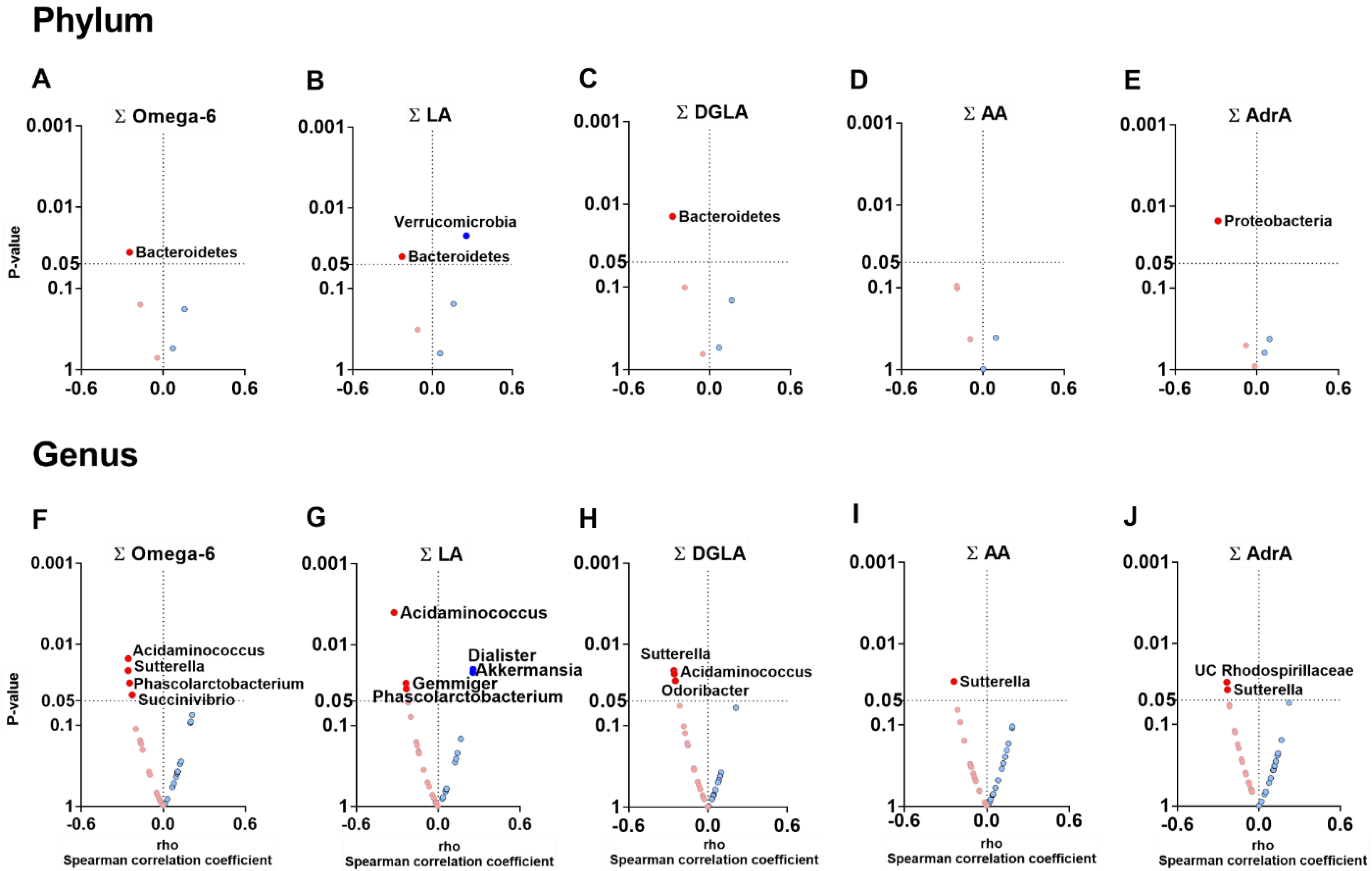
3.3. The Plasma Levels of Omega-6 Oxylipins Are Negatively Associated with the Relative Abundance of Genera Belonging to Firmicutes and Proteobacteria Phyla

At the phylum level, the total plasma levels of omega-6, LA- and DGLA-derived oxylipins were negatively associated with the relative abundances of Bacteroidetes (all rho ≥ −0.274, p ≤ 0.040; Figure 2A–C), whereas the total plasma levels of AdrA-derived oxylipins were negatively associated with the relative abundance of Proteobacteria (rho = −0.284, p = 0.013; Figure 2E). However, the total plasma levels of LA-derived oxylipins were positively associated with the relative abundance of Verrucomicrobia phylum (rho = 0.255, p = 0.022; Figure 2B). At the genus level, we found that the total plasma levels of omega-6, LA-, and DGLA-derived oxylipins were negatively associated with the relative abundance of Acidaminococcus and Phascolarctobacterium genera (Firmicutes phylum; all rho ≥ −0.326, p ≤ 0.034; Figure 2F–H). We also observed that the total plasma levels of omega-6, DGLA-, AA-, and AdrA-derived oxylipins levels were negatively associated with the relative abundance of Sutterella, Succinivibrio, and Gemmiger genera (Proteobacteria phylum; all rho ≥ −0.258, p ≤ 0.042; Figure 2F–J). Similarly, the total plasma levels of DGLA-derived oxylipins were negatively associated with the relative abundance of the Odoribacter genus (Bacteroidetes phylum; rho = −0.246, p = 0.028; Figure 2H).
The plasma levels of individuals omega-6 oxylipins LA, DGLA, AA, 5-HETE and AdrA, were negatively associated with the relative abundance of the Sutterella genus (all rho ≥ −0.313, p ≤ 0.027; Figure S1B). All these associations remained significant after adjusting for BMI, PUFA, and fish intake (Tables S3 and S4).

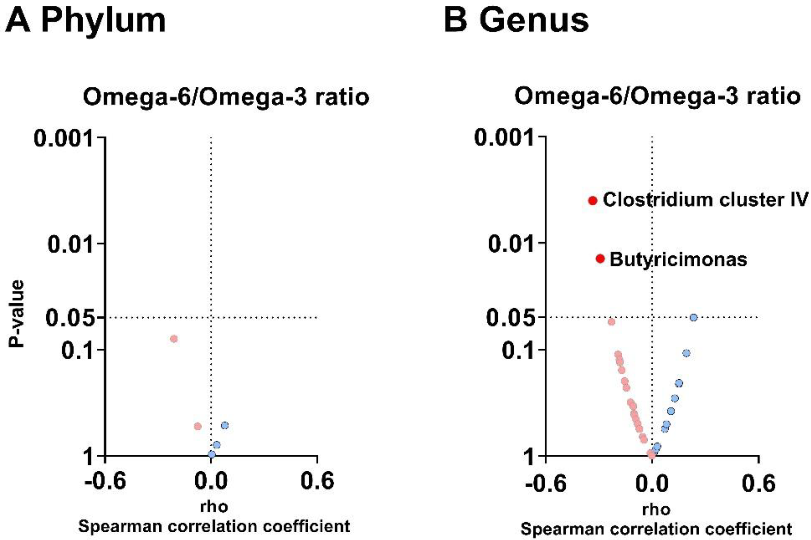
3.4. The Plasma Omega-6/Omega-3 Oxylipin Ratio Is Negatively Associated with the Relative Abundance of Clostridium Cluster IV and Butyricimonas Genera

At the phylum level, we found no associations between the omega-6/omega-3 oxylipin ratio in plasma and fecal microbiota composition (all p > 0.05; Figure 3A). On the other hand, at the genus level, the omega-6/omega-3 oxylipin ratio was negatively associated with the relative abundance of Clostridium cluster IV (Firmicutes phylum; rho = −0.334, p = 0.004; Figure 3B) and Butyricimonas genera (Bacteroidetes phylum; rho = −0.292, p = 0.014; Figure 3B) independently of BMI, PUFA, and fish intake (Table S3).

4. Discussion

In the present study, we show that plasma levels of oxylipins are linked to the fecal microbiota composition of young adults. Specifically, plasma levels of omega-3 derived oxylipins were positively associated with the relative abundance of Clostridium cluster IV and negatively associated with the relative abundance of Sutterella genera. Additionally, plasma levels of omega-6 derived oxylipins were negatively associated with the relative abundance of several genera. In contrast, the ratio of omega-6/omega-3 oxylipins in plasma was negatively associated with the relative abundance of Clostridium cluster IV and Butyricimonas genera. These findings suggest that the plasma levels of omega-3 and omega-6 derived oxylipins may modulate the relative abundance of gut microbiota composition, or vice versa, in young adults.
We found that the plasma levels of omega-3 derived oxylipins and the omega-6/omega-3 oxylipin ratio in plasma are positively and negatively associated, respectively, with the relative abundance of Clostridium cluster IV genus, which is composed of Clostridium, Eubacterium, Ruminococcus, and Anaerofilum genera. Similarly, the FAT-1 mice, capable of transforming omega-6 into omega-3 PUFAs, also showed a higher relative abundance of Clostridium cluster IV genus compared to WT mice [21]. The Clostridium cluster IV genus bacteria have been shown to exert anti-inflammatory effects and maintain intestinal health via the production of short-chain fatty acids, such as butyrate [32]. These findings suggest that higher plasma levels of omega-3 derived oxylipins could improve systemic and intestinal health through an increased relative abundance of Clostridium cluster IV genus, which produces butyrate and ultimately enhances intestinal barrier function and immunity [33]. In contrast, gut microbiota influences the production of omega-6 oxylipins, conferring host resistance to high-fat diet-induced obesity and adipose tissue inflammation in mice [34]. The above-mentioned pre-clinical studies are confirmed in the present human cohort, reinforcing the clinical benefits of omega-3 PUFAs, specifically its oxylipins, on human health.
Additionally, we identified that plasma levels of omega-3 and omega-6 oxylipins are negatively associated with the relative abundance of the Sutterella genus in feces. In line with these results, rats consuming a diet rich in the omega-6 PUFA LA for 10 weeks showed a decrease in the relative abundance of the Sutterella genus [35]. In contrast, daily oral supplementation with omega-3 PUFAs (i.e., 4 g of EPA and DHA) for eight weeks increased the relative abundance of Sutterellaceae family in feces of in middle-aged healthy humans [36]. After a wash-out period of eight weeks, the relative abundance of the Sutterellaceae family returned to baseline levels [36]. These studies show that supplementing omega-3 or omega-6 PUFAs can modulate the relative abundance of the specific bacteria in the gut. This effect might be explained by the direct effects of dietary PUFAs in the intestine since PUFAs and their downstream oxylipins can modify the fatty acid composition of the intestinal brush border membrane, increasing the intestinal alkaline phosphatase (IAP) activity in the small intestine [37,38]. Thus, we hypothesize that oxylipins could modulate IAP activity similarly to their PUFA precursors. In this regard, both supplementation of omega-3 PUFAs [21] and overproduction of omega-6 PUFAs [39] have been shown to induce the release of IAP from enterocytes, either through the modulation of resolvins (i.e., distal mediators of the anti-inflammatory/pro-resolution cascade derived from omega-3 oxylipins) [40] or the increase in the relative abundance of certain bacteria (e.g., Sutterella genus [22] that can produce lipopolysaccharides [LPS]). IAP dephosphorylates LPS [41], preventing LPS from binding to Toll-like receptor-4 (TLR4) [41], consequently avoiding the activation of the inflammatory cascade [41]. Since gut microbiota could contribute to the levels of oxylipins in response to inflammation [23], we hypothesize that plasma oxylipins could translocate to the gut lumen, as with other inflammatory mediators. Therefore, the relationship observed between omega-3 and omega-6 oxylipins plasma levels and the relative abundance of the Sutterella genus could be partially explained by the modulation of IAP activity in humans. Although this hypothesis is based on pre-clinical studies, it should be explored in future studies specifically designed. However, our results describe the relationship between the omega-3 and omega-6 oxylipins with these specific bacteria for the first time in humans. Since oxylipins are the primary mediators of PUFAs physiological effects, mainly in regulating the inflammatory and immune response, our results are of great clinical interest.

Limitations and Strengths

This study shows the following limitations: the cross-sectional study design does not allow to establishment a cause-effect relationship. Our results should be interpreted with caution due to the lack of specific sample size and power calculation for the current study. The analyses were not controlled for false discovery rate (FDR) since this approach is hypothesis-generating, and adjusting for FDR might overcorrect potentially meaningful findings. Lastly, while these results generate new hypotheses, further research is needed to elucidate the direction of the crosstalk between plasma levels of oxylipins and fecal microbiota since plasma oxylipins might influence fecal microbiota composition and vice versa. On the other hand, two major strengths in this study are: (i) we performed DNA sequencing with one of the latest technologies (Illumina platform) and using the DADA2 program that uses amplicon sequence variants instead of operational taxonomic units [42]; and (ii) RDP conducted the annotation step until the genus taxon, a methodology with an annotation error less than 10% [43].

5. Conclusions

Our study reveals that plasma omega-3 oxylipins are positively associated with the relative abundance of Clostridium cluster IV genus. In contrast, plasma levels of omega-3 and omega-6 oxylipins are negatively associated with the relative abundance of the Sutterella genus in fecal samples of young adults. These results may suggest that plasma levels of omega-3 and omega-6 oxylipins modulate human gut microbiota composition. Based on the well-known influence of omega-3 and omega-6 oxylipins on human health, our results suggest that these oxylipins may be involved in regulating the gut barrier function in humans. However, future studies should address the influence of omega-3 and omega-6 supplementation in gut microbiota health, as well as the effect of different probiotics on the regulation of oxylipins in the plasma and the gut barrier.

Supplementary Materials

The following supporting information can be downloaded at: https://www.mdpi.com/article/10.3390/nu14234991/s1, Table S1: Overview of the labelled internal standards used in the LC-MS/MS method. Table S2: Overview of the detected omega-3 and omega-6 oxylipins, including the variability observed in QC samples (expressed using RSD). Analytes showing an RSD < 30% in the QC samples were kept for further analysis. Table S3: Partial Spearman correlation between total omega-3 and omega-6 oxylipins and the relative abundance of fecal microbiota composition at the genus level. Table S4: Partial Spearman correlation between plasma levels of individual omega-3 and omega-6 oxylipins and the relative abundance of fecal microbiota composition at the genus level. Figure S1: Spearman correlations between plasma levels of individual omega-3 (Panel A) and omega-6 (Panel B) oxylipins and the abundance relative of Clostridium IV and Sutterella genera [44,45,46].

Author Contributions

H.X., L.J.-F., A.G., P.C.N.R., J.R.R. and B.M.-T. contributed to the study conception and design. H.X., L.J.-F., L.O.-A., F.J.O.-P., I.K., X.D., R.V.-V., A.L. and J.P.-D. performed material preparation and data collection. H.X., L.J.-F. and B.M.-T. performed the statistical analysis. J.R.R. and B.M.-T. have the primary responsibility for the final content. All authors have read and agreed to the published version of the manuscript.

Funding

The study was supported by the Spanish Ministry of Economy and Competitiveness via Fondo de Investigación Sanitaria del Instituto de Salud Carlos III (PI13/01393) and PTA 12264-I, Retos de la Sociedad (DEP2016- 79512-R), and European Regional Development Funds (ERDF), by the Spanish Ministry of Education (FPU16/05159, FPU16/02828, FPU17/01523 and FPU19/01609), the Fundación Iberoamericana de Nutrición (FINUT), the University of Granada Plan Propio de Investigación 2016-Excellence actions: Unit of Excellence on Exercise and Health (UCEES), AstraZeneca HealthCare Foundationand by the Junta de Andalucía, Consejería de Conocimiento, Investigación y Universidades (ERDF, SOMM17/6107/UGR), the Chinese Scholarship Council (CSC, No. 201707060012 to XD), Fundación Alfonso Martin Escudero, and Maria Zambrano fellowship by the Ministerio de Universidades y la Unión Europea –NextGenerationEU (RR_C_2021_04). AL is supported by the funds of the European Commission through the “European funds for regional development” (EFRE) as well as by the regional Ministry of Economy, Science and Digitalization of Saxony-Anhalt as part of the Autonomy in old Age “(AiA) research group for “LiLife” Project (Project ID: ZS/2018/11/95324).

Institutional Review Board Statement

The study protocol and experimental design were applied by the last revised ethical guidelines of the Declaration of Helsinki. The study was approved by the Ethics Committee on Human Research of the University of Granada (no. 924) and the Servicio Andaluz de Salud (Centro de Granada, CEI-Granada).

Informed Consent Statement

Informed consent was obtained from all subjects involved in the study.

Data Availability Statement

The data supporting this study’s findings are available from the corresponding author upon reasonable request, as the study consists of a high number of participants and outcomes and requires specific knowledge for data interpretation.

Acknowledgments

This study is part of a PhD thesis conducted within the Biomedicine Doctoral Studies Program of the University of Granada, Spain. The authors would like to thank the Data Integration Center of University Medicine Magdeburg team for local data-analysis solutions supported by MIRACUM and funded by the German Federal Ministry of Education and Research (BMBF) (FKZ 01ZZ1801H).

Conflicts of Interest

The authors declare no conflict of interest.

References

  1. Hotamisligil, G. Inflammation and Metabolic Disorders. Nature 2006, 444, 860–867. [Google Scholar] [CrossRef]
  2. Ruiz-Núñez, B.; Pruimboom, L.; Dijck-Brouwer, D.A.J.; Muskiet, F.A.J. Lifestyle and Nutritional Imbalances Associated with Western Diseases: Causes and Consequences of Chronic Systemic Low-Grade Inflammation in an Evolutionary Context. J. Nutr. Biochem. 2013, 24, 1183–1201. [Google Scholar] [CrossRef] [Green Version]
  3. Nathan, C.; Ding, A. Nonresolving Inflammation. Cell 2010, 140, 871–882. [Google Scholar] [CrossRef] [PubMed] [Green Version]
  4. Leaf, A.; Weber, P.C. A New Era for Science in Nutrition. Am. J. Clin. Nutr. 1987, 45, 1048–1053. [Google Scholar] [CrossRef]
  5. Simopoulos, A. The Importance of the Omega-6/Omega-3 Fatty Acid Ratio in Cardiovascular Disease and Other Chronic Diseases. Exp. Biol. Med. 2008, 233, 674–688. [Google Scholar] [CrossRef] [PubMed]
  6. Yeung, J.; Hawley, M.; Holinstat, M. The Expansive Role of Oxylipins on Platelet Biology. J. Mol. Med. 2017, 95, 575–588. [Google Scholar] [CrossRef] [Green Version]
  7. Gabbs, M.; Leng, S.; Devassy, J.G.; Monirujjaman, M.; Aukema, H.M. Advances in Our Understanding of Oxylipins Derived from Dietary PUFAs 1,2. Adv. Nutr. 2015, 6, 513–540. [Google Scholar] [CrossRef] [Green Version]
  8. Calder, P.C. Omega-3 Fatty Acids and Inflammatory Processes. Nutrients 2010, 2, 355–374. [Google Scholar] [CrossRef] [PubMed] [Green Version]
  9. Dyall, S.C.; Balas, L.; Bazan, N.G.; Brenna, J.T.; Chiang, N.; da Costa Souza, F.; Dalli, J.; Durand, T.; Galano, J.-M.; Lein, P.J.; et al. Polyunsaturated Fatty Acids and Fatty Acid-Derived Lipid Mediators: Recent Advances in the Understanding of Their Biosynthesis, Structures, and Functions. Prog. Lipid Res. 2022, 86, 101165. [Google Scholar] [CrossRef]
  10. Robertson, R.C.; Kaliannan, K.; Strain, C.R.; Ross, R.P.; Stanton, C.; Kang, J.X. Maternal Omega-3 Fatty Acids Regulate Offspring Obesity through Persistent Modulation of Gut Microbiota. Microbiome 2018, 6, 95. [Google Scholar] [CrossRef]
  11. Sekirov, I.; Finlay, B.B. Human and Microbe: United We Stand. Nat. Med. 2006, 12, 736–737. [Google Scholar] [CrossRef]
  12. Arumugam, M.; Raes, J.; Pelletier, E.; Le Paslier, D.; Yamada, T.; Mende, D.R.; Fernandes, G.R.; Tap, J.; Bruls, T.; Batto, J.M.; et al. Enterotypes of the Human Gut Microbiome. Nature 2011, 473, 174–180. [Google Scholar] [CrossRef] [Green Version]
  13. Segata, N.; Haake, S.K.; Mannon, P.; Lemon, K.P.; Waldron, L.; Gevers, D.; Huttenhower, C.; Izard, J. Composition of the Adult Digestive Tract Bacterial Microbiome Based on Seven Mouth Surfaces, Tonsils, Throat and Stool Samples. Genome Biol. 2012, 13, R42. [Google Scholar] [CrossRef] [PubMed] [Green Version]
  14. Montalto, M.; D’Onofrio, F.; Gallo, A.; Cazzato, A.; Gasbarrini, G. Intestinal Microbiota and Its Functions. Dig. Liver Dis. Suppl. 2009, 3, 30–34. [Google Scholar] [CrossRef]
  15. van de Guchte, M.; Blottière, H.M.; Doré, J. Humans as Holobionts: Implications for Prevention and Therapy. Microbiome 2018, 6, 81. [Google Scholar] [CrossRef]
  16. Hill, D.A.; Artis, D. Intestinal Bacteria and the Regulation of Immune Cell Homeostasis. Annu. Rev. Immunol. 2010, 28, 623–667. [Google Scholar] [CrossRef] [Green Version]
  17. Tomasello, G.; Mazzola, M.; Leone, A.; Sinagra, E.; Zummo, G.; Farina, F.; Damiani, P.; Cappello, F.; Geagea, A.G.; Jurjus, A.; et al. Nutrition, Oxidative Stress and Intestinal Dysbiosis: Influence of Diet on Gut Microbiota in Inflammatory Bowel Diseases. Biomed. Pap. 2016, 160, 461–466. [Google Scholar] [CrossRef] [PubMed] [Green Version]
  18. David, L.A.; Maurice, C.F.; Carmody, R.N.; Gootenberg, D.B.; Button, J.E.; Wolfe, B.E.; Ling, A.V.; Devlin, A.S.; Varma, Y.; Fischbach, M.A.; et al. Diet Rapidly and Reproducibly Alters the Human Gut Microbiome. Nature 2014, 505, 559–563. [Google Scholar] [CrossRef] [Green Version]
  19. Moszak, M.; Szulińska, M.; Bogdański, P. You Are What You Eat—The Relationship between Diet, Microbiota, and Metabolic Disorders—A Review. Nutrients 2020, 12, 1096. [Google Scholar] [CrossRef] [Green Version]
  20. Fu, Y.; Wang, Y.; Gao, H.; Li, D.; Jiang, R.; Ge, L.; Tong, C.; Xu, K. Associations among Dietary Omega-3 Poly-unsaturated Fatty Acids, the Gut Microbiota, and Intestinal Immunity. Mediat. Inflamm. 2021, 2021, 8879227. [Google Scholar] [CrossRef]
  21. Kaliannan, K.; Wang, B.; Li, X.-Y.; Kim, K.-J.; Kang, J.X. A Host-Microbiome Interaction Mediates the Opposing Effects of Omega-6 and Omega-3 Fatty Acids on Metabolic Endotoxemia. Sci. Rep. 2015, 5, 11276. [Google Scholar] [CrossRef] [Green Version]
  22. Kaliannan, K.; Li, X.-Y.; Wang, B.; Pan, Q.; Chen, C.-Y.; Hao, L.; Xie, S.; Kang, J.X. Multi-Omic Analysis in Transgenic Mice Implicates Omega-6/Omega-3 Fatty Acid Imbalance as a Risk Factor for Chronic Disease. Commun. Biol. 2019, 2, 276. [Google Scholar] [CrossRef] [Green Version]
  23. Ávila-Román, J.; Arreaza-Gil, V.; Cortés-Espinar, A.J.; Soliz-Rueda, J.R.; Mulero, M.; Muguerza, B.; Arola-Arnal, A.; Arola, L.; Torres-Fuentes, C. Impact of Gut Microbiota on Plasma Oxylipins Profile under Healthy and Obesogenic Conditions. Clin. Nutr. 2021, 40, 1475–1486. [Google Scholar] [CrossRef] [PubMed]
  24. Sanchez-Delgado, G.; Martinez-Tellez, B.; Olza, J.; Aguilera, C.M.; Labayen, I.; Ortega, F.B.; Chillon, P.; Fernandez-Reguera, C.; Alcantara, J.M.A.A.; Martinez-Avila, W.D.; et al. Activating Brown Adipose Tissue through Exercise (ACTIBATE) in Young Adults: Rationale, Design and Methodology. Contemp. Clin. Trials 2015, 45, 416–425. [Google Scholar] [CrossRef] [PubMed]
  25. Jurado-Fasoli, L.; Di, X.; Kohler, I.; Osuna-Prieto, F.J.; Hankemeier, T.; Krekels, E.; Harms, A.C.; Yang, W.; Garcia-Lario, J.V.; Fernández-Veledo, S.; et al. Omega-6 and Omega-3 Oxylipins as Potential Markers of Cardiometabolic Risk in Young Adults. Obesity 2022, 30, 50–61. [Google Scholar] [CrossRef]
  26. R Core Team. R: A Language and Environment for Statistical Computing, v. 4.2.2; R Core Team: Vienna, Austria, 2019. [Google Scholar]
  27. Cole, J.R.; Wang, Q.; Fish, J.A.; Chai, B.; McGarrell, D.M.; Sun, Y.; Brown, C.T.; Porras-Alfaro, A.; Kuske, C.R.; Tiedje, J.M. Ribosomal Database Project: Data and Tools for High Throughput RRNA Analysis. Nucleic Acids Res. 2014, 42, D633–D642. [Google Scholar] [CrossRef] [Green Version]
  28. Jurado-Fasoli, L.; Amaro-Gahete, F.J.; Merchan-Ramirez, E.; Labayen, I.; Ruiz, J.R. Relationships between Diet and Basal Fat Oxidation and Maximal Fat Oxidation during Exercise in Sedentary Adults. Nutr. Metab. Cardiovasc. Dis. 2021, 31, 1087–1101. [Google Scholar] [CrossRef]
  29. Jurado-Fasoli, L.; Amaro-Gahete, F.J.; Arias-Tellez, M.J.; Gil, A.; Labayen, I.; Ruiz, J.R. Relationship between Dietary Factors and S-Klotho Plasma Levels in Young Sedentary Healthy Adults. Mech. Ageing Dev. 2021, 194, 111435. [Google Scholar] [CrossRef]
  30. Jurado-Fasoli, L.; Merchan-Ramirez, E.; Martinez-Tellez, B.; Acosta, F.M.; Sanchez-Delgado, G.; Amaro-Gahete, F.J.; Muñoz Hernandez, V.; Martinez-Avila, W.D.; Ortiz-Alvarez, L.; Xu, H.; et al. Association between Dietary Factors and Brown Adipose Tissue Volume/18F-FDG Uptake in Young Adults. Clin. Nutr. 2021, 40, 1997–2008. [Google Scholar] [CrossRef]
  31. Vioque, J.; Navarrete-Muñoz, E.-M.; Gimenez-Monzó, D.; García-de-la-Hera, M.; Granado, F.; Young, I.S.; Ramón, R.; Ballester, F.; Murcia, M.; Rebagliato, M.; et al. Reproducibility and Validity of a Food Frequency Questionnaire among Pregnant Women in a Mediterranean Area. Nutr. J. 2013, 12, 26. [Google Scholar] [CrossRef]
  32. Guo, P.; Zhang, K.; Ma, X.; He, P. Clostridium Species as Probiotics: Potentials and Challenges. J. Anim. Sci. Biotechnol. 2020, 11, 24. [Google Scholar] [CrossRef]
  33. Liu, H.; Wang, J.; He, T.; Becker, S.; Zhang, G.; Li, D.; Ma, X. Butyrate: A Double-Edged Sword for Health? Adv. Nutr. 2018, 9, 21–29. [Google Scholar] [CrossRef] [PubMed] [Green Version]
  34. Miyamoto, J.; Igarashi, M.; Watanabe, K.; Karaki, S.I.; Mukouyama, H.; Kishino, S.; Li, X.; Ichimura, A.; Irie, J.; Sugimoto, Y.; et al. Gut Microbiota Confers Host Resistance to Obesity by Metabolizing Dietary Poly-unsaturated Fatty Acids. Nat. Commun. 2019, 10, 4007. [Google Scholar] [CrossRef] [Green Version]
  35. Shrestha, N.; Sleep, S.L.; Cuffe, J.S.M.; Holland, O.J.; McAinch, A.J.; Dekker Nitert, M.; Hryciw, D.H. Pregnancy and Diet-Related Changes in the Maternal Gut Microbiota Following Exposure to an Elevated Linoleic Acid Diet. Am. J. Physiol.-Endocrinol. Metab. 2020, 318, E276–E285. [Google Scholar] [CrossRef] [PubMed]
  36. Watson, H.; Mitra, S.; Croden, F.C.; Taylor, M.; Wood, H.M.; Perry, S.L.; Spencer, J.A.; Quirke, P.; Toogood, G.J.; Lawton, C.L.; et al. A Randomised Trial of the Effect of Omega-3 Polyunsaturated Fatty Acid Supplements on the Human Intestinal Microbiota. Gut 2018, 67, 1974–1983. [Google Scholar] [CrossRef]
  37. Sharma, U.; Singh, S.K.; Pal, D.; Khajuria, R.; Mandal, A.K.; Prasad, R. Implication of BBM Lipid Composition and Fluidity in Mitigated Alkaline Phosphatase Activity in Renal Cell Carcinoma. Mol. Cell. Biochem. 2012, 369, 287–293. [Google Scholar] [CrossRef]
  38. Stenson, W.F.; Seetharam, B.; Talkad, V.; Pickett, W.; Dudeja, P.; Brasitus, T.A. Effects of Dietary Fish Oil Supplementation on Membrane Fluidity and Enzyme Activity in Rat Small Intestine. Biochem. J. 1989, 263, 41–45. [Google Scholar] [CrossRef] [PubMed] [Green Version]
  39. Ghosh, S.; DeCoffe, D.; Brown, K.; Rajendiran, E.; Estaki, M.; Dai, C.; Yip, A.; Gibson, D.L. Fish Oil Attenuates Omega-6 Polyunsaturated Fatty Acid-Induced Dysbiosis and Infectious Colitis but Impairs LPS Dephosphorylation Activity Causing Sepsis. PLoS ONE 2013, 8, e55468. [Google Scholar] [CrossRef] [PubMed]
  40. Campbell, E.L.; MacManus, C.F.; Kominsky, D.J.; Keely, S.; Glover, L.E.; Bowers, B.E.; Scully, M.; Bruyninckx, W.J.; Colgan, S.P. Resolvin E1-Induced Intestinal Alkaline Phosphatase Promotes Resolution of Inflammation through LPS Detoxification. Proc. Natl. Acad. Sci. USA 2010, 107, 14298–14303. [Google Scholar] [CrossRef] [Green Version]
  41. Lallès, J.-P. Intestinal Alkaline Phosphatase: Multiple Biological Roles in Maintenance of Intestinal Homeostasis and Modulation by Diet. Nutr. Rev. 2010, 68, 323–332. [Google Scholar] [CrossRef]
  42. Chiarello, M.; McCauley, M.; Villéger, S.; Jackson, C.R. Ranking the Biases: The Choice of OTUs vs. ASVs in 16S RRNA Amplicon Data Analysis Has Stronger Effects on Diversity Measures than Rarefaction and OTU Identity Threshold. PLoS ONE 2022, 17, e0264443. [Google Scholar] [CrossRef] [PubMed]
  43. Edgar, R. Taxonomy Annotation and Guide Tree Errors in 16S RRNA Databases. PeerJ 2018, 6, e5030. [Google Scholar] [CrossRef] [PubMed] [Green Version]
  44. Di Zazzo, A.; Yang, W.; Coassin, M.; Micera, A.; Antonini, M.; Piccinni, F.; De Piano, M.; Kohler, I.; Harms, A.C.; Hankemeier, T.; et al. Signaling lipids as diagnostic biomarkers for ocular surface cicatrizing conjunctivitis. J. Mol. Med. 2020, 98, 751–760. [Google Scholar] [CrossRef] [PubMed] [Green Version]
  45. Van Der Kloet, F.M.; Bobeldijk, I.; Verheij, E.R.; Jellema, R.H. Analytical error reduction using single point calibration for accurate and precise metabolomic phenotyping. J. Proteome Res. 2009, 8, 5132–5141. [Google Scholar] [CrossRef] [PubMed]
  46. McMurdie, P.J.; Holmes, S. Phyloseq: An R Package for Reproducible Interactive Analysis and Graphics of Microbiome Census Data. PLoS ONE 2013, 8, e61217. [Google Scholar] [CrossRef] [PubMed]
Figure 1. Volcano plots show associations between plasma levels of omega-3 oxylipins and the relative abundance of fecal microbiota composition at the phylum and genus levels. Total omega-3 (A,E), ALA- (B,F), EPA- (C,G), and DHA- (D,H) derived oxylipins were computed from the individual peak area ratio of each oxylipin within each group. The X-axis represents Spearman’s correlation coefficients, whereas the Y-axis represents the p values of the correlations. Only significant associations (p < 0.05) were annotated with the name of the phylum or genus. Blue dots represent positive associations, whereas red dots represent negative associations. Abbreviations: ALA: α-linolenic acid; DHA: docosahexaenoic acid; EPA: eicosapentaenoic acid.
Nutrients 14 04991 g001
Figure 2. Volcano plots showing associations between the plasma levels of omega-6 oxylipins and the relative abundance of fecal microbiota composition at phylum and genus levels. Total omega-6 (A,F), LA- (B,G), DGLA- (C,H), AA- (D,I), and AdrA- (E,J) derived oxylipins groups were computed from the individual area peak ratio of each oxylipins group. The X-axis represents Spearman’s correlation coefficients, whereas the Y-axis represents the p values of the correlations. Only significant correlations (p < 0.05) were annotated with the name of the phylum or genus. Blue dots represent positive correlations, whereas red dots represent negative correlations. Abbreviations: AA: arachidonic acid; AdrA: adrenic acid; DGLA: dihomo-γ-linolenic acid; LA: linoleic acid; UC: unclassified.
Nutrients 14 04991 g002
Figure 3. Volcano plots showing the associations between the omega-6/omega-3 oxylipin ratio in plasma and the relative abundance of fecal microbiota composition at the phylum (A) and genus levels (B). The X-axis represents Spearman’s correlation coefficients, whereas the Y-axis represents the p values of the correlations. Only significant correlations (p < 0.05) were annotated with the name of the phylum or genus. Blue dots represent positive correlations, whereas red dots represent negative correlations.
Nutrients 14 04991 g003
Table 1. Characteristics of the study participants.
NMean±SD
Sex (women, %)8059 (73.8%)
Age (years)8021.9±2.2
Body composition
Lean mass (kg)8041.0±8.9
Fat mass (kg)8036.3±7.8
Body mass index (kg/m2)8024.7±4.7
Dietary intake
Energy (kcal/day)801920±489
PUFAs (g/day)8013±5
Fish (servings/week)775±3
Total plasma levels of derived-oxylipins (peak area ratio)
Omega-380172.8±65.6
ALA8012.0±5.1
EPA8019.0±10.3
DHA80141.7±54.6
Omega-68099.5±27.8
LA8028.6±10.6
DGLA801.0±0.5
AA8066.4±22.2
AdrA801.0±0.6
Omega-6/omega-3 oxylipin ratio800.6±0.1
Fecal microbiota composition (phylum, %)
Actinobacteria801.6±1.6
Bacteroidetes8039.6±9.0
Firmicutes8048.8±9.7
Proteobacteria806.5±4.8
Verrucomicrobia802.3±4.3
Data are presented as mean and standard deviation (SD), otherwise stated. Total oxylipins in each group from the individual peak area ratio of each oxylipin; AA: arachidonic acid; AdrA: adrenic acid; ALA: α-linolenic acid; DHA: docosahexaenoic acid; DGLA: dihomo-γ-linolenic acid; EPA: eicosapentaenoic acid; LA: linoleic acid; PUFAs: polyunsaturated fatty acids.
Publisher’s Note: MDPI stays neutral with regard to jurisdictional claims in published maps and institutional affiliations.

