Article

The Effects of Persimmon (Diospyros kaki L.f.) Oligosaccharides on Features of the Metabolic Syndrome in Zebrafish

1
Department of Oriental Medicine Biotechnology, College of Life Sciences, Kyung Hee University Global Campus, Yongin-si 17104, Korea
2
Department of Pharmacognosy and Pharmaceutical Botany, Faculty of Pharmaceutical Sciences, Prince of Songkla University, Hat Yai 90112, Thailand
3
School of Pharmacy, Sungkyunkwan University, Suwon-si 16419, Korea
*
Author to whom correspondence should be addressed.
Nutrients 2022, 14(16), 3249; https://doi.org/10.3390/nu14163249
Submission received: 25 April 2022 / Revised: 27 July 2022 / Accepted: 3 August 2022 / Published: 9 August 2022
(This article belongs to the Section Carbohydrates)

Abstract

:
Metabolic syndrome has become a global health care problem since it is rapidly increasing worldwide. The search for alternative natural supplements may have potential benefits for obesity and diabetes patients. Diospyros kaki fruit extract and its oligosaccharides, including gentiobiose, melibiose, and raffinose, were examined for their anti-insulin resistance and obesity-preventing effect in zebrafish larvae. The results show that D. kaki oligosaccharides improved insulin resistance and high-fat-diet-induced obesity in zebrafish larvae, evidenced by enhanced β-cell recovery, decreased abdominal size, and reduced the lipid accumulation. The mechanism of the oligosaccharides, molecular docking, and enzyme activities of PTP1B were investigated. Three of the oligosaccharides had a binding interaction with the catalytic active sites of PTP1B, but did not show inhibitory effects in an enzyme assay. The catalytic residues of PTP1B were typically conserved and the cellular penetration of the cell membrane was necessary for the inhibitors. The results of the mechanism of action study indicate that D. kaki fruit extract and its oligosaccharides affected gene expression changes in inflammation- (TNF-α, IL-6, and IL-1β), lipogenesis- (SREBF1 and FASN), and lipid-lowering (CPT1A)-related genes. Therefore, D. kaki fruit extract and its oligosaccharides may have a great potential for applications in metabolic syndrome drug development and dietary supplements.

1. Introduction

Recently, metabolic syndrome has become a common occurrence in the world, with a prevalence of 10–40% [1]. The consumption of quality fats and carbohydrates is pivotal to improve blood lipids and glycemic index in metabolic syndrome patients [2]. Type 2 diabetes is related to hyperglycemia and insulin resistance, which are mainly caused by obesity [3]. Insulin resistance is the early stage of type 2 diabetes, when high glucose blood levels lead to an increase in pancreatic insulin production [4], which results in increased glucose output and lipogenesis via the liver [5].
There is considerable evidence showing that the intracellular protein PTP1B (Protein Tyrosine Phosphatase 1B) is upregulated in muscle and adipose tissues of obese and insulin-resistant mice [6]. The inhibition of PTP1B improved energy expenditure, reduced fat deposition, and prevented weight gain in obese mice [7]. PTP1B is able to impair the glucose uptake by interacting with IR and IRS-1 to induce the tyrosine phosphorylation hydrolysis of insulin activity [8].
Using zebrafish (Danio rerio) as an animal model, as it has a rapid reproduction and morphology development, allows drug screening in a short period of time, lower maintenance costs, and small amount of test samples. Moreover, zebrafish share 84% similarity to mammalian disease genes [9]. The similarity of the zebrafish pancreas structure to that of mammals [10], of normal blood glucose levels (human: 70–120 mg/dL; adult zebrafish: 50–75 mg/dL) [11], and adipocytes in larvae make zebrafish a potential tool to investigate metabolic syndrome [12]. Several previous studies used zebrafish as a model of metabolic syndromes, such as Moss et al. [13] and Intine et al. [14], who induced diabetes in zebrafish with streptozotocin and observed the regeneration of the pancreas in adult zebrafish.
Diospyros kaki (persimmon) fruit is known as a valuable nutrient source. The dried pulp of D. kaki decreased the plasma glucose levels and triglycerides of and inhibited digestive enzymes in rats [15]. Moreover, D. kaki leaf has been reported to lower blood glucose levels in a rodent model by inhibiting the pancreas alpha-amylase enzyme [16]. The triterpenoids from their leaves showed an inhibitory activity of PTP1B [17] and the proanthocyanidins from the peel had an effect on α-glucosidase or α-amylase enzyme [18].
Oligosaccharides are natural products that have gained attention in the treatment of metabolic syndrome [19]. Because the glycosidic bond between the monomeric sugar units of oligosaccharides cannot be broken down by human gastrointestinal digestive enzymes, they are nondigestible in the human digestive system [20]. Previous research showed several beneficial effects of functional oligosaccharides, such as improving pancreas function, insulin and leptin resistance, inflammation, inhibiting α-glucosidase, and regulating gut microbiota and hormones, though largely the mechanism still remains unclear. Thus, studies on the mode of action of oligosaccharides are beneficial to metabolic syndrome drug development [19].

2. Materials and Methods

2.1. Preparation and Extraction of D. kaki Fruit Extract and Source of the Oligosaccharides

D. kaki fruits were purchased from the market in Suwon, South Korea. Fruit flesh was cut into small pieces. A total of 100 g of the flesh and 100 mL of distilled water were blended using a food processor and then filtered. The juice of D. kaki fruit was evaporated by Smart Evaporator S Spiral Plug (BioChromato, Kanagawa-ken, Japan) at the temperature of 55 °C until completely dried to yield 7.64% based on the fresh weight. Gentiobiose, melibiose, and raffinose were purchased from Tokyo Chemical Industry Co., Ltd. (Tokyo, Japan).

2.2. The Investigation of the Chemical Constituent of D. kaki Fruit Extract

The chemical constituent analysis of D. kaki was performed using ultra-high performance liquid chromatograph-mass spectrometer/mass spectrometer 1290 Infinity II UHPLC-6545 Q-TOF (Agilent Technologies, Santa Clara, CA, USA) with an Agilent EclipsePlus C18 column (2.1 mm × 50 mm i.d., 1.8 μm; flow rate: 0.2 mL/min) maintained at 35 °C and the injection volume was 10 µL. To optimize the separation of the sample, two types of mobile phase included 0.1% formic acid–water (A) and acetonitrile (B). The optimized linear gradient elution was performed as follows: 0–20 min, 18% B; 21–35 min, 20% B; and 36–70 min, 30% B. Mass spectroscopy was performed on quadruple time-of-flight mass spectrometer (Q-TOF MS) and characterization equipped with electrospray ionization Dual AJS ESI. Mass spectrometry data were acquired in the negative and positive ionization modes covering the range from m/z 100 to m/z 1500 with scan rate of 2 spectra/s, nebulization with nitrogen at 35 psi, dry gas flow of 13 L/min at a temperature of 325 °C, and skimmer 1 voltage 65 V, and collision energy was set to 10%, 20%, and 40%. The instrument was operated and analyzed using Agilent Technologies. Mass spectra were analyzed by MS/MS spectral mass library.

2.3. Animals

Zebrafish (Danio rerio) were maintained in S type system (1500 × 400 × 2050 mm, Woojung Bio, Inc., Suwon, Korea) at 28.5 °C under a 10/14 dark–light cycle. Three pairs of adult zebrafish were breeding overnight, and the embryos were collected three hours post-fertilization (hpf). The embryos were washed and renewed with 0.03% sea salt solution or treatment solution every day until the experiment was performed. The zebrafish maintenance was followed standard protocols approved by the Animal Care and Use Committee of Kyung Hee University (KHUASP[SE]-15-10).

2.4. Anti-Insulin Resistance in Zebrafish

Three-day post-fertilization (dpf) wild-type zebrafish larvae (20 larvae/group) were used for the experiments. A total of 10 µM of the biosynthetic human insulin solution (Novo Nordisk, Kalundborg, Denmark) was used for degenerating the pancreatic islet (PI) of the zebrafish larvae. After 48 h of induction, the larvae were rinsed with 0.03% sea salt solution and treated by 10 µg/mL of fruit extracts (contained approximate amounts of gentiobiose, melibiose, and raffinose at 11.7, 7, 1.3 ng/mL, respectively) and 1 µM of pure compounds (342.3 ng/mL of gentiobiose, 342.3 ng/mL of melibiose, and 504.4 ng/mL of raffinose) for another 48 h.
Finally, the larvae were stained with 40 µM 2-NBDG (Invitrogen, Life Technologies, Grand Island, NY, USA) for 30 min and imaging PI picture by a fluorescence microscope (Olympus, Tokyo, Japan). PI size was measured and analyzed by Focus Lite software (Focus Co., Daejeon, Korea) to monitor β-cell ablation [21].

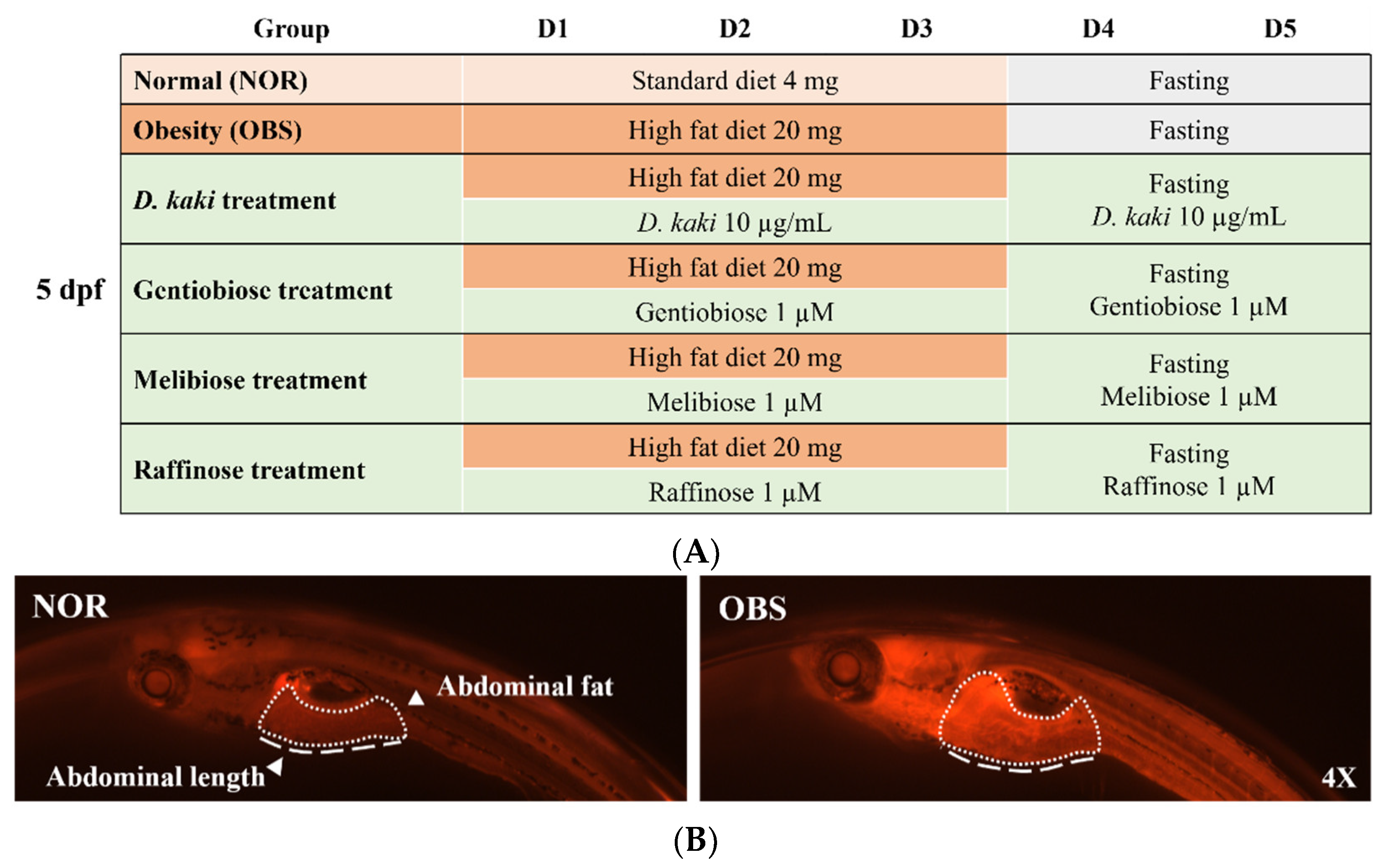
2.5. Obesity-Preventing Effect in Zebrafish

A total of 5 dpf of wide-type zebrafish were placed in 20 mL of 0.03% sea salt solution (20 larvae per group). The normal group (NOR) was fed by 4 mg of commercial standard diet (ACE KOREA, Korea), and the obesity group (OBS) was fed by 20 mg of high-fat diet (hard-boiled egg yolk). A total of 10 µg/mL of D. kaki fruit extract and 1 µM of the oligosaccharides were provided when start feeding induction to explore the obesity-preventing effect. After three days of feeding, all groups were starved and the solution was renewed. The treatment groups remained with the D. kaki fruit extract and oligosaccharide maintenance. The fasting and treatment took another two days (the experimental setup is shown in Figure 1A). To monitor initial adiposity, larvae belly length was measured and imaged the abdominal part of larvae using 0.5 µg/mL of Nile red staining and fluorescence microscopy on the fifth day of the experiment (image shown in Figure 1B).

2.6. Molecular Docking of Isolated Compounds and PTP1B

The analysis of molecular binding interactions of oligosaccharide structures with PTP1B enzyme were performed using AutoDock Vina version 1.1.2. The structure of PTP1B (PDB code: 1T49) was downloaded from RCSB Protein Data Bank. The 3D structure of gentiobiose (CID: 441422), melibiose (CID: 440658), and raffinose (CID: 439242) were obtained from PubChem Compound (NCBI). Discovery Studio 2019 was used for removing ligands and water molecules. Hydrogen atoms and charges were then added using the AutoDock Tool. The grid maps were generated with a default spacing of 0.375 Å and 50 × 50 × 50 grid box size. The X, Y, Z grid maps centers were 51.013, 27.372, and 22.603, respectively. All of the molecular bonds of the isolated compounds were set to rotatable. All torsions were also allowed to rotate. The interactions of molecular docking were analyzed using Discovery Studio 2019. The best affinity of the molecular interaction was determined by the lowest binding energy of hydrogen bond between the active residues and compounds [22].

2.7. Enzyme Activity Assays of PTP1B

A total of 1 mM EDTA (Sigma-Aldrich Co., St. Louis, MO, USA), 0.15 M NaCl (Sigma-Aldrich Co., St. Louis, MO, USA), and 3 units/mL of human-recombinant PTP1B (BioVision, Inc., Milpitas, CA, USA) were dissolved in sodium acetate buffer (pH 5.5) to prepare the reaction mixture. Suramin (Sigma-Aldrich Co., St. Louis, MO, USA) was used as a positive control. A total of 100 µL of the samples were mixed with 50 µL of reaction mixture and incubated at 30 °C for 10 min. A total of 50 µL of the substrate 10 mM ρ-nitrophenyl phosphate (PNPP) (Sigma-Aldrich Co., St. Louis, MO, USA) was added and incubated at 30 °C for 20 min. Then, 100 mM of NaHCO3 (Sigma-Aldrich Co., St. Louis, MO, USA) was added to stop the reaction and the absorbance was measured at 415 nm [23].

2.8. Real Time-Quantitative Polymerase Chain Reaction (RT-qPCR)

Zebrafish larvae were fed with a high-fat diet for three days and then treated with the D. kaki fruit extract and oligosaccharides for another two days. The extraction of total RNA was performed using TRIzolTM reagent (Thermo Fisher Scientific, Seoul, Korea). Reverse transcription was performed by Reverse Aid First Strand cDNA Synthesis Kit (Thermo Fisher Scientific, Seoul, Korea) for the synthesis of cDNA. The qPCR was performed in 10 μL reactions containing 5 μL of SYBR Select Master Mix (Applied Biosystems, Waltham, MA, USA, Thermo Fisher Scientific), 1 μL of cDNA template, 1 μL of forward primer (10 pmol), 1 μL of reverse primer (10 pmol), and 2 μL of RNase free water. The qPCR parameters were initial denaturation at 95 °C for 5 min, followed by 45 cycles of 95 °C for 15 s, 60 °C for 15 s and 72 °C for 20 s, and then 73 °C for 5 min. Primer sequences are listed in Table 1. The expression of genes was analyzed by the 2−ΔΔCt method.

2.9. Statistical Analysis

The result data were expressed as mean with standard error of the mean (±SEM). A one-way ANOVA with Dunnett’s post hoc test of GraphPad Prism version 8 was used to verifying the statistical significance. A p value < 0.05 (*) was considered statistically significant.

3. Results and Discussions

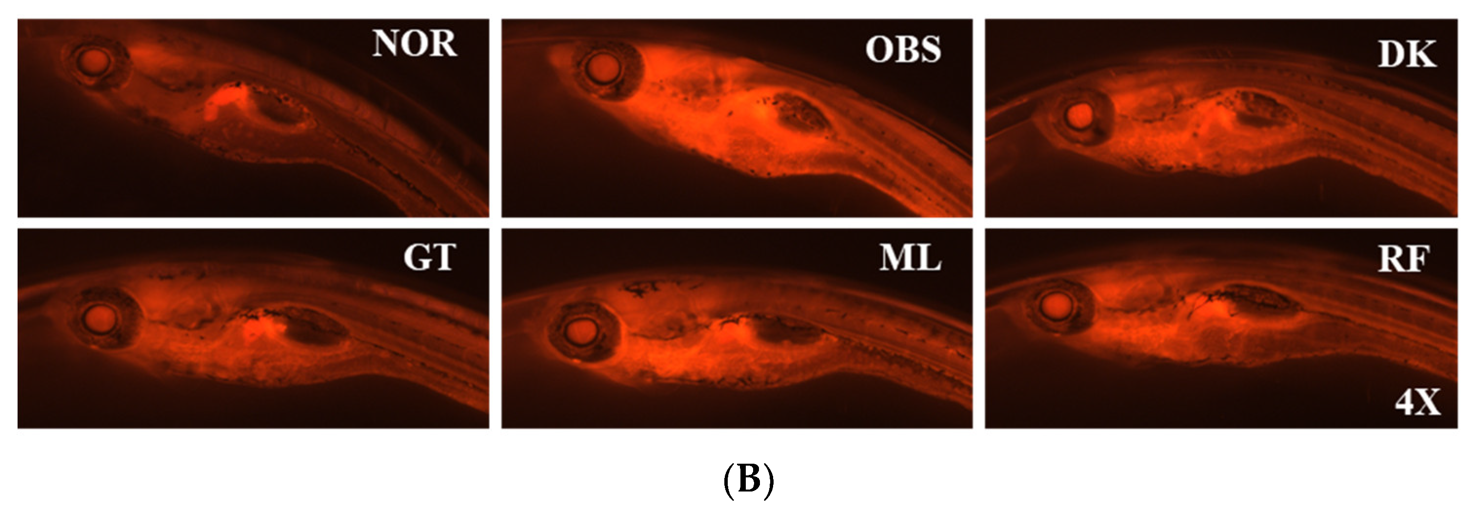
3.1. Screening of D. kaki Fruits Extracts for Anti-Insulin Resistance in Zebrafish

The investigation of anti-insulin resistance in larvae zebrafish was established to screen the natural products for the treatment of metabolic syndrome. A high dose of human insulin (10 µM) was provided to stimulated insulin resistance in zebrafish larvae by downregulating the immune system and insulin signaling pathway. The PI size of zebrafish larvae were measured to evaluate the β-cell existence [20]. The human insulin significantly lowered the size of the larvae PI compared to the normal group (p < 0.0001), while the treatment with 10 µg/mL of D. kaki fruit extract significantly increased the PI size compared to the insulin-induced group (p < 0.01) (Figure 2). Thus, the fruit extract of D. kaki was considered for the further investigation of anti-metabolic syndrome activities by their enhancing β-cell subsistence in insulin-resistant larvae zebrafish.

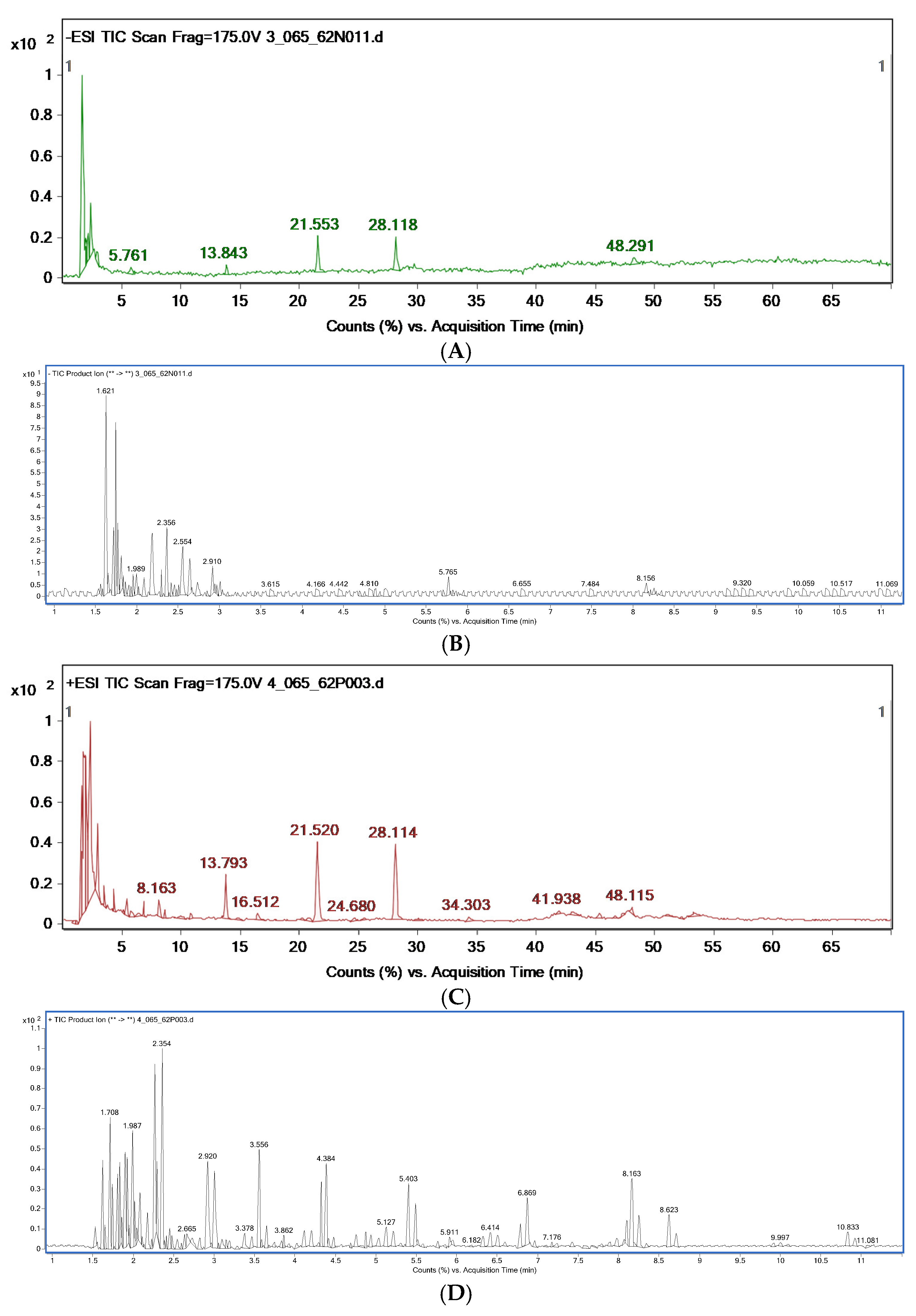
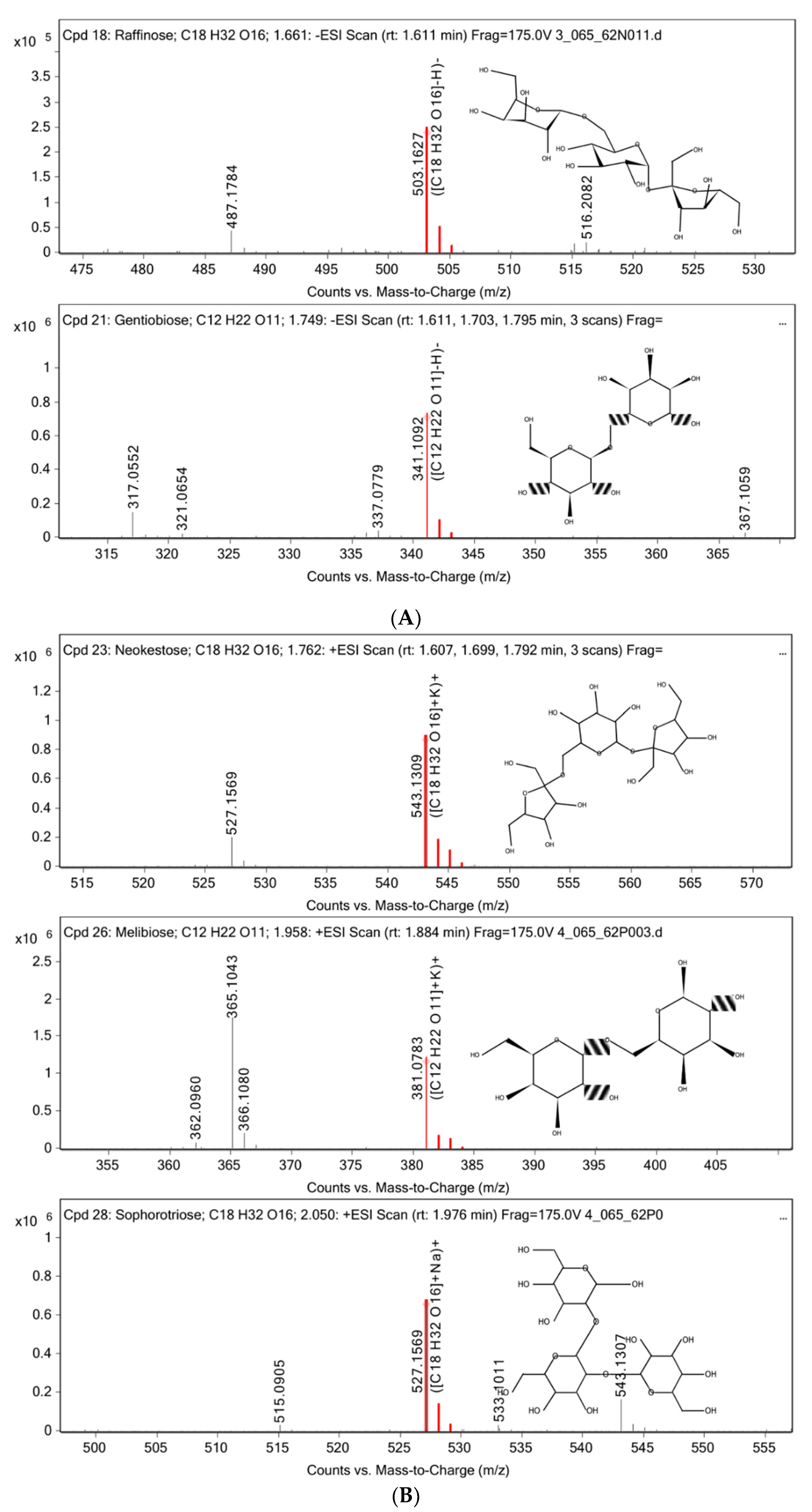
3.2. Chemical Constituent Analysis by HPLC-MS/MS

The chemical constituents of the D. kaki fruit extract were analyzed using HPLC-MS/MS via ESI(−) (Figure 3A) and ESI(+) (Figure 3C) modes. A total of 10 compounds were detected in ESI(−) and 28 compounds were detected in ESI(+). The compounds’ name, retention times, peak high, and mass of the compounds are shown in Table 2. Various classes of natural compounds were found, especially several types of oligosaccharides, including, raffinose, gentiobiose, neokestose, melibiose, and sophorotriose (the approximate percent contents are 0.13, 1.17, 1.21, 0.70, and 0.72, respectively). Mass spectrum of the oligosaccharides found in D. kaki fruit extract were shown in Figure 4. According to the non-digestible and probiotic features of functional oligosaccharides, such as galacto-oligosaccharides, xylo-oligosaccharides, and fructo-oligosaccharides. Their anti-diabetic effect has been reported [19]. However, limitations in the knowledge of oligosaccharides’ structure and their efficiency remain. Therefore, the study of their mechanism in metabolic syndrome is required to evaluate the ability of oligosaccharides.

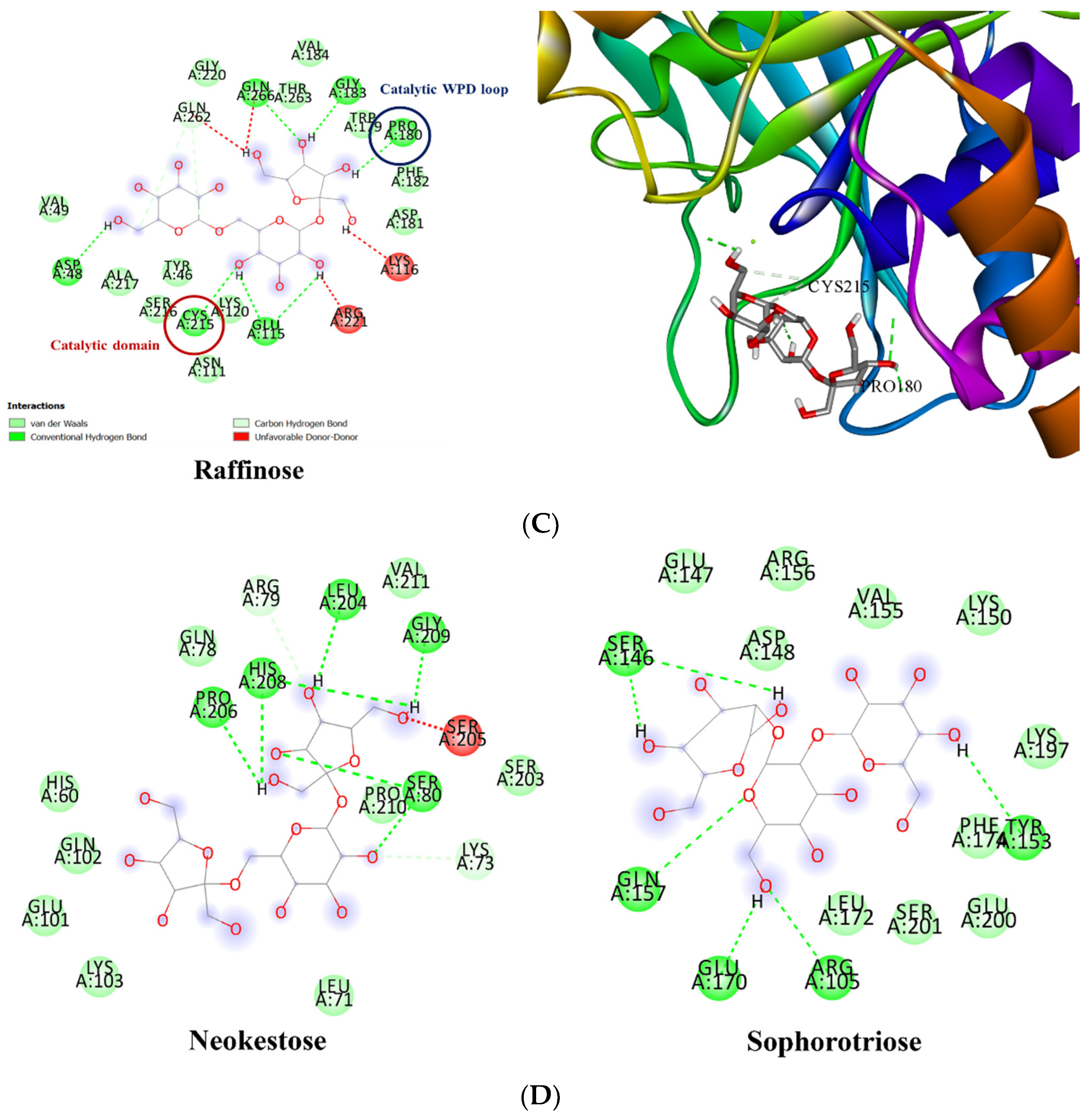
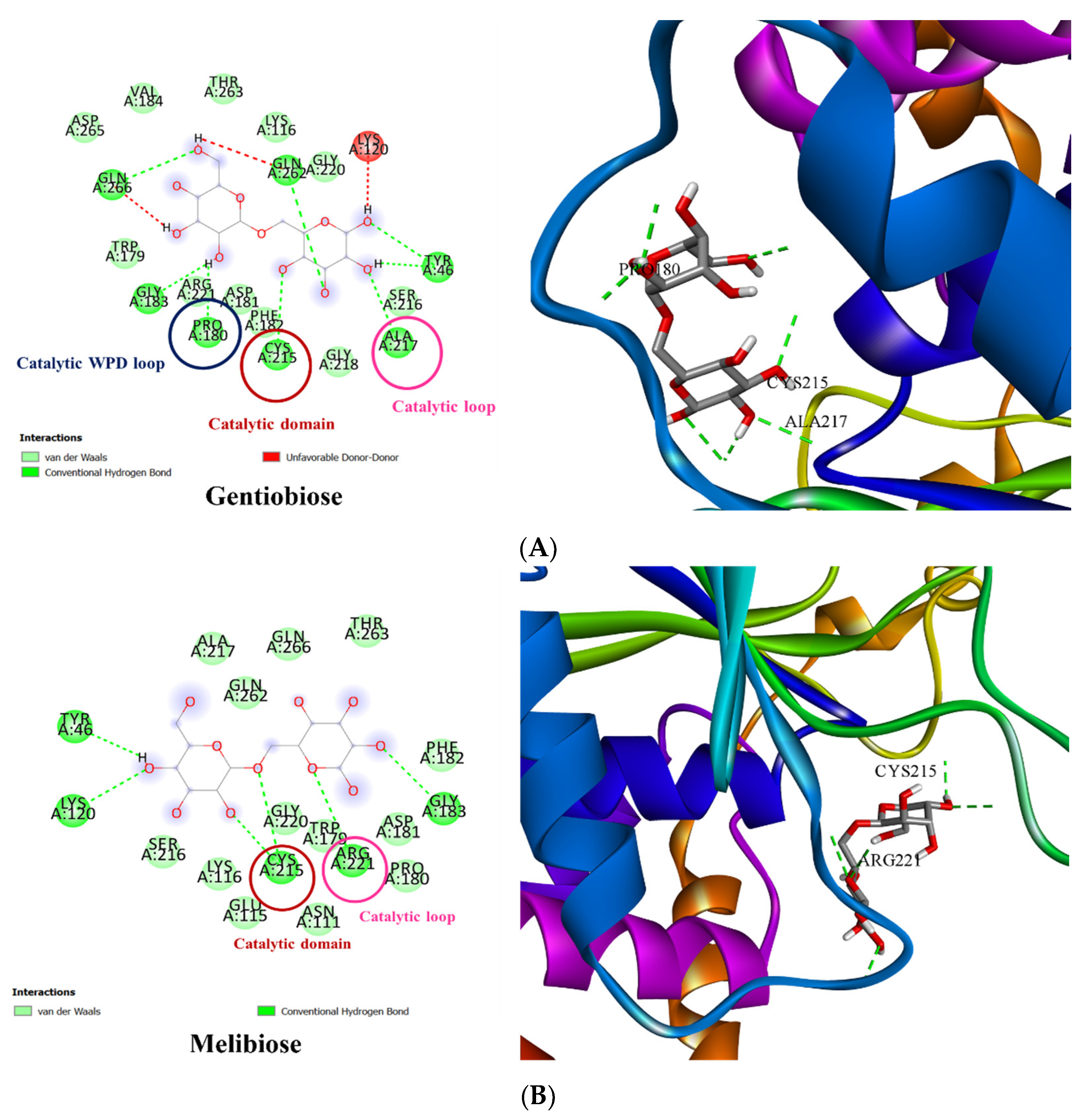
3.3. Molecular Docking of PTP1B to the Oligosaccharide from D. kaki Fruit

To understand the mechanism of the selected oligosaccharides with PTP1B, molecular docking studies were performed. The binding interaction of oligosaccharides’ structure and PTP1B was performed via AutoDock Vina 1.1.2. The active site of PTP1B includes the allosteric site and the catalytic domain, which consist of the catalytic site, catalytic loop, and catalytic WPD loop [24]. The results of in silico molecular docking show that the hydroxyl groups of gentiobiose binding to Pro180 residue of the catalytic WPD loop, Cys215 of the catalytic domain, and Ala217 of the catalytic loop with the hydrogen bond interaction and binding energy of −6.9 kcal/mol. The O-glycosidic bond of melibiose binding with Cys215 of the catalytic domain and O in the backbone bound to the Arg221 residue of the catalytic loop with a binding energy of −7.3 kcal/mol. The hydroxyl groups of raffinose were bound to the Pro180 and Cys215 amino acid residues of the catalytic WPD loop and catalytic domain, respectively. The affinity of the binding reaction is −7.1 kcal/mol. Neokestose and sophorotriose did not show a hydrogen bond interaction with the active residues of PTP1B. All of the results are shown in Figure 5. Therefore, the oligosaccharides gentiobiose, melibiose, and raffinose were selected for further experiments.

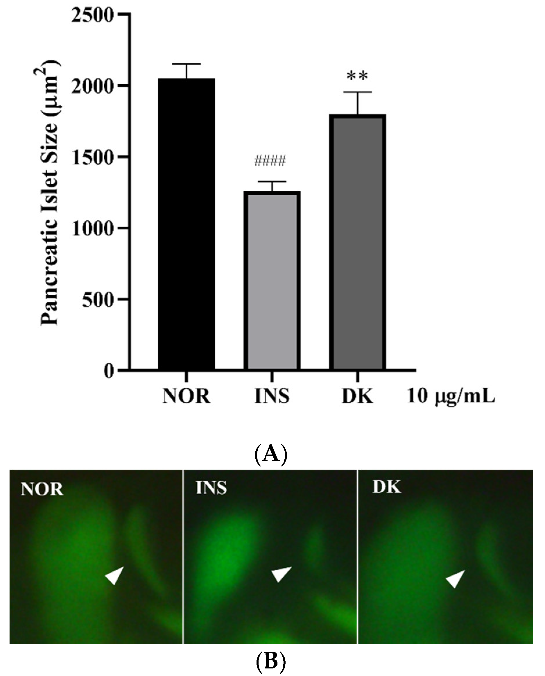
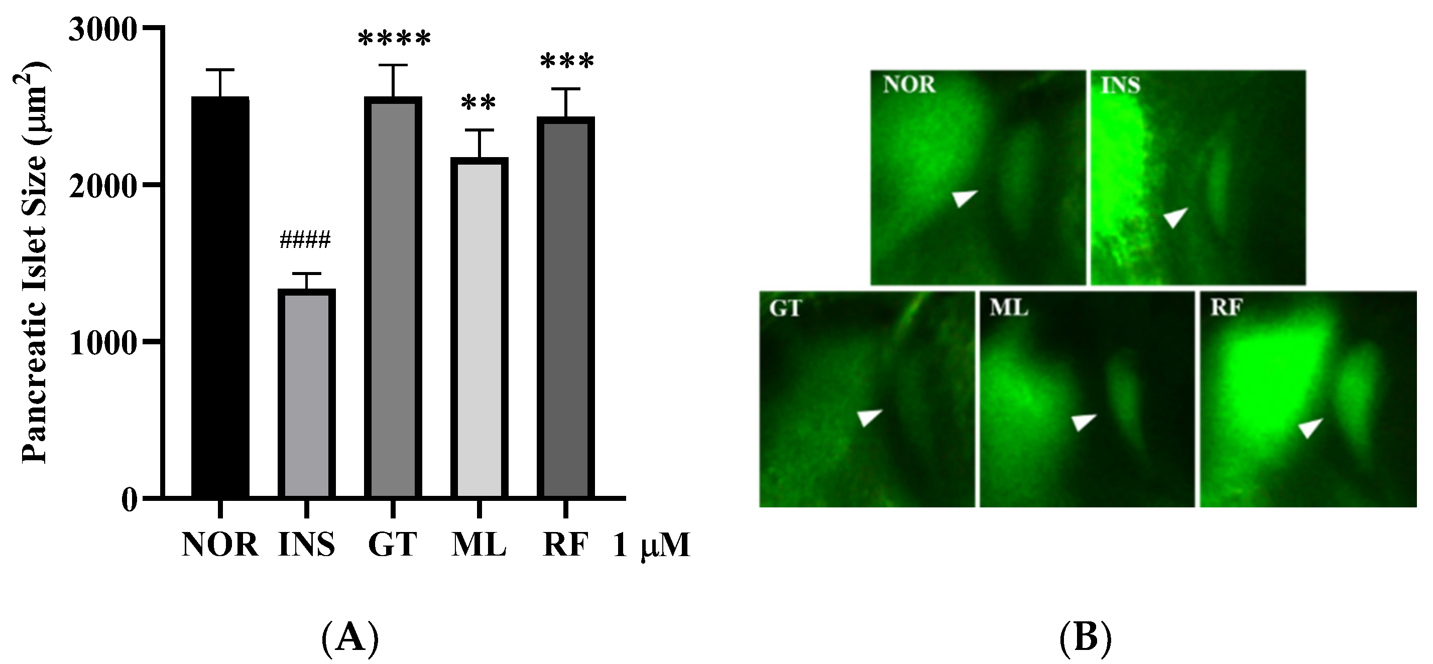
3.4. Anti-Insulin Resistance in Zebrafish Induced by Oligosaccharides from D. kaki Fruit

The oligosaccharides gentiobiose, melibiose, and raffinose were investigated against insulin resistance in larvae zebrafish with a concentration of 1 µM. The results show that gentiobiose, melibiose, and raffinose had a potential effect to increase the PI size of the larvae significantly (p < 0.01–0.0001). The results are shown in Figure 6.

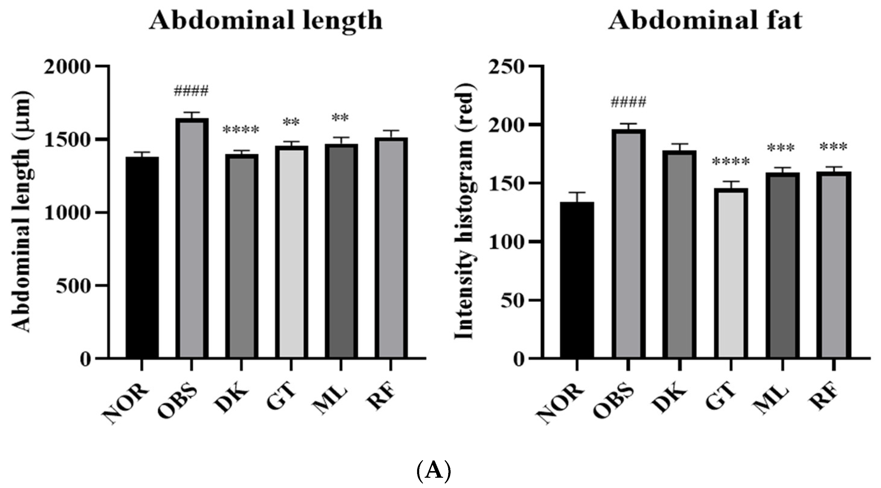
3.5. The Investigation of the Obesity-Preventing Effect of the D. kaki Fruit Extract and Its Oligosaccharides

Zebrafish is an efficient tool for the rapid identification of cellular morphology and genetic studies. High-fat-diet induction increased whole-body adipocytes of zebrafish larvae by egg yolk overfeeding [25]. To hasten natural product research and development for metabolic syndrome, zebrafish is a predictive and convenient tool with a low cost and a short period of drug screening [26]. D. kaki and its oligosaccharides were examined by co-treatment to investigate their obesity-preventing effects. A total of 9 dpf of zebrafish larvae were imaged and determined the belly size by measuring the abdominal length. The overfeeding of a high-fat diet increased the size of the zebrafish bodies as evidenced by the significant increase in abdominal length (p < 0.0001), which was prevented in the treatment groups. The D. kaki extract and oligosaccharide exposure groups showed a significantly decreased size of zebrafish abdominal length, p < 0.01–0.0001 (Figure 7A). Lipid accumulation was monitored by the intensity histogram of Nile red staining fluorescence image. The obesity group increased the intensity in the abdominal area significantly (p < 0.0001) and the treatment groups lowered the lipid accumulation by decreasing the intensity, as shown in Figure 7B (p < 0.001–0.0001). As a result of this experiment, D. kaki and its oligosaccharides exhibited an obesity-preventing effect in the zebrafish model via lowering the lipid accumulation in the larvae body.

3.6. Genes Expression on High-Fat-Diet-Induced Obesity in Zebrafish Larvae

The mechanism studies of high-fat-diet-induced obesity in zebrafish larvae were performed via RT-qPCR to evaluate the expression of inflammatory, lipogenesis, and lipid-lowering-related gene. The inflammatory-related mRNA tumor necrosis factor alpha (TNF-α), interleukin 6 (IL-6), and interleukin (IL-1β) were increased significantly in the obesity group. Gentiobiose and melibiose reduced the expression of three of the inflammatory-related genes. TNF-α and IL-6 were reduced significantly in the D. kaki fruit extract treatment group. The results show that lipogenesis-related fatty acid synthase (FASN) gene expression significantly increased by all treatments, while the D. kaki fruit extract, gentiobiose, and melibiose reduced sterol regulatory element binding transcription factor 1 (SREBF1). Lipid-lowering-related gene, carnitine palmitoyltransferase 1A (CPT1A), was reduced by the D. kaki fruit extract and gentiobiose significantly. The results are shown in Figure 8.

3.7. Enzyme Activity Assays of PTP1B to D. kaki Fruit and Oligosaccharides

The inhibition of PTP1B was performed to investigate in vitro activity. D. kaki showed PTP1B inhibition as the IC50 value of 693.3 µg/mL. In contrast, at the concentration of 10 mg/mL, none of the oligosaccharides showed an inhibitory effect against PTP1B. From the results of molecular docking, the three oligosaccharides gentiobiose, melibiose, and raffinose interacted with the catalytic sites of PTP1B, but were inactive in the enzyme assay. The active catalytic site of PTP1B was the phosphotyrosine (pTyr)-binding pocket, which is located in the intracellular region [27]. Therefore, the cellular penetration of the cell membrane was necessary for the PTP1B inhibitors [28]. On the other hand, the allosteric site was less conserved and may enhance the selectivity and bioavailability of the inhibitors. This may explain why the function of the catalytic site of PTP1B had a poor inhibitory effect in vitro study [29].

4. Conclusions

D. kaki fruit extract, gentiobiose, melibiose, and raffinose showed anti-insulin resistance and obesity-preventing effects by protecting β cells from high doses of human insulin induction, decreasing the abdominal size, and reducing the lipid accumulation in obese zebrafish. The mechanism of action of the oligosaccharides was studied via molecular docking and enzyme activities of PTP1B. Three of the oligosaccharides showed a binding interaction with the active catalytic sites of PTP1B, but did not show an inhibition effect in the enzyme assay. This result confirmed that the catalytic sites of PTP1B are highly conserved structures from bacteria to mammals [30], and cellular penetration was essential for the inhibitors [28]. In addition, the large uncharged polar molecule of oligosaccharides cannot diffuse through the cell membrane [31]; this may be the reason of enzyme activity results. On the other hand, there is evidence for the intestinal transportation of non-digestible oligosaccharides in clinical studies. Non-digestible oligosaccharides might directly contact with systemic immune cell enter through the gut barrier when the gut permeability was increased, such as in inflammatory bowel disease (IBD), obesity, type 1 diabetes, and non-alcoholic fatty liver disease [32].
Previous literature studies have shown that raffinose and melibiose promoted the growth of bifidobacteria in overweight children. Due to their non-digestibility, these oligosaccharides may have beneficial effects in the obese patient’s colon [33]. Moreover, raffinose was reported to inhibit lipid accumulation in HepG2 and 3T3-L1 cells and increase glucose uptake in vivo study [34]. To apply the effects of D. kaki and oligosaccharides on the metabolic syndrome to humans, studies of metabolic-related gene expression are required. The pathological characteristics of the inflammation pathway (IL-6, IL-1β, and apolipoprotein H (APOH)) and lipid metabolism (SREBF1), peroxisome proliferator-activated receptors α/γ (PPARα/γ), nuclear receptor subfamily 1 group H member 3 (NR1H3), and leptin (LEP)) between human and zebrafish have been divulged [35]. Thus, further studies on the role of the mechanism of function of these oligosaccharides will contribute to the development of dietary supplements.
From our studies, the D. kaki fruit extract and oligosaccharides resulted in gene expression changing inflammation, lipogenesis, and lipid-lowering-related genes. The fruit extract decreased the expression of TNF-α and IL-6, which impair the insulin action in metabolic tissues [36], and SREBF1 and FASN, which regulate lipogenesis [37], and CPT1A, which promote the fatty acid metabolism by improving lipid levels and glucose [38]. The oligosaccharide gentiobiose downregulated the proinflammatory cytokines TNF-α, IL-6, IL-1β (interleukin 1β is the production of β-cell oxidative stress that promote macrophages activation [39]), lipogenesis-related SREBF1, FASN, and lipid-lowering-related CPT1A. Moreover, the regulation of TNF-α, IL-6, IL-1β, SREBF1, and FASN expression was decreased by melibiose. In contrast, raffinose only downregulated FASN expression. In conclusion, gentiobiose, melibiose, and the D. kaki fruit extract have a great potential for applications in metabolic syndrome drug development.

Author Contributions

Conceptualization, T.H.K. and W.N.; investigation and formal analysis, W.N., S.W.S. and M.C.K.; resources, H.K.L., Y.H.N., J.H.S., N.W.K., S.W.S., M.C.K. and S.Y.S.; writing—original draft preparation, W.N.; writing—review and editing, T.H.K., J.H.K., B.N.H. and S.D.-a. All authors have read and agreed to the published version of the manuscript.

Funding

This research was funded by Korea Institute of Planning and Evaluation for Technology in Food, Agriculture, Forestry and Fisheries (KR), funded by Ministry of Agriculture, Food and Rural Affairs (317071-03-3-CG000).

Institutional Review Board Statement

The animal study protocol was approved by the Animal Care and Use Committee of Kyung Hee University (KHUASP[SE]-15-10).

Informed Consent Statement

Not applicable.

Data Availability Statement

Not applicable.

Acknowledgments

HPLC-MS/MS was performed by Office of Scientific Instrument and Testing, Prince of Songkla University, Thailand.

Conflicts of Interest

The authors declare no conflict of interest.

References

  1. Grundy, S.M. Metabolic syndrome pandemic. Arterioscler. Thromb. Vasc. Biol. 2008, 28, 629–636. [Google Scholar] [CrossRef] [PubMed] [Green Version]
  2. Ley, S.H.; Hamdy, O.; Mohan, V.; Hu, F.B. Prevention and management of type 2 diabetes: Dietary components and nutritional strategies. Lancet 2014, 383, 1999–2007. [Google Scholar] [CrossRef] [Green Version]
  3. Wellen, K.E.; Hotamisligil, G.S. Inflammation, stress, and diabetes. J. Clin. Investig. 2005, 115, 1111–1119. [Google Scholar] [CrossRef] [PubMed] [Green Version]
  4. Kahn, S.E.; Hull, R.L.; Utzschneider, K.M. Mechanisms linking obesity to insulin resistance and type 2 diabetes. Nature 2006, 444, 840–846. [Google Scholar] [CrossRef]
  5. Martyn, J.A.; Kaneki, M.; Yasuhara, S. Obesity-induced insulin resistance and hyperglycemia: Etiologic factors and molecular mechanisms. Anesthesiology 2008, 109, 137–148. [Google Scholar] [CrossRef] [Green Version]
  6. Ali, M.I.; Ketsawatsomkron, P.; Belin de Chantemele, E.J.; Mintz, J.D.; Muta, K.; Salet, C.; Black, S.N.; Tremblay, M.L.; Fulton, D.V.; Marrero, M.B.; et al. Deletion of Protein Tyrosine Phosphatase 1b Improves Peripheral Insulin Resistance and Vascular Function in Obese, Leptin-Resistant Mice via Reduced Oxidant Tone. Circ. Res. 2009, 105, 1013–1022. [Google Scholar] [CrossRef]
  7. Banno, R.; Zimmer, D.; De Jonghe, B.C.; Atienza, M.; Rak, K.; Yang, W.; Bence, K.K. PTP1B and SHP2 in POMC neurons reciprocally regulate energy balance in mice. J. Clin. Investig. 2010, 120, 720–734. [Google Scholar] [CrossRef] [Green Version]
  8. Yip, S.C.; Saha, S.; Chernoff, J. PTP1B: A double agent in metabolism and oncogenesis. Trends Biochem. Sci. 2010, 35, 442–449. [Google Scholar] [CrossRef] [Green Version]
  9. Guillot, R.; Cortés, R.; Navarro, S.; Mischitelli, M.; García-Herranz, V.; Sánchez, E.; Cal, L.; Navarro, J.C.; Míguez, J.M.; Afanasyev, S.; et al. Behind melanocortin antagonist overexpression in the zebrafish brain: A behavioral and transcriptomic approach. Horm. Behav. 2016, 82, 87–100. [Google Scholar] [CrossRef] [Green Version]
  10. Chen, S.; Li, C.; Yuan, G.; Xie, F. Anatomical and histological observation on the pancreas in adult zebrafish. Pancreas 2007, 34, 120–125. [Google Scholar] [CrossRef]
  11. Zang, L.; Shimada, Y.; Nishimura, Y.; Tanaka, T.; Nishimura, N. Repeated blood collection for blood tests in adult zebrafish. J. Vis. Exp. 2015, 102, e53272. [Google Scholar] [CrossRef]
  12. Anderson, J.L.; Carten, J.D.; Farber, S.A. Zebrafish lipid metabolism: From mediating early patterning to the metabolism of dietary fat and cholesterol. Methods Cell Biol. 2011, 101, 111–141. [Google Scholar]
  13. Moss, J.B.; Koustubhan, P.; Greenman, M.; Parsons, M.J.; Walter, I.; Moss, L.G. Regeneration of the pancreas in adult zebrafish. Diabetes 2009, 58, 1844–1851. [Google Scholar] [CrossRef] [Green Version]
  14. Intine, R.V.; Olsen, A.S.; Sarras, M.P. A zebrafish model of diabetes mellitus and metabolic memory. J. Vis. Exp. 2013, 72, e50232. [Google Scholar] [CrossRef] [Green Version]
  15. Lee, Y.A.; Cho, E.J.; Tanaka, T.; Yokozawa, T. Inhibitory activities of proanthocyanidins from persimmon against oxidative stress and digestive enzymes related to diabetes. J. Nutr. Sci. Vitaminol. 2007, 53, 287–292. [Google Scholar] [CrossRef] [Green Version]
  16. Ramasubbu, N.; Ragunath, C.; Mishra, P.J.; Thomas, L.M.; Gyémánt, G.; Kandra, L. Human salivary alpha-amylase Trp58 situated at subsite -2 is critical for enzyme activity. Eur. J. Biochem. 2004, 271, 2517–2529. [Google Scholar] [CrossRef]
  17. Thuong, P.T.; Lee, C.H.; Dao, T.T.; Nguyen, P.H.; Kim, W.G.; Lee, S.J.; Oh, W.K. Triterpenoids from the leaves of Diospyros kaki (persimmon) and their inhibitory effects on protein tyrosine phosphatase 1B. J. Nat. Prod. 2008, 71, 1775–1778. [Google Scholar] [CrossRef]
  18. Lee, Y.A.; Kim, Y.J.; Cho, E.J.; Yokozawa, T. Ameliorative effects of proanthocyanidin on oxidative stress and inflammation in streptozotocin-induced diabetic rats. J. Agrie. Food Chem. 2008, 55, 9395–9400. [Google Scholar] [CrossRef]
  19. Zhu, D.; Yan, Q.; Liu, J.; Wu, X.; Jiang, Z. Can functional oligosaccharides reduce the risk of diabetes mellitus? FASEB J. 2019, 33, 11655–11667. [Google Scholar] [CrossRef] [Green Version]
  20. Swennen, K.; Courtin, C.M.; Delcour, J.A. Non-digestible oligosaccharides with prebiotic properties. Crit. Rev. Food Sci. Nutr. 2006, 46, 459–471. [Google Scholar] [CrossRef]
  21. Nuankaew, W.; Heemman, A.; Wattanapiromsakul, C.; Shi, J.H.; Kim, N.W.; Yasmin, T.; Jeong, S.Y.; Nam, Y.H.; Hong, B.N.; Dej-adisai, S.; et al. Anti-insulin resistance effect of constituents from Senna siamea on zebrafish model, its molecular docking, and structure–activity relationships. J. Nat. Med. 2021, 75, 520–531. [Google Scholar] [CrossRef]
  22. Ha, M.; Seong, S.; Nguyen, T.; Cho, W.; Ah, K.; Ma, J.; Woo, M.; Choi, J.; Min, B. Chalcone derivatives from the root bark of Morus alba L. act as inhibitors of PTP1B and α-glucosidase. Phytochemistry 2018, 155, 114–125. [Google Scholar] [CrossRef]
  23. Zhang, Z.Y.; Walsh, A.B.; Wu, L.; McNamarai, D.J.; Dobrusini, E.M.; Miller, W.T. Determinants of substrate recognition in the protein-tyrosine phosphatase, PTP1. J. Biol. Chem. 1996, 271, 5386–5392. [Google Scholar] [CrossRef] [Green Version]
  24. Jin, T.; Yu, H.; Huang, X.F. Selective binding modes and allosteric inhibitory effects of lupane triterpenes on protein tyrosine phosphatase 1B. Sci. Rep. 2016, 6, 20766. [Google Scholar] [CrossRef] [Green Version]
  25. Tingaud-Sequeira, A.; Ouadah, N.; Babin, P.J. Zebrafish obesogenic test: A tool for screening molecules that target adiposity. J. Lipid Res. 2011, 52, 1765–1772. [Google Scholar] [CrossRef] [Green Version]
  26. Zhou, J.; Xu, Y.Q.; Guo, S.Y.; Li, C.Q. Rapid analysis of hypolipidemic drugs in a live zebrafish assay. J. Pharmacol. Toxicol. Methods 2015, 72, 47–52. [Google Scholar] [CrossRef]
  27. Zhang, S.; Zhang, Z.Y. PTP1B as a drug target: Recent developments in PTP1B inhibitor discovery. Drug Discov. Today 2007, 12, 373–381. [Google Scholar] [CrossRef] [PubMed]
  28. Johnson, T.O.; Ermolieff, J.; Jirousek, M.R. Protein tyrosine phosphatase 1B inhibitors for diabetes. Nat. Rev. Drug Discov. 2002, 1, 696–709. [Google Scholar] [CrossRef] [PubMed]
  29. Wiesmann, C.; Barr, K.; Kung, J.; Zhu, J.; Erlanson, D.A.; Shen, W.; Fahr, B.J.; Zhong, M.; Taylor, L.; Randal, M.; et al. Allosteric inhibition of protein tyrosine phosphatase 1B. Nat. Struct. Mol. Biol. 2004, 11, 730–737. [Google Scholar] [CrossRef] [PubMed]
  30. Li, S.; Zhang, J.; Lu, S.; Huang, W.; Geng, L.; Shen, Q.; Zhang, J. The mechanism of allosteric inhibition of protein tyrosine phosphatase 1B. PLoS ONE 2014, 9, e97668. [Google Scholar]
  31. Cooper, J.M. The Cell: A Molecular Approach, 2nd ed.; Sinauer Associates: Sunderland, MA, USA, 2000. [Google Scholar]
  32. Del Fabbro, S.; Calder, P.; Childs, C. Microbiota-independent immunological effects of non-digestible oligosaccharides in the context of inflammatory bowel diseases. Proc. Nutr. Soc. 2020, 79, 468–478. [Google Scholar] [CrossRef]
  33. Kaarel, A.; Signe, A.; Karin, E.; Anneli, L.; Tiia, V.; Madis, J.; Triinu, V.; Tiina, A. Composition and metabolism of fecal microbiota from normal and overweight children are differentially affected by melibiose, raffinose and raffinose-derived fructans. Anaerobe 2018, 52, 100–110. [Google Scholar]
  34. Padmanaban, M.; Gopal, T.; Rajendran, A.B.; Baddireddi, S.L. Raffinose from Costus speciosus attenuates lipid synthesis through modulation of PPARs/SREBP1c and improves insulin sensitivity through PI3K/AKT. Chem. Biol. Interact. 2018, 284, 80–89. [Google Scholar]
  35. Oka, T.; Nishimura, Y.; Zang, L.; Hirano, M.; Shimada, Y.; Wang, Z.; Umemoto, N.; Kuroyanagi, J.; Nishimura, N.; Tanaka, T. Diet-induced obesity in zebrafish shares common pathophysiological pathways with mammalian obesity. BMC Physiol. 2010, 10, 21. [Google Scholar] [CrossRef] [Green Version]
  36. Kern, L.; Mittenbühler, M.J.; Vesting, A.J.; Ostermann, A.L.; Wunderlich, C.M.; Wunderlich, F.T. Obesity-Induced TNFα and IL-6 Signaling: The Missing Link between Obesity and Inflammation—Driven Liver and Colorectal Cancers. Cancers 2019, 11, 24. [Google Scholar] [CrossRef] [Green Version]
  37. Guo, D.; Bell, E.H.; Mischel, P.; Chakravarti, A. Targeting SREBP-1-driven lipid metabolism to treat cancer. Curr. Pharm. Des. 2014, 20, 2619–2626. [Google Scholar] [CrossRef] [Green Version]
  38. Das, M.; Sha, J.; Hidalgo, B.; Aslibekyan, S.; Do, A.N.; Zhi, D.; Sun, D.; Zhang, T.; Li, S.; Chen, W.; et al. Association of DNA Methylation at CPT1A Locus with Metabolic Syndrome in the Genetics of Lipid Lowering Drugs and Diet Network (GOLDN) Study. PLoS ONE 2016, 11, e0145789. [Google Scholar] [CrossRef]
  39. Donath, M.Y.; Dalmas, E.; Sauter, N.S.; Böni-Schnetzler, M. Inflammation in obesity and diabetes: Islet dysfunction and therapeutic opportunity. Cell Metab. 2013, 17, 860–872. [Google Scholar] [CrossRef] [Green Version]
Figure 1. Zebrafish as an obesity model. Experimental setup (A). Comparison of the abdominal length and the abdominal fat between the normal group (NOR) and obesity group (OBS) stained with Nile red staining and observed under florescence microscope at day five of the experiment (B).
Nutrients 14 03249 g001
Figure 2. Screening of the D. kaki fruit extract for anti-insulin resistance activity in the zebrafish larvae. The treatment group with the D. kaki fruit extract significantly recovered, as shown by the PI size (A). Image of PI (pancreatic islet) staining by 40 µM 2-NBDG (B), normal (NOR), insulin induction (INS), and D. kaki (DK). #### p < 0.0001 compared to the control (NOR). ** p < 0.01 compared to insulin induction (INS). The statistical significance was determined using a one-way ANOVA with Dunnett’s post hoc test, expressed as means ± SEM (n = 20).
Nutrients 14 03249 g002
Figure 3. LC-ESI-MS/MS (A) and the expanded chromatogram created by plotting the total ion current in a series of product ions recorded as a function of the retention time of LC-ESI-MS/MS (B). LC+ESI-MS/MS (C) chromatograms of the D. kaki water extract and the expanded chromatogram created by plotting the total ion current in a series of product ions recorded as a function of the retention time of LC+ESI-MS/MS (D).
Nutrients 14 03249 g003
Figure 4. Mass spectrum of raffinose and gentiobiose from −ESI mode (A). Neokestose, melibiose, and sophorotriose from +ESI mode (B).
Nutrients 14 03249 g004
Figure 5. The crystallographic structure of PTP1B molecular docking related five oligosaccharides. Hydrogen bond of gentiobiose binding to the catalytic active sites of PTP1B Pro180, Cys215, and Ala217 residue of the catalytic WPD loop, catalytic domain, and catalytic loop, respectively (A). Melibiose showed ligand binding to Cys215 of the catalytic domain and Arg221 of the catalytic loop (B). Pro180 and Cys215 of the catalytic WPD loop and catalytic domain were bound to raffinose with hydrogen bonding (C). Neokestose and sophorotriose were not bound to any of the active residues (D).
Nutrients 14 03249 g005aNutrients 14 03249 g005b
Figure 6. Oligosaccharides from D. kaki significantly showed the recovery of β cells by decreasing the size of zebrafish PI (A). The images of 2-NBDG staining of PI (B), normal (NOR), insulin induction (INS), gentiobiose (GT), melibiose (MB), and raffinose (RF). #### p < 0.0001 compared to NOR. **** p < 0.0001, *** p < 0.001, ** p < 0.01 compared to INS. The statistical significance was determined using a one-way ANOVA with Dunnett’s post hoc test, expressed as means ± SEM (n = 20).
Nutrients 14 03249 g006
Figure 7. A total of 10 µg/mL of D. kaki (DK) and 1 µM of the oligosaccharides significantly decreased the abdominal size of and lipid accumulation in obese zebrafish by co-treatment (A). The images of 0.5 µg/mL Nile red staining of zebrafish body (B), normal (NOR), obesity induction (OBS), D. kaki (DK), gentiobiose (GT), melibiose (MB), and raffinose (RF). #### p < 0.0001 compared to NOR. **** p < 0.0001, *** p < 0.001, ** p < 0.01 compared to OBS. The statistical significance was determined using a one-way ANOVA with Dunnett’s post hoc test, expressed as means ± SEM (n = 20).
Nutrients 14 03249 g007aNutrients 14 03249 g007b
Figure 8. Gene expression on high-fat-diet-induced obesity in zebrafish larvae. The results of RT-qPCR evaluation on inflammatory-related genes TNF-α, IL-6, and IL-1β (A). The results of RT-qPCR evaluation on the lipogenesis-related genes SREBF1 and FASN (B). The results of RT-qPCR evaluation on lipid-lowering-related genes CPT1A (C). Normal (NOR), obesity induction (OBS), D. kaki (DK), gentiobiose (GT), melibiose (MB), and raffinose (RF). ### p < 0.001, ## p < 0.01 compared to NOR. *** p < 0.001, ** p < 0.01, * p < 0.05 compared to OBS. The statistical significance was determined using a one-way ANOVA with Dunnett’s post hoc test, expressed as means ± SEM (n = 20).
Nutrients 14 03249 g008
Table 1. Primers for RT-qPCR.
GeneForward SequenceReverse Sequence
TNF-αGCT TAT GAG CCA TGC AGT GATGC CCA GTC TGT CTC CTT CT
IL6AAG GGG TCA GGA TCA GCA CGCT GTA GAT TCG CGT TAG ACA TC
IL1BTGG CGA ACG TCA TCC AAGGGA GCA CTG GGC GAC GCA TA
SREBF1CAT CCA CAT GGC TCT GAG TGCTC ATC CAC AAA GAA GCG GT
FASNGAG AAA GCT TGC CAA ACA GGGAG GGT CTT GCA GGA GAC AG
CPT1ACAT GAG GCT CTT CGG CAAAAG AGC AGG CCT AAG GAT G
Table 2. Chemical constituents of the D. kaki extract detected in LC-ESI-MS/MS and LC+ESI-MS/MS chromatograms.
RTHeightMassIdentification Result
LC-ESI-MS/MS chromatograms
1.661497,154504.17Raffinose
1.7494,200,859342.1166Gentiobiose
1.991482,479342.1165Gentiobiose
2.1881,329,997646.1966beta-D-4-Deoxy-delta4-GlcA-(1->4)-beta-D-Glc-(1->4)-alpha-L-Rha-(1->3)-beta-D-Glc
2.318294,628342.1169Gentiobiose
2.3373,904192.02682,5-Didehydro-D-gluconate
2.464252,822219.1112Pantothenic Acid
2.6021,051,653224.0327Dehydrochorismic acid
2.811145,798342.1157D-(+)-Cellobiose
5.836415,037382.18391,2,10-Trihydroxydihydro-trans-linalyl oxide 7-O-beta-D-glucopyranoside
46.14208,962294.1834Myrsinone
48.25409,723328.2251(9R,10S,12Z)-9,10-Dihydroxy-8-oxo-12-octadecenoic acid
LC+ESI-MS/MS chromatograms
1.59142,819317.1212N-(1-Deoxy-1-fructosyl) histidine
1.7624,633,119504.1678Neokestose
1.9336,561,444261.1204Epidermin
1.9581,734,862342.1152Melibiose
2.051,517,744504.1676Sophorotriose
2.234735,399438.1151Loquatoside
2.3016,245,852271.1647Prolyl-Arginine
2.519998,209342.1153Sucrose
2.7031,104,006366.1413N-(1-Deoxy-1-fructosyl) tryptophan
3.0136,275,606187.0633-Amino-2-naphthoic acid
3.4861,147,920422.21176alpha-Fluoro-17-hydroxycorticosterone 21-acetate
3.615266,556418.1823Glu Arg Asp
3.841595,699478.1672Kelampayoside A
4.406758,433444.2559Arg Asn Arg
4.9121,230,160361.233Lys Lys Ser
5.131,574,844510.2643Nebramycin factor 5′
5.4984,669,055436.2273Flurandrenolide
5.82568,261404.1644Asp Asp Arg
6.4181,185,915532.30825β-Cyprinolsulfate
6.8793,665,061458.2718Arg Gln Arg
8.7192,455,730486.3239Docosahexaenoyl Serotonin
10.9411,224,227568.3056Ceanothine E
16.685162,831538.2945Euphorbia factor Ti2
21.6092,415,031672.4863PE-Cer(d14:2(4E,6E)/20:1(11Z)(2OH))
28.065,609,787775.6128PE(21:0/17:0)
42.0381,513,417273.2659C16 Sphinganine
43.051,422,944229.24Xestoaminol C
48.2051,502,361317.2921Phytosphingosine
Publisher’s Note: MDPI stays neutral with regard to jurisdictional claims in published maps and institutional affiliations.

