Article

Prenatal Iron Deficiency and Choline Supplementation Interact to Epigenetically Regulate Jarid1b and Bdnf in the Rat Hippocampus into Adulthood

1
Department of Pediatrics, University of Minnesota, Minneapolis, MN 55455, USA
2
Department of Psychology, University of Minnesota, Minneapolis, MN 55455, USA
3
Biostatistical Design and Analysis Center, Clinical and Translational Science Institute, University of Minnesota, Minneapolis, MN 55455, USA
*
Author to whom correspondence should be addressed.
Nutrients 2021, 13(12), 4527; https://doi.org/10.3390/nu13124527
Submission received: 19 November 2021 / Revised: 10 December 2021 / Accepted: 14 December 2021 / Published: 17 December 2021
(This article belongs to the Special Issue Iron and Infant Development)

Abstract

:
Early-life iron deficiency (ID) causes long-term neurocognitive impairments and gene dysregulation that can be partially mitigated by prenatal choline supplementation. The long-term gene dysregulation is hypothesized to underlie cognitive dysfunction. However, mechanisms by which iron and choline mediate long-term gene dysregulation remain unknown. In the present study, using a well-established rat model of fetal-neonatal ID, we demonstrated that ID downregulated hippocampal expression of the gene encoding JmjC-ARID domain-containing protein 1B (JARID1B), an iron-dependent histone H3K4 demethylase, associated with a higher histone deacetylase 1 (HDAC1) enrichment and a lower enrichment of acetylated histone H3K9 (H3K9ac) and phosphorylated cAMP response element-binding protein (pCREB). Likewise, ID reduced transcriptional capacity of the gene encoding brain-derived neurotrophic factor (BDNF), a target of JARID1B, associated with repressive histone modifications such as lower H3K9ac and pCREB enrichments at the Bdnf promoters in the adult rat hippocampus. Prenatal choline supplementation did not prevent the ID-induced chromatin modifications at these loci but induced long-lasting repressive chromatin modifications in the iron-sufficient adult rats. Collectively, these findings demonstrated that the iron-dependent epigenetic mechanism mediated by JARID1B accounted for long-term Bdnf dysregulation by early-life ID. Choline supplementation utilized a separate mechanism to rescue the effect of ID on neural gene regulation. The negative epigenetic effects of choline supplementation in the iron-sufficient rat hippocampus necessitate additional investigations prior to its use as an adjunctive therapeutic agent.

Graphical Abstract

1. Introduction

Iron deficiency (ID) is a common micronutrient deficiency worldwide, disproportionately affecting 40–50% of pregnant women and preschool-aged children in middle-to-low-income countries [1,2]. Human studies show an association between early-life ID and persistent neurodevelopmental effects in formerly iron-deficient individuals in adolescence [3] and adulthood [4]. Low maternal iron intake during pregnancy is also associated with increased risk for schizophrenia [5] and autism [6] in the offspring. Collectively, early-life ID can produce lasting negative effects on neurocognitive function and adult mental health. These long-term neurological effects are also observed in animal models of fetal-neonatal ID. In animal models, ID during the fetal and early postnatal periods compromises neurodevelopment [7,8,9]. Brain development during fetal and early postnatal life is an energy-intensive process [10,11,12] and is disrupted in a rat model of fetal-neonatal ID [13,14,15]. In addition, formerly iron-deficient rats show long-term behavioral deficits in hippocampal-dependent learning and memory [16,17,18] associated with altered expression of genes critical for neuron development [14,19,20] and gene networks implicated in schizophrenia, autism and mood disorders [21].
The presence of long-term behavioral abnormalities in formerly iron-deficient humans and rodent models despite early treatment with iron following diagnosis indicates the need to understand the mechanisms underlying the effects to ultimately identify adjunct treatments for early-life ID. One attractive candidate is maternal choline supplementation. Choline is an essential nutrient that is critically involved in early brain development [22,23], and is readily found in over 630 food sources [24], which makes it more accessible than iron [25]. Choline supplementation during specific pre- and postnatal time windows reverses some cognitive deficits observed in a rodent model of fetal-neonatal ID associated with increased expression of neurotrophic factors such as brain-derived neurotrophic factor (BDNF) [21,26,27]. However, the mechanism by which choline reverses these effects is unknown. One possibility is that choline acts as a methyl donor and thereby modifies the epigenetic landscape through DNA methylation and histone methylation [28,29].
Epigenetic modifications can contribute to the long-term effects of fetal-neonatal ID on neural gene dysregulation [30,31]. Of particular interest is the family of iron-containing JmjC-ARID (JARID) domain containing dioxygenases, which require iron as a cofactor to remove methyl groups from lysine residues of histones [32]. Previously, we demonstrated that persistent dysregulation of JARIDs was accompanied by changes in histone methylation at the Bdnf locus in adult hippocampus following early-life ID [31,33]. In the present study, we assessed the regulatory effects of fetal-neonatal ID and prenatal choline supplementation through a hypothesized iron-mediated epigenetic mechanism involving JmjC-ARID domain-containing protein 1B (JARID1B) and its target Bdnf in the rat hippocampus across postnatal ages. Although ID also reduces expression of other iron-containing JARIDs [33], we elected to analyze JARID1B due to its function in demethylating tri- and di-methylated histone H3K4, which is canonically associated with transcriptional activation, especially of Bdnf, and its important regulatory effects in neural development [32,34]. We hypothesized that ID dysregulates Jarid1b expression, consequently modifying epigenetic signatures at the Bdnf promoters and that prenatal choline supplementation mitigates these regulatory changes.

2. Materials and Methods

2.1. Animals

Gestational day (G) 2 pregnant Sprague–Dawley rats were purchased from Charles River Laboratories (Wilmington, MA, USA). Rats were maintained in a 12-h:12-h light/dark cycle with ad lib food and water. ID was induced by dietary manipulation as described previously [26,27,35]. In brief, to generate iron-deficient pups, pregnant dams were given a purified iron-deficient diet (4 mg Fe/kg, TD.80396; Harlan Teklad) from G2 to postnatal day (P) 7 when lactating dams were switched to a purified iron-sufficient diet (200 mg Fe/kg, TD.09256; Harlan Teklad). Control iron-sufficient pups were generated from pregnant dams maintained on the iron-sufficient diet. Both diets were similar in all respects with the exception of the iron (ferric citrate) content. Details of diet contents have been described in our previous study [26]. Half of the dams on an iron-sufficient or iron-deficient diet received dietary choline supplementation (5.0 g/kg choline chloride supplemented [36,37]; iron-sufficient with choline: TD.1448261, iron-deficient with choline: TD.110139; Harlan Teklad) from G11-18, while the other half of the dams received their iron-sufficient or iron-deficient diet with standard choline content (1.1 g/kg). Thus, dams and their litters were assigned to one of four groups based on maternal diet: iron-deficient with G11-18 choline supplementation (IDCh), iron-deficient without supplemental choline (ID), always iron-sufficient with G11-18 choline supplementation (ISCh), always iron-sufficient without supplemental choline (IS) (Figure 1). All litters were culled to 8 pups with six males and two females at birth. Only male offspring were used in experiments, because all prior extant data on long-term effects are in males [21,27,30,31]. To avoid litter-specific effects, two male rats per litters and ≥3 L/group were used in the experiments. The University of Minnesota Institutional Animal Care and Use Committee approved all experiments in this study (Protocol # 2001-37802A).

2.2. Hippocampal Dissection

Rats were euthanized by injection of pentobarbital (100 mg/kg, intraperitoneal). Brains were removed and bisected along the midline on an ice-cold metal block. Hippocampus from P0 (end of proliferation), P15 (start of robust dendrite differentiation), and P65 (mature) rat was dissected and immediately flash-frozen in liquid nitrogen and stored at −80 °C for further use.

2.3. RNA Isolation and cDNA Synthesis

Total RNA was isolated from 1 hippocampal lobe per rat using RNAqueous Total RNA Isolation Kit (Invitrogen). cDNA synthesis was performed using 1 µg isolated RNA and the High-Capacity RNA-to-cDNA Kit (Applied Biosystems), as previously described [7].

2.4. Nuclear Protein Isolation

Nuclear proteins were isolated from hippocampal tissue using the Epiquik Nuclear Extraction Kit II (Epigentek), as per the manufacturer’s protocol. Protein concentration was determined by a standard Bradford protein assay using serial dilutions of bovine serum albumin (BioRad) as standards.

2.5. JARID Activity

JARID1 enzymatic activity was assessed from 20 µg of nuclear protein using the Epigenase JARID Demethylase Activity Assay Kit (Epigentek), following the manufacturer’s protocol. This assay quantified total JARID1, including JARID1A, 1B, 1C, and 1D, demethylase activity. In this assay, histone H3K4me3 substrate was coated onto microplate wells. Active JARID1 bound and removed the methyl group from the substrate. Demethylated products were recognized by a specific antibody, which was quantified by colorimetric absorbance at 450 nm (Epigentek). JARID1 activity was quantified in the hippocampus from 6 animals per treatment group. All samples were analyzed in technical duplicate. Results were calculated from the average of the duplicates.

2.6. Western Blot

Western blot analysis was used to quantify levels of JARID1B using a previously described protocol [35]. In brief, 30 µg of hippocampal protein lysate were separated using a 4–20% SDS-PAGE gel (Novex, Life Technologies). Proteins were blotted onto a nitrocellulose membrane (Pierce Protein Biology), blocked with Blocking Buffer for Fluorescent Western Blotting (Rockland Immunochemicals), and incubated with antibodies against JARID1B (Abcam) and beta actin (Sigma). Following PBS + 0.1% Tween–20 washes, blots were incubated in Alexa–700 anti-mouse (Invitrogen) and IR Dye-800 anti-rabbit (Rockland) and scanned by Near-Infrared Fluorescent using the Odyssey Infrared Imaging System (Li-Cor Biosciences). Integrated intensity of protein bands normalized to beta actin was determined using Photoshop CS 4 (Adobe, San Jose, CA, USA).

2.7. Chromatin Immunoprecipitation (ChIP) Assay

ChIP experiments were performed as previously described [31]. In brief, chromatin was prepared from hippocampal tissue following the manufacturer’s recommendation (Millipore). Hippocampi were homogenized in ice-cold PBS (500 µL). Chromatin was cross-linked in 1% formaldehyde solution (Sigma), pelleted by centrifugation, resuspended in 500 µL lysis buffer (1% SDS, 10 mM EDTA, 50 mM Tris pH 8.1, 1 mM PMSF, 10 µL 10× protease inhibitor cocktails (Roche)), and fragmented by sonication (Bioruptor Pico, Diagenode). Chromatin fragments were visualized by agarose gel electrophoresis following a cross-linking reversal (0.2 M NaCl, 65 °C overnight) to ensure optimal fragment sizes. Fragmented chromatin was diluted 10-fold with ChIP dilution buffer (0.01% SDS, 1.1% Triton X-100, 1.2 mM EDTA, 16.7 mM Tris-pH 8.1, 167 mM NaCl), pre-cleared with 75 µL of Protein A agarose (50% slurry, Sigma), and immunoprecipitated by ChIP-grade antibody (4 °C, overnight). ChIP-grade antibodies used in this study included JARID1B (Abcam), histone deacetylase 1 (HDAC1) (Diagenode), H3K9ac (Diagenode), H3K9me3 (Diagenode), upstream stimulatory factor 1 (USF1) (Santa Cruz Biotechnologies), and phosphorylated cAMP response element-binding protein (pCREB) (Millipore Sigma). Normal IgG was used as a negative control. The antibody-histone complex was collected by the addition of 60 µL of Protein A agarose slurry with mixing (4 °C, ≥1 h) and subsequently eluted in 500 µL of elution buffer (1% SDS, 0.1 M NaHCO3) following buffer (as per the manufacturer’s protocol) rinses. Reverse cross-linking was achieved by incubation in NaCl (0.2 M, 65 °C, overnight). A protease digestion (20 µg proteinase K, 20 mM EDTA, 100 mM Tris-pH 6.5, 45 °C, 1 h) was performed to recover DNA, which was further purified using phenol/chloroform extraction and ethanol precipitation (0.1 × 3 M sodium acetate, pH 5.2, 2 × 100% ethanol). Levels of enriched Gapdh (active) and Myod1 (inactive) loci were used to validate ChIP experiments.

2.8. Real-Time Quantitative PCR (RT-qPCR)

For mRNA quantitation, TaqMan Universal PCR Master Mix (Applied Biosystem) was used with gene expression assays (Applied Biosystem; G9a, Assay ID: Rn01758882_m1; Jarid1b, Assay ID: Rn01758882_m1; Suv39h1, Assay ID: Rn01528294_g1). Genes that encode TATA-box binding protein (Tbp, Assay ID: Rn01455646_m1) or ribosomal protein s18 (Rps18, Assay ID: Rn01428915_g1), the expressions of which were not altered by ID, were used as normalizers. For analysis of precipitated DNA from ChIP experiments, Fast SYBR green master mix (Applied Biosystem) was used to amplify the Bdnf4, Bdnf6, and Jarid1b promoters using validated primers (Bdnf4, For-TCGAGGCAGAGGAGGTATCA, Rev- CCTCTCCTCGGTGAATGGGA; Bdnf6, For-GATGAAAGGTTTGGCTTCTGTG, Rev- TCGGTGAATGGGAAAGTGG; Jarid1b, For-TGGGAGAGTTCCTGCCT, Rev- GCCGGATCTTGTGGATGAA). Input DNA (10%) was used as a normalizer to account for input amount (ΔCt). Data were expressed as fold change (2-ΔΔCt) relative to IS control using one of the IS samples as a calibrator (ΔΔCt). Real-time PCR was performed with QuantStudio™ 3 (Thermo Fisher, Waltham, MA, USA).

2.9. Experimental Design and Statistical Analysis

For data that were collected from IS, ID, ISCh and IDCh groups, a two-way ANOVA with interaction (iron status × choline supplementation) was used at each time point. If there was a significant interaction, the simple effect of iron status or choline supplementation was analyzed at each choline supplementation condition or iron status, respectively. Completed two-way ANOVA results are shown in Table 1. α was set at 0.05 for all comparisons. Data were graphed and statistically analyzed using GraphPad Prism 9 (GraphPad Software, Inc., San Diego, CA, USA) and SAS V9.4 (SAS Institute Inc., Cary, NC, USA).

3. Results

3.1. Iron Deficiency Downregulates Hippocampal Jarid1b Expression

To determine the effects of ID on Jarid1b expression across critical periods of hippocampal development, Jarid1b mRNA levels were compared among IS, ID, ISCh and IDCh groups. An interaction between iron status and choline supplementation on Jarid1b expression was found at P15 (p = 0.0341) and P65 (p = 0.0313). Compared to the iron-sufficient group, the effects of iron status and choline supplementation were significant at P15 (ID vs. IS, p = 0.0003; ISCh vs. IS, p = 0.0096). At P65, the effect of iron status and choline supplementation in the iron-deficient groups remained significant (IS vs. ID, p = 0.0029; IDCh vs. ID, p = 0.0051). Compared to the iron-sufficient control group, hippocampal Jarid1b expression was lower in the iron-deficient group at P15 (Figure 2B, ID vs. IS) and P65 (Figure 2C, ID vs. IS). Prenatal choline supplementation ameliorated the effect of ID at P65 (Figure 2C, IDCh vs. ID), but not at P15 (Figure 2B, IDCh vs. ID, p = 0.7). Choline supplementation also reduced Jarid1b expression in the P15 ISCh group (Figure 2B, ISCh vs. IS). Western blot analysis corroborated the lower hippocampal Jarid1b expression caused by ID at P15 and P65 (Figure 2D, ID vs. IS). While we could not specifically determine JARID1B activity, total JARID1 (JARID1A, B, C, and D) enzymatic activity was assessed and showed a lower activity level in the P15 ID group and a higher activity level in the P65 iron-replete ID group (Figure 2E, ID vs. IS).

3.2. Iron Deficiency and Prenatal Choline Supplementation Alter Jarid1b’s Epigenetic Signatures

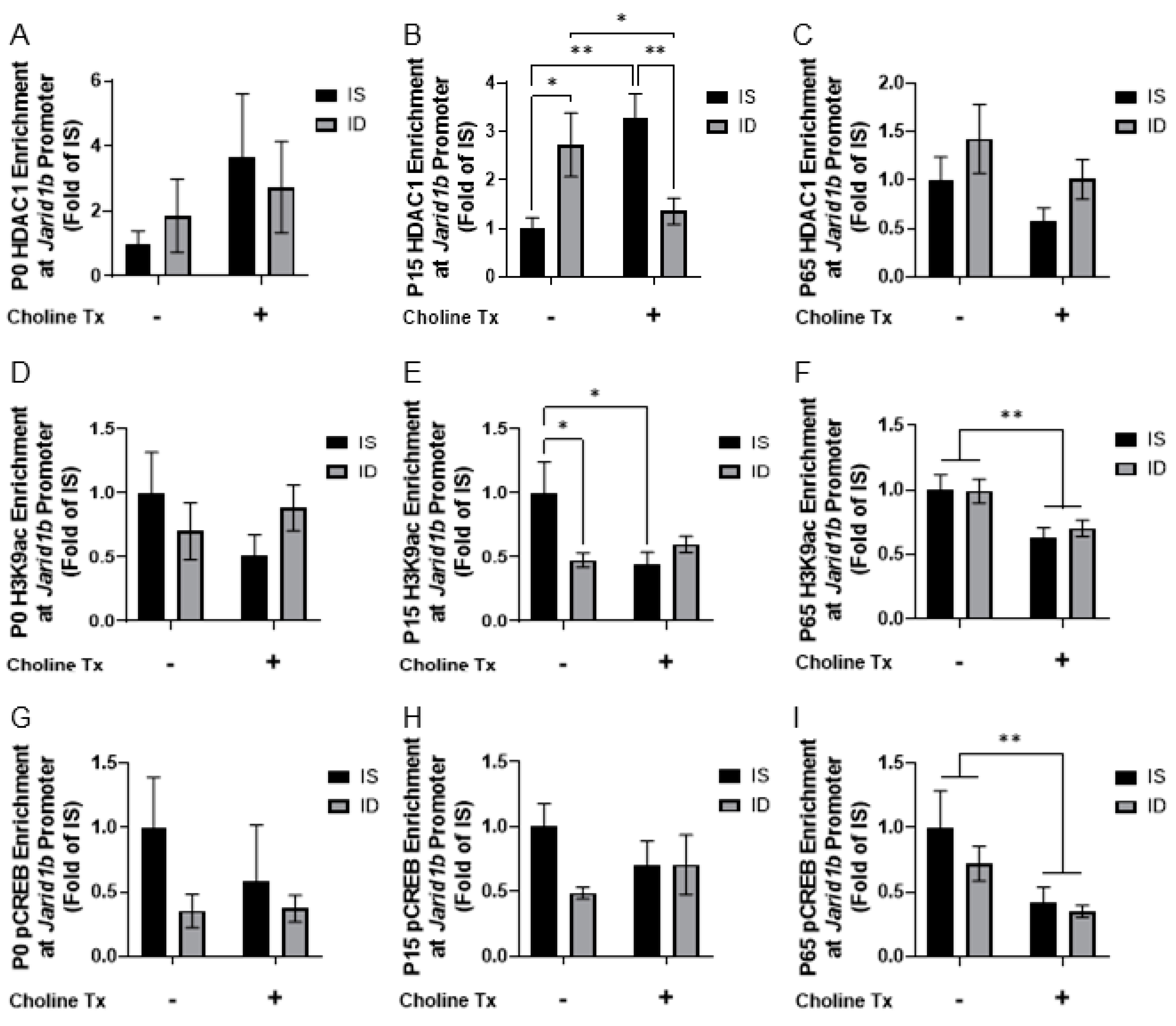
We then tested whether ID and prenatal choline supplementation were associated with epigenetic regulation at the Jarid1b promoter. Since histone deacetylase 1 (HDAC1) has been reported to downregulate Jarid1b expression [38], HDAC1 enrichment was compared among the 4 groups. An interaction between iron status and choline supplementation on HDAC1 enrichment was found at P15 (p = 0.0012). The effects of iron status and choline supplementation were significant (Figure 3B; ID vs. IS, p = 0.0194; IDCh vs. ISCh, p = 0.0086; ISCh vs. IS, p = 0.0051; IDCh vs. ID, p = 0.0363). No effects were found at P0 or P65 (Figure 3A,C). The level of HDAC1 enrichment was greater in the P15 iron-deficient group (Figure 3B, IS vs. ID). Choline supplementation ameliorated the effect of ID (Figure 3B, IDCh vs. ID) and induced a higher level of HDAC1 enrichment in the ISCh group (Figure 3B, ISCh vs. IS). These findings were further corroborated by the concomitant changes in H3K9 acetylation (H3K9ac). An interaction between iron status and choline supplementation on H3K9ac enrichment was found at P15 (p = 0.0331), where both ID and choline supplementation reduced H3K9ac enrichment at P15 (Figure 3E; ID vs. IS, ISCh vs. IS). At P65, only a main effect of choline supplementation was found with a reduced H3K9ac enrichment (Figure 3F).
We further tested whether the changes in HDAC1 recruitment and histone H3 acetylation would reduce the recruitment of phosphorylated cAMP response element-binding protein (pCREB), which can reflect neuronal activity [39,40,41]. While no clear effect was found at P0 or P15 (Figure 3G,H), a main effect of choline supplementation was found with a lower pCREB enrichment at P65 (Figure 3I).

3.3. Iron Deficiency and Prenatal Choline Supplementation Alter Hippocampal Histone H3K9 Methylation at the Jarid1b Locus

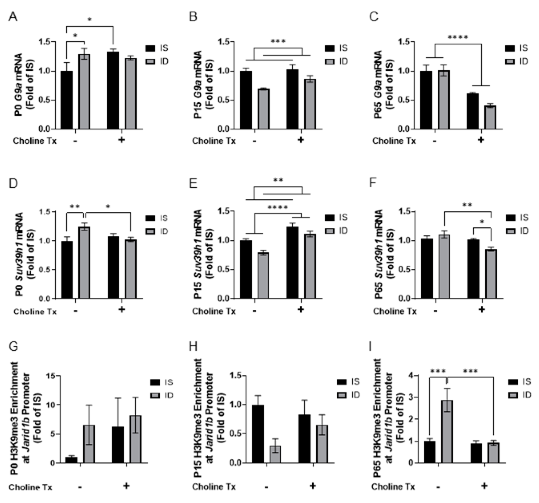
Given the documented effects of choline supplementation on expression of histone H3K9 methyltransferases and histone H3K9 methylation [29,42], we determined the effects of ID and prenatal choline supplementation on the hippocampal expression of H3K9 methyltransferases G9a and Suv39h1. For G9a expression, an interaction between iron status and choline supplementation was found at P0 (p = 0.0491). ID and choline supplementation induced a higher G9a expression in P0 hippocampus (Figure 4A; ID vs. IS, ISCh vs. IS). A main effect of iron status was found with a reduced G9a expression at P15 (Figure 4B). A main effect of choline supplementation was found with a lower G9a expression at P65 (Figure 4C). For Suv39h1 expression, an interaction between iron status and choline supplementation was found at P0 (p = 0.0165). ID induced a higher Suv39h1 expression and choline supplementation removed this effect (Figure 4D; ID vs. IS, IDCh vs. ID). An interaction between iron status and choline supplementation was also found at P65 (p = 0.0152), where choline supplementation reduced Suv39h1 expression (Figure 4F; IDCh vs. ISCh, IDCh vs. ID). The main effects of both iron status and choline supplementation were found at P15, where ID reduced and choline supplementation induced Suv39h1 expression (Figure 4E).
Since both G9A and SUV39H1 target histone H3K9 [43], we quantified H3K9me3 enrichment at the Jarid1b promoter by quantitative chromatin immunoprecipitation (qChIP) to determine the relevance of the changes in histone methyltransferase expression in the ID hippocampus. No clear difference was found among comparison groups at P0 or P15 (Figure 4G,H). An interaction between iron status and choline supplementation (p = 0.0081) was found at P65. ID induced a higher H3K9me3 enrichment (Figure 4I, ID vs. IS) and a choline supplementation ameliorated the effect of ID (Figure 4I, IDCh vs. ID).

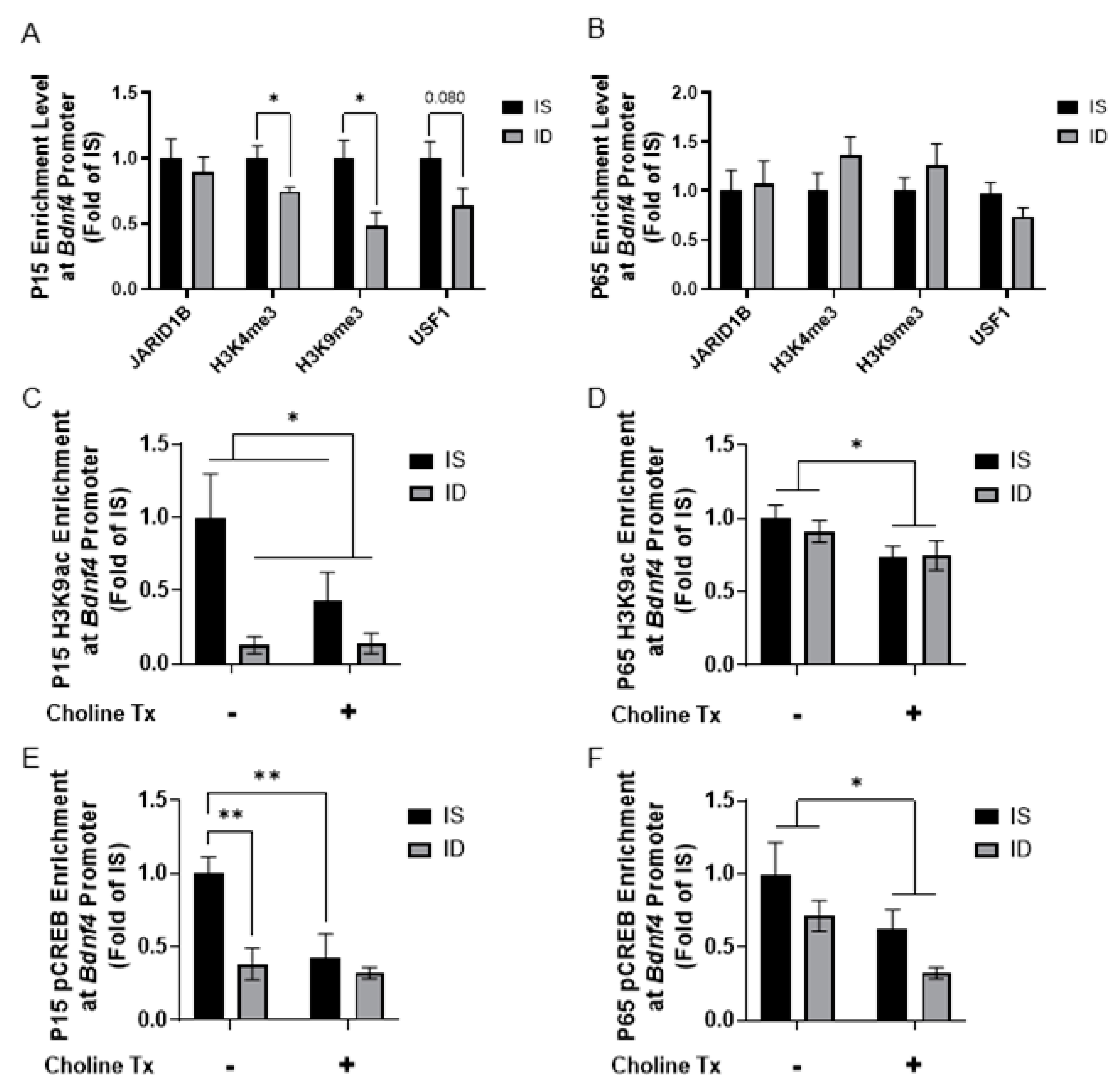
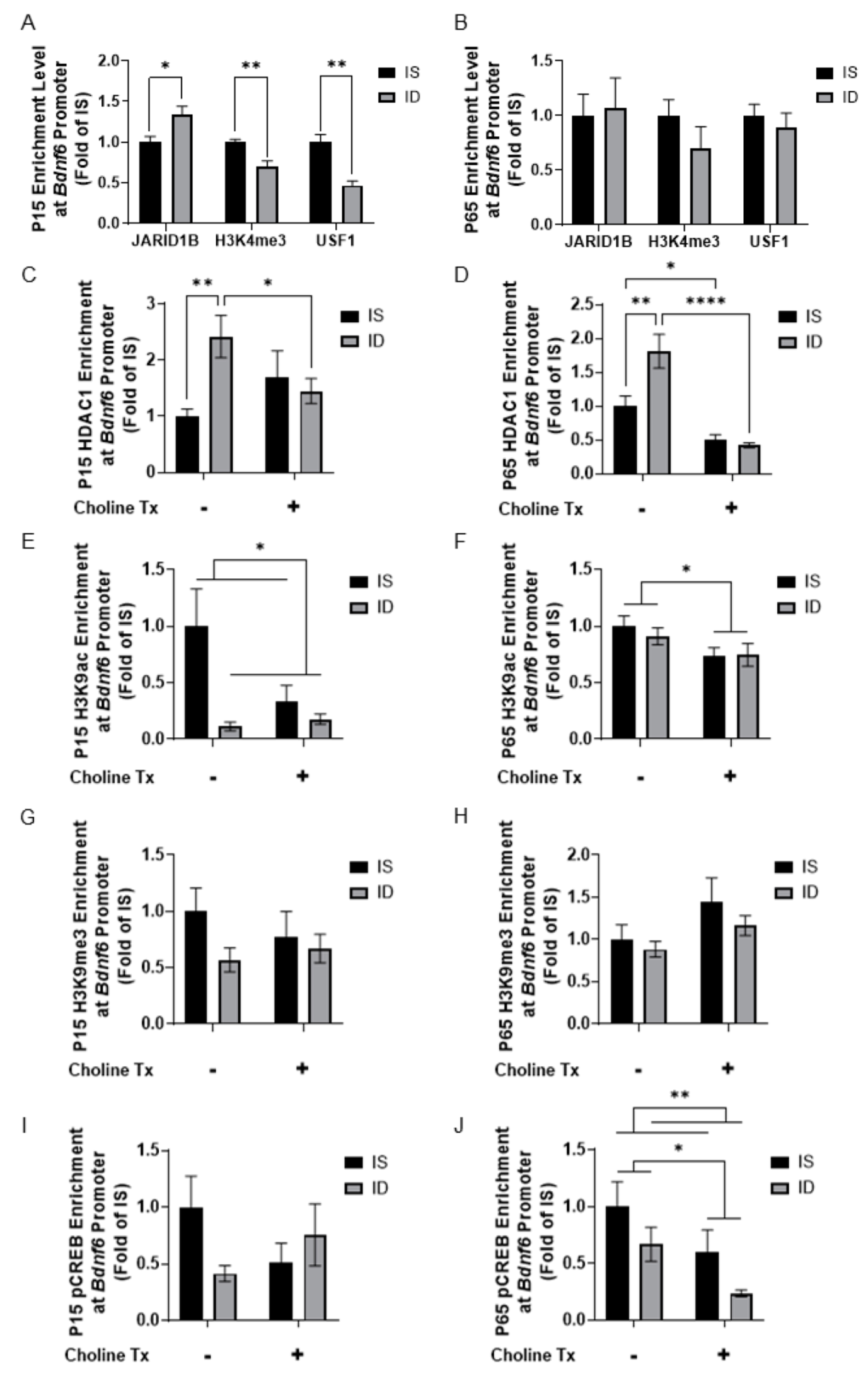
3.4. Iron Deficiency and Choline Supplementation Alter Histone Modification at the Bdnf Promoters

To further analyze how fetal-neonatal ID alters the epigenetic regulation of Bdnf, a target of JARID1B [31,44,45,46,47,48,49,50], we quantified changes in histone modifications and binding of known transcription factors at the Bdnf4 and Bdnf6 promoters by qChIP. Initial studies between ID and IS groups not supplemented with choline showed that, during the peak of hippocampal ID at P15 [51], enrichment levels of H3K4me3 were lower at both the Bdnf4 and Bdnf6 promoters (Figure 5A and Figure 6A), which was accompanied by a lower enrichment of upstream stimulating factor 1 (USF1) and a higher JARID1B enrichment at the Bdnf6 promoter (Figure 6A). These epigenetic marks were not different between P65 iron-replete ID and IS groups (Figure 5B and Figure 6B). Subsequent analyses revealed a main effect of iron status with a lower H3K9ac enrichment at the Bdnf4 promoter at P15 (Figure 5C). A main effect of choline supplementation on H3K9ac enrichment at the Bdnf4 promoter was also found at P65 (Figure 5D). Analysis of pCREB enrichment at the Bdnf4 promoter found an interaction between iron status and choline supplementation (p = 0.0417). ID and choline supplementation lowered pCREB enrichment at P15 (Figure 5E; ID vs. IS, ISCh vs. IS). A main effect of choline supplementation was found with a lower pCREB enrichment at P65 (Figure 5F).
The increased JARID1B enrichment prompted us to assess HDAC1 recruitment at the Bdnf6 promoter. An interaction between iron status and choline supplementation was found with HDAC1 enrichment at P15 (p = 0.0199) and P65 (p = 0.0063). At P15, ID induced a higher HDAC1 enrichment and choline supplementation removed this effect (Figure 6C; ID vs. IS, IDCh vs. ID). Similar effects were found for HDAC1 enrichment at P65 (Figure 6D; ID vs. IS, IDCh vs ID). Despite these changes, only the main effect of iron status was found with a lower H3K9ac enrichment at P15 (Figure 6E). At P65, a main effect of choline supplementation was found with lower H3K9ac (Figure 6F) and pCREB (Figure 6J) enrichments. A main effect of iron status was also found with a reduced pCREB enrichment at P65 (Figure 6J).

4. Discussion

Long-term gene dysregulation, particularly of the Bdnf [49] in adult rat hippocampus [20,31], suggests a possible epigenetic mechanism whereby early-life iron ID exerts an enduring influence into adulthood. Nevertheless, the link between ID and Bdnf dysregulation remains unknown. The present study provides evidence that early-life ID alters the regulation and enzymatic activity of JARID1B, an iron-dependent histone demethylase. These changes could contribute to ID-altered epigenetic regulation of the Bdnf gene, which is a known target of JARID1B [31,44,45,46,47,48,50]. The current findings provide an iron-specific mechanism to underlie the neural gene dysregulation caused by ID in the developing hippocampus.
JARID1B is an iron-containing demethylase that removes methyl groups from histone H3K4me2/3 and plays a role in cellular oxygen sensing [52,53,54]. Hippocampal JARID1B expression was lowered during (P15) and beyond (P65) the period of ID, the latter indicating a long-term change in its regulation mediated by an epigenetic modification of the Jarid1b gene. The early-life downregulatory effect might function as an adaptive response to low oxygen or reduced energetic environment caused by ID to inappropriately alter expression of genes regulating neural growth and differentiation [55,56,57]. Consequently, this adaptation could lead to an accelerated hippocampal aging [19], while attempting to improve the fitness of the animal beyond the fetal period [11]. Mechanistically, a greater HDAC1 recruitment and lower H3K9 acetylation could only account for Jarid1b downregulation in the iron-deficient hippocampus (Figure 3D,E). The mechanism of long-term Jarid1b downregulation likely involves additional epigenetic modifications (e.g., H3K27me3) and requires further investigation. The recovery of overall JARID1 activity with iron treatment across postnatal ages, despite the persistent Jarid1b downregulation suggests that iron therapy following a period of ID could potentially restore iron-containing JARID by incorporating the appropriate metal [58,59]. However, we speculate that the window of opportunity for such restoration may be time-limited during hippocampal development [13].
Given the documented beneficial effects of prenatal choline supplementation on normalizing gene expression (e.g., Bdnf) and neurocognitive function in adult rats that were iron-deficient during the fetal-neonatal period [21,26], it is not surprising that choline supplementation normalized Jarid1b long-term downregulation in the formerly iron-deficient adult rat hippocampus (Figure 2C, ID vs. IDCh). It is noteworthy that iron treatment alone was not sufficient to restore Jarid1b expression. These findings provide a functional link between this methyl diet and Jarid1b regulation under ID environment. The lower hippocampal expression of histone H3 methyltransferases, G9a and Suv39h1, and H3K9me3 enrichment at the Jarid1b promoter supports the epigenetic regulation of choline supplementation with long-term implications. Thus, the interaction between iron status and choline supplementation will need further investigation to optimize the utility of choline as an adjunctive therapy for early-life exposures.
Our previous findings showed that ID and choline supplementation epigenetically modified the Bdnf gene [31]. In the present study, we provide a potential mechanistic link between the iron-containing JARID1B histone demethylase, HDAC1 recruitment, and Bdnf gene regulation [44,45,46,47,48,60,61,62,63,64]. Our assessment of the effects of ID on the Bdnf4 and Bdnf6 (previously designated as BdnfIII and BdnfIV in rats, respectively) promoters revealed differential regulatory changes, with a greater short-term JARID1B binding and a corresponding lower H3K4me3 enrichment only at the Bdnf6 promoter in the P15 ID hippocampus. In light of the concurrent lower Jarid1b expression and enzymatic activity, the findings suggest promoter-specific effects. Enrichment of a transcriptional repressive H3K9me3 marker was also different, with a lower level at the Bdnf4 promoter and no change at the Bdnf6 promoter. As such, both promoters were less transcriptionally poised or active in the iron-deficient hippocampus as evidenced by reduced enrichment of H3K9ac, pCREB, and USF1. Taken together, these findings suggest that the Bdnf6 promoter, but not the Bdnf4 promoter, was likely regulated by the iron-specific JARID1B mechanism. These epigenetic modifications were normalized in the P65 iron-replete ID hippocampus, indicating the short-term nature of both iron-dependent and -independent histone modifications in the developing ID rat hippocampus. These findings further add to the body of work demonstrating the dynamic and plastic properties of the hippocampus and epigenetic changes in response to external environments [65,66,67,68,69].
Choline supplementation is also known to modify histone methylation [29,42] and rescue long-term Bdnf6 downregulation during fetal-neonatal ID [31]. However, here we provide evidence that choline supplementation to IS animals had a negative effect on histone H3K9 acetylation and pCREB enrichment at the Bdnf promoters. These findings suggest an interaction between choline and iron status in gene regulation during hippocampal development that is consistent with transcriptomic changes previously observed in the adult ISCh hippocampus [21]. In this context, the mode of choline action regulating specific histone modifications could involve distinct histone demethylases and methyltransferases identified in previous studies [29,42]. Alternatively, it is possible that the timing of analysis following choline supplementation to IS animals could account for the disparate findings in the present study and those previously reported [29,42], where analyses of histone methylation were performed in embryonic tissues immediately following the final day of choline supplementation. Likewise, the effects of choline supplementation on epigenetic regulation of Bdnf in the developing iron-deficient hippocampus could involve DNA methylation [70,71], which was not measured in the present study.
In summary, the present study provides evidence for an epigenetic regulation of the iron-dependent mechanism that could underlie the previously documented dysregulation of the Bdnf gene in the adult rat hippocampus caused by early-life ID. Given that JARID1B plays a critical role in neural growth and differentiation [34,52,56], perturbed expression of this protein during neural development provides a molecular mechanism whereby early-life brain iron depletion alters the regulation of synaptic plasticity genes that lasts beyond the period of ID [14,19,20,21,72]. Changes in histone modifications associated with early-life ID or choline supplementation were dynamic and changeable across postnatal age in the rat hippocampus. Mechanisms underlying these dynamic changes in the nervous system may involve epigenetic modifiers similar to studies that assessed the relationship between the epigenome and postnatal life neuropathology such as Alzheimer’s disease, an associated risk of early-life ID anemia [73,74]. While there are substantial numbers of interactions among epigenetic factors in reprogramming gene expression, this study represents a novel assessment of the effects of iron, choline, and their potential interaction, laying down a foundation for additional study in this field.

Author Contributions

Conceptualization, P.V.T. and M.K.G.; methodology, P.V.T. and M.K.G.; investigation, S.X.L., A.K.B. and P.V.T.; formal analysis, S.X.L., A.K.B., S.L., J.C.G. and P.V.T.; writing—original draft preparation, S.X.L. and P.V.T.; writing—review and editing, S.X.L., A.K.B., S.L., J.C.G., M.K.G. and P.V.T.; funding acquisition, P.V.T., M.K.G. and S.L. All authors have read and agreed to the published version of the manuscript.

Funding

This work was funded by NIH R01NS099178 to P.V.T., R01HD29421 to M.K.G., and UL1TR002494 to S.L.

Institutional Review Board Statement

The University of Minnesota Institutional Animal Care and Use Committee approved all experiments in this study (Protocol # 2001-37802A).

Informed Consent Statement

Not applicable.

Data Availability Statement

Not applicable.

Conflicts of Interest

The authors declare that they have no conflict of interest with the contents of this article.

References

  1. McLean, E.; Cogswell, M.; Egli, I.; Wojdyla, D.; De Benoist, B. Worldwide prevalence of anaemia, WHO Vitamin and Mineral Nutrition Information System, 1993–2005. Public Health Nutr. 2009, 12, 444–454. [Google Scholar] [CrossRef] [Green Version]
  2. Stevens, G.A.; Finucane, M.M.; De-Regil, L.M.; Paciorek, C.J.; Flaxman, S.R.; Branca, F.; Peña-Rosas, J.P.; Bhutta, Z.A.; Ezzati, M. Global, regional, and national trends in haemoglobin concentration and prevalence of total and severe anaemia in children and pregnant and non-pregnant women for 1995-2011: A systematic analysis of population-representative data. Lancet Glob. Health 2013, 1, e16–e25. [Google Scholar] [CrossRef] [Green Version]
  3. Lozoff, B.; Jimenez, E.; Hagen, J.; Mollen, E.; Wolf, A.W. Poorer behavioral and developmental outcome more than 10 years after treatment for iron deficiency in infancy. Pediatrics 2000, 105, e51. [Google Scholar] [CrossRef] [Green Version]
  4. Lozoff, B.; Smith, J.B.; Kaciroti, N.; Clark, K.M.; Guevara, S.; Jimenez, E. Functional significance of early-life iron deficiency: Outcomes at 25 years. J. Pediatr. 2013, 163, 1260–1266. [Google Scholar] [CrossRef] [PubMed] [Green Version]
  5. Insel, B.J.; Schaefer, C.A.; McKeague, I.W.; Susser, E.S.; Brown, A.S. Maternal iron deficiency and the risk of schizophrenia in offspring. Arch. Gen. Psychiatry 2008, 65, 1136–1144. [Google Scholar] [CrossRef] [PubMed] [Green Version]
  6. Schmidt, R.J.; Tancredi, D.J.; Krakowiak, P.; Hansen, R.L.; Ozonoff, S. Maternal intake of supplemental iron and risk of autism spectrum disorder. Am. J. Epidemiol. 2014, 180, 890–900. [Google Scholar] [CrossRef] [PubMed] [Green Version]
  7. Barks, A.; Fretham, S.J.B.; Georgieff, M.K.; Tran, P.V. Early-Life Neuronal-Specific Iron Deficiency Alters the Adult Mouse Hippocampal Transcriptome. J. Nutr. 2018, 148, 1521–1528. [Google Scholar] [CrossRef] [Green Version]
  8. Barks, A.; Hall, A.M.; Tran, P.V.; Georgieff, M.K. Iron as a model nutrient for understanding the nutritional origins of neuropsychiatric disease. Pediatr. Res. 2019, 85, 176–182. [Google Scholar] [CrossRef] [Green Version]
  9. Fretham, S.J.B.; Carlson, E.S.; Georgieff, M.K. The role of iron in learning and memory. Adv. Nutr. 2011, 2, 112–121. [Google Scholar] [CrossRef] [Green Version]
  10. Fukumitsu, K.; Hatsukano, T.; Yoshimura, A.; Heuser, J.; Fujishima, K.; Kengaku, M. Mitochondrial fission protein Drp1 regulates mitochondrial transport and dendritic arborization in cerebellar Purkinje cells. Mol. Cell. Neurosci. 2016, 71, 56–65. [Google Scholar] [CrossRef]
  11. Kuzawa, C.W. Adipose Tissue in Human Infancy and Childhood: An Evolutionary Perspective. Yearb. Phys. Anthropol. 1998, 41, 177–209. [Google Scholar] [CrossRef]
  12. Li, Z.; Okamoto, K.I.; Hayashi, Y.; Sheng, M. The importance of dendritic mitochondria in the morphogenesis and plasticity of spines and synapses. Cell 2004, 119, 873–887. [Google Scholar] [CrossRef] [Green Version]
  13. Fretham, S.J.B.; Carlson, E.S.; Wobken, J.; Tran, P.V.; Petryk, A.; Georgieff, M.K. Temporal manipulation of transferrin-receptor-1-dependent iron uptake identifies a sensitive period in mouse hippocampal neurodevelopment. Hippocampus 2012, 22, 1691–1702. [Google Scholar] [CrossRef] [Green Version]
  14. Brunette, K.E.; Tran, P.V.; Wobken, J.D.; Carlson, E.S.; Georgieff, M.K. Gestational and Neonatal Iron Deficiency Alters Apical Dendrite Structure of CA1 Pyramidal Neurons in Adult Rat Hippocampus. Dev. Neurosci. 2010, 32, 238–248. [Google Scholar] [CrossRef] [PubMed] [Green Version]
  15. Carlson, E.S.; Tkac, I.; Magid, R.; O’Connor, M.B.; Andrews, N.C.; Schallert, T.; Gunshin, H.; Georgieff, M.K.; Petryk, A. Iron Is Essential for Neuron Development and Memory Function in Mouse Hippocampus. J. Nutr. 2009, 139, 672–679. [Google Scholar] [CrossRef] [PubMed] [Green Version]
  16. Pisansky, M.T.; Wickham, R.J.; Su, J.; Fretham, S.; Yuan, L.L.; Sun, M.; Gewirtz, J.C.; Georgieff, M.K. Iron deficiency with or without anemia impairs prepulse inhibition of the startle reflex. Hippocampus 2013, 23, 952–962. [Google Scholar] [CrossRef] [Green Version]
  17. Schmidt, A.T.; Alvarez, G.C.; Grove, W.M.; Rao, R.; Georgieff, M.K. Early iron deficiency enhances stimulus-response learning of adult rats in the context of competing spatial information. Dev. Cogn. Neurosci. 2012, 2, 174–180. [Google Scholar] [CrossRef] [Green Version]
  18. Schmidt, A.T.; Waldow, K.J.; Grove, W.M.; Salinas, J.A.; Georgieff, M.K. Dissociating the Long-Term Effects of Fetal/Neonatal Iron Deficiency on Three Types of Learning in the Rat. Behav. Neurosci. 2007, 121, 475–482. [Google Scholar] [CrossRef] [PubMed] [Green Version]
  19. Carlson, E.S.; Stead, J.D.H.; Neal, C.R.; Petryk, A.; Georgieff, M.K. Perinatal iron deficiency results in altered developmental expression of genes mediating energy metabolism and neuronal morphogenesis in hippocampus. Hippocampus 2007, 17, 679–691. [Google Scholar] [CrossRef]
  20. Tran, P.V.; Fretham, S.J.B.; Carlson, E.S.; Georgieff, M.K. Long-term reduction of hippocampal brain-derived neurotrophic factor activity after fetal-neonatal iron deficiency in adult rats. Pediatr. Res. 2009, 65, 493–498. [Google Scholar] [CrossRef] [PubMed] [Green Version]
  21. Tran, P.V.; Kennedy, B.C.; Pisansky, M.T.; Won, K.J.; Gewirtz, J.C.; Simmons, R.A.; Georgieff, M.K. Prenatal choline supplementation diminishes early-life iron deficiency-induced reprogramming of molecular networks associated with behavioral abnormalities in the adult rat hippocampus. J. Nutr. 2016, 146, 484–493. [Google Scholar] [CrossRef] [PubMed] [Green Version]
  22. Fisher, M.C.; Zeisel, S.H.; Mar, M.H.; Sadler, T.W. Perturbations in choline metabolism cause neural tube defects in mouse embryos in vitro. FASEB J. 2002, 16, 619–621. [Google Scholar] [CrossRef] [PubMed] [Green Version]
  23. Zeisel, S.H. Nutritional Importance of Choline for Brain Development. J. Am. Coll. Nutr. 2004, 23, 621S–626S. [Google Scholar] [CrossRef]
  24. Zeisel, S.H.; Da Costa, K.A. Choline: An essential nutrient for public health. Nutr. Rev. 2009, 67, 615–623. [Google Scholar] [CrossRef] [PubMed] [Green Version]
  25. Verna, G.; Sila, A.; Liso, M.; Mastronardi, M.; Chieppa, M.; Cena, H.; Campiglia, P. Iron-enriched nutritional supplements for the 2030 pharmacy shelves. Nutrients 2021, 13, 378. [Google Scholar] [CrossRef]
  26. Kennedy, B.C.; Dimova, J.G.; Siddappa, A.J.M.; Tran, P.V.; Gewirtz, J.C.; Georgieff, M.K. Prenatal choline supplementation ameliorates the long-term neurobehavioral effects of fetal-neonatal iron deficiency in rats. J. Nutr. 2014, 144, 1858–1865. [Google Scholar] [CrossRef] [Green Version]
  27. Kennedy, B.C.; Tran, P.V.; Kohli, M.; Maertens, J.J.; Gewirtz, J.C.; Georgieff, M.K. Beneficial effects of postnatal choline supplementation on long-Term neurocognitive deficit resulting from fetal-Neonatal iron deficiency. Behav. Brain Res. 2018, 336, 40–43. [Google Scholar] [CrossRef]
  28. Krzysztof Blusztajn, J.; Mellott, T.J. Choline Nutrition Programs Brain Development Via DNA and Histone Methylation. Cent. Nerv. Syst. Agents Med. Chem. 2012, 12, 82–94. [Google Scholar] [CrossRef] [PubMed]
  29. Davison, J.M.; Mellott, T.J.; Kovacheva, V.P.; Blusztajn, J.K. Gestational choline supply regulates methylation of histone H3, expression of histone methyltransferases G9a (Kmt1c) and Suv39h1 (Kmt1a), and DNA methylation of their genes in rat fetal liver and brain. J. Biol. Chem. 2009, 284, 1982–1989. [Google Scholar] [CrossRef] [PubMed] [Green Version]
  30. Lien, Y.C.; Condon, D.E.; Georgieff, M.K.; Simmons, R.A.; Tran, P.V. Dysregulation of neuronal genes by fetal-neonatal iron deficiency anemia is associated with altered DNA methylation in the rat hippocampus. Nutrients 2019, 11, 1191. [Google Scholar] [CrossRef] [PubMed] [Green Version]
  31. Tran, P.V.; Kennedy, B.C.; Lien, Y.C.; Simmons, R.A.; Georgieff, M.K. Fetal iron deficiency induces chromatin remodeling at the Bdnf locus in adult rat hippocampus. Am. J. Physiol. Regul. Integr. Comp. Physiol. 2015, 308, R276–R282. [Google Scholar] [CrossRef] [Green Version]
  32. Kouzarides, T. Chromatin Modifications and Their Function. Cell 2007, 128, 693–705. [Google Scholar] [CrossRef] [Green Version]
  33. Blegen, M.B.; Kennedy, B.C.; Thibert, K.A.; Gewirtz, J.C.; Tran, P.V.; Georgieff, M.K. Multigenerational effects of fetal-neonatal iron deficiency on hippocampal BDNF signaling. Physiol. Rep. 2013, 1, e00096. [Google Scholar] [CrossRef] [PubMed] [Green Version]
  34. Albert, M.; Schmitz, S.U.; Kooistra, S.M.; Malatesta, M.; Morales Torres, C.; Rekling, J.C.; Johansen, J.V.; Abarrategui, I.; Helin, K. The Histone Demethylase Jarid1b Ensures Faithful Mouse Development by Protecting Developmental Genes from Aberrant H3K4me3. PLoS Genet. 2013, 9, e1003461. [Google Scholar] [CrossRef] [PubMed] [Green Version]
  35. Tran, P.V.; Fretham, S.J.B.; Wobken, J.; Miller, B.S.; Georgieff, M.K. Gestational-neonatal iron deficiency suppresses and iron treatment reactivates IGF signaling in developing rat hippocampus. Am. J. Physiol. Endocrinol. Metab. 2012, 302, 316–324. [Google Scholar] [CrossRef]
  36. Glenn, M.J.; Kirby, E.D.; Gibson, E.M.; Wong-Goodrich, S.J.; Mellott, T.J.; Blusztajn, J.K.; Williams, C.L. Age-related declines in exploratory behavior and markers of hippocampal plasticity are attenuated by prenatal choline supplementation in rats. Brain Res. 2008, 1237, 110–123. [Google Scholar] [CrossRef] [Green Version]
  37. Wong-Goodrich, S.J.E.; Glenn, M.J.; Mellott, T.J.; Blusztajn, J.K.; Meck, W.H.; Williams, C.L. Spatial memory and hippocampal plasticity are differentially sensitive to the availability of choline in adulthood as a function of choline supply in utero. Brain Res. 2008, 1237, 153–166. [Google Scholar] [CrossRef] [PubMed] [Green Version]
  38. Wang, H.; Song, C.; Ding, Y.; Pan, X.; Ge, Z.; Tan, B.H.; Gowda, C.; Sachdev, M.; Muthusami, S.; Ouyang, H.; et al. Transcriptional regulation of JARID1B/KDM5B histone demethylase by ikaros, histone deacetylase 1 (HDAC1), and casein kinase 2 (CK2) in B-cell acute lymphoblastic leukemia. J. Biol. Chem. 2016, 291, 4004–4018. [Google Scholar] [CrossRef] [Green Version]
  39. Alberini, C.M. Transcription factors in long-term memory and synaptic plasticity. Physiol. Rev. 2009, 89, 121–145. [Google Scholar] [CrossRef]
  40. Landeira, B.S.; Santana, T.T.D.S.; Araújo, J.A.D.M.; Tabet, E.I.; Tannous, B.A.; Schroeder, T.; Costa, M.R. Activity-Independent Effects of CREB on Neuronal Survival and Differentiation during Mouse Cerebral Cortex Development. Cereb. Cortex 2018, 28, 538–548. [Google Scholar] [CrossRef] [PubMed] [Green Version]
  41. Kitagawa, H.; Sugo, N.; Morimatsu, M.; Arai, X.; Yanagida, T.; Yamamoto, N. Activity-dependent dynamics of the transcription factor of cAMP-response element binding protein in cortical neurons revealed by single-molecule imaging. J. Neurosci. 2017, 37, 1–10. [Google Scholar] [CrossRef]
  42. Mehedint, M.G.; Niculescu, M.D.; Craciunescu, C.N.; Zeisel, S.H. Choline deficiency alters global histone methylation and epigenetic marking at the Rel site of the calbindin 1 gene. FASEB J. 2010, 24, 184–195. [Google Scholar] [CrossRef] [Green Version]
  43. Fritsch, L.; Robin, P.; Mathieu, J.R.R.; Souidi, M.; Hinaux, H.; Rougeulle, C.; Harel-Bellan, A.; Ameyar-Zazoua, M.; Ait-Si-Ali, S. A Subset of the Histone H3 Lysine 9 Methyltransferases Suv39h1, G9a, GLP, and SETDB1 Participate in a Multimeric Complex. Mol. Cell 2010, 37, 46–56. [Google Scholar] [CrossRef] [Green Version]
  44. Cascante, A.; Klum, S.; Biswas, M.; Antolin-Fontes, B.; Barnabé-Heider, F.; Hermanson, O. Gene-specific methylation control of H3K9 and H3K36 on neurotrophic BDNF versus astroglial GFAP genes by KDM4A/C regulates neural stem cell differentiation. J. Mol. Biol. 2014, 426, 3467–3477. [Google Scholar] [CrossRef] [Green Version]
  45. Klose, R.J.; Yan, Q.; Tothova, Z.; Yamane, K.; Erdjument-Bromage, H.; Tempst, P.; Gilliland, D.G.; Zhang, Y.; Kaelin, W.G. The Retinoblastoma Binding Protein RBP2 Is an H3K4 Demethylase. Cell 2007, 128, 889–900. [Google Scholar] [CrossRef] [Green Version]
  46. Li, G.; Margueron, R.; Ku, M.; Chambon, P.; Bernstein, B.E.; Reinberg, D. Jarid2 and PRC2, partners in regulating gene expression. Genes Dev. 2010, 24, 368–380. [Google Scholar] [CrossRef] [Green Version]
  47. Liefke, R.; Oswald, F.; Alvarado, C.; Ferres-Marco, D.; Mittler, G.; Rodriguez, P.; Dominguez, M.; Borggrefe, T. Histone demethylase KDM5A is an integral part of the core Notch-RBP-J repressor complex. Genes Dev. 2010, 24, 590–601. [Google Scholar] [CrossRef] [Green Version]
  48. Pasini, D.; Cloos, P.A.C.; Walfridsson, J.; Olsson, L.; Bukowski, J.P.; Johansen, J.V.; Bak, M.; Tommerup, N.; Rappsilber, J.; Helin, K. JARID2 regulates binding of the Polycomb repressive complex 2 to target genes in ES cells. Nature 2010, 464, 306–310. [Google Scholar] [CrossRef]
  49. Zagrebelsky, M.; Korte, M. Form follows function: BDNF and its involvement in sculpting the function and structure of synapses. Neuropharmacology 2014, 76, 628–638. [Google Scholar] [CrossRef]
  50. Arifuzzaman, S.; Khatun, M.R.; Khatun, R. Emerging of lysine demethylases (KDMs): From pathophysiological insights to novel therapeutic opportunities. Biomed. Pharmacother. 2020, 129, 110392. [Google Scholar] [CrossRef]
  51. Jorgenson, L.A.; Wobken, J.D.; Georgieff, M.K. Perinatal Iron Deficiency Alters Apical Dendritic Growth in Hippocampal CA1 Pyramidal Neurons. Dev. Neurosci. 2003, 25, 412–420. [Google Scholar] [CrossRef] [PubMed]
  52. Shmakova, A.; Batie, M.; Druker, J.; Rocha, S. Chromatin and oxygen sensing in the context of JmjC histone demethylases. Biochem. J. 2014, 462, 385–395. [Google Scholar] [CrossRef] [Green Version]
  53. Xia, X.; Lemieux, M.E.; Li, W.; Carroll, J.S.; Brown, M.; Shirley Liu, X.; Kung, A.L. Integrative analysis of HIF binding and transactivation reveals its role in maintaining histone methylation homeostasis. Proc. Natl. Acad. Sci. USA 2009, 106, 4260–4265. [Google Scholar] [CrossRef] [PubMed] [Green Version]
  54. Roesch, A.; Fukunaga-Kalabis, M.; Schmidt, E.C.; Zabierowski, S.E.; Brafford, P.A.; Vultur, A.; Basu, D.; Gimotty, P.; Vogt, T.; Herlyn, M. A Temporarily Distinct Subpopulation of Slow-Cycling Melanoma Cells Is Required for Continuous Tumor Growth. Cell 2010, 141, 583–594. [Google Scholar] [CrossRef] [Green Version]
  55. Zhou, Q.; Obana, E.A.; Radomski, K.L.; Sukumar, G.; Wynder, C.; Dalgard, C.L.; Doughty, M.L. Inhibition of the histone demethylase Kdm5b promotes neurogenesis and derepresses Reln (reelin) in neural stem cells from the adult subventricular zone of mice. Mol. Biol. Cell 2016, 27, 627–639. [Google Scholar] [CrossRef] [Green Version]
  56. Schmitz, S.U.; Albert, M.; Malatesta, M.; Morey, L.; Johansen, J.V.; Bak, M.; Tommerup, N.; Abarrategui, I.; Helin, K. Jarid1b targets genes regulating development and is involved in neural differentiation. EMBO J. 2011, 30, 4586–4600. [Google Scholar] [CrossRef] [PubMed] [Green Version]
  57. Dey, B.K.; Stalker, L.; Schnerch, A.; Bhatia, M.; Taylor-Papidimitriou, J.; Wynder, C. The Histone Demethylase KDM5b/JARID1b Plays a Role in Cell Fate Decisions by Blocking Terminal Differentiation. Mol. Cell. Biol. 2008, 28, 5312. [Google Scholar] [CrossRef] [PubMed] [Green Version]
  58. Cotruvo, J.A.; Stubbe, J. Metallation and mismetallation of iron and manganese proteins in vitro and in vivo: The class i ribonucleotide reductases as a case study. Metallomics 2012, 4, 1020–1036. [Google Scholar] [CrossRef] [Green Version]
  59. Foster, A.W.; Osman, D.; Robinson, N.J. Metal preferences and metallation. J. Biol. Chem. 2014, 289, 28095–28103. [Google Scholar] [CrossRef] [PubMed] [Green Version]
  60. Chang, B.; Chen, Y.; Zhao, Y.; Bruick, R.K. JMJD6 is a histone arginine demethylase. Science 2007, 318, 444–447. [Google Scholar] [CrossRef] [Green Version]
  61. Chicas, A.; Kapoor, A.; Wang, X.; Aksoy, O.; Evertts, A.G.; Zhang, M.Q.; Garcia, B.A.; Bernstein, E.; Lowe, S.W. H3K4 demethylation by Jarid1a and Jarid1b contributes to retinoblastoma-mediated gene silencing during cellular senescence. Proc. Natl. Acad. Sci. USA 2012, 109, 8971–8976. [Google Scholar] [CrossRef] [PubMed] [Green Version]
  62. Ramadoss, S.; Chen, X.; Wang, C.Y. Histone demethylase KDM6B promotes epithelial-mesenchymal transition. J. Biol. Chem. 2012, 287, 44508–44517. [Google Scholar] [CrossRef] [PubMed] [Green Version]
  63. Suh, S.W.; Aoyama, K.; Matsomori, Y.; Liu, J.; Swanson, R.A. Pyruvate administered after severe hypoglycemia reduces neuronal death and cognitive impairment. Diabetes 2005, 54, 1452–1458. [Google Scholar] [CrossRef] [PubMed] [Green Version]
  64. Barrett, A.; Santangelo, S.; Tan, K.; Catchpole, S.; Roberts, K.; Spencer-Dene, B.; Hall, D.; Scibetta, A.; Burchell, J.; Verdin, E.; et al. Breast cancer associated transcriptional repressor PLU-1/JARID1B interacts directly with histone deacetylases. Int. J. Cancer 2007, 121, 265–275. [Google Scholar] [CrossRef]
  65. Cooper, C.; Moon, H.Y.; Van Praag, H. On the run for hippocampal plasticity. Cold Spring Harb. Perspect. Med. 2018, 8, a029736. [Google Scholar] [CrossRef]
  66. de Meireles, L.C.F.; Galvão, F.; Walker, D.M.; Cechinel, L.R.; de Souza Grefenhagen, Á.I.; Andrade, G.; Palazzo, R.P.; Lovatel, G.A.; Basso, C.G.; Nestler, E.J.; et al. Exercise Modalities Improve Aversive Memory and Survival Rate in Aged Rats: Role of Hippocampal Epigenetic Modifications. Mol. Neurobiol. 2019, 56, 8408–8419. [Google Scholar] [CrossRef]
  67. Ieraci, A.; Mallei, A.; Musazzi, L.; Popoli, M. Physical exercise and acute restraint stress differentially modulate hippocampal brain-derived neurotrophic factor transcripts and epigenetic mechanisms in mice. Hippocampus 2015, 25, 1380–1392. [Google Scholar] [CrossRef] [Green Version]
  68. Seo, M.K.; Kim, Y.H.; McIntyre, R.S.; Mansur, R.B.; Lee, Y.; Carmona, N.E.; Choi, A.J.; Kim, G.M.; Lee, J.G.; Park, S.W. Effects of antipsychotic drugs on the epigenetic modification of brain-derived neurotrophic factor gene expression in the hippocampi of chronic restraint stress rats. Neural Plast. 2018, 2018, 2682037. [Google Scholar] [CrossRef] [Green Version]
  69. Li, C.; Meng, F.; Lei, Y.; Liu, J.; Liu, J.; Zhang, J.; Liu, F.; Liu, C.; Guo, M.; Lu, X.Y. Leptin regulates exon-specific transcription of the Bdnf gene via epigenetic modifications mediated by an AKT/p300 HAT cascade. Mol. Psychiatry 2020. [Google Scholar] [CrossRef]
  70. Niculescu, M.D.; Craciunescu, C.N.; Zeisel, S.H. Dietary choline deficiency alters global and gene-specific DNA methylation in the developing hippocampus of mouse fetal brains. FASEB J. 2006, 20, 43–49. [Google Scholar] [CrossRef] [Green Version]
  71. Waterland, R.A.; Jirtle, R.L. Transposable Elements: Targets for Early Nutritional Effects on Epigenetic Gene Regulation. Mol. Cell. Biol. 2003, 23, 5293–5300. [Google Scholar] [CrossRef] [PubMed] [Green Version]
  72. Fretham, S.J.B.; Carlson, E.S.; Georgieff, M.K. Neuronal-specific iron deficiency dysregulates mammalian target of rapamycin signaling during hippocampal development in nonanemic genetic mouse Models 1, 2. J. Nutr. 2013, 143, 260–266. [Google Scholar] [CrossRef] [PubMed] [Green Version]
  73. Faux, N.G.; Rembach, A.; Wiley, J.; Ellis, K.A.; Ames, D.; Fowler, C.J.; Martins, R.N.; Pertile, K.K.; Rumble, R.L.; Trounson, B.; et al. An anemia of Alzheimer’s disease. Mol. Psychiatry 2014, 19, 1227–1234. [Google Scholar] [CrossRef] [PubMed] [Green Version]
  74. Lee, M.Y.; Lee, J.; Hyeon, S.J.; Cho, H.; Hwang, Y.J.; Shin, J.Y.; McKee, A.C.; Kowall, N.W.; Kim, J.I.; Stein, T.D.; et al. Epigenome signatures landscaped by histone H3K9me3 are associated with the synaptic dysfunction in Alzheimer’s disease. Aging Cell 2020, 19, e13153. [Google Scholar] [CrossRef] [PubMed]
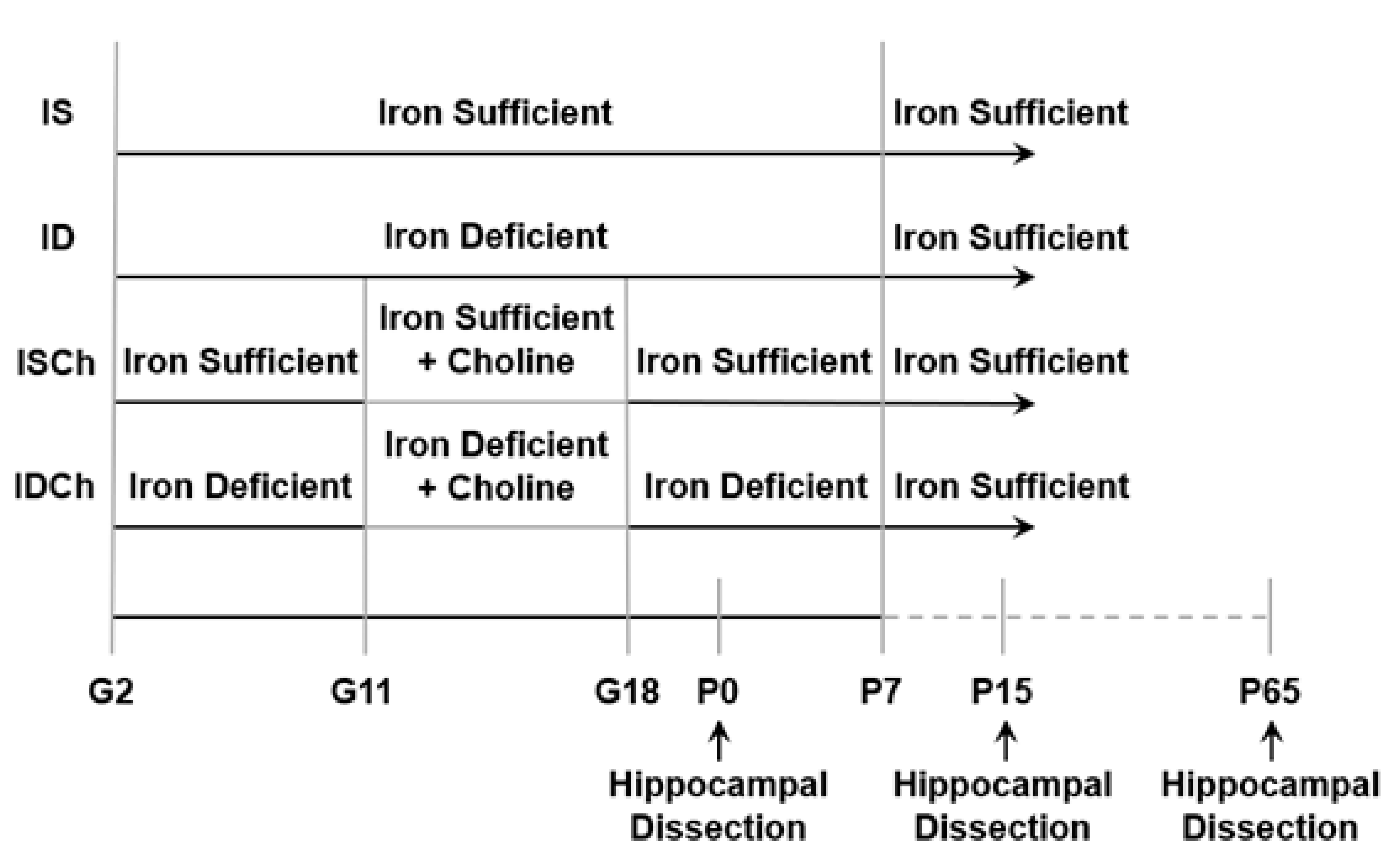
Figure 1. Graphical diagram of experimental groups. Abbreviations: Gestational day (G), iron-deficient group (ID), iron-deficient with choline supplementation group (IDCh), iron-sufficient group (IS), iron-sufficient with choline supplementation group (ISCh), and postnatal day (P).
Nutrients 13 04527 g001
Figure 2. Iron deficiency alters expression and enzymatic activity of hippocampal JARID1 in the ID rats. (AC) Hippocampal Jarid1b RNA expression in IS, ID, ISCh and IDCh groups at P0 (A), P15 (B) and P65 (C) quantified by Real-time quantitative PCR(RT-qPCR). (D) Hippocampal JARID1B protein expression in IS and ID groups at P15 and P65 measured with Western blot. Representative Western blot image of JARID1B at P15 is shown. (E) Hippocampal JARID1 activity in IS and ID groups at P0, P15 and P65 assessed with enzyme-linked immunosorbent assay. All data were normalized to the age-matched IS group. Tx: Treatment (Supplementation). Values are mean ± SEM; n = 5–6/group; * p < 0.05, ** p < 0.01, *** p < 0.001.
Nutrients 13 04527 g002
Figure 3. Iron deficiency and choline supplementation alter rat hippocampal Jarid1b regulation. (AC) HDAC1 enrichment at the Jarid1b promoter in the 4 groups at P0 (A), P15 (B), and P65 (C). (DF) H3K9ac enrichment at the Jarid1b promoter in the 4 groups at P0 (D), P15 (E), and P65 (F). (GI) Phosphorylated cAMP response element-binding protein (pCREB) enrichment at the Jarid1b promoter in the 4 groups at P0 (G), P15 (H), and P65 (I). All data were assessed with quantitative chromatin immunoprecipitation (qChIP) and normalized to the age-matched IS group. Tx: Treatment (Supplementation). Values are mean ± SEM, n = 5–6/group, * p < 0.05, ** p < 0.01.
Nutrients 13 04527 g003
Figure 4. Iron deficiency and choline supplementation alter hippocampal histone H3K9 methylation. (AC) Expression levels of G9a in the 4 groups at P0 (A), P15 (B) and P65 (C). (DF) Expression levels of Suv39h1 in the 4 groups at P0 (D), P15 (E) and P65 (F). (GI) H3K9me3 enrichment at the Jarid1b promoter in the 4 groups at P0 (G), P15 (H) and P65 (I). All data were assessed with RT-qPCR or qChIP and normalized to the age-matched IS group. Tx: Treatment (Supplementation). Values are mean ± SEM; n = 5–6/group; * p < 0.05, ** p < 0.01, *** p < 0.001, **** p < 0.0001.
Nutrients 13 04527 g004
Figure 5. Iron deficiency and choline supplementation induce epigenetic changes at the hippocampal Bdnf4 promoter. (A,B) Enrichment levels of JARID1B, H3K4me3, H3K9me3 and upstream stimulatory factor 1(USF1) at the Bdnf4 promoter in P15 (A) and P65 (B) IS and ID groups. (C,D) H3K9ac enrichment at the Bdnf4 promoter in the 4 groups at P15 (C) and P65 (D). (E,F) pCREB enrichment at the Bdnf4 promoter in the 4 groups at P15 (E) and P65 (F). All data were assessed by qChIP and normalized to the age-matched IS group. Tx: Treatment (Supplementation). Values are mean ± SEM; n = 5–6/group; * p < 0.05, ** p < 0.01.
Nutrients 13 04527 g005
Figure 6. Iron deficiency and choline supplementation induce epigenetic changes at the hippocampal Bdnf6 promoter. (A,B) Enrichment levels of JARID1B, H3K4me3 and USF1 at the Bdnf6 promoter in P15 (A) and P65 (B) IS and ID groups. (C,D) HDAC1 enrichment at the Bdnf6 promoter in the 4 groups at P15 (C) and P65 (D). (E,F) H3K9ac enrichment at the Bdnf6 promoter in the 4 groups at P15 (E) and P65 (F). (G,H) H3K9me3 enrichment at the Bdnf6 promoter in the 4 groups at P15 (G) and P65 (H). (I,J) pCREB enrichment at the Bdnf6 promoter in the 4 groups at P15 (I) and P65 (J). All data were assessed by qChIP and normalized to the age-matched IS group. Tx: Treatment (Supplementation). Values are mean ± SEM; n = 5–6/group; * p < 0.05, ** p < 0.01, **** p < 0.0001.
Nutrients 13 04527 g006
Table 1. Two-way ANOVA results. Significant p-values are in bold text.
2-Way ANOVA p-Value Table
FigureInteractionMain Effect of Iron StatusMain Effect of Choline SupplementationSimple Effect for Iron in Non-Choline-Supplemented GroupSimple Effect for Iron in Choline-Supplemented GroupSimple Effect for Choline Supplementation in Iron-Deficient GroupSimple Effect for Choline Supplementation in Iron-Sufficient Group
2A0.11060.68710.9212----
2B0.0341--0.00030.24560.72780.0096
2C0.0313--0.00290.93650.00510.9858
3A0.51590.96540.1919----
3B0.0012--0.01940.00860.03630.0051
3C0.99730.09780.1032----
3D0.16350.89010.5326----
3E0.0331--0.02090.47250.58180.0116
3F0.66430.70630.0014----
3G0.48940.16900.5148----
3H0.16330.15360.9047----
3I0.51030.31820.0079----
4A0.0491--0.03610.46020.59530.0274
4B0.20160.00020.0577----
4C0.17260.2513<0.0001----
4D0.0165--0.00590.54260.01120.3749
4E0.36640.0025<0.0001----
4F0.0152--0.24690.01910.00120.8812
4G0.61540.26730.3485----
4H0.19660.05620.5823----
4I0.0081--0.00040.92570.00030.7840
5C0.15560.01400.1612----
5D0.58680.62250.0190----
5E0.0417--0.00200.48980.69520.0037
5F0.96670.05570.0142----
6C0.0199--0.00730.60040.04410.1584
6D0.0063--0.00120.6758<0.00010.0293
6E0.06380.01140.1402----
6F0.58680.62250.0190----
6G0.36140.14000.7255----
6H0.65550.26840.0563----
6I0.07930.32730.8499----
6J0.91430.04990.0231----
Publisher’s Note: MDPI stays neutral with regard to jurisdictional claims in published maps and institutional affiliations.