Review

The Importance of the Fatty Acid Transporter L-Carnitine in Non-Alcoholic Fatty Liver Disease (NAFLD)

1
Radcliffe Department of Medicine, Oxford Centre for Magnetic Resonance Research, John Radcliffe Hospital, University of Oxford, Oxford OX3 9DU, UK
2
Radcliffe Department of Medicine, Oxford Centre for Diabetes, Endocrinology & Metabolism, Churchill Hospital, University of Oxford, Oxford OX3 7LE, UK
3
Oxford NIHR Biomedical Research Centre, University of Oxford, Oxford OX3 7LE, UK
4
Translational Gastroenterology Unit, University of Oxford, Oxford OX3 9DU, UK
*
Author to whom correspondence should be addressed.
Nutrients 2020, 12(8), 2178; https://doi.org/10.3390/nu12082178
Submission received: 19 June 2020 / Revised: 17 July 2020 / Accepted: 20 July 2020 / Published: 22 July 2020
(This article belongs to the Special Issue Diet and Nutrition for Hepatitis)

Abstract

:
L-carnitine transports fatty acids into the mitochondria for oxidation and also buffers excess acetyl-CoA away from the mitochondria. Thus, L-carnitine may play a key role in maintaining liver function, by its effect on lipid metabolism. The importance of L-carnitine in liver health is supported by the observation that patients with primary carnitine deficiency (PCD) can present with fatty liver disease, which could be due to low levels of intrahepatic and serum levels of L-carnitine. Furthermore, studies suggest that supplementation with L-carnitine may reduce liver fat and the liver enzymes alanine aminotransferase (ALT) and aspartate transaminase (AST) in patients with Non-Alcoholic Fatty Liver Disease (NAFLD). L-carnitine has also been shown to improve insulin sensitivity and elevate pyruvate dehydrogenase (PDH) flux. Studies that show reduced intrahepatic fat and reduced liver enzymes after L-carnitine supplementation suggest that L-carnitine might be a promising supplement to improve or delay the progression of NAFLD.

1. Introduction

One-third of the world’s Western population suffers from Non-Alcoholic Fatty Liver Disease (NAFLD) [1]. NAFLD develops when liver fat content exceeds 5% [2], and consists of a spectrum of pathologies ranging from simple steatosis (>5% liver fat) to Non-Alcoholic Steatohepatitis (NASH; fat + inflammation), fibrosis and cirrhosis. Liver fibrosis is a strong predictor of long-term mortality in patients with NAFLD [3,4]. Steatosis is associated with obesity and the metabolic syndrome [5] and develops when there is an imbalance between fatty acid uptake by the liver, synthesis (de novo lipogenesis) within the liver and disposal from the liver (Very Low Density Lipoprotein (VLDL) secretion and fatty acid oxidation). To enter the oxidation pathways fatty acids need to be coupled with L-carnitine in order to be transported into the mitochondria. Relative lack of L-carnitine may therefore lead to reduced fatty acid oxidation and triglyceride accumulation, resulting in NAFLD. Supplementation with L-carnitine may be a potential therapeutic option for lowering the risk of NAFLD by promoting fatty acid oxidation. Furthermore, studies show that L-carnitine is reduced in patients with liver disease, diabetes and cardiovascular disease [6,7,8] and supplementation with L-carnitine has been reported to improve liver inflammation and reduce liver enzymes and liver fat in these patients [9,10,11,12]. The aim of this review is to provide an overview of the published literature on the effects of L-carnitine supplementation in patients with NAFLD.

2. The Importance of L-Carnitine

L-carnitine (beta-hydroxy-gamma-N-trimethyl-aminobutyric acid) was discovered in 1905 as a constituent of muscle [13,14]. The name carnitine comes from the Latin word “carnis” (meat). The chemical structure was first described 20 years later, and it was in the 1970s that L-carnitine deficiencies were first discovered in humans [15].
The total body pool of L-carnitine is made up of several esters, including the short-chain ester, acetyl-carnitine [7]. L-carnitine homeostasis is regulated at multiple levels including intestinal absorption, de novo biosynthesis and renal reabsorption [16]. The human body contains approximately 300 mg/kg of L-carnitine, 98% of which is intracellular [17]. It is unevenly distributed between tissues and organs, with 80% present in muscle, and 5–10% present in the gastrointestinal tract. The liver contains about 3% and the blood contains 0.25% of the body’s L-carnitine pool [17].

2.1. Dietary Intake of L-carnitine

Dietary derived L-carnitine mostly comes from meat, therefore the Latin word “carnis”, with much smaller quantities from avocado and dairy products. In carnivores around 75% of the total body L-carnitine comes from the diet, but vegetarians mostly biosynthesize L-carnitine [18]. This is usually sufficient to maintain physiological function, as vegetarians also tend to have lower levels of long-chain fatty acids that need transportation to the mitochondria [19,20,21].
An average 70 kg person can absorb 23 to 135 mg/day of dietary L-carnitine, while a 70 kg person on a strictly vegetarian diet only takes up 1 mg/day of L-carnitine from the diet [22]. Infants are introduced to exogenous L-carnitine and acetyl-carnitine through breast milk, although it is known that L-carnitine and acetyl-carnitine have no specific immediate effects in the breast-feeding child [23]. It is speculated that L-carnitine in the breast milk serves as preparation for further uptake of L-carnitine through the diet, later in life.
For many years it was believed that all L-carnitine that was eaten through the diet was totally absorbed; however between 54–86% of L-carnitine from the food is absorbed, while only 5–25% of L-carnitine is absorbed if given through oral supplementation [24]. The efficiency of the absorption tends to diminish as the dose of L-carnitine increases, with some studies finding absorption of L-carnitine is saturated beyond doses of 2 g/12 h [25,26]. L-carnitine is absorbed partly through passive diffusion and partly via carrier-mediated transport in the colon and in the small intestines [22]. Dietary L-carnitine is degraded in the gut partly by trimethylamine N-oxide and partly by gamma-butyrobetaine [27]. The majority of these studies have investigated absorption in healthy humans and it is not clear whether there are any differences in L-carnitine absorption in individuals with NAFLD.

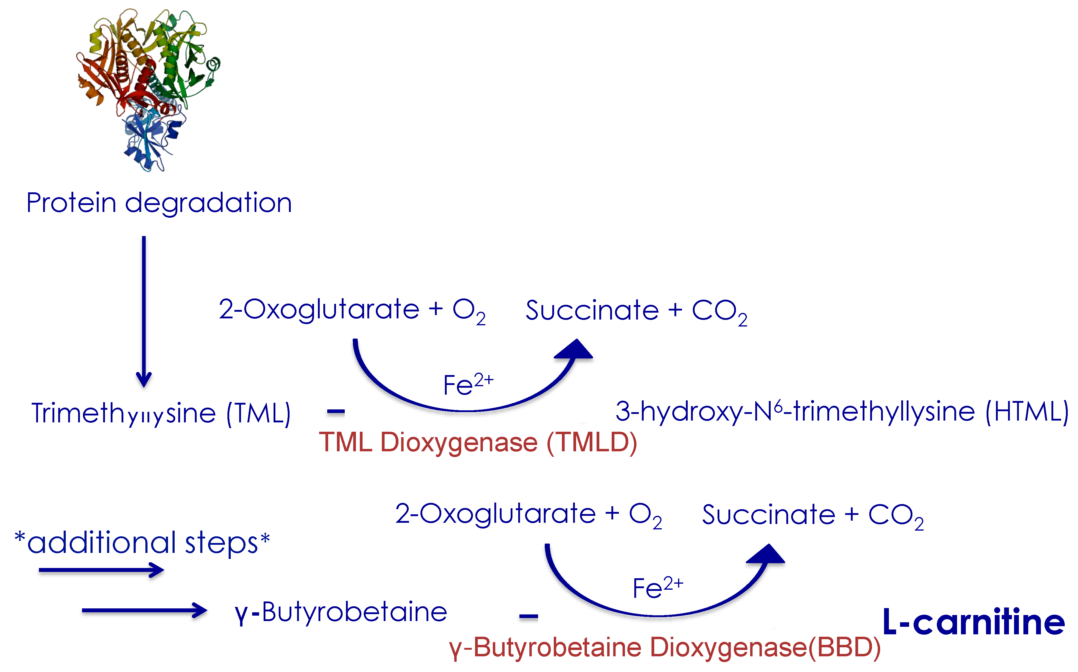
2.2. Endogenous L-carnitine Synthesis

L-carnitine is synthesized from the two amino-acids, lysine and methionine. A healthy human body can synthesize from 11 to 34 mg of L-carnitine per day. L-carnitine synthesis starts with the precursor trimethyl-lysine (TML), which is released from lysosomal protein degradation and ends with hydroxylation of gamma-butyrobetaine (BB) by gamma-butyrobetaine dioxygenase (BBD) producing L-carnitine (Figure 1). Trimethyl-lysine dioxygenase (TMLD) is the only enzyme involved in L-carnitine synthesis that is localized in the mitochondrial matrix. The other enzymes reside in the cytosol [28]. As the final enzyme in the synthesis pathway (BBD) is located in the liver, kidneys and in the brain, only these organs finalize the formation of L-carnitine from BB [29]. If the liver is metabolically compromised, then the last step of the synthesis pathway may be inhibited (Figure 1).

2.3. L-Carnitine Absorption

L-carnitine is transported into the cell by the high-affinity organic cation/carnitine transporter 2 (OCTN2), which mediates active absorption from the intestinal lumen into the enterocytes and the reabsorption into the kidneys. OCTN2 also mediates uptake into other tissues like adipose tissue, liver, cardiac myocytes, muscle cells, lymphocytes, and the brain.

2.4. Transport of Fatty Acids into Mitochondria

Within cells, long-chain fatty acids are dependent on esterification with L-carnitine to form acetyl-carnitine in order to be transported from the cytoplasm to the mitochondrial matrix for oxidation and energy production (Figure 2). The enzymes carnitine-palmitoyl-transferase-1 (CPT I), CPT II and carnitine-acylcarnitine translocase (CACT) are essential in catalyzing these reactions (Figure 2). CPT I allows binding of acyl-CoA to L-carnitine to form acyl-carnitine for entry into the mitochondrial intermembrane space. From there, CACT transports the acyl-carnitine across to the mitochondrial matrix in exchange for free L-carnitine. Once acyl-carnitine is inside the mitochondrial matrix CPT II separates it into L-carnitine and acyl-CoA. The free L-carnitine can then be exported to the cytosol by CACT. There is evidence that the composition of dietary fatty acids affects the CACT, by increasing the transcriptional rate of CACT RNA, and therefore fatty acid oxidation, in the mitochondria of livers in rats [30]. Within the liver, acyl-CoA then undergoes β-oxidation to produce acetyl-CoA for oxidative phosphorylation or ketone body production [31].

2.5. L-Carnitine as a Buffer for Excess Acetyl-CoA

In a reaction catalyzed by CPT I, the free form of L-carnitine binds to a fatty acid to form acyl-carnitine. Acyl-carnitines have varying chain-lengths, depending on the cellular location and metabolic purpose. Even though L-carnitine binds to all chain-lengths of fatty acids, it is only the most abundant form of long-chain fatty acids that are dependent on L-carnitine for transportation to the mitochondria [32]. However, intramitochondrial L-carnitine can export both short-chain and medium-chain acyl-CoA out of the mitochondria, that otherwise could lead to the production of free radicals which can destabilize the cell membrane; thus a reduced level of L-carnitine may lead to oxidative damage [33,34]. Both the tissue and the plasma will have a significant free and bound pool of L-carnitine. L-carnitine can be re-circulated once fatty acids enter the mitochondrial matrix, therefore only a small amount of L-carnitine is required in order to allow fatty acid to enter the mitochondria. However, L-carnitine has another important function as a buffer of excess acetyl-CoA in the mitochondria, through the formation of acetyl-carnitine [35]. A larger amount of L-carnitine is needed for this function, since acetyl-CoA either needs to be metabolized by the TCA cycle or exported with the use of L-carnitine in form of acetyl-carnitine. Once acetyl-carnitine is transported it can either be excreted in the urine or split from L-carnitine, which can be re-circulated again. L-carnitine reserves can be used up completely if there is a larger amount of excess acetyl-CoA that needs to be buffered out of the mitochondria [15].

3. Fatty Liver Disease and the Role of L-Carnitine

3.1. Drivers of Non-Alcoholic Fatty Liver Disease (NAFLD)

NAFLD is associated with obesity, insulin resistance [36,37], diabetes and cardiovascular disease (CVD) [38,39]. The main drivers in NAFLD are inflammation and accumulation of lipid [40]. Individuals with NAFLD have an intrahepatic accumulation of several lipid species, including diacylglycerides, triglycerides, ceramides and cholesterols [41]. Kupffer cells in the liver respond to alterations in lipid accumulation and can activate inflammatory pathways [42], that then can drive NASH progression. L-carnitine could be relevant to NAFLD pathology in two ways. Firstly, reduced levels of L-carnitine may lower fatty acid oxidation and be a contributing factor in the accumulation of liver fat. L-carnitine has been shown to have anti-inflammatory effects by upregulating the Peroxisome Proliferator Activator Receptor-γ (PPAR-γ) in the liver [43].
Other circumstantial evidence for the importance of L-carnitine in NAFLD comes from studies that have examined L-carnitine in associated metabolic conditions and that have shown that L-carnitine is reduced in obesity, insulin resistance, diabetes and advanced age [6,16,44,45]. Patients with obesity may present with reduced CPT I levels leading to reduced levels of intracellular L-carnitine, which prevents them from using certain fatty acids for energy production, resulting in lipid accumulation in the cells especially in the adipose tissue and the liver [46,47].

3.2. Fatty Liver Disease Is a Feature of Primary Carnitine Deficiency

Primary Carnitine Deficiency (PCD) is an autosomal recessive disorder of fatty acid oxidation due to the lack of organic cation/carnitine transporter (OCTN). The lack of OCTN, which is needed for the absorption of L-carnitine results in L-carnitine deficiency. Patients with PCD have low serum levels of L-carnitine and low intracellular levels of L-carnitine, thus fatty acids are not utilized as an energy source and accumulate [48], whilst patients rely exclusively on glucose for energy metabolism. As a consequence glucose stores are depleted rapidly and hypoglycemia is often seen in patients with PCD [49,50,51]. The inability to oxidize fatty acids also leads to an elevated production of reactive oxygen species (ROS) [52]. Half the patients with this condition have hepatomegaly and elevated transaminase and this condition is therefore an exemplar of the link between L-carnitine and liver health.
Patients with PCD have similarities in their liver profile to patients with NAFLD [51,53]. Patients with PCD show accumulation of fat in their liver and often they show hepatic encephalopathy, which is one of the major complications of advanced liver disease [50,54]. Furthermore in both diseases there are elevated liver enzymes like alanine transaminase (ALT) and aspartate transaminase (AST)—an indicator of liver injury [55]. A few cases in small children with PCD showed that liver size and enzymes were normalized after treatment with L-carnitine [52,56,57]

3.3. Other Evidence Linking L-Carnitine Deficiency to Liver Disease

Further evidence that lack of L-carnitine or dysfunction in the L-carnitine shuttle leads to a fatty liver comes from the effects of the drug etomoxir. Etomoxir is an irreversible inhibitor of CPT I and therefore switches energy metabolism from fatty acid to glucose oxidation in humans [58]. Etomoxir leads to elevated food intake and reduces liver energy status by reducing adenosine triphosphate/adenosine diphosphate (ATP/ADP) levels in rats [59]. Furthermore, in mice that were fed a short-term high fat (45%) diet, etomoxir stimulated glucose oxidation, peripheral glucose disposal, and led to elevated circulating fatty acids and circulating triglycerides (TGs) within 5 h of treatment and after several days hepatic steatosis was induced [60]. Another study that used etomoxir showed that inhibition of CPT I activity by 50% specifically in the liver led to an enlarged liver in a murine model [61].
These studies suggest that inhibition of the L-carnitine shuttle by pharmacological inhibition of CPT I, as in the case of etomoxir, results in liver steatosis. Reduced levels of L-carnitine would be expected to have a similar effect as they could equally result in an inability to transport fatty acids into the mitochondria for oxidation.

3.4. Patients with Chronic Liver Disease have Low Levels of L-Carnitine

There are several pieces of evidence pointing to low levels of L-carnitine in patients with chronic liver disease, particularly those with cirrhosis. This is critical since the liver is the primary site for L-carnitine synthesis and therefore impaired L-carnitine synthesis due to liver disease can result in whole-body impairment of L-carnitine metabolism [62]. It is unlikely that a reduction in L-carnitine causes NAFLD but having reduced levels of L-carnitine could exacerbate liver steatosis, and contribute to overall disease progression. The accumulation of fat can lead to a dysfunction in the biosynthesis of L-carnitine (Figure 1) in the liver thus inducing a negative feedback loop, whereby less L-carnitine is produced which leads to enhanced impairment of fatty acid oxidation re-enforcing fatty liver disease.
In a study of 68 children with chronic liver disease, 38 of whom had liver cirrhosis, serum L-carnitine levels were significantly lower in those with cirrhosis compared to those with early liver disease and healthy controls [62]. A correlation between TG levels and L-carnitine concentration in the plasma was found in patients with chronic liver disease, although this relationship was not detected in the patients with cirrhosis possibly due to already too low and sometimes absent levels of L-carnitine [62]. This could indicate that early liver disease progression might be evaluated not just based on fat levels in the liver but also on reduced levels of L-carnitine; however, once the stage of liver disease is severe, there is a complete depletion of L-carnitine and it is no longer of value.
Rudment et al. found that hospitalized patients with cirrhosis had significantly reduced levels of free and total L-carnitine in the serum compared to healthy controls [63]. In the same study, post-mortem examination of patients with cirrhosis that died also found reduced tissue L-carnitine levels in heart, liver, kidney, brain and muscle [63].

3.5. Acyl-Carnitine Chain Length can be Associated with Liver Disease

A study in 241 patients with biopsy-proven NAFLD and 23 patients with hepatocellular carcinoma (HCC) showed an inverse relation between disease severity and acyl-carnitine length. Plasma long-chain acyl-carnitine, but not free L-carnitine, was associated with fibrosis, inflammation and HCC. Medium-chain acyl-carnitines were reduced with worsening severity of NAFLD [64]. This inverse relationship is also supported by previous studies that have shown that replacement of dietary long-chain fatty acids with medium-chain fatty acids reduces triglyceride accumulation by 50% in the murine liver [65].
Another study in patients with cirrhosis also showed up to a three-fold elevation of short-chain acyl-carnitine and long-chain acyl-carnitine in the plasma compared to healthy volunteers [66], and this has been supported by others [62,67]. These studies suggest that the length of the acyl-carnitine might be critical for predicting the severity of patients with NAFLD; however, larger studies are needed to confirm these findings.
In urinary metabolomics Dong et al. showed that, compared to healthy controls, patients with NAFLD and NASH had reduced levels of urine free L-carnitine but not acetyl-carnitine [33]. This suggests that the body is conserving free L-carnitine to be able to bind to long-chain fatty acids, but it also shows an elevated excretion of short-chain acetyl-CoA. This study did not measure other lengths of acyl-carnitine.

4. The Importance of L-Carnitine Supplementation

L-carnitine supplementation has beneficial effects in patients with fatty liver disease, where elevations in high-density lipoprotein (HDL) cholesterol and reductions in liver fat have been reported [68]. L-carnitine has been shown to elevate activity and transcription of hepatic CPT I [69], leading to a reduced amount of fat in the liver [70]. Several studies have shown improvement in hepatic steatosis and cirrhosis after L-carnitine supplementation [11,50,71,72]. Furthermore, L-carnitine supplementation in humans and animal models has been shown to modulate insulin sensitivity and glucose uptake and also to have an antioxidant effect in hepatocytes [71,73,74].

4.1. L-Carnitine Supplementation is Beneficial to the Liver

Several studies have examined L-carnitine’s ability to reduce fat accumulation in the liver in patients with NAFLD, generally with positive results (Table 1). These studies show that liver enzymes that are most commonly used as a laboratory test for detecting an abnormal liver can be normalized with supplementation of L-carnitine [75].
In one animal study, plasma L-carnitine was reduced in obese animals that were fed a high-fat diet for a full year, and this was associated with reduced expression of hepatic regulatory L-carnitine genes. These effects were reversible with L-carnitine treatment [16]. We have also previously shown that streptozotocin (STZ)-induced diabetic rats treated with L-carnitine (3 g/kg/day for five weeks) showed improved liver enzymes as well as choline levels in the liver while reducing TGs in the plasma [76].
Just as L-carnitine induces improvements in NASH [77], it also may induce regression of cirrhosis. Patients with cirrhosis have an acquired L-carnitine deficiency [12] and levels of free fatty acids progressively elevate with the severity of liver cirrhosis [78].
There are some discrepancies in findings, and it is not entirely understood when L-carnitine elevates free fatty acids [81] and when L-carnitine reduces free fatty acid [82]. For example in patients with obesity, L-carnitine directly reduces all free fatty acids in the plasma by transporting them to the mitochondria [83]. One study investigated 13 patients with liver cirrhosis before and after four weeks of L-carnitine treatment (1800 mg/day), and found that, after L-carnitine treatment, free fatty acid levels increased, whole-body carbohydrate oxidation increased, whilst whole-body fatty acid and protein oxidation significantly decreased [81]. Others have reported no differences in energy metabolism when patients with liver cirrhosis were treated with L-carnitine, although improvements in exercise tolerance were observed, which alludes to improved energy metabolism [84]. Data from animal models of diabetes point towards an effect of L-carnitine on energy metabolism with one study showing that L-carnitine supplementation elevates fatty acid oxidation [85].
Intestinal microbiota of trimethylamine (TMA) is metabolized to trimethylamine-N-oxide (TMAO), which is associated with CVD risk and could promote atherosclerosis [86]. Koeth et al. found that L-carnitine supplementation in mice enhanced synthesis of TMA and TMAO. Unlike other studies included in this review, where patients were given oral treatments of pure L-carnitine, Koeth et al. investigated the production of TMAO in the plasma and the urine by feeding patients an eight-ounce steak followed by a capsule of 250 mg heavy isotope-labelled L-carnitine [86]. The increase in TMAO was modest as the authors suggest but in five subjects TMAO production could be suppressed by giving oral broad-spectrum antibiotics, suggesting how intestinal microbiota contribute to the link between red meat consumption and CVD risk [86]. Since these patients were given meat, the dietary uptake of L-carnitine would have been much higher compared to if the same dose of oral L-carnitine was given [24], possibly leading to higher TMAO levels. The reason why a larger consumption of L-carnitine occurs if taken through the diet, rather than as a supplement, is still unclear and need to be further investigated.

4.2. Effects of L-Carnitine in Ketogenesis

Studies have shown how L-carnitine stimulates ketogenesis in the liver of mice [87,88]. A study undertaken in perfused livers showed that ketogenesis increases with an infusion of free L-carnitine in the liver. A subsequent infusion of long-chain fatty acids had no effect on ketone production, which implies that the substrate of ketone production in the presence of L-carnitine is endogenous fatty acids [87]. Nakajima et al. showed the rate-limiting step in ketogenesis is L-carnitine and not fatty acid supply [87]. Reduced L-carnitine levels in the liver result in an impairment of ketogenesis after a fatty diet (80%) in children with primary carnitine deficiencies and episodes of hepatic and cerebral dysfunction [89]. Oral L-carnitine supplementation improved these children’s clinical outcome and restored L-carnitine levels in the plasma [89]. Several studies have also shown that L-carnitine lowers the ketone body β-hydroxybutyrate [24,88,90] and that it is dose dependent [91]. It is not yet clear how L-carnitine regulates ketone utilizations or production, but these studies would suggest that L-carnitine can activate different pathways [90,92,93].

4.3. L-Carnitine has Significant Effect on Insulin and Glucose Levels

Insulin resistance is often observed in individuals with liver disease, specifically in patients with NASH [94]. Patients with NAFLD have been shown to have an impaired ability to oxidize glucose and non-oxidative glucose disposal with insulin stimulation [95].
L-carnitine does not only affect fatty acids, but it also showed a significant effect on glucose and insulin levels in several studies [8,96]. Randle et al. proposed in 1963 that the interaction between carbohydrate and fatty acid metabolism takes the form of a glucose-fatty-acid cycle that controls blood glucose levels, fatty acid concentrations and insulin sensitivity [97,98]. The Randle cycle essentially describes that when fatty acid oxidation is active there is a significant reduction in the uptake and utilization of glucose by inhibition of PDH flux, but it also works the other way around, so the production of acetyl-CoA from glycolysis will inhibit fatty acid oxidation through the generation of malonyl-CoA which can inhibit CPT I [99].
A study in 25 healthy subjects evaluated the effects of L-carnitine on glucose metabolism where subjects were initially infused with 5% glucose solution and 48 h later the same subjects were infused with 2 g of L-carnitine with 5% of glucose solution [100]. It was found that L-carnitine reduced glucose levels by reducing insulin levels; notably, glucose was maintained in the normal range [100]. Furthermore, Bae et al. showed that patients with NAFLD and diabetes improved their glycemic control (using HbA1c as a marker) after treatment with carnitine-orotate for 12 weeks [11].
There are several proposed mechanisms for L-carnitine’s effect on carbohydrate metabolism including: (i) the regulation of the ratio of acetyl-CoA/CoA in the mitochondria and thereby PDH flux [101], (ii) by modulating glycolytic and gluconeogenic enzymes [99,102], and (iii) by stimulating insulin-like growth factor-1 (IGF-1) signaling cascades [103]. Infusion of L-carnitine in healthy people attenuates a rise in plasma glucose levels with a 10% glucose infusion for 3 h [104]. Furthermore, insulin can stimulate free L-carnitine as measured in the muscle [105]. Insulin sensitivity was reported to be improved by inducing hepatic autophagy through PPAR-γ, a possible new mechanism of L-carnitine treatment [106].
Recently it was reported that inflammation can affect insulin sensitivity [107]; L-carnitine has been shown to improve insulin-stimulated disposal of glucose [108], therefore it is plausible that L-carnitine may play a role in reducing inflammation via improvements in insulin-stimulated disposal of glucose.
Despite the above findings, treatment with L-carnitine does not always show changes in insulin levels even though the majority of studies report reduced serum glucose levels, TGs, fatty acids and ketone levels [8,109], sometimes without insulin changes. The discrepancy remains unclear in findings of studies in insulin levels and the mechanisms underpinning the effect of L-carnitine on plasma insulin remain to be elucidated.
Associations have been shown between insulin resistance and elevated long-chain acyl-carnitines in the plasma [110,111], and patients with NAFLD have higher levels of long-chain acyl-carnitines [111]. Progression of NAFLD has been shown to correlate with long-chain acyl-carnitine species [64,111] with patients with cirrhosis also often have elevated plasma insulin levels. Mihalik et al. showed that insulin infusion could reduce all species of plasma acyl-carnitine in healthy people, but in patients with diabetes this function was blunted [112]. Plasma long-chain acyl-carnitine species are elevated in patients with NAFLD, obesity and type 2 diabetes, suggesting that more fatty acids can enter the mitochondria, but since there is also an elevation of short-chain acyl-carnitines there might be a defect in the oxidation pathways.

5. Summary and Conclusions

L-carnitine is a critical co-factor for transporting long-chain fatty acids into mitochondria for β-oxidation and to export excess acetyl-CoA from the mitochondrial matrix. It is relevant to liver disease in two ways. Firstly, the liver is critical in synthesizing L-carnitine and, if diseased, L-carnitine biosynthesis is reduced which may affect whole body fatty acid metabolism. Secondly, it might be a potential treatment for liver fat accumulation as it promotes fat oxidation and can also have beneficial effects on carbohydrate metabolism.
Treatment with L-carnitine can improve outcomes in patients with fatty liver disease (Figure 3) and has been shown to reduce ALT and AST levels, as well as liver fat accumulation. L-carnitine administration has also been shown to improve markers of glycemic control in patients with NAFLD and diabetes, most likely by regulating the ratio of acetyl-CoA/CoA in the mitochondria and thereby the PDH flux.
Patients with chronic liver disease often have reduced levels of L-carnitine. The length of acyl-carnitine species found in plasma and urine might be of importance for disease outcomes. Therefore, it might not be sufficient to solely investigate free L-carnitine, but rather all L-carnitine species should be studied. Several studies show elevated plasma long-chain acyl-carnitine but not free L-carnitine and medium-chain acyl-carnitine to be associated with fibrosis, inflammation and cirrhosis in patients.
Larger and more comprehensive studies are needed to confirm whether L-carnitine has the beneficial effects observed in these small-scale studies in patients with NAFLD. Studies generally only measure free L-carnitine, acetyl-carnitine, or both. If in vivo imaging or biopsies are available, a more comprehensive analysis of the L-carnitine species could be possible before and after treatment with L-carnitine, to fully understand the effects of L-carnitine supplementation. Measuring L-carnitine species in a range of phenotypes might provide an insight into their use as a biomarker for liver disease.

Author Contributions

D.S. and M.P. wrote the manuscript, L.H. and S.N. edited, discussed and read the manuscript. All authors have read and agreed to the published version of the manuscript.

Funding

D.S. is a Novo Nordisk (NN) Research Fellow. L.H. is a British Heart Foundation Senior Research Fellow in Basic Science.

Acknowledgments

We acknowledge the support and help from Damian J. Tyler, including other colleagues from the Oxford Centre for Magnetic Resonance Research. We also acknowledge Novo Nordisk Mentor Anni Moorsing, NN International Medical Director and Dorthe Lundsgaard, NN Senior Alliance Director for their support, help and discussions.

Conflicts of Interest

The authors declare no conflict of interest.

References

  1. Younossi, Z.M.; Marchesini, G.; Pinto-Cortez, H.; Petta, S. Epidemiology of nonalcoholic fatty liver disease and nonalcoholic steatohepatitis. Transplantation 2019, 103, 22–27. [Google Scholar] [CrossRef] [PubMed]
  2. European Association for the Study of the Liver (EASL); European Association for the Study of Diabetes (EASD); European Association for the Study of Obesity (EASO). EASL-EASD-EASO clinical practice guidelines for the management of non-alcoholic fatty liver disease. Obes. Facts 2016, 9, 65–90. [Google Scholar] [CrossRef] [PubMed] [Green Version]
  3. Angulo, P.; Kleiner, D.E.; Dam-Larsen, S.; Adams, L.A.; Bjornsson, E.S.; Charatcharoenwitthaya, P.; Mills, P.R.; Keach, J.C.; Lafferty, H.D.; Stahler, A.; et al. Liver fibrosis, but no other histologic features, is associated with long-term outcomes of patients with nonalcoholic fatty liver disease. Gastroenterology 2015, 149, 389–397. [Google Scholar] [CrossRef] [PubMed] [Green Version]
  4. Fielding, C.M.; Angulo, P. Hepatic steatosis and steatohepatitis: Are they really two distinct entities? Curr. Hepatol. Rep. 2014, 13, 151–158. [Google Scholar] [CrossRef] [PubMed] [Green Version]
  5. Marchesini, G.; Brizi, M.; Bianchi, G.; Tomassetti, S.; Bugianesi, E.; Lenzi, M.; McCullough, A.J.; Natale, S.; Forlani, G.; Melchionda, N. Nonalcoholic fatty liver disease: A feature of the metabolic syndrome. Diabetes 2001, 50, 1844–1850. [Google Scholar] [CrossRef] [PubMed] [Green Version]
  6. Tamamoğullari, N.; Siliğ, Y.; Içağasioğlu, S.; Atalay, A. Carnitine deficiency in diabetes mellitus complications. J. Diabetes Complicat. 1999, 13, 251–253. [Google Scholar] [CrossRef]
  7. Pekala, J.; Patkowska-Sokola, B.; Bodkowski, R.; Jamroz, D.; Nowakowski, P.; Lochynski, S.; Librowski, T. L-Carnitine-Metabolic functions and meaning in humans life. Curr. Drug Metab. 2011, 12, 667–668. [Google Scholar] [CrossRef]
  8. Rodrigues, B.; Xiang, H.; McNeill, J.H. Effect of L-Carnitine treatment on lipid metabolism and cardiac performance in chronically diabetic rats. Diabetes 1988, 37, 1358–1364. [Google Scholar] [CrossRef]
  9. Cecere, A.; Ciaramella, F.; Tancredi, L.; Romano, C.; Gattoni, A. Efficacy of L-Carnitine in reducing hyperammonaemia and improving neuropsychological test performance in patients with hepatic cirrhosis results of a randomised Trial. Clin. Drug Investig. 2002, 22, 7–14. [Google Scholar] [CrossRef]
  10. Malaguarnera, M.; Gargante, M.P.; Russo, C.; Antic, T.; Vacante, M.; Malaguarnera, M.; Avitabile, T.; Li Volti, G.; Galvano, F. L-Carnitine supplementation to diet: A new tool in treatment of nonalcoholic steatohepatitis—A randomized and controlled clinical trial. Am. J. Gastroenterol. 2010, 105, 1338–1345. [Google Scholar] [CrossRef]
  11. Bae, J.C.; Lee, W.Y.; Yoon, K.H.; Park, J.Y.; Son, H.S.; Han, K.A.; Lee, K.W.; Woo, J.T.; Ju, Y.C.; Lee, W.J.; et al. Improvement of nonalcoholic fatty liver disease with carnitine-orotate complex in type 2 diabetes (CORONA): A randomized controlled trial. Diabetes Care 2015, 38, 1245–1252. [Google Scholar] [CrossRef] [PubMed] [Green Version]
  12. Hassan, A.; Tsuda, Y.; Asai, A.; Yokohama, K.; Nakamura, K.; Sujishi, T.; Ohama, H.; Tsuchimoto, Y.; Fukunishi, S.; Abdelaal, U.M.; et al. Effects of oral L-carnitine on liver functions after transarterial chemoembolization in intermediate-stage HCC patients. Mediat. Inflamm. 2015, 2015, 608216. [Google Scholar] [CrossRef] [PubMed] [Green Version]
  13. Gulewitsch, W.; Krimberg, R. Zur kenntnis der extraktivstoffe der muskeln: II. mitteilung: Über das carnitin. Hoppe Seyler’s Z. Physiol. Chem. 1905, 45, 326–330. [Google Scholar] [CrossRef] [Green Version]
  14. Kutscher, F. Zur kenntnis des novains. Hoppe Seyler’s Z. Physiol. Chem. 1906, 49, 47–49. [Google Scholar] [CrossRef]
  15. Harmeyer, J. The physiological role of L-carnitine. Lohmann Inf. 2002, 27, 15–21. [Google Scholar]
  16. Noland, R.C.; Koves, T.R.; Seiler, S.E.; Lum, H.; Lust, R.M.; Ilkayeva, O.; Stevens, R.D.; Hegardt, F.G.; Muoio, D.M. Carnitine insufficiency caused by aging and overnutrition compromises mitochondrial performance and metabolic control. J. Biol. Chem. 2009, 284, 22840–22852. [Google Scholar] [CrossRef] [Green Version]
  17. Adeva-Andany, M.M.; Calvo-Castro, I.; Fernández-Fernández, C.; Donapetry-García, C.; Pedre-Piñeiro, A.M. Significance of L-carnitine for human health. IUBMB Life 2017, 69, 578–594. [Google Scholar] [CrossRef] [Green Version]
  18. Stephens, F.B.; Marimuthu, K.; Cheng, Y.; Patel, N.; Constantin, D.; Simpson, E.J.; Greenhaff, P.L. Vegetarians have a reduced skeletal muscle carnitine transport capacity. Am. J. Clin. Nutr. 2011, 94, 938–944. [Google Scholar] [CrossRef] [Green Version]
  19. Lombard, K.A.; Olson, A.L.; Nelson, S.E.; Rebouche, C.J. Carnitine status of lactoovovegetarians and strict vegetarian adults and children. Am. J. Clin. Nutr. 1989, 50, 301–306. [Google Scholar] [CrossRef]
  20. Krajčovičová-Kudláčková, M.; Šimončič, R.; Béderová, A.; Babinská, K.; Béder, I. Correlation of carnitine levels to methionine and lysine intake. Physiol. Res. 2000, 49, 399–402. [Google Scholar]
  21. Rosell, M.S.; Lloyd-Wright, Z.; Appleby, P.N.; Sanders, T.A.; Allen, N.E.; Key, T.J. Long-chain n-3 polyunsaturated fatty acids in plasma in british meat-eating, vegetarian, and vegan men. Am. J. Clin. Nutr. 2005, 82, 327–334. [Google Scholar] [CrossRef] [PubMed]
  22. Evans, A.M.; Fornasini, G. Pharmacokinetics of L-Carnitine. Clin. Pharmacokinet. 2003, 42, 941–967. [Google Scholar] [CrossRef] [PubMed]
  23. Levocarnitine. Drugs and Lactation Database (LactMed); National Library of Medicine (US): Bethesda, MD, USA, 2006. Available online: https://www.ncbi.nlm.nih.gov/books/NBK501864/ (accessed on 19 June 2020).
  24. Rebouche, C.J.; Paulson, D.J. Carnitine Metabolism and Function in Humans. Annu. Rev. Nutr. 1986, 6, 41–66. [Google Scholar] [CrossRef] [PubMed]
  25. Sahajwalla, C.G.; Helton, E.D.; Purich, E.D.; Hoppel, C.L.; Cabana, B.E. Multiple-dose pharmacokinetics and bioequivalence of L-carnitine 330-mg tablet versus 1-g chewable tablet versus enteral solution in healthy adult male volunteers. J. Pharm. Sci. 1995, 84, 627–633. [Google Scholar] [CrossRef] [PubMed]
  26. Segre, G.; Bianchi, E.; Corsi, M.; D’Iddio, S.; Ghirardi, O.; Maccari, F. Plasma and urine pharmacokinetics of free and of short-chain carnitine after administration of carnitine in man. Arzneim. Forsch. Drug Res. 1988, 38, 1830. [Google Scholar]
  27. Rebouche, C.J.; Chenard, C.A. Metabolic fate of dietary carnitine in human adults: Identification and quantification of urinary and fecal metabolites. J. Nutr. 1991. [Google Scholar] [CrossRef] [PubMed] [Green Version]
  28. Strijbis, K.; Vaz, F.M.; Distel, B. Enzymology of the carnitine biosynthesis pathway. IUBMB Life 2010. [Google Scholar] [CrossRef]
  29. Englard, S. Hydroxylation of γ-butyrobetaine to carnitine in human and monkey tissues. FEBS Lett. 1979. [Google Scholar] [CrossRef] [Green Version]
  30. Priore, P.; Stanca, E.; Gnoni, G.V.; Siculella, L. Dietary fat types differently modulate the activity and expression of mitochondrial carnitine/acylcarnitine translocase in rat liver. Biochim. Biophys. Acta Mol. Cell Biol. Lipids 2012. [Google Scholar] [CrossRef]
  31. Longo, N.; Frigeni, M.; Pasquali, M. Carnitine transport and fatty acid oxidation. Biochim. Biophys. Acta 2016, 1863, 2422–2435. [Google Scholar] [CrossRef]
  32. Schönfeld, P.; Wojtczak, L. Short- and medium-chain fatty acids in energy metabolism: The cellular perspective. J. Lipid Res. 2016, 57, 943–954. [Google Scholar] [CrossRef] [PubMed] [Green Version]
  33. Dong, S.; Zhan, Z.-Y.; Cao, H.-Y.; Wu, C.; Bian, Y.-Q.; Li, J.-Y.; Cheng, G.-H.; Liu, P.; Sun, M.-Y. Urinary metabolomics analysis identifies key biomarkers of different stages of nonalcoholic fatty liver disease. World J. Gastroenterol. 2017, 23, 2771–2784. [Google Scholar] [CrossRef] [PubMed]
  34. Mohammadi, M.; Hajhossein Talasaz, A.; Alidoosti, M. Preventive effect of L-carnitine and its derivatives on endothelial dysfunction and platelet aggregation. Clin. Nutr. ESPEN 2016, 15, 1–10. [Google Scholar] [CrossRef] [PubMed]
  35. Rinaldo, P.; Cowan, T.M.; Matern, D. Acylcarnitine profile analysis. Genet. Med. 2008, 10, 151–156. [Google Scholar] [CrossRef] [Green Version]
  36. Bae, J.C.; Cho, Y.K.; Lee, W.Y.; Seo, H.I.; Rhee, E.J.; Park, S.E.; Park, C.Y.; Oh, K.W.; Sung, K.C.; Kim, B.I. Impact of nonalcoholic fatty liver disease on insulin resistance in relation to HbA1c levels in nondiabetic subjects. Am. J. Gastroenterol. 2010, 105, 2389–2395. [Google Scholar] [CrossRef] [PubMed]
  37. Rector, R.S.; Thyfault, J.P.; Wei, Y.; Ibdah, J.A. Non-alcoholic fatty liver disease and the metabolic syndrome: An update. World J. Gastroenterol. 2008, 14, 185. [Google Scholar] [CrossRef] [PubMed]
  38. Bae, J.C.; Rhee, E.J.; Lee, W.Y.; Park, S.E.; Park, C.Y.; Oh, K.W.; Park, S.W.; Kim, S.W. Combined effect of nonalcoholic fatty liver disease and impaired fasting glucose on the development of type 2 diabetes: A 4-year retrospective longitudinal study. Diabetes Care 2011, 34, 727–729. [Google Scholar] [CrossRef] [PubMed] [Green Version]
  39. Stepanova, M.; Younossi, Z.M. Independent association between nonalcoholic fatty liver disease and cardiovascular disease in the US population. Clin. Gastroenterol. Hepatol. 2012, 10, 646–650. [Google Scholar] [CrossRef]
  40. Gruben, N.; Shiri-Sverdlov, R.; Koonen, D.P.Y.; Hofker, M.H. Nonalcoholic fatty liver disease: A main driver of insulin resistance or a dangerous liaison? Biochim. Biophys. Acta Mol. Basis Dis. 2014, 1842, 2329–2343. [Google Scholar] [CrossRef] [Green Version]
  41. Cheung, O.; Sanyal, A. Abnormalities of lipid metabolism in nonalcoholic fatty liver disease. Semin. Liver Dis. 2008, 28, 351–359. [Google Scholar] [CrossRef]
  42. Baffy, G. Kupffer cells in non-alcoholic fatty liver disease: The emerging view. J. Hepatol. 2009. [Google Scholar] [CrossRef] [Green Version]
  43. El-Sheikh, A.A.; Rifaai, R.A. Peroxisome proliferator activator receptor (PPAR)-γ ligand, but not PPAR-α, ameliorates cyclophosphamide-induced oxidative stress and inflammation in rat liver. PPAR Res. 2014, 2014. [Google Scholar] [CrossRef] [PubMed] [Green Version]
  44. Mamoulakis, D.; Galanakis, E.; Dionyssopoulou, E.; Evangeliou, A.; Sbyrakis, S. Carnitine deficiency in children and adolescents with type 1 diabetes. J. Diabetes Complicat. 2004, 18, 271–274. [Google Scholar] [CrossRef]
  45. la Marca, G.; Malvagia, S.; Toni, S.; Piccini, B.; Di Ciommo, V.; Bottazzo, G.F. Children who develop type 1 diabetes early in life show low levels of carnitine and amino acids at birth: Does this finding shed light on the etiopathogenesis of the disease? Nutr. Diabetes 2013, 3, e94. [Google Scholar] [CrossRef] [PubMed]
  46. Fucho, R.; Casals, N.; Serra, D.; Herrero, L. Ceramides and mitochondrial fatty acid oxidation in obesity. FASEB J. 2017, 31, 1263–1272. [Google Scholar] [CrossRef] [PubMed] [Green Version]
  47. Costa, C.C.G.; De Almeida, I.T.; Jakobs, C.; Poll-The, B.-T.; Duran, M. Dynamic changes of plasma acylcarnitine levels induced by fasting and sunflower oil challenge test in children. Pediatr. Res. 1999, 46, 440. [Google Scholar] [CrossRef] [Green Version]
  48. Chapoy, P.R.; Angelini, C.; Brown, W.J.; Stiff, J.E.; Shug, A.L.; Cederbaum, S.D. Systemic carnitine deficiency—A treatable inherited lipid-storage disease presenting as Reye’s Syndrome. N. Engl. J. Med. 1980, 303, 1389–1394. [Google Scholar] [CrossRef]
  49. CA, S. Carnitine deficiency disorders in children. Ann. N. Y. Acad. Sci. 2004, 1033, 42–51. [Google Scholar]
  50. Longo, N.; Amat di San Filippo, C.; Pasquali, M. Disorders of carnitine transport and the carnitine cycle. In American Journal of Medical Genetics Part C: Seminars in Medical Genetics; Wiley: Hoboken, NJ, USA, 2006; Volume 142, pp. 77–85. [Google Scholar]
  51. Jun, J.S.; Lee, E.J.; Park, H.D.; Kim, H.S. Systemic primary carnitine deficiency with hypoglycemic encephalopathy. Ann. Pediatr. Endocrinol. Metab. 2016, 21, 226. [Google Scholar] [CrossRef] [Green Version]
  52. Pike, L.S.; Smift, A.L.; Croteau, N.J.; Ferrick, D.A.; Wu, M. Inhibition of fatty acid oxidation by etomoxir impairs NADPH production and increases reactive oxygen species resulting in ATP depletion and cell death in human glioblastoma cells. Biochim. Biophys. Acta Bioenerg. 2011, 1807, 726–734. [Google Scholar] [CrossRef] [Green Version]
  53. Deswal, S.; Bijarnia-Mahay, S.; Manocha, V.; Hara, K.; Shigematsu, Y.; Saxena, R.; Verma, I.C. Primary carnitine deficiency—A rare treatable cause of cardiomyopathy and massive hepatomegaly. Indian J. Pediatr. 2017, 84, 83–85. [Google Scholar] [CrossRef] [PubMed]
  54. Rasmussen, J.; Nielsen, O.W.; Lund, A.M.; Køber, L.; Djurhuus, H. Primary carnitine deficiency and pivalic acid exposure causing encephalopathy and fatal cardiac events. J. Inherit. Metab. Dis. 2013, 36, 35–41. [Google Scholar] [CrossRef] [PubMed]
  55. Han, L.; Wang, F.; Wang, Y.; Ye, J.; Qiu, W.; Zhang, H.; Gao, X.; Gong, Z.; Gu, X. Analysis of genetic mutations in Chinese patients with systemic primary carnitine deficiency. Eur. J. Med. Genet. 2014. [Google Scholar] [CrossRef] [PubMed]
  56. Treem, W.A.; Stanley, C.A. Massive hepatomegaly, steatosis, and secondary plasma carnitine deficiency in an infant with cystic fibrosis. Pediatrics 1989, 83, 993–997. [Google Scholar] [PubMed]
  57. Ravindranath, A.; Pai, G.; Srivastava, A.; Poddar, U.; Yachha, S.K. Infant with hepatomegaly and hypoglycemia: A setting for fatty acid oxidation defects. Indian J. Gastroenterol. 2017, 36, 429–434. [Google Scholar] [CrossRef] [PubMed]
  58. Hinderling, V.B.; Schrauwen, P.; Langhans, W.; Westerterp-Plantenga, M.S. The effect of etomoxir on 24-h substrate oxidation and satiety in humans. Am. J. Clin. Nutr. 2002, 76, 141–147. [Google Scholar] [CrossRef] [Green Version]
  59. Horn, C.C.; Ji, H.; Friedman, M.I. Etomoxir, a fatty acid oxidation inhibitor, increases food intake and reduces hepatic energy status in rats. Physiol. Behav. 2004, 81, 157–162. [Google Scholar] [CrossRef]
  60. Lundsgaard, A.M.; Fritzen, A.M.; Nicolaisen, T.S.; Carl, C.S.; Sjøberg, K.A.; Raun, S.H.; Klein, A.B.; Sanchez-Quant, E.; Langer, J.; Ørskov, C.; et al. Glucometabolic consequences of acute and prolonged inhibition of fatty acid oxidation. J. Lipid Res. 2020. [Google Scholar] [CrossRef]
  61. Coort, S.; Niessen, H.; Koonen, D.; Coumans, W.; Bonen, A.; Vandervusse, G.; Glatz, J.; Luiken, J. 204 effects of the hypertrophy-inducing agent etomoxir on fatty acid utilization and fatty acid transporters in rat heart and liver. Eur. J. Hear. Fail. Suppl. 2003, 2, 42. [Google Scholar] [CrossRef]
  62. Selimoglu, M.A.; Aydogdu, S.; Yagci, R.V.; Huseyinov, A. Plasma and liver carnitine status of children with chronic liver disease and cirrhosis. Pediatr. Int. 2001, 43, 391–395. [Google Scholar] [CrossRef]
  63. Rudman, D.; Sewell, C.W.; Ansley, J.D. Deficiency of carnitine in cachectic cirrhotic patients. J. Clin. Investig. 1977, 60, 716–723. [Google Scholar] [CrossRef] [PubMed]
  64. Enooku, K.; Nakagawa, H.; Fujiwara, N.; Kondo, M.; Minami, T.; Hoshida, Y.; Shibahara, J.; Tateishi, R.; Koike, K. Altered serum acylcarnitine profile is associated with the status of nonalcoholic fatty liver disease (NAFLD) and NAFLD-related hepatocellular carcinoma. Sci. Rep. 2019, 9, 10663. [Google Scholar] [CrossRef] [PubMed]
  65. Lieber, C.S.; Lefèvre, A.; Spritz, N.; Feinman, L.; DeCarli, L.M. Difference in hepatic metabolism of long- and medium-chain fatty acids: The role of fatty acid chain length in the production of the alcoholic fatty liver. J. Clin. Invest. 1967, 46, 1451–1460. [Google Scholar] [CrossRef] [PubMed] [Green Version]
  66. Fuller, R.K.; Hoppel, C.L. Elevated plasma carnitine in hepatic cirrhosis. Hepatology 2007, 3, 554–558. [Google Scholar] [CrossRef] [PubMed]
  67. Krahenbuhl, S.; Reichen, J. Carnitine metabolism in patients with chronic liver disease. Hepatology 1997, 25, 148–153. [Google Scholar] [CrossRef]
  68. Su, C.C.; Chang, C.S.; Chou, C.H.; Wu, Y.H.S.; Yang, K.T.; Tseng, J.K.; Chang, Y.Y.; Chen, Y.C. L-carnitine ameliorates dyslipidemic and hepatic disorders induced by a high-fat diet via regulating lipid metabolism, self-antioxidant capacity, and inflammatory response. J. Funct. Foods 2015. [Google Scholar] [CrossRef]
  69. Karlic, H.; Lohninger, S.; Koeck, T.; Lohninger, A. Dietary L-carnitine stimulates carnitine acyltransferases in the liver of aged rats. J. Histochem. Cytochem. 2002. [Google Scholar] [CrossRef] [PubMed] [Green Version]
  70. Kim, C.-W.; Addy, C.; Kusunoki, J.; Anderson, N.N.; Deja, S.; Fu, X.; Burgess, S.C.; Li, C.; Ruddy, M.; Chakravarthy, M.; et al. Acetyl CoA carboxylase inhibition reduces hepatic steatosis but elevates plasma triglycerides in mice and humans: A bedside to bench investigation. Cell Metab. 2017, 26, 394–406. [Google Scholar] [CrossRef]
  71. Somi, M.H.; Fatahi, E.; Panahi, J.; Havasian, M.R.; Judaki, A. Data from a randomized and controlled trial of LCarnitine prescription for the treatment for Non-Alcoholic Fatty Liver Disease. Bioinformation 2014, 10, 575–579. [Google Scholar] [CrossRef] [Green Version]
  72. Dai, J.; Liang, K.; Zhao, S.; Jia, W.; Liu, Y.; Wu, H.; Lv, J.; Cao, C.; Chen, T.; Zhuang, S.; et al. Chemoproteomics reveals baicalin activates hepatic CPT1 to ameliorate diet-induced obesity and hepatic steatosis. Proc. Natl. Acad. Sci. USA 2018. [Google Scholar] [CrossRef] [Green Version]
  73. Rinella, M.; Alonso, E.; Rao, S.; Whitington, P.; Fryer, J.; Abecassis, M.; Superina, R.; Flamm, S.L.; Blei, A.T. Body mass index as a predictor of hepatic steatosis in living liver donors. Liver Transplant. 2001, 7, 409–414. [Google Scholar] [CrossRef] [PubMed]
  74. Angulo, P.; Keach, J.C.; Batts, K.P.; Lindor, K.D. Independent predictors of liver fibrosis in patients with nonalcoholic steatohepatitis. Hepatology 1999, 30, 1356–1362. [Google Scholar] [CrossRef] [PubMed]
  75. LaBrecque, D.R.; Abbas, Z.; Anania, F.; Ferenci, P.; Khan, A.G.; Goh, K.-L.; Hamid, S.S.; Isakov, V.; Lizarzabal, M.; Peñaranda, M.M.; et al. World gastroenterology organisation global guidelines. J. Clin. Gastroenterol. 2014, 48, 467–473. [Google Scholar] [CrossRef]
  76. Savic, D.; Ball, V.; Pavlides, M.; Heather, L.C.; Tyler, D.J. Linking diabetic cardiovascular disease with non-alcoholic fatty liver disease through L-carnitine: A hyperpolarized MRS study. In Proceedings of the International Liver Conference, Vienna, Austria, 14 April 2019. [Google Scholar]
  77. Malaguarnera, M.; Pistone, G.; Elvira, R.; Leotta, C.; Scarpello, L.; Liborio, R. Effects of L-carnitine in patients with hepatic encephalopathy. World J. Gastroenterol. 2005, 11, 7197–7202. [Google Scholar] [CrossRef] [PubMed]
  78. Hanai, T.; Shiraki, M.; Nishimura, K.; Imai, K.; Suetsugu, A.; Takai, K.; Shimizu, M.; Naiki, T.; Moriwaki, H. Free fatty acid as a marker of energy malnutrition in liver cirrhosis. Hepatol. Res. 2014. [Google Scholar] [CrossRef]
  79. Alavinejad, P.; Alavinejad, P.; Zakerkish, M.; Hajiani, E.; Hashemi, S.J.; Chobineh, M.; Moghaddam, E.K. Evaluation of L-carnitine efficacy in the treatment of non-alcoholic fatty liver disease among diabetic patients: A randomized double blind pilot study. J. Gastroenterol. Hepatol. Res. 2016, 5, 2191–2195. [Google Scholar] [CrossRef]
  80. Lim, C.Y.; Jun, D.W.; Jang, S.S.; Cho, W.K.; Chae, J.D.; Jun, J.H. Effects of carnitine on peripheral blood mitochondrial DNA copy number and liver function in non-alcoholic fatty liver disease. Korean J. Gastroenterol. 2010, 55, 384–389. [Google Scholar] [CrossRef] [Green Version]
  81. Sakai, Y.; Nishikawa, H.; Enomoto, H.; Yoh, K.; Iwata, Y.; Hasegawa, K.; Nakano, C.; Kishino, K.; Shimono, Y.; Takata, R.; et al. Effect of L-carnitine in patients with liver cirrhosis on energy metabolism using indirect calorimetry: A pilot study. J. Clin. Med. Res. 2016. [Google Scholar] [CrossRef] [Green Version]
  82. Schwenk, W.F.; Hale, D.E.; Haymond, M.W. Decreased fasting free fatty acids with L-carnitine in children with carnitine deficiency. Pediatr. Res. 1988, 23, 491–494. [Google Scholar] [CrossRef] [Green Version]
  83. Isaeva, A.P.; Gapparova, K.M. The effect of L-carnitine on lipid metabolism in patients with obesity. Clin. Nutr. 2018, 37, S38–S39. [Google Scholar] [CrossRef]
  84. Zillikens, M.C.; Swart, G.R.; Wilson, J.H.P. Metabolic Effects of L-Carnitine in Alcoholic Cirrhosis. J. Hepatol. 1988, 7, S200. [Google Scholar] [CrossRef]
  85. Xia, Y.; Li, Q.; Zhong, W.; Dong, J.; Wang, Z.; Wang, C. L-carnitine ameliorated fatty liver in high-calorie diet/STZ-induced type 2 diabetic mice by improving mitochondrial function. Diabetol. Metab. Syndr. 2011. [Google Scholar] [CrossRef] [Green Version]
  86. Koeth, R.A.; Wang, Z.; Levison, B.S.; Buffa, J.A.; Org, E.; Sheehy, B.T.; Britt, E.B.; Fu, X.; Wu, Y.; Li, L.; et al. Intestinal microbiota metabolism of L-carnitine, a nutrient in red meat, promotes atherosclerosis. Nat. Med. 2013, 19, 576–585. [Google Scholar] [CrossRef] [PubMed] [Green Version]
  87. Nakajima, T.; Horiuchi, M.; Yamanaka, H.; Kizaki, Z.; Inoue, F.; Kodo, N.; Kinugasa, A.; Saheki, T.; Sawada, T. The effect of carnitine on ketogenesis in perfused livers from juvenile visceral steatosis mice with systemic carnitine deficiency. Pediatr. Res. 1997, 42, 108–113. [Google Scholar] [CrossRef] [PubMed] [Green Version]
  88. Mcgarry, J.D.; Robles-Valdes, C.; Foster, D.W. Role of carnitine in hepatic ketogenesis. Proc. Natl. Acad. Sci. USA 1975, 72, 4385–4388. [Google Scholar] [CrossRef] [Green Version]
  89. Karpati, G.; Carpenter, S.; Engel, A.G.; Watters, G.; Allen, J.; Rothman, S.; Klassen, G.; Mamer, O.A. The syndrome of systemic carnitine deficiency. Clinical, morphologic, biochemical, and pathophysiologic features. Neurology 1975, 25, 16–24. [Google Scholar] [CrossRef] [Green Version]
  90. Gravina, E.; Gravina-Sanvitale, G. Effect of carnitine on blood acetoacetate in fasting children. Clin. Chim. Acta 1969, 23, 376–377. [Google Scholar] [CrossRef]
  91. Blanchi, P.B.; Lehotay, D.C.; Davis, A.T. Carnitine supplementation ameliorates the steatosis and ketosis induced by pivalate in rats. J. Nutr. 1996, 126, 2873–2879. [Google Scholar]
  92. Paulson, D.J.; Hoganson, G.E.; Traxler, J.; Sufit, R.; Peters, H.; Shug, A.L. Ketogenic effects of carnitine in patients with muscular dystrophy and cytochrome oxidase deficiency. Biochem. Med. Metab. Biol. 1988. [Google Scholar] [CrossRef]
  93. Waber, L.J.; Valle, D.; Neill, C.; DiMauro, S.; Shug, A. Carnitine deficiency presenting as familial cardiomyopathy: A treatable defect in carnitine transport. J. Pediatr. 1982. [Google Scholar] [CrossRef]
  94. Chalasani, N.; Deeg, M.A.; Persohn, S.; Crabb, D.W. Metabolic and anthropometric evaluation of insulin resistance in nondiabetic patients with nonalcoholic steatohepatitis. Am. J. Gastroenterol. 2003, 98, 1849–1855. [Google Scholar] [CrossRef]
  95. Bugianesi, E.; Gastaldelli, A.; Vanni, E.; Gambino, R.; Cassader, M.; Baldi, S.; Ponti, V.; Pagano, G.; Ferrannini, E.; Rizzetto, M. Insulin resistance in non-diabetic patients with non-alcoholic fatty liver disease: Sites and mechanisms. Diabetologia 2005, 48, 634–642. [Google Scholar] [CrossRef] [Green Version]
  96. Broderick, T.L.; Quinney, H.A.; Lopaschuk, G.D. Carnitine stimulation of glucose oxidation in the fatty acid perfused isolated working rat heart. J. Biol. Chem. 1992, 267, 3758–3763. [Google Scholar]
  97. Randle, P.J.; Garland, P.B.; Hales, C.N.; Newsholme, E.A. The glucose fatty-acid cycle its role in insulin sensitivity and the metabolic disturbances of diabetes mellituS. Lancet 1963, 281, 785–789. [Google Scholar] [CrossRef]
  98. Hue, L.; Taegtmeyer, H. The Randle cycle revisited: A new head for an old hat. AJP Endocrinol. Metab. 2009, 297, E578–E591. [Google Scholar] [CrossRef] [PubMed] [Green Version]
  99. Ruggenenti, P.; Cattaneo, D.; Loriga, G.; Ledda, F.; Motterlini, N.; Gherardi, G.; Orisio, S.; Remuzzi, G. Ameliorating hypertension and insulin resistance in subjects at increased cardiovascular risk. Hypertension 2009, 54, 567–574. [Google Scholar] [CrossRef] [PubMed]
  100. Grandi, M.; Pederzoli, S.; Sacchetti, C. Effect of acute carnitine administration on glucose insulin metabolism in healthy subjects. Int. J. Clin. Pharmacol. Res. 1997, 17, 143–147. [Google Scholar] [PubMed]
  101. Uziel, G.; Garavaglia, B.; Di Donato, S. Carnitine stimulation of pyruvate dehydrogenase complex (PDHC) in isolated human skeletal muscle mitochondria. Muscle Nerve 1988. [Google Scholar] [CrossRef] [PubMed]
  102. Keller, K.; Ringseis, R.; Priebe, S.; Guthke, R.; Kluge, H.; Eder, K. Transcript profiling in the liver of piglets fed L-carnitine. Nutr. Metab. 2011, 8, 76. [Google Scholar] [CrossRef] [Green Version]
  103. Molfino, A.; Cascino, A.; Ramaccini, C.; Conte, C.; Fanelli, F.R.; Laviano, A. L-carnitine administration improves insulin sensitivity in patients with impaired glucose metabolism. Eur. J. Intern. Med. 2008, 19, S48. [Google Scholar] [CrossRef]
  104. Angelini, A.; Imparato, L.; Landi, C.; Porfido, F.A.; Ciarimboli, M.; Marro, A. Variation in levels of glycaemia and insulin after infusion of glucose solutions with or without added L-carnitine. Drugs Exp. Clin. Res. 1993, 19, 219–222. [Google Scholar] [PubMed]
  105. Stephens, F.B.; Constantin-Teodosiu, D.; Laithwaite, D.; Simpson, E.J.; Greenhaff, P.L. Insulin stimulates L-carnitine accumulation in human skeletal muscle. FASEB J. 2006, 20, 377–379. [Google Scholar] [CrossRef] [PubMed]
  106. Choi, J.W.; Ohn, J.H.; Jung, H.S.; Park, Y.J.; Jang, H.C.; Chung, S.S.; Park, K.S. Carnitine induces autophagy and restores high-fat diet-induced mitochondrial dysfunction. Metabolism. Clin. Exp. 2018. [Google Scholar] [CrossRef] [PubMed]
  107. de Luca, C.; Olefsky, J.M. Inflammation and insulin resistance. FEBS Lett. 2008, 582, 97–105. [Google Scholar] [CrossRef] [PubMed] [Green Version]
  108. Mingrone, G.; Greco, A.V.; Capristo, E.; Benedetti, G.; Giancaterini, A.; De Gaetano, A.; Gasbarrini, G. L-carnitine improves glucose disposal in type 2 diabetic patients. J. Am. Coll. Nutr. 1999. [Google Scholar] [CrossRef] [PubMed]
  109. Paulson, D.J.; Schmidt, M.J.; Traxler, J.S.; Ramacci, M.T.; Shug, A.L. Improvement of myocardial function in diabetic rats after treatment with L-carnitine. Metabolism 1984, 33, 358–363. [Google Scholar] [CrossRef]
  110. Aguer, C.; McCoin, C.S.; Knotts, T.A.; Thrush, A.B.; Ono-Moore, K.; McPherson, R.; Dent, R.; Hwang, D.H.; Adams, S.H.; Harper, M.-E. Acylcarnitines: Potential implications for skeletal muscle insulin resistance. FASEB J. 2015, 29, 336–345. [Google Scholar] [CrossRef] [Green Version]
  111. Schooneman, M.G.; Vaz, F.M.; Houten, S.M.; Soeters, M.R. Acylcarnitines: Reflecting or inflicting insulin resistance? Diabetes 2013, 62, 1–8. [Google Scholar] [CrossRef] [Green Version]
  112. Mihalik, S.J.; Goodpaster, B.H.; Kelley, D.E.; Chace, D.H.; Vockley, J.; Toledo, F.G.S.; DeLany, J.P. Increased levels of plasma acylcarnitines in obesity and type 2 diabetes and identification of a marker of glucolipotoxicity. Obesity 2010, 18, 1695–1700. [Google Scholar] [CrossRef] [Green Version]
  113. Koo, S.-H. Nonalcoholic fatty liver disease: Molecular mechanisms for the hepatic steatosis. Clin. Mol. Hepatol. 2013, 19, 210–215. [Google Scholar] [CrossRef]
  114. Liu, Q.; Bengmark, S.; Qu, S. The role of hepatic fat accumulation in pathogenesis of non-alcoholic fatty liver disease (NAFLD). Lipids Health Dis. 2010, 9, 42. [Google Scholar] [CrossRef] [PubMed] [Green Version]
  115. Sanyal, D.; Mukherjee, P.; Raychaudhuri, M.; Ghosh, S.; Mukherjee, S.; Chowdhury, S. Profile of liver enzymes in non-alcoholic fatty liver disease in patients with impaired glucose tolerance and newly detected untreated type 2 diabetes. Indian J. Endocrinol. Metab. 2015, 19, 597–601. [Google Scholar] [CrossRef] [PubMed]
  116. Go, Y.; Jeong, J.Y.; Jeoung, N.H.; Jeon, J.-H.; Park, B.-Y.; Kang, H.-J.; Ha, C.-M.; Choi, Y.-K.; Lee, S.J.; Ham, H.J.; et al. Inhibition of pyruvate dehydrogenase kinase 2 protects against hepatic steatosis through modulation of tricarboxylic acid cycle anaplerosis and ketogenesis. Diabetes 2016, 65, 2876–2887. [Google Scholar] [CrossRef] [PubMed] [Green Version]
Figure 1. L-carnitine Synthesis. Endogenous L-carnitine synthesis. L-carnitine is biosynthesized from trimethyl-lysine (TML). At least four enzymes are involved in the overall biosynthesis pathway. These are trimethyl-lysine dioxygenase (TMLD), 3-hydroxy-N-trimethyllysine aldolase (HTMLA), 4-N-trimethylaminobutyraldehyde dehydrogenase (TMABA-DH) and γ-butyrobetaine dioxygenase (BBD). * The double arrow represents additional steps between HTML and γ-butyrobetaine, which are 3-hydroxy-N-trimethyllysine aldolase (HTMLA) that catalyzes 4-N-trimethylaminobutyraldehyde, which then is catalyzed by 4-N-trimethylaminobutyraldehyde dehydrogenase (TMABA-DH) into γ-butyrobetaine.
Nutrients 12 02178 g001
Figure 2. The L-carnitine shuttle. L-carnitine binds to acyl-CoA to help transportation to the mitochondria for β-oxidation. L-carnitine also binds to excess acetyl-CoA to be exported from the mitochondria. The outer mitochondrial membrane contains Carnitine Acyltransferase I/Carnitine Palmitoyl Transferase (CAT I/CPT I), that binds L-carnitine to acyl-CoA. Acyl-carnitine can then enter the intermembrane space of the mitochondria. The inner mitochondrial membrane contains Carnitine Acyl Carnitine Translocase (CACT), which both can transport acyl-carnitine into the mitochondrial matrix, and can export L-carnitine. The inter mitochondrial membrane also contains Carnitine Acyl Transferase II/Carnitine Palmitoyl Transferase II (CAT II/CPT II), which can separate L-carnitine from the acyl-CoA, so that it can undergo β-oxidation. L-carnitine can also bind to acetyl-CoA forming acetyl-carnitine in the mitochondrial matrix, which allows for export of acetyl-CoA, if not used for oxidative phosphorylation or ketone production. Figure created in BioRender.com.
Nutrients 12 02178 g002
Figure 3. Simplified schematic of the processes in Non-Alcoholic Fatty Liver Disease (NAFLD) with and without L-carnitine treatment. Left is an illustration of a fatty liver, where there is reduced oxidation of fatty acids (FA) [113], which results in accumulation of FA [114], that can lead to a less functioning liver, with elevated liver enzymes [115], possibly resulting in poor biosynthesis of L-carnitine in the liver [29], which has a negative feedback loop on transport of FA into the mitochondria. Furthermore patients with NAFLD have inhibited pyruvate dehydrogenase (PDH) flux [116]. Right is an illustration of a fatty liver treated with L-carnitine. L-carnitine treatment will improve the transport of long-chain FA to the mitochondria, which will increase the oxidation of FA and therefore reduce accumulation of fat in the liver [11,71]. By having less fat in the liver, the biosynthesis of L-carnitine will not be inhibited by the liver, and it will have a positive feedback-loop and improve transport of FA to the mitochondria by L-carnitine. L-carnitine will therefore reduce liver enzymes and improve liver function [11]. L-carnitine treatment stimulates PDH flux by improving the acetyl-CoA/CoA ratio [101].
Nutrients 12 02178 g003
Table 1. Research studies in humans. Effects of L-carnitine supplementation in liver disease. Most results are undertaken in blood samples unless otherwise stated. ALT = alanine aminotransferase, AST = aspartate transaminase, BW = body weight, CRP = C-Reactive Protein, CT = Computed Tomography, FA = fatty acids, HbA1c = glycated hemoglobin, TNF = Tumor Necrosis Factor, LDL = low-density lipoprotein.
AuthorType of StudyPatientsDurationDoseDiseaseResults
Alavinejadet al. [79]Randomized double blind pilot study603 months750 mg of L-carnitine tabletsNAFLD + Diabetes Reduced ALT levels
Somi et al. [71]Placebo controlled trial8024 weeks500 mg of L-carnitine twice/dayNAFLD Reduced ALT, AST, BW
Improved sonographic grade
Lim et al. [80]Pilot Study453 months600 mg/day of L-carnitineNAFLD Reduced ALT, AST, bilirubin.
Elevated peripheral mitochondrial copy number
Bae et al. [11]Randomized control trial7812 weeks150 mg of carnitine-orotateNAFLD + diabetes Reduced liver attenuation index on CT
Reduced ALT, AST and HbA1c levels
Malaguarna et al. [10]Randomized double-blind placebo-controlled trial7424 weeks1 g of oral L-carnitine twice/dayNASH Reduced liver inflammation (biopsies)
Reduced serum TNF-alpha & CRP
Reduced insulin, LDL, cholesterol, TG.
Cecere et al. [9]Randomized controlled clinical trial314 weeks6 g/day of L-carnitineCirrhosis Reduced ammonia levels, improved psychometric test. Improved neurological function
Sakai et al. [81]Pilot Study134 weeks1800 mg/day Reduced Free FA
Elevated whole-body carbohydrate oxidation. Decreased FA and protein oxidation

