Article

Study of Genetic Variation in Bermuda Grass along Longitudinal and Latitudinal Gradients Using Spectral Reflectance

1
College of Animal Science and Technology, Yangzhou University, Yangzhou 225000, China
2
Ottawa Research and Development Centre, Agriculture and Agri-Food Canada, Ottawa, ON K1A 0C6, Canada
3
College of Hydraulie Science and Engineering, Yangzhou University, Yangzhou 225000, China
*
Authors to whom correspondence should be addressed.
These authors contributed equally to this work.
Remote Sens. 2023, 15(4), 896; https://doi.org/10.3390/rs15040896
Submission received: 5 December 2022 / Revised: 22 January 2023 / Accepted: 31 January 2023 / Published: 6 February 2023

Abstract

:
Genetic variation among populations within plant species can have huge impact on canopy biochemistry and structure across broad spatial scales. Since canopy spectral reflectance is determined largely by canopy biochemistry and structure, spectral reflectance can be used as a means to capture the variability of th genetic characteristics of plant species. In this study, we used spectral measurements of Bermuda grass [Cynodon dactylon (L.) Pers.] at both the leaf and canopy levels to characterize the variability of plant traits pertinent to phylogeographic variation along the longitudinal and latitudinal gradients. An integration of airborne multispectral and hyperspectral data allows for the exploitation of spectral variations to discriminate between the five different genotypic groups using ANOVA and RF models. We evaluated the spectral variability among high-latitude genotypic groups and other groups along the latitudinal gradients and assessed spectral variability along longitudinal gradients. Spectral difference was observed between genetic groups from the northern regions and those from other regions along the latitudinal gradient, which indicated the usefulness of spectral signatures for discriminating between genetic groups. The canopy spectral reflectance was better suited to discriminate between genotypes of Bermuda grass across multiple scales than leaf spectral data, as assessed using random forest models. The use of spectral reflectance, derived from remote sensing, for studying genetic variability across landscapes is becoming an emerging research topic, with the potential to monitor and forecast phenology, evolution and biodiversity.

1. Introduction

Leaf and canopy biochemical, physiological, and structural properties influence how electromagnetic energy is reflected, transmitted, and absorbed [1,2,3,4]. Leaf biochemical properties can affect light absorption in various spectral regions, while leaf and canopy structures determine light scattering processes inside the leaves and among canopy components [5]. Spectral signatures have the potential to reveal the differences in biochemical and structural composition among plant populations distributed over different geographical regions and at different levels of the phylogeny. Several vegetation functional traits at different organizational levels have been assessed using multispectral and hyperspectral remote sensing data [2,6,7]. Spectral vegetation indices, derived from optical remote sensing data, are sensitive to vegetation photosynthetic capacity and bio-chemical properties [8,9], which can be used to quantify functional and biological diversity of leaves and canopies.
The emergence of functional biogeography provides a way of studying the relationship between functional identity and the geographical distribution of individuals within vegetation species. Environmental and climatic conditions have considerable influence on biodiversity. They are important for understanding the evolution patterns among populations and predicting plant biological variation across spatial scales [10]. Different biochemical pathways may be associated with differences in genetics and genotypes. Genetic variation drives phenotypic variation in order to adapt to different environmental conditions, which leads to successful genotypes [11]. Genetic diversity could promote plant populations to adapt to new environments, and the selection, mutation, gene flow, and drift may cause different allele frequencies in populations under climate change conditions [12,13]. Some traditional quantitative genetic models are only used for the artificial selection of traits in domesticated species, with limited molecular mechanisms being studied. Some molecular genetic datasets could be used for predictive modeling of Arabidopsis spp responses to different environmental factors [14]. Ecological genome niche modeling has been established to predict the distribution of genetic variation among adaptive variants [15].
Optical spectral characteristics are related to functional and taxonomic diversity, and evolutionary processes can affect the relationship between taxonomic diversity and spectral variability [16,17,18]. Remote sensing could provide rapid measurements of genotypic variation related to genetic diversity and plant structure. Quaking aspen genotypes have been discriminated using airborne spectral data [19,20,21]. Imaging spectroscopy data have recently been used to map the within-species population genetic variation of trembling aspen Populus tremuloides in two ecoregions of the USA [21]. In addition, imaging spectroscopy fingerprints have been used to detect different populations of Quercus oleoides under randomized common garden conditions [22]. Using remote sensing data, genetic variation could be characterized for the detection of within-species population variation across varying geographical locations. Some studies have revealed that leaf spectra can be measured to investigate evolutionary relationships within species [21,22,23]. More recently, remote sensing has been frequently used for retrieving plant biophysical and biochemical variables such as plant height, LAI, biomass and productivity [24,25,26,27]. The variables measured using remote sensing can be used to study ecosystem functioning or community composition and diversity (such as phylogenetic and taxonomic diversity, phenology, and habitat structure), which can help to predict the responses of plant communities to different environments at large spatial scales (continental) [28]. Many spectral indices offer new ways to explore the relationships between remote sensing and species diversity, functional diversity, and genetic diversity across a range of spatial scales from sub-meter to kilometer resolutions [29]. With more remote sensing data providing different and complementary information, there has been a rapid development in data processing technique and information analysis methodology. Among many remote sensing methods, machine learning approaches tend to provide higher accuracy than traditional parametric classifiers, and they can deal with complex data that have high-dimensional feature space [30].
Bermuda grass [Cynodon dactylon (L.) Pers.] is distributed mostly in the temperate and tropical zones between latitudes of 45°N and 45°S in the world. The species has a high ecological service value and is widely used for pasture, turf grass, and soil stabilization [31,32]. Bermuda grass is a genetically and morphologically diverse warm-season turf grass that grows in spatially extensive clones. A lot of genetic analysis methods, such as microsatellite analysis and genotyping-by-sequencing, have been used to investigate the genetic variation and phylogeography of Bermuda grass in a biogeographical context [33,34,35]. Different environmental conditions including climate, geologic substrate, and soils along longitudinal and latitudinal gradients could create diversity among different populations [36,37,38,39]. Some studies have been conducted to link spectral data to taxonomic hierarchies of plant species, but fewer attempts have been made to study phylogenetic relationships among populations and the association between environment and the spectroscopy. Despite the environmental effects on spectral variations of different populations at their geographic origins, spectral characteristics were dominated by phylogeny among populations grown in a garden experiment. Therefore, spectral variation has the potential to differentiate between taxa because genetically-driven variation dominates environmentally-driven variation.
In this study, we used UAV-based multispectral optical image data and hyperspectral data to address two aspects of questions related to the genotypic recognition for 28 populations of Bermuda grass along longitudinal and latitudinal gradients, asking the following questions: (1) Are spectral signals able to differentiate genetic variations among populations and genotypic groups of Bermuda grass along longitudinal and latitudinal gradients? (2) Whether spectral data has great accuracy in detecting phylogeographic difference of Bermuda grass. We analyzed the spectral differences in respect to genetic variation in Bermuda grass along the longitudinal and latitudinal gradient using a combination of genetic and remote sensing data.

2. Materials and Methods

2.1. Study Area and Sample Collection

Plants were collected from 28 geographic regions in southeastern China along both longitudinal and latitudinal gradients (105°E to 119°E and 22°N to 36°N) in 2015 (Figure 1). Approximately 20 individual plants were randomly chosen in each of the 28 geographic regions, with a distance of at least 50 m between neighboring plant sample locations, far away from home lawns, public parks, golf courses, and sports fields. Each plant sample consisted of both roots and stems. All 560 samples were later planted at an experimental farm at Yangzhou University, Yangzhou, Jiangsu Province, China. Excluding plants that did not survive, a total of 209 plants over 12 sites along the longitudinal gradient and 246 plants from 16 different sites along the latitudinal gradient were used to conduct the study. There were 455 plots in total within a 500 m2 area at the experimental farm and each plot had a ground area of 20 cm in radius. The shortest distance between two neighboring plots was 1 m. All plots were managed with the same practices.

2.2. Genotypic Analysis

A molecular phylogeny for the populations was generated using three chloroplast DNA (cpDNA) sequences in order to explore the evolutionary patterns. The cpDNA sequences for all individuals were aligned using MUSCLE [40] implemented in MEGA 7.0.21 (Auckland, New Zealand) [41]. Haplotypes and genetic diversity indices were then determined by using DNASP version 5.10 [42]. Maximum likelihood (ML) analysis with default parameters was conducted using MEGA 7.0 [43] to analyze individual Bermuda grass from 28 different geographic sites and to generate phylogenetic trees and different genotypic groups.

2.3. Multispectral Image Acquisition and Processing

Multispectral images were obtained using a snapshot multispectral camera (RedEdge-MX, MicaSense, Seattle, DC, USA) on a gimbal mount. The Micasense RedEdge MX sensor includes five spectral bands: blue (475 ± 20 nm), green (560 ± 20 nm), red (668 ± 21 nm), red edge (717 ± 10 nm), and near-infrared (840 ± 40 nm) simulated with 3.6 Megapixel (MP) (Table 1). Image resolution was about 8 cm at the altitude of 120 m above the ground. UAV Flights were carried out under clear sky conditions between 11:00 and 13:00 local time on 17 May and 1 June 2021, at an altitude of 90–120 m above-ground level. Immediately before and after each flight, reference images were taken over a calibrated grey reflectance panel placed on the ground (Micasense, Calibrated Reflectance Panel). These were then used to convert target images from digital numbers into reflectance using empirical line (EL) calibration [44]. Geometric correction was achieved by positioning and marking ground control points (GCPs) for the multispectral images present throughout the experimental field.
The multispectral images were mosaicked together using the PhotoScan Software (Agisoft LLC, ST, St. Petersburg, Russia) and then saved in Tagged Image Format (TIF). Pixel values were transformed into reflectance using measured down-welling radiation and references from the grey reference panel. For each plot, pixels inside a circle of 20 cm radius in the images were tagged with the plot identifier for subsequent analysis. Soil background effects were removed by creating a segmentation mask containing only plant pixels of NDVI > 0.2, while all other parts were considered to be soil and excluded from subsequent analysis [45,46]. Selected spectral vegetation indices (VIs) were used to characterize the growth conditions of Bermuda grass and investigate the influence of different genotypes on spectral traits. The indices include the normalized difference vegetation index (NDVI), ratio spectral index (RSI), difference spectral index (DSI), red edge chlorophyll index (CI red edge), MERIS terrestrial chlorophyll index (MTCI), enhanced vegetation index (EVI), and optimized soil-adjusted vegetation index (OSAVI) (Table 2).

2.4. Hyperspectral Data Acquisition

Leaf reflectance was measured in situ using a handheld field spectrometer (PSR+3500, Spectral evolution, Inc., Haverhill, MA, USA, with a leaf clip and internal light source) covering the spectral range 400–2500 nm, with spectral resolution of 2.8 nm at 700 nm (FWHM), 8 nm at 1500 nm, and 6 nm at 2100 nm. Measurements were made at leaf adaxial surface, avoiding the main vein. A minimum of ten scans were taken for each leaf measurement, and three leaves were measured for each individual plant. The mean reflectance spectrum was then calculated by averaging the spectral data for each individual plant. Target spectral measurements were calibrated using a white and a black reference measured before each leaf measurement. Canopy and soil spectral reflectance were also collected in the field in the same wavelength range, with three spectral replicates per sample. The reflectance spectra of the Bermuda grass canopy were measured by the handheld field spectrometer at a field-of-view (FOV) of 25°, with the fiber-optic probe placed approximately 30–40 cm above the canopy. The FOV is smaller than the area of the plot, so spectral data captured only the plant information without the bare-soil buffers among different plots. As the plants were very dense, there was no background soil exposed within each plot; hence, the effect of background soil is negligible. We used reflectance data in the 410–1300 nm wavelength range due to high signal-to-noise ratio and further smoothed this signal with a Savitzky–Golay filter [54].

2.5. Data Analysis and Model Development

Principal component analysis (PCA) was conducted on the spectral data, and analysis of variance (ANOVA) was used to assess differences among different populations and genotypic groups for each principal component (PC) using the SPSS Statistics software (version 20.0, IBM Corporation, Armonk, NY, USA). Metric multidimensional scaling analysis (MDS) was performed for all populations distinguished by genotypic groups, a judgement based on reflectance spectra for both the hyperspectral and multispectral datasets. We conducted MDS analyses to summarize spectral variation using the “vegan” package 2.6–4 in R [55].
The random forest (RF) classifier was used to predict different genotypic groups based on hyperspectral and multispectral data. All samples were randomly partitioned into calibration (70%) and validation sets (30%). A total of 10 random forest models were built in each analysis, and each was constructed after resampling data to the same number of observations for different genotypic group due to unbalanced samples among the groups. Classification accuracy was assessed by summarizing three types of statistics across 10 random forest model replicates: F1 score (the harmonic mean of precision and recall) [56], Cohen’s kappa (the improvement in the model relative to the null expectation of random guessing) [57], and classification error rate (the predictive accuracy in out-of-bag predictions). All analyses (statistical, image, and GIS) were conducted in R (R 3.5.1, R Foundation for Statistical Computing, Vienna, Austria). Random forest models and classification statistics were, respectively, implemented in the random forest (4.6–14) package and caret (6.0–80) package in R using default parameters. The overall workflow of the data analysis is shown in Figure 2.

3. Results

3.1. Spectral Variability among Populations at the Phylogeographic Level

Based on their genetic background differences (Figure S1), all Bermuda grass samples collected from 28 different locations were divided into five genotypic groups: Group 1 at low latitude (Zhongshan, Guangzhou, Yingde, Renhua, Guidong), Group 2 at mid latitude (Youxian, Liuyang, Linxiang, Xiantao), Group 3 at high latitude (Xiao-chang, Xinyang, Zhumadian, Xuchang, Zhengzhou, Huixian, Cixian), and Group 4 (Tianshui, Baoji, Fufeng, Jingyang, Luoyang, Shanxian, Lianyungang) and Group 5 (Tongguan, Sanmenxia, Lankao, Zaozhuang and Tancheng,) along the longitudinal gradient. There is a phylogeographic relationship among the five different genotypic groups. Genetic variation occurred mainly among three clusters along the latitudinal gradient due to environmental variation and geographical isolation (Dabieshan and Nanling Mountains). According to the result of PCA, vegetation indices, including RSI and MTCI, contribute a large part of variability to leaf and canopy spectral differentiation, which may indicate that the canopy or leaf features such as chlorophyll, LAI, plant height have genetic variations. Each spectral dataset showed capability of discriminating genotypic variation of populations. For the hyperspectral dataset, variation in leaf reflectance was obvious among different genotypic groups in different spectral regions (Figure 3a), and variation in the near-infrared region was the most apparent. However, the lowest spectral variation between Groups 4 and 5 along longitudinal gradient was observed in the visible spectral range. The difference was most apparent between Groups 1, 2 (Hubei, Hunan and Guangdong provinces) and Groups 3, 4, and 5 (Henan, Hebei, Shandong and Shanxi provinces). Among all groups, Group 1 (Guangdong) had the lowest reflectance in the SWIR2 and NIR range. The standard deviation of leaf spectral reflectance showed the highest within-group spectral variation in the NIR region and the lowest in the visible region. The great canopy hyperspectral reflectance variability among the genotypic groups 1, 2 and 3 was also observed in the VIS region, followed by variation between Group 3 and the two other latitudinal groups in the NIR regions (Figure 3b). Populations in Group 5 had the highest NIR reflectance, and the spectral variances in the NIR were highest between Groups 4 and 5 along the longitudinal gradient.
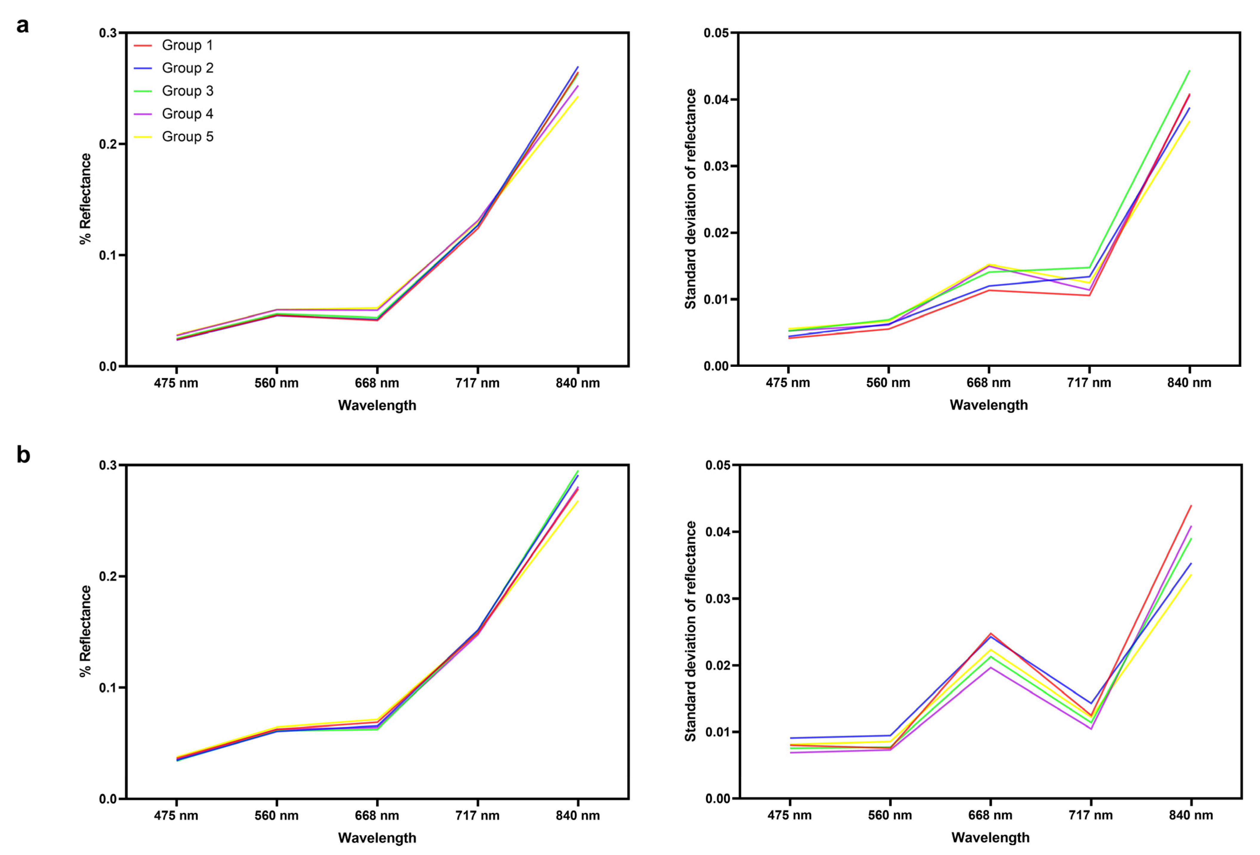
For the multispectral dataset, variation of reflectance among the five genotypic groups was the largest in Band 5 (NIR), followed by Band 3 (red) (Figure 4a,b).
According to the principal component analysis results shown in Table S1, the first component consists of the dominant spectral variability; thus, it was used in ANOVA analysis among the population groups and the genotypic groups. According to the ANOVA results for the first PC (Table 3), spectral variability was observed for both the hyperspectral and the multispectral dataset among the 28 population groups and the five genotypic groups. Thus, PC 1 could be used in differentiating between the genotypic groups. For the first PC of the hyperspectral dataset, DSI and RSI contribute a large part of the variability of PC 1, while RSI and MTCI play an important role in canopy spectral differentiation.
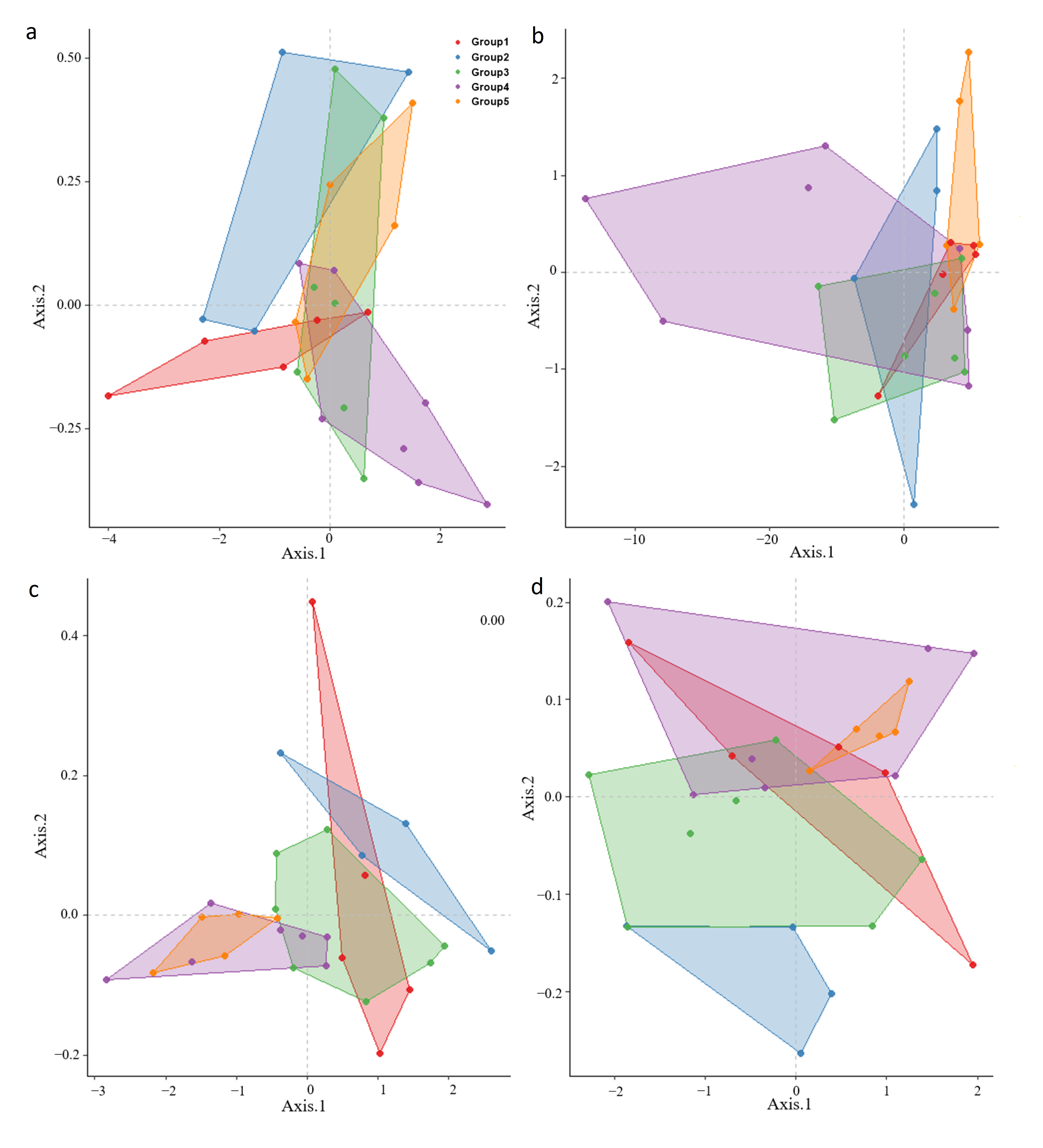
The leaf hyperspectral dataset showed that Group 3 overlapped with a portion of the spectral space held by Groups 4 and 5, and so there was a high spectral confusion among different genotypic groups (Figure 5a) as canopy hyperspectral datasets and the longitudinal groups (Groups 4, 5) were separable from latitudinal groups (Groups 1, 2 and 3) (Figure 5b). According to the multispectral measurements, the separability was higher among the 5 genotypic groups, although Group 5 appeared to occupy a subset of the spectral space of Group 4 (Figure 5c,d).

3.2. Classification of Major Genetic Groups Using Spectral Reflectance

We calculated the variable importance of multispectral and hyperspectral data for discriminating between the genotype groups using the random forest algorithm and performed genotype classification using the remote sensing data. Satisfactory results were achieved in discriminating between the genotype groups. However, the classification results among the five different genotypic groups using the important variables were the same as those obtained using all hyperspectral and multispectral reflectance and vegetation indices. Using the leaf and canopy hyperspectral dataset, the classifier showed high classification error rates for all five genotypic groups (Table 4), and the F1 scores and the Cohen’s kappa scores were both low. Classification of genotypic groups using airborne multispectral canopy reflectance had a better performance than the hyperspectral data using the RF model. With lower predictive error rates for each genotypic group, the F1 scores and the Cohen’s kappa were higher for all five genotypic groups. Thus, the five genotypic groups can be classified from canopy spectral reflectance using the RF classifiers, which suggests that canopy spectral reflectance has the potential to reveal genetic variations.
Using the leaf hyperspectral dataset, the longitudinal groups (Group 4, 5) and the latitudinal groups (Group 1, 2, 3) can be discriminated with the F1 scores of 0.81 ± 0.04 (Mean ± SD) and Cohen’s kappa scores of 0.62 ± 0.08 (Table 4). The classifier showed F1 scores of 0.80 ± 0.03 and Cohen’s kappa scores of 0.58 ± 0.08 for discriminating between the two longitudinal groups. The classifier produced low classification error rates of 0.32 ± 0.03 for discriminating between the three genotypic groups along latitudinal gradients. The F1 score was 0.61 ± 0.07, and the Cohen’s kappa score was 0.47 ± 0.09 for Group 1, 2, and 3. Using canopy hyperspectral dataset, the results for discriminating between longitudinal groups and latitudinal groups were F1 scores of 0.72 ± 0.05 and Cohen’s kappa scores of 0.43 ± 0.10 for longitude and latitude. Results showed that the hyperspectral dataset produced high classification accuracy and failed to discriminate the five different genotypic groups.

4. Discussions

The results from our study indicate that hyperspectral and multispectral dataset both have the potential capability to detect and map different Bermuda grass genotypic classes across the landscapes along the longitudinal and latitudinal gradients. Multiple leaf traits can influence spectral variations in a complex way, e.g., different spectral regions respond to different traits with various sensitivities [58,59,60]. Leaf spectral variation in the NIR and SWIR regions is sensitive to variations in leaf water and dry matter content and leaf structure [58]. For canopy spectral data, canopy hyperspectral variation is apparent in the VIS spectral range for Bermuda grass along longitudinal and latitudinal gradients. Canopy multispectral variation among genotypic groups is more apparent in the red and near-infrared bands. Spectral reflectance in the 700–900 nm range is sensitive to plant cell structure and biochemistry. A few vegetation indices (VIs) combining reflectance in the visible, red edge, or NIR spectral ranges can be used to assess the structural and biochemical characteristics of vegetation [61,62].
The spectral variability of genetic patterns remains strong in Bermuda grass along both the longitudinal and latitudinal gradients. The spectral variation is pronounced for genotypic variation because genetic variation can lead to phenotypic variation in the functional traits of plant tissues adapted to different geographic regions [20]. It is clear from our data that variations along the latitudinal gradients were captured well by certain spectral features, while spectral variation along the longitudinal gradients was low. Group 3, at a high latitude, showed larger spectral differences from other groups (Group 1 and 2) at low and mid-latitude conditions. These results are consistent with genetic divergence of populations at high latitude reported in our previous work [63], and so spectral variation of populations of Bermuda grass may increase with increasing genetic divergence along the latitudinal gradients. Populations at high latitudes could adapt to low temperatures due to genetic diversity among different genotypic groups, and local adaptation to low temperature conditions may affect the spectral variation of populations at high latitudes, which promotes evolutionary diversification [64]. Populations in Group 3, 4, and 5 all adapted to the arid environments, a change which may cause different populations at different geographic sites to have similar reflectance spectra. Regions having similar spectra along the longitudinal gradients may have been under slow rates of evolution [18]. Remote sensing for large-scale monitoring can be used to detect prevalent rapid evolution of different populations within species in natural different environments [65].
Random forests, an ensemble learning method, could be used to assemble a large set of decision trees using random subsets of training data and then make predictions based on votes of classification from a set of decision trees [66]. Lawrence and Moran (2015) found that RF had the highest average classification accuracy based on a study using 30 different datasets to compare the performance of many machine learning classification algorithms [67]. In this study, the RF classifier had great potential to discriminate between genotypic groups among the population of a single species. Leaf spectral data are promising for classifying genotypic groups at the population level within a species. The classification error based on canopy hyperspectral data was about 27%, with F1 values close to 0.72 between longitudinal groups and latitudinal groups. This was better than the results of the classification from perusing leaf hyperspectral data. Both the two multispectral datasets had good predictive ability to classify populations among the five genotypic groups along longitudinal and latitudinal gradients with a lower error rate, with high values of F1 and Cohen’s kappa. It is worth noting that we can achieve a much higher accuracy in classifying genotypic groups by using canopy multispectral data than using leaf hyperspectral data. A study also reported that ground-based leaf spectra classification of aspen by ploidy level is poorer than that performed with airborne canopy spectra [67]. We linked plant optical properties with phylogeographic and genetic information under experimental conditions where environmental variation is controlled. The possible explanation is that, though the canopy data are limited by ground pixels and other issues, NDVI with a threshold of 0.2 is used to extract canopy pixels for multispectral image, while there is no image to guide the selection for hyperspectral data. In addition, variability is more apparent at the canopy level than at the leaf level. While healthy mature leaves are collected, the hyperspectral leaf data may include more undesired and unavoidable variation. Multispectral data collection was carried out at a flight altitude of 90–120 m above ground level. However, the hyperspectral data of the Bermuda grass canopy were measured by the portable spectral radiometer at a field of view (FOV) of 25°m, with the fiber-optic probe placed at approximately 15–20 cm above the canopy. For the other interpretation, spectral variation in genotypic group with phylogeographic pattern was captured by reflectance in red and NIR regions, showing that genetic variation may have an influence on spectral properties in the red and NIR regions. Using a small number of spectral features in the red and NIR regions is better for achieving a good classification performance than using the whole wavelength. A small number of spectral features may provide better performance in discriminating genotypical groups due to the information redundancy of additional features and limited number of training data. This result is supported by the similar performance of classification models using important variables and all available spectral reflectance. Therefore, future applications for phylogeographic classification may depend on airborne canopy data rather than leaf hyperspectral data. It may be anticipated airborne multispectral image will soon be used for mapping the geographical distribution of genotype of plants over larger spatial regions.
In our study, we used both hyperspectral and multispectral data to study the spectral variation among different genotypic groups. We investigated the relationships between spectral variation and genetic diversity, which can translate into processes and mechanisms in the evolution history of different populations. While remote sensing studies can provide plant functional traits across different spatial regions [68,69], it is still challenging to reveal the mechanistic basis of biodiversity. Imaging spectroscopy data could be used to map genotypes and to identify geographic regions of genetic diversity [21]. Leaf spectral reflectance data have the potential to determine the phylogenetic patterns of biochemical traits when adapted to different environmental conditions [70]. Integrating remote sensing techniques with genomic analysis may help to monitor large-scale biodiversity at different levels and map the dynamics of ecological systems [71]. In addition, several optical vegetation indices can be selected to estimate spatial variability of some phenotypic traits, including plant area index (PAI) and leaf chlorophyll content in Northern Ontario [72,73]. Spectral signature databases have been established in the literature [74,75,76]. A similar database can be established to support the study of plant genetic diversity. However, there are still a number of challenges in respect to a full understanding of the relationships between plant or canopy properties and spectral signatures, and spectral data collection and processing protocols. Statistical analysis of spectral data collected in a typical experiment allows for identifying spectral signatures in characterization of genetic diversity and for discrimination of different genetic groups. This could lead to the establishment of knowledge or databases in this new area of research. The integration of spectral libraries for species mapping could be a future effort.

5. Conclusions

Spectroscopy could provide richer information than traditional field and laboratory methods for studying intraspecific genotypic diversity over large spatial scales. As demonstrated in this study, spectral variation existing among the five genotypic groups could be due to structural or biochemical characteristics associated with different genotypes along longitudinal and latitudinal gradients. Besides similarities in the spatial distribution of spectral diversity along the longitudinal gradient, there are also apparent spectral differences between a genotypic group at high latitude and other groups along the latitudinal gradient. The large spectral variation of different genotypes along the latitudinal gradient indicates that an evolutionary basis for spectral delineation of phylogeography may be possible. Genotypic groups can be classified using canopy spectral reflectance at a higher degree of accuracy than leaf spectral reflectance. The study shows the need for the next step of studying to better understand how well genotype can be classified spectrally for phylogeography when estimated using the whole genome sequence. Remotely sensed optical properties have great potential to assess a wide range of biological attributes, which is valuable for biodiversity conservation and management over large areas repeatedly.

Supplementary Materials

The following supporting information can be downloaded at: https://www.mdpi.com/article/10.3390/rs15040896/s1, Figure S1: Maximum likelihood (ML) phylogenetic tree inferred from longitude and latitude data for 283 Bermuda grass individuals and two outgroups (Typha latifolia L. and Anomochloa marantoidea). Numbers on branches are bootstrap frequency values for 1000 bootstrap replicates. Table S1: Proportion of variance of the first ten principle components using PCA analysis in Bermuda grass.

Author Contributions

Conceptualization, J.Z., J.S. and X.Y.; methodology, J.Z., J.L., J.S. and X.Y.; software and resources, J.Z.; data curation, J.Z.; Investigation, J.Z., M.H., L.W., M.C., C.C., S.S. and C.Z.; writing—original draft preparation, J.Z.; writing—and revision, J.Z., J.L., J.S. and X.Y.; supervision, X.Y. and J.S. All authors have read and agreed to the published version of the manuscript.

Funding

This work was supported by National Natural Science Foundation of China (32171672). This work was also supported by Graduate Scientific Research Innovation Project in Jiangsu (KYCX21-3282).

Data Availability Statement

The data that support the findings of this study are available from the corresponding author upon reasonable request.

Conflicts of Interest

The authors declare no conflict of interest.

References

  1. Couture, J.J.; Serbin, S.P.; Townsend, P.A. Spectroscopic sensitivity of real-time, rapidly induced phytochemical change in response to damage. New Phytol. 2013, 198, 311–319. [Google Scholar] [CrossRef] [PubMed]
  2. Townsend, P.A.; Serbin, S.P.; Kruger, E.L.; Gamon, J.A. Disentangling the contribution of biological and physical properties of leaves and canopies in imaging spectroscopy data. Proc. Natl. Acad. Sci. USA 2013, 110, E1074. [Google Scholar] [CrossRef]
  3. Asner, G.P.; Martin, R.E.; Carranza-Jiménez, L.; Sinca, F.; Tupayachi, R.; Anderson, C.B.; Martinez, P. Functional and biological diversity of foliar spectra in tree canopies throughout the Andes to Amazon region. New Phytol. 2014, 204, 127–139. [Google Scholar] [CrossRef]
  4. Serbin, S.P.; Singh, A.; McNeil, B.E.; Kingdon, C.C.; Townsend, P.A. Spectroscopic determination of leaf morphological and biochemical traits for northern temperate and boreal tree species. Ecol. Appl. 2014, 24, 1651–1669. [Google Scholar] [CrossRef]
  5. Yoder, B.J.; Pettigrew-Crosby, R.E. Predicting nitrogen and chlorophyll content and concentrations from reflectance spectra (400–2500 nm) at leaf and canopy scales. Remote Sens. Environ. 1995, 53, 199–211. [Google Scholar] [CrossRef]
  6. Asner, G.P.; Martin, R.E. Airborne spectranomics: Mapping canopy chemical and taxonomic diversity in tropical forests. Front. Ecol. Environ. 2009, 7, 269–276. [Google Scholar] [CrossRef]
  7. Singh, A.; Serbin, S.P.; McNeil, B.E.; Kingdon, C.C.; Townsend, P.A. Imaging spectroscopy algorithms for mapping canopy foliar chemical and morphological traits and their uncertainties. Ecol. Appl. 2015, 25, 2180–2197. [Google Scholar] [CrossRef]
  8. Gutierrez, M.; Reynolds, M.P.; Klatt, A.R. Association of water spectral indices with plant and soil water relations in contrasting wheat genotypes. J. Exp. Bot. 2010, 61, 3291–3303. [Google Scholar] [CrossRef]
  9. Fritsche-Neto, R.; Borém, A.; Cobb, J.N. Phenomics; Springer International Publishing: New York, NY, USA, 2015. [Google Scholar]
  10. Larigauderie, A.; Prieur-Richard, A.-H.; Mace, G.M.; Lonsdale, M.; Mooney, H.A.; Brussaard, L.; Cooper, D.; Cramer, W.; Daszak, P.; Díaz, S.; et al. Biodiversity and ecosystem services science for a sustainable planet: The DIVERSITAS vision for 2012–20. Curr. Opin. Environ. Sustain. 2012, 4, 101–105. [Google Scholar] [CrossRef] [PubMed]
  11. Alberto, F.J.; Aitken, S.N.; Alía, R.; González-Martínez, S.C.; Hänninen, H.; Kremer, A.; Lefèvre, F.; Lenormand, T.; Yeaman, S.; Whetten, R.; et al. Potential for evolutionary responses to climate change—Evidence from tree populations. Glob. Chang. Biol. 2013, 19, 1645–1661. [Google Scholar] [CrossRef] [PubMed] [Green Version]
  12. Urban, M.C.; Bocedi, G.; Hendry, A.P.; Mihoub, J.-B.; Peer, G.; Singer, A.; Bridle, J.R.; Crozier, L.G.; Meester, L.D.; Godsoe, W.; et al. Improving the forecast for biodiversity under climate change. Science 2016, 353, aad8466. [Google Scholar] [CrossRef]
  13. Messer, P.W.; Ellner, S.P.; Hairston, N.G. Can population genetics adapt to rapid evolution? Trends Genet. 2016, 32, 408–418. [Google Scholar] [CrossRef] [PubMed]
  14. Richards, C.L.; Rosas, U.; Banta, J.; Bhambhra, N.; Purugganan, M.D. Genome-wide patterns of Arabidopsis gene expression in nature. PloS Genet. 2012, 8, e1002662. [Google Scholar] [CrossRef]
  15. Morinaga, S.; Iwasaki, T.; Suyama, Y. Integrative Observations and Assessments; Springer: Tokyo, Japan, 2014. [Google Scholar]
  16. Schweiger, A.K.; Cavender-Bares, J.; Townsend, P.A.; Hobbie, S.E.; Madritch, M.D.; Wang, R.; Tilman, D.; Gamon, J.A. Plant spectral diversity integrates functional and phylogenetic components of biodiversity and predicts ecosystem function. Nat. Ecol. Evol. 2018, 2, 976–982. [Google Scholar] [CrossRef] [PubMed]
  17. Wang, R.; Gamon, J.A. Remote sensing of terrestrial plant biodiversity. Remote Sens. Environ. 2019, 231, 111218. [Google Scholar] [CrossRef]
  18. Ollinger, S.V. Sources of variability in canopy reflectance and the convergent properties of plants. New Phytol. 2011, 189, 375–394. [Google Scholar] [CrossRef]
  19. Ustin, S.L.; Roberts, D.A.; Gamon, J.A.; Asner, G.P.; Green, R.O. Using imaging spectroscopy to study ecosystem processes and properties. AIBS Bull. 2004, 54, 523–534. [Google Scholar] [CrossRef]
  20. Asner, G.P.; Martin, R.E.; Knapp, D.E.; Tupayachi, R.; Anderson, C.B.; Sinca, F.; Vaughnand, N.R.; Llactayo, W. Airborne laser-guided imaging spectroscopy to map forest trait diversity and guide conservation. Science 2017, 355, 385–389. [Google Scholar] [CrossRef]
  21. Madritch, M.D.; Kingdon, C.C.; Singh, A.; Mock, K.E.; Lindroth, R.L.; Townsend, P.A. Imaging spectroscopy links aspen genotype with below-ground processes at landscape scales. Philosophical Transactions of the Royal Society of London. Series B Biol. Sci. 2014, 369, 20130194. [Google Scholar] [CrossRef]
  22. Cavender-Bares, J.; Meireles, J.E.; Couture, J.J.; Kaproth, M.A.; Kingdon, C.C.; Singh, A.; Serbin, S.P.; Center, A.; Zuniga, E.; Pilz, G.; et al. Associations of leaf spectra with genetic and phylogenetic variation in oaks: Prospects for remote detection of biodiversity. Remote Sens. 2016, 8, 221. [Google Scholar] [CrossRef] [Green Version]
  23. Meireles, J.E.; Cavender-Bares, J.; Townsend, P.A.; Ustin, S.; Gamon, J.A.; Schweiger, A.K.; Schaepman, M.E.; Asner, G.P.; Martin, R.E.; Singh, A.; et al. Leaf reflectance spectra capture the evolutionary history of seed plant. New Phytol. 2020, 228, 485–493. [Google Scholar] [CrossRef] [PubMed]
  24. Jung, J.; Maeda, M.; Chang, A.; Bhandari, M.; Ashapure, A.; Landivar-Bowles, J. The potential of remote sensing and artificial intelligence as tools to improve the resilience of agriculture production systems. Curr. Opin. Biotechnol. 2021, 70, 15–22. [Google Scholar] [CrossRef]
  25. Bendig, J.; Yu, K.; Aasen, H.; Bolten, A.; Bennertz, S.; Broscheit, J.; Gnyp, M.L.; Bareth, G. Combining UAV-based plant height from crop surface models, visible, and near infrared vegetation indices for biomass monitoring in barley. Int. J. Appl. Earth Obs. Geoinf. 2015, 39, 79–87. [Google Scholar] [CrossRef]
  26. Tang, H.; Brolly, M.; Zhao, F.; Strahler, A.H.; Schaaf, C.L.; Ganguly, S.; Zhang, G.; Dubayah, R. Deriving and validating Leaf Area Index (LAI) at multiple spatial scales through lidar remote sensing: A case study in Sierra National Forest, CA. Remote Sens. Environ. 2014, 143, 131–141. [Google Scholar] [CrossRef]
  27. Jay, S.; Maupas, F.; Bendoula, R.; Gorretta, N. Retrieving LAI, chlorophyll and nitrogen contents in sugar beet crops from multi-angular optical remote sensing: Comparison of vegetation indices and PROSAIL inversion for field phenotyping. Field Crops Res. 2017, 210, 33–46. [Google Scholar] [CrossRef]
  28. Pereira, H.M.; Ferrier, S.; Walters, M.; Geller, G.N.; Jongman, R.H.G.; Scholes, R.J.; Bruford, M.W.; Brummitt, N.; Butchart, S.H.M.; Cardoso, A.C.; et al. Essential biodiversity variables. Science 2013, 339, 277–278. [Google Scholar] [CrossRef]
  29. Ustin, S.L.; Gamon, J.A. Remote sensing of plant functional types. New Phytol. 2010, 186, 795–816. [Google Scholar] [CrossRef] [PubMed]
  30. Maxwell, A.E.; Warner, T.A.; Fang, F. Implementation of machine-learning classification in remote sensing: An applied review. Int. J. Remote Sens. 2018, 39, 2784–2817. [Google Scholar] [CrossRef]
  31. Burton, G.W. Breeding Bermuda grass for the Southeastern United States. Agron. J. 1947, 39, 8. [Google Scholar] [CrossRef]
  32. Casler, M.D.; Duncan, R.R.; Casler, M.D.; Duncan, R.R. Turfgrass biology, genetics and breeding. Q. Rev. Biol. 1976, 51, 286–287. [Google Scholar]
  33. Wu, Y.Q.; Taliaferro, C.M.; Bai, G.H.; Anderson, M.P. AFLP analysis of Cynodon dactylon (L.) Pers. Var. dactylon genetic variation. Genome 2004, 47, 689–696. [Google Scholar] [CrossRef] [PubMed] [Green Version]
  34. Huang, C.Q.; Liu, G.D.; Bai, C.J.; Wang, W.Q.; Zhou, S.Y.; Yu, D. Estimation of genetic variation in Cynodon dactylon accessions using the ISSR technique. Biochem. Syst. Ecol. 2010, 38, 993–999. [Google Scholar] [CrossRef]
  35. Jewell, M.; Frère, C.H.; Harris-Shultz, K.; Anderson, W.F.; Godwin, I.D.; Lambrides, C.J. Phylogenetic analysis reveals multiple introductions of Cynodon species in Australia. Mol. Phylogenetics Evol. 2012, 65, 390–396. [Google Scholar] [CrossRef]
  36. Zhang, J.X.; Wang, M.L.; Guo, Z.P.; Guan, Y.Z.; Guo, Y.X.; Yan, X.B. Variations in morphological traits of Bermuda grass and relationship with soil and climate along latitudinal gradient. Hereditas 2018, 155, 31. [Google Scholar] [CrossRef] [PubMed]
  37. Wang, M.L.; Zhang, J.X.; Guo, Z.P.; Guan, Y.Z.; Qu, G.; Liu, J.Y.; Guo, Y.X.; Yan, X.B. Morphological variation in Cynodon dactylon (L.) Pers. And its relationship with the environment along a longitudinal gradient. Hereditas 2020, 157, 4. [Google Scholar] [CrossRef] [PubMed]
  38. Zhang, J.X.; Wang, M.L.; Guo, Z.P.; Guan, Y.Z.; Liu, J.Y.; Guo, Y.X.; Yan, X.B. Genetic diversity and population structure of Bermuda grass [Cynodon dactylon (L.) Pers.] along latitudinal gradient and the relationship with polyploidy level. Diversity 2019, 11, 135. [Google Scholar] [CrossRef]
  39. Zhang, J.X.; Wang, M.L.; Fan, J.; Guo, Z.P.; Guan, Y.Z.; Qu, G.; Zhang, C.J.; Guo, Y.X.; Yan, X.B. Non-linear genetic diversity and notable population differentiation caused by low gene flow of Bermuda grass [Cynodon dactylon (L.) Pers.] along longitude gradient. Peer J. 2021, 9, e11953. [Google Scholar] [CrossRef]
  40. Edgar, R.C. MUSCLE: Multiple sequence alignment with high accuracy and high throughput. Nucleic Acids Res. 2004, 32, 1792–1797. [Google Scholar] [CrossRef]
  41. Kumar, S.; Stecher, G.; Tamura, K. MEGA7: Molecular evolutionary genetics analysis version 7.0 for bigger datasets. Mol. Biol. Evol. 2016, 33, 1870–1874. [Google Scholar] [CrossRef]
  42. Librado, P.; Rozas, J. DnaSP v5: A software for comprehensive analysis of DNA polymorphism data. Bioinformatics 2009, 25, 1451–1452. [Google Scholar] [CrossRef]
  43. Stamatakis, A. RaxML version 8: A tool for phylogenetic analysis and post-analysis of large phylogenies. Bioinformatics 2014, 30, 1312–1313. [Google Scholar] [CrossRef]
  44. Smith, G.M.; Milton, E.J. The use of the empirical line method to calibrate remotely sensed data to reflectance. Int. J. Remote Sens. 1999, 20, 2653–2662. [Google Scholar] [CrossRef]
  45. Liebisch, F.; Kirchgessner, N.; Schneider, D.; Walter, A.; Hund, A. Remote, aerial phenotyping of maize traits with a mobile multi-sensor approach. Plant Methods 2015, 11, 19. [Google Scholar] [CrossRef] [PubMed]
  46. Duan, T.; Chapman, S.C.; Guo, Y.; Zheng, B. Dynamic monitoring of NDVI in wheat agronomy and breeding trials using an unmanned aerial vehicle. Field Crops Res. 2017, 210, 71–80. [Google Scholar] [CrossRef]
  47. Tucker, C.J. Red and photographic infrared linear combinations for monitoring vegetation. Remote Sens. Environ. 1979, 8, 127–150. [Google Scholar] [CrossRef]
  48. Blackburn, G.A. Quantifying chlorophylls and 2arotenoids at leaf and canopy scales: An evaluation of some hyperspectral approaches. Remote Sens. Environ. 1998, 66, 273–285. [Google Scholar] [CrossRef]
  49. Hollberg, J.; Schellberg, J. Distinguishing Intensity Levels of Grassland Fertilization Using Vegetation Indices. Remote Sens. 2017, 9, 81. [Google Scholar] [CrossRef]
  50. Gitelson, A.A.; Gritz, Y.; Merzlyak, M.N. Relationships between leaf chlorophyll content and spectral reflectance and algorithms for non-destructive chlorophyll assessment in higher plant leaves. J. Plant Physiol. 2003, 160, 271–282. [Google Scholar] [CrossRef] [PubMed]
  51. Dash, J.; Curran, P.J. The MERIS terrestrial chlorophyll index. Int. J. Remote Sens. 2004, 25, 5403–5413. [Google Scholar] [CrossRef]
  52. Jiang, Z.Y.; Huete, A.R.; Didan, K.; Miura, T. Development of a two-band enhanced vegetation index without a blue band. Remote Sens. Environ. 2008, 112, 3833–3845. [Google Scholar] [CrossRef]
  53. Rondeaux, G.; Steven, M.; Baret, F. Optimization of soil-adjusted vegetation indices. Remote Sens. Environ. 1996, 55, 95–107. [Google Scholar] [CrossRef]
  54. Savitzky, A.; Golay, M.J.E. Smoothing and differentiation of data by simplified least squares procedures. Anal. Chem. 1964, 36, 1627–1639. [Google Scholar] [CrossRef]
  55. Gauch, H.G. Multivariate Analysis in Community Ecology; Cambridge University Press: Cambridge, UK, 1982. [Google Scholar]
  56. Sammut, C.; Webb, G.I. Encyclopedia of Machine Learning; Springer: Boston, MA, USA, 2010. [Google Scholar]
  57. McHugh, M.L. Interrater reliability: The kappa statistic. Biochem. Med. 2012, 22, 276–282. [Google Scholar] [CrossRef]
  58. Curran, P.J. Remote sensing of foliar chemistry. Remote Sens. Environ. 1989, 30, 271–278. [Google Scholar] [CrossRef]
  59. Jacquemoud, S.; Baret, F. PROSPECT: A model of leaf optical properties spectra. Remote Sens. Environ. 1990, 34, 75–91. [Google Scholar] [CrossRef]
  60. F’eret, J.B.; Gitelson, A.A.; Noble, S.D.; Jacquemoud, S. PROSPECT-D: Towards modeling leaf optical properties through a complete lifecycle. Remote Sens. Environ. 2017, 193, 204–215. [Google Scholar] [CrossRef]
  61. Cozzolino, D. Use of Infrared Spectroscopy for In-Field Measurement and Phenotyping of Plant Properties: Instrumentation, Data Analysis, and Examples. Appl. Spectrosc. Rev. 2014, 49, 564–584. [Google Scholar] [CrossRef]
  62. AOAC. Official Methods and Analysis, 15th ed.; Method 990-03; AOAC: Arlington, VA, USA, 1990. [Google Scholar]
  63. Zhang, J.X.; Chen, M.H.; Gan, L.; Zhang, C.J.; Shen, Y.; Qian, J.; Han, M.L.; Guo, Y.X.; Yan, X.B. Diversity Patterns of Bermuda Grass along Latitudinal Gradients at Different Temperatures in Southeastern China. Plants 2020, 9, 1778. [Google Scholar] [CrossRef]
  64. Jorde, P.E.; SoVik, G.; Westgaard, J.I.; Albretsen, J.; André, C.; Hvingel, C.; Johansen, T.; Sandvik, A.D.; Kingsley, M.; Jørstad, K.E. Genetically distinct populations of northern shrimp, Pandalus borealis, in the North Atlantic: Adaptation to different temperatures as an isolation factor. Mol. Ecol. 2015, 24, 1742–1757. [Google Scholar] [CrossRef]
  65. Yamasaki, E.; Altermatt, F.; Cavender-Bares, J.; Schuman, M.C.; Zuppinger-Dingley, D.; Garonna, I.; Schneider, F.D.; Escribà, C.G.; Moorsel, S.J.; Hahl, T.; et al. Genomics meets remote sensing in global change studies: Monitoring and predicting phenology, evolution and biodiversity. Curr. Opin. Environ. Sustain. 2017, 29, 177–186. [Google Scholar] [CrossRef]
  66. Breiman, L. Random forests. Mach. Learn. 2001, 45, 5–32. [Google Scholar] [CrossRef] [Green Version]
  67. Lawrence, R.L.; Moran, C.J. The AmericaView Classification Methods Accuracy Project: A Rigorous Approach for Model Selection. Remote Sens. Environ. 2015, 170, 115–120. [Google Scholar] [CrossRef]
  68. Han, L.; Yang, G.; Yang, H.; Xu, B.; Li, Z.; Yang, X. Clustering field-based maize phenotyping of plant-height growth and canopy spectral dynamics using a UAV remote-sensing approach. Front. Plant Sci. 2018, 9, 1638. [Google Scholar] [CrossRef] [PubMed]
  69. Schneider, F.D.; Morsdorf, F.; Schmid, B.; Petchey, O.L.; Hueni, A.; Schimel, D.S.; Schaepman, M.E. Mapping functional diversity from remotely sensed morphological and physiological forest traits. Nat. Commun. 2017, 8, 1441. [Google Scholar] [CrossRef]
  70. Kursar, T.A.; Dexter, K.G.; Lokvam, J.; Pennington, R.T.; Richardson, J.E.; Weber, M.G.; Murakamia, E.T.; Draked, C.; McGregord, R.; Coleya, P.D. The evolution of antiherbivore defenses and their contribution to species coexistence in the tropical tree genus Inga. Proc. Natl. Acad. Sci. USA 2009, 106, 18073–18078. [Google Scholar] [CrossRef]
  71. Bush, A.; Sollmann, R.; Wilting, A.; Bohmann, K.; Cole, B.; Balzter, H.; Martius, C.; Zlinszky, A.; Calvignac-Spencer, S.; Cobbold, C.A.; et al. Connecting Earth observation to high-throughput biodiversity data. Nat. Ecol. Evol. 2017, 1, 176. [Google Scholar] [CrossRef]
  72. Shang, J.; Liu, J.; Ma, B.; Zhao, T.; Jiao, X.; Geng, X.; Huffman, T.; Kovacsc, J.M.; Walters, D. Mapping spatial variability of crop growth conditions using RapidEye data in Northern Ontario, Canada. Remote Sens. Environ. 2015, 168, 113–125. [Google Scholar] [CrossRef]
  73. Dong, T.; Liu, J.; Shang, J.; Qian, B.; Ma, B.; Kovacs, J.M.; Walters, D.; Jiao, X.; Geng, X.; Shi, Y. Assessment of red-edge vegetation indices for crop leaf area index estimation. Remote Sens. Environ. 2019, 222, 133–143. [Google Scholar] [CrossRef]
  74. Kokaly, F.; Clark, N.; Swayze, A.; Livo, E.; Hoefen, M.; Pearson, C.; Wise, A.; Benzel, M.; Lowers, A.; Driscoll, L.; et al. Usgs Spectral Library Version 7 Data: Us Geological Survey Data Release; United States Geological Survey (USGS): Reston, VA, USA, 2017.
  75. Hueni, A.; Nieke, J.; Schopfer, J.; Kneubuhler, M.; Itten, K. The spectral database SPECCHIO for improved long-term usability and data sharing. Comput. Geosci. 2009, 35, 557–565. [Google Scholar] [CrossRef]
  76. Awad, M.M.; Alawar, B.; Jbeily, R. A New Crop Spectral Signatures Database Interactive Tool (CSSIT). Data 2019, 4, 77. [Google Scholar] [CrossRef] [Green Version]
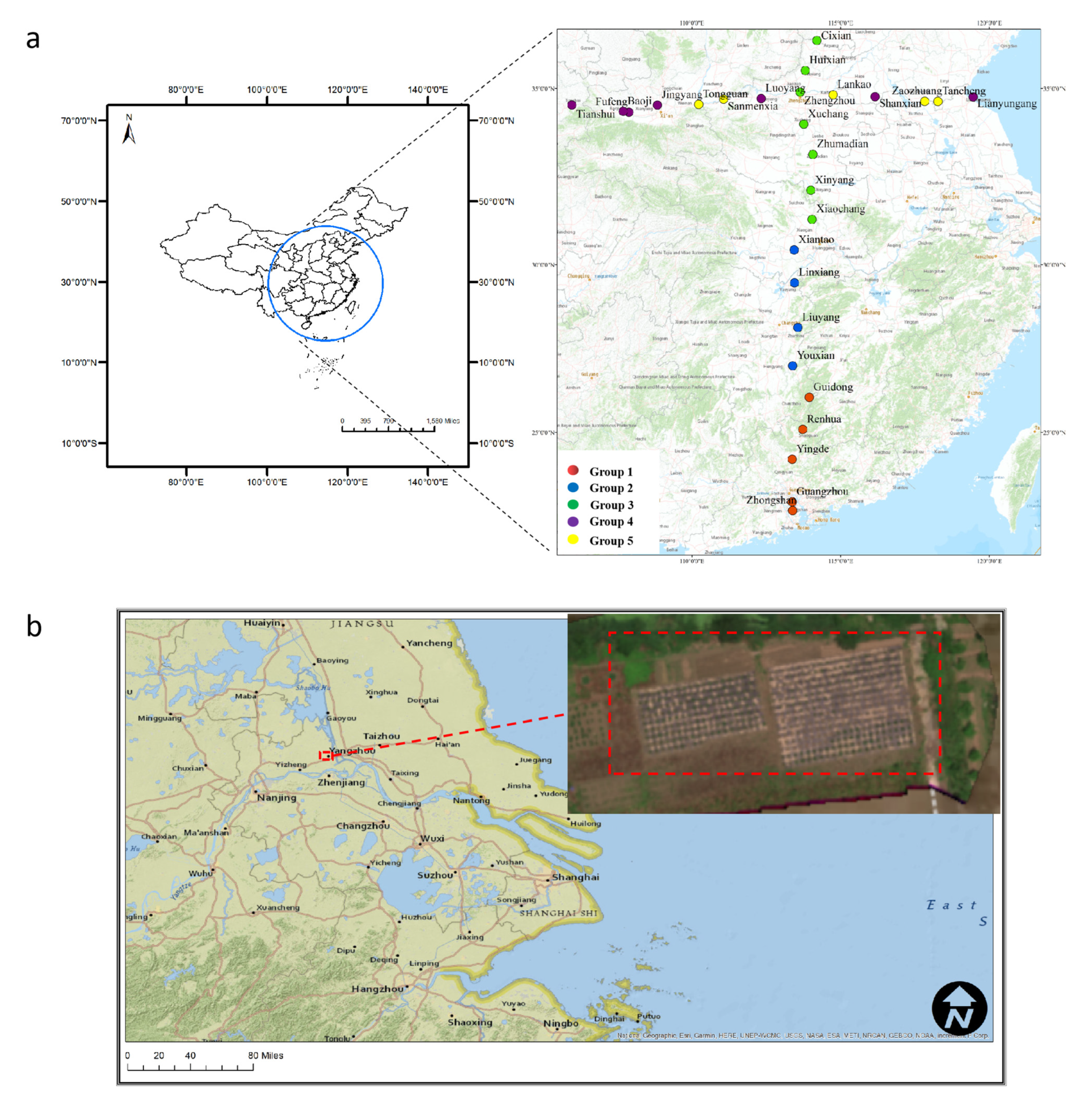
Figure 1. (a) Study area and sampling locations of Bermuda grass cpDNA sequences from 28 populations along longitudinal and latitudinal gradients. Points represent the locations where samples were collected, and colors illustrate five distinct genotypic groups (1–5). (b) Location of the study area—an experimental field in the campus of Yangzhou University.
Remotesensing 15 00896 g001
Figure 2. The workflow for analyzing spectral variation and evaluating the genotypic difference.
Remotesensing 15 00896 g002
Figure 3. Reflectance variations among the five genotypic groups for (a) leaf hyperspectral measurement, and (b) canopy hyperspectral measurement. The left panels show mean % reflectance of each group indicate 95% quantiles, and the right panels show the standard deviation of leaf and canopy reflectance.
Remotesensing 15 00896 g003
Figure 4. Reflectance variations of multispectral data among the five genotypic groups for (a) the early acquisition (17 May), and (b) the late acquisition (1 June). The right panels show the standard deviation of canopy reflectance.
Remotesensing 15 00896 g004
Figure 5. Metric multidimensional scaling analysis (MDS) of the spectral data for (a) leaf hyperspectral measurement, (b) canopy hyperspectral measurement, (c) early multispectral measurement and (d) late multispectral measurement (via metric dimensional scaling into k = 5 dimensions), with boundaries indicating the use of alpha hulls.
Remotesensing 15 00896 g005
Table 1. Details of hyperspectral and multispectral data acquisition.
Spectral Data Number of Genetic Samples
TotalGroup 1Group 2Group 3Group 4Group 5Wavelength Range Spatial Resolution
Hyperspectral leaf data446586611811589400 to 2500 nm
Hyperspectral canopy data3103947788363410 to 1300 nmField of view (FOV) of 25° at approximately 30–40 cm height
Multispectral day 1445576611811589475 nm, 560 nm, 668 nm, 717 nm and 840 nm8 cm (3.1in) per pixel
Multispectral day 2438576511511487476 nm, 560 nm, 668 nm, 717 nm and 840 nm8 cm (3.1in) per pixel
Table 2. Vegetation indices calculated from spectral data in this study. R679, R796 and R719 indicated the spectral bands at 679, 796 and 719 nm wavelength, respectively.
Vegetation IndexReference
Indices calculated from multispectral data
DSI = NIR − Red
[47]
RSI = NIR/Red[48]
NDVI = (NIR − Red)/(NIR + Red)[49]
CI red edge = (NIR/Rededge) − 1[50]
MTCI = (NIR − Rededge)/(Rededge − Red)[51]
EVI = 2.5 × (NIR − Red)/(NIR+2.4 × Red + 1)[52]
OSAVI = 1.16 × (NIR − Red)/(NIR + Red + 0.16)[53]
Indices calculated from hyperspectral data
DSI = R796 − R679
[47]
RSI = R796 / R679[48]
NDVI = (R796 − R679)/(R796 + R679)[49]
CI red edge = R796/R719 − 1[50]
MTCI = (R796 − R719)/(R719 + R679)[51]
EVI = 2.5 × (R796 − R679)/(R796 + 2.4 × R679 + 1)[52]
OSAVI = 1.16 × (R796 − R679)/(R796 + R679 + 0.16)[53]
Table 3. Analysis of variance (ANOVA) for the 28 population groups and the 5 genotypic groups of Bermuda grass based on hyperspectral, early (17 May) and late (1 June) multispectral datasets, with the first PC of the PCA as a response variable. The F value is the ratio of between-group variation and within-group variation for both the population groups and the genotypic groups.
LevelDataDfSum of SquaresMean of SquaresF Valuep Value
Among populationsLeaf hyperspectral data2723.0560.8544.1110.000
Canopy hyperspectral data27431.62315.9862.8320.000
Early multispectral data (17 May)277.0140.2603.1090.000
Late multispectral data (1 June)275.8760.2182.9000.000
Among groupsLeaf hyperspectral data47.9601.9908.6110.000
Canopy hyperspectral data4107.02726.7574.2580.002
Early multispectral data (17 May)41.5980.3994.3660.002
Late multispectral data (1 June)42.4900.6227.6400.000
Note: Df denotes Degree of freedom.
Table 4. Classification performance of random forest models based on different spectral dataset.
Classification Error Rates: Mean (SD)F1 Scores: Mean (SD)Cohen’s Kappa Scores: Mean (SD)
Leaf hyperspectral datasetAmong 5 groups0.45 (0.02)0.52 (0.03)0.45 (0.04)
Between longitude and latitude0.19 (0.01)0.81 (0.04)0.62 (0.08)
Among 2 groups at longitude0.18 (0.01)0.80 (0.03)0.58 (0.08)
Among 3 groups at latitude0.32 (0.03)0.61 (0.07)0.47 (0.09)
Canopy hyperspectral datasetAmong 5 groups0.69 (0.02)0.31 (0.06)0.16 (0.07)
Between longitude and latitude0.27 (0.02)0.72 (0.05)0.43 (0.10)
Among 2groups at longitude0.26 (0.03)0.74 (0.04)0.48 (0.08)
Among 3groups at latitude0.57 (0.05)0.42 (0.04)0.14 (0.05)
Early multispectral dataset (May 17)
Among 5 groups0.04 (0.02)0.96 (0.02)0.95 (0.03)
Late multispectral dataset (June 1)
Among 5 groups0.03 (0.02)0.97 (0.02)0.96 (0.03)
Note: SD denotes standard deviation.
Disclaimer/Publisher’s Note: The statements, opinions and data contained in all publications are solely those of the individual author(s) and contributor(s) and not of MDPI and/or the editor(s). MDPI and/or the editor(s) disclaim responsibility for any injury to people or property resulting from any ideas, methods, instructions or products referred to in the content.