Article

LED Light Quality Drives In Vitro Development of Bletilla striata: Toward Sustainable Orchid Propagation


1
Department of Ornamental Plants and Garden Art, Faculty of Biotechnology and Horticulture, University of Agriculture in Krakow, Mickiewicza 21 Ave., 31-120 Krakow, Poland
2
Department of Botany, Physiology and Plant Protection, Faculty of Biotechnology and Horticulture, University of Agriculture in Krakow, Mickiewicza 21 Ave., 31-120 Krakow, Poland
3
Faculty of Biotechnology and Horticulture, University of Agriculture in Krakow, Mickiewicza 21 Ave., 31-120 Krakow, Poland
4
Research Centre for Vegetable and Ornamental Crops, Council for Agricultural Research and Economics, Corso Degli Inglesi 508, 18038 Sanremo, Italy
*
Authors to whom correspondence should be addressed.
Sustainability 2026, 18(3), 1522; https://doi.org/10.3390/su18031522
Submission received: 22 December 2025 / Revised: 21 January 2026 / Accepted: 29 January 2026 / Published: 3 February 2026
(This article belongs to the Section Sustainable Agriculture)

Abstract

This study examined the effects of different LED light spectra on the in vitro development of plantlets of Bletilla striata (Orchidaceae), a frost-hardy ornamental orchid with increasing horticultural relevance outside its native East Asian range. The objective was to optimize growth conditions using energy-efficient lighting to support sustainable cultivation practices. Plantlets approximately 4 cm in length with 0.5 cm leaves were cultured on standard Orchimax medium in 200 mL Erlenmeyer flasks and exposed to five LED treatments: 100% blue (B), 100% red (R), red/blue at 70:30 (RB), 50% yellow + RB (7:3), and 50% green + RB (7:3). Fluorescent light served as controls. The photosynthetic photon flux density (PPFD) was maintained at approximately 40 µmol m−2 s−1 across all light treatments. After seven weeks, selected LED spectra improved plantlet performance compared with the control. Leaf number remained stable, while RB light promoted leaf expansion, resulting in the widest leaves. Root formation occurred under all LED treatments, supporting subsequent acclimatization. Light quality strongly affected photosynthetic pigments and secondary metabolism. The highest total chlorophyll content was recorded under RB illumination (581 µg g−1 FW), whereas monochromatic red light resulted in the lowest pigment levels. Carotenoid accumulation was significantly enhanced under RB and RBG spectra. Blue-containing treatments (B and RB) markedly stimulated the accumulation of phenolic compounds, including flavonols and anthocyanins, while red light suppressed phenolic biosynthesis. Total soluble sugars showed an organ-specific response, with red light promoting sugar accumulation in shoots and blue light in roots. These findings demonstrate that targeted LED lighting not only improves plant quality but also offers an environmentally sustainable and economically viable approach for commercial micropropagation and conservation of B. striata.

1. Introduction

The genus Bletilla is a small group of terrestrial orchids (Orchidaceae) native to the temperate and subtropical zones of East Asia, especially China, Japan, Korea, and northern Myanmar. This genus comprises several species with great economic value for their ornamental beauty and traditional medicinal uses. Species include Bletilla striata, B. ochracea, B. formosana, B. guizhouensis, and B. chartacea [1,2]. New species, such as B. guizhouensis, have been described based on distinct floral morphology and molecular data [3]. Based on both molecular and morphological evidence, B. foliosa has been transferred to the newly established genus Mengzia [4]. Bletilla species are characterized by pseudobulbs, leafy stems, and showy flowers. Key distinguishing features include variations in floral bracts and in sepal and lip shapes and the number of lamellae on the lip [5].
The most widespread and best-known representative is Bletilla striata (Thunb.) Rchb. f., the type species of the genus. It is a perennial native to China, Japan, and Tibet but is increasingly cultivated outside its native range as an attractive ornamental and useful plant [2]. It is distinguished by its high cold hardiness (down to about −15 °C), making it the only orchid in this group that can be permanently cultivated outdoors in a temperate climate [6]. In the horticulture cultivation, B. striata is valued as a winter-hardy orchid (in temperate climate zones), making it interesting for both hobbyists and greenhouse production. B. striata is a model species for biotechnological research and micropropagation in the genus Bletilla [7].
Bletilla striata is also known as hyacinth orchid or “Bai-Ji”. Its underground rhizomes (tubers) are traditionally used in Chinese medicine for treating mucous membrane injuries, ulcers, and burns and in research on anticancer effects [8]. Recent studies confirm the presence of 294 compounds identified in B. striata, including 272 non-polysaccharide compounds and 22 polysaccharides [9]. Extracts and bioactive compounds derived from B. striata possess diverse pharmacological properties, including anticancer, antiviral, antibacterial, antioxidant, and wound-healing activities, among others; however, efficacy and safety remain inconclusive due to insufficient scientific research [8,10,11]. These plants are traditionally used to treat respiratory diseases and as hemostatic agents, but their sustainable conservation and commercialization require urgent attention due to the ongoing destruction of natural habitats [2].
Bletilla striata reproduces through both sexual (seed) and asexual (vegetative) methods, with modern techniques greatly improving propagation efficiency. In the wild and in amateur cultivation, B. striata is mainly propagated vegetatively by division of clumps or division of bi- or multituberculates, which allows one to produce new individuals retaining the characteristics of the parent plant. However, this method of propagation has limited efficiency on a commercial scale and may not meet the needs of cultivar preservation, production, or selection of new genotypes with optimized ornamental and pharmaceutical value [6]. Therefore, the development of biotechnological techniques, such as complete tissue regeneration through in vitro techniques, is becoming important [12]. For example, Kulpa and Katroń [13] described methods for in vitro seed germination of B. striata and plant regeneration from seedlings. Furthermore, there are reports of callus induction and suspension culture for B. striata, which enables both mass propagation and the production of secondary bioactive metabolites [14]. Moreover, in vitro techniques themselves offer many advantages: rapid clonal propagation, obtaining healthy, pathogen-free plants; the ability to form acclimatization material; the preservation of genetic resources; and the rapid introduction of new varieties. In the context of species conservation and the commercial production of orchids and crop plants, micropropagation represents a significant alternative to conventional methods.
In vitro light conditions (intensity, spectral quality, photoperiod) are factors influencing morphogenesis, shoot growth, leaf development, rooting, and the content of pigments and secondary metabolites. However, the best results depend on species, developmental stage, and precise light parameters [15,16]. Traditional fluorescent lamps are still widely used but are increasingly being replaced by light-emitting diodes (LEDs) because of their many advantages. In the case of orchids, studies on the influence of monochromatic or mixed LED spectra on in vitro development are available [17]. For example, for the species Cattleya walkeriana, it has been shown that monochromatic light (red, blue, green) affects the development of shoots in vitro and subsequent acclimatization [18]. Furthermore, broad-spectrum LED studies have shown that appropriately selected spectra can outperform classic fluorescent lighting in terms of plant proliferation and quality [19]. To date, research on the use of LED light in the cultivation of Bletilla orchids has been limited to the species B. ochracea. The wavelength of LED light has been shown to have a significant impact on the germination process and seedling morphogenesis in vitro [20]. Therefore, in the context of B. striata micropropagation, optimizing the LED spectrum is a key element—both from the perspective of production efficiency and sustainable energy and resource management—which is in line with the goal of sustainable cultivation of this orchid plant.
This study focused on assessing the effect of different LED spectra on in vitro plantlets development of B. striata in standard Orchimax medium. The goal was to optimize light conditions (spectral quality) while considering economic and ecological aspects, i.e., the use of energy-efficient LED lighting to support the quality of plants obtained through micropropagation.

2. Materials and Methods

2.1. Plant Material

In vitro plant material of Bletilla striata Rchb.f. used in this study was obtained from seeds collected from the ornamental plant collection maintained at CREA (Council for Agricultural Research and Economics) in Sanremo, Italy. Mature seed capsules were harvested in October from healthy, well-established mother plants grown under standard horticultural conditions. After collection, the capsules were transported to the laboratory of the Department of Ornamental Plants and Garden Art, University of Agriculture in Krakow, Poland, and stored under dry, room temperature conditions (approximately 22–25 °C) until further processing for a period of two months. Fruits were washed under running tap water, surface-sterilized in alcohol for 1 min, and then treated with 5% sodium hypochlorite solution for 20 min. After three rinses with distilled water, the fruit was aseptically opened under a laminar flow hood, and the seeds were transferred onto the culture medium. Sterile seedlings obtained on Orchimax medium (Duchefa, Haarlem, The Netherlands) with 20 g/L sucrose, pH of 5.8, and stabilized with 8 g/L agar (Biocorp, Warsaw, Poland). The seedlings originated from seeds of a single fruit collected from one mother plant and were used as the starting material for the establishment of shoot cultures, resulting in genetically related but non-clonal, sexually derived individuals. Plants were cultivated in a growth chamber illuminated with white light during the day, following a 16 h light/8 h dark photoperiod, and maintained at 23–25 °C (night/day).

2.2. Light-Treatment Experiment Using LED Illumination

Following successful propagation of orchid shoots, an experiment was conducted to assess the effect of different types of LED light on the growth and development of Bletilla striata plantlet cultures. From the propagated cultures, uniform plantlets of comparable size (around 4 cm) and developmental stage were selected and transferred to fresh medium Orchimax under sterile conditions. The cultures were then placed in growth chambers equipped with LED lighting systems. The LED panels provided a standardized spectral composition suitable for orchid tissue culture. The plantlets cultures were exposed to LED light of various wavelengths, as shown in Table 1. The control conditions were a fluorescent light lamp (abbreviation C). All other environmental parameters, including temperature (25/23 °C–day/night), light intensity (photosynthetic photon flux density (PPFD) of ~40 μmol m−2 s−1), and relative humidity were maintained constant among the treatments. Each spectral combination was supplied using LED panels calibrated to equal PPFD, ensuring that only spectral composition differed among treatments. The cultures, 10 plantlets in 250 mL Erlenmeyer flasks in 4 replicates, were then placed in growth chambers equipped with appropriate LED lighting systems. This configuration of spectral variants allowed for the assessment of the effect of varying LED light exposure on plant growth. The experiment lasted for 7 weeks. After 7 weeks, the plant material was subjected to biometric analyses (aboveground height, number of leaves, and width of the widest leaf) and subsequently preserved in liquid nitrogen and stored at −80 °C for further biochemical assessments, including the determination of photosynthetic pigments, phenolic compounds, and total soluble sugars. Plant material for biochemical analyses was randomly collected from independent plants within each experimental treatment to ensure representative sampling and to avoid selection bias.

2.3. Assessing Metabolic Parameters

2.3.1. Photosynthetic Pigments

All the spectrophotometrical analyses were performed using a Hitachi U-2900 UV–Vis spectrophotometer (Hitachi High-Tech Science Corporation, Westford, MA, USA). The quantification of chlorophylls and total carotenoids was carried out using a modified version of the protocol described by Lichtenthaler and Wellburn [21]. Approximately 0.1 g of fresh leaf tissue was ground thoroughly in a mortar and pestle, and the resulting homogenate was extracted with 80% acetone that had been chilled to −20 °C. The mixture was vortexed to ensure complete pigment solubilization and subsequently centrifuged at 13,000× g for 15 min at 4 °C. Following centrifugation, the clear supernatant was transferred to clean tubes, and its absorbance was recorded at 663, 646, and 470 nm.
Pigment concentrations were then determined using the Lichtenthaler and Wellburn equations [21]:
  • Chlorophyll a (µg cm−3) = 12.21 × A663 − 2.81 × A646;
  • Chlorophyll b (µg cm−3) = 20.13 × A646 − 5.03 × A663;
  • Total chlorophyll = Chl a + Chl b;
  • Total carotenoids (µg cm−3) = (1000 × A470 − 3.27 × Chl a − 104 × Chl b)/229.
For consistency across samples, the resulting pigment contents were finally expressed as milligrams per 100 g of fresh weight (µg g−1 FW).

2.3.2. Phenolic Profile

The phenolic profile—comprising total phenolic content (TPC), phenylpropanoids, flavonols, and anthocyanins—was determined using UV–Vis spectrophotometric methods adapted from standard protocols [22] with modifications aimed at phenolic compound quantification rather than antioxidant activity assessment. Chlorogenic acid (CGA), caffeic acid (CA), and quercetin (QC) were used as calibration standards for TPC, phenylpropanoids, and flavonols, respectively, while anthocyanins were quantified as cyanidin (CY) equivalents based on their molar extinction coefficients. Calibration curves were prepared using a series of standard solutions covering the linear concentration ranges typically between 10 and 100 µg mL−1, depending on the compound. Approximately 0.1 g of fresh leaf tissue was homogenized in 1 mL of 80% methanol, followed by centrifugation for 15 min at 3000× g to obtain a clear extract. A 0.25 mL aliquot of the supernatant was mixed with 0.25 mL of 0.1% HCl in 96% ethanol and 4.50 mL of 2% aqueous HCl to ensure pigment stabilization and optimal chromophore development. After a 30 min incubation in the dark, absorbance was recorded at 280 nm (total phenolics), 320 nm (phenylpropanoids), 360 nm (flavonols), and 520 nm (anthocyanins). Calibration curves prepared with analytical standards enabled quantification within linear response ranges, and all measurements were performed in triplicate to ensure analytical precision. Phenolic contents were expressed as mg g−1 fresh weight (FW) of the respective standard equivalents.

2.3.3. Total Soluble Sugars

Total soluble sugars (TSSs) were quantified using the colorimetric phenol–sulfuric acid method described by Dubois et al. [23], with minor adjustments to optimize extraction efficiency. Fresh plant material (leaves and roots separately) was finely ground and extracted with 3 mL of 80% methanol, followed by continuous agitation on a rocker shaker for 32 h to ensure complete solubilization of low-molecular-weight sugars. After extraction, the samples were centrifuged, and an aliquot of the supernatant was transferred to clean tubes. A freshly prepared 5% phenol solution and concentrated sulfuric acid were then added sequentially, producing a characteristic yellow–orange chromophore as a result of carbohydrate dehydration and reaction with phenol. The reaction mixture was allowed to develop for 20–30 min at room temperature before absorbance was measured at 490 nm. Glucose was used to construct a standard calibration curve, and TSS values were expressed as milligrams of glucose equivalents per gram of dry weight (mg GE g−1 FW). This method provides a sensitive and widely validated quantification of total soluble sugars, including mono- and oligosaccharides present in plant extracts.

2.4. Statystical Analysis

Growth and biochemical datasets were analyzed using one-way ANOVA (p < 0.05). The significance of variation among experimental treatments for both growth and biochemical parameters was assessed with NIR post hoc test at p = 0.05. Four replicates (four flasks) were used for all biometric measurements. Quantitative measurements of biochemical constituents were derived from three independent biological replicates. All results are expressed as the mean ± 95% confidence interval. Data were statistically analyzed using STATISTICA 13.0 software (StatSoft, Tulsa, OK, USA).

3. Results

3.1. Biometric Measurements

After seven weeks of growing Bletilla striata shoots on Orchimax medium, the aboveground average height or average leaf number did not significantly differ according to the light combinations used. For shoot height, mean values ranged from 4.82 to 5.88 cm, with the lowest value recorded in the control (C) and the highest in the RBY combination. A similar relation was observed for leaf number, which ranged from 2.90 to 3.15 per plant. Differences between light treatments were small and did not reach significance. Significant differences were found for mean average leaf width. The narrowest leaves were obtained in the R combination (0.46 ± 0.14 cm), while the widest were in the RB combination (0.74 ± 0.28 cm). Comparative analysis showed that these different variants belong to different homogeneous groups (A–D), which confirms the significant influence of the light spectrum on the development of the leaf blade (Figure 1; Table 2). Each plantlet developed several roots, forming a well-developed root system, which indicates that the plantlets are suitable for subsequent acclimatization. The root structures are clearly visible in Figure 1.

3.2. Metabolic Parameters

3.2.1. Photosynthetic Pigments Content

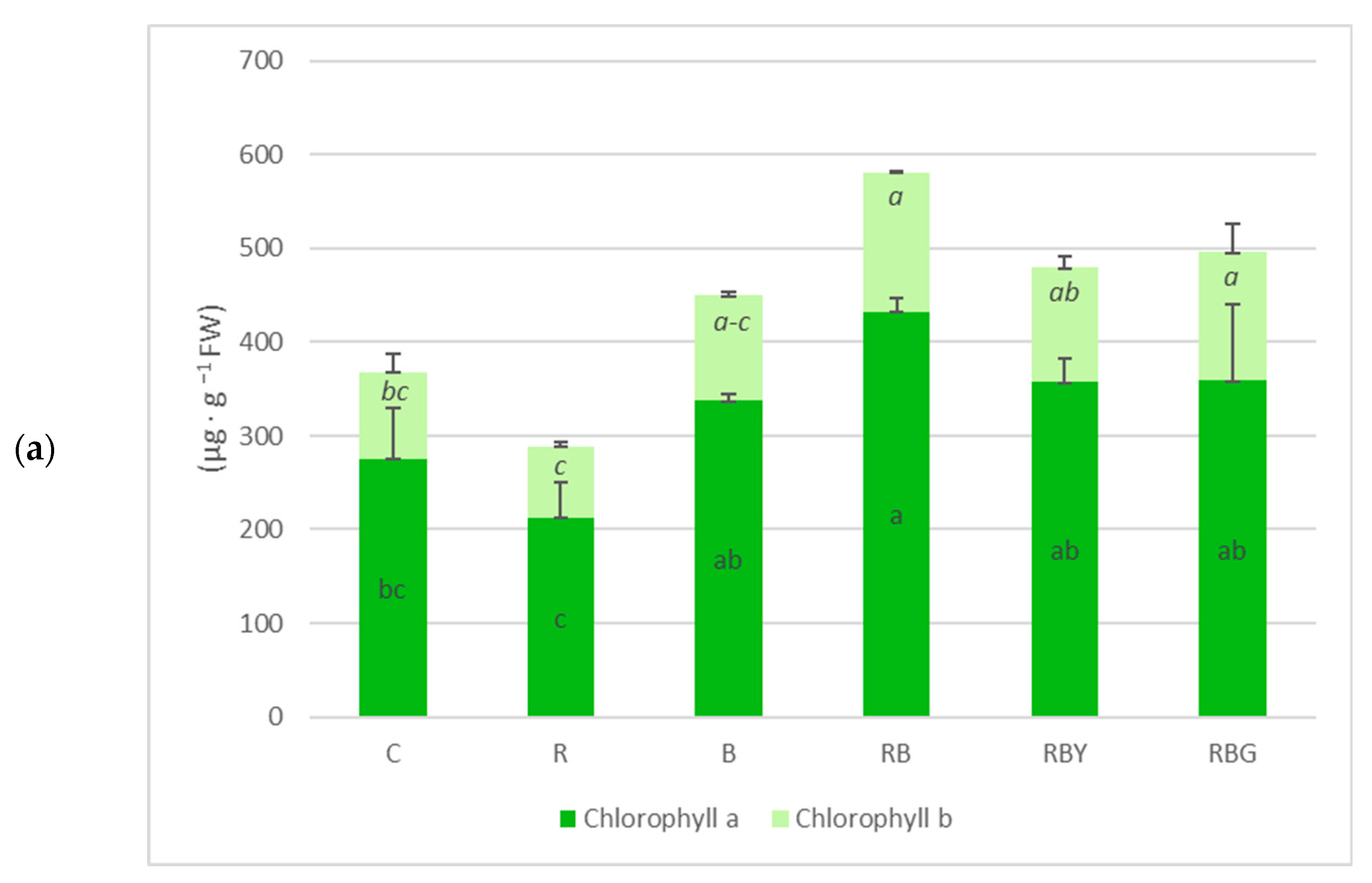
Exposure to different light treatments resulted in substantial variation in total chlorophyll content, as well as in the proportions of chlorophyll a and b. The statistically significant differences in total chlorophyll content among the individual experimental variants followed the same pattern as those observed for chlorophyll a. The control plants (C) exhibited moderate pigment levels, whereas red light alone (R) produced the lowest concentrations of both chlorophyll a and b. Blue light (B) significantly increased chlorophyll accumulation relative to R and C. The highest content of total chlorophyll (581 µg g−1 FW) was recorded under the combined red–blue treatment (RB), which exceeded all other treatments and was statistically distinct. Treatments combining red, blue, and additional wavelengths (RBY and RBG) also increased chlorophyll content compared with the control; however, their values were lower than those under RB, although the difference was not statistically significant (Figure 2a).
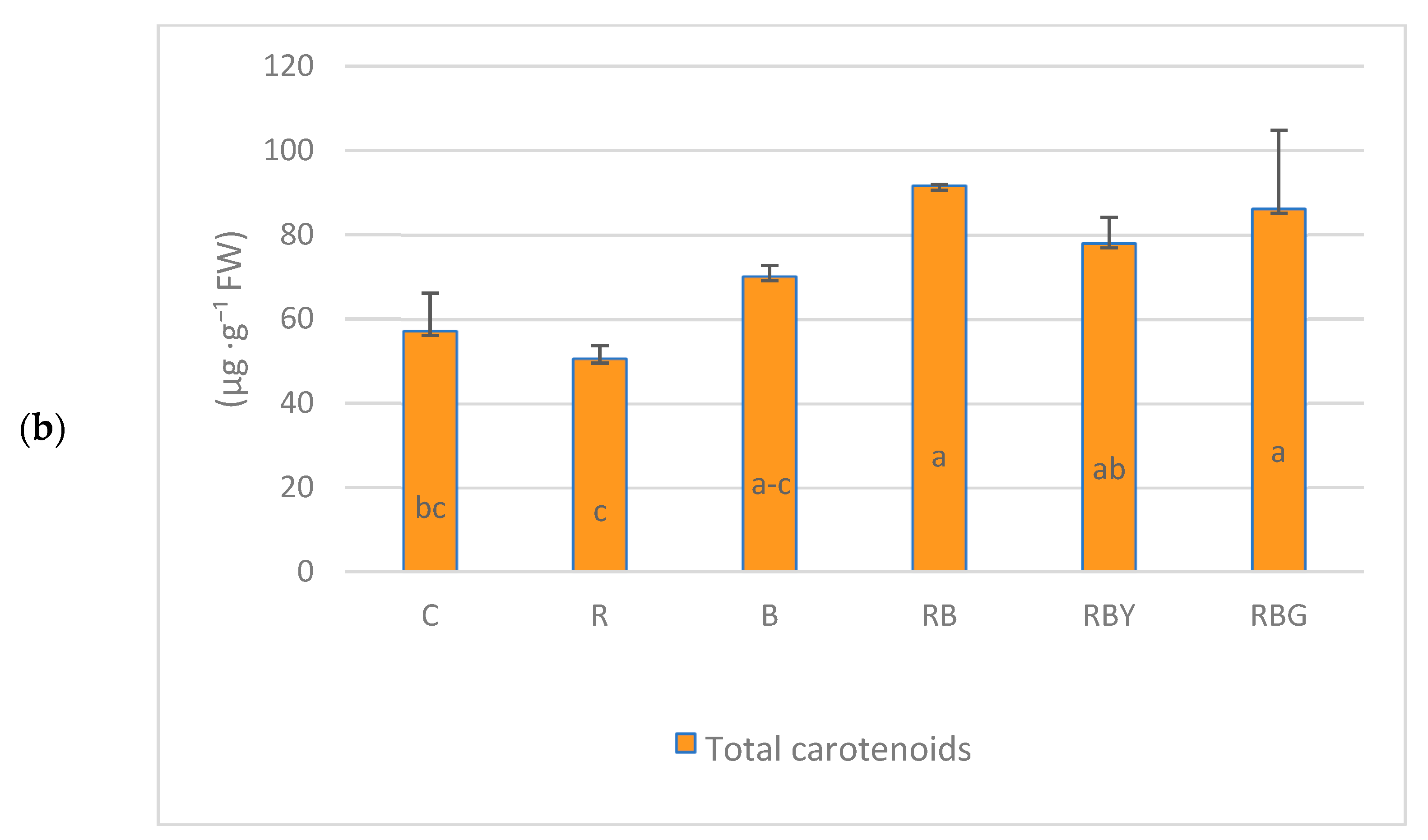
The total carotenoid content in B. striata shoots varied among the LED light treatments. The highest carotenoid levels were recorded under red + blue (RB) and red + blue + green (RBG) illumination, reaching 92 µg g−1 FW and 86 µg g−1 FW, respectively. These values were significantly higher than those observed under control conditions and under monochromatic red light (R), which resulted in the lowest carotenoid content (around 50 µg g−1 FW).
Although the yellow-supplemented spectrum (RBY) and blue light (B) produced numerically higher carotenoid levels than the control, these increases were not statistically significant. Carotenoid accumulation under blue light was comparable to that under red light, with no significant differences observed. However, the RB and RBG treatments significantly enhanced carotenoid content in B. striata relative to the control (p < 0.05) (Figure 2b).

3.2.2. Phenolic Profile Content

The accumulation of phenolic compounds in Bletilla striata was strongly dependent on light quality (Table 3). Total phenolics were highest under blue (B) and red–blue (RB) illumination, which formed the top statistical group, reaching 65.0 and 61.4 mg·g−1 FW, respectively. These values were significantly higher than those observed in the control (C) and all other treatments. Mixed-spectrum treatments containing additional wavelengths (RBY and RBG) produced intermediate phenolic levels, while red light (R) resulted in the lowest concentrations, indicating a suppressive effect of monochromatic red illumination on overall phenolic biosynthesis. A similar trend was observed for phenylpropanoids; i.e., B, RB, and RBG treatments produced the highest concentrations (31.0–35.7 mg·g−1 FW), whereas red light consistently yielded the lowest levels. The control and RBY treatments formed an intermediate statistical group. Flavonols were also strongly stimulated by blue-containing treatments. Both B and RB exhibited the highest flavonol levels (21.5 and 20.8 mg·g−1 FW, respectively), clearly exceeding all other treatments. The control (C), RBY, and RBG produced intermediate values, whereas the lowest concentrations again occurred under red light, highlighting the limited capacity of monochromatic red light to support flavonol biosynthesis. Anthocyanin accumulation followed the same general pattern, with RB producing the highest concentration (5.4 mg·g−1 FW), followed by B (4.4 mg·g−1 FW) and the two broad-spectrum treatments (RBY, RBG), which formed a shared intermediate group. The control and red treatments resulted in the lowest anthocyanin levels, confirming a strong dependence of anthocyanin biosynthesis on blue and mixed-spectral illumination.

3.2.3. Total Soluble Sugars Content

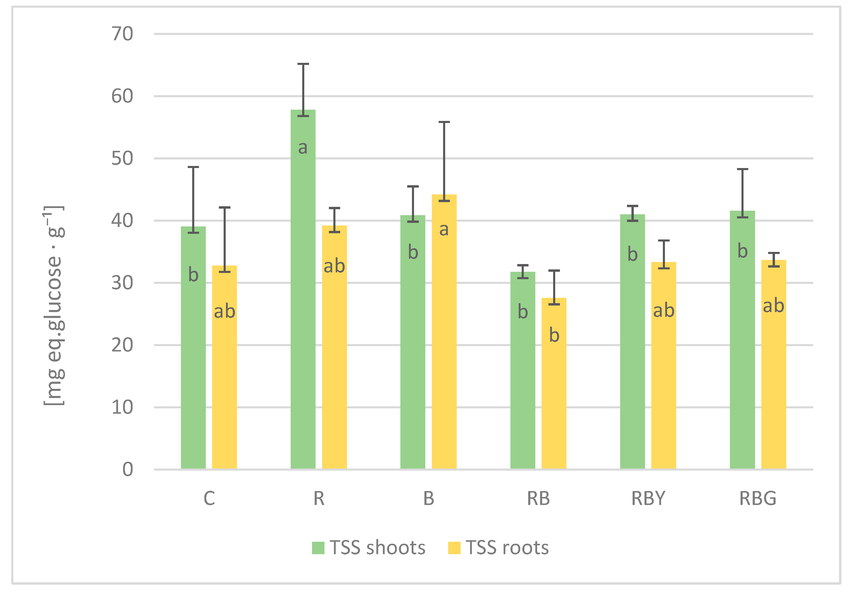
Light quality markedly affected the accumulation of total soluble sugars (TSSs) in both shoots and roots of Bletilla striata. In shoots, the highest TSS levels were observed under red light (R), which formed a distinct statistical group and exceeded all other treatments. The control (C) and mixed red–blue–green light (RBG) produced intermediate values, whereas the lowest TSS content occurred under the red–blue (RB) treatment, indicating that RB illumination did not stimulate sugar accumulation to the same extent as monochromatic red light or broad-spectrum mixtures.
In roots, the highest sugar content was observed under blue light (B). Interestingly, the combination of red and blue light (RB) suppressed the synthesis of these metabolites. Overall, TSS levels in roots were lower than in shoots, with treatments incorporating broader spectral compositions (RBY and RBG) resulting in intermediate values (Figure 3).

4. Discussion

Culture medium composition and light regime are key determinants influencing the efficiency and quality of in vitro plant propagation. In recent years, light-emitting diodes (LEDs) have gained particular prominence as a controllable and energy-efficient alternative to conventional fluorescent lighting systems. Owing to their narrow, customizable spectral profiles and the ability to precisely select the wavelength and adapt the spectrum to the needs of plants, as well as low heat emission and high electrical efficiency, LEDs have become increasingly integrated into both research laboratories and large-scale micropropagation facilities. Their application has been shown to improve morphogenetic responses, enhance photosynthetic performance, and reduce production costs, thereby contributing to greater standardization and sustainability of in vitro culture protocols [16,17,24,25]. As a result, LED-based illumination is now considered one of the most important technological advancements supporting the development of modern plant tissue culture.
Both light quality and plant growth regulators exert a strong influence on in vitro plant proliferation; however, inconsistent findings across studies and the absence of standardized protocols make it challenging to identify the most suitable light spectrum for each species [26]. The effect of LED light quality on shoot elongation has been investigated in several orchid species, revealing considerable variation in plant height responses depending on the spectral composition. In Cattleya crispata, white LEDs produced the longest and most vigorous shoots after 90 days of culture, outperforming red, blue, and red/blue LED treatments [27]. In contrast, for Vanilla planifolia, shoot length exceeded 3 cm under blue, red, and blue + red (1:1) LEDs, while cultures grown under fluorescent lamps and white LEDs produced shoots shorter than 3 cm [24]. In Phalaenopsis, blue + red LED combinations and fluorescent lamps resulted in the greatest shoot lengths, whereas white and red LEDs generated noticeably shorter shoots [19]. In Cymbidium cultivars, red LED light produced the greatest number of leaves per shoot, while alternating green and red LEDs also stimulated leaf development [28]. A broader review of plant micropropagation indicates that combined red–blue LED treatments frequently enhance leaf number and, in some cases, leaf length, whereas monochromatic red or blue light alone may lead to reductions in leaf area or leaf number [26]. In our study, shoot lengths and number of leaves were higher compared with the control conditions, however, these differences were not statistically significant.
To date, only one study has examined the effects of LED lighting on the growth of orchids in the genus Bletilla. In that study, leaf blade width increased significantly when Bletilla ochracea seedlings were cultivated under blue (470 nm) or white LED light compared with seedlings grown under green, orange, or red LEDs. The latter wavelengths did not promote leaf blade expansion and resulted in reduced leaf width. Seedlings grown under blue and white LEDs also formed thicker pseudobulbs, demonstrating improved overall growth and vigor under these light conditions [20]. Importantly, the study did not include treatments combining blue and red light. Studies conducted on Brassavola nodosa demonstrated that the use of the LED-3 light source (red LED illumination at 77 ± 5 µmol PPFD, composed of 13% blue, 26% green, and 61% red light) resulted in the most favorable in vitro growth and development of this species. Notably, this lower-intensity LED configuration also promoted 100% ex vitro survival of the plants [29]. Shin et al. [30] reported that leaf area was greatest in Doritaenopsis hort. (Orchidaceae) plants grown under a combination of red and blue light-emitting diodes (LEDs). In our study, this light treatment likewise produced the most favorable results. In Gerbera jamesonii, leaf blade morphology responds strongly to the spectral composition of LED lighting under in vitro conditions. The application of either blue (B) or red (R) light alone resulted in a reduced leaf blade area [31]. In other studies conducted on Gerbera, growth under an R:B ratio of 7:3 promoted an increase in the number of leaves, leaf blade width, and root number. This light combination also enhanced superoxide dismutase and catalase activities, stimulated chlorophyll synthesis, and increased the accumulation of both Chl a and Chl b. Additionally, it resulted in the highest chlorophyll fluorescence parameters [32]. In our study, both B and R illumination produced noticeably narrower leaf blades compared to the red–blue combination (70% red + 30% blue). This mixed spectrum promoted greater lateral expansion of the leaf blade, indicating a synergistic effect between the two wavelengths. These findings support the strategic use of specific LED spectra to optimize leaf morphology in plant tissue culture.
The wavelength of blue light is associated with higher quantum efficiency and photosynthetic activity, which promotes cellular material production. The results of the present study support these findings, indicating that blue LED light positively influences growth performance and the synthesis of selected metabolites and bioactive compounds in B. striata compared with other LED light treatments. Chlorophyll a, chlorophyll b, and β-carotene contents were significantly (p < 0.05) affected by the different LED light spectra. The highest levels of chlorophyll and β-carotene were consistently observed in all treatments where blue light was present.
Blue light (430 nm) is known to stimulate chlorophyll biosynthesis by upregulating genes involved in chlorophyll production and promoting chloroplast development. Optimal pigment synthesis often occurs at moderate blue light intensities or under specific photoperiods [33,34,35]. In contrast, red light (670 nm) primarily drives photosynthetic activity but has a weaker effect on chlorophyll accumulation. In our study, plants exposed solely to red light exhibited the lowest chlorophyll content. Exposure to blue light alone resulted in intermediate chlorophyll levels, whereas the combination of red and blue light (7:3 ratio) provided both efficient photosynthetic energy and enhanced chlorophyll synthesis, leading to the highest chlorophyll a and b content. These results highlight the synergistic effect of red and blue wavelengths in optimizing chlorophyll accumulation. For example, in vitro Doritaenopsis plants grown under red plus blue LEDs show higher growth parameters and increased carbohydrate and leaf pigment biosynthesis compared to red or blue LED and fluorescent light treatments [30]. Our findings are consistent with previous studies, where combining red and blue light often resulted in greater pigment accumulation than red light alone, enhancing both chlorophyll and carotenoid levels in various vegetables and microgreens [35,36]. The optimal red-to-blue light ratio is species-dependent. Blue LED light is efficiently absorbed by photosynthetic pigments and acts as a key stimulus for pigment synthesis [37]. The activity of enzymes associated with pigment biosynthesis can be stimulated by blue light, which enhances pigment accumulation and activates cryptochrome-mediated signaling [38].
Research on the influence of light quality on the morphogenesis of hybrid Petunia [39] also indicates that the highest chlorophyll accumulation in this species is stimulated by blue light. Blue light rapidly and reversibly regulates stomatal aperture, resulting in greater stomatal opening [40]. It is also likely that zeaxanthin [41], cryptochromes, and phototropins [42] participate in blue-light signaling in guard cells. The positive effect of RB treatments on chlorophyll content observed in this study is consistent with the findings of Naznin [33], who reported that chlorophyll a, chlorophyll b, and total chlorophyll content in lettuce, spinach, basil, and pepper were significantly increased under RB light. However, these results contrast with reports showing that continuous white LED lighting increased chlorophyll content in lettuce [43] or that red LED lighting enhanced photosynthetic pigments in Scots pine [44]. Such differences may be attributed to genetic variation among species, as well as to differences in pigment composition.
The results presented here for Bletilla striata clearly demonstrate that red–blue light combinations are capable of stimulating synthesis of the highest level of total phenols, flavonoids, and phenylpropanoids, as well as anthocyanins, while red light acting alone significantly reduces the biosynthesis of these compounds. A red–blue (RB) LED spectrum stimulates both photochemistry and blue-light signaling, which together promote phenylpropanoid/anthocyanin pathways. Red alone can maximize growth but provides weak activation of these defense-related pathways, leading to the lowest phenolic accumulation. Enhanced blue increases phenolic acids and flavonoids via upregulation of PAL, CHS, F3H, F3′H, and related genes in barley, lettuce, basil, and other species. Under red alone, these blue-light pathways are poorly activated, so phenolic synthesis is minimal [45,46,47]. Red efficiently drives photosynthesis and biomass accumulation, providing carbohydrates and malonyl-CoA precursors for phenylpropanoid synthesis [48]. The combined red–blue light creates optimal metabolic and signaling conditions for phenolic compound accumulation, whereas red light alone leads to the lowest levels of these secondary metabolites. When combined with blue, plants have both (i) a signal to make phenolics and (ii) metabolic resources to do so, increasing total phenols, flavonoids, and anthocyanins [49,50]. It should also be mentioned that anthocyanin accumulation is important for the sustainable cultivation of frost-tolerant ornamental orchids because these pigments enhance tolerance to low-temperature and light-induced oxidative stress by acting as antioxidants and photoprotective compounds [51]. Increased anthocyanin levels can also improve visual coloration, thereby increasing the ornamental value of the plants without the need for additional chemical inputs [52]. Consequently, anthocyanins support both stress resilience and aesthetic quality under environmentally friendly cultivation conditions. We conclude that in B. striata, RB LEDs outperform red alone for phenolics because blue-light signaling (cryptochromes) plus red-driven carbon supply jointly upregulate phenylpropanoid and anthocyanin pathways and route more flux to these compounds, while red alone mainly supports biomass with weak induction of these secondary metabolites. In our study, we also observed that the inclusion of green light in combination with blue and red wavelengths resulted in the highest flavonoid accumulation in B. striata. These findings contrast with previous reports indicating that green LED light often reduces or does not enhance flavonoid content in actively growing plants when compared with blue or red–blue light treatments [50,53]. Conversely, other studies have demonstrated that green LEDs in the 500–600 nm range can stimulate flavonoid synthesis in several stored leafy vegetables. For production systems, blue or blue-rich spectra remain the most reliable drivers of flavonoid accumulation; green is most promising as a postharvest treatment [54]. Additionally, it has been reported that the presence of yellow–green components in mixed light spectra may be associated with lower total flavonoid levels in some species compared with red–blue light alone [50,53].
The accumulation of total soluble sugars (TSSs) in ornamental orchids plays a crucial role in both their commercial value and aspects of sustainable horticulture. Soluble sugars, such as sucrose, glucose, and fructose, serve as key energy sources and metabolic signals that influence plant development and physiological functions, including stress responses and cell turgor, which directly affect tissue quality and resilience under cultivation and storage conditions. Higher TSS levels are often associated with longer shelf life, improved appearance, and greater consumer appeal, enhancing market value. Moreover, soluble sugars act as natural osmoprotectants, helping plants cope with various abiotic stresses, such as low temperatures and drought, which is particularly relevant in the context of sustainable horticulture, where reducing chemical inputs and increasing plant resilience are priorities [55,56]. In our study, we observed that the highest accumulation of total soluble sugars in B. striata shoots occurred under red light, whereas the highest sugar content in roots was recorded under blue light. This organ-specific response can be explained by the distinct roles of red- and blue-light photoreceptors in regulating carbon metabolism and assimilating partitioning: red light, perceived mainly by phytochromes, promotes photosynthetic activity and shoot growth, favoring sugar accumulation in aboveground tissues, while blue light, mediated by cryptochromes and phototropins, enhances root metabolic activity and source–sink signaling, leading to increased sugar allocation and accumulation in roots [57]
These findings are consistent with previous studies reporting that red-enriched light, particularly red combined with far-red, generally promotes total soluble sugar and related carbohydrate pools in shoots, whereas higher proportions of blue light often shift metabolism toward reduced sucrose/starch accumulation or altered sugar profiles. The specific responses, however, are both species-dependent and light-regime dependent, although red light is typically stimulatory for shoot sugar accumulation [58,59]. For instance, in licorice seedlings, exposure to pure red LEDs markedly increased soluble sugar content compared with blue light, and a mixed red/blue ratio of 4:1 also enhanced soluble sugar levels during early development [60]. Similarly, in Mesona chinensis, red light elevated soluble sugar and pectin content relative to blue light [61], while in Allium victorialis sprouts, red light increased leaf area, dry mass, and soluble sugar accumulation compared with green light [62].

5. Conclusions

The results of this study clearly demonstrate the significant role of LED lighting in the cultivation of Bletilla striata. LED technology provides several advantages—energy efficiency, long lifespan, low heat emission, customizable light spectra, and environmental sustainability—which collectively make it a valuable tool for modern, controlled plant production systems. Importantly, the obtained results indicate that properly selected LED spectra can effectively enhance the quality of plant material, highlighting their strong application potential.
The LED variants used in this study, particularly the red–blue (RB) and blue (B) spectra, significantly improved plant morphology by increasing leaf blade width compared with fluorescent lighting. At the same time, LED treatments influenced key metabolic parameters, including chlorophyll, carotenoid, and phenolic compound accumulation (phenylpropanoids, flavonols, and anthocyanins), with the RB and B combinations producing the most favorable responses. Red–blue (RB) LED light resulted in an approximately 60% increase in the content of photosynthetic pigments (chlorophyll a, chlorophyll b, and carotenoids) compared to the fluorescent light control. Moreover, RB light significantly enhanced the accumulation of secondary metabolites, with total phenolics increasing by nearly 60%, phenylpropanoids by about 68%, and flavonols and anthocyanins by more than 85% relative to the control. These results highlight red–blue LED lighting as a highly effective strategy for improving both photosynthetic efficiency and the biosynthesis of bioactive compounds in B. striata cultured in vitro. In contrast, red light (R) promoted increased sugar accumulation in shoots, indicating that B. striata exhibits distinct metabolic adjustments depending on the wavelength composition. It highlights the importance of designing the light spectra in accordance with the intended outcomes.
From an application-oriented perspective, these findings are particularly relevant given the growing interest in B. striata as both an ornamental species and a plant of pharmacological importance. LED lighting can be used not only to enhance physiological quality but also to modify the visual attributes of in vitro-grown plantlets, for example, narrower leaves under red light or broader leaves under RB light, allowing producers to shape plant appearance in a controlled manner. Among the tested LED variants, the RB spectrum proved to be particularly effective in promoting desirable morphological traits, such as broader leaves, and enhancing the accumulation of key metabolites. These results indicate that the RB light combination can be recommended as a practical strategy for in vitro propagation of B. striata, providing a reliable approach to improve both plant quality and appearance in controlled cultivation systems.
Overall, this study confirms that targeted use of LED lighting represents an effective strategy for improving the quality of B. striata in controlled cultivation and may be successfully implemented in commercial production systems to optimize selected morphological and metabolic traits. Future research could focus on the acclimation of B. striata plantlets in greenhouse or open-field conditions, as well as on extending similar studies to other species within the genus Bletilla, to further evaluate the practical applicability and generalizability of LED-mediated propagation strategies.

Author Contributions

Conceptualization, D.K.; methodology, D.K. and A.K.; software, D.K.; validation, D.K. and A.K.; formal analysis, D.K., A.K. and A.F.; investigation, D.K., A.K. and A.F.; resources, D.K. and A.V.; data curation, D.K., A.K. and A.F.; writing—original draft preparation, D.K. and A.K.; writing—review and editing, D.K., A.K. and A.V.; visualization, D.K.; supervision, D.K. and A.V.; project administration, D.K.; funding acquisition, D.K. and A.K. All authors have read and agreed to the published version of the manuscript.

Funding

This research was funded by the Ministry of Science and Higher Education of Poland—statutory funding of the research activity held at the University of Agriculture in Kraków, Poland (050012-D011).

Institutional Review Board Statement

Not applicable.

Informed Consent Statement

Not applicable.

Data Availability Statement

The original contributions presented in this study are included in the article. Further inquiries can be directed to the corresponding author.

Conflicts of Interest

The authors declare no conflicts of interest.

References

  1. Wu, Z.; Hong, D. Flora of China; Missouri Botanical Garden Press: St. Louis, MO, USA, 2009; Volume 25, pp. 1–570. [Google Scholar]
  2. Devkota, H.P.; Logesh, R.; Adhikari-Devkota, A.; Paudel, M.R. Orchids of Genus Bletilla: Traditional Uses, Phytochemistry, Bioactivities, and Commercial Importance. In Orchids Phytochemistry, Biology and Horticulture. Reference Series in Phytochemistry; Mérillon, J.M., Kodja, H., Eds.; Springer: Cham, Switzerland, 2021; pp. 573–590. [Google Scholar] [CrossRef]
  3. Huang, J.; Wang, M.; Chen, L.; Huang, Z.; Rao, W.; Zhang, Y.; Chen, J.; Chen, G. Bletilla guizhouensis (Orchidaceae; Epidendroideae), a new species from Guizhou China: Evidence from morphological and molecular analyses. Phytotaxa 2019, 406, 279–286. [Google Scholar] [CrossRef]
  4. Huang, W.; Liu, Z.; Jiang, K.; Luo, Y.; Jin, X.; Zhang, Z.; Xu, R.; Muchuku, J.; Musungwa, S.; Yukawa, T.; et al. Phylogenetic analysis and character evolution of tribe Arethuseae (Orchidaceae) reveal a new genus Mengzia. Mol. Phylogenet. Evol. 2021, 167, 107362. [Google Scholar] [CrossRef]
  5. Huang, J.; Yuan, F.; Zhou, M.; Huang, T.; Zhang, Y.; Liang, Q. Phenotype correlation analysis and excellent germplasm screening of herb Bletilla Rchb. f. based on comprehensive evaluation from thirty-three geographic populations. BMC Plant Biol. 2022, 22, 154. [Google Scholar] [CrossRef]
  6. Zhang, M.; Shao, Q.; Xu, E.; Wang, Z.; Wang, Z.; Yin, L. Bletilla striata: A review of seedling propagation and cultivation modes. Physiol. Mol. Biol. Plants 2019, 25, 601–609. [Google Scholar] [CrossRef]
  7. Wei, X.; Liu, Y.; Wang, X.; Gao, Z.; Yao, S.; Han, J. Progress on research of tissue culture of Bletilla striata. Chin. Herb. Med. 2017, 10, 23–26. [Google Scholar] [CrossRef]
  8. He, X.; Wang, X.; Fang, J.; Zhao, Z.; Huang, L.; Guo, H.; Zheng, X. Bletilla striata: Medicinal uses, phytochemistry and pharmacological activities. J. Ethnopharmacol. 2017, 195, 20–38. [Google Scholar] [CrossRef]
  9. Zhang, L.; Shi, X.; Zhang, X.; Chen, T.; Liao, S.; Gao, J.; Zhou, Z.; Zhang, M.; Peng, F. A comprehensive review of ethnopharmacology, phytochemistry, pharmacological effects, and quality control of Bletilla striata. J. Ethnopharmacol. 2025, 353, 120273. [Google Scholar] [CrossRef]
  10. Xu, D.; Pan, Y.; Chen, J. Chemical Constituents, Pharmacologic Properties, and Clinical Applications of Bletilla striata. Front. Pharmacol. 2019, 10, 1168. [Google Scholar] [CrossRef]
  11. Jiang, S.; Wang, M.; Jiang, L.; Xie, Q.; Yuan, H.; Yang, Y.; Zafar, S.; Liu, Y.; Jian, Y.; Li, B.; et al. The medicinal uses of the genus Bletilla in traditional Chinese medicine: A phytochemical and pharmacological review. J. Ethnopharmacol. 2021, 280, 114263. [Google Scholar] [CrossRef]
  12. Fan, L.; Wang, R.-R.; Yu, M.; Liu, J.-H.; Hou, D.-B.; Tang, Z.-K. Shoot regeneration from transverse thin cell layers of pseudobulbs and micropropagation of Bletilla striata. Bull. Bot. Res. 2020, 40, 368–375. [Google Scholar] [CrossRef]
  13. Kulpa, D.; Katroń, J. Seed germination and plant development of Bletilla striata in vitro. Folia Pomer. Univ. Technol. Stetin. Agric. Aliment. Pisc. Zootech. 2012, 293, 51–60. [Google Scholar]
  14. Pan, Y.; Li, L.; Xiao, S.; Chen, Z.; Sarsaiya, S.; Zhang, S.; ShangGuan, Y.; Liu, H.; Xu, D. Callus growth kinetics and accumulation of secondary metabolites of Bletilla striata Rchb.f. using a callus suspension culture. PLoS ONE 2020, 15, e0220084. [Google Scholar] [CrossRef]
  15. Billore, V.; Jain, M.; Suprasanna, P. Monochromic radiation through light-emitting diode (LED) positively augments in vitro shoot regeneration in Orchid (Dendrobium sonia). Can. J. Biotech. 2017, 1, 50–58. [Google Scholar] [CrossRef]
  16. Nhut, D.T.; Tung, H.T.; Tanaka, M. Enhanced Growth and Development of Cymbidium and Phalaenopsis Plantlets Cultured In Vitro Under Light-Emitting Diodes. In Orchid Propagation: From Laboratories to Greenhouses—Methods and Protocols. Springer Protocols Handbooks; Lee, Y.I., Yeung, E.T., Eds.; Humana Press: New York, NY, USA, 2018; pp. 209–223. [Google Scholar] [CrossRef]
  17. Hanus-Fajerska, E.; Wojciechowska, R. Impact of Light-Emitting Diodes (LEDs) on Propagation of Orchids in Tissue Culture. In Light Emitting Diodes for Agriculture; Dutta Gupta, S., Ed.; Springer: Singapore, 2017; pp. 305–320. [Google Scholar] [CrossRef]
  18. Nadal, M.; Machado, N.; Santos, C.; Nunes, F.; José, H.; Dória, J.; Pasqual, M. Impact of monochromatic lights on the in vitro development of Cattleya walkeriana and effects on acclimatization. Ornam. Hortic. 2023, 29, 238–248. [Google Scholar] [CrossRef]
  19. Bello-Bello, J.; Sato, J.; Cruz-Cruz, C.; Martínez-Estrada, E. Light-Emitting Diodes: Progress in Plant Micropropagation. In Chlorophyll; Jacob-Lopes, E., Queiroz Zepka, L., Maria Isabel Queiroz, M.I., Eds.; IntechOpen: London, UK, 2017. [Google Scholar] [CrossRef]
  20. Godo, T.; Fujiwara, K.; Guan, K.; Miyoshi, K. Effects of wavelength of LED-light on in vitro asymbiotic germination and seedling growth of Bletilla ochracea Schltr. (Orchidaceae). Plant Biotechnol. 2011, 28, 397–400. [Google Scholar] [CrossRef]
  21. Lichtenthaler, H.K.; Wellburn, A.R. Determinations of total carotenoids and chlorophylls a and b of leaf extracts in different solvents. Biochem. Soc. Trans. 1983, 11, 591–592. [Google Scholar] [CrossRef]
  22. Fukumoto, L.R.; Mazza, G. Assessing antioxidant and prooxidant activities of phenolic compounds. J. Agric. Food Chem. 2000, 48, 3597–3604. [Google Scholar] [CrossRef]
  23. Dubois, M.; Gilles, K.A.; Hamilton, J.K.; Rebers, P.A.; Smith, F. Colorimetric method for determination of sugars and related substances. Anal. Chem. ACS 1956, 26, 350. [Google Scholar] [CrossRef]
  24. Bello-Bello, J.; Inez-Estrada, E.; Azquez, J.; Morales-Ramos, V. Effect of LED light quality on in vitro shoot proliferation and growth of vanilla (Vanilla planifolia Andrews). Afr. J. Biotechnol. 2016, 15, 272–277. [Google Scholar] [CrossRef]
  25. Ramos, J.; Ribeiro, L.; Nunes, G.; Soares, J.; Sorgato, J. In vitro and ex vitro production of Schomburgkia crispa: Effect of flask sealing systems and different light sources. Rodriguésia 2023, 74, e01062022. [Google Scholar] [CrossRef]
  26. Cavallaro, V.; Pellegrino, A.; Muleo, R.; Forgione, I. Light and Plant Growth Regulators on In vitro Proliferation. Plants 2022, 11, 844. [Google Scholar] [CrossRef] [PubMed]
  27. Souza, D.; Fernandes, S.; Molinari, L.; Avelar, M.; Gonçalves, D.; Faria, J.; Teixeira, G.; Brondani, G. The influence of light quality in the in vitro cultivation of Cattleya crispata (Thunb.) Van den Berg. Nativa 2021, 9, 582–588. [Google Scholar] [CrossRef]
  28. Ona, A.; Shimasaki, K.; Emteas, M.; Uddin, A. Effects of Different LED Lights on the Organogenesis of a Cymbidium Cultivar. Environ. Control Biol. 2021, 59, 197–201. [Google Scholar] [CrossRef]
  29. Vendrame, W.; Xu, J.; Beleski, D. Evaluation of the Effects of Culture Media and Light Sources on In vitro Growth of Brassavola nodosa (L.) Lindl. Hybrid. Horticulturae 2022, 8, 450. [Google Scholar] [CrossRef]
  30. Shin, K.; Murthy, H.; Heo, J.; Hahn, E.; Paek, K. The effect of light quality on the growth and development of in vitro cultured Doritaenopsis plants. Acta Physiol. Plant. 2008, 30, 339–343. [Google Scholar] [CrossRef]
  31. Pawłowska, B.; Żupnik, M.; Szewczyk-Taranek, B.; Cioć, M. Impact of LED light sources on morphogenesis and levels of photosynthetic pigments in Gerbera jamesonii grown in vitro. Hortic. Environ. Biotechnol. 2018, 59, 115–123. [Google Scholar] [CrossRef]
  32. Meng, X.; Wang, Z.; He, S.; Shi, L.; Song, Y.; Lou, X.; He, D. LED-Supplied Red and Blue Light Alters the Growth, Antioxidant Status, and Photochemical Potential of in vitro-Grown Gerbera jamesonii Plantlets. Korean J. Hortic. Sci. Technol. 2019, 37, 473–489. [Google Scholar] [CrossRef]
  33. Naznin, M.T.; Lefsrud, M.; Gravel, V.; Azad, M.O.K. Blue Light added with Red LEDs Enhance Growth Characteristics, Pigments Content, and Antioxidant Capacity in Lettuce, Spinach, Kale, Basil, and Sweet Pepper in a Controlled Environment. Plants 2019, 8, 93. [Google Scholar] [CrossRef]
  34. Da Cruz, E.; Varjão, C.; Souza, Ê.; Da Silva, R.; Almeida, R.; De Magalhães Cabral Albuquerque, E. Effect of Blue Led Light Photoperiod on the Synthesis of Bioactive Pigments in Arugula Microgreens. Rev. Gestão Soc. Ambient. 2025, 19, e013088. [Google Scholar] [CrossRef]
  35. Nguyen, T.; Sung, J. Light Spectral-Ranged Specific Metabolisms of Plant Pigments. Metabolites 2024, 15, 1. [Google Scholar] [CrossRef]
  36. Zhang, Y.; Lau, N.; Subramaniam, S. Illuminating the harvest: The regulatory effects of LEDs on pigment accumulation in various food crops. Physiol. Mol. Biol. Plants 2025, 31, 693–708. [Google Scholar] [CrossRef]
  37. Atta, M.; Idris, A.; Bukhari, A.; Wahidin, S. Intensity of blue LED light: A potential stimulus for biomass and lipid content in fresh water microalgae Chlorella vulgaris. Bioresour. Technol. 2013, 148, 373–378. [Google Scholar] [CrossRef]
  38. Ma, G.; Zhang, L.; Kato, M.; Yamawaki, K.; Kiriiwa, Y.; Yahata, M.; Ikoma, Y.; Matsumoto, H. Effect of Blue and Red LED Light Irradiation on β-Cryptoxanthin Accumulation in the Flavedo of Citrus Fruits. J. Agric. Food Chem. 2012, 60, 197–201. [Google Scholar] [CrossRef] [PubMed]
  39. Park, Y.G.; Jeong, B.R. Shift in the Light Quality of Night Interruption Affects Flowering and Morphogenesis of Petunia hybrida. Plants 2023, 12, 2049. [Google Scholar] [CrossRef]
  40. Meidner, H.; Mansfield, T.A. Physiology of Stomata, 1st ed.; McGraw-Hill: London, UK, 1968. [Google Scholar]
  41. Zeiger, E.; Talbott, L.D.; Frechilla, S.; Srivastava, A.; Zhu, J. The guard cell chloroplast: A perspective for the twenty-first century. New Phytol. 2002, 153, 415–424. [Google Scholar] [CrossRef] [PubMed]
  42. Kinoshita, T.; Doi, M.; Suetsugu, N.; Kagawa, T.; Wada, M.; Shimazaki, K. Photo1 and photo 2 mediate blue light regulation of stomatal opening. Nature 2001, 414, 656–660. [Google Scholar] [CrossRef]
  43. Viršilė, A.; Brazaitytė, A.; Vaštakaitė-Kairienė, V.; Miliauskienė, J.; Jankauskienė, J.; Novičkovas, A.; Laužikė, K.; Samuolienė, G. The distinct impact of multi-color LED light on nitrate, amino acid, soluble sugar and organic acid contents in red and green leaf lettuce cultivated in controlled environment. Food Chem. 2020, 310, 125799. [Google Scholar] [CrossRef]
  44. Pashkovskiy, P.; Ivanov, Y.; Ivanova, A.; Kreslavski, V.; Vereshchagin, M.; Tatarkina, P.; Kuznetsov, V.; Allakhverdiev, S. Influence of Light of Different Spectral Compositions on Growth Parameters, Photosynthetic Pigment Contents and Gene Expression in Scots Pine Plantlets. Int. J. Mol. Sci. 2023, 24, 2063. [Google Scholar] [CrossRef] [PubMed]
  45. Pech, R.; Volná, A.; Hunt, L.; Bartas, M.; Červeň, J.; Pečinka, P.; Špunda, V.; Nezval, J. Regulation of Phenolic Compound Production by Light Varying in Spectral Quality and Total Irradiance. Int. J. Mol. Sci. 2022, 23, 6533. [Google Scholar] [CrossRef]
  46. Son, K.; Oh, M. Leaf Shape, Growth, and Antioxidant Phenolic Compounds of Two Lettuce Cultivars Grown under Various Combinations of Blue and Red Light-emitting Diodes. Hortscience 2013, 48, 988–995. [Google Scholar] [CrossRef]
  47. Jung, W.; Chung, I.; Hwang, M.; Kim, S.; Yu, C.; Ghimire, B. Application of Light-Emitting Diodes for Improving the Nutritional Quality and Bioactive Compound Levels of Some Crops and Medicinal Plants. Molecules 2021, 26, 1477. [Google Scholar] [CrossRef]
  48. Zhu, Y.; Patil, B.; Zhen, S. From ultraviolet-B to red photons: Effects of end-of-production supplemental light on anthocyanins, phenolics, ascorbic acid, and biomass production in red leaf lettuce. PLoS ONE 2025, 20, e0328303. [Google Scholar] [CrossRef]
  49. Taulavuori, K.; Hyöky, V.; Oksanen, J.; Taulavuori, E.; Julkunen-Tiitto, R. Species-specific differences in synthesis of flavonoids and phenolic acids under increasing periods of enhanced blue light. Environ. Exp. Bot. 2016, 121, 145–150. [Google Scholar] [CrossRef]
  50. Ptak, A.; Szewczyk, A.; Simlat, M.; Pawłowska, B.; Warchoł, M. LED light improves shoot multiplication, steviol glycosides and phenolic compounds biosynthesis in Stevia rebaudiana Bertoni in vitro culture. Sci. Rep. 2024, 14, 30860. [Google Scholar] [CrossRef]
  51. Li, Z.; Ahammed, G. Plant stress response and adaptation via anthocyanins: A review. Plant Stress 2023, 10, 100230. [Google Scholar] [CrossRef]
  52. Mekapogu, M.; Vasamsetti, B.; Kwon, O.; Ahn, M.; Lim, S.; Jung, J. Anthocyanins in Floral Colors: Biosynthesis and Regulation in Chrysanthemum Flowers. Int. J. Mol. Sci. 2020, 21, 6537. [Google Scholar] [CrossRef] [PubMed]
  53. Gam, D.; Khoi, P.; Ngoc, P.; Linh, L.; Hưng, N.; Anh, P.; Thư, N.; Hien, N.; Khanh, T.; Ha, C. LED Lights Promote Growth and Flavonoid Accumulation of Anoectochilus roxburghii and Are Linked to the Enhanced Expression of Several Related Genes. Plants 2020, 9, 1344. [Google Scholar] [CrossRef] [PubMed]
  54. Salehinia, S.; Didaran, F.; Aliniaeifard, S.; Zohrabi, S.; Macpherson, S.; Lefsrud, M. Green light enhances the phytochemical preservation of lettuce during postharvest cold storage. PLoS ONE 2024, 19, e0311100. [Google Scholar] [CrossRef]
  55. van Doorn, W.G. Role of Soluble Carbohydrates in Flower Senescence. Acta Hortic. 2006, 543, 123–130. [Google Scholar] [CrossRef]
  56. Ouyang, L.; Leus, L.; De Keyser, E.; Van Labeke, M.-C. Dehydrins and Soluble Sugars Involved in Cold Acclimation of Rosa wichurana and Rose Cultivar ‘Yesterday’. Horticulturae 2021, 7, 379. [Google Scholar] [CrossRef]
  57. Krahmer, J.; Ganpudi, A.; Abbas, A.; Romanowski, A.; Halliday, K.J. Phytochrome, Carbon Sensing, Metabolism, and Plant Growth Plasticity. Plant Physiol. 2018, 176, 1039–1048. [Google Scholar] [CrossRef]
  58. Liu, J.; Liu, W. Regulation of accumulation and metabolism circadian rhythms of starch and sucrose in two leaf-color lettuces by red:blue ratios of LED continuous light. Environ. Exp. Bot. 2022, 196, 104811. [Google Scholar] [CrossRef]
  59. Driesen, E.; Saeys, W.; De Proft, M.; Lauwers, A.; Van Den Ende, W. Far-Red Light Mediated Carbohydrate Concentration Changes in Leaves of Sweet Basil, a Stachyose Translocating Plant. Int. J. Mol. Sci. 2023, 24, 8378. [Google Scholar] [CrossRef] [PubMed]
  60. Jiang, Y.; Zhang, Z.; Zhang, S.; Chen, X.; Li, B.; Ma, S.; Wang, Y.; Sun, Z. Transcriptome and Metabolomics Analysis Reveal the Effects of Red and Blue Light on the Physiology and Primary Medicinal Components (Liquiritin and Glycyrrhizic Acid) of Glycyrrhiza uralensis Seedlings. Int. J. Mol. Sci. 2025, 26, 4641. [Google Scholar] [CrossRef] [PubMed]
  61. Tang, D.; Huang, Q.; Wei, K.; Yang, X.; Wei, F.; Miao, J. Identification of Differentially Expressed Genes and Pathways Involved in Growth and Development of Mesona chinensis Benth Under Red- and Blue-Light Conditions. Front. Plant Sci. 2021, 12, 761068. [Google Scholar] [CrossRef] [PubMed]
  62. Zhou, C.; Cui, W.; Yuan, T.; Cheng, H.; Su, Q.; Guo, P. Water content, carbohydrate accumulation, and secondary metabolites in Allium victorialis sprouts exposed to shoot cutting in varied irradiations. Not. Bot. Horti Agrobot. Cluj-Napoca 2021, 49, 12524. [Google Scholar] [CrossRef]
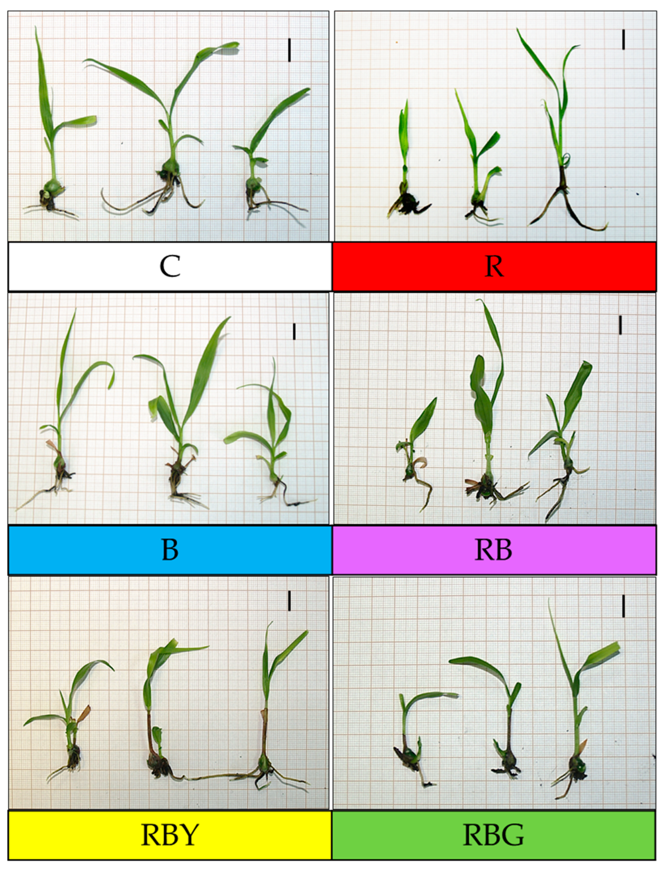
Figure 1. The appearance of Bletilla striata plantlets after 7 weeks of cultivation under different LED light variants and in controlled conditions; bar = 1 cm. C—control, fluorescent light; R—100% red–monochromatic red light; B—100% blue–monochromatic blue light; RB—70% red + 30% blue–standard red–blue LED mixture; RBY—50% RB 7:3 red-to-blue ratio supplemented with 50% yellow light; RBG—50% RB 7:3 red-to-blue ratio supplemented with 50% green light.
Sustainability 18 01522 g001
Figure 2. (a) Photosynthetic pigments—chlorophyll a and chlorophyll b in Bletilla striata plantlets grown under different light regimes after 7 weeks of cultivation on Orchimax medium; (b) total carotenoids measured under the same conditions. Means followed by the same letters within a column are not significantly different (p ≤ 0.05).
Sustainability 18 01522 g002aSustainability 18 01522 g002b
Figure 3. Total soluble sugars (TSSs) in shoots and roots of Bletilla striata grown under different light regimes after 7 weeks of cultivation on Orchimax medium. Means followed by the same letters within a column are not significantly different (p ≤ 0.05).
Sustainability 18 01522 g003
Table 1. LED light treatments used in the experiment.
LED Light VariantWavelength of Light [nm]AbbreviationSymbolic Color
100% red—monochromatic red light670R
100% blue—monochromatic blue light430B
70% red + 30% blue—standard red–blue LED mixture(7:3—670, 430)RB
50% RB 7:3 red-to-blue ratio supplemented with 50% yellow light(7:3—670, 430) + 600RBY
50% RB 7:3 red-to-blue ratio supplemented with 50% green light(7:3—670, 430) + 528RBG
Table 2. Biometric measurement results after 7 weeks of cultivation of Bletilla striata plantlets on Orchimax medium. Statistical analysis using Statistica software based on one-way analysis of variance (ANOVA); NIR test at a significance level of α = 0.05.
The Combination of Light Average Height of the Aboveground Part [cm]Average Number
of Leaves
Average Leaf Width [cm]
C4.82 ± 1.76 a *2.90 ± 0.92 a0.51 ± 0.16 cd
R5.19 ± 2.07 a3.08 ± 0.89 a0.46 ± 0.14 d
B5.37 ± 2.50 a3.15 ± 0.86 a0.60 ± 0.20 b
RB5.74 ± 2.83 a3.03 ± 0.99 a0.74 ± 0.28 a
RBY5.88 ± 2.20 a3.03 ± 0.95 a0.60 ± 0.26 b
RBG5.74 ± 1.87 a2.93 ± 0.94 a0.58 ± 0.21 bc
* a, b, c, d: means for the studied traits marked with the same letters in the column do not differ significantly.
Table 3. Total contents of total phenols, phenylpropanoids, flavonols, and anthocyanins (mg·g−1 FW) in Bletilla striata cultivated under different light regimes for 7 weeks on Orchimax medium. Statistical analysis using Statistica software based on one-way analysis of variance (ANOVA); NIR test at a significance level of α = 0.05.
Total Phenolics [mg·g−1 FW]Phenylopropanoids [mg·g−1 FW]Flavonols [mg·g−1 FW] Anthocyanins [mg·g−1 FW]
C 38.4 ± 8.6 ab *21.3 ± 4.2 bc11.2 ± 3.2 bc 2.9 ± 1.0 b
R 30.0 ± 10.9 b15.6 ± 6.1 c7.2 ± 2.5 c2.7 ± 0.5 b
B 65.0 ± 0.8 a 32.0 ± 0.3 a21.5 ± 1.2 a4.4 ± 1.4 ab
RB 61.4 ± 23.4 a35.7 ± 0.3 a20.8 ± 2.1 a5.4 ± 1.1 a
RBY 46.5 ± 2.7 ab26.5 ± 4.5 ab10.8 ± 1.0 bc3.9 ± 0.9 ab
RBG 53.5 ± 7.0 ab31.0 ± 3.5 a12.5 ± 1.7 b3.2 ± 0.1 ab
* a, b, c: means for the studied traits marked with the same letters in the column do not differ significantly.
Disclaimer/Publisher’s Note: The statements, opinions and data contained in all publications are solely those of the individual author(s) and contributor(s) and not of MDPI and/or the editor(s). MDPI and/or the editor(s) disclaim responsibility for any injury to people or property resulting from any ideas, methods, instructions or products referred to in the content.