Article

Efficacy of Entomopathogenic Fungi for Sustainable Biocontrol of Fungus Gnat (Bradysia difformis) in Peat-Free Substrates: A Laboratory Study

1
Research Centre for Horticultural Crops (FGK), Erfurt University of Applied Sciences, Kühnhäuser Straße 101, 99090 Erfurt, Germany
2
Institute of Microbiology, Friedrich Schiller University Jena, Neugasse 25, 07743 Jena, Germany
3
Department of Horticulture, Faculty of Landscape Architecture, Horticulture and Forestry, Erfurt University of Applied Sciences, Leipziger Straße 77, 99085 Erfurt, Germany
*
Author to whom correspondence should be addressed.
Sustainability 2025, 17(13), 5897; https://doi.org/10.3390/su17135897 (registering DOI)
Submission received: 8 May 2025 / Revised: 22 June 2025 / Accepted: 23 June 2025 / Published: 26 June 2025

Abstract

Bradysia difformis Frey (Diptera: Sciaridae) is a fungus gnat that poses a significant threat to greenhouse cultures, and is attracted to soils devoid of peat. Fungal strains from the German Collection of Microorganisms and Cell Culture (DSMZ), such as Beauveria bassiana, Metarhizium flavoviride, Mucor hiemalis, and Niesslia tinuis, as well as Serendipita indica, were screened for entomopathogenic activity against B. difformis and their capacity to colonize Petunia hybrida cv. “Mitchell” and Ocimum basilicum plants. The survival rates of Bradysia difformis (three instar larvae) treated with Metarhizium flavoviride were 45.33% at 14 days following inoculation with 1 × 10⁶ spores/mL of each fungal strain, when compared to others. We concluded that the fungal strain M. flavoviride could serve as an entomopathogenic fungus with the highest virulence against B. difformis larvae. Although M. flavoviride did not show a beneficial effect as an endophyte, interestingly, the strain Niesslia tinuis exhibited plant growth benefits in Petunia hybrida cv. “Mitchell” by enhancing its shoot length up to 13.18 ± 0.72 cm, whereas the control treatment had a shoot length up to 10.68 ± 0.39. Enzymatic assays confirmed the ability of M. flavoviride to produce cuticle-degrading enzymes such as chitinase and protease. Together, these findings highlight the potential of EMPF—particularly M. flavoviride—as a sustainable biocontrol tool well-suited for peat-free horticultural systems, offering an eco-friendly alternative to chemical insecticides where fungus gnat pressure is typically high.

1. Introduction

Black fungus gnats, also known as sciarids (Diptera, Sciaridae), are small insects with adult lengths of up to 8 mm. They are predominantly dark in color, and their larvae typically develop among rotting plant remains that have been infested with fungal hyphae [1]. They are among the most prevalent insect pests of greenhouse ornamentals and production nurseries, especially Bradysia sp. (Diptera: Sciaridae) [2,3]. Two fungus gnat species, Bradysia coprophila Lintner and Bradysia difformis Frey (=B. impatiens Johansen), are regarded as significant greenhouse pests in Europe and the United States [4]. Although their primary food sources are fungi and organic materials, fungus gnat maggots can harm a variety of ornamental plants [5]. Plant roots and tunnel stems are frequently chewed or stripped by these maggots [6]. Severe damage to plants affects their ability to absorb water and nutrients, which causes them to lose vitality, turn off-color, and eventually die. Furthermore, adult flies are a nuisance to humans and spread fungal spores from plants to plants as they move around the greenhouse [7], whereas maggots can spread fungal pathogens (Fusarium, Phoma, Pythium, and Verticillium) during feeding [8]. During their lifetime, adult females frequently produce up to 1000 eggs, which they then deposit on the growth media surface [9]. Within four to six days, eggs hatch; within 12 to 14 days, maggots move through four instars, pupate in the soil for three to four days, and then emerge as adults. Accordingly, at 20–25 °C, the egg-to-egg life cycle can be finished in 20–25 d [9,10].
Bradysia species continue to be major greenhouse pests due to their capacity to transmit various plant pathogens and cause direct root damage, leading to significant crop losses. Their preference for moist, microbially rich growing media makes peat-free substrates, increasingly used for sustainability reasons, especially susceptible to infestations by B. difformis [11,12]. These organic-rich media provide ideal conditions for fungus gnat oviposition and larval development, highlighting the urgent need for effective biological control methods compatible with environmentally friendly horticultural practices. The horticultural industry’s transition to peat-free substrates, driven by environmental sustainability objectives, has inadvertently resulted in a marked increase in the population of fungus gnats (Diptera: Sciaridae) in greenhouse cultures. The high organic content and microbial activity of peat-free media, such as composted green waste and other organic-rich materials, attract these pests, especially species like Bradysia difformis. It has been demonstrated by means of empirical observation that the substrates in question provide a favorable environment for fungus gnat oviposition and larval development, thus resulting in infestations in greenhouse environments. Notwithstanding the fact that fungus gnat larvae primarily consume decomposing organic matter, they have been observed to cause harm to plant roots and root hairs. This has the potential to compromise the health and vitality of the plants cultivated in such systems. This phenomenon assumes particular significance in systems devoid of peat, where the abundance of organic waste serves to stimulate the proliferation of gnats. Consequently, the management of fungus gnat populations has become a significant challenge in the context of environmentally sustainable horticultural practices [12].
Administering organophosphates, carbamates, and neonicotinoid pesticides is currently the most widely used method for controlling fungus gnat larvae [13]. However, owing to the development of pesticide resistance in larvae following years of treatment, the level of control has been inadequate [14]. Concerns about human health and environmental contamination have led to growing restrictions on the use of conventional chemical pesticides [15]. Thus, investigating biological control options is crucial for reducing the use of pesticides, lowering environmental pollution levels, and enhancing the control of pest populations.
A fundamental component of Integrated Pest Management (IPM) is biological control, which includes the use of natural enemies such as predators, parasitoids, entomopathogenic fungi, nematodes, bacteria, and insect viruses, due to their effectiveness and low environmental impact [16,17,18]. Entomopathogenic fungi (EMPF) are parasitic microorganisms that can infect and significantly increase the mortality of arthropod populations. They are widely used in the biological control of insects because of their biology, which allows them to infect them through touch, pierce their cuticles, and multiply in the hemocoel [19]. A diverse group of EMPF includes potential biocontrol agents targeting agriculturally important pest species [20,21]. Entomopathogenic fungi used for pest control include Beauveria bassiana (Balsamo), Paecilomyces fumosoroseus (Wize), Nomuraea rileyi (Farlow), Verticillium lecanii (Zimmerman), and Metarhizium anisopliae (Metschnikoff) [22]. Only a few entomopathogenic fungal species have been successfully developed into commercial biopesticides, where they have shown promising results against fungus gnats (Diptera: Sciaridae), which are significant pests in greenhouse and nursery production. For example, Andreadis et al. [23] reported that Beauveria bassiana formulations caused 100% mortality in adult Lycoriella ingenua, demonstrating strong pathogenicity in mushroom cultivation environments. In greenhouse trials, Metarhizium brunneum was found to significantly reduce larval survival and adult emergence of Bradysia impatiens when applied to growing substrates. Furthermore, the combined application of M. brunneum with botanical compounds enhanced control efficacy, indicating potential for integrated biopesticide strategies [24]. These studies highlight the direct applicability of entomopathogenic fungi for controlling fungus gnats, supporting their integration into sustainable pest management programs. These examples illustrate the diverse potential of entomopathogenic fungi to manage different arthropod pests through various mechanisms, making them valuable tools in integrated pest management strategies. As highlighted by Tóthné Bogdányi et al. (2019) [25], M. flavoviride exhibits broad host specificity, effective cuticle degradation, and a notable potential for sustainable pest control. However, its performance in peat-free horticultural systems remains underexplored.
In addition to their role as biological insecticides, increasing evidence suggests that numerous EMPF species have the ability to colonize the tissues of specific plants. A plethora of endeavors have been undertaken to artificially induce the presence of electromagnetic pulses (EMPs) in plants, employing a range of methodologies. However, only a limited number of EMPF species have been identified as naturally occurring endophytes [26,27]. As it has been shown to increase plant growth and decrease pest infestation in a variety of economically important crops, this natural or artificial colonization may be advantageous for the plant [28,29,30,31]. EMPF endophytes have been found in hundreds of plants, such as a number of significant crops, including wheat, soybeans, bananas, and tomatoes [30,32,33], and as beneficial rhizosphere colonizers [34,35] and plant growth promoters [36,37,38,39]. Nevertheless, the diverse functions of fungal entomopathogens can also be economically and subsequently utilized in sustainable agriculture, such as microbial control agents against plant diseases and arthropod pests [39,40,41,42] as vertically transmitted fungal endophytes that can enhance host plant fitness, induce systemic resistance, and provide long-term pest and disease suppression [38,43,44,45].
The objective of this study was to evaluate the potential of selected fungal strains obtained from the German Collection of Microorganisms and Cell Cultures (DSMZ), namely Beauveria bassiana (strain 1344), Metarhizium flavoviride (strain 21704), Mucor hiemalis (strain 2655), and Niesslia tinuis (strain 3481). In this group, B. bassiana, M. flavoviride, and M. hiemalis are well-established entomopathogenic fungi, whereas N. tinuis represents a novel species that has not previously been investigated for its potential as either an entomopathogen or an endophyte. The objective of the present study was to evaluate the potential of the fungal strains under investigation, in conjunction with Serendipita indica, a well-documented plant endophyte, to regulate the population dynamics of Bradysia difformis and to establish a colonizing presence within the root systems of Petunia hybrida cv. “Mitchell” and Ocimum basilicum plants.

2. Materials and Methods

2.1. Efficacy of Entomopathogenic Fungi to Manage Fungus Gnat Larvae from Becoming Adults

Five fungal strains such as Beauveria bassiana (1344), Metarhizium flavoviride (21704), Mucor hiemalis (2655), and Niesslia tinuis (3481), obtained from DSMZ and Serendipita indica, were assessed for their ability to affect the population of fungus gnat larvae to become adults. The sterile regular coconut block with potting soil substrate used for the fungus gnat culture was placed in 750 mL plastic cups and treated with fungal spore solution at a concentration of 1 × 10⁶ spores/mL. A negative control, consisting of sterile water, and a positive control using 0.1% Neemazal were included in the experimental design. The boxes were incubated for 24 h at 16:8 light: dark and 25 ± 1 °C. After 24 h of incubation, a fine paintbrush was used to place 25 freshly molted third-instar fungus gnat larvae into each treated control cup. The cups were kept in a growth room with a 16:8 light-dark photoperiod at 25 ± 1 °C and 65 ± 5% relative humidity (RH). A lid was placed on each cup to prevent contamination and larval escape. A yellow sticky trap was attached to the top of each cup to monitor adult emergence, and the moisture content of the treatments was maintained by spraying sterilized distilled water every other day. The number of adults caught in the traps was recorded throughout the observation period. Observations were made over a two-week period to evaluate adult emergence, fungal effectiveness, and larval development in adults, where six replicates of each treatment were used. The trial was terminated when no new adults were counted within five days. The mortality rate and control efficiency were calculated according to Equations (1) and (2) [46,47]. Prior to conducting ANOVA, data were tested for normality using the Shapiro–Wilk test, and for homogeneity of variances using the Brown–Forsythe test. Statistical analyses were performed using GraphPad Prism version 10.5, and treatment means were compared using Tukey’s HSD test at a significance level of p ≤ 0.05.
M o r t a l i t y   r a t e = Number   of   dead   larave   Total   Number   of   treated   larvae × 100
C o n t r o l   e f f i c i e n c y =   Mortality   rate   of   control   group Mortlity   rate   of   treatment   group 100 Mortality   rate   of   control   group × 100

2.2. Larval Survival on Pure Fungal Cultures

Initially, 25 third-instar larvae of Bradysia difformis, were placed on water agar plates (Carl Roth GmbH and Co. KG, Karlsruhe, Germany) for 6 h to cleanse their guts. The larvae were then moved with a small brush to fungal cultures, where they stayed to finish growing and become adults. Each control treatment consisted of PDA plates without fungal growth. Every day, larval death and adult emergence were carefully observed with a binocular microscope, while all plates were kept in a climate chamber at 25 ± 1 °C in darkness. Eight replicates, each containing 25 larvae, were created for each treatment, with 200 larvae per treatment. Fungal plates of each strain without B. difformis larvae and sterile PDA plates with larvae inoculated without fungal inoculation were also used as controls.
Additionally, the dead larvae and adults obtained from the petri dish underwent two rinses in sterile distilled water and a 30 s immersion in 0.5% sodium hypochlorite for surface sterilization. Following surface sterilization, the larvae were placed on PDA medium to observe the fungal outgrowth.

2.3. Chitinase Production Assay

2.3.1. Preparation of Culture Filtrates

A basal medium composed of (per liter): 0.3 g MgSO4 × 7H2O, 3.0 g (NH4)2SO4, 2.0 g KH2PO4, 0.91 g citric acid, 200 μL Tween-80, and 4.5 g colloidal chitin was prepared, the pH was adjusted to 5.0, and the mixture was autoclaved at 121 °C for 20 min. After the sterilization process, the medium was inoculated with a culture disk (6 mm diameter) taken from the growing margins of fungi on a PDA plate. The inoculated flasks were then placed on a rotary shaker at 200 rpm and 28 °C. Each treatment was replicated five times. The culture filtrate (250 mL) was used as the crude enzyme extract after incubation for 5, 10, and 20 d. The culture broths from both media were filtered through a 0.22 μM bacterial-proof filter. Culture filtrates were used as sources of crude chitinase and kept throughout the experiment at −20 °C for further experiments.

2.3.2. Preparation of Colloidal Chitin

A modified protocol from Roberts and Selitrennikoff [48] was used to make colloidal chitin. Briefly, 5 g of chitin powder derived from crab shells (Carl Roth GmbH and Co. KG, Karlsruhe, Germany) was slowly added to 60 mL of concentrated HCl (Carl Roth GmbH and Co. KG, Karlsruhe, Germany) under vigorous stirring and allowed to stand at room temperature for at least three hours. The resulting mixture was then poured into 200 mL of ice-cold 95% ethanol and stirred vigorously overnight at room temperature to facilitate precipitation. The precipitated colloidal chitin was collected by centrifugation at 4000× g for 20 min at 4 °C. The pellet was transferred to a glass funnel fitted with filter paper and washed repeatedly with distilled water using a vacuum filtration system until the pH of the filtrate reached 5.0. The washed and pH adjusted colloidal chitin retained on the filter paper was scraped off, weighed, and stored at 4 °C in the dark until further use.

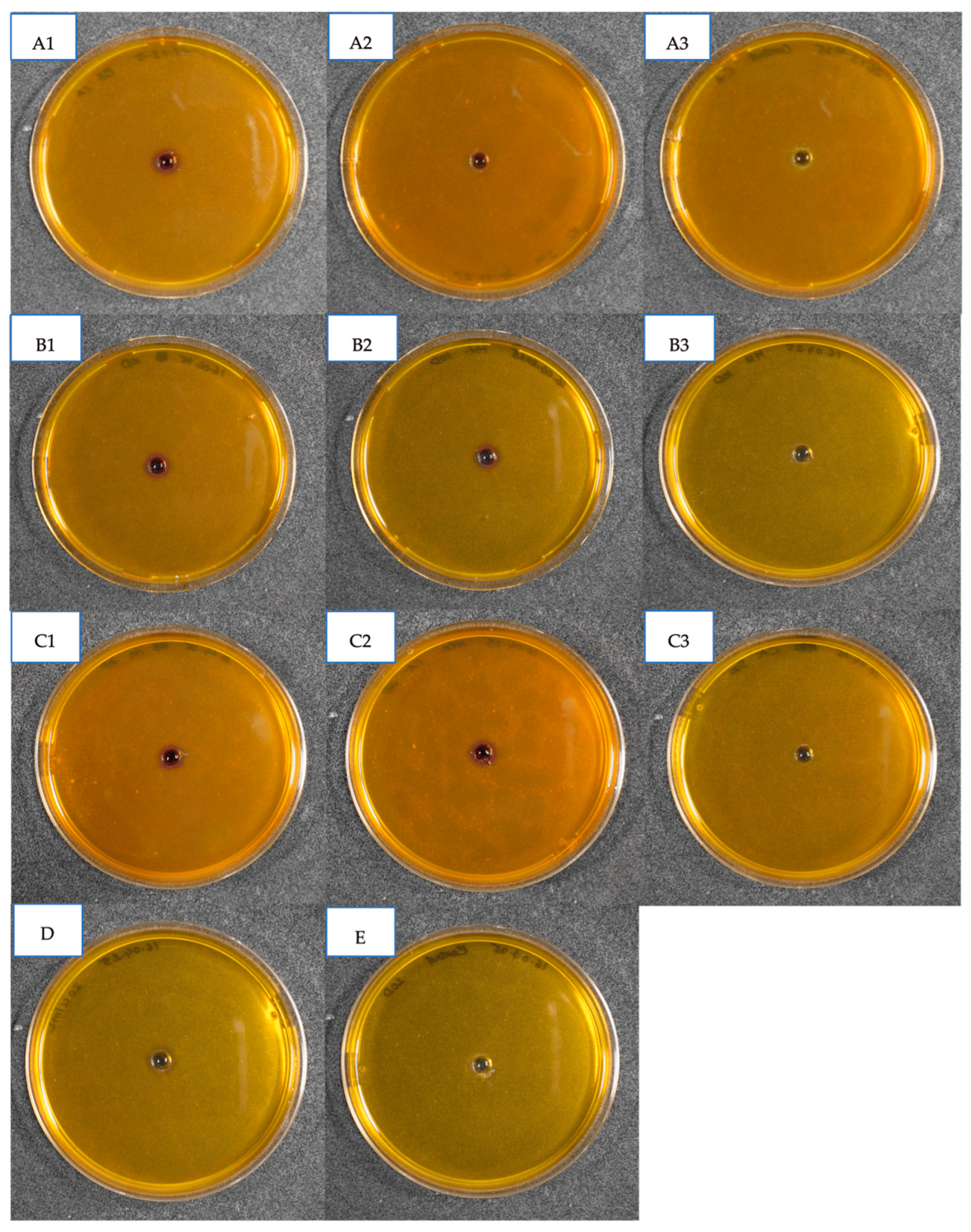
2.3.3. Qualitative Detection of Chitinase Enzyme

Qualitative detection of chitinase enzyme was performed using the protocol described by [49] with minor modifications. A chitinase detection medium was prepared using a basal medium composed of (per liter): 0.3 g MgSO4·7H2O, 3.0 g (NH4)2SO4, 2.0 g KH2PO4, 0.91 g citric acid, 15 g agar, 200 μL Tween-80, 4.5 g colloidal chitin, and 0.15 g bromocresol purple. The pH was adjusted to 5.0, and the mixture was autoclaved at 121 °C for 15 min. The sterilized lukewarm medium was poured into Petri dishes and allowed to solidify. To assess chitinase activity, fresh culture plugs of the isolates were introduced into the solidified medium and incubated at 25 ± 2 °C. Plates were observed for the formation of purple-colored zones around the colonies. In parallel, isolates were inoculated into colloidal chitin broth without bromocresol purple and incubated for 20 days. The resulting supernatant was filtered through a 0.22 μm bacterial-proof filter on the 5th, 10th, and 20th days to verify the reproducibility of the results. Wells were prepared in solid media Petri plates supplemented with colloidal chitin, and each well was filled with the filtered supernatant. A commercially available chitinase enzyme derived from Trichoderma harzianum (ASA Spezial enzyme GmbH, Wolfenbüttel, Germany) was used as the positive control, while colloidal chitin broth without fungal inoculation served as the negative control. Both controls were incubated at room temperature for 15 min. The appearance of a purple zone surrounding the wells was considered indicative of chitinase activity.

2.3.4. Assay for Chitinase Activity

Quantification of endochitinase activity was determined by mixing colloidal chitin with the enzyme solution in a 1:1 ratio and incubated at 37 °C for 1, 2.5, 4, 5.5, 7, and 8.5 h. The reduction in turbidity of the colloidal chitin suspension, measured as absorbance at 510 nm was used as an indicator of enzyme activity, following the method of Tronsmo and Harman [50]. The colloidal chitin suspensions, sterilized by autoclaving, consisted of 1% (w/v) colloidal chitin in 100 mM acetate buffer at pH 5.0. Chitinase activity was expressed as the percentage decrease in turbidity relative to a control suspension containing water in place of the enzyme solution. One enzyme unit was defined as the amount of enzyme required to reduce the turbidity of a chitin suspension by 5%, as described by [50].

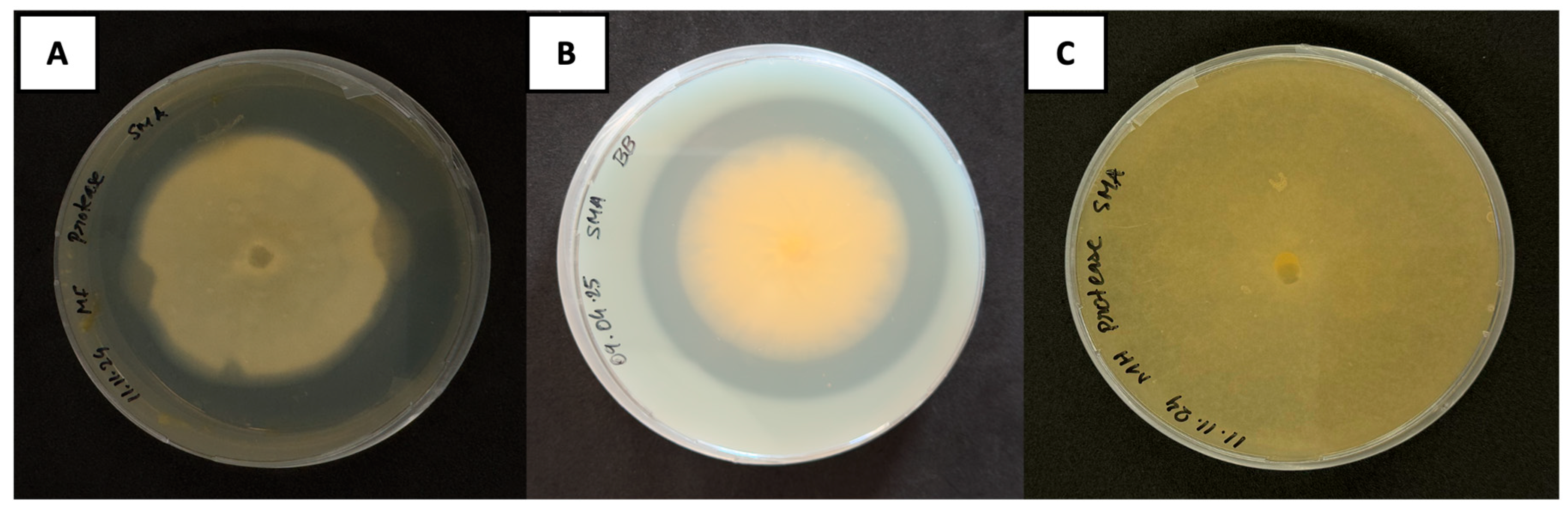
2.4. Protease Production Assay

The method described by Zghair [51] was performed to detect protease production in fungi with slight modification. Skim milk agar was prepared by dissolving 10 g of skim milk powder (Carl Roth GmbH and Co. KG, Karlsruhe, Germany) in 100 mL distilled water with gentle stirring at approximately 37–40 °C to aid solubility without denaturing milk proteins. The basal media consisting of 10 g of agar and 2 g of yeast extract were dissolved separately in 900 mL of distilled water. Subsequently, the pH of yeast extract-agar solution was adjusted to 7 and autoclaved at 121 °C for 20 min. The skim milk solution was sterilized separately by filter sterilization using a 0.22 μm bacterial-proof filter. Once the autoclaved agar media was cooled to approximately 45 °C, sterile skim milk solution was added aseptically and mixed thoroughly and poured into the Petri plates. To assess protease activity, fresh culture plugs of the isolates were introduced into the solidified medium and incubated at 26 °C for 3 to 7 days. Protein breakdown (casein in milk) by the production of protease enzyme was detected when a transparent halo around the fungal colonies appeared.

2.5. Root Colonizing Ability of EMPFs

2.5.1. Preparation of ½ Murashige and Skoog (MS) Medium

To prepare Murashige and Skoog (½ MS) agar medium, 1.1 g of MS vitamins, 5 g of sucrose, and 3.5 g of plant agar purchased from Duchefa Biochemie, Haarlem, The Netherlands were dissolved in 500 mL of distilled water. The pH was set to 5.8 before autoclaving at 121 °C for 20 min. The sterilized material was then poured into sterile Petri plates and 24 well plates (5 mL per well) and stored aseptically until needed.

2.5.2. Seed Sterilization and Germination

Seeds of Petunia hybrida cv “Mitchell” and Ocimum basilicum underwent surface sterilization with immersion in 70% ethanol for 3 minutes followed by a 10 min treatment with 10% of sodium hypochlorite (NaOCl). After treating with NaOCl, seeds were rinsed three to four times with sterile distilled water. Sterilized seeds (20–25 per plate) were germinated on ½ MS agar plates under controlled conditions in a phyto-chamber at 24 °C, 16:8 light/dark (L:D) photoperiod for ten days. After ten days of incubation, uniform healthy seedlings were selected for further study.

2.5.3. Inoculation of Test Fungi

In this study, direct mycelial inoculation method was used for the analysis of fungal root colonization and plant growth benefits. Fungal discs of test fungi measuring 6 mm were excised from the actively growing margin of the freshly cultivated fungal plate with PDA as culture media. Each seedling was inoculated with a single fungal disc in the root zone ensuring the fungal mycelia and the root tissues are in intimate contact. The plant saplings were planted in 24-well plates containing 5 mL of ½ MS agar medium in each well. The fungal strains acquired from DSMZ were used as test treatments, and the control groups included a negative control that was not inoculated with any fungi, and seedlings treated with Serendipita indica were used as a positive control. Each treatment was replicated six times. The plants were incubated at 24 °C with a 16:8 L:D photoperiod in a climate-controlled growth chamber for 20 d.

2.5.4. Assessment of Root Colonization and Plant Growth Effects

The plants were harvested and subjected to growth examination for confirming the fungal colonization in the root, which included measurement of shoot length, root length, and the number of leaves produced. After being sequentially washed with sterile distilled water, the root samples were treated with 70% ethanol for 3 min, 2.5% NaOCl for 6 min, and three additional rinses with sterile distilled water. Surface sterilized roots were chopped into 1 cm long pieces and placed on PDA plates. The plates were incubated at 26 °C in the dark, and fungal development from the roots was observed. To confirm colonization, DNA was extracted from the outgrown fungi according to Cenis methodology [52] and sequenced for identity confirmation (Eurofins, Genomics, GmbH, Konstanz, Germany and NCBI; Blast search).

3. Results

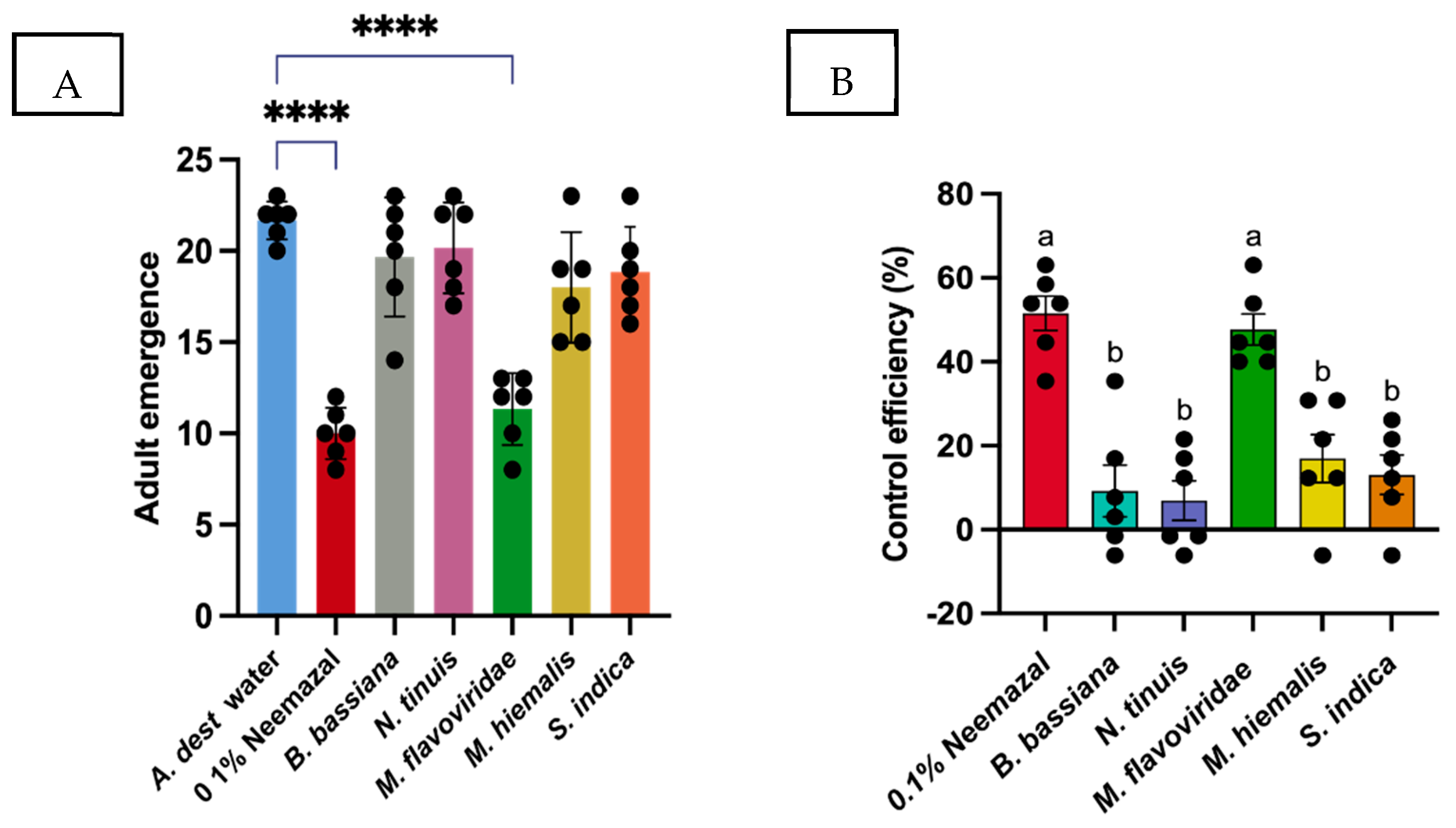
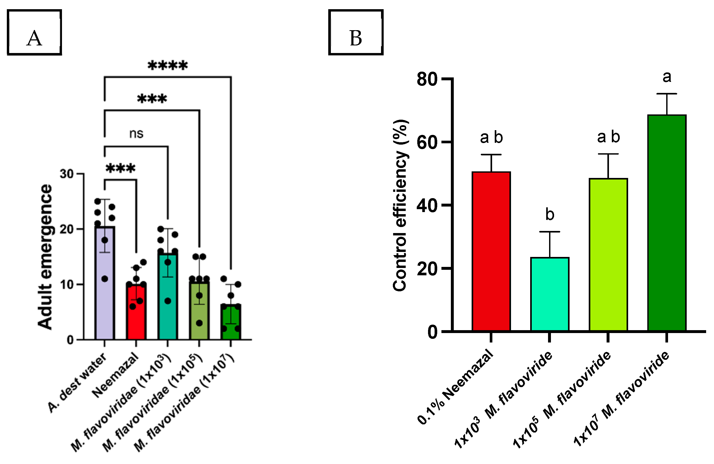
3.1. Effect of Entomopathogenic Fungi to Control the Bradysia Difformis Larvae

The four DSMZ strains that were selected for this experiment, Beauveria bassiana (1344), Metarhizium flavoviride (21704), Mucor hiemalis (2655), and Niesslia tinuis (3481) along with Serendipita indica were examined for their ability to lower the survival rate of B. difformis larvae, with fewer adults emerging from the treated substrate containing pure fungal spores than from the sterile water control and 0.1% Neemazal. When compared to the spore solutions (1 × 10⁶ spores/mL) of other fungal strains, B. difformis larvae were significantly susceptible to the treatment fungal culture M. flavoviride (Table 1). The emergence of adults in substrate was significantly inhibited by M. flavoviride up to 44.61% in comparison to the control treatments (0.1% Neemazal = 51.54%, p < 0.001). In terms of control efficiency, the number of fungus gnat adults that emerged from B. bassiana (9.23%), M. hiemalis (12.27%), N. tinuis (6.92%), and S. indica (13.08%) was substantially higher than the number of adults that emerged from the control treatment (0.1% Neemazal = 51.54, p < 0.001) and did not significantly lower the survival rate of larvae, resulting in a higher emergence rate of adults with a survival rate of 78.66% (p < 0.4057), 72% (p < 0.102), 80.66% (p < 0.6640), and 13.08% (p < 0.132). In contrast, the number of adults emerging from M. flavoviride treatment was lower with a survival rate of 45.33% (p < 0.001), while 0.1% Neemazal, the positive treatment of this study, showed a survival rate of 42% (p < 0.001) (Figure 1A,B). The outcomes were confirmed by examining the impact of varying M. flavoviride concentrations (1 × 10³ spores/mL, 1 × 10⁵ spores/mL, and 1 × 10⁷ spores/mL) on the emergence of B. difformis larvae into adults. As shown in Table 2, the highest activity in controlling the larvae to emerge as adults was significantly exhibited by 1 × 10⁷ spores/mL with control efficiency 68.75%, which showed 25.71% of survival rate, followed by 1 × 10⁵ with control efficiency 48.61%, which showed 48.28% of survival rate, while 1 × 10³ spores/mL with control efficiency 23.61%, which showed 62.85% of survival rate, did not show any significant reduction in adult emergence (Figure 2A,B).

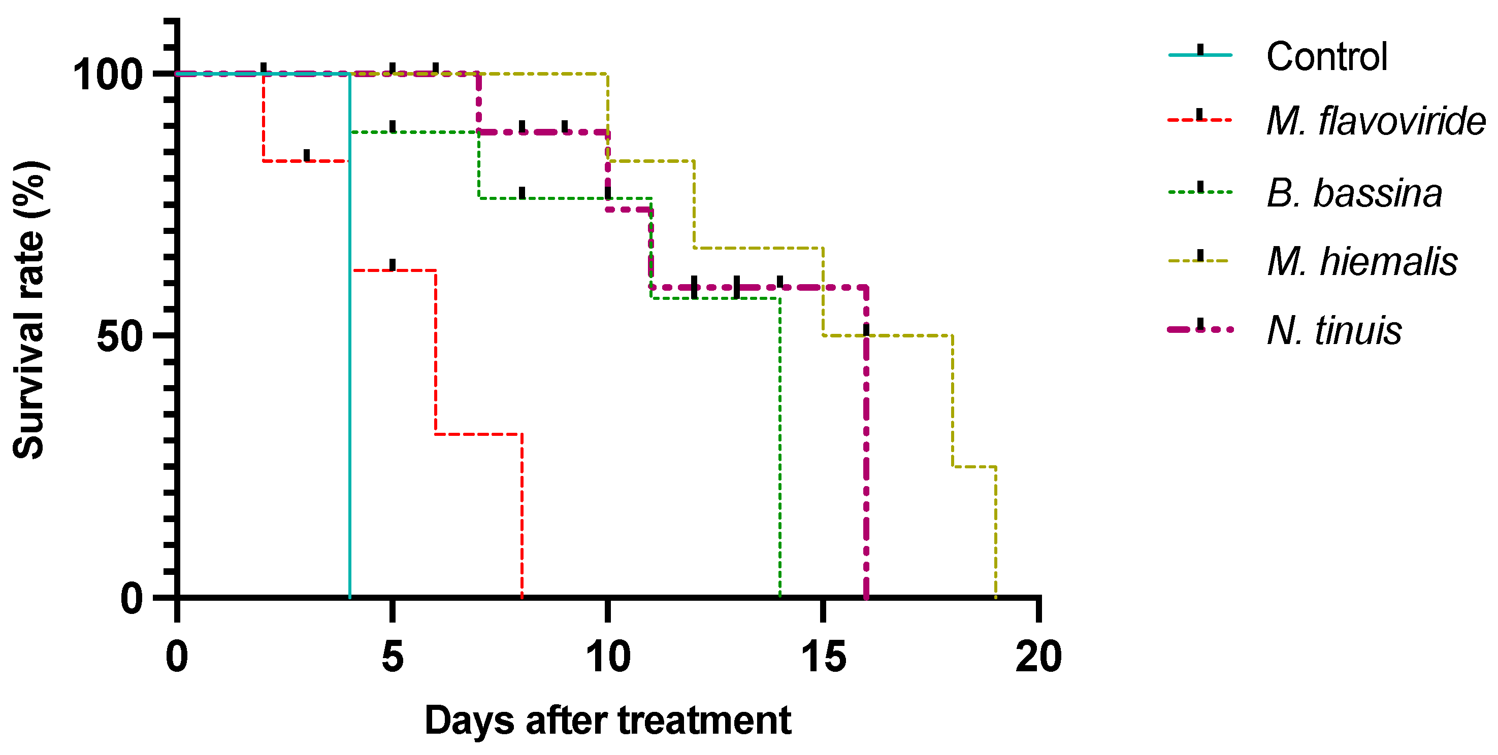
3.2. Larval Survival on Pure Fungal Cultures

Survival rates of larvae progressively declined from the first day after treatment when exposed to the different fungal cultures (Chisq = 25, df = 4, p < 0.001; Figure 1). The control group of larvae, reared on pure PDA, survived only for four days, which was significantly shorter than in all other fungal treatments. In comparison, larvae exposed to fungal cultures of M. flavoviride (21704), B. bassiana (1344), M. hiemalis (2655), and N. tinuis (3481) survived for 8, 14, 19, and 16 days post-exposure, respectively. Larvae treated with M. flavoviride were the most susceptible, with only 7.85 ± 0.95% adult emergence (total N = 200), significantly lower than all other treatments. Exposure to B. bassiana resulted in intermediate survivorship, with 18.62 ± 1.71% (N = 200) of larvae successfully completing their life cycle to adult flies. A significant difference in adult emergence rates was observed between these two treatments (p = 0.0013). Larvae exposed to N. tinuis and M. hiemalis exhibited prolonged survival for up to 16 days post-infection, with adult emergence rates of 20.37% and 20.87% (total N = 200), respectively. Although differences in emergence rates between these two treatments were not significant, both demonstrated significantly higher survival rates compared to the M. flavoviride treatment (p = 0.0002) (Figure 3).

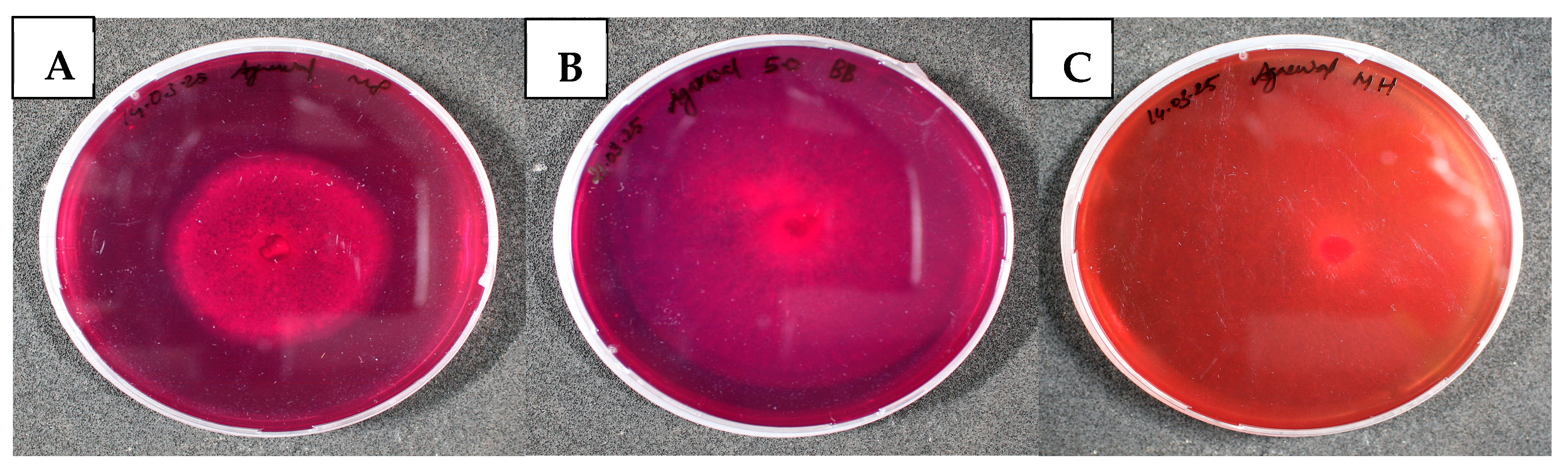
3.2.1. Qualitative Detection of Chitinase Activity

Chitinase activity was assessed by inoculating fresh fungal plugs and culture filtrates into basal chitinase detection medium, as described by Agrawal and Kotasthane [49], supplemented with colloidal chitin (4.5 g/L) and bromocresol purple (0.15 g/L). As illustrated in Figure 4 and Figure 5, the plate assay revealed that Beauveria bassiana and Metarhizium flavoviride, both culture plugs and supernatants, exhibited a pronounced purple coloration, indicating high chitinase activity. In contrast, Mucor hiemalis culture plugs displayed minimal activity. Additionally, culture supernatants collected at different incubation intervals (5th, 10th, and 20th days) were analyzed to confirm chitinase production. The results confirmed preliminary screening with fresh fungal plugs, revealing that B. bassiana produced a more intense purple zone around the wells compared to M. flavoviride, while no zone formation was observed for the supernatant from M. hiemalis. Notably, the intensity of the purple zones surrounding the wells of M. flavoviride increased progressively with incubation time, with supernatants collected on day 20 producing the highest intensity, followed by those obtained on days 10 and 5 (Figure 4 and Figure 5).

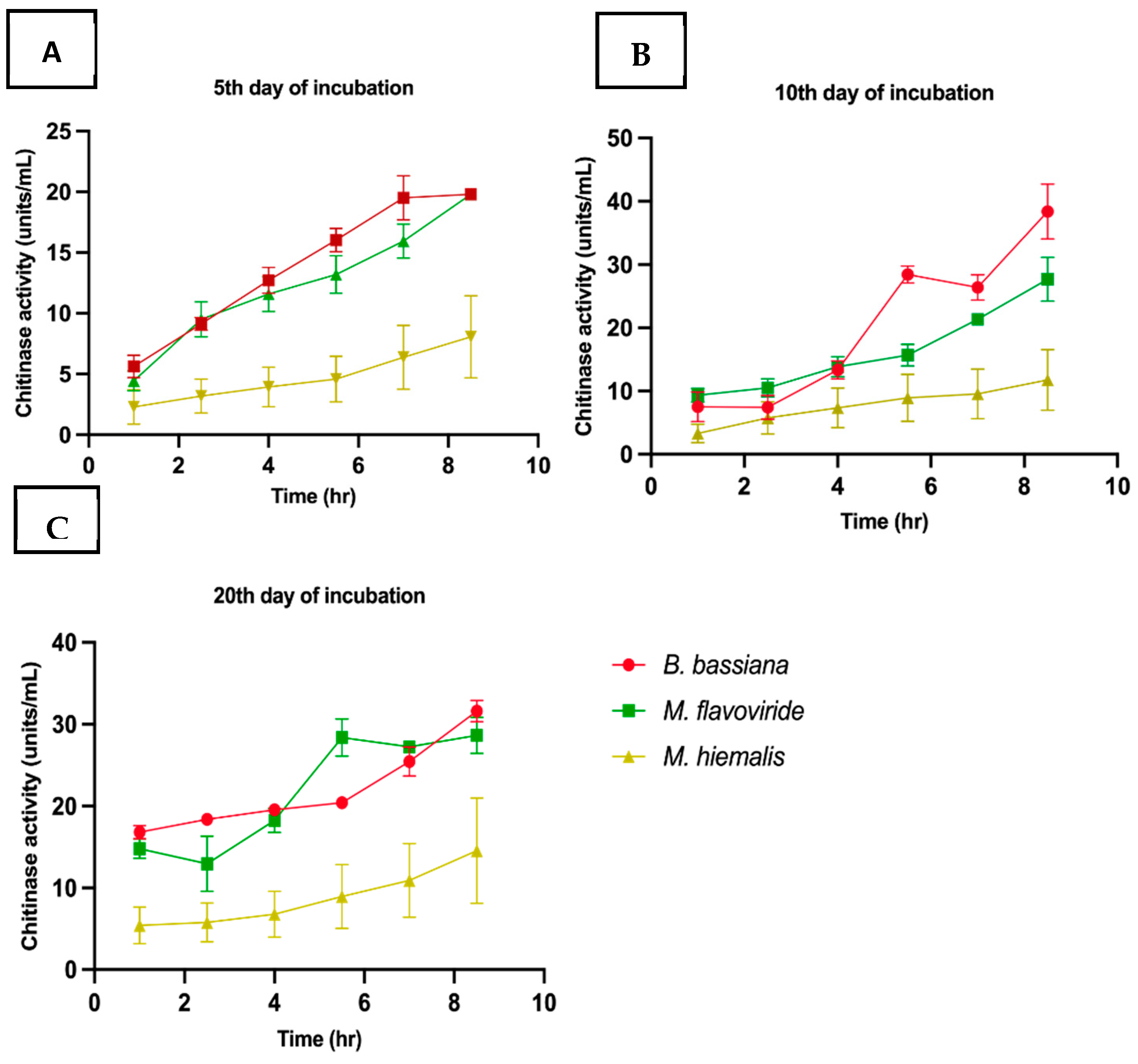
3.2.2. Assay for Chitinase Activity

Chitinase activity in culture filtrates was assessed using colloidal chitin as the substrate and incubating the reaction mixture at 37 °C for time intervals ranging from 1 to 8.5 h. A consistent increase in chitinase activity was observed across all incubation durations (5th, 10th, and 20th day filtrates). Among the tested strains, Beauveria bassiana exhibited the highest chitinase activity, increasing from 19.8 ± 0.05 to 31.6 ± 2.9 U/mL, with the 10-day culture filtrate reaching a peak activity of 38.4 ± 4.3 U/mL, although a notable decline was recorded at the 7 h time point, followed by M. flavoviride showing activity ranging from 19.8 ± 0.16 to 28.64 ± 2.1 U/mL, as well as a similar drop at the 7 h mark. In contrast, Mucor hiemalis demonstrated the lowest chitinase activity among the three fungi, with values increasing from 8.7 ± 1.3 to 24.5 ± 4.5 U/mL across the tested incubation periods. Overall, B. bassiana filtrates (10 and 20 days) exhibited higher chitinase activity, followed by M. flavoviride, while M. hiemalis consistently showed the lowest activity levels (Figure 6, Table 3).

3.3. Qualitative Identification of Protease Production

Protease activity was assessed by inoculating fresh fungal plugs onto skim milk agar plates. Protease producing fungi among the DSMZ strains Beauveria bassiana, Metarhizium flavoviride and Mucor hiemalis were identified by the formation of a clear zone around the fungal colonies, indicating casein hydrolysis. As shown in Figure 7, the skim milk agar plate assay revealed that M. flavoviride exhibited the largest clear zone around its colony (17 ± 0.57 mm), followed by B. bassiana (8 ± 0.57 mm), both indicating significant protease activity. In contrast, M. hiemalis produced only a narrow zone of clearance, indicating minimal protease secretion. The pronounced difference in the diameter of the clear zones suggests that M. flavoviride and B. bassiana possess higher extracellular protease activity under the tested conditions compared to the other isolates.

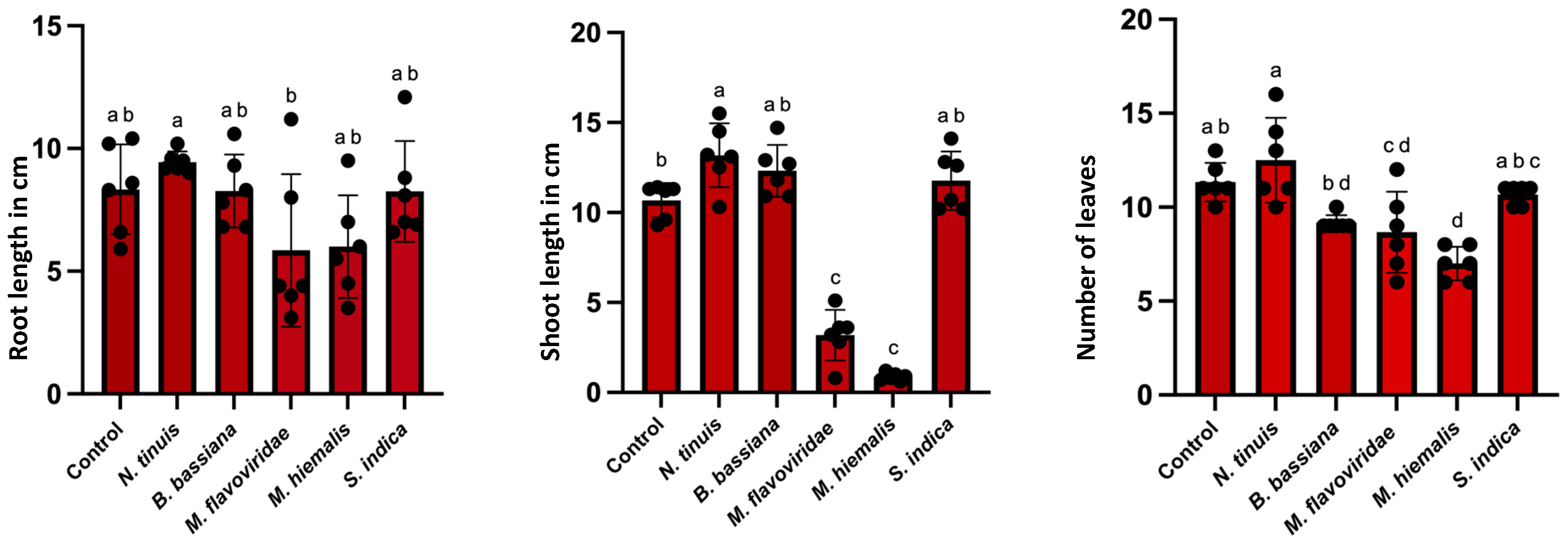
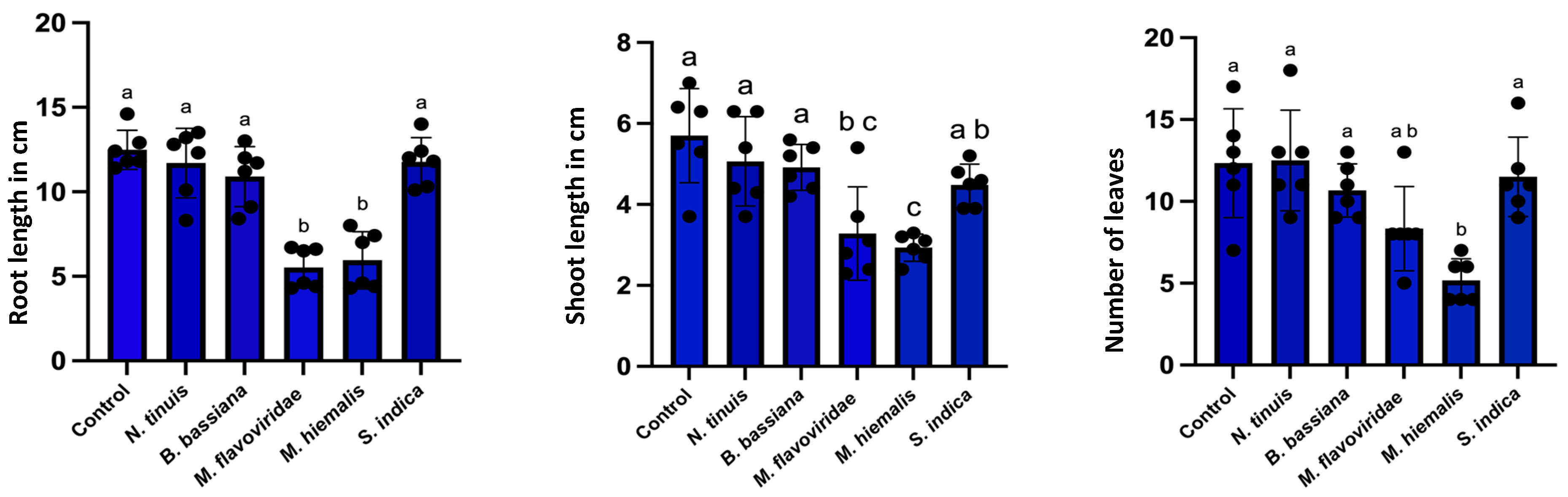
3.4. Effect of Fungal Colonization in Petunia and Basil Plants

The impact of different fungal inoculations on Petunia hybrida cv. “Mitchell” growth was evaluated by measuring shoot length, root length, and number of leaves after 20 days of incubation (Figure 8, Supplementary Table S2). A significant increase in shoot length was observed in seedlings treated with N. tinuis (13.18 ± 0.72 cm) compared to the control (10.68 ± 0.39 cm; p ≤ 0.05). In contrast, M. flavoviride and M. hiemalis treatments significantly reduced shoot length to 3.18 ± 0.57 cm and 0.88 ± 0.08 cm, respectively (p ≤ 0.05). No statistically significant differences in root length were detected between the control and any of the fungal treatments (p > 0.05). In terms of leaf production, M. hiemalis inoculation resulted in a significant reduction in the number of leaves (7.00 ± 0.36) compared to the control (11.33 ± 0.42; p ≤ 0.05). Other treatments, including N. tinuis and M. flavoviride, did not show a significant impact on leaf number. Overall, N. tinuis promoted shoot elongation in Petunia hybrida, while M. flavoviride and M. hiemalis exerted inhibitory effects on growth parameters. Similarly, in Ocimum basilicum, a significant reduction in shoot length was recorded in seedlings treated with M. flavoviride (3.28 ± 0.47) and M. hiemalis (2.93 ± 0.38 cm) compared to the control (5.70 ± 0.47; p ≤ 0.05). Both fungal strains also significantly reduced root length, with values of 5.51 ± 0.48 cm for M. flavoviride and 5.95 ± 0.69 cm for M. hiemalis, relative to the control (12.48 ± 0.47; p ≤ 0.05). Moreover, the number of leaves was significantly decreased in plants inoculated with M. flavoviride (8.33 ± 1.05) and M. hiemalis (5.16 ± 0.54) compared to control seedlings (12.33 ± 1.33; p ≤ 0.05). These results suggest that while M. flavoviride and M. hiemalis negatively impacted Ocimum basilicum growth, other tested fungi did not significantly alter growth parameters (Figure 9, Supplementary Table S3). Surface sterilization and subsequent incubation of plant roots confirmed that M. flavoviride and N. tinuis were capable of colonizing the plant roots. However, among these, only N. tinuis demonstrated a significant influence on the growth of Petunia hybrida cv. “Mitchell”, specifically by promoting an increase in shoot length. Molecular identification through DNA extraction and sequencing further validated the successful colonization of these fungal strains within the plant root tissues (Supplementary Table S1).

4. Discussion

In this study, we checked selected fungal strains from German Collection of Microorganisms and Cell Culture (DSMZ) namely Beauveria bassiana, Metarhizium flavoviride, Mucor hiemalis, and Niesslia tinuis for their ability to act as entomopathogens against the fungus gnat, Bradysia difformis along with its ability to act as endophytes in Petunia hybrida cv. “Mitchell” and Ocimum basilicum. The results of the bioassay revealed that the significant entomopathogenic potential of M. flavoviride in controlling the population of B. difformis, a species of fungus gnat. Among the four selected DSMZ fungal strains and Serendipita indica tested, only M. flavoviride exhibited a significant inhibitory effect on adult emergence, leading to a significant reduction in the survival rate of larvae. This finding confirms previous findings that M. anisophile, a well-documented entomopathogen belonging to the same genus as M. flavoviride, is effective against the insect pests including those within the order Diptera (Fungus gnats) [53]. The effectiveness of B. bassiana and M. anisopliae against the spiny bollworm (Earias insulana) on cotton plants was assessed in a study published in the Egyptian Journal of Biological Pest Control [54]. The results indicate that in some situations, Metarhizium sp. may provide better control than B. bassiana. In this case, M. flavoviride’s considerable pathogenicity was noticeably greater than that of the other evaluated fungal strains. In line with previous research indicating that these fungi might not always be as efficient against specific pest species under controlled settings, B. bassiana and M. hiemalis did not exhibit a substantial decrease in larval survival or adult emergence [47]. Remarkably, in the current investigation, the DSMZ-obtained strain of Mucor hiemalis showed no pathogenicity towards fungus gnat populations which stands in contrast to research conducted [55] where they who found that the isolated strain of M. hiemalis BO-1 was significantly harmful to the fungus gnat Bradysia odoriphaga (Diptera: Sciaridae). Notably, higher spore concentrations (1 × 10⁷ spores/mL) of M. flavoviride significantly reduced adult emergence of B. difformis, which was accompanied by a lower survival rate of 25.71%. This concentration-dependent effect of M. flavoviride was also noted in line with recent research that found that larger concentrations of M. flavoviride result in better control of insect populations, where it shows that the concentration of the entomopathogenic fungus is a critical factor in increasing its efficacy [56]. Finding the ideal spore concentration for efficient pest control is crucial, as lower concentrations (1 × 10³ spores/mL) did not exhibit any discernible effects.
The direct fungal feed assay results support the pathogenicity assay results because M. flavoviride (21704) exhibits high virulence, which results in a significantly reduced adult emergence rate of 7.85%. This emphasizes the potential of the strain as an efficient biocontrol agent against fungus gnats. This is in line with earlier studies showing M. flavoviride’s effectiveness in managing a range of insect pests. Liu et al. (2022) [57], for example, demonstrated the broad-spectrum insecticidal abilities of M. flavoviride by reporting significant pathogenicity against Spodoptera litura. On the contrary, B. bassiana (1344), which had an adult emergence rate of 18.62%, exhibited moderate efficacy. This is compatible with the results of Cloyd et al. [58], who observed that although B. bassiana can be useful against specific fungus gnat stages, its overall efficacy varies based on the developmental stage that is targeted. Additionally, as mentioned earlier, Mucor hiemalis (2655) exhibited an adult emergence rate of 20.87% and demonstrated little pathogenicity in our research. However, Ref. [55] discovered a strain of M. hiemalis (BO-1) that was significantly virulent against Bradysia odoriphaga. At concentrations more than 1 × 10⁷ spores/mL, the strain achieved over 80% control efficiency in just five days. This disparity emphasizes how crucial strain-specific analyses are to biocontrol studies.
A qualitative assessment of the chitinase synthesis in the entomopathogenic fungi B. bassiana and M. flavoviride was conducted using bromocresol purple indicator agar medium. After 5, 10, and 20 days of incubation, a distinct purple ring appeared around the wells containing fungal extracts, confirming the isolates’ capacity to manufacture the extracellular chitinase enzyme. Commercially available chitinase from Trichoderma sp. worked well as a positive control, but M. hiemalis notably had no such activity during any of the incubation periods. Chitinase activity in M. flavoviride and B. bassiana is in line with a number of other discoveries. There is a strong correlation between chitinase synthesis and insecticidal capacity, as Juliya [59] showed in a study that isolates of Beauveria and Metarhizium with increased extracellular chitinase activity exhibited enhanced pathogenicity against the cowpea aphid (Aphis craccivora). Further supporting the critical role of chitinase in fungal virulence, a study by Gebremariam et al. [60] found that higher chitinase activity in M. anisopliae and B. bassiana corresponded with increased mortality in whiteflies (Bemisia tabaci and Trialeurodes vaporariorum). Furthermore, the purple ring’s increasing intensity over longer incubation times in this study is consistent with the chitinase genes’ temporal expression pattern, which was studied by Fang et al. [61]. They found that the late exponential and stationary phases of growth were when B. bassiana produced the most chitinase. Fungal biomass accumulation and the adaptive need for chitin degradation during host invasion and nutrition acquisition are probably correlated with the steady rise in enzyme synthesis. The chitinase activity of Beauveria bassiana, which reached a peak of 38.4 ± 4.3 Units/mL in 10-day culture filtrates, is consistent with previous findings demonstrating the role of chitinolytic enzymes in enhancing fungal virulence. Notably, the Bbchit1 gene encoding an endochitinase was purified and characterized from B. bassiana, and its overexpression significantly increased virulence against aphids by reducing both the lethal concentration and the time to mortality [61]. In contrast, M. flavoviride exhibited moderate chitinase activity, ranging from 19.8 ± 0.16 to 28.64 ± 2.1 U/mL, while Mucor hiemalis showed the lowest activity levels (8.7 ± 1.3 to 24.5 ± 4.5 U/mL). These findings indicate that while B. bassiana’s strong chitinolytic activity likely contributes to its entomopathogenicity, M. flavoviride and M. hiemalis may utilize alternative or complementary mechanisms, as their lower chitinase activity does not directly correspond to reduced pathogenic effects. The decline in chitinase activity observed at the 7 h incubation point across all three fungi may indicate a regulatory mechanism or substrate depletion, warranting further investigation. The higher chitinase activity observed in B. bassiana and M. flavoviride may contribute significantly to their larvicidal effects. Chitinases are hydrolytic enzymes that degrade the chitin polymer found in the insect cuticle and peritrophic matrix, facilitating fungal penetration and ultimately causing host death. Previous research has shown a direct correlation between chitinase activity and fungal virulence [61]. The increasing enzyme activity over incubation time also aligns with the progressively enhanced mortality seen in bioassays, suggesting that enzymatic degradation plays a critical role in larval susceptibility [62]. The exogenous production of cuticle-degrading enzymes such as chitinase by Metarhizium flavoviride, as demonstrated in this study, offers a practical advantage over gene-driven pest control strategies that require endogenous genetic manipulation. While genetic approaches aimed at inducing chitinase expression in insects face challenges related to population-level gene regulation, entomopathogenic fungi achieve similar outcomes externally, simplifying implementation and avoiding potential ecological complications [63]. Overall, the comparative analysis underscores the significance of chitinase activity in fungal virulence and highlights potential avenues for enhancing biocontrol efficacy through genetic or environmental modulation of enzyme production.
Protease activity assays demonstrated significant variability among the tested fungal strains. M. flavoviride exhibited the highest proteolytic activity, followed by B. bassiana, while Mucor hiemalis showed no detectable casein hydrolysis. These findings are consistent with previous studies highlighting the importance of extracellular proteases in entomopathogenic fungi for degrading host cuticles and facilitating infection [64]. In particular, B. bassiana is known to secrete subtilisin-like proteases, which are critical for both pathogenicity and nutrient acquisition [64]. Conversely, the minimal activity observed in M. hiemalis aligns with earlier reports indicating that protease production in Mucor species tends to be cell-associated or exhibits limited activity under similar experimental conditions [65]. These findings align with Tóthné Bogdányi et al. (2019) [25], who reported that M. flavoviride relies heavily on extracellular enzymes such as chitinases and proteases for cuticle degradation, which facilitates host infection and contributes to its broad insecticidal efficacy. These functional disparities suggest that the fungi under study employ distinct ecological and physiological strategies, with potential implications for their application in plant–microbe interaction studies and biological control initiatives.
The ability of selected endophytic and entomopathogenic fungi from DSMZ to colonize and their effects on plant growth regulation in Ocimum basilicum and Petunia hybrida cv. “Mitchell” were also examined in this study. When compared to the control, N. tinuis inoculation significantly increased the shoot length in Petunia hybrida. This result is consistent with past studies showing that specific endophytes improved plant growth by influencing hormone levels, enhancing nutrient uptake, and fostering systemic resistance to biotic and abiotic stressors [66,67]. To the best of our knowledge, this is the first publication detailing the influence of N. tinuis on Petunia hybrida cv. “Mitchell”; however, the beneficial effect seen in this study points to its possible function as a plant growth-promoting endophyte (PGPE). While the exact colonization mechanisms of N. tinuis remain unclear, endophytic fungi typically enter plants via root hairs, epidermal cracks, or lateral root emergence sites, forming mutualistic associations by modulating phytohormones and enhancing nutrient uptake. We are planning controlled pot experiments using microscopic imaging and molecular markers to investigate N. tinuis’s colonization patterns, entry routes, and tissue localization. Elucidating its physiological interactions and potential activation of plant defense responses will be essential to understand its endophytic role. These studies will help clarify the mechanisms of N. tinuis endophytism and support its potential use in sustainable horticulture. Similar to their inhibitory effects in Petunia hybrida cv. “Mitchell”, inoculation with M. flavoviride and M. hiemalis in Ocimum basilicum consistently led to considerable reductions in shoot and root length as well as leaf production including the damage to the plants. According to research by Sena et al. [68] and Porras-Alfaro and Baymann [69], some soil fungi and fungal endophytes can have a negative impact on growth, depending on the host species, fungal strain, and environmental conditions, even if they can be advantageous in some plant–fungal systems. Interestingly, other plant development in both species was not significantly impacted by the tested fungal treatments in this study, indicating a strain-and host-specific connection that needs more research. According to Rodriques et al. [70] the increasing understanding that fungal symbioses operate on a mutualism–pathogenicity continuum is reflected in the variation in plant responses to several endophytic and soil fungi that were seen here. These findings highlight the potential of M. flavoviride as effective entomopathogenic agents against fungus gnats, pests that are increasingly problematic in horticultural systems transitioning to peat-free substrates. Peat-free substrates, while environmentally favorable, are known to enhance fungus gnat attraction and infestation due to their higher organic content and moisture retention. Therefore, identifying biological control agents that are not only effective against fungus gnats but also compatible with these substrates is of urgent practical relevance. The substantial chitinase activity observed in M. flavoviride aligns with its insecticidal efficacy in vitro and suggests potential for further development as a biocontrol agent. However, additional studies under greenhouse and field conditions are essential to validate its effectiveness and safety before considering commercial application. Further research is needed to elucidate the plant–fungus interaction mechanisms, especially concerning root colonization and growth promotion. In addition, greenhouse trials should evaluate the performance of these fungal strains in terms of reducing its affinity across various peat-free or peat-reduced substrates under different environmental conditions. These findings lay foundational insights for the possible strategic use of entomopathogenic fungi in sustainable pest management systems. Nonetheless, such applications must be supported by future studies evaluating efficacy across diverse environmental conditions and substrate types.

5. Conclusions

The findings of this study indicate that certain entomopathogenic and endophytic fungal strains, particularly Metarhizium flavoviride, may serve as effective candidates for the environmentally sustainable management of Bradysia difformis (fungus gnats), particularly within peat-free horticultural systems. Among the tested strains, M. flavoviride exhibited superior entomopathogenic activity, significantly reducing larval survival and adult emergence, especially at higher spore concentrations. Concurrently, both M. flavoviride and B. bassiana demonstrated extracellular enzymatic activities, chitinase and protease production, linked to their virulence and insecticidal capabilities. Importantly, the findings also contribute to the growing understanding of plant–fungus interactions. While Niesslia tinuis showed potential as a plant growth-promoting endophyte in Petunia hybrida cv. “Mitchell”, other strains like M. hiemalis demonstrated host-specific pathogenic effects, underlining the complexity of fungal colonization and its outcomes. These observations affirm the ecological and physiological diversity of fungal strains and underscore the need for tailored application strategies. Fungus gnats are a significant greenhouse pest, and their attraction to peat-free substrates that are commonly used for their environmental sustainability poses an increasing challenge for growers. As the horticultural industry transitions away from peat to more eco-friendly alternatives, there is a growing need for effective, substrate-compatible pest control solutions. The entomopathogenic fungi evaluated in this study present a promising biological alternative to conventional insecticides, particularly in systems utilizing peat-free or peat-reduced media.
To maximize their practical application, future research should investigate the complex interactions between fungal strains, host plants, and diverse substrate types under varying environmental conditions. Although the present findings are derived from controlled greenhouse experiments, comprehensive field studies are necessary to substantiate the efficacy and reliability of these fungal strains under practical agricultural conditions. Such investigations will be critical to assess their suitability for incorporation into integrated pest management (IPM) strategies. Advancing such knowledge will support the development of integrated, low-input pest management strategies that align with sustainable horticultural practices. Furthermore, building on the promising results demonstrated in controlled greenhouse conditions, future research should focus on evaluating the efficacy of Metarhizium flavoviride under open-field crop environments to better understand its potential in diverse agricultural settings. An expanding the scope of investigation to include other species of mushroom moth and various soil types will provide valuable insights into the broader applicability of this entomopathogenic fungus. Considering its tropical origin and mode of action, further studies exploring formulation improvements and application strategies could enhance the practical implementation of this biocontrol agent. Additionally, future research should also be focused on testing its efficacy not only against fungus gnats but also against a wider range of insect pests to explore its broader applicability. Finally, integrating molecular and genetic analyses alongside ecological assessments may deepen our understanding of the fungus’s interaction with target pests and contribute to sustainable pest management approaches.

Supplementary Materials

The following supporting information can be downloaded at: https://www.mdpi.com/article/10.3390/su17135897/s1, Section S1, Table S1: DNA/Protein Sequence Data; Section S2, Table S2: Means of root, shoot lengths and number of leaves produced by Petunia hybrida cv. “Mitchell” inoculated with selected fungal isolates of DSMZ along with S. indica as positive control; Table S3: Means of root and shoot lengths and number of leaves produced by Ocimum basilicum inoculated with selected fungal isolates of DSMZ along with S. indica as positive control.

Author Contributions

The study was jointly conceived by S.S., K.B. and P.F. Experiments were designed by S.S. and K.B. S.S. prepared the manuscript. S.S., K.B. and P.L. edited the manuscript, and S.S. carried out the experiments. All authors have read and agreed to the published version of the manuscript.

Funding

This research was funded by Deutscher Akademischer Austauschdienst (DAAD), Research Grants-Doctoral Programmes in Germany, 2022/23 (57588370) as part of the requirements of a PhD thesis.

Institutional Review Board Statement

Not applicable.

Informed Consent Statement

Not applicable.

Data Availability Statement

The original contributions presented in this study are included in the article/Supplementary Material. Additional data presented in this study are available on request from the corresponding author. Further inquiries can be directed to the corresponding author.

Acknowledgments

The authors acknowledge the use of ChatGPT (GPT-4-turbo model, OpenAI, San Francisco, CA, USA) for language refinement and paraphrasing assistance during manuscript preparation. All content was subsequently reviewed and verified by the authors. The authors acknowledge the technical support provided by the technical assistants, Sabine Czekalla and Natalie Hauswald, whose expertise greatly contributed to the success of this work.

Conflicts of Interest

The authors declare no conflicts of interest.

Abbreviations

The following abbreviations are used in this manuscript:
EMPFEntomopathogenic fungi
MSMurashige and Skoog
PDAPotato Dextrose Agar
ANOVAAnalysis of Variance
DSMZGerman Collection of Microorganisms and Cell Cultures
DNADeoxyribonucleic Acid
NCBINational Center for Biotechnology Information
rpmRevolutions per minute
NaOClSodium hypochlorite
(NH4)2SO4Ammonium sulfate
MgSO4·7H2OMagnesium sulfate heptahydrate
KH2PO4Monopotassium phosphate
°CDegrees Celsius
RHRelative humidity
IPMIntegrated Pest Management
UUnits
HSDHonestly Significance Difference
L:DLight:Dark
SEStandard error
dDays
×gRelative centrifugal force (times gravity)
gGram
L, mLLiter, milliliter
cm, µmCentimeter, micrometer

References

  1. Babytskiy, A.I.; Moroz, M.S.; Kalashnyk, S.O.; Bezsmertna, O.O.; Voitsekhivska, O.V.; Dudiak, I.D. New findings of pest sciarid species (Diptera, Sciaridae) in Ukraine, with the first record of Bradysia difformis. Biosyst. Divers. 2019, 27, 131–141. [Google Scholar] [CrossRef]
  2. Harris, M.A.; Oetting, R.D.; Gardner, W.A. Use of entomopathogenic nematodes and a new monitoring technique for control of fungus gnats, Bradysia coprophila (Diptera: Sciaridae), in floriculture. Biol. Control 1995, 5, 412–418. [Google Scholar] [CrossRef]
  3. Tomalak, M.; Piggott, S.; Jagdale, G.B. Glasshouse applications. In Nematodes as Biocontrol Agents; CABI Publishing Wallingford: Oxfordshire, UK, 2005; pp. 147–166. [Google Scholar]
  4. Menzel, F.; Smith, J.E.; Colauto, N.B. Bradysia difformis Frey and Bradysia ocellaris (Comstock): Two additional neotropical species of black fungus gnats (Diptera: Sciaridae) of economic importance: A redescription and review. Ann. Entomol. Soc. Am. 2003, 96, 448–457. [Google Scholar] [CrossRef]
  5. Freeman, P. Sciarid flies, Diptera; Sciaridae. In Handbooks for the Identification of British Insects 9, Part 6; The Royal Entomological Society of London: St Albans, UK, 1983; p. 68. [Google Scholar]
  6. Binns, E.S. Fungus gnats (Diptera: Mycetophilidae/Sciaridae) and the role of mycophagy in soil: A review. Revue d'Écologie et de Biologie du Sol 1981, 18, 77–90. [Google Scholar]
  7. Gillespie, D.R.; Menzies, J.G. Fungus gnats vector Fusarium oxysporum f. sp. radicislycopersici 1. Ann. Appl. Biol. 1993, 123, 539–544. [Google Scholar] [CrossRef]
  8. Ludwig, S.W.; Oetting, R.D. Evaluation of medium treatments for management of Frankliniella occidentalis (Thripidae: Thysanoptera) and Bradysia coprophila (Diptera: Sciaridae). Pest. Manag. Sci. 2001, 57, 1114–1118. [Google Scholar] [CrossRef]
  9. Nielsen, G.R. Fungus Gnats. Available online: https://ipm.ucanr.edu/PMG/PESTNOTES/pn7448.html (accessed on 29 April 2025).
  10. Wilkinson, J.D.; Daugherty, D.M. Comparative development of Bradysia impatiens (Diptera: Sciaridae) under constant and variable temperatures. Ann. Entomol. Soc. Am. 1970, 63, 1079–1083. [Google Scholar] [CrossRef]
  11. Cloyd, R.A. Management of fungus gnats (Bradysia spp.) in greenhouses and nurseries. Floric. Ornam. Biotechnol. 2008, 2, 84–89. [Google Scholar]
  12. Kekkilä Professional. The Control of Fungus Gnats in Your Greenhouse. Available online: https://www.kekkilaprofessional.com/growing-tips/the-control-of-fungus-gnats-in-your-greenhouse/ (accessed on 18 April 2025).
  13. Ma, J.; Chen, S.; Moens, M.; Han, R.; De Clercq, P. Efficacy of entomopathogenic nematodes (Rhabditida: Steinernematidae and Heterorhabditidae) against the chive gnat, Bradysia odoriphaga. J. Pest Sci. 2013, 86, 551–561. [Google Scholar] [CrossRef]
  14. Bai, G.-Y.; Xu, H.; Fu, Y.-Q.; Wang, X.-Y.; Shen, G.-S.; Ma, H.-K.; Feng, X.; Pan, J.; Gu, X.-S.; Guo, Y.-Z.; et al. A comparison of novel entomopathogenic nematode application methods for control of the chive gnat, Bradysia odoriphaga (Diptera: Sciaridae). J. Econ. Entomol. 2016, 109, 2006–2013. [Google Scholar] [CrossRef]
  15. Shan, T.; Zhang, H.; Chen, C.; Chen, A.; Shi, X.; Gao, X. Low expression levels of nicotinic acetylcholine receptor subunits Boα1 and Boβ1 are associated with imidacloprid resistance in Bradysia odoriphaga. Pest Manag. Sci. 2020, 76, 3038–3045. [Google Scholar] [CrossRef] [PubMed]
  16. Shah, P.A.; Pell, J.K. Entomopathogenic fungi as biological control agents. Appl. Microbiol. Biotechnol. 2003, 61, 413–423. [Google Scholar] [CrossRef] [PubMed]
  17. Hajek, A.E.; McManus, M.L.; Junior, I.D. A review of introductions of pathogens and nematodes for classical biological control of insects and mites. Biol. Control 2007, 41, 1–13. [Google Scholar] [CrossRef]
  18. Lacey, L.A.; Frutos, R.; Kaya, H.K.; Vail, P. Insect pathogens as biological control agents: Do they have a future? Biol. Control 2001, 21, 230–248. [Google Scholar] [CrossRef]
  19. Amobonye, A.; Bhagwat, P.; Singh, S.; Pillai, S. Enhanced xylanase and endoglucanase production from Beauveria bassiana SAN01, an entomopathogenic fungal endophyte. Fungal Biol. 2021, 125, 39–48. [Google Scholar] [CrossRef]
  20. Bale, J.S.; Van Lenteren, J.C.; Bigler, F. Biological control and sustainable food production. Philos. Trans. R. Soc. B Biol. Sci. 2008, 363, 761–776. [Google Scholar] [CrossRef]
  21. Gabarty, A.; Salem, H.M.; Fouda, M.A.; Abas, A.A.; Ibrahim, A.A. Pathogencity induced by the entomopathogenic fungi Beauveria bassiana and Metarhizium anisopliae in Agrotis ipsilon (Hufn.). J. Radiat. Res. Appl. Sci. 2014, 7, 95–100. [Google Scholar] [CrossRef]
  22. Coombes, C.A.; Hill, M.P.; Moore, S.D.; Dames, J.F. Entomopathogenic fungi as control agents of Thaumatotibia leucotreta in citrus orchards: Field efficacy and persistence. BioControl 2016, 61, 729–739. [Google Scholar] [CrossRef]
  23. Andreadis, S.S.; Cloonan, K.R.; Bellicanta, G.S.; Paley, K.; Pecchia, J.; Jenkins, N.E. Efficacy of Beauveria bassiana formulations against the fungus gnat Lycoriella ingenua. Biol. Control 2016, 103, 165–171. [Google Scholar] [CrossRef]
  24. Dehghani, M.; Vidal, J.R.A. Combined application of a biopesticide and a botanical compound to control fungus gnats Bradysia impatiens on different host plants. Combined Application of a Biopesticide and a Botanical Compound to Control Fungus Gnats Bradysia impatiens, p. 83. Available online: https://ediss.uni-goettingen.de/handle/21.11130/00-1735-0000-0008-59C5-6 (accessed on 20 June 2022).
  25. Tóthné Bogdányi, F.; Petrikovszki, R.; Balog, A.; Putnoky-Csicsó, B.; Gódor, A.; Bálint, J.; Tóth, F. Current knowledge of the entomopathogenic fungal species Metarhizium flavoviride Sensu Lato and its potential in sustainable pest control. Insects 2019, 10, 385. [Google Scholar] [CrossRef]
  26. Vega, F.E.; Posada, F.; Aime, M.C.; Pava-Ripoll, M.; Infante, F.; Rehner, S.A. Entomopathogenic fungal endophytes. Biol. Control 2008, 46, 72–82. [Google Scholar] [CrossRef]
  27. Vega, F.E. The use of fungal entomopathogens as endophytes in biological control: A review. Mycologia 2018, 110, 4–30. [Google Scholar] [CrossRef] [PubMed]
  28. Akutse, K.S.; Maniania, N.K.; Fiaboe, K.K.M.; Van den Berg, J.; Ekesi, S. Endophytic colonization of Vicia faba and Phaseolus vulgaris (Fabaceae) by fungal pathogens and their effects on the life-history parameters of Liriomyza huidobrensis (Diptera: Agromyzidae). Fungal Ecol. 2013, 6, 293–301. [Google Scholar] [CrossRef]
  29. Gathage, J.W.; Lagat, Z.O.; Fiaboe, K.K.M.; Akutse, K.S.; Ekesi, S.; Maniania, N.K. Prospects of fungal endophytes in the control of Liriomyza leafminer flies in common bean Phaseolus vulgaris under field conditions. BioControl 2016, 61, 741–753. [Google Scholar] [CrossRef]
  30. Jaber, L.R. Grapevine leaf tissue colonization by the fungal entomopathogen Beauveria bassiana sl and its effect against downy mildew. BioControl 2015, 60, 103–112. [Google Scholar] [CrossRef]
  31. Dash, A.; Singh, A.P.; Chaudhary, B.R.; Singh, S.K.; Dash, D. Effect of silver nanoparticles on growth of eukaryotic green algae. Nano-micro Lett. 2012, 4, 158–165. [Google Scholar] [CrossRef]
  32. Sasan, R.K.; Bidochka, M.J. Antagonism of the endophytic insect pathogenic fungus Metarhizium robertsii against the bean plant pathogen Fusarium solani f. sp. phaseoli. Can. J. Plant Pathol. 2013, 35, 288–293. [Google Scholar] [CrossRef]
  33. Jaber, L.R.; Salem, N. Endophytic colonisation of squash by the fungal entomopathogen Beauveria bassiana (Ascomycota: Hypocreales) for managing Zucchini yellow mosaic virus in cucurbits. Biocontrol Sci. Technol. 2014, 24, 1096–1109. [Google Scholar] [CrossRef]
  34. Bruck, D.J. Fungal entomopathogens in the rhizosphere. BioControl 2010, 55, 103–112. [Google Scholar] [CrossRef]
  35. Pava-Ripoll, M.; Angelini, C.; Fang, W.; Wang, S.; Posada, F.J.; Leger, R.S. The rhizosphere-competent entomopathogen Metarhizium anisopliae expresses a specific subset of genes in plant root exudate. Microbiology 2011, 157, 47–55. [Google Scholar] [CrossRef]
  36. Liao, X.; O’Brien, T.R.; Fang, W.; Leger, R.J.S. The plant beneficial effects of Metarhizium species correlate with their association with roots. Appl. Microbiol. Biotechnol. 2014, 98, 7089–7096. [Google Scholar] [CrossRef] [PubMed]
  37. Lopez, D.C.; Sword, G.A. The endophytic fungal entomopathogens Beauveria bassiana and Purpureocillium lilacinum enhance the growth of cultivated cotton (Gossypium hirsutum) and negatively affect survival of the cotton bollworm (Helicoverpa zea). Biol. Control 2015, 89, 53–60. [Google Scholar] [CrossRef]
  38. Jaber, L.R.; Enkerli, J. Effect of seed treatment duration on growth and colonization of Vicia faba by endophytic Beauveria bassiana and Metarhizium brunneum. Biol. Control 2016, 103, 187–195. [Google Scholar] [CrossRef]
  39. Zheng, Y.; Liu, Y.; Zhang, J.; Liu, X.; Ju, Z.; Shi, H.; Mendoza-Mendoza, A.; Zhou, W. Dual role of endophytic entomopathogenic fungi: Induce plant growth and control tomato leafminer Phthorimaea absoluta. Pest. Manag. Sci. 2023, 79, 4557–4568. [Google Scholar] [CrossRef]
  40. Lacey, L.A. Microbial Control of Insect and Mite Pests: From Theory to Practice; Academic Press: Cambridge, MA, USA, 2016. [Google Scholar]
  41. Mantzoukas, S.; Lagogiannis, I. Endophytic colonization of pepper (Capsicum annum) controls aphids (Myzus persicae Sulzer). Appl. Sci. 2019, 9, 2239. [Google Scholar] [CrossRef]
  42. Mantzoukas, S.; Grammatikopoulos, G. The effect of three entomopathogenic endophytes of the sweet sorghum on the growth and feeding performance of its pest, Sesamia nonagrioides larvae, and their efficacy under field conditions. Crop Prot. 2020, 127, 104952. [Google Scholar] [CrossRef]
  43. Quesada-Moraga, E.; Herrero, N.; Zabalgogeazcoa, Í. Entomopathogenic and nematophagous fungal endophytes. In Advances in Endophytic Research; Springer: New Delhi, India, 2014; pp. 85–99. [Google Scholar]
  44. Lefort, M.-C.; McKinnon, A.C.; Nelson, T.L.; Glare, T. Natural occurrence of the entomopathogenic fungi Beauveria bassiana as a vertically transmitted endophyte of Pinus radiata and its effect on above-and below-ground insect pests. N. Z. Plant Prot. 2016, 69, 68–77. [Google Scholar] [CrossRef]
  45. Kabaluk, J.T.; Ericsson, J.D. Metarhizium anisopliae seed treatment increases yield of field corn when applied for wireworm control. Agron. J. 2007, 99, 1377–1381. [Google Scholar] [CrossRef]
  46. Schneider, P.; Aarau, O. Entomologisches Praktikum; Verlag H. R. Sauerländer & Co.: Aarau, Switzerland, 1947. [Google Scholar]
  47. Mkiga, A.M.; Mohamed, S.A.; Plessis, H.D.; Khamis, F.M.; Akutse, K.S.; Ekesi, S. Metarhizium anisopliae and Beauveria bassiana: Pathogenicity, horizontal transmission, and their effects on reproductive potential of Thaumatotibia leucotreta (Lepidoptera: Tortricidae). J. Econ. Entomol. 2020, 113, 660–668. [Google Scholar] [CrossRef]
  48. Roberts, W.K.; Selitrennikoff, C.P. Plant and bacterial chitinases differ in antifungal activity. Microbiology 1988, 134, 169–176. [Google Scholar] [CrossRef]
  49. Agrawal, T.; Kotasthane, A.S. Chitinolytic Assay of Indigenous Trichoderma Isolates Collected from Different Geographical Locations of Chhattisgarh in Central India. 2012. Available online: http://www.springerplus.com/content/1/1/73 (accessed on 10 March 2025).
  50. Tronsmo, A.; Harman, G.E. Detection and quantification of N-acetyl-β-D-glucosaminidase, chitobiosidase, and endochitinase in solutions and on gels. Anal. Biochem. 1993, 208, 74–79. [Google Scholar] [CrossRef] [PubMed]
  51. Zghair, A. Enzymatic efficacy of some types of Aspergillus fungi isolated from some manuscripts and its effect on some of the physical and chemical properties of the manuscripts. In IOP Conference Series: Materials Science and Engineering; Institute of Physics Publishing: Bristol, UK, 2019. [Google Scholar] [CrossRef]
  52. Cenis, J.L. Rapid extraction of fungal DNA for PCR amplification. Nucleic Acids Res. 1992, 20, 2380. [Google Scholar] [CrossRef]
  53. Ajvad, F.T.; Madadi, H.; Michaud, J.P.; Zafari, D.; Khanjani, M. Combined applications of an entomopathogenic fungus and a predatory mite to control fungus gnats (Diptera: Sciaridae) in mushroom production. Biol. Control 2020, 141, 104101. [Google Scholar]
  54. Lotfy, D.E.S.; Moustafa, H.Z. Field evaluation of two entomopathogenic fungi; Beauveria bassiana and Metarhizium anisopliae as a biocontrol agent against the spiny bollworm, Earias insulana Boisduval (Lepidoptera: Noctuidae) on cotton plants. Egypt. J. Biol. Pest Control 2021, 31, 78. [Google Scholar] [CrossRef]
  55. Zhu, G.; Ding, W.; Xue, M.; Zhao, Y.; Li, M.; Li, Z. Identification and Pathogenicity of a New Entomopathogenic Fungus, Mucor hiemalis (Mucorales: Mucorales), on the Root Maggot, Bradysia odoriphaga (Diptera: Sciaridae). J. Insect Sci. 2022, 22, 2. [Google Scholar] [CrossRef] [PubMed]
  56. Ge, W.; Du, G.; Zhang, L.; Li, Z.; Xiao, G.; Chen, B. The time–concentration–mortality responses of western flower thrips, Frankliniella occidentalis, to the synergistic interaction of entomopathogenic fungus Metarhizium flavoviride, insecticides, and diatomaceous earth. Insects 2020, 11, 93. [Google Scholar] [CrossRef] [PubMed]
  57. Liu, S.; Lai, Y.; Du, G.; Chen, B. Investigation on tolerance of Metarhizium flavoviride Ma130821 to environmental stress factors and responses on biological control of larvae of Potosia brevitarsis Lewis. Entomol. Res. 2022, 52, 459–475. [Google Scholar] [CrossRef]
  58. Cloyd, R.A.; Dickinson, A. Effect of Bacillus thuringiensis subsp. israelensis and neonicotinoid insecticides on the fungus gnat Bradysia sp nr. coprophila (Lintner) (Diptera: Sciaridae). Pest Manag. Sci. 2006, 62, 171–177. [Google Scholar] [CrossRef]
  59. Juliya, R.F. Phylogeny, chitinase activity, and pathogenicity of Beauveria, Metarhizium and Lecanicillium species against cowpea aphid, Aphis craccivora Koch. Int. J. Trop. Insect Sci. 2020, 40, 309–314. [Google Scholar] [CrossRef]
  60. Gebremariam, A.; Chekol, Y.; Assefa, F. Extracellular enzyme activity of entomopathogenic fungi, Beauveria bassiana and Metarhizium anisopliae and their pathogenicity potential as a bio-control agent against whitefly pests, Bemisia tabaci and Trialeurodes vaporariorum (Hemiptera: Aleyrodidae). BMC Res. Notes 2022, 15, 117. [Google Scholar] [CrossRef]
  61. Fang, W.; Leng, B.; Xiao, Y.; Jin, K.; Ma, J.; Fan, Y.; Feng, J.; Yang, X.; Zhang, Y.; Pei, Y. Cloning of Beauveria bassiana chitinase gene Bbchit1 and its application to improve fungal strain virulence. Appl. Env. Microbiol. 2005, 71, 363–370. [Google Scholar] [CrossRef] [PubMed]
  62. Dhawan, M.; Joshi, N. Enzymatic comparison and mortality of Beauveria bassiana against cabbage caterpillar Pieris brassicae LINN. Braz. J. Microbiol. 2017, 48, 522–529. [Google Scholar] [CrossRef] [PubMed]
  63. Roberts, D.W.; Hajek, A.E. Entomopathogenic fungi as bioinsecticides. In Frontiers in Industrial Mycology; Springer: Berlin/Heidelberg, Germany, 1992; pp. 144–159. [Google Scholar]
  64. Bidochka, M.J.; Khachatourians, G.G. Purification and properties of an extracellular protease produced by the entomopathogenic fungus Beauveria bassiana. Appl. Env. Microbiol. 1987, 53, 1679–1684. [Google Scholar] [CrossRef]
  65. Fer, H.L.W. Release of Proteinase from Mycelitrn of Mucor hiemalis. 1967. Available online: https://journals.asm.org/journal/jb (accessed on 12 April 2025).
  66. Schulz, B.; Boyle, C. The endophytic continuum. Mycol. Res. 2005, 109, 661–686. [Google Scholar] [CrossRef]
  67. Hardoim, P.R.; van Overbeek, L.S.; Berg, G.; Pirttilä, A.M.; Compant, S.; Campisano, A.; Döring, M.; Sessitsch, A. The hidden world within plants: Ecological and evolutionary considerations for defining functioning of microbial endophytes. Microbiol. Mol. Biol. Rev. 2015, 79, 293–320. [Google Scholar] [CrossRef]
  68. Sena, L.; Mica, E.; Valè, G.; Vaccino, P.; Pecchioni, N. Exploring the potential of endophyte-plant interactions for improving crop sustainable yields in a changing climate. Front. Plant Sci. 2024, 15, 1349401. [Google Scholar] [CrossRef]
  69. Porras-Alfaro, A.; Bayman, P. Hidden fungi, emergent properties: Endophytes and microbiomes. Annu. Rev. Phytopathol. 2011, 49, 291–315. [Google Scholar] [CrossRef]
  70. Rodriguez, R.J.; White, J.F., Jr.; Arnold, A.E.; Redman, R.S. Fungal endophytes: Diversity and functional roles. New Phytol. 2009, 182, 314–330. [Google Scholar] [CrossRef]
Figure 1. Pathogenic effects of selected DSMZ strains and S. indica against B. difformis (A) Data in the figure explain the significant difference (****) exhibited by M. flavoviride in terms of controlling fungus gnat (B. difformis) populations when compared to the control treatments such as sterile distilled water and 0.1% Neemazal. (B) Data represent the mean ± SE of control efficiency of selected DSMZ strains in terms of controlling the emergence of B. difformis larvae into adults. Columns within the treatments bearing same letters are not significantly different (ANOVA followed by Tukey’s HSD test p < 0.05).
Sustainability 17 05897 g001
Figure 2. Pathogenic effects of different concentrations of M. flavoviride against B. difformis (A) Different concentrations of M. flavoviride showed statistically significant effects (****) in reducing fungus gnat (B. difformis) populations compared to the control treatments (sterile distilled water and 0.1% Neemazal) where ‘ns’ indicates no significance. (B) Data represent the mean ± standard error (SE) of control efficiency at each concentration, based on the reduction in larval emergence to adults. Columns sharing the same letter are not significantly different (determined by one-way analysis of variance [ANOVA] followed by Tukey’s Honestly Significant Difference [HSD] post hoc test, p < 0.05). Significance levels: *** p < 0.001, **** p < 0.001.
Sustainability 17 05897 g002
Figure 3. Survival rate of fungus gnat larvae following direct exposure to the entomopathogenic fungal cultures or PDA (control) analyzed using Kaplan–Meier survival analysis. Data represent the mean survival ratio of larvae (Chisq = 25, df = 4, p < 0.001). Pairwise comparisons were conducted using Tukey’s HSD post hoc test (α = 0.05), indicating that M. flavoviride treatment resulted in significantly reduced survival compared to other treatments.
Sustainability 17 05897 g003
Figure 4. Determination of chitinase production exhibited by the fungal strains obtained from DSMZ after 14 days of incubation. Purple coloration indicated chitinase enzyme production exhibited by M. flavoviride (A), B. bassiana (B), and M. hiemalis (C), which indicates no production of chitinase enzyme.
Sustainability 17 05897 g004
Figure 5. Detection of chitinase production by fungal extracts using a colloidal chitin agar plate assay. Formation of a purple halo around the wells indicates enzymatic degradation of chitin. Chitinase activity exhibited by Beauveria bassiana and Metarhizium flavoviride extracts from 5-day-old fungal cultures (A1,A2), 10 days (B1,B2), and 20 days (C1,C2). (A3,B3,C3) Absence of purple halo formation indicates no detectable chitinase activity by M. hiemalis after 5, 10, and 20 days of incubation. (D) Positive control containing 20 U/mL of commercially available chitinase from Trichoderma sp., showing a distinct purple halo. (E) Negative control comprising uninoculated medium, showing no halo formation.
Sustainability 17 05897 g005
Figure 6. Determination of chitinase activity. Chitinase activity exhibited by the DSMZ strains after 5 days (A), 10 days (B), and 20 days (C). Each point represents the mean of five independent experiments, and error bars indicate ± SE.
Sustainability 17 05897 g006
Figure 7. Determination of protease activity exhibited by the strains obtained from DSMZ after 10 days of incubation. (A) M. flavoviride exhibiting clear zone around the colony, indicating the production of protease enzyme. (B) B. bassiana exhibiting clear zone around the colony, indicating the production of protease enzyme. (C) M. hiemalis showing a narrow zone, almost no zone, suggesting very less or no production of protease.
Sustainability 17 05897 g007
Figure 8. Means ± SE of root and shoot lengths and number of leaves of hybrida cv. “Mitchell” inoculated with selected fungal isolates of DSMZ along with S. indica as positive control. Data represent the mean ± SE of root and shoot lengths and number of leaves produced in Petunia hybrida cv. “Mitchell” by the fungal treatments. Columns within the treatments bearing same letters are not significantly different (ANOVA followed by Tukey’s multiple comparison test p < 0.05).
Sustainability 17 05897 g008
Figure 9. Means ± SE of root and shoot lengths and number of leaves of Ocimum basilicum inoculated with selected fungal isolates of DSMZ along with S. indica as positive control. Data represent the mean ± SE of root and shoot lengths and number of leaves produced in Ocimum basilicum by the fungal treatments. Columns within the treatments bearing same letters are not significantly different (ANOVA followed by Tukey’s multiple comparison test p < 0.05).
Sustainability 17 05897 g009
Table 1. Means of survival and mortality rates and control efficiency by 1 × 10⁶ spores/mL of selected fungal isolates of DSMZ along with S. indica with estimated control efficiency third-instar larval stages of B. difformis. (SE = standard error).
TreatmentsMortality Rate (%) ± SEControl Efficiency (%) ± SE
A. dest (sterile)13.33 ± 1.80
0.1% Neemazal58.00 ± 3.551.54 ± 4.08
B. bassiana21.33 ± 5.39.23 ± 6.7
N. tinuis19.33 ± 4.056.92 ± 4.7
M. flavoviride54.66 ± 3.244.61 ± 3.7
M. hiemalis24.20 ± 4.912.27 ± 5.7
S. indica24.66 ± 4.0513.08 ± 4.6
Table 2. Means of survival and mortality rates by different concentrations of M. flavoviride (1 × 10³ spores/mL, 1 × 10⁵ spores/mL and 1 × 10⁷ spores/mL), with estimated control efficiency in third-instar larval stages of B. difformis. (SE = standard error).
TreatmentsMortality Rate (%) ± SEControl Efficiency (%) ± SE
A. dest (sterile)17.71 ± 7.20
0.1% Neemazal59.42 ± 4.450.69 ± 5.3
M. flavoviride 1 × 10337.14 ± 6.523.61 ± 7.9
M. flavoviride 1 × 10⁵ 57.71 ± 6.348.61 ± 7.6
M. flavoviride 1 × 10774.28 ± 5.368.75 ± 6.5
Table 3. Chitinase activity (mean ± SE, five independent replicates) exhibited by the selected DSMZ strains for 5 d, 10 d and 20 d of incubation (SE = Standard Error).
Culture FiltratesChitinase Activity (Units/mL)
5 d10 d20 d
B. bassiana19.8 ± 0.05 a38.4 ± 4.3 a31.6 ± 2.9 a
M. flavoviride19.8 ± 0.16 a27.70 ± 3.4 a28.64 ± 2.1 a
M. hiemalis8.7 ± 1.3 a11.78 ± 0.25 b14.5 ± 4.5 b
Means in the same columns followed by different lowercase superscripts are significantly different from each other (Tukey’s HSD, p ≤ 0.05).
Disclaimer/Publisher’s Note: The statements, opinions and data contained in all publications are solely those of the individual author(s) and contributor(s) and not of MDPI and/or the editor(s). MDPI and/or the editor(s) disclaim responsibility for any injury to people or property resulting from any ideas, methods, instructions or products referred to in the content.

