Article

Differences in Pathogenesis-Related Protein Expression and Polyphenolic Compound Accumulation Reveal Insights into Tomato–Pythium aphanidermatum Interaction

1
Plant Protection and Biomolecular Diagnosis Department, Arid Lands Cultivation Research Institute, City of Scientific Research and Technological Applications, Alexandria 21934, Egypt
2
Department of Botany and Microbiology, College of Science, King Saud University, P.O. Box 2455, Riyadh 11451, Saudi Arabia
3
Department of Agricultural Microbiology, National Research Centre, Cairo 12622, Egypt
4
Utah Water Research Laboratory, Department of Civil and Environmental Engineering, Utah State University, Logan, UT 84341, USA
5
Agricultural Botany Department, Faculty of Agriculture (Saba Basha), Alexandria University, Alexandria 21531, Egypt
*
Authors to whom correspondence should be addressed.
Sustainability 2023, 15(8), 6551; https://doi.org/10.3390/su15086551
Submission received: 25 February 2023 / Revised: 25 March 2023 / Accepted: 4 April 2023 / Published: 12 April 2023

Abstract

:
Plant diseases significantly reduce crop yields, threatening food security and agricultural sustainability. Fungi are the most destructive type of phytopathogen, and they are responsible for major yield losses in some of the most crucial crops grown across the world. In this study, a fungus isolate was detected from infected tomato plants and molecularly identified as Pythium aphanidermatum (GenBank accession number MW725032). This fungus caused damping-off disease and was shown to be pathogenic. Moreover, the expression of five pathogenesis-related genes, namely PR-1, PR-2, PR-3, PR-4, and PR-5, was quantitatively evaluated under the inoculation of tomato with P. aphanidermatum. The quantitative polymerase chain reaction (qPCR) showed that the expression levels of PR-1, PR-2, and PR-5 genes went up significantly at 5 days post-inoculation (dpi). The expression of the PR-1 gene also increased the variably, which reached its highest value at 20 dpi, with a reported relative expression level 6.34-fold higher than that of the control. At 15 dpi, PR-2 and PR-5 increased the most, while PR-1, PR-3, and PR-5 also increased noticeably at 20 dpi. On the contrary, PR-4 gene expression significantly decreased after inoculation, at all time intervals. Regarding PR-5 gene expression, the data showed a variable change in PR-5 gene expression at a different sample collection period. Still, it was highly expressed at 15 dpi and reached 3.99-fold, followed by 20 dpi, where the increasing percentage reached 3.70-fold, relative to the untreated control. The HPLC analysis indicated that the total concentration of all detected polyphenolic compounds was 3858 µg/g and 3202.2 µg/g in control and infected plant leaves, respectively. Moreover, the HPLC results concluded that Pythium infection decreased phenolic acids, such as chlorogenic and ellagic acids, which correlated with the infection–plant complex process. Based on the results, P. aphanidermatum could be a biotic stress pathogen that causes the expression of pathogen-related genes and stops the regulation of defensin phenolic compounds.

1. Introduction

Every year, plant diseases cause massive economic crop losses all over the world [1]. The diseases caused by fungi are the most common, and they have been a persistent threat to food and feed security since the domestication of agricultural plants [2]. The use of synthetic fungicides is the most common method used to prevent and treat diseases caused by fungi. The overuse of pesticides harms land, water, and humans. This threatens the sustainability of agriculture by harming farmers, consumers, and agricultural resources. Among fungal pathogens, Pythium spp. produce pectinases and cellulases to partially puree cellular structure in order to gain entry to simple sugars inside of plant tissues [3]; thus, it is called “sugar fungi” because it degrades simple carbohydrate polymers [4] and appears to rely on them for growth. Root rot and damping-off diseases caused by Pythium aphanidermatum are considered the most prominent diseases in all tomato-growing areas [5].
Tomato (Solanum lycopersicum L.) is one of the essential vegetable crops globally, and it can be used in diverse ways, including fresh as well as processed [6]. In Egypt, tomato accounts for about 16% of cumulative vegetable production, despite only occupying about 32% of the total vegetable growing area [7]. Tomato production is facing a threat from multiple fungal diseases. It is worth noting that plants employ various immune mechanisms, which begin with recognizing pathogens, triggering defense signal pathways, and generating antifungal substances, such as pathogenesis-related (PR) proteins [8]. These proteins help prevent pathogen infiltration and reduce its growth. [9]. The pathogenesis-related (PR) proteins consist of different molecules stimulated by both phytopathogens and signaling molecules related to defense. The PRs are widely used as diagnostic molecular markers for defense signaling pathways because they comprise the bulk of the plant’s innate immune system [10]. PR genes have been shown to increase resistance to both biotic and abiotic stresses, making them a promising strategy for creating crop varieties that can withstand a wide range of environmental stresses [11].
It has been reported that many polyphenolic, phenolic, and flavonoid compounds in herbal-originated extracts possess higher anti-oxidative activity than those of vitamins C and E. Their anti-oxidant properties stop reactive oxygen species from being cleaned up or indirectly chelating transition metals [12]. In plants, it is well known that polyphenolics play a significant role in the plant’s ability to protect itself from both biotic and abiotic stresses [13,14,15]. Plants make phenolic compounds, including polyphenols, flavonoids, anthocyanins, phenolic acids, and phenolic terpenes, to control important physiological processes and protect themselves from biotic stress, such as oxidative stress caused by pathogens [16]. Often, these phenolic compounds build up at the site of an infection to slow the growth of invading microorganisms and stop them from spreading through the plant tissue [17]. Cheynier et al. [18] suggest that this is achieved by increasing the number of reactive species and free radicals.
From a scientific point of view, studying how the Pythium pathogen affects the tomato can help us learn more about how plants and pathogens interact and how plants fight off pathogens. This can lead to the creation of new technologies and ideas that can help keep plant diseases under sustainable control. In the case of P. aphanidermatum infecting tomato plants, the expression of several PR genes involved in plant defense was examined to determine how well plants and pathogens get along. This analysis was performed during the early stages of infection, specifically at 5, 10, 15, and 20 days post-inoculation. HPLC analysis also showed that Pythium infection caused changes in the plant’s metabolic activity and the number of polyphenolics.

2. Materials and Methods

2.1. Sample Collection

The tomato plant samples were collected from the local open fields at Borg EL Arab city in Egypt’s Alexandria Governorate. Tomato plants that suffer from these symptoms (chlorosis, wilting, damping-off, and root rot) were harvested and transported to the laboratory in sterilized plastic bags.

2.2. Isolation and Purification of the Pathogen

Once at the laboratory, the affected tomato plants were carefully separated into their root and aerial parts using a sterilized scalpel. The sample parts were washed gently with tap water and scissored into small pieces measuring about 0.5–1.0 cm. These pieces were then superficially sterilized for 1 min in a 0.5% NaOCl (w/v) solution before being washed several times with sterile double distilled water (SDDW) and dried. The sterilized small pieces were then cultured on the surface of potato dextrose agar (PDA) plates for 7 days at 28 ± 2 °C. To purify the fungus isolate, the hyphal tip technique [19] was employed, and each fungal colony was placed on fresh PDA plates for 7 days at 28 ± 2 °C to confirm its purity. The cleaned fungal isolates were then stored on PDA slants until they were used in other experiments.

2.3. Fungus Characterization

2.3.1. Morphological Identification

The primary identification of each isolate was performed by analyzing its morphological and cultural characteristics [20,21]. The morphological identification of the fungus isolate was performed using a lactophenol cotton blue solution stain. The stain was applied to the slide, followed by the transfer of the mycelia growth; the stain and mycelia were then covered by a slip. Finally, the prepared slide was observed under the microscope [22].

2.3.2. Molecular Identification of Fungi Using PCR

DNA Isolation of Fungal Cells

The method used to isolate the DNA from the fungus was based on that of Tiwari et al. [23]. The crushed fungus mycelium was treated with 1.5 mL of cetyltrimethylammonium bromide (CTAB) buffer before being placed in an Eppendorf tube with 10 μL of beta-mercaptoethanol. The mixture was vortexed and held at 65–70 °C for half an hour. Next, the tube mixture was allowed to spin at 12,000 rpm for 10 min, the supernatant lysate was moved to a new tube, and the same amount of phenol was added. The tube was spun at 12,000 rpm, the top layer was transferred to a fresh tube, and the same volume of chloroform was added to eliminate the phenol residue. DNA was precipitated by combining the sample with isopropyl alcohol and freezing it at −80 °C for 1 h before centrifuging it at 12,000 rpm for 20 min. The aqueous layer was discarded, and the pellet was washed with 750 μL of 75% ethanol before centrifuging it at 12,000 rpm for 10 min. The pellet was resuspended in 70 μL of SDDW following the removal of the supernatant, and the DNA was subsequently analyzed by gel electrophoresis and quantification.

Agarose Gel Electrophoresis

The fungus’ genomic DNA was analyzed using a 1% agarose gel prepared in tris-borate EDTA (0.5X TBE), as described by Sambrook et al. [24], with the gel being prepared in a microwave. The agarose gel was loaded onto an electrophoresis apparatus in TBE 0.5X, and the DNA was visualized on an ultraviolet transilluminator (UV) after staining with ethidium bromide. The gel was photographed using a gel documentation system.

Detection of the Fungus by ITS-PCR

A primer was utilized to detect and amplify the rDNA-ITS region of the fungal isolate. The PCR reaction mixture consisted of 10 μL of master mix, 1 μL of DNA (30 ng), universal primer ITS (forward TCC GTA GGT GAA CCT GCG G and reverse TCC TCC GCT TAT TGA TAT G), 1 μL for each primer (10 pmol/μL), and sterile dH2O, up to a final volume of 20 μL. The PCR program involved initial denaturation at 95 °C for 3 min, followed by 35 cycles of 94 °C for 1 min, annealing at 50 °C for 1 min, and extension at 72 °C for 1 min. The final extension step was performed at 72 °C for 5 min. Subsequently, 5 μL of the PCR products were separated on 2% (w/v) agarose gel electrophoresis, and the gel was photographed using a gel documentation system.

PCR Product Purification

A PCR clean-up column kit (Koma Biotech, Seoul, Republic of Korea) was used to purify the amplified PCR products, according to the manufacturing procedures. The DNA fragment was excised from the gel and weighed in a 1.5-mL Eppendorf tube. For each 100 mg of gel weight, 400 μL of binding buffer II was added and incubated in a water bath at 65 °C until it dissolved. The mixture was transferred to the EZ-10 column and allowed to stand at room temperature for 2 min before being centrifuged at 10,000 rpm for 2 min, and the flow-through was discarded. In a repeated step, 750 μL of wash solution was added to the column and then spun for 1 min at 10,000 rpm. The free columns were spun at 10,000 rpm for 1 min to remove the buffer residues. The columns were placed in a clean 1.5 mL Eppendorf tube, and 50 μL elution buffer was added to the column center and left for 2 min before being spun at 10,000 rpm for 2 min to elute DNA. The collected DNA was stored at −20 °C until use.

DNA Sequencing and Phylogenetic Construction

The sequence was submitted to the NCBI GenBank database, the DNA sequence was aligned with other organism strains of the same species available in the GeneBank database, and the phylogenetic tree was constructed with MEGA 11 software [25].

2.4. Greenhouse Experiment

To estimate the effect of Pythium spp. on tomato plants, a greenhouse experiment was carried out. For a detailed pathogenicity experiment, Pythium inocula were prepared at a concentration of 5%. To prepare the inoculated soil, 5 g of grass blade (Echinochloa colonum) leaf segments measuring 1 × 0.5 cm and 2 g of glucose were mixed with 10 mL of SDDW in an Erlenmeyer flask. Each flask was autoclaved at 121 °C for 20 min and inoculated with three 5 mm diameter discs of Pythium mycelium cultured on PDA medium. The inoculated flasks were incubated at 28 °C for 10 days. A 5% inoculum concentration was achieved by grinding 2 g of colonized grass leaf segments with 50 g of sterilized clay–sand soil (1:1 w/w). In a plastic bag, 5 g of the inoculated soil were combined with 95 g of moistened sterilized clay–sand soil and incubated at 28 °C for 2 weeks before use. The experiment was distributed among five replicate pots (500 g of clay-sand mixture soil), and the inocula were added to the soil after the seedlings were planted in each pot. The tomato seedlings not inoculated with Pythium served as the controls.

2.5. Sample Collection and RNA Isolation

Fungal-inoculated tomato plant leaf samples were collected 5, 10, 15, and 20 days post-inoculation (dpi). The samples were weighted and kept at −80 °C for RNA isolation. Total RNA was extracted from the samples, according to the methods of Chomczynski and Sacchi [26]. Frozen tissues were ground in liquid nitrogen immediately after dissection. A total of 1 mL of denaturing solution (4 M guanidinium thiocyanate, 25 mM sodium citrate, pH 7.0, 0.1 M 2-mercaptoethanol, and 0.5% (w/v) N-laurosyl-sarcosine) was added to 100 mg of pulverized tissue and transferred to a new Eppendorf tube. Then, 0.1 mL of 2 M sodium acetate, pH 4.0, was added to 1 mL of lysate and inverted 2–3 times; after that, 1 mL of water-saturated phenol was added and inverted again; then, 0.2 mL of chloroform/isoamyl alcohol (49:1) was added. The mixture was mixed by hand for 10 s, put on ice for 15 min, and centrifuged for 20 min at 10,000× g at 4 °C. The aqueous layer was transferred to the new Eppendorf tube, and 1 mL of isopropanol was added to precipitate the RNA. The mixture was incubated at −80 °C for 1 h and centrifuged at 10,000× g for 20 min at 4 °C; the supernatant was discarded. The RNA pellet was resuspended in 750 μL of 75% ethanol and vortexed for a few seconds, centrifuged at 10,000× g for 5 min at 4 °C, and the supernatant was discarded. The RNA pellet was air-dried for 5–10 min at room temperature. The RNA concentration was measured using a Nanodrop 2000c spectrophotometer (Thermo, Waltham, MA, USA). The purity of the RNA was calculated, and it was stored at −80 °C until use.

2.6. Reverse Transcription–Polymerase Chain Reaction (RT-PCR)

A process was carried out to create complementary DNA (cDNA) by the reverse transcription of total RNA. The reaction mixture contained 3 μL of total RNA, 5 μL of oligo (-dT) primer (10 pmol), 2.5 μL of dNTPs, 2.5 μL of buffer (10×), 0.3 μL of reverse transcriptase (M-MULV, Biolabs, London, UK), and up to 20 μL of sterile dH2O. The reaction mixture was gently mixed by shaking and subjected to amplification, which was programmed as follows: 42 °C for 1.5 h, followed by inactivation at 80 °C for 10 min in a thermocycler (MJ Research, Inc., PTC-100TM Programmable Thermal Controller, Deltona, FL, USA). The resulting cDNA-RNA was then stored at −20 °C.

2.7. Real-Time PCR

The qRT-PCR was performed in triplicate using SYBR Green master mix in 25 μL total reaction volume. Primers for five specific defense genes were used in this study, as shown in (Table 1). The reaction mixture contained: 12.5 µL SYBR Green, 1 µL of 10 pmol forward primer, 1 µL of 10 pmol reverse primer, and 1 µL of cDNA (50 ng), and the volume was completed up to 25 μL with sterile dH2O. The samples were spun in the centrifuge before running. The reaction conditions were as follows: initial denaturation at 95 °C for 15 min; 45 cycles of 95 °C for 10 s; annealing at 60 °C for 15 s; elongation at 72 °C for 30 s. Data acquisition was performed during the extension step. This reaction analysis was performed by Rotor-Gene-6000-system analysis (Qiagen, Germantown, MD, USA). The threshold of the cycle equal to each identified gene was determined by automated threshold analysis on the ABI System. The equations for the quantity gene expression were calculated according to the 2−ΔΔCT algorithm [27].

2.8. HPLC Analysis

Using a modified version of the technique provided by Ćetković et al. [28], 20 μL of the extract was injected onto a 0.45 μm nylon membrane for HPLC resolution with the use of an automated injector and an automatic degassing system from a Waters 2690 diode array detector. At a flow rate of 1 mL/min, a 4.6 mm inner diameter by 250 mm long Zorbax SB-C18 column packed with 5 μm particles size was utilized, with a mobile-phase combination of 1% formic acid in water v/v (A) and 100% methanol (B). The procedure involved a step gradient from 20% (B) and 80% (A) at 0–40 min to 65% (B) and 35% (A) at 40 min, 92% (B) and 8% (A) at 45 min, and 100% (B) and 0% (A) at 50 min. At last, the column was restored to working order. Absorption was measured at 254 and 324 nm. The chemicals were recognized by comparing the information included in their UV spectra to those found in a library of known standards. The phenolic content reported as µg/g was determined using standard curves.

2.9. Statistical Analysis

Statistical analysis was performed using the analysis of variance technique and the CoStat software v.6.311(CoHort software, Monterey, CA, USA). The attained experimental result was statistically analyzed according to the method of Gomez and Gomez [29]. The PRs expression levels were presented as means ± S.E., and statistical significance was considered when the p-value was less than or equal to 0.05, using Duncan’s multiple range test [30].
Table 1. Nucleotide sequences of qPCR primers used in this study.
GeneAbbreviationSequence 5′-----3′FunctionReferences
Beta-actinβ-actinFrw-ATGCCATTCTCCGTCTTGACTTG
Rev-GAACCTAAGCCACGATACCA
Housekeeping gene used for normalizing gene expression levels in many different types of cells.[31]
Pathogenesis-related protein-1PR-1Frw-TTCTTCCCTCGAAAGCTCAA
Rev-CGCTACCCCAGGCTAAGTTT
Plays a role in the plant’s defense against pathogens. PR-1 can bind to fungal cell walls and inhibit fungal growth; considered as a biomarker for plant stress response.[32]
β-1,3-glucanasesPR-2Frw-TCACCAAACTATTGGATTTCAA
Rev-GACTCAATTTTTGACTTCTTAATCC
Encodes the production of β-1,3-glucanase enzyme, which plays a role in defense against fungal pathogens.[33]
ChitinasePR-3Frw-ACTGGAGGATGGGCTTCAGCA
Rev-TGGATGGGGCCTCGTCCGAA
Encoding the production of chitinase, the enzyme that breaks down chitin, found in the cell walls of fungi (cell wall degrading enzyme).
Chitin binding proteinPR-4Frw-GACAACAATGCGGTCGTCAAGG
Rev-AGCATGTTTCTGGAATCAGGCTG
Encodes a protein called chitinase-binding protein in plants, which binds to chitin and prevents fungal growth.[34]
Thaumatin-like proteinPR-5Frw-ATGGGGTAAACCACCAAACA
Rev-GTTAGTTGGGCCGAAAGACA
Thaumatin-like proteins (TLPs) are part of the family of PR proteins. Plants make more of these proteins when they are stressed by both biotic and abiotic factors. The PR-5 gene encodes for a type of TLP in plants, and it may act by disrupting the integrity of the cell walls of pathogens.[32]

3. Results and Discussion

In the current study, research into the interaction between the Pythium pathogen and the tomato plant can contribute to sustainability by addressing challenges related to sustainable agriculture. By understanding the mechanisms of the Tomato–Pythium interaction, researchers can develop strategies to manage plant diseases in a sustainable manner, minimizing the negative impacts on agricultural productivity, human health, and the environment.

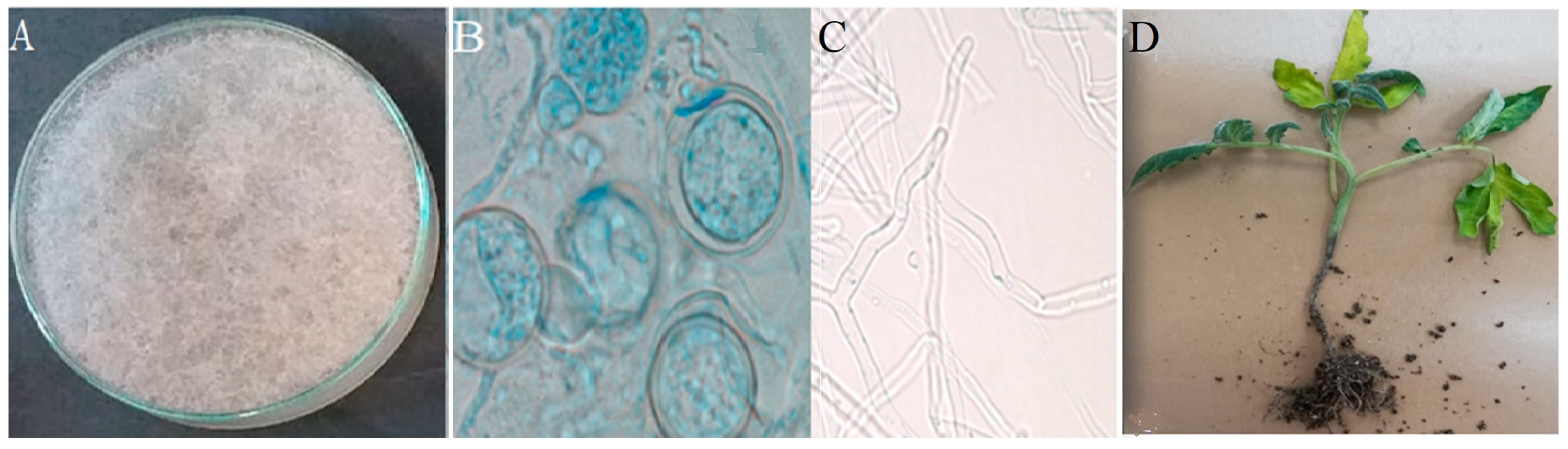
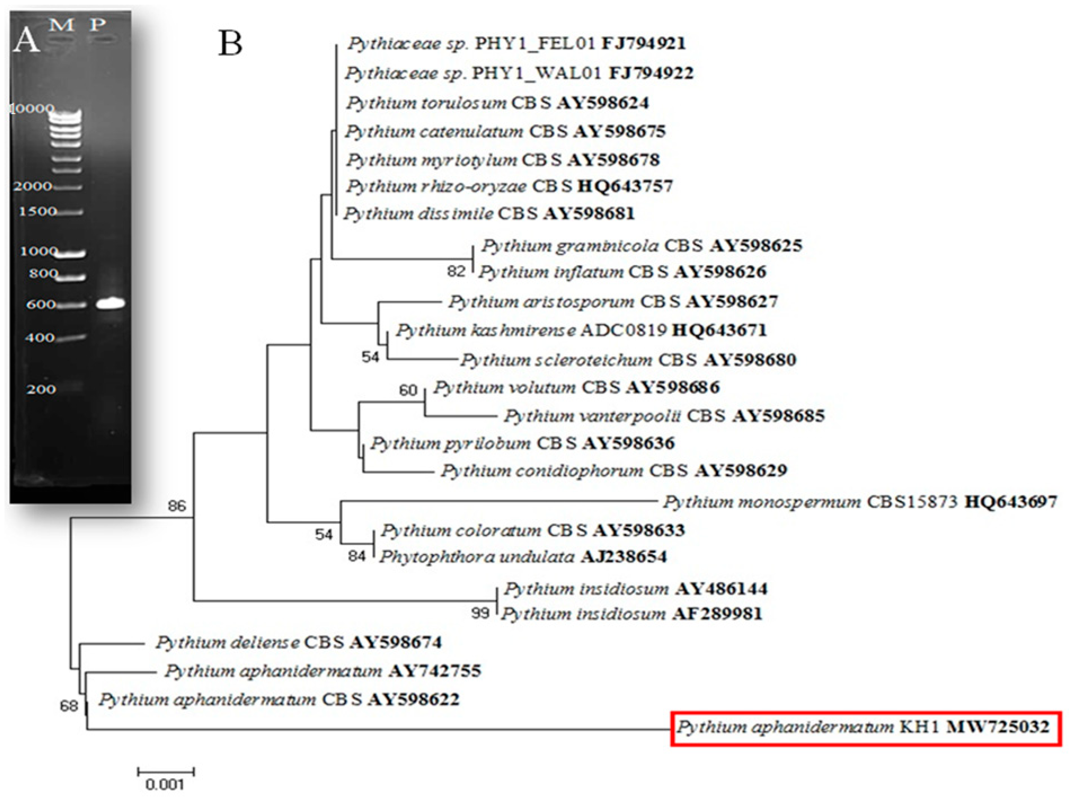
3.1. Isolation and Identification of the Fungus Isolate

In our study, tomato samples exhibited damping-off symptoms, which led us to isolate a pathogen from the infected tissues and purify it on PDA media (Figure 1A). Morphological analysis revealed that the fungus had branched, filamentous hyphae that were generally non-septate, lacking cross-walls, as well as spherically or pear-shaped oogonia that contain female gametes. Upon fertilization, the oogonia produce thick-walled oospores [20,21]. We identified the fungus using lactophenol cotton blue solution stain under a light microscope, and Figure 1B,C show the hyphae of Pythium spp., as well as the oogonia, oospores, and antheridia.
The internal transcribed spacer (ITS) region of fungal ribosomal DNA has gained popularity as a molecular marker for fungal identification, systematics, and phylogenetics. This is mainly due to its high sequence variability within and between fungal species, its conserved flanking regions, and the availability of a large number of reference sequences in public databases. Consequently, ITS has become known as a “fungal barcode” and is widely used for various fungal research applications [35]. The genomic DNA of the fungus isolate was subjected to PCR amplification using the ITS region; by comparing the PCR product and DNA marker, the size of the PCR product was 550–600 bp, as shown in Figure 2A.
The DNA nucleotide sequence of the fungus ITS region was produced at Macrogen Company (Seoul, Republic of Korea). After that, the nucleotide sequence was compared with the global recorded database in the NCBI, using the BLAST tool. The highest similarity of the fungus isolate determined it to be P. aphanidermatum. The fungus isolate nucleotide was then submitted to the GenBank database submission portal and assigned to the accession number (MW725032). The phylogenetic tree of the obtained fungus nucleotide (ITS) was constructed based on Pythium-fungal sequences obtained from the GenBank database, closely related to P. aphanidermatum. The constructed tree assigned all the Pythium species to two clusters. P. aphanidermatum isolates were divided into two sub-climbs. Sub-climb two was split into two groups; group two included P. aphanidermatum, similar to the investigated isolate with different percentages (Figure 2B). In the same manner, a study conducted by Matsumoto et al. [36] revealed that the ITS region was a suitable target for designing primers specific to Pythium species, which enabled their identification and detection. Analysis of the 5.8S rDNA sequence tree indicated that oomycetes, including Pythium, are evolutionarily distant from other fungi. The findings of Barboza et al. [37] also agree with our results of the phylogenetic tree, which grouped the same species in one clade of BLASTn ITS regions.

3.2. Greenhouse Trail and Pathogenicity Test

This study sheds some light on changes during inoculation between the tomato and the necrotrophic fungal pathogen Pythium aphanidermatum. The obtained results revealed that P. aphanidermatum was implicated as a pathogen in tomato plants (Figure 1D). P. aphanidermatum causes damping-off disease symptoms in the inoculated tomato plant 25 days post-inoculation. The symptoms that appeared include leaf drop and wilting, and this finding may be attributed to the cell wall-degrading enzymes produced by the fungus, which soften the plant cell wall [4]. Moreover, the results also showed chlorotic leaves and stunted tomato growth. This result may be accredited to the need of this fungus for carbohydrates, as it depends on them for growth, disturbing metabolism, and enzyme action. The infection begins at the root tip and can cause the infected region to lose its protective outer layer [4]. Our findings concur with those of Muthukumar et al. [38], who evaluated the pathogenicity of several Pythium isolates, including P. aphanidermatum, P. deliense, P. graminicola, P. heterothallicum, and P. ultimum in chili plants. The results indicated that P. aphanidermatum was the most virulent isolate, causing a significant level of post-emergence damping-off (65%). According to the findings of Garibaldi et al. [39], pathogenicity tests were carried out to evaluate the impact of P. aphanidermatum on Beta vulgaris subsp. vulgaris at two different temperatures: 27 °C and 20 °C. The results demonstrated that after a period of 20 days, a significantly higher percentage of plants exhibited damping-off symptoms at 27 °C (70%) in comparison to those seen at 20 °C (10%).

3.3. The Expression Levels of PR-Related Genes

Real-time PCR was used to detect the relative amounts of mRNA for five PR-related protein genes in tomato leaves infected with P. aphanidermatum. The results were normalized to the β-actin gene, as a reference or housekeeping gene. Among PR proteins, PR-1, PR-2, PR-3, PR-4, and PR-5 have been evaluated as the most potent antifungal proteins in plants. The over-enhancement of these genes in several crops leads to enhanced disease resistance against biotrophic and necrotrophic fungal phytopathogens [40].
In the present study, the expression of the PR-1 gene increased variably and reached its highest value at 20 dpi, with a reported relative expression level 6.34-fold higher than that of the control (Figure 3). Larsen et al. [41] reported similar findings, indicating that upon inoculation with P. aphanidermatum, tomato plants exhibited a high expression of the PR-1 gene. The pronounced rise in PR-1 gene expression relative to that of the uninoculated control was consistent with the findings of Moghaddam et al. [42], where up-regulation of the PR-1 gene, encoding pathogenesis-related protein 1, was strongly elevated in tomato plants as a result of Alternaria alternata infection, supporting the possible involvement of these PR genes in the regulation and response to early fungal infection. Early research suggested that PR-1 proteins have antimicrobial activity, with PR-1 derived from Nicotiana tabacum being able to inhibit the growth of Phytophthora infestans zoospores [43]; this role may be due to PR-1’s ability to sequester sterols from pathogen membranes, resulting in growth inhibition [44]. Accordingly, the increase in PR-1 expression is frequently used as an indicator for resistance responses, including pattern-associated triggered immunity (PTI) and systemic acquired resistance (SAR) [45].
Moreover, data also showed that PR-2 gene (β-1,3-glucanase) expression was modified slightly at 5, 15, and 20 dpi, while it decreased significantly at 10 dpi relative to the control (Figure 3). Glucanases, abundant proteins in plants, play chief roles in many physiological processes, such as cell division, transferring materials through plasmodesmata, flower formation through seed maturation, and amending abiotic stress effects [46]. They also defend plants against fungal pathogens, alone or in combination with many genes, such as thaumatin-like proteins, peroxidases, and a-1-puro thionin, where they develop strong resistance against fungal pathogens in crop plants [46]. The obtained results were in agreement with many previous reports that showed the induction of PR-2 upon plant fungal infections [47,48,49]. Thus, it is assumed that the up-regulation of tomato PR-2 in the current study could be related to the defense response activity of tomato plants against P. aphanidermatum infection.
Chitinases, also known as PR-3, are a group of proteins that protect plants against fungal diseases by cleaving the bonds between chitin molecules that are found in the fungal cell wall [50,51]. In the present study, compared to the uninfected control, a remarkable rise in PR-3, with a relative expression level of 5.76-fold at 20 dpi was reported (Figure 3). Many researchers found that A. alternata and A. solani infections caused PR-2 and PR-3 expression to increase, showing that they protect against early blight disease [52,53]. Moreover, Jongedijk et al. [54] and Wubben et al. [55] evaluated the way in which inoculating tomato plants with Fusarium oxysporum and Cladosporium fulvum made PR-2 and PR-3 work better together. In the same regard, the antifungal activity of chitinases can be synergistically enhanced by β-1,3-glucanases, both in vitro and in vivo, and these findings are also supported by many studies [56,57], in which the co-expression of chitinase and glucanase genes in tobacco enhanced resistance against Cercospora nicotianae. On the contrary, the obtained result demonstrated a decrease in the chitin binding protein (PR-4) gene, and this result was a mismatch with that of Hafez et al. [58], where PR-4 expression was increased in tomato plants in response to F. oxysporum and R. solani inoculation; therefore, the plant under Pythium inoculation may undergo a defense pathway other than that of the PR-4 gene.
Regarding PR-5 gene expression, the data showed a variable change in PR-5 (Thaumatin-like proteins, TLPs) gene expression at different sample collection periods. Still, it was highly expressed at 15 dpi and reached 3.99-fold, followed by 20 dpi, where the increasing percentage reached 3.70-fold, relative to the untreated control (Figure 3). The TLPs may have the ability to increase membrane permeability through two possible mechanisms. First, they may cause the destruction of β-1,3-glucans, which are important components of fungal cell walls. Second, they may inhibit fungal enzymes, such as xylanases. This can lead to increased membrane permeability and the subsequent death of the fungus [59]. This finding might support the role of TLPs in the resistance mechanism toward this pathogen, or it may be ascribed to the PR role, which further restricts fungal invasion and its reproduction [9]. Previous studies have indicated that TLPs are induced following fungal infection in plants, including in fruit species. However, there is a significant lack of understanding regarding the genotypic variability of TLPs and their downstream effects. Further research is needed to elucidate the specific roles of TLPs in response to fungal infection in different plant species, particularly in fruits [46,60,61,62]. Overall, the pathological effects of P. aphanidermatum on tomato plants evaluated by the expression of PR genes at different time intervals may require more insights into their role in disease resistance in the future.

3.4. HPLC Profiling of Tomato Leaves

Figure 4 displays the HPLC chromatograms of the ethanolic extracts from tomato leaves infected with P. aphanidermatum at 20 dpi and those which were uninfected (control). The HPLC analysis indicated that the total concentration of 19 polyphenolic compounds was 3858 µg/g and 3202.2 µg/g in the control and infected plant leaves, respectively, which means that the leaves showed a low amount of phenolic content after infection. According to Khatri et al. [63], rice leaves infected with Entyloma oryzae showed a reduction in the amount of phenols. Naik et al. [64] observed a quick decrease in phenolic compounds in betel vine leaves infected with Colletotrichum glocosporioides. Nema [65] believed that phenolic compounds could inhibit the growth of the pathogen, but in cases where the pathogen caused disease, the ratio of phenolic compounds changed. Highly susceptible cultivars were more likely to experience a depletion of phenolic compounds.
The main compounds detected are listed in Table 2. The most abundant phenolic compounds in terms of µg/g were chlorogenic acid, ellagic acid, pyrocatechol, and gallic acid in both the control and infected leaf extracts. However, vanillin, a flavonoid compound, was expressed at a lower level in infected plants (299.3 µg/g) than in control plants (421 µg/g) (Table 2). Contrarily, other investigators found that the most abundant flavonoids were vanillin, daidzein, naringenin, and kaempferol [66]. In contrast, Behiry et al. [67] reported that the antifungal activities of the extracted tomato plant leaves were linked to the functional groups and chemical composition of the phenolic compounds, such as caffeic and chlorogenic acids. The same findings were also noticed by several authors who found potent biological activities of identified plant phenolic substances towards various pathogenic microbes [68,69].
HPLC analysis indicated an increase in pyrocatechol compound content and a decrease in chlorogenic acid in infected plants compared to control plants. Sharifi-Rad et al. [70] noticed that tomato leaves contained more species of flavonoids, which have toxic activities against a broad range of pathogens and pests. Our research shows that when P. aphanidermatum is present in tomato plants, several phenolic and flavonoid compounds, such as chlorogenic, syringic, cinnamic, ferulic, ellagic, and coumaric acids, vanillin, catechin, rutin, methyl gallate, naringenin, daidzein, and quercetin, decrease or disappear at 20 dpi. These results suggest that the infection may reduce phenolic acid levels typically produced by infected plant cells. Interestingly, our results also reveal that certain flavonoid compounds (apigenin, kaempferol, and hesperetin) are up-regulated and accumulate in uninfected plants. These findings are supported by previous studies, which have shown that viral infections can promote the expression of flavonoid biosynthesis genes in various plants, including squash, potato, tomato, and grapevine [16,71,72,73]. These data suggest that flavonoids may affect the physiological responses to fungal infections. Our data somewhat agrees with that of Ortega-Garca et al. [74], who found leaf extracts containing phenolic compounds could protect onions plants from R. solani infection.

4. Conclusions

Fungi are a major cause of crop losses worldwide; thus, this study focused on the impact of P. aphanidermatum on tomato plants. The researchers identified the fungus and then examined the expression of five pathogenesis-related genes (PR-1, PR-2, PR-3, PR-4, and PR-5) using qPCR. They found that the expression of PR-1, PR-2, and PR-5 increased significantly 5 days after inoculation, with the most pronounced increase in PR-2 and PR-5 observed at 15 dpi. PR-1, PR-3, and PR-5 also showed an increase at 20 dpi, while PR-4 expression decreased significantly at all time points. In addition, the researchers used HPLC to analyze polyphenolic compounds in the tomato leaves and found that the fungus infection led to a decrease in certain phenolic acids, such as chlorogenic and ellagic acids. Overall, the findings suggest that P. aphanidermatum is a biotic stress pathogen that induces the expression of pathogen-related genes and disrupts the regulation of defensin phenolic compounds in tomato plants. Finally, this research can help in the creation of tools and metrics for measuring sustainability that can be used to judge the sustainability of agricultural systems. By determining the degree to which plant diseases affect agricultural productivity, human health, and the environment, researchers can come up with ways to measure the sustainability of farming practices and find ways to make them better.

Author Contributions

Conceptualization, S.A.S., A.A.A.-A., S.S., M.A.S., E.H., O.A.S., Y.S., S.I.B. and A.A.; methodology, S.A.S., A.A.A.-A., S.S., M.A.S., E.H., O.A.S., Y.S., S.I.B. and A.A.; software, A.A. and S.I.B.; validation, S.A.S., A.A.A.-A., S.S., M.A.S., E.H., O.A.S., Y.S., S.I.B. and A.A.; formal analysis, S.A.S., A.A.A.-A., S.S., M.A.S., E.H., O.A.S., Y.S., S.I.B. and A.A.; investigation, S.S., M.A.S. and E.H.; resources, S.A.S., A.A.A.-A., S.S., M.A.S., E.H., O.A.S., Y.S., S.I.B. and A.A.; writing—original draft preparation, S.A.S., A.A.A.-A., S.S., M.A.S., E.H., O.A.S., Y.S., S.I.B. and A.A.; writing—review and editing, S.A.S., A.A.A.-A., S.S., M.A.S., E.H., O.A.S., Y.S., S.I.B. and A.A.; supervision, A.A.A.-A. and A.A.; project administration, A.A. and A.A.A.-A.; funding acquisition, A.A.A.-A. All authors have read and agreed to the published version of the manuscript.

Funding

This research was financially supported by the Researchers Supporting Project (RSP2023R505), King Saud University, Riyadh, Saudi Arabia.

Institutional Review Board Statement

Not applicable.

Informed Consent Statement

Not applicable.

Data Availability Statement

The data that support the findings of this study are available on request from the corresponding author.

Acknowledgments

The authors express their sincere thanks to the City of Scientific Research and Technological Applications (SRTA-City) and the Faculty of Agriculture (Saba Basha), Alexandria University, Egypt, for providing the necessary research facilities. The authors would like to extend their appreciation to the Researchers Supporting Project (RSP2023R505), King Saud University, Riyadh, Saudi Arabia.

Conflicts of Interest

The authors declare no conflict of interest.

References

  1. Chellemi, D.O.; Gamliel, A.; Katan, J.; Subbarao, K.V. Development and deployment of systems-based approaches for the management of soilborne plant pathogens. Phytopathology 2016, 106, 216–225. [Google Scholar] [CrossRef] [Green Version]
  2. Buerstmayr, M.; Schweiger, W.; Steiner, B. Breeding for resistance to head blight caused by Fusarium spp. in wheat. CABI Rev. 2014, 9, 1–13. [Google Scholar] [CrossRef]
  3. Elshahawy, I.; Abouelnasr, H.M.; Lashin, S.M.; Darwesh, O.M. First report of Pythium aphanidermatum infecting tomato in Egypt and its control using biogenic silver nanoparticles. J. Plant Prot. Res. 2018, 58, 137–151. [Google Scholar] [CrossRef] [PubMed]
  4. Deacon, J. Cellulose Decomposition by Pythium and Its Relevance to Substrate-Groups of Fungi. Trans. Br. Mycol. Soc. 1979, 72, 469–477. [Google Scholar] [CrossRef]
  5. Kipngeno, P.; Losenge, T.; Maina, N.; Kahangi, E.; Juma, P. Efficacy of Bacillus subtilis and Trichoderma asperellum against Pythium aphanidermatum in tomatoes. Biol. Control 2015, 90, 92–95. [Google Scholar] [CrossRef]
  6. Gupta, S.; Nawaz, K.; Parween, S.; Roy, R.; Sahu, K.; Kumar Pole, A.; Khandal, H.; Srivastava, R.; Kumar Parida, S.; Chattopadhyay, D. Draft genome sequence of Cicer reticulatum L., the wild progenitor of chickpea provides a resource for agronomic trait improvement. DNA Res. 2017, 24, 1–10. [Google Scholar] [PubMed] [Green Version]
  7. Siam, G.; Abdelhakim, T. Analysis of the Tomato Value Chain in Egypt and Establishment of an Action Plan to Increase Its Efficiency. Ph.D. Thesis, CIHEAM-IAMM, Montpellier, France, 2018; p. 118. [Google Scholar]
  8. Hafez, E.E.; Abdelkhalek, A.A.; Abd El-Wahab, A.S.E.-D.; Galal, F.H. Altered gene expression: Induction/suppression in leek elicited by Iris Yellow Spot Virus infection (IYSV) Egyptian isolate. Biotechnol. Biotechnol. Equip. 2013, 27, 4061–4068. [Google Scholar] [CrossRef] [Green Version]
  9. Sels, J.; Mathys, J.; De Coninck, B.M.A.; Cammue, B.P.A.; De Bolle, M.F.C. Plant pathogenesis-related (PR) proteins: A focus on PR peptides. Plant Physiol. Biochem. 2008, 46, 941–950. [Google Scholar] [CrossRef]
  10. Behiry, S.I.; Ashmawy, N.A.; Abdelkhalek, A.A.; Younes, H.A.; Khaled, A.E.; Hafez, E.E. Compatible- and incompatible-type interactions related to defense genes in potato elucidation by Pectobacterium carotovorum. J. Plant Dis. Prot. 2018, 125, 197–204. [Google Scholar] [CrossRef]
  11. Ali, S.; Ganai, B.A.; Kamili, A.N.; Bhat, A.A.; Mir, Z.A.; Bhat, J.A.; Tyagi, A.; Islam, S.T.; Mushtaq, M.; Yadav, P. Pathogenesis-related proteins and peptides as promising tools for engineering plants with multiple stress tolerance. Microbiol. Res. 2018, 212, 29–37. [Google Scholar] [CrossRef]
  12. Barzegar, P.E.F.; Ranjbar, R.; Yazdanian, M.; Tahmasebi, E.; Alam, M.; Abbasi, K.; Tebyaniyan, H.; Barzegar, K.E.F. The current natural/chemical materials and innovative technologies in periodontal diseases therapy and regeneration: A narrative review. Mater. Today Commun. 2022, 32, 104099. [Google Scholar] [CrossRef]
  13. Abdelkhalek, A.; Al-Askar, A.A.; Hafez, E. Differential induction and suppression of the potato innate immune system in response to Alfalfa mosaic virus infection. Physiol. Mol. Plant Pathol. 2020, 110, 101485. [Google Scholar] [CrossRef]
  14. Abo-Zaid, G.; Abdelkhalek, A.; Matar, S.; Darwish, M.; Abdel-Gayed, M. Application of Bio-Friendly Formulations of Chitinase-Producing Streptomyces cellulosae Actino 48 for Controlling Peanut Soil-Borne Diseases Caused by Sclerotium rolfsii. J. Fungi 2021, 7, 167. [Google Scholar] [CrossRef] [PubMed]
  15. Akyol, H.; Riciputi, Y.; Capanoglu, E.; Caboni, M.; Verardo, V. Phenolic compounds in the potato and its byproducts: An overview. Int. J. Mol. Sci. 2016, 17, 835. [Google Scholar] [CrossRef]
  16. Abdelkhalek, A.; Király, L.; Al-Mansori, A.N.A.; Younes, H.A.; Zeid, A.; Elsharkawy, M.M.; Behiry, S.I. Defense Responses and Metabolic Changes Involving Phenylpropanoid Pathway and PR Genes in Squash (Cucurbita pepo L.) following Cucumber mosaic virus Infection. Plants 2022, 11, 1908. [Google Scholar] [CrossRef]
  17. Abdelkhalek, A.; Al-Askar, A.A.; Alsubaie, M.M.; Behiry, S.I. First Report of Protective Activity of Paronychia argentea Extract against Tobacco Mosaic Virus Infection. Plants 2021, 10, 2435. [Google Scholar] [CrossRef]
  18. Cheynier, V.; Comte, G.; Davies, K.M.; Lattanzio, V.; Martens, S. Plant phenolics: Recent advances on their biosynthesis, genetics, and ecophysiology. Plant Physiol. Biochem. 2013, 72, 1–20. [Google Scholar] [CrossRef]
  19. Gillings, M.R.; Tesoriero, L.A.; Gunn, L.A. Detection of double-stranded RNA and virus-like particles in Australian isolates of Pythium irregular. Plant Pathol. 1993, 1, 6–15. [Google Scholar] [CrossRef]
  20. Sneh, B.; Burpee, L.; Qgoshi, A. Identification of Rhizocto-Nia Species; APS Press: Saint Paul, MN, USA, 1991; p. 133. [Google Scholar]
  21. Van der Plaats-Niterink, A.J. Monogrpah of the Genus Pythium. In Studies Mycology; No. 21; Centra Albareau vor Schimmel Cultures: Baarh, The Netherlands, 1981. [Google Scholar]
  22. Leck, A. Lactophenol Cotton Blue Slide Mounts. Community Eye Health 1999, 12, 24. [Google Scholar]
  23. Tiwari, K.L.; Jadhav, S.K.; Gupta, S. Modified CTAB technique for isolation of DNA from some medicinal plants. Res. J. Med. Plant 2012, 6, 65–73. [Google Scholar] [CrossRef] [Green Version]
  24. Sambrook, J.; Fritsch, E.F.; Maniatis, T. Molecular Cloning: A Laboratory Manual; University of Texas South Western Medical Center: Dallas, TX, USA, 1989. [Google Scholar]
  25. Tamura, K.; Stecher, G.; Kumar, S. MEGA11: Molecular Evolutionary Genetics Analysis Version 11. Mol. Biol. Evol. 2021, 38, 3022–3027. [Google Scholar] [CrossRef]
  26. Chomczynski, P.; Sacchi, N. The single-step method of RNA isolation by acid guanidinium thiocyanate–phenol–chloroform extraction: Twenty-something years on. Nat. Protoc. 2006, 1, 581–585. [Google Scholar] [CrossRef] [PubMed]
  27. Livak, K.J.; Schmittgen, T.D. Analysis of Relative Gene Expression Data Using Real-Time Quantitative PCR and the 2−ΔΔCT Method. Methods 2001, 25, 402–408. [Google Scholar] [CrossRef] [PubMed]
  28. Ćetković, G.; Savatović, S.; Čanadanović-Brunet, J.; Djilas, S.; Vulić, J.; Mandić, A.; Četojević-Simin, D. Valorisation of phenolic composition, antioxidant and cell growth activities of tomato waste. Food Chem. 2012, 133, 938–945. [Google Scholar] [CrossRef]
  29. Gomez, K.A.; Gomez, A.A. Statistical Procedures for Agricultural Research; John Wiley & Sons: New York, NY, USA, 1984; ISBN 0471870927. [Google Scholar]
  30. McDonald, J.H. Handbook of Biological Statistics; Sparky House Publishing: Baltimore, MD, USA, 2009; Volume 2. [Google Scholar]
  31. Pasumarthy, K.K.; Mukherjee, S.K.; Choudhury, N.R. The presence of tomato leaf curl Kerala virus AC3 protein enhances viral DNA replication and modulates virus induced gene-silencing mechanism in tomato plants. Virol. J. 2011, 8, 178. [Google Scholar] [CrossRef] [Green Version]
  32. Wang, X.; El Hadrami, A.; Adam, L.R.; Daayf, F. Differential activation and suppression of potato defence responses by Phytophthora infestans isolates representing US-1 and US-8 genotypes. Plant Pathol. 2008, 57, 1026–1037. [Google Scholar] [CrossRef]
  33. Floryszak-Wieczorek, J.; Arasimowicz-Jelonek, M. Contrasting regulation of NO and ROS in potato defense-associated metabolism in response to pathogens of different lifestyles. PLoS ONE 2016, 11, e0163546. [Google Scholar] [CrossRef] [Green Version]
  34. Mukherjee, A.K.; Lev, S.; Gepstein, S.; Horwitz, B.A. A compatible interaction of Alternaria brassicicola with Arabidopsis thaliana ecotype DiG: Evidence for a specific transcriptional signature. BMC Plant Biol. 2009, 9, 31. [Google Scholar] [CrossRef] [Green Version]
  35. Badotti, F.; de Oliveira, F.S.; Garcia, C.F.; Vaz, A.B.M.; Fonseca, P.L.C.; Nahum, L.A.; Oliveira, G.; Góes-Neto, A. Effectiveness of ITS and sub-regions as DNA barcode markers for the identification of Basidiomycota (Fungi). BMC Microbiol. 2017, 17, 42. [Google Scholar] [CrossRef] [Green Version]
  36. Matsumoto, C.; Kageyama, K.; Suga, H.; Hyakumachi, M. Phylogenetic relationships of Pythium species based on ITS and 5.8 S sequences of the ribosomal DNA. Mycoscience 1999, 40, 321–331. [Google Scholar] [CrossRef]
  37. Barboza, E.A.; Cabral, C.S.; Rossato, M.; Martins, F.; Reis, A. Pythium and Phytopythium species associated with weeds collected in vegetable production fields in Brazil. Lett. Appl. Microbiol. 2022, 74, 796–808. [Google Scholar] [CrossRef] [PubMed]
  38. Muthukumar, A.; Eswaran, A.; Sangeetha, G. Occurrence, virulence and pathogenicity of species of Pythium inciting damping-off disease in chilli. J. Mycol. Plant Pathol. 2010, 40, 67. [Google Scholar]
  39. Garibaldi, A.; Gilardi, G.; Ortu, G.; Gullino, M.L. First report of damping-off caused by Pythium aphanidermatum on leaf beet (Beta vulgaris subsp. vulgaris) in Italy. Plant Dis. 2013, 97, 292. [Google Scholar] [CrossRef]
  40. Dai, L.; Wang, D.; Xie, X.; Zhang, C.; Wang, X.; Xu, Y.; Wang, Y.; Zhang, J. The novel gene VpPR4-1 from Vitis pseudoreticulata increases powdery mildew resistance in transgenic Vitis vinifera L. Front. Plant Sci. 2016, 7, 695. [Google Scholar] [CrossRef] [PubMed] [Green Version]
  41. Larsen, J.; Graham, J.H.; Cubero, J.; Ravnskov, S. Biocontrol traits of plant growth suppressive arbuscular mycorrhizal fungi against root rot in tomato caused by Pythium aphanidermatum. Eur. J. Plant Pathol. 2012, 133, 361–369. [Google Scholar] [CrossRef]
  42. Moghaddam, G.A.; Rezayatmanda, Z.; Esfahani, M.N.; Khozaei, M. Genetic defense analysis of tomatoes in response to early blight disease, Alternaria alternata. Plant Physiol. Biochem. 2019, 142, 500–509. [Google Scholar] [CrossRef] [PubMed]
  43. Niderman, T.; Genetet, I.; Bruyere, T.; Gees, R.; Stintzi, A.; Legrand, M.; Fritig, B.; Mosinger, E. Pathogenesis-related PR-1 proteins are antifungal (isolation and characterization of three 14-kilodalton proteins of tomato and of a basic PR-1 of tobacco with inhibitory activity against Phytophthora infestans). Plant Physiol. 1995, 108, 17–27. [Google Scholar] [CrossRef] [Green Version]
  44. Gamir, J.; Darwiche, R.; Van’t Hof, P.; Choudhary, V.; Stumpe, M.; Schneiter, R.; Mauch, F. The sterol-binding activity of pathogenesis-related protein 1 reveals the mode of action of an antimicrobial protein. Plant J. 2017, 89, 502–509. [Google Scholar] [CrossRef] [Green Version]
  45. van Loon, L.C.; Rep, M.; Pieterse, C.M.J. Significance of inducible defense-related proteins in infected plants. Annu. Rev. Phytopathol. 2006, 44, 135–162. [Google Scholar] [CrossRef] [Green Version]
  46. Balasubramanian, V.; Vashisht, D.; Cletus, J.; Sakthivel, N. Plant β-1,3-glucanases: Their biological functions and transgenic expression against phytopathogenic fungi. Biotechnol. Lett. 2012, 34, 1983–1990. [Google Scholar] [CrossRef]
  47. Al Daoude, A.; Shoaib, A.; Al-Shehadah, E.; Jawhar, M.; Arabi MI, E. Pathogenesis—Related genes responses in barley plants challenged with pathogenic fungi with different lifestyles. Cereal Res. Commun. 2020, 48, 341–346. [Google Scholar] [CrossRef]
  48. Anisimova, O.K.; Shchennikova, A.V.; Kochieva, E.Z.; Filyushin, M.A. Pathogenesis-related genes of PR1, PR2, PR4, and PR5 families are involved in the response to Fusarium infection in garlic (Allium sativum L.). Int. J. Mol. Sci. 2021, 22, 6688. [Google Scholar] [CrossRef] [PubMed]
  49. Kim, Y.J.; Hwang, B.K. Isolation of a basic 34 kiloDalton β-1, 3-glucanase with inhibitory activity against Phytophthora capsicifrom pepper stems. Physiol. Mol. Plant Pathol. 1997, 50, 103–115. [Google Scholar] [CrossRef]
  50. Sierra-Gomez, Y.; Annia Rodrıguez-Hernandez, P.C.-S.; Homero Gomez-Velasco, A.H.-S.; Siliqi, D.; Rodrıguez-Romero, A. A biophysical and structural study of two chitinases from Agave tequilana and their potential role as defense proteins. FEBS J. 2019, 286, 4778–4796. [Google Scholar] [CrossRef]
  51. Hamid, R.; Khan, M.A.; Ahmad, M.; Ahmad, M.M.; Abdin, M.Z.; Musarrat, J.; Javed, S. Chitinases: An update. J. Pharm. Bioallied Sci. 2013, 5, 21. [Google Scholar]
  52. Singh, R.; Tiwari, J.K.; Sharma, V.; Rawat, S. Role of Pathogen related protein families in defence mechanism with potential role in applied biotechnology. Int. J. Adv. Res. 2014, 2, 210–226. [Google Scholar]
  53. El-Hadary, M.H.; Tayel, A.A. Differential expression of tomato chitinases upon Alternaria solani inoculation with special reference to a modified purification zymogram. Egypt. J. Exp. Biol. (Bot.) 2013, 9, 9–17. [Google Scholar]
  54. Jongedijk, E.; Tigelaar, H.; van Roekel, J.S.C.; Bres-Vloemans, S.A.; Dekker, I.; van den Elzen, P.J.M.; Cornelissen, B.J.C.; Melchers, L.S. Synergistic activity of chitinases and β-1,3-glucanases enhances fungal resistance in transgenic tomato plants. Euphytica Int. J. Plant Breed. 1995, 85, 173–180. [Google Scholar] [CrossRef] [Green Version]
  55. Wubben, J.P.; Lawrence, C.B.; De Wit, P.J.G.M. Differential induction of chitinase and 1,3-β-glucanase gene expression in tomato by Cladosporium fulvum and its race-specific elicitors. Physiol. Mol. Plant Pathol. 1996, 48, 105–116. [Google Scholar] [CrossRef]
  56. Zhu, Q.; Maher, E.A.; Masoud, S.; Dixon, R.A.; Lamb, C.J. Enhanced protection against fungal attack by constitutive co--expression of chitinase and glucanase genes in transgenic tobacco. Bio/Technology 1994, 12, 807–812. [Google Scholar] [CrossRef]
  57. Jach, G.; Görnhardt, B.; Mundy, J.; Logemann, J.; Pinsdorf, E.; Leah, R.; Schell, J.; Maas, C. Enhanced quantitative resistance against fungal disease by combinatorial expression of different barley antifungal proteins in transgenic tobacco. Plant J. 1995, 8, 97–109. [Google Scholar] [CrossRef] [PubMed]
  58. Hafez, E.; Hashem, M.; Balbaa, M.M.; Ma, E.; Ahmed, S.A. Induction of New Defensin Genes in Tomato Plants via Pathogens-Biocontrol Plant Pathology & Microbiology Induction of New Defensin Genes in Tomato Plants via Pathogens-Biocontrol Agent Interaction. Plant Pathol. Microbiol. 2013, 4, 167. [Google Scholar] [CrossRef] [Green Version]
  59. de Jesús-Pires, C.; Ferreira-Neto, J.R.C.; Pacifico Bezerra-Neto, J.; Kido, E.A.; de Oliveira Silva, R.L.; Pandolfi, V.; Wanderley-Nogueira, A.C.; Binneck, E.; da Costa, A.F.; Pio-Ribeiro, G. Plant thaumatin-like proteins: Function, evolution and biotechnological applications. Curr. Protein Pept. Sci. 2020, 21, 36–51. [Google Scholar] [CrossRef] [PubMed]
  60. Koiwa, H.; Kato, H.; Nakatsu, T.; Oda, J.; Yamada, Y.; Sato, F. Purification and characterization of tobacco pathogenesis-related protein PR-5d, an antifungal thaumatin-like protein. Plant Cell Physiol. 1997, 38, 783–791. [Google Scholar] [CrossRef] [PubMed] [Green Version]
  61. Wang, H.; Ng, T.B. Isolation of an antifungal thaumatin-like protein from kiwi fruits. Phytochemistry 2002, 61, 1–6. [Google Scholar] [CrossRef] [PubMed]
  62. Krebitz, M.; Wagner, B.; Ferreira, F.; Peterbauer, C.; Campillo, N.; Witty, M.; Kolarich, D.; Steinkellner, H.; Scheiner, O.; Breiteneder, H. Plant-based heterologous expression of Mal d 2, a thaumatin-like protein and allergen of apple (Malus domestica), and its characterization as an antifungal protein. J. Mol. Biol. 2003, 329, 721–730. [Google Scholar] [CrossRef]
  63. Khatri, R.K.; Shastry, R.P.; Reddy, P.N.; Nema, K.G. Metabolic changes in rice leaves infected by Entyloma oryzae. Indian Phytopathol. 1985, 38, 769–771. [Google Scholar]
  64. Naik, M.K.; Hiremath, P.C.; Hiremath, S.V. Post Infectional Changes in the Betelvine Leaves Infected with Colletotrichum gloeosporioides. Indian Phytopathol. 1988, 41, 370–372. [Google Scholar]
  65. Nema, A.G. Sugar and phenol contents of betelvine leaves after inoculation with leaf spot bacterium. Indian Phytopathol. 1989, 42, 31–37. [Google Scholar]
  66. Ejike, C.E.C.C.; Gong, M.; Udenigwe, C.C. Phytoalexins from the Poaceae: Biosynthesis, function and prospects in food preservation. Food Res. Int. 2013, 52, 167–177. [Google Scholar] [CrossRef]
  67. Behiry, S.; Soliman, S.A.; Massoud, M.A.; Abdelbary, M.; Kordy, A.M.; Abdelkhalek, A.; Heflish, A. Trichoderma pubescens Elicit Induced Systemic Resistance in Tomato Challenged by Rhizoctonia solani. J. Fungi 2023, 9, 167. [Google Scholar] [CrossRef] [PubMed]
  68. Kim, D.S.; Na, H.; Kwack, Y.; Chun, C. Secondary metabolite profiling in various parts of tomato plants. Hortic. Sci. Technol. 2014, 32, 252–260. [Google Scholar] [CrossRef] [Green Version]
  69. Kim, D.S.; Kwack, Y.; Lee, J.H.; Chun, C. Antimicrobial activity of various parts of tomato plants varied with different solvent extracts. plant Pathol. J. 2019, 35, 149. [Google Scholar] [CrossRef] [PubMed]
  70. Sharifi-Rad, M.; Varoni, E.M.; Iriti, M.; Martorell, M.; Setzer, W.N.; del Mar Contreras, M.; Salehi, B.; Soltani-Nejad, A.; Rajabi, S.; Tajbakhsh, M. Carvacrol and human health: A comprehensive review. Phyther. Res. 2018, 32, 1675–1687. [Google Scholar] [CrossRef] [PubMed]
  71. Abdelkhalek, A.; Al-Askar, A.A.; Behiry, S.I. Bacillus licheniformis strain POT1 mediated polyphenol biosynthetic pathways genes activation and systemic resistance in potato plants against Alfalfa mosaic virus. Sci. Rep. 2020, 10, 1–16. [Google Scholar] [CrossRef] [PubMed]
  72. Abdelkhalek, A.; Behiry, S.I.; Al-Askar, A.A. Bacillus velezensis PEA1 Inhibits Fusarium oxysporum Growth and Induces Systemic Resistance to Cucumber Mosaic Virus. Agronomy 2020, 10, 1312. [Google Scholar] [CrossRef]
  73. Gutha, L.R.; Casassa, L.F.; Harbertson, J.F.; Naidu, R.A. Modulation of flavonoid biosynthetic pathway genes and anthocyanins due to virus infection in grapevine (Vitis vinifera L.) leaves. BMC Plant Biol. 2010, 10, 187. [Google Scholar] [CrossRef] [Green Version]
  74. Ortega-García, J.G.; Montes-Belmont, R.; Rodríguez-Monroy, M.; Ramírez-Trujillo, J.A.; Suárez-Rodríguez, R.; Sepúlveda-Jiménez, G. Effect of Trichoderma asperellum applications and mineral fertilization on growth promotion and the content of phenolic compounds and flavonoids in onions. Sci. Hortic. 2015, 195, 8–16. [Google Scholar] [CrossRef]
Figure 1. (A) Morphological mycelial growth of Pythium aphanidermatum grown on a PDA plate; (B) light microscope images showing oospores, oogonia, and antheridia; (C) P. aphanidermatum hyphae; (D) an infected tomato plant.
Sustainability 15 06551 g001
Figure 2. (A) PCR product of the ITS region of the fungus [M, 10 kb DNA marker, Lane 1 (P), Pythium aphanidermatum KH1 (MW725032)]; (B) phylogenetic tree of the ITS region of the P. aphanidermatum nucleotide sequence compared with other GenBank sequences.
Sustainability 15 06551 g002
Figure 3. The relative expression level of pathogenesis-related genes (PR-1, PR-2, PR-3, PR-4, and PR-5) of tomato plants infected with Pythium aphanidermatum, collected at different time intervals (5, 10, 15, and 20 days) post inoculation (dpi). Different letters reveal statistically significant variations (p ≤ 0.05).
Sustainability 15 06551 g003
Figure 4. (A) HPLC profiles of the major polyphenolic compounds in healthy tomato leaves and (B) infected tomato plant leaves at 20 dpi.
Sustainability 15 06551 g004
Table 2. The polyphenolic compounds detected in the control and infected tomato plant leaves.
Polyphenolic Compounds Concentration (µg/g)
ControlInfected
Gallic acid192.4195.6
Chlorogenic acid1110.4617.2
Catechin72.430.7
Methyl gallate45.835.8
Caffeic acid182.1219.2
Syringic acid62.845.0
Pyro catechol476.7603.3
Rutin30.60.0
Ellagic acid609.8578.9
Coumaric acid28.613.7
Vanillin421.0299.3
Ferulic acid54.523.3
Naringenin57.954.3
Daidzein403.5177.6
Querectin102.367.9
Cinnamic acid7.20.0
Apigenin0.086.8
Kaempferol0.0134.1
Hesperetin0.019.5
Disclaimer/Publisher’s Note: The statements, opinions and data contained in all publications are solely those of the individual author(s) and contributor(s) and not of MDPI and/or the editor(s). MDPI and/or the editor(s) disclaim responsibility for any injury to people or property resulting from any ideas, methods, instructions or products referred to in the content.

