Article

The Stimulation of Superoxide Dismutase Enzyme Activity and Its Relation with the Pyrenophora teres f. teres Infection in Different Barley Genotypes

1
Agricultural Institute, Centre for Agricultural Research, 2462 Martonvásár, Hungary
2
Plant Protection Institute, Centre for Agricultural Research, 1022 Budapest, Hungary
3
Institute of Agronomy, Hungarian University of Agriculture and Life Sciences, 2100 Gödöllő, Hungary
*
Author to whom correspondence should be addressed.
Sustainability 2022, 14(5), 2597; https://doi.org/10.3390/su14052597
Submission received: 30 December 2021 / Revised: 18 February 2022 / Accepted: 21 February 2022 / Published: 23 February 2022
(This article belongs to the Special Issue Plant Breeding Supporting the Sustainable Field Crop Production)

Abstract

:
Changes in superoxide dismutase (SOD) enzyme activity were examined in infected barley seedlings of five cultivars with the goal to study the role of SOD in the defense mechanism induced by Pyrenophora teres f. teres (PTT) infection. Our results showed that although there were differences in the responses of the cultivars, all three PTT isolates (H-618, H-774, H-949) had significantly increased SOD activity in all examined barley varieties at the early stages of the infection. The lowest SOD activity was observed in the case of the most resistant cultivar. Our results did not show a clear connection between seedling resistance of genotypes and SOD enzyme activity; however, we were able to find strong significant correlations between the PTT infection scores on the Tekauz scale and the SOD activity. The measurement of the SOD activity could offer a novel perspective to detect the early stress responses induced by PTT. Our results suggest that the resistance of varieties cannot be estimated based on SOD enzyme activity alone, because many antioxidant enzymes play a role in fine-tuning the defense response, but SOD is an important member of this system.

1. Introduction

Food security highly depends on successful plant breeding activity and the production of adaptive, disease-resistant crops. Cultivation of tolerant or resistant varieties is one of the most effective and eco-friendly methods of controlling plant diseases [1]; therefore, the resistance of breeding lines against pathogens is among the main selection criteria in plant breeding. Controlling foliar diseases is essential in barley (Hordeum vulgare L.) production, especially in the case of the damaging fungal pathogen Pyrenophora teres f. teres Drechsler (anamorph Drechslera teres (Sacc.) Shoem.) (PTT) [2]. This ascomycete fungus causes the typical net-like leaf symptoms of the net blotch disease and necrotic lesions with chlorotic borders on sensitive barley genotypes [3]. PTT infection could cause significant damage both in spring and winter barley genotypes with an average of 20–30% grain yield loss, especially in rainy weather [4]. In case of very sensitive barley genotypes, the damage can be up to 100% [5,6].
Pathogen infection-induced oxidative stress has been observed in several cases such as the powdery mildew on cereals [7] and the PTT and Rhynchosporium secalis on barley [8]; Lehmann [9] also summarizes several other relevant results. During oxidative stress, reactive oxygen species (ROS) appear in plant cells and have important roles in controlling signaling, metabolic and developmental processes [10,11,12,13,14]. ROS are able to act as signaling molecules at low concentrations, but they can cause oxidative stress at high concentrations [8,15,16,17,18,19]. The balance between ROS generation and its scavenging has a key role to combat the effects of pathogen attack in plants [20]. Production of O2• is considered as the first response in a cell because it is involved in the generation of other ROS [21]. High concentrations of ROS activate ROS-dependent programmed cell death (PCD) pathways [22,23]. Avoiding cell damage and expanding the range of ROS scavenging mechanisms is necessary [24,25]. Superoxide dismutase (SOD) is one of the several defense-related antioxidant enzymes involved in the elimination of ROS [26]. The lifetime of superoxide anion (O2•) depends on the enzymatic activity of CuZn superoxide dismutase (SOD) [27]. SOD enzymes are responsible for the conversion of the superoxide anion (O2−) to molecular oxygen and hydrogen peroxide (H2O2) [28]. Among the reactive oxygen species, H2O2 is the only one able to cross the plant membrane, therefore being essential in cell signaling [10,25,29]. Enhanced H2O2 production was observed as a response to pathogen attack [30]. The pathogens trigger the ROS production mainly in apoplasts [29]. Several papers have described the effects of biotic stress on the antioxidant system in cereals [31,32,33]. SOD is the first barrier of oxidative damage [34] and plays a significant role in the control of ROS resulting from both abiotic and biotic environmental stresses [35,36]. The role of SOD was also proven under abiotic stress conditions of cereal, such as higher or lower light intensity, drought or waterlogging [37], salinity [38], heavy metal toxicity, mineral nutrient deficiencies [26] and other environmental stresses [26,39,40,41]. There are two distinct peaks of ROS production during an oxidative burst caused by multiple abiotic stresses. The initial burst of ROS triggers the cell-to-cell communication and activates the specific signals. The second is the ROS burst at target [13]. Biphasic ROS accumulation with a low-amplitude, transient first phase is followed by a sustained phase of much higher magnitude that correlates with disease resistance [42,43]. Reactive oxygen species (ROS) can reduce the symptoms and block pathogen growth in plants [44]. The influence of ROS was reversed by the SOD, which indicated that O2− and H2O2 were the most relevant reactive oxygen species during the pathogen infection [45]. Early elevations of ROS levels and increased SOD activity were observed after the appearance of net blotch symptoms in stressed barley cultivars [12,46]. In the study of Able [8], three barley cultivars were tested with virulent and avirulent PTT isolates, and an increased level of SOD activity was observed in the resistance response of barley varieties. The timely recognition of an invading microorganism coupled with the rapid and effective induction of defense responses appears to make a key difference between resistance and susceptibility [47]. The resistance of barley varieties against PTT depends on the pathotype of fungus. However, the effects of various pathotypes of PTT infection on SOD activity in the case of different barley genotypes with different resistance against PTT have not yet been investigated widely.
In this study, the aim was to investigate the relationship between SOD enzyme activity stimulated by PTT infection and the susceptibility of barley cultivars. According to these aims, we examined the change in super-oxide dismutase enzyme activity caused by the Pyrenophora teres f. teres infection in the first 72 h after infection and the subsequent two weeks on barley varieties with different PTT resistance. Five barley cultivars were inoculated in the seedling stage with three different monosporic PTT isolates from Hungary to study the defense response triggered by different PTT pathotypes in each variety. Changes in SOD enzyme activity and the severity of PTT infection were determined in inoculated barley seedlings to study the role of SOD in the defense mechanism induced by PTT pathogenesis.

2. Materials and Methods

Our experiments were performed between 2018 and 2019 in the laboratory and the greenhouse of the Agricultural Institute of the Centre for Agricultural Research, under controlled conditions.

2.1. Preparation of Inoculum

Three PTT isolates (H-618, H-774, H-949), originating from Hungary, were used in the experiment. Previously infected leaves were placed into moist chambers (water-logged filter papers in glass Petri dishes), which were incubated under white light (OSRAM model L36W/640) in a 16 h light/8 h dark cycle for 24 h at 20–22 °C to induce conidiogenesis. Monoconidial isolates were made aseptically in a laminar air flow device by transferring conidia from the leaves to V8 juice agar (V8A; 16 g agar, 3 g CaCO3, 100 mL Campbell’s V8 juice, 900 mL distilled water) with a sterile needle, using a Leica MZ6 stereomicroscope at 40× magnification. Plates were incubated for 10 days in the dark at 17–19 °C. To produce inoculum for the artificial inoculation experiments, isolates were grown on V8A and/or autoclaved maize leaves in 90 mm diameter plastic Petri plates under white light (OSRAM model L36W/640) in a 16 h light/8 h dark cycle at 17–19 °C for 10 days. Then, sterile distilled water containing 0.01% Tween 20 was added to the sporulating cultures (10 mL solution per plate), and conidia were removed from the conidiophores by gently agitating the mycelium mat with a sterile paint brush. Finally, the suspension was filtered through a fine sieve with 100 µm diameter pore size and the concentration of conidia was adjusted to 10.000/mL.

2.2. Plant Material

Five barley varieties were selected based on their different susceptibility to PTT, which we determined in our previous greenhouse experiments and field trials [48]. The following genotypes were selected: cv. ”Canela”, cv. “Harrington”, cv. “Manas”, cv. “Mv-Initium” and cv. “Antonella”.

2.3. Setup of Treatments

Plants were grown up until the two-leaf stage at 22 °C on average in a greenhouse under a 12 h photoperiod in five replications. Plants were inoculated later with the conidial suspension of the selected PTT isolates by spraying with a hand sprayer on the surface of the leaves with a concentration of 10.000 conidia/mL until runoff. After inoculation, the plants were kept in a greenhouse chamber under a transparent plastic tent for 48 h at 22 °C and 100% humidity, which provided suitable environmental conditions and appropriate humidity for the development of the fungus.
The leaf samples were collected 0 h (as the control), 24 h, 48 h and 72 h after infection. The sampling dates were chosen based on Able’s previous study [8]. In the case of treatment with H-949, infected barley seedlings were sampled on 7 and 15 DAI (days after inoculation); the longer incubation time was based on the work of Pál et al. [49] to investigate the SOD activity at a later stage in the infection. In all cases, 0.2 g of the middle part of second leaf tissue was collected in five repetitions from different pots. After that, the samples were snap-frozen in liquid nitrogen and kept at −70 °C until processing and measurement.
The evaluation of the PTT responses was based on the infection recorded on the second leaves using the 10-point scale of Tekauz [50] for single plants evaluated from each pot in three replicates until the fifteenth day.

2.4. Measurement of SOD Activity

The xanthine oxidase (EC 1.1.3.22) assay, the most frequently used method [8,28], was performed for the analysis of SOD activity according to Sigma-Aldrich’s manufacturer instructions, based on the work of Bergmeyer et al. [51]. Measurements were performed with a Shimadzu UV-160A UV-VIS spectrophotometer (Shimadzu, Japan) at 550 nm with 1 cm glass cuvettes and the absorbance followed for 1 min in 10 s intervals. During the data processing, the measured changes in absorbance values were converted to enzyme activity according to the work of Zhang et al. [52].

2.5. Data Analysis

One-way ANOVA was conducted to compare the effect of infection with different PTT isolates on the different barley genotypes and the length of the contagion on the SOD activity in the tissue samples. The Tekauz scoring points were statistically evaluated. ANOVA was performed at the p = 0.05 level of significance. Post-hoc comparisons using the least significant difference (LSD) test were made at p < 0.05. For the statistical evaluation of the results, we used the Explore and ANOVA modules of the IBM SPSS V.23 software.

3. Results

3.1. Results of the SOD Activity Measurements

3.1.1. Results of the Treatment with Isolate H-618

In the case of infection with H-618, significant differences were found between the genotypes and the SOD activity in the leaf samples at 0 h [F(4, 10) = 4189, p = 0.03], 24 h [F(4, 10) = 4.681, p = 0.22], 48 h [F(4, 10) = 12.191, p = 0.001] and 72 h [F(4, 10) = 22.610, p = 0.000] (Figure 1). We recorded slight differences between the cultivars in the first 24 h, and SOD activity increased significantly in all genotypes in the 48 h after inoculation; the largest jump in SOD activity was shown by cv. “Canela” with 678.25 ΔA/min, followed by cv. “Mv-Initium” with 340.60 ΔA/min. At 72 h, the highest SOD activity (528.02 ΔA/min) was measured in cv. “Harrington”, followed by cv. “Canela” (490.84 ΔA/min). On the other hand, the SOD activity of cv. “Manas”, cv. “Canela” and cv. “Mv-Initium” genotypes decreased in 72 h of the measurement with 112 ΔA/min, 188 ΔA/min and 70 ΔA/min, respectively, while cv. “Antonella” and cv. “Harrington” had further increases of 128 ΔA/min and 160 ΔA/min in the last 24 h, respectively. It is clear from the data that inoculation with H-618 influenced the SOD activity of all cultivars and that cv. “Harrington” and cv. “Canela” reacted most sensitively to the isolate. The SOD activity of cv. “Harrington” ranged between 122.98 and 528.02 ΔA/min, while cv. “Canela” had minimum activity of 101.21 ΔA/min and maximum of 782.26 ΔA/min.
We also performed the LSD post-hoc test on all the data to explore differences in SOD activity between barley varieties inoculated with isolate H-618 (Table 1). The post-hoc comparison showed at 0 h that the average SOD activity of cv. “Manas” (156.89 ± 8.80) was significantly lower than cv. “Harrington” (222.60 ± 38.34) and cv. “Mv-Initium” (207.71 ± 22.89). The cv. “Harrington” (222.60 ± 38.34) showed a difference only from cv. “Antonella” (149.25 ± 15.96), which had also lower SOD activity than cv. “Mv-Initium” (207.71 ± 22.89). The genotype cv. “Canela” (185.40 ± 35.35) did not show a significant difference in SOD activity in opposition to the other examined genotypes at the beginning of the experiment at 0 h.
Measurements after 24 h showed that the average SOD activity of cv. “Manas” (184.95 ± 4.00) was significantly higher only than cv. “Harrington” (117.43 ± 18.49). In comparison, the cv. “Harrington” (117.43 ± 18.49) was significantly different from cv. “Canela” (180.14 ± 51.28), cv. “Mv-Initium” (206.95 ± 23.22) and cv. “Antonella” (176.59 ± 5.92) because of its lower SOD activity. No statistically significant change in SOD activity was found in other combinations of the genotypes after the first 24 h.
After 48 h of inoculation, only cv. “Canela” (678.25 ± 142.17) showed significantly higher differences from the other examined genotypes, the cv. “Manas” (250.25 ± 127.78), the cv. “Harrington” (250.72 ± 85.86), the cv. “Mv-Initium” (340.60 ± 46.06) and cv. “Antonella” (170.22 ± 54.28). No statistically significant change in SOD activity was found in other combinations of the genotypes after the first 48 h.
The cv. “Manas” (194.98 ± 10.66) after 72 h showed a significantly lower difference in opposition to cv. “Harrington” (528.02 ± 107.73), cv. “Canela” (490.84 ± 30.52) and cv. “Antonella” (298.52 ± 38.03), but there was no difference between cv. “Manas” and cv. “Mv-Initium” (260.59 ± 13.42). However, the cv. “Mv-Initium” showed a significant difference against cv. “Harrington” (528.02 ± 107.73) and cv. “Canela” (490.84 ± 30.52) because they showed higher SOD activity. The cv. “Antonella” had a different SOD activity than all of the other examined genotypes except the cv. “Mv-Initium”. No statistically significant change in SOD activity was found in other combinations of the genotypes after 72 h.

3.1.2. Results of the Treatment with Isolate H-774

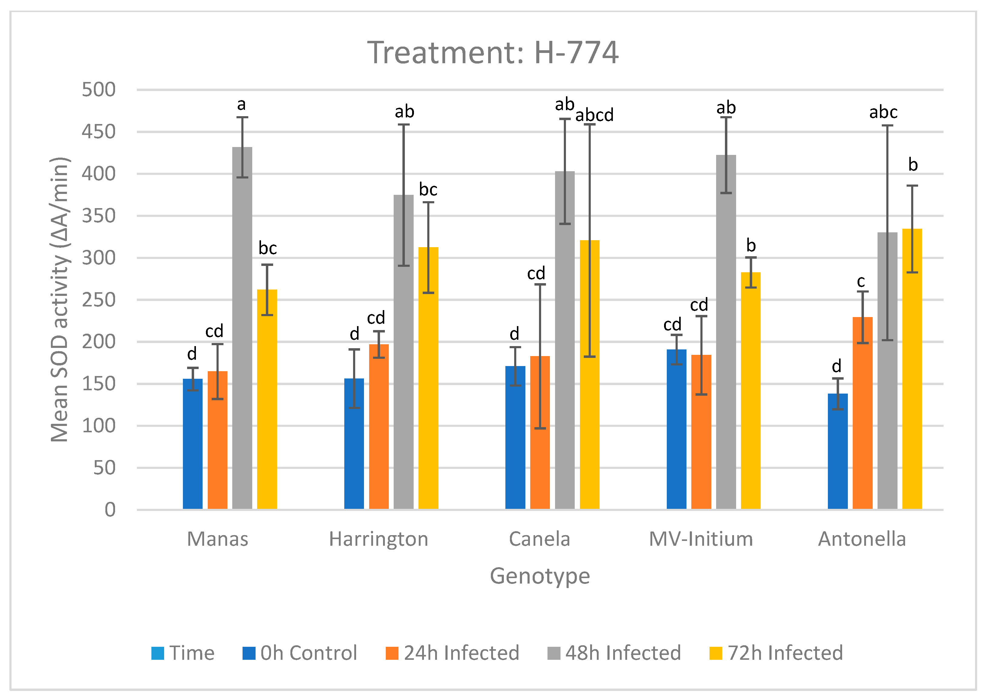
The H-774 treatment induced no significant differences between SOD activity of genotypes at 0 h [F(4, 10) = 3.026, p = 0.71], 24 h [F(4, 10) = 0.982, p = 0.46], 48 h [F(4, 10) = 1.104, p = 0.406] and 72 h [F(4, 10) = 0.673, p = 0.626]; however, it is important to note that after 48 h, SOD activity increased for all genotypes, just like in the case of isolate H-618, but it decreased in the last 24 h (Figure 2).
For isolate H-774, the largest jump in SOD activity was measured in the 48 h after inoculation in cv. “Manas” (+267 ΔA/min), followed by cv. “Mv-Initium” (+238 ΔA/min) and cv. “Canela” (+221 ΔA/min), although the change was also significant for the varieties cv. “Harrington” (+178 ΔA/min) and cv. “Antonella” (+100 ΔA/min). The 72 h measurement showed a decrease in the SOD activity in all genotypes except cv. “Antonella”, where a slight increase was observed. The largest decreases in activity of 170 ΔA/min and 140 ΔA/min occurred in the cases of cv. “Manas” and cv. “Mv-Initium”, respectively.

3.1.3. Results of the Treatment with Isolate H-949

To study the changes in SOD activity in the longer term in different barley genotypes, we measured the absorbance at 0 h and on the 7th and 15th days after infection with the H-949 isolate. Although there was no significant difference between the genotypes at the beginning of the experiment at 0 h [F (4, 20) = 1.714, p = 0.186], later, a significant genotype effect was found on the SOD activity on the 7th day [F (4, 20) = 16.903, p = 0.000] and on the 15th day [F (4, 20) = 5.571, p = 0.004] (Figure 3). On the 7th day, there were significant changes in the SOD activity of the studied genotypes. The SOD activity of cv. “Manas”, cv. “Harrington” and cv. “Antonella” increased greatly by 444 ΔA/min, 254 ΔA/min and 154 ΔA/min, respectively, in the first 7 days. In contrast, the cv. “Canela” and cv. “Mv-Initium” showed a significant decrease in their SOD activity by 257 ΔA/min and 322 ΔA/min, respectively.
On the 15th day, the SOD activity of cv. “Canela” almost reached its initial values, because in the last 7 days, its SOD activity increased by 198 ΔA/min, while cv. “Mv-Initium” exceeded its initial value by 285 ΔA/min.
All genotypes, except cv. “Canela”, showed an increasing SOD response to isolate H-949 on the 15th day compared to the initial values. The cv. “Antonella” reached its final SOD activity at 657 ΔA/min on the 15th DAI with a total change of 376 ΔA/min. In total, cv. “MV-Initium” reached the 669 ΔA/min of SOD activity for the 15th DAI and showed a total of 296 ΔA/min change in SOD activity. The cv. “Harrington” and the cv “Manas” had 743 ΔA/min and 754 ΔA/min of SOD activity at the end of the experiment, with total changes of 448 ΔA/min and 393 ΔA/min, respectively.
The LSD post-hoc test was performed to explore differences between varieties in the case of H-949 treatment (Table 2) during the 15 days of the experiment. There were no significant differences among the SOD activities at 0 h control samples of the genotypes.
The SOD activity of cv. “Manas” (805.58 ± 219.32) at the 7th day was significantly higher from cv. “Harrington” (548.81 ± 203.48), cv. “Canela” (92.86 ± 55.15), cv. “Mv-Initium” (52.06 ± 26.74) and cv. “Antonella” (434.47 ± 235.58). Furthermore, the SOD activity of cv. “Harrington” (548.81 ± 203.48) was significantly higher than cv. “Canela” (92.86 ± 55.15) and cv. “Mv-Initium” (52.06 ± 26.74). Moreover, cv. “Antonella” (434.47 ± 235.58) had a significantly different SOD activity from cv. “Canela” (92.86 ± 55.15) and cv. “Mv-Initium” (52.06 ± 26.74) because of their lower results.
At the measurements on the 15th day, cv. “Canela” (289.68 ± 69.33) showed significantly lower SOD activity, in contrast to the genotypes of cv. “Manas” (757.25 ± 94.05), cv. “Harrington” (734.40 ± 150.40), cv. “Mv-Initium” (668.60 ± 333.44) and cv. “Antonella” (657.00 ± 128.04). There was no statistically significant difference found between the examined genotypes in any other cases.

3.2. Infection of Barley Cultivars Due to Inoculation with Different PTT Isolates

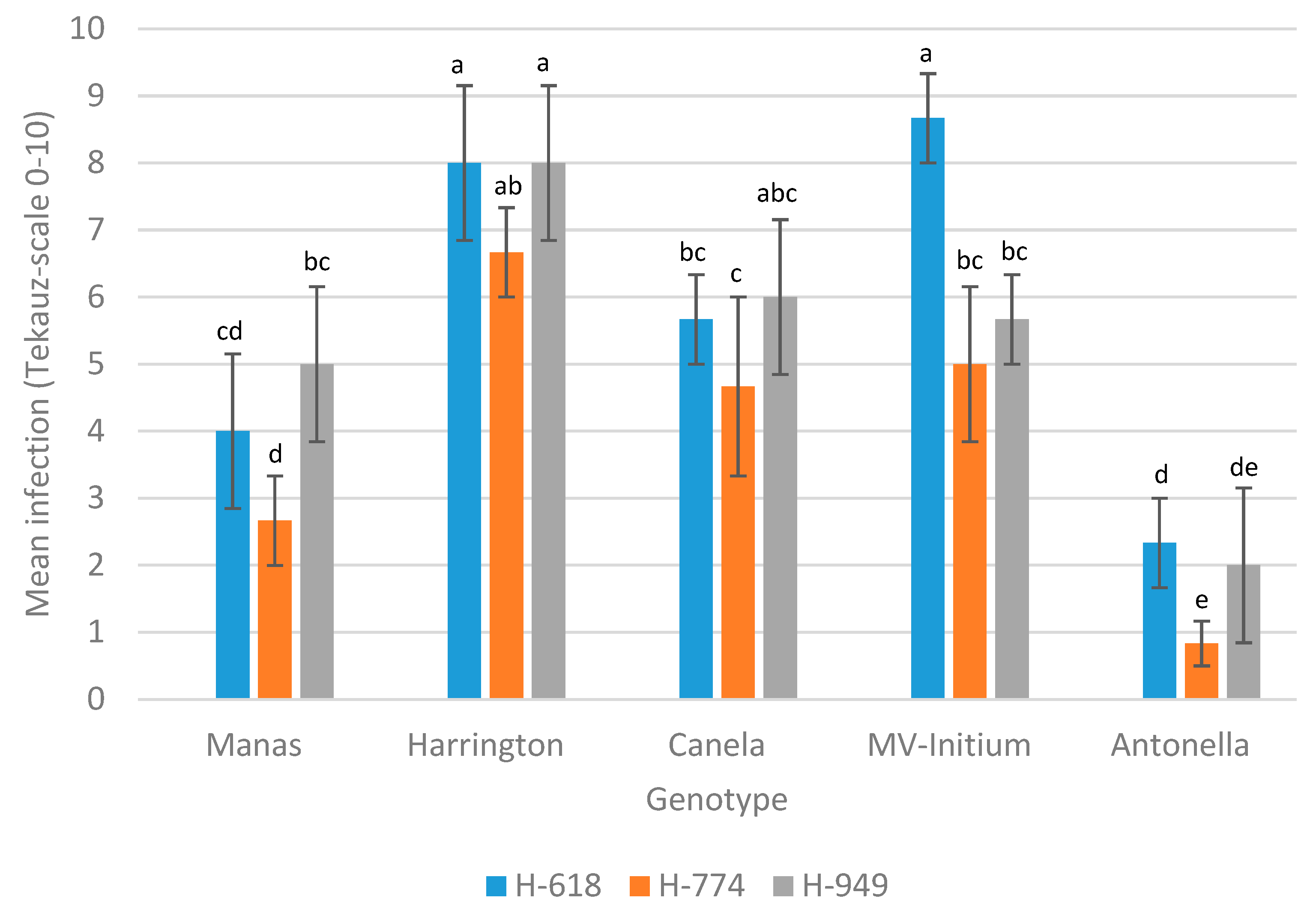
We found statistically significant differences between the examined isolates while measuring on the Tekauz scoring scale the severity of the infections H-618 [F(4, 10) = 35.389, p = 0.000], H-774 [F(4, 10) = 24.757, p = 0.000] and H-949 [F(4, 10) = 16.346, p = 0.000]. We could evaluate the degree of infection after 3 days of inoculation when the first symptoms appeared. Our results are based on the Tekauz [27] scale. The infection rates of cv. “Antonella” were significantly lower in the case of every PTT isolate, except in the case of cv. “Manas” with isolates H-618 (Figure 4).
The LSD post-hoc test was performed to explore differences between varieties in the case of isolate H-618 infection scoring on the Tekauz (1985) scale 15 days after infection (Table 3). The LSD test showed that the infection scoring was significantly different in each case. The lowest scoring points were showed by cv. “Antonella” (2.33 ± 0.577) and cv. “Manas” (4.00 ± 1.00). The only two varieties which did not differ significantly from each other were cv. “Harrington” (8.00 ± 1.00) and cv. “MV-Initium” (8.67 ± 0.578), and these two had the highest measured scoring points (9) against isolate H-618.
In the case of isolate H-774, the LSD test showed that the infection scoring was also significantly different in each case (Table 4). The only two varieties which were not significantly different from each other were cv. “Canela” (4.67 ± 1.15) and cv. “MV-Initium” (5.00 ± 1.00). The lowest scoring points were shown by cv. “Antonella” (1.00 ± 0.289) and cv. “Manas” (2.00 ± 0.575).
The results of isolate H-949 showed a slight degree of susceptibility. The cv. “Manas” (5.00 ± 1.00) did not show a significant difference from cv. “Canela” (6.00 ± 1.00) and cv. “MV-Initium” (0.56 ± 0.55), and cv. “Canela” and cv. “MV-Initium” did not differ from each other in this case. The highest scoring points were obtained by cv. “Harrington” (9). The lowest scoring points were shown by cv. “Antonella” (3) (Table 5).
Overall, our scoring point results show that the isolates had different degrees of severity on each genotype. The most severe symptoms were caused by the H-618 (5.73 ± 2.54) followed by the H-949 (5.33 ± 2.16) and the H-774 (3.97 ± 2.19) on average. The least susceptible genotype overall was cv. “Antonella”, followed by cv. “Manas”, and the most susceptible genotypes were cv. “Harrington” and cv. “MV-Initium” (Figure 4).
We examined the correlation between the SOD activity and the registered Tekauz infection scoring points by genotypes and isolates (Table 6). In the case of cv. “Manas”, we found a significant, positive, strong correlation between the SOD activity and the Tekauz infection scores at the treatment with the H-774 isolate (r = 0.778) and we found a strong but negative correlation (r = −0.914) at the treatment with the H-949 isolate. The cv. “Harrington” showed a strong, significant, positive correlation between the SOD activity and the Tekauz infection scoring points in the case of the treatments with H-774 and H-949 isolates. On the other hand, cv. “Canela” and cv. “MV-Initium” showed a significantly strong but inverse correlation in the case of treatments with H-618 (rCanela = −0.787, rInitium = −0.917) and H-949 (rCanela = −0.956, rInitium = −0.853) isolates. In relation to cv. “Antonella”, we registered a strong positive correlation between the SOD activity and the Tekauz infection scorings at the treatments with isolates H-618 and H-774, which were r = 0.99 and r = 0.99, respectively. 
We did not find significant correlations between the SOD activity and the Tekauz infection scorings in cv. “Manas” and cv. “Harrington” in the case of the isolate H-618, in cv. “Canela” and cv. “MV-Initium” in the case of the isolate H-774 or in cv. “Antonella” in the case of the isolate H-949.

4. Discussion

Changes in environmental factors, which include abiotic and biotic stress, cause modified ROS homeostasis within the plant cell. To protect plant cells from oxidative stress, ROS removal mechanisms have been developed that include antioxidant enzymes. The reactive oxygen species also play a key role as signal molecules in initiating plant defense mechanisms [19,24]. Fine-tuning this system is essential for effective defense. Although SOD, along with several other antioxidants, regulates ROS, its role is still dominant because it is the first barrier to oxidative damage. In the present study, we investigated the change in SOD activity after PTT infections in barley seedlings. Our results showed that all three PTT isolates caused significant changes in the SOD activity of all examined barley varieties in the early stages of the infection.
The treatment with H-618 resulted in significant increases in SOD activity in the examined genotypes after 48 h. The measured SOD activity highly increased in the case of every genotype except cv. “Antonella”, which showed a slight decrease, but this result was not significant. Cv. “Antonella” was the most resistant genotype to PTT. Able et al. [8] observed a lower ROS production in resistant reaction to necrotrophic pathogens, and in larger amounts in susceptible plants [12]. The ROS induces the activation of different antioxidant enzymes and defense mechanism against pathogens [12,53]. On the other hand, the accumulation of ROS blocked the early growing of fungus in [23,54]. Apart from cv. “Antonella” and cv. “Harrington”, the other examined genotypes showed decreases in SOD activity at the 72 h measurements. In the last 24 h, the cv. “Antonella” and cv. “Harrington” increased the measured activity significantly. Despite our results, cv. “Harrington” did not show a significant correlation between the H-618-induced PTT infection and the SOD activity; however, cv. “Antonella” had a strong significant correlation with its PTT infection with H-618, as did cv. “Canela” and cv. “MV-Initium”.
The characteristic of SOD activity change was quite similar in the case of H-774 isolate to the case of H-618. For the 48 h measurements, the SOD activity reached the maximum regarding every examined genotype, expect cv. “Antonella”, which showed a further increase in SOD activity in the last 24 h. However, we did not find any statistically valid differences in SOD activity between genotypes in the treatment with H-774. We found a significant correlation between the severity of the H-774-induced PTT infection and the measured SOD activity in genotypes cv. “Manas”, cv. “Harrington” and cv. “Antonella”.
If we compare the two isolates which were examined in the same timeframe (H-618 and H-774), we can conclude that the SOD activity was higher in cv. “Canela” in the case of H-618 infection than H-774 infection. The cv. “Harrington” was sensitive to both PTT isolates, and its SOD activity was also one of the highest in both cases on average. In contrast, the most resistant genotype to both isolates was cv. “Antonella”, although its SOD activity was the lowest on average, followed by cv. “Manas” and cv. “Mv-Initium”. These results support the fact that the susceptibility of barley cultivars to individual PTT isolates varies [9].
The subsequent effect of PTT infection on SOD activity was examined on days 7 and 15 after infection with the H-949 isolate. The SOD activity of cv. “MV-Initium” and cv. “Canela” decreased between 0 h and the 7th day, while significant or near significant increases in SOD activity were observed in other varieties. Further increases in activity were observed in cv. “Harrington”, cv. “Canela”, cv. “MV-Initium” and cv. “Antonella”, and no significant decrease in SOD activity was observed in cv. “Manas”. Cv. “Antonella“ and cv. “MV-Initium” showed the lowest SOD activity in the case of H-949 treatment right after cv. “Canela”. A negative significant correlation was observed between the infection level based on the Tekauz scale and SOD activity in the case of all cultivars expect cv. “Antonella”. This result contradicts the findings of previous research that resistant varieties have higher SOD activity [8]. In our experiment, cv. “Antonella”, which is the most resistant to PTT, had the lowest SOD activity in all isolates. However, this coincides with the findings that reported a positive association between the presence of ROS and disease resistance [12,55,56].
In conclusion, in our experiment, we observed a significant increase in SOD activity upon PTT inoculation, although the extent of this was genotype- and isolate-dependent. An increase in SOD activity presupposes the appearance of reactive oxygen species as a result of infection, as Able’s [8] hypotheses has noted. Although he stated that a higher SOD activity suggests resistance to PTT infection, as previously advocated by Urbanek [57] and Gil-ad et al. [58], the opposite was observed in our experiment—the SOD activity of susceptible cultivars increased to a greater extent. Shetty et al. [30] state that removal of H2O2 by catalase at both early and late stages made wheat plants more susceptible to Septoria tritici, whereas H2O2 formation made them more resistant. Furthermore, our results also support those which show the inoculation-dependent increase in SOD activity at the 24 h stage of powdery mildew infection mentioned by Vanacker [59]. Higher SOD activity was detected in the susceptible genotype than in the resistant genotype, just like in our experiment. Increased activity of all three SOD isoforms was observed in increased Pseudomonas susceptible double mutant Arabidopsis thaliana (At-pao1-1 x Atpao2-1) after inoculation [55]. The previous results are in contradiction with those of Lightfoot et al. [12], who detected higher SOD activity in resistant barley genotypes, and smaller symptoms were detected in transgenic HcCSD1 knock-down lines. However, when the expression pattern of SOD2 gene was investigated, it was found that except for clear activation of expression at 8 h after infection measured in the resistant genotype, the expression pattern fluctuated more in the sensitive ones, and there was a second expression peak at 120 h in resistance one after a decline at 48 h [56].
Based on this, our findings could offer a novel perspective of the measurement of the SOD activity to detect the early stress responses induced by PTT.
However, it should be noted that our results do not show a clear connection between seedling resistance of genotypes and SOD enzyme activity, although we did find significant correlations between the PTT infection scores and the SOD activity in several cases.
Overall, our results suggest that the SOD activity induced by the PTT infection will increase with elapsed time of infection, especially after 48 h. Increased SOD activity due to pathogenic attack promotes H2O2 formation and protects the plant cell from superoxide accumulation. In the case of increased production of O2−, enhanced SOD activity reduces the risk of hydroxyl radical formation due to Fenton-type chemistry [57]. These results suggest that although many antioxidant enzymes play a role in fine-tuning the defense response, the resistance of varieties cannot be estimated based on SOD enzyme activity alone.
Studying the other members of the antioxidant enzyme system is necessary to clarify their role during biotic stresses, such as pathogen infection. We would like to continue our work and extend our studies beyond 3 and 15 DAI to learn more about the association between PTT infection and SOD activity, as we were unable to examine the full phenomenon because we assumed that the highest SOD activity could be observed in the early phase of the PTT infection. Furthermore, we may extend this work by involving more genotypes and isolates to understand the connection between ROS accumulation and an early pathogen infection.

Author Contributions

Conceptualization, K.M. and J.B. (József Bakonyi); methodology, K.M.; software, Z.K.; validation, M.P., V.K. and M.C.; formal analysis, A.E.; investigation, M.C., V.K. and K.M.; resources, M.P., J.B. (József Bakonyi) and D.S.; data curation, M.P. and V.K.; writing—original draft preparation, V.K.; writing—review and editing, V.K., J.B. (Judit Bányai), A.U. and K.M.; visualization, Z.K.; funding acquisition, K.M. All authors have read and agreed to the published version of the manuscript.

Funding

This research was funded by the Hungarian National Research, Development and Innovation Office, Grant Numbers: NKFIH K119276 and GINOP-2.3.2-15-2016-00029.

Institutional Review Board Statement

Not applicable.

Informed Consent Statement

Not applicable.

Data Availability Statement

The data presented in this study are available on request from the corresponding author.

Acknowledgments

We thank to Klára Illés, Ágnes Bencze and Ildiko Csorba for their help in carrying out the experiment and preparation of the semples for measuring the enzyme activity.

Conflicts of Interest

The authors declare no conflict of interest. The funders had no role in the design of the study, in the collection, analyses or interpretation of data, in the writing of the manuscript, or in the decision to publish the results.

References

  1. Afanasenko, O.; Mironenko, N.; Filatova, O.; Kopahnke, D.; Krämer, I.; Ordon, F. Genetics of host-pathogen interactions in the Pyrenophora teres f. teres (net form)—Barley (Hordeum vulgare) pathosystem. Eur. J. Plant Pathol. 2007, 117, 267–280. [Google Scholar] [CrossRef]
  2. Liu, Z.; Ellwood, S.R.; Oliver, R.P.; Friesen, T.L. Pyrenophora teres: Profile of an increasingly damaging barley pathogen. Mol. Plant Pathol. 2011, 12, 1–19. [Google Scholar] [CrossRef] [PubMed] [Green Version]
  3. Smedegård-Petersen, V. Pyrenophora teres f. maculata f. nov. and Pyrenophora teres f. teres on Barley in Denmark; Royal Veterinary and Agricultural University; Copenhagen, Denmark, 1971; pp. 124–144. [Google Scholar]
  4. Tomcsányi, A.; Szeőke, K.; Tóth, Á. TOMCSÁNYI, A., SZEŐKE, K., TÓTH, Á. (2006): Az őszi árpa védelme. Növényvédelem 2006, 42, 87–106. [Google Scholar]
  5. Mathre, D.E. Compendium of Barley Diseases; American Phytopathological Society: St. Paul, MN, USA, 1982. [Google Scholar]
  6. Steffenson, B.J. Reduction in Yield Loss Using Incomplete Resistance to Pyrenophora teres f. teres in Barley. Plant Dis. 1991, 75, 96. [Google Scholar] [CrossRef]
  7. Heitefuss, R. Defence reactions of plants to fungal pathogens: Principles and perspectives, using powdery mildew on cereals as an example. Naturwissenschaften 2001, 88, 273–283. [Google Scholar] [CrossRef]
  8. Able, A.J. Role of reactive oxygen species in the response of barley to necrotrophic pathogens. Protoplasma 2003, 221, 137–143. [Google Scholar] [CrossRef]
  9. Lehmann, S.; Serrano, M.; L’Haridon, F.; Tjamos, S.E.; Metraux, J.P. Reactive oxygen species and plant resistance to fungal pathogens. Phytochemistry 2015, 112, 54–62. [Google Scholar] [CrossRef] [Green Version]
  10. Baxter, A.; Mittler, R.; Suzuki, N. ROS as key players in plant stress signalling. J. Exp. Bot. 2014, 65, 1229–1240. [Google Scholar] [CrossRef]
  11. Groß, F.; Durner, J.; Gaupels, F. Nitric oxide, antioxidants and prooxidants in plant defence responses. Front. Plant Sci. 2013, 4. [Google Scholar] [CrossRef] [Green Version]
  12. Lightfoot, D.J.; Mcgrann, G.R.D.; Able, A.J. The role of a cytosolic superoxide dismutase in barley–pathogen interactions. Mol. Plant Pathol. 2017, 18, 323–335. [Google Scholar] [CrossRef] [PubMed]
  13. Mittler, R.; Vanderauwera, S.; Suzuki, N.; Miller, G.; Tognetti, V.B.; Vandepoele, K.; Gollery, M.; Shulaev, V.; Van Breusegem, F. ROS signaling: The new wave? Trends Plant Sci. 2011, 16, 300–309. [Google Scholar] [CrossRef] [PubMed]
  14. Torres, M.A.; Dangl, J.L. Functions of the respiratory burst oxidase in biotic interactions, abiotic stress and development. Curr. Opin. Plant Biol. 2005, 8, 397–403. [Google Scholar] [CrossRef]
  15. Kranner, I.; Roach, T.; Beckett, R.P.; Whitaker, C.; Minibayeva, F.V. Extracellular production of reactive oxygen species during seed germination and early seedling growth in Pisum sativum. J. Plant Physiol. 2010, 167, 805–811. [Google Scholar] [CrossRef]
  16. Tyagi, S.; Sharma, S.; Taneja, M.; Shumayla; Kumar, R.; Sembi, J.K.; Upadhyay, S.K. Superoxide dismutases in bread wheat (Triticum aestivum L.): Comprehensive characterization and expression analysis during development and, biotic and abiotic stresses. Agri Gene 2017, 6, 1–13. [Google Scholar] [CrossRef]
  17. Berens, M.L.; Berry, H.M.; Mine, A.; Argueso, C.T.; Tsuda, K. Evolution of Hormone Signaling Networks in Plant Defense. Annu. Rev. Phytopathol. 2017, 55, 401–425. [Google Scholar] [CrossRef]
  18. Sies, H. On the history of oxidative stress: Concept and some aspects of current development. Curr. Opin. Toxicol. 2018, 7, 122–126. [Google Scholar] [CrossRef]
  19. Waszczak, C.; Carmody, M.; Kangasjärvi, J. Reactive Oxygen Species in Plant Signaling. Annu. Rev. Plant Biol. 2018, 69, 209–236. [Google Scholar] [CrossRef] [Green Version]
  20. Wang, Y.; Ji, D.; Chen, T.; Li, B.; Zhang, Z.; Qin, G.; Tian, S. Production, signaling, and scavenging mechanisms of reactive oxygen species in fruit–pathogen interactions. Int. J. Mol. Sci. 2019, 20, 2994. [Google Scholar] [CrossRef] [Green Version]
  21. Saed-Moucheshi, A.; Shekoofa, A.; Pessarakli, M. Reactive Oxygen Species (ROS) Generation and Detoxifying in Plants. J. Plant Nutr. 2014, 37, 1573–1585. [Google Scholar] [CrossRef]
  22. Gechev, T.S.; Hille, J. Hydrogen peroxide as a signal controlling plant programmed cell death. J. Cell Biol. 2005, 168, 17–20. [Google Scholar] [CrossRef] [PubMed] [Green Version]
  23. Künstler, A.; Bacsó, R.; Gullner, G.; Hafez, Y.M.; Király, L. Staying alive—Is cell death dispensable for plant disease resistance during the hypersensitive response? Physiol. Mol. Plant Pathol. 2016, 93, 75–84. [Google Scholar] [CrossRef]
  24. García-Caparrós, P.; De Filippis, L.; Gul, A.; Hasanuzzaman, M.; Ozturk, M.; Altay, V.; Lao, M.T. Oxidative Stress and Antioxidant Metabolism under Adverse Environmental Conditions: A Review. Bot. Rev. 2021, 87, 421–466. [Google Scholar] [CrossRef]
  25. Pucciariello, C.; Perata, P. New insights into reactive oxygen species and nitric oxide signalling under low oxygen in plants. Plant Cell Environ. 2017, 40, 473–482. [Google Scholar] [CrossRef] [PubMed]
  26. Yu, Q.; Worth, C.; Rengel, Z. Using capillary electrophoresis to measure Cu/Zn superoxide dismutase concentration in leaves of wheat genotypes differing in tolerance to zinc deficiency. Plant Sci. 1999, 143, 231–239. [Google Scholar] [CrossRef]
  27. Takagi, D.; Takumi, S.; Hashiguchi, M.; Sejima, T.; Miyake, C. Superoxide and singlet oxygen produced within the thylakoid membranes both cause photosystem I photoinhibition. Plant Physiol. 2016, 171, 1626–1634. [Google Scholar] [CrossRef] [Green Version]
  28. Acar, O.; Türkan, I.; Özdemir, F. Superoxide dismutase and peroxidase activities in drought sensitive and resistant barley (Hordeum vulgare L.) varieties. Acta Physiol. Plant. 2001, 23, 351–356. [Google Scholar] [CrossRef]
  29. Lam, E.; Kato, N.; Lawton, M. Programmed cell death, mitochondria and the plant hypersensitive response. Nature 2001, 411, 848–853. [Google Scholar] [CrossRef]
  30. Shetty, N.P.; Mehrabi, R.; Lütken, H.; Haldrup, A.; Kema, G.H.J.; Collinge, D.B.; Jørgensen, H.J.L. Role of hydrogen peroxide during the interaction between the hemibiotrophic fungal pathogen Septoria tritici and wheat. New Phytol. 2007, 174, 637–647. [Google Scholar] [CrossRef]
  31. Ivanov, S.; Miteva, L.; Alexieva, V.; Karjin, H.; Karanov, E. Alterations in some oxidative parameters in susceptible and resistant wheat plants infected with Puccinia recondita f.sp. tritici. J. Plant Physiol. 2005, 162, 275–279. [Google Scholar] [CrossRef]
  32. Harrach, B.D.; Fodor, J.; Pogány, M.; Preuss, J.; Barna, B. Antioxidant, ethylene and membrane leakage responses to powdery mildew infection of near-isogenic barley lines with various types of resistance. Eur. J. Plant Pathol. 2008, 121, 21–33. [Google Scholar] [CrossRef]
  33. Asthir, B.; Koundal, A.; Bains, N.S.; Mann, S.K. Stimulation of antioxidative enzymes and polyamines during stripe rust disease of wheat. Biol. Plant. 2010, 54, 329–333. [Google Scholar] [CrossRef]
  34. Chung, W.H. Unraveling new functions of superoxide dismutase using yeast model system: Beyond its conventional role in superoxide radical scavenging. J. Microbiol. 2017, 55, 409–416. [Google Scholar] [CrossRef]
  35. Bowler, C.; Van Camp, W.; Van Montagu, M.; Inzé, D. Superoxide Dismutase in Plants. CRC. Crit. Rev. Plant Sci. 1994, 13, 199–218. [Google Scholar] [CrossRef]
  36. Monk, L.S.; Fagerstedt, K.V.; Crawford, R.M.M. Oxygen toxicity and superoxide dismutase as an antioxidant in physiological stress. Physiol. Plant. 1989, 76, 456–459. [Google Scholar] [CrossRef]
  37. Deng, B.; Du, W.; Liu, C.; Sun, W.; Tian, S.; Dong, H. Antioxidant response to drought, cold and nutrient stress in two ploidy levels of tobacco plants: Low resource requirement confers polytolerance in polyploids? Plant Growth Regul. 2012, 66, 37–47. [Google Scholar] [CrossRef]
  38. Miller, G.; Schlauch, K.; Tam, R.; Cortes, D.; Torres, M.A.; Shulaev, V.; Dangl, J.L.; Mittler, R. The plant NADPH oxidase RBOHD mediates rapid systemic signaling in response to diverse stimuli. Sci. Signal. 2009, 2. [Google Scholar] [CrossRef] [Green Version]
  39. Singh, R.; Rathore, D. Oxidative stress defence responses of wheat (Triticum aestivum L.) and chilli (Capsicum annum L.) cultivars grown under textile effluent fertilization. Plant Physiol. Biochem. 2018, 123, 342–358. [Google Scholar] [CrossRef]
  40. Suzuki, N.; Miller, G.; Salazar, C.; Mondal, H.A.; Shulaev, E.; Cortes, D.F.; Shuman, J.L.; Luo, X.; Shah, J.; Schlauch, K.; et al. Temporal-spatial interaction between reactive oxygen species and abscisic acid regulates rapid systemic acclimation in plants. Plant Cell 2013, 25, 3553–3569. [Google Scholar] [CrossRef] [PubMed] [Green Version]
  41. Tounsi, S.; Feki, K.; Kamoun, Y.; Saïdi, M.N.; Jemli, S.; Ghorbel, M.; Alcon, C.; Brini, F. Highlight on the expression and the function of a novel MnSOD from diploid wheat (T. monococcum) in response to abiotic stress and heavy metal toxicity. Plant Physiol. Biochem. 2019, 142, 384–394. [Google Scholar] [CrossRef]
  42. Lamb, C.; Dixon, R.A. The oxidative burst in plant disease resistance. Annu. Rev. Plant Biol. 1997, 48, 251–275. [Google Scholar] [CrossRef] [PubMed]
  43. Torres, M.A.; Jones, J.D.G.; Dangl, J.L. Reactive oxygen species signaling in response to pathogens. Plant Physiol. 2006, 141, 373–378. [Google Scholar] [CrossRef] [PubMed] [Green Version]
  44. Künstler, A.; Bacsó, R.; Hafez, Y.M.; Király, L. Reactive Oxygen Species and Plant Disease Resistance. In Reactive Oxygen Species and Oxidative Damage in Plants under Stress; Gupta, D.K., Palma, J.M., Corpas, F.J., Eds.; Springer International Publishing: Cham, Switzerland, 2015; pp. 269–303. ISBN 978-3-319-20421-5. [Google Scholar]
  45. El-Zahaby, H.M.; Hafez, Y.M.; Király, Z. Effect of Reactive Oxygen Species on Plant Pathogens in planta and on Disease Symptoms. Acta Phytopathol. Entomol. Hung. 2004, 39, 325–345. [Google Scholar] [CrossRef]
  46. Abdelaal, K.A.A.; El-Shawy, E.S.A.A.; Hafez, Y.M.; Abdel-Dayem, S.M.A.; Chidya, R.C.G.; Saneoka, H.; Sabagh, A. El Nano-silver and non-traditional compounds mitigate the adverse effects of net blotch disease of barley in correlation with up-regulation of antioxidant enzymes. Pak. J. Bot. 2020, 52, 1065–1072. [Google Scholar] [CrossRef]
  47. Bari, R.; Jones, J.D.G. Role of plant hormones in plant defence responses. Plant Mol. Biol. 2009, 69, 473–488. [Google Scholar] [CrossRef] [PubMed]
  48. Mészáros, K.; Cséplő, M.; Kunos, V.; Búza, Z.; Bányai, J.; Seress, D.; Csorba, I.; Pál, M.; Vida, G.; Bakonyi, J. Investigation of net blotch resistance of barley and preliminary data on Hungarian pathotypes of Pyrenophora teres f. teres. In Proceedings of the Resistance Breeding: From Pathogen Epidemiology to Molecular Breeding; Brandsetter, A., Geppner, M., Eds.; Druck und Verlag: Sankt Pölten, Austria, 2019; pp. 27–29. [Google Scholar]
  49. Pál, M.; Kovács, V.; Vida, G.; Szalai, G.; Janda, T. Changes induced by powdery mildew in the salicylic acid and polyamine contents and the antioxidant enzyme activities of wheat lines. Eur. J. Plant Pathol. 2013, 135, 35–47. [Google Scholar] [CrossRef]
  50. Tekauz, A. A numerical scale to classify reactions of barley to Pyrenophora teres. Can. J. Plant Pathol. 1985, 7, 181–183. [Google Scholar] [CrossRef]
  51. Bergmeyer, H.U.; Gawehn, K.; Grassl, M. Methods of Enzymatic Analysis. Verlag Chemie 1974, 1, 481–482. [Google Scholar]
  52. Zhang, C.; Bruins, M.E.; Yang, Z.Q.; Liu, S.T.; Rao, P.F. A new formula to calculate activity of superoxide dismutase in indirect assays. Anal. Biochem. 2016, 503, 65–67. [Google Scholar] [CrossRef]
  53. O’Brien, J.A.; Daudi, A.; Butt, V.S.; Bolwell, G.P. Reactive oxygen species and their role in plant defence and cell wall metabolism. Planta 2012, 236, 765–779. [Google Scholar] [CrossRef]
  54. Liu, X.; Williams, C.E.; Nemacheck, J.A.; Wang, H.; Subramanyam, S.; Zheng, C.; Chen, M.S. Reactive oxygen species are involved in plant defense against a gall midge. Plant Physiol. 2010, 152, 985–999. [Google Scholar] [CrossRef] [PubMed] [Green Version]
  55. Jasso-Robles, F.I.; Gonzalez, M.E.; Pieckenstain, F.L.; Ramírez-García, J.M.; de la Luz Guerrero-González, M.; Jiménez-Bremont, J.F.; Rodríguez-Kessler, M. Decrease of Arabidopsis PAO activity entails increased RBOH activity, ROS content and altered responses to Pseudomonas. Plant Sci. 2020, 292, 110–372. [Google Scholar] [CrossRef]
  56. Pandey, C.; Großkinsky, D.K.; Westergaard, J.C.; Jørgensen, H.J.L.; Svensgaard, J.; Christensen, S.; Schulz, A.; Roitsch, T. Identification of a bio-signature for barley resistance against Pyrenophora teres infection based on physiological, molecular and sensor-based phenotyping. Plant Sci. 2021, 313, 111072. [Google Scholar] [CrossRef] [PubMed]
  57. Urbanek, H.; Gajewska, E.; Karwowska, R.; Wielanek, M. Generation of superoxide anion and induction of superoxide dismutase and peroxidase in bean leaves infected with pathogenic fungi. Acta Biochim. Pol. 1996, 43, 679–685. [Google Scholar] [CrossRef]
  58. Gil-ad, N.L.; Bar-Nun, N.; Noy, T.; Mayer, A.M. Enzymes of Botrytis cinerea capable of breaking down hydrogen peroxide. FEMS Microbiol. Lett. 2000, 190, 121–126. [Google Scholar] [CrossRef] [PubMed] [Green Version]
  59. Vanacker, H.; Harbinson, J.; Ruisch, J.; Carver, T.L.W.; Foyer, C.H. Antioxidant defences of the apoplast. Protoplasma 1998, 205, 129–140. [Google Scholar] [CrossRef]
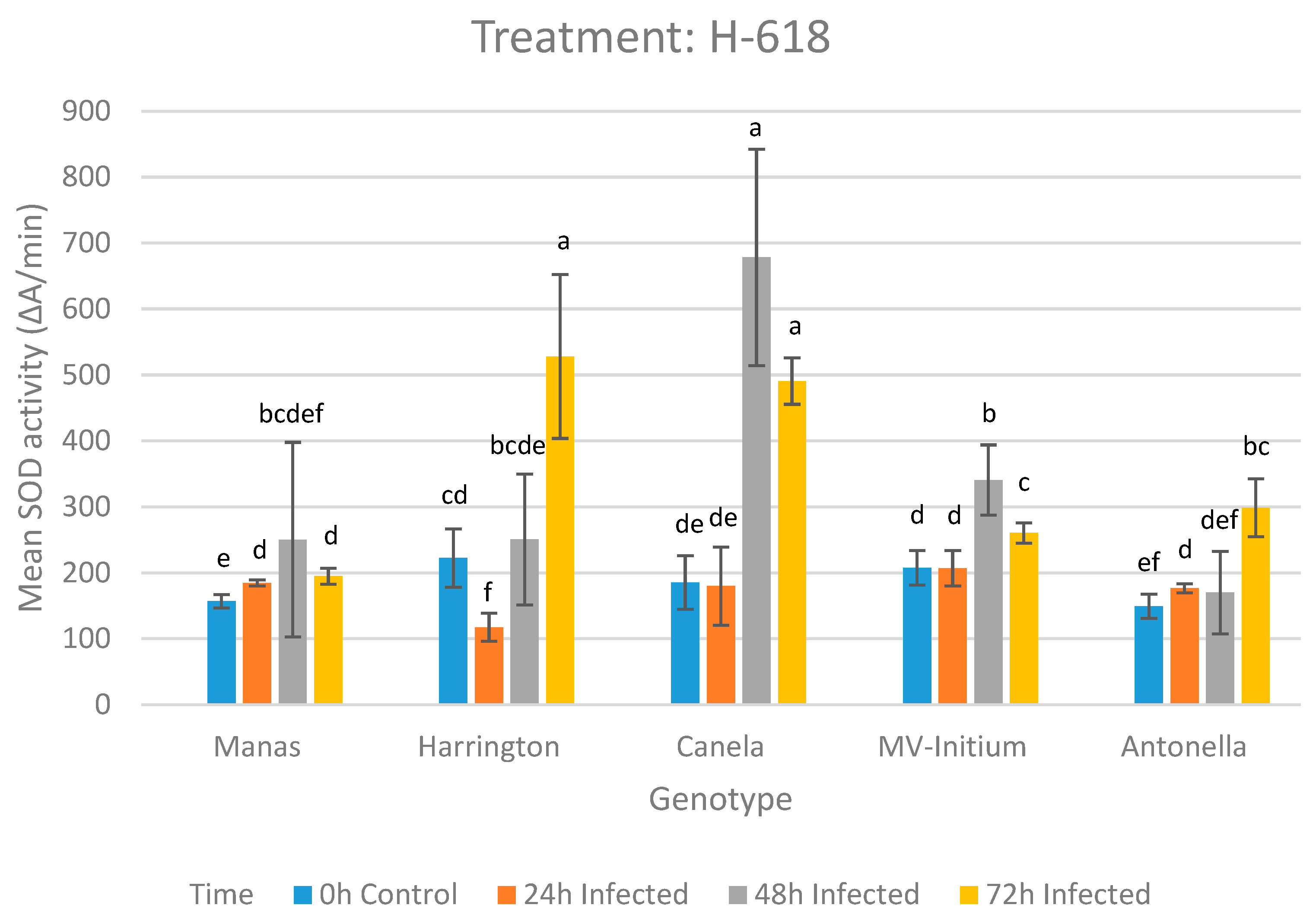
Figure 1. SOD activity of the tissue samples by the infection time in the case of H-618 treatment at 0, 1, 2 and 3 DAI. Error bars indicate the standard error (SE) of the mean instead of standard deviation (SD) at the p = 0.05 level. Values denoted with different letters are significantly different at p < 0.05.
Sustainability 14 02597 g001
Figure 2. SOD activity of the tissue samples in the case of H-774 treatment at 0, 1, 2 and 3 DAI. Error bars indicate the standard error (SE) of the mean instead of standard deviation (SD) at p = 0.05 level. Values denoted with different letters are significantly different at p < 0.05.
Sustainability 14 02597 g002
Figure 3. SOD activity of the tissue samples in the case of H-949 treatment at 0, 7 and 15 DAI. Error bars indicate the standard error (SE) of the mean instead of standard deviation (SD) at the p = 0.05 level. Values denoted with different letters are significantly different at p < 0.05.
Sustainability 14 02597 g003
Figure 4. Infection scoring on the Tekauz scale by genotypes 15 DAI. Error bars indicate the standard error (SE) of the mean instead of standard deviation (SD) at the p = 0.05 level. Values denoted with different letters are significantly different at p < 0.05.
Sustainability 14 02597 g004
Table 1. Mean differences in SOD activity of genotypes after inoculation with H-618 according to LSD test for multiple comparisons.
0 h ManasHarringtonCanelaMV-InitiumAntonella
Manas-−65.707 *−28.511−50.817 *7.638
Harrington--37.19614.88973.345 *
Canela---−22.30636.149
MV-Initium----58.455 *
Antonella-----
24 h ManasHarringtonCanelaMV-InitiumAntonella
Manas-67.516 *4.814−21.9978.355
Harrington--−62.702 *−89.514 *−59.160 *
Canela---−26.8123.541
MV-Initium----−30.353
Antonella-----
48 h ManasHarringtonCanelaMV-InitiumAntonella
Manas-−0.468−427.996 *−90.34980.028
Harrington--−427.527 *−89.88080.497
Canela---337.646 *508.025 *
MV-Initium----170.378
Antonella-----
72 h ManasHarringtonCanelaMV-InitiumAntonella
Manas-−333.04 *−295.857 *−65.614−103.541 *
Harrington--37.183267.425 *229.498 *
Canela---230.242 *192.315 *
MV-Initium----−37.927
Antonella-----
* The mean difference is significant at the p = 0.05 level. Significant differences shown in the table are different than those shown in Figure 1 because in the figure, error bars indicate the standard error (SE) of the mean instead of standard deviation (SD) at the p = 0.05 level.
Table 2. Post-hoc comparisons of genotypes in the case of H-949 treatment at 0, 7 and 15 DAI according to LSD test for multiple comparisons. Mean differences are shown.
0 h ManasHarringtonCanelaMV-InitiumAntonella
Manas-66.30911.583−13.35480.508
Harrington--−54.726−79.66314.199
Canela---24.93793.863
MV-Initium----−93.863
Antonella-----
7th day ManasHarringtonCanelaMV-InitiumAntonella
Manas-256.77 *712.713 *753.514 *371.11 *
Harrington--455.943 *496.744 *114.34
Canela---40.8−341.602 *
MV-Initium----−382.403 *
Antonella-----
15th day ManasHarringtonCanelaMV-InitiumAntonella
Manas-10.84464.567 *85.65097.25
Harrington--453.722 *74.80586.405
Canela---−378.916 *−367.317 *
MV-Initium----11.599
Antonella-----
* The mean difference is significant at the 0.05 level. Significant differences shown in the table are different than those shown in Figure 3 because in the figure, error bars indicate the standard error (SE) of the mean instead of standard deviation (SD) at p = 0.05 level.
Table 3. Post-hoc comparisons of the isolate H-618 infection scoring on the Tekauz scale by genotypes 3 DAI using Tamhane LSD. Mean differences are shown.
ManasHarringtonCanelaMV-InitiumAntonella
Manas-−4.00 *−1.66 *−4.66 *1.66 *
Harrington -2.33 *−0.665.66 *
Canela -−3.00 *3.33 *
MV-Initium -6.33 *
Antonella -
* The mean difference is significant at the 0.05 level. Significant differences shown in the table are different than those shown in Figure 4 because in the figure, error bars indicate the standard error (SE) of the mean instead of standard deviation (SD) at the p = 0.05 level.
Table 4. Post-hoc comparisons of the isolate H-774 infection scoring on the Tekauz scale by genotypes 3 DAI using Tamhane LSD. Mean differences are shown.
ManasHarringtonCanelaMV-InitiumAntonella
Manas-−4.00 *−2.00* −2.33 *1.83 *
Harrington -2.00 *1.66 *5.83 *
Canela -−0.333.83 *
MV-Initium -4.16 *
Antonella -
* The mean difference is significant at the 0.05 level. Significant differences shown in the table are different than those shown in Figure 4 because in the figure, error bars indicate the standard error (SE) of the mean instead of standard deviation (SD) at the p = 0.05 level.
Table 5. Post-hoc comparisons of the isolate H-949 infection scoring on the Tekauz scale by genotypes 15 DAI using Tamhane LSD. Mean differences are shown.
ManasHarringtonCanelaMV-InitiumAntonella
Manas-−3.00 *−1.00−0.663.00 *
Harrington -2.00 *2.33 *6.00 *
Canela -0.334.00 *
MV-Initium -3.66 *
Antonella -
* The mean difference is significant at the 0.05 level. Significant differences shown in the table are different than those shown in Figure 4 because in the figure, error bars indicate the standard error (SE) of the mean instead of standard deviation (SD) at the p = 0.05 level.
Table 6. Pearson’s correlation matrix between SOD activity (ΔA/min) and infection scoring on the Tekauz scale by genotypes after 3 DAI in the case of H-618 and H-774 and after 14 DAI in the case of H-949.
GenotypeIsolateH-618 TekauzH-774 TekauzH-949 Tekauz
ManasH-618 SOD0.143--
H-774 SOD-0.778 *-
H-949 SOD--−0.914 *
HarringtonH-618 SOD0.662--
H-774 SOD-0.938 *-
H-949 SOD--0.997 *
CanelaH-618 SOD−0.787 *--
H-774 SOD-0.144-
H-949 SOD--−0.956 *
MV-InitiumH-618 SOD−0.917 *--
H-774 SOD-−0.584-
H-949 SOD--−0.853 *
AntonellaH-618 SOD0.999 *--
H-774 SOD-0.999 *-
H-949 SOD--0.588
* The correlations are significant at the 0.05 level.
Publisher’s Note: MDPI stays neutral with regard to jurisdictional claims in published maps and institutional affiliations.