Article

Bacteriocin from Lacticaseibacillus rhamnosus sp. A5: Isolation, Purification, Characterization, and Antibacterial Evaluation for Sustainable Food Processing

1
Jiangsu Key Laboratory of Regional Resource Exploitation and Medicinal Research, Huaiyin Institute of Technology, Huai’an 223003, China
2
School of Life Science and Food Engineering, Huaiyin Institute of Technology, Huai’an 223003, China
3
Faculty of Chemical Engineering, Huaiyin Institute of Technology, Huai’an 223003, China
4
Tecnologico de Monterrey, School of Engineering and Sciences, Monterrey 64849, Mexico
5
Department of Microbiology and Immunology, Faculty of Pharmacy, Future University in Egypt, New Cairo 11835, Egypt
*
Author to whom correspondence should be addressed.
Sustainability 2022, 14(15), 9571; https://doi.org/10.3390/su14159571
Submission received: 16 June 2022 / Revised: 27 July 2022 / Accepted: 28 July 2022 / Published: 4 August 2022
(This article belongs to the Special Issue Environmental Sustainability in IR 4.0)

Abstract

:
A new Lacticaseibacillus rhamnosus strain A5 was isolated from pickle soup and characterized for its probiotic suitability. Strain A5 was Gram-positive, catalase-negative, acid-producing, and exhibited potential antibacterial activity against Escherichia coli (inhibition zone 17.3 mm), Bacillus subtilis (inhibition zone 14.5 mm), Salmonella enterica (zone of inhibition 16.1 mm) and Staphylococcus aureus (zone of inhibition 14.2 mm) by performing investigations on the disc diffusion. The cell-free supernatant of newly isolated strain A5 retained its inhibition ability of the growth of test bacteria at pH 2.0 to 5.0, temperature 121 °C for 30 min and UV irradiation for 8 h. However, the inhibitory effects of cell-free supernatant disappeared when subjected to papain, trypsin, and pepsin enzymatic treatments. By eliminating the interferences of organic acid and hydrogen peroxide, the cell-free supernatant possessed antibacterial activity against two indicator bacteria (E. coli and B. subtilis) and showed high thermal tolerance. These results indicated that the antibacterial substances produced by strain A5 were proteinaceous in nature, namely bacteriocin. The antibacterial bacteriocins in the supernatant of the strain A5 culture were further purified by ammonium sulfate fractionation and gel filtration chromatography. The purified bacteriocins also showed a pronounced inhibitory effect against E. coli and B. subtilis. The approximated molecular weight of bacteriocins was less than 14 kDa after determining by sodium dodecyl sulfate-polyacrylamide gel electrophoresis. In conclusion, the newly isolated strain A5 and its bacteriocins could be potentially applied in food preservation to prevent the risk of foodborne illness.

1. Introduction

Antibiotics have been widely used in animal feed and food processing industries to control pathogenic infection and extend shelf life. However, serious problems, such as the emergence of bacterial antibiotic resistance, spreading of antibiotic resistance genes with pathogenic bacteria and residue of potentially harmful antibiotics in foods diminish antibiotic usage and encourage manufacturers to search for suitable alternatives from natural sources [1,2]. In addition, and increasing number of people are inclined to consume foods that are natural and healthy with no artificial additives or preservatives. Recent research focuses on the replacement possibility of commonly used synthetic preservatives, such as sodium benzoate with natural compounds in various foods [3]. Therefore, it is meaningful to explore alternative methods and substitutive antibacterial substances to restrain and inhibit the growth of food pathogens. Probiotics have been considered promising substitute materials to control bacterial decay because they are living microorganisms and intended to have beneficial influences on the host cells [4,5]. The screening of novel probiotic strains from fermented foods has become an emerging research field in recent years owing to their foregrounded properties in the field of healthcare and food processing technologies [6,7,8]. Lactic acid bacteria (LAB) are an important source of probiotics with the proven safe and effective application.
The fermentation process is an ancient and commercial method, which is widely applied in the food preservation industry. Fermented food products are vital to the human diet around the world and provide a broad variety of nutrients and flavors [9,10]. The pickle is a preferred source for a balanced and healthy diet because it offers good flavor and nutritional requirements essential for human consumption [11]. LAB producing antibacterial substances, including bacteriocins, organic acids, hydrogen peroxide, and diacetyl compounds represent a dominant community in fermented pickles and can be used as a food preservative in the fermentation industry [12]. Many Gram-positive and Gram-negative bacteria can secrete substances possessing antibacterial characteristics, called bacteriocins, during their lifecycle [13,14]. Bacteriocins are polypeptides or proteins produced by bacteria that demonstrate antibacterial capabilities against other microbes, which are often genetically close to the producing strain [2,15]. The utilization of these antimicrobial peptides offers a number of advantageous features, such as high efficacy, low possibility to non-specific toxicity or inducing resistance, and the absence of residue generation. The biosynthesis of bacteriocins is controlled by the bacterial genome [16]. The bacteriocins synthesized by LAB were categorized into the following four types by the recommended classifying protocol for bacteriocins: class I, small, thermo-stable lanthionine-containing peptides (<5 kDa) [17]; class II, small, thermo-stable non-lanthionine containing peptides (<10 kDa) [13]; class III, large, thermo-sensitive proteins (>30 kDa); and class IV compound proteins embracing one or more chemical moieties, either carbohydrate or lipid [18]. The bacteriocins of class I and class II are suitably applied as bio-preservatives in the food processing industry and as a partial replacement of chemical preservatives [19]. A comprehensive focus on the prospective applications of bacteriocins from LAB in the food processing industry resulted in the screenings of new LAB and bacteriocins [19].
Bacteriocins produced by LAB are promising antimicrobials and may be potentially utilized as natural bio-preservatives in the food industry because of their beneficial properties, such as safe compounds, pH and heat tolerance [20]. Some bacteriocins, including Nisin from Lactococcus lactis [21], Plantaricin from Lactobacillus plantarum LR/14 [22], Lichenin from Bacillus licheniformis [23], Cerein from Bacillus cereus [24], Haloduracin from Bacillus halodurans [25], Thuricin from Bacillus thuringiensis [26] and Divergicin M35 from Carnobacterium divergens [27] were intensively studied in earlier works. Within the numerous strains of bacteria, lactobacilli LAB species were extensively exploited for their probiotic benefits and excellent antibacterial and antifungal characteristics. However, the number of successfully applied bacteriocins is few because of the narrow inhibition spectrum, low thermo-tolerance, and environmental instability [28]. Consequently, identification of putative bacteriocin-producing strains and the purification of excellent environmental-adaptability bacteriocins are highly important processes to the development of natural bio-preservative in the food processing industry. In this paper, Lacticaseibacillus rhamnosus sp. producing bacteriocins were isolated from pickles and their antibacterial activities were identified. The antibacterial peptide produced by this strain was purified and characterized.

2. Materials and Methods

2.1. Materials

All the reagents and chemicals were procured from Sangon (Shanghai, China). Primer synthesis was carried out by Sangon (Shanghai, China). DNA Marker, Taq polymerase and PCR kit were purchased from Generay (Shanghai, China). The pickle was procured from the local supermarket of Huaian.

2.2. Bacterial Strains, Media and Growth Conditions

The bacterial strains, Escherichia coli (ATCC 11229), Bacillus subtilis (ATCC 21556), Salmonella enterica (ATCC 12002), and Staphylococcus aureus (ATCC 27154) were streaked out freshly from slants reserved in our Laboratory and used as indicator strains in this study. MRS (%, w/v) for LAB growth consists of 1.0 g peptone, 1.0 g beef extract, 0.5 g yeast extract, 0.2 g di-ammonium citrate, 2 g glucose, 0.1 g Tuwen-80, 0.5 g sodium acetate, 0.2 g K2HPO4, 0.058 g MgSO4·7H2O, 0.025 g MnSO4·4H2O (pH 6.2–6.4). Where required, 1.5 g agar was added to prepare solid MRS. All the strains were cultivated in 5 mL MRS broth and incubated at 37 °C for 48 h. LB broth (%, w/v) for the growth of indicator strains comprised 1 g tryptone, 1 g yeast extract, 0.5 g NaCl, pH 7.0–7.2. 2 g agar was added for LB agar. Strains were cultivated in 5 mL LB broth and incubated at 37 °C for 12 h.

2.3. Sample Collection, Isolation, and Identification of LAB

The fermented pickles were procured from the supermarket. Precisely 0.1 mL of pickle soup was aseptically collected, added into 0.9 mL of sterile water, and mixed well. The resulting mixture was diluted in a range of 10−1 to 10−7 and the 0.1 mL diluent mixture was plated onto the MRS agar plates containing 3% CaCO3 (w/v) following incubation at 37 °C for 48 h. The bacterial colonies forming clear CaCO3-dissolving circles were putatively regarded as LAB and streaked on MRS agar plates for purification. To obtain LAB, the morphology of all the colonies and strains was analyzed based on catalase reaction, Gram staining, and acid production. The strains or isolates, which were Gram-positive, catalase-negative and exhibited acid production, were selected, streaked on MRS agar slants, and stored at 4 °C.
After the original screening, a colony producing a larger clear circle was chosen as experimental strain and characterized by its physiological and metabolic characteristics such as cell morphology, growth ability under different temperatures (10 °C, 40 °C and 45 °C) and salinity (4%, and 6.5% NaCl (w/v)), sugar fermentation reactions (lactose, glucose, sucrose, and maltose), litmus milk test, gelatin test and starch hydrolysis test. The characterization was further authenticated by 16S rDNA sequencing. The 16S rRNA gene was PCR-amplified using the following set of forward and reverse primers: 27F (5-AGA GTT TGA TCC TGG CTC AG-3) and 1492R (5-GGC TAC CTT GTT ACG ACTT-3). The PCR thermal program involved an automatic hot start at 94 °C for 5 min, 30 cycles of 94 °C denaturation for 30 s, 55 °C annealing for 30 s and 72 °C polymerization for 1 min, and a final extension at 72 °C for 10 min (Bio-Rad CFX Connect). The amplified gene (about 1500 bp) was purified using the PCR purification kit (Shanghai Betting Biological Technology Co., Shanghai, China) and sequenced at Shanghai Sangon Biotech. The homology analysis was performed and a phylogenetic tree was constructed with BLAST NCBI and MEGA5.0 Software using the neighbor-joining method.

2.4. Optimization of Growth Conditions for LAB Fermentation

The starting culture was prepared by inoculating the strain into 200 mL MRS broth followed by incubation at 37 °C for 12 h. The 1% (v/v) starting culture was inoculated into triplicate flasks (250 mL) containing 200 mL test media and was allowed to incubate at 37 °C for 24 h. The cell density of the strain was determined and indicated by OD600nm. Likewise, the antibacterial potential was also evaluated by measuring the size of the zone of inhibition. Different factors such as initial pH (4.0, 5.0, 6.0, 6.3, 7.0, 8.0 and 9.0), time (6, 12, 24, 30, 36, 48, and 60 h), temperature (24, 28, 30, 37, 40, 45 and 50 °C), and loading volume of the MRS broth (250 mL flask): (50, 100, 150, 200, and 250 mL) were optimized. Similarly, different sugars (20 g/L) (glucose, lactose, maltose, and sucrose), and nitrogen resources (25 g/L): (beef extract, tryptone, yeast extract, beef extract + tryptone (2:3), beef extract + tryptone (3:2), beef extract + yeast extract (2:3), beef extract + yeast extract (3:2), tryptone + yeast extract (2:3), tryptone + yeast extract (3:2), beef extract + tryptone + yeast extract (1:2:2), beef extract + tryptone + yeast extract (2:1:2) and beef extract + tryptone + yeast extract (2:2:1) were also tested and optimized.

2.5. LAB Strains and Evaluation of Antibacterial Activity

Each LAB strain was cultivated in 50 mL MRS broth at 37 °C for 24 h, and then this activated culture was centrifuged at 9500× g for 15 min (Beckman Avanti J-26S XP). The supernatant was sterilized using a 0.45 μm pore-size filter to obtain cell-free supernatant for further experiments [29]. The indicators were inoculated in 5 mL LB broth for 12 h at 37 °C, and these cultures were then diluted to maintain 1~5 × 10⁸ CFU/mL with sterilized water. The diluted culture (0.1 mL) was separately plated onto LB agar plate and three Oxford cups were placed on each plate. The 0.1 mL test solution was filled in oxford cup and the agar plate was incubated at 37 °C for 12 h. After stipulated incaution, the detectable zone of inhibition was measured which denoted the antibacterial activity of the test solution.

2.6. Impact of Temperature, pH, and UV Irradiation on Bacteriocins Antibacterial Activity

To examine the thermal stability of bacteriocins, cell-free supernatants were incubated at varying temperatures (−20 °C, 4 °C, 40 °C, 60 °C, 80 °C, 90 °C, 100 °C and 121 °C) for different periods (10 min, 20 min, and 30 min). Sterile MRS broth was served as a control, and then the remaining antibacterial activity was determined. Likewise, the sensitivity of bacteriocins to pH was assessed by maintaining the pH value of cell-free supernatants to 2, 3, 4, 5, 6, 7, 8 and 9. The sterile MRS broth adjusted with a suitable pH value served as a control. To detect the tolerance of bacteriocins to UV irradiation, the antibacterial activity was determined by exposing cell-free supernatants to UV light (40 W, 30 cm, 320~360 nm) for 1 to 8 h. The antibacterial activity of the cell-free supernatant was indicated by the diameters of the inhibition zones.

2.7. Proteinaceous Nature Verification of Bacteriocins

To eliminate the influence of organic acids, the pH value of the cell-free supernatant was adjusted to 4.0 with 1 M NaOH. Meanwhile, the pH value of MRS broth was adjusted to 4.0 with 1 M HCl as a control, and then their antibacterial activities were compared. To overcome the influence of H2O2, the cell-free supernatant (pH 4.0) was incubated at 80 °C for 10 min and the cell-free supernatant untreated by heating served as a control, and then their antibacterial activities were compared. After eliminating the impact of hydrogen peroxide and organic acids, the cell-free supernatants (pH 4.0) were treated with trypsin, pepsin, and papain (Shanghai Sangon Biotech, Shanghai, China, 1 mg/mL) at 37 °C for 2 h. The supernatants without enzyme treatment were used as controls, which were prepared by adding distilled water. The residual enzymatic activity was inactivated by sterilization (121 °C for 15 min) and their antibacterial activities were compared to verify the proteinaceous nature of antibacterial substances (bacteriocins).

2.8. Extraction and Purification of Bacteriocins

A 50 mL supernatant was concentrated using a rotary evaporator at 60 °C with 50 mL methanol and placed overnight for stratification. The upper- and lower-layer solutions were collected and evaporated to remove residual methanol with the rotary evaporator, and then condensed down to 1~2 mL by adding sterile water. The activity of bacteriocins in cell-free supernatant was determined by calculating the size of the inhibition zone against indicators. After double dilution, the concentrated sample was passed through the Sephadex G-75 column to eliminate impurities. The samples were eluted with sterile water at a flow rate of 0.5 mL/min, and 60 fractions of 0.5 mL per tube were collected measure the antibacterial activity. The protein profiling of the active fraction, which showed antibacterial activity, was performed using SDS-PAGE (15% isolating agar, 5% concentrating agar (w/v)) [30]. The concentration of protein was analyzed using Folin–Ciocalteu’s phenol method [31].

2.9. Data Analysis

Data were analyzed using a completely randomized design with the factorial test and verified using ANOVA at a 95% confidence level. The data analysis and graphs were constructed by Excel software version 2010. Orthogonal optimization was analyzed using SPSS software. All data were obtained from triplicate experiments.

3. Results and Discussion

3.1. Isolation and Identification of LAB

Five colony-forming clear CaCO3-dissolving circles were obtained and purified from pickle soup and designated as A1, A2, A3, A4, and A5. All these isolates were Gram-positive and catalase-negative, and their cell-free supernatants had obvious inhibitory activity against E. coli, B. subtilis, S. aureus and S. enterica (Table 1). Among these isolates, strain A3 and A5 exhibited better antibacterial activity against all four tested indicators. Finally, the A5 strain was shortlisted for further studies owing to larger zones of inhibition on the indicator agar plates (Figure 1). Many antimicrobial strains were isolated and characterization was performed from some sorts of traditionally fermented dairy products, such as cheese and yogurt. These strains revealed prosperous functional characteristics and balanced-diet benefits for human health [26].
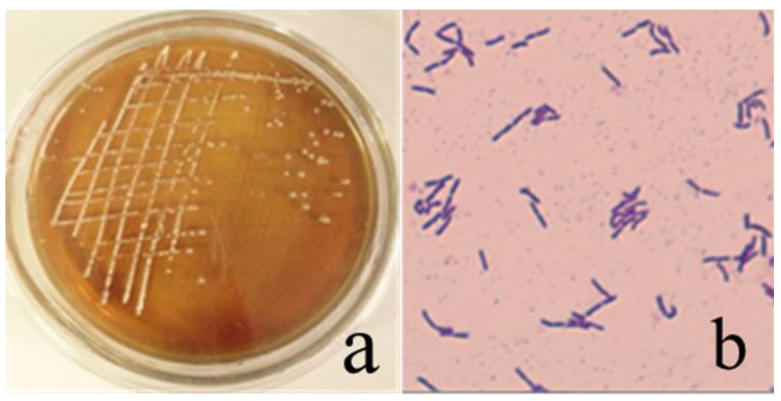
Gram staining, catalase reaction, and acid production tests were performed to analyze the morphological and metabolic characteristics of all the colonies and LAB isolates. The strains or isolates, which were catalase-negative, Gram-positive, and exhibited acid production, were streaked on MRS agar slants. The Gram-stained isolates were then submitted to a microscopic analysis for morphological characterization that revealed the following characteristics. The morphology of A5 isolate was observed to be round (Figure 2a, 1~2 mm), and cells were rod (0.4 µm × 2 µm), Gram-positive (Figure 2b), facultative anaerobic, and did not form any intracellular spores. In a second screening, physiological and metabolic characteristics were examined by different biochemical analysis methods. It grew well at 40 °C and 45 °C but its growth was retarded at 10 °C. The cells were endurable to the 4% (w/v) concentration of NaCl but growth was hampered when the concentration of NaCl increased to 6.5%. The utilization screening of carbohydrates such as lactose, glucose, sucrose, and maltose was positive; whereas the results of arabinose, sorbitol and mannitol were negative. The litmus milk test was observed to be positive, while the starch hydrolysis and catalase tests were negative. Lactobacillus strain P68 grew under the conditions of NaCl concentrations (1–7%), pH (4.5–7.5), and temperatures (28–40 °C). LAB have a higher survival capability under stressful environmental conditions. The strain A5 has some similar characteristics to P68.
Strain A5 was further characterized by conducting a 16S rDNA sequencing analysis. Results revealed high similarity with the species of genus Lacticaseibacillus rhamnosus [32]. According to the analysis, it was named Lacticaseibacillus rhamnosus sp. A5. Phylogenetic analysis of the 16S rRNA gene also clustered strain A5 within the clade of Lacticaseibacillus rhamnosus bacterium in the phylogenetic tree (Figure 3). The 16S rDNA sequence was submitted to GenBank (NCBI Accession No. MK329243). The strain A5 was a suitable LAB with desirable properties such as antibacterial activity and securable obtainment.

3.2. Verification of Bacteriocin Produced by Strain A5

Organic acids, hydrogen peroxide, and bacteriocins are some of the well-known characterized antimicrobial agents synthesized by LAB [16]. The pH value of MRS broth was adjusted to 4.0 by adding lactic acid and hydrochloric acid. It had no substantial inhibitory effect on E. coli and B. subtilis. In contrast, the cell-free supernatant of strain A5 (pH 4.0) had an inhibitory activity to both indicators (Table 2). Therefore, the antibacterial activity of cell-free supernatant was not caused by organic acids, and the antibacterial substances may be bacteriocin or hydrogen peroxide. The antibacterial activity of the L. plantarum strain IMAU80076 was observed to be pH-dependent. At pH 6.0, the inhibitory activity of cell-free supernatants was lower against E. coli O157: H7 and S. typhimurium, but not Shigella flexneri, indicating that some other inhibitory substances were involved. Although it was previously been considered that the organic acid effect is responsible for LAB antibacterial activities against Gram-negative bacteria, consequences from strains A5 suggested that other inhibitory mechanisms may exist.
The inhibitory activity of Lactobacillus LAB strains was mainly attributed to the production of antimicrobial metabolites, such as hydrogen peroxide, organic acids, ethanol, carbon dioxide, and diacetyl acid. Hydrogen peroxide produced by LAB can also impede the growth of bacteria, especially the growth of Gram-negative bacteria, so it is necessary to exclude the over-interference of hydrogen peroxide. After the cell-free supernatant of strain A5 was incubated at 80 °C for 10 min to eliminate the influence of H2O2, its antibacterial activity was almost unchanged (Table 3). Therefore, it might be inferred that the antibacterial activity of the cell-free supernatant was also not caused by hydrogen peroxide.
The antimicrobial activities of some probiotic strains were suitably governed by an exclusive competition mechanism, in which probiotic strains contended against pathogens for attachment sites and nutrients, whereas pathogenic bacteria were formed from colonizing. The lower concentrations of hydrogen peroxide influenced the physiological levels of proteins; however, higher concentrations induced DNA damage and cell degradation. However, the antibacterial activity of L. plantarum IMAU80184 and IMAU70104 was possibly dependent on hydrogen peroxide, while the antibacterial property was significantly reduced after treatment with the catalase enzyme [20]. The antibacterial activities of the strains A5 were obtained by the synthesis of organic acid, hydrogen peroxide, or bacteriocins. However, the antibacterial activities of strains A5 were not due to the production of hydrogen peroxide. These findings revealed that the mechanism of antibacterial properties of strain A5 mainly derived from the synthesis of other antibacterial substances different from organic acid and hydrogen peroxide.
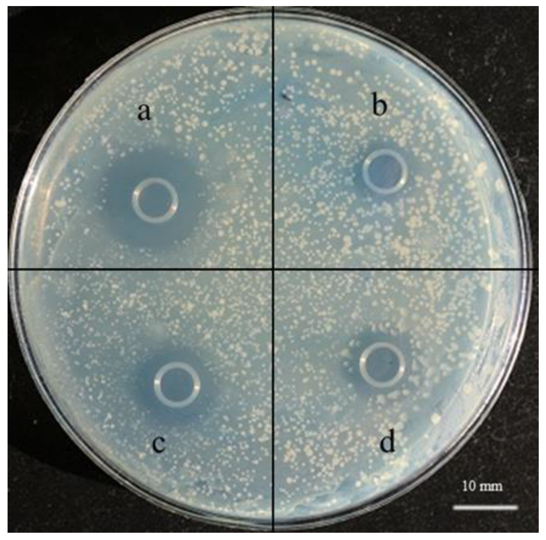
After treatment with three kinds of protease, the inhibition zones of the supernatants of the strain A5 significantly decreased (Figure 4), which indicated the liability of antibacterial substance to proteases. In conclusion, the antibacterial substances produced by strain A5 were verified to be proteinaceous. LAB could synthesize different compounds, such as bacteriocins, hydrogen peroxide, organic acids, and valuable enzymes during the fermentation procedure. The cell-free supernatants of LAB strains were treated with trypsin and pepsin and the inhibitory capability disappeared, which reflected that their antibacterial compounds were bioactive peptides/proteins in nature.

3.3. Strain Growth Curve and Antibacterial Activity

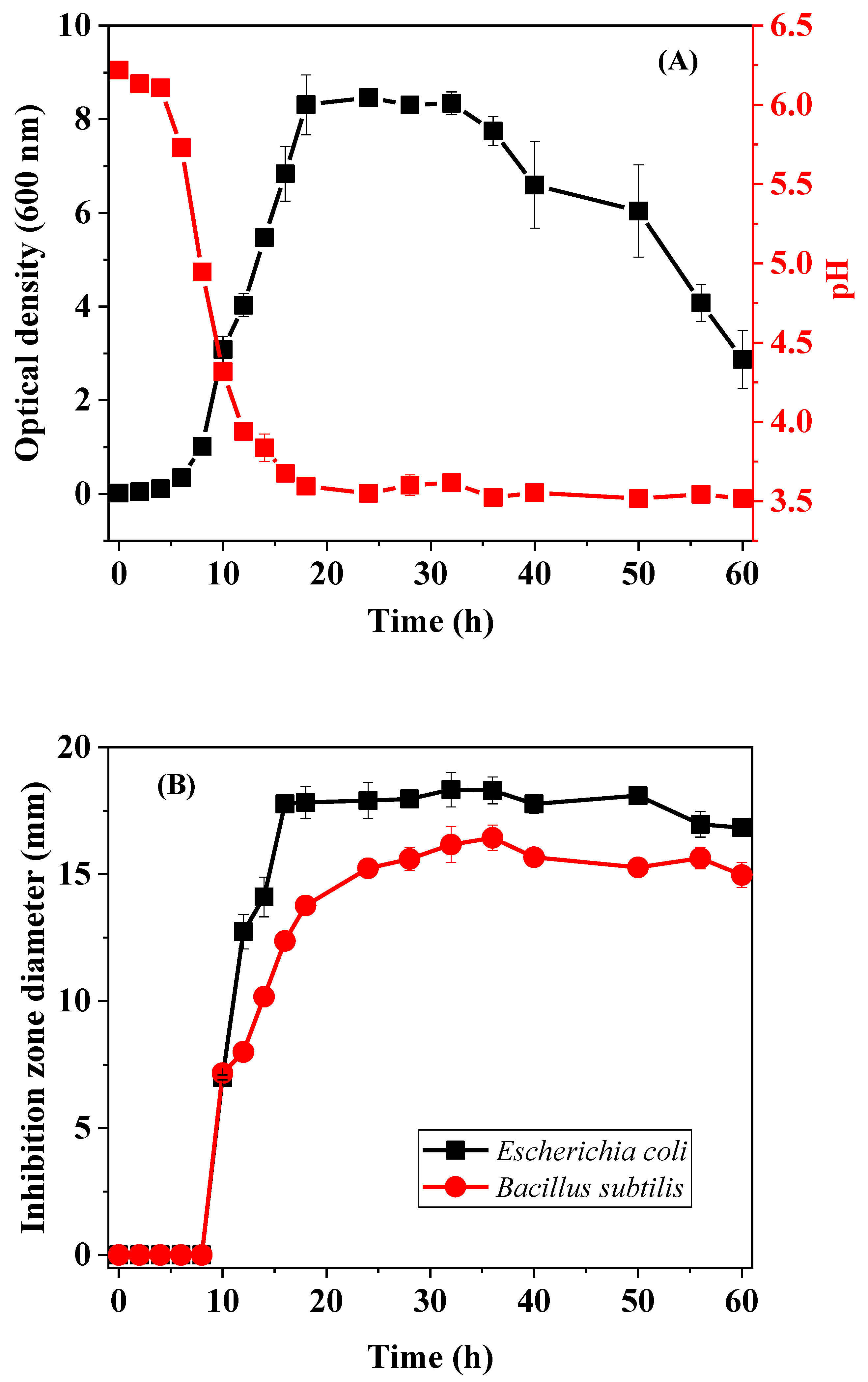
The growth and pH curve is portrayed in Figure 5A, whereas the antibacterial activity curve of the supernatant of strain A5 to both indicators is shown in Figure 5B. A lag phase of A5 for 6 h indicated its quick adaptability to growth conditions. During this period, the change in pH was negligible and the antibacterial substances were not produced. Afterward, the strain developed into the logarithmic phase with a significant change in pH, and the production of antibacterial substances. After 18 h, the cells entered the stationary phase, where the pH remained stabilized at around 3.5 h. It was found that the diameters of inhibition zones to both indicators were equal to the largest records (18.5 mm for E. coli and 16.5 mm against B. subtilis). A lot of nutrients were consumed with the rapid growth and reproduction of microorganisms, and hence the growth rate declined after 36 h cultivation leading to cells in the decline phase. However, pH and antibacterial activity were unchanged. Therefore, the antibacterial substances in the supernatant were found to be stable under acidic conditions. The probiotic strain synthesized the maximum amount of a bacteriocin-like compound at the early stationary phase which was incubated for 16 h at 37 °C and pH 4. LABs are a broad span of microbes possessing common metabolic properties, such as lactic acid production, and antibacterial activities in the fermentation of carbohydrates. All of them can survive in a stressful environment owing to their strong resistance and inhibition to other bacteria.

3.4. Sensitivity of Bacteriocin in the Cell-Free Supernatant to pH, Temperature, and UV Irradiation

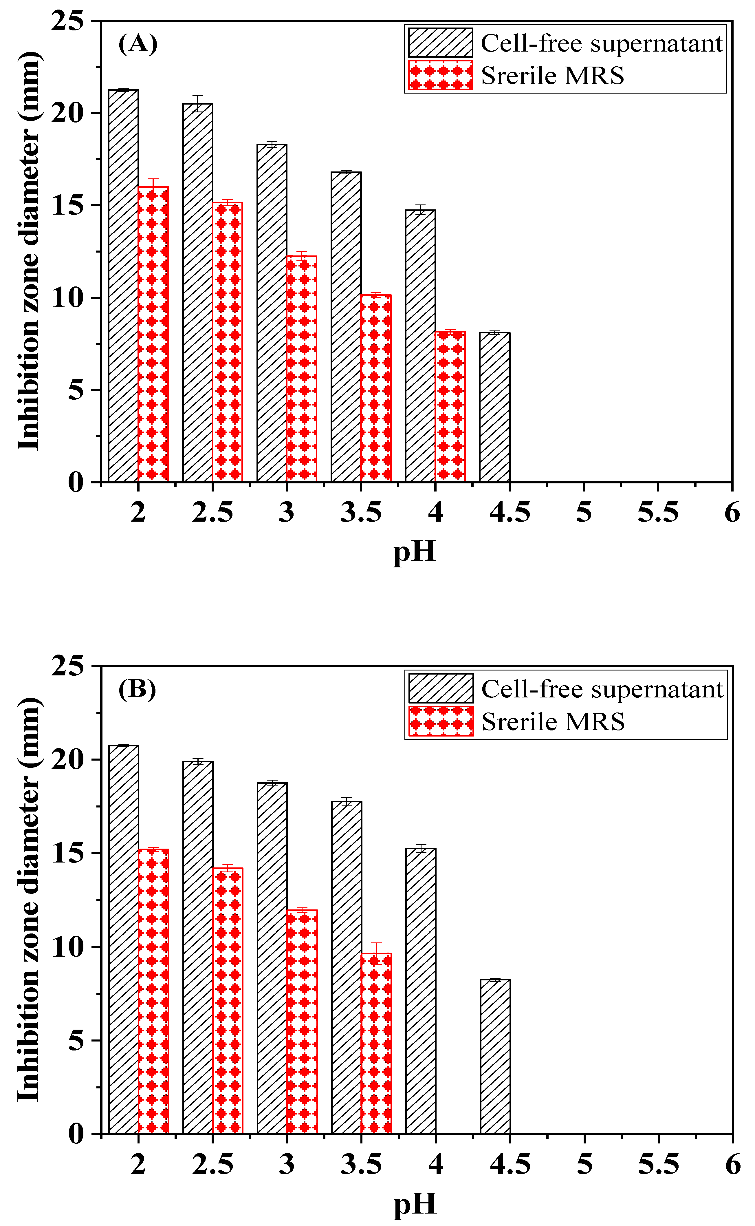
Cell-free supernatants of strain A5 with different pH values were investigated to carry out antibacterial tests (Figure 6). Results showed that the antibacterial activity was strong at pH 2.0, which might be due to the effect of organic acids. These acids can pass the membrane of the bacterium in an undissociated form, thereby decreasing the internal pH of the cell and causing considerable levels of additional stress. With the increase in the pH values, the antibacterial activity of the cell-free supernatant declined and was completely lost at a pH above 5.0. It was demonstrated that the bacteriocin in supernatant possessed better antimicrobial activity under acidic conditions. The purified bacteriocin SLG10 sustained antibacterial activity in a wide pH spectrum of 2.0–7.0, but the activity was reduced at pH 8.0 and above. The bacteriocin RC20975, plantaricin GZ1-27, and plantaricin JLA-9 were inactivated when confronted by the alkaline medium. The plantaricin P1053, belonging to Class II, was pH resistant and had a low molecular mass. These results suggested that the bioactive compounds secreted by the strain A5 possessed pH resistance, which was notably advantageous to the food processing industry.
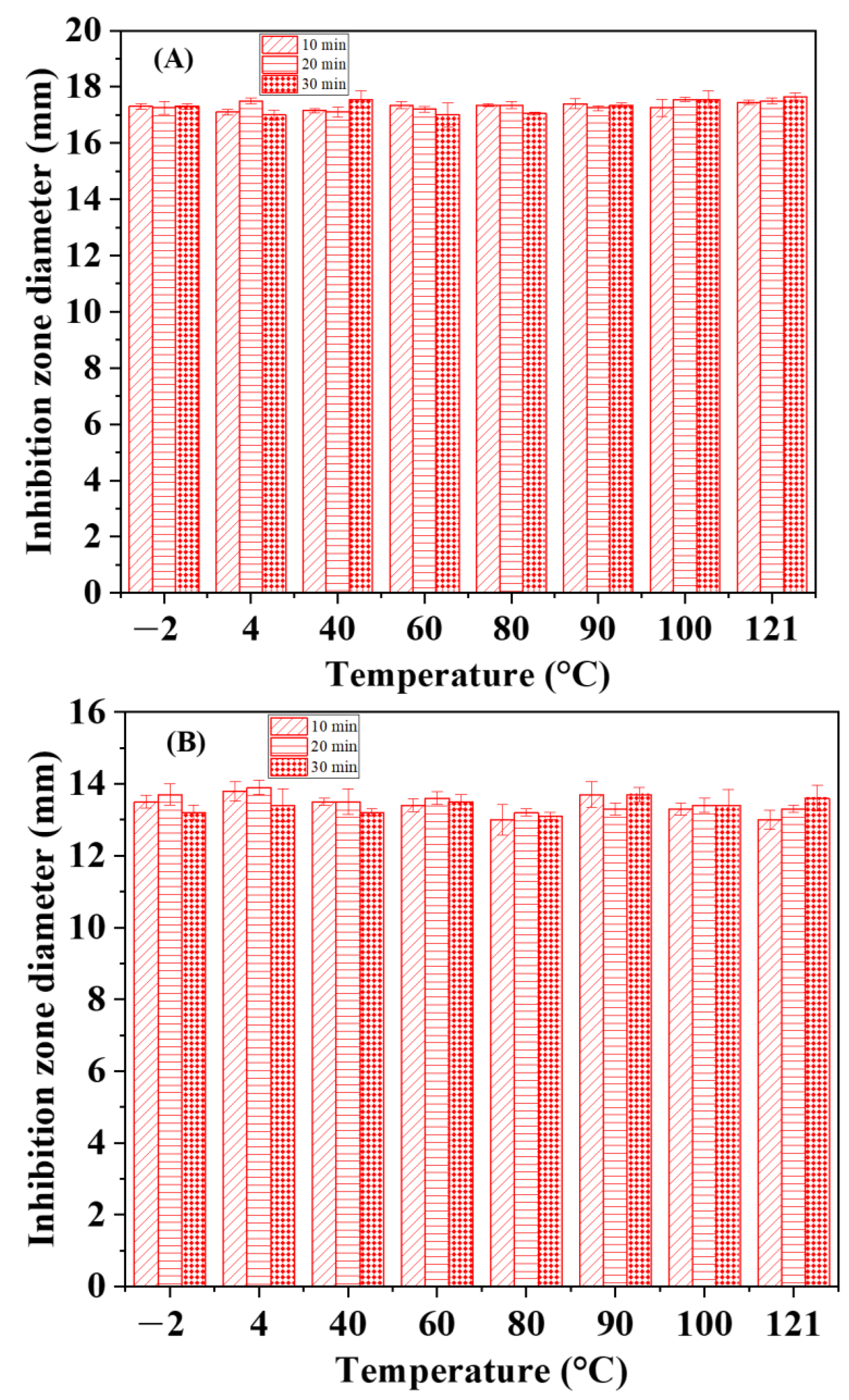
The bacteriocin produced by strain A5 possessed relatively high thermo-stability at lower and moderate temperatures or at 100 °C for 10, 20 or 30 min. After treatment at 121 °C for 30 min, the antibacterial activity did not change (Figure 7). The cell-free supernatant of L. crispatus 156 completely lost its antibacterial activity by incubating at 100 °C for 30 min and after autoclaving. However, some small-mass peptides, such as plantaricin JLA-9 (1004 Da), plantaricin K25 (1772 Da), and bacteriocin SLG10 (1422 Da) were insensitive to the heating attributed to their antibacterial activities [22]. The elevated thermal stability is vital as a food preservative because of the involvement of the heating process in many food processing industries. The cell-free supernatants were subjected to ultraviolet irradiation at different times and the resultant inhibitory activity profile is shown in Figure 8. Based on the findings, the inhibitory ability of bacteriocin in the supernatant against E. coli and B. subtilis was almost unchanged indicating the stability of bacteriocin towards UV denaturation. UV irradiation could trigger the disruption of the cell but not influence the antibacterial activity of the cell-free supernatant of strain A5, so the bioactive compounds have potential uses in the food industry.

3.5. Extraction and Purification of Bacteriocins

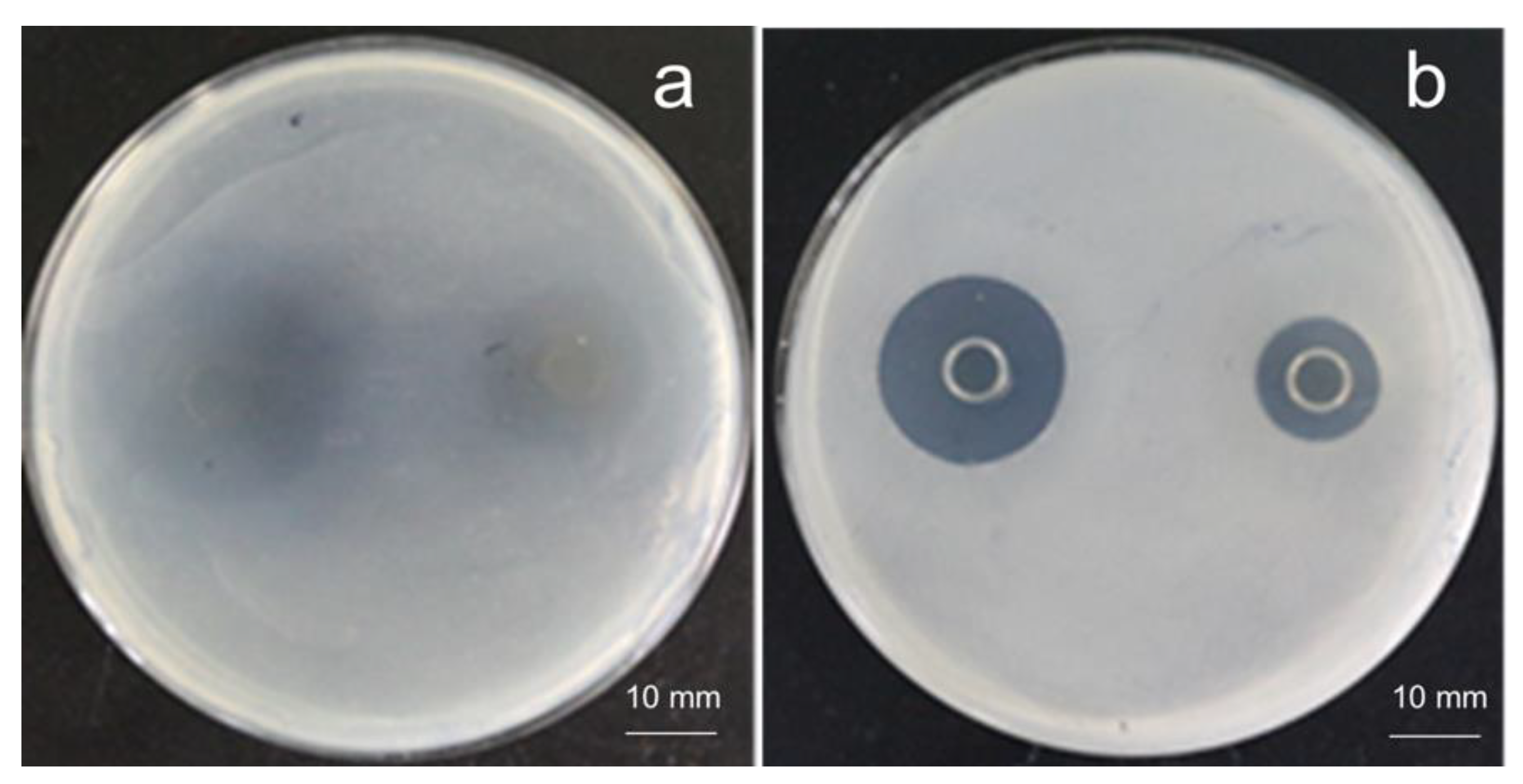
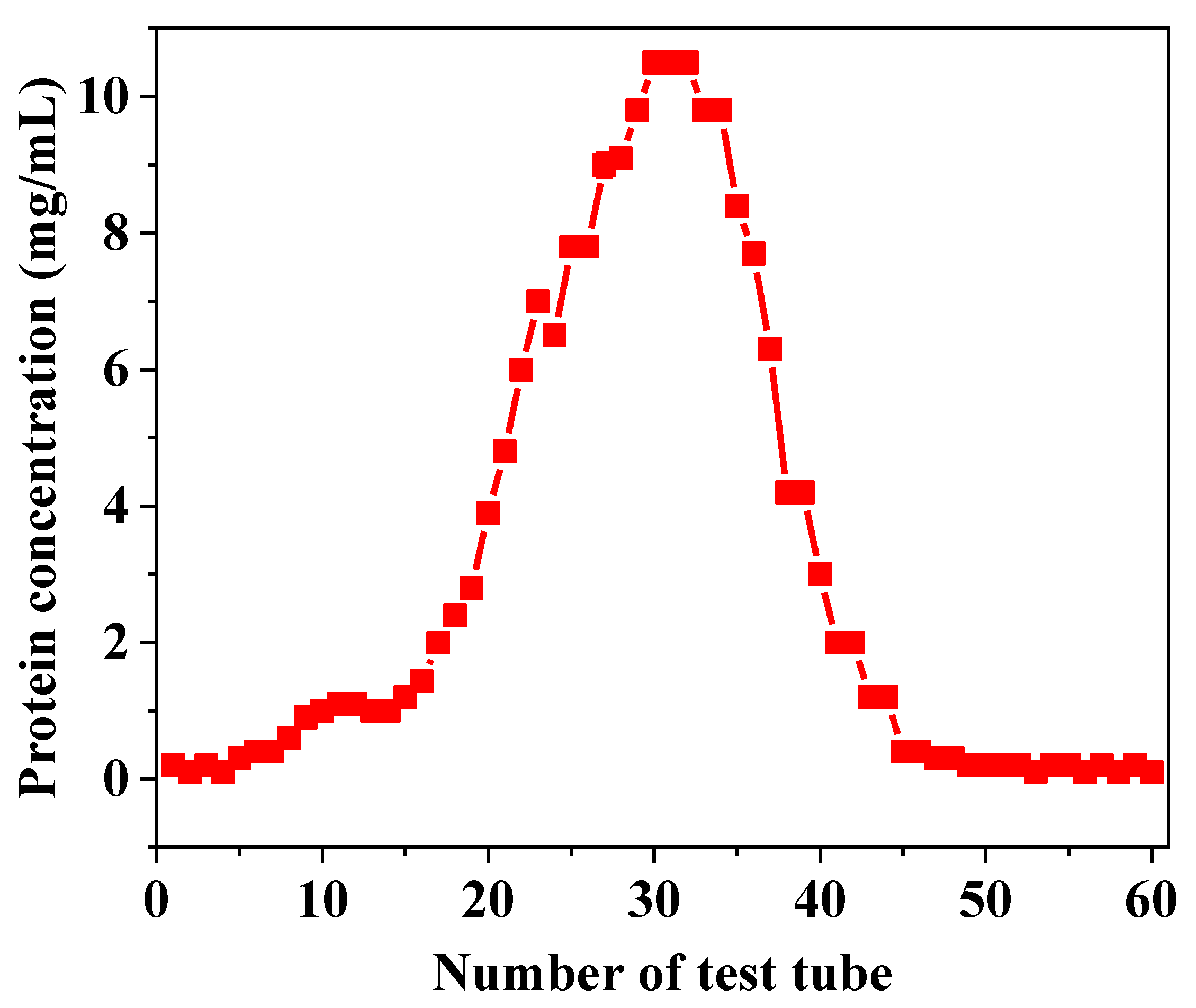
After the cell-free supernatant of strain A5 was extracted by methanol, the upper layer fractions were found to be inhibitory against E. coli and B. subtilis owing to the condensation of lactic acid in the upper layer. The clear inhibition zones were observed in plates of E. coli and B. subtilis, which were inoculated with the lower layer fractions (Figure 9). The lower layer fraction in which the peptides/proteins were precipitated by methanol was further purified using Sephadex G-75. The concentration after condensation was 80 mg/mL and the concentration of peptides/proteins per tube after elution can be seen in Figure 10. The pigment was concentrated in tube 8~18 and proteins were mainly in tube 19~40, while the highest concentration was in tube 30~32.
Six tubes containing a higher concentration of peptides/proteins and stronger antibacterial activity against B. subtilis and E. coli were determined using SDS-PAGE (Table 4). The molecular mass of peptides/proteins was smaller than 14 kDa corresponding to the bands of SDS-PAGE (Figure 11). The detailed components or structures require further study. The molecular mass of Limosilactobacillus mucosae AN1 was 10.66 kDa. The bacteriocin was comprehensively understood; however, only pediocin PA-1 and nisin have been commercially applied. The nisin-resistance continuously emerged and prompted people to discover novel bioactive proteinaceous compounds [30]. Additionally, the exploration of new bacteriocins with broad inhibition activities can be applied in controlling unwanted pathogenic microorganisms in food.

4. Conclusions

Among the five LAB strains isolated from pickles, strain A5 was able to produce a bacteriocin that strongly inhibited the growth of Bacillus subtilis, Escherichia coli, Salmonella enterica, and Staphylococcus aureus. This strain was classified as Lacticaseibacillus rhamnosus based on phenotypical and physiological identification and characterization. The bacteriocin produced by strain A5 displayed potential antibacterial properties against a wide range of food spoilage bacteria. After diminishing the influence of hydrogen peroxide and organic acids, the antibacterial activity was unchanged; however, it was sensitive to protease. The bacteriocin produced by the strain A5 possessed relatively high acidic endurance and thermal stability at lower and moderate temperatures or 100 °C for 10, 20 or 30 min. After treatment at 121 °C for 30 min, the antibacterial activity did not change. The heat stability and inhibitory characteristic indicated that it might be a new bacteriocin synthesized by Genus Lacticaseibacillus rhamnosus. Antibacterial activity of cell-free supernatant of strain A5 was substantiated by the synthesis of bacteriocin and further corroborated by a molecular weight of smaller than 14 kDa by SDS-PAGE. These findings might establish the basis for the future application of bacteriocins as bio-preservatives in the food processing industry. Future studies should purify this bacteriocin as a bio-preservative for food storage, substituting chemical preservatives for consideration in food security and human health. Therefore, finding the right method to purify bacteriocin is a key factor behind this work.

Author Contributions

Conceptualization, S.R. and F.L.; Data curation, S.R. and X.Y.; Formal analysis, X.Y., F.L. and F.F.; Investigation, S.R.; Methodology, S.R., M.B. and F.F.; Supervision, F.L.; Writing—Original draft, S.R., M.B., F.F. and H.M.N.I.; Writing—Review and editing, S.A.Z., M.B., X.Y., F.L. and H.M.N.I. All authors have read and agreed to the published version of the manuscript.

Funding

This work was financially supported by the Open project of Jiangsu Key Laboratory of Regional Resource Exploitation and Medicinal Research (No. LPRK201806).

Institutional Review Board Statement

Not applicable.

Informed Consent Statement

Not applicable.

Data Availability Statement

The data and contributions presented in the study are included in the article. Further inquiries can be directed to the corresponding author.

Conflicts of Interest

The authors declare no conflict of interest.

References

  1. Braïek, O.B.; Morandi, S.; Cremonesi, P.; Smaoui, S.; Hani, K.; Ghrairi, T. Safety, potential biotechnological and probiotic properties of bacteriocinogenic Enterococcus lactis strains isolated from raw shrimps. Microb. Pathog. 2018, 117, 109–117. [Google Scholar] [CrossRef] [PubMed]
  2. Meade, E.; Slattery, M.A.; Garvey, M. Bacteriocins, potent antimicrobial peptides and the fight against multi drug resistant species: Resistance is futile? Antibiotics 2020, 9, 32. [Google Scholar] [CrossRef] [PubMed] [Green Version]
  3. Khodaei, M.; Sh, S.N. Isolation and molecular identification of bacteriocin-producing enterococci with broad antibacterial activity from traditional dairy products in kerman province of Iran. Korean J. Food Sci. Anim. Resour. 2018, 38, 172–179. [Google Scholar]
  4. Wasfi, R.; El-Rahman, O.A.A.; Mai, M.Z.; Ashour, H.M. Probiotic Lactobacillus sp. inhibit growth, biofilm formation and gene expression of caries-inducing Streptococcus mutans. J. Cell. Mol. Med. 2018, 22, 1972–1983. [Google Scholar] [CrossRef] [Green Version]
  5. Simons, A.; Alhanout, K.; Duval, R.E. Bacteriocins, antimicrobial peptides from bacterial origin: Overview of their biology and their impact against multidrug-resistant bacteria. Microorganisms 2020, 8, 639. [Google Scholar] [CrossRef]
  6. Cleveland, J.; Montville, T.J.; Nes, I.F.; Chikindas, M.L. Bacteriocins: Safe, natural antimicrobials for food preservation. Int. J. Food Microbiol. 2001, 71, 1–20. [Google Scholar] [CrossRef]
  7. Rajoka, M.S.R.; Zhao, H.; Lu, Y.; Lian, Z.; Li, N.; Hussain, N.; Shi, J. Anticancer potential against cervix cancer (HeLa) cell line of probiotic Lactobacillus casei and Lactobacillus paracasei strains isolated from human breast milk. Food Funct. 2018, 9, 2705–2715. [Google Scholar] [CrossRef]
  8. Rajoka, M.S.R.; Hayat, H.F.; Sarwar, S.; Mehwish, H.M.; Ahmad, F.; Hussain, N.; Shi, J. Isolation and evaluation of probiotic potential of lactic acid bacteria isolated from poultry intestine. Microbiology 2018, 87, 116–126. [Google Scholar] [CrossRef]
  9. Altay, F.; Karbancıoglu-Güler, F.; Daskaya-Dikmen, C.; Heperkan, D. A review on traditional Turkish fermented non-alcoholic beverages: Microbiota, fermentation process and quality characteristics. Int. J. Food Microbiol. 2013, 67, 44–56. [Google Scholar] [CrossRef] [Green Version]
  10. Arasu, M.V.; Al-Dhabi, N.A.; Rejiniemon, T.S. Identification and characterization of Lactobacillus brevis P68 with antifungal, antioxidant and probiotic functional properties. Indian J. Microbiol. 2015, 55, 19–28. [Google Scholar] [CrossRef]
  11. Zhou, F.; Zhao, H.; Bai, F.; Dziugan, P.; Zhang, B. Purification and characterisation of the bacteriocin produced by Lactobacillus plantarum, isolated from chinese pickle. Czech J. Food Sci. 2014, 32, 430–436. [Google Scholar] [CrossRef] [Green Version]
  12. Karim, N.; Djamel, D.; Ismail, F. Purification of pediocin PA-1 by immunoaffinity chromatography. J. AOAC Int. 2008, 91, 828–832. [Google Scholar]
  13. Moračanin, S.M.V.; Dukić, D.A.; Memiši, N.R. Bacteriocins produced by lactic acid bacteria—A review. APCBEE Procedia 2012, 2, 50–56. [Google Scholar]
  14. Zhang, J.; Bilal, M.; Liu, S.; Lu, H.; Luo, H.; Zhao, Y. Isolation, identification and antimicrobial evaluation of bactericides secreting Bacillus subtilis Natto as a biocontrol agent. Processes 2020, 8, 259. [Google Scholar] [CrossRef] [Green Version]
  15. Elyass, M.E.; Shigidi, M.T.; Attitalla, I.H.; Mahdi, A.A. Characterization and optimization of bacteriocin from Lactobacillus plantarum isolated from fermented beef (Shermout). Open J. Appl. Sci. 2017, 7, 83–97. [Google Scholar] [CrossRef]
  16. Abbasiliasi, S.; Tan, J.; Tengkuibrahim, T.; Bashokouh, F. Fermentation factors influencing the production of bacteriocins by lactic acid bacteria: A review. RSC Adv. 2017, 7, 29395–29420. [Google Scholar] [CrossRef]
  17. Altena, K.; Guder, A.; Cramer, C.; Bierbaum, G. Biosynthesis of the lantibiotic mersacidin: Organization of a type B lantibiotic gene cluster. Appl. Environ. Microbiol. 2000, 66, 2565–2571. [Google Scholar] [CrossRef] [Green Version]
  18. Klaenhammer, T.R. Genetics of bacteriocins produced by lactic acid bacteria. FEMS Microbiol. Rev. 1993, 12, 39–85. [Google Scholar] [CrossRef]
  19. Gao, Y.; Jia, S.; Gao, Q.; Tan, Z. A novel bacteriocin with a broad inhibitory spectrum produced by Lactobacillus sake C2, isolated from traditional Chinese fermented cabbage. Food Control 2010, 21, 76–81. [Google Scholar] [CrossRef]
  20. Zhou, B.; Zhang, D. Antibacterial effects of bacteriocins isolated from Lactobacillus rhamnosus (ATCC 53103) in a rabbit model of knee implant infection. Exp. Ther. Med. 2018, 15, 2985–2989. [Google Scholar] [CrossRef] [Green Version]
  21. Ramya, S.; Mythili, A. Sathiavelu. Recovery of bacteriocin (NISIN) from Lactococcus lactis and testing its ability to increase the shelf life of vegetables (carrot and beans). Res. J. Biol. Sci. 2010, 5, 727–730. [Google Scholar]
  22. Tiwari, S.K.; Srivastava, S. Purification and characterization of plantaricin LR14: A novel bacteriocin produced by Lactobacillus plantarum LR/14. Appl. Microbiol. Biotechnol. 2008, 79, 759–767. [Google Scholar] [CrossRef]
  23. Dischinger, J.; Josten, M.; Szekat, C.; Sahl, H.G.; Bierbaum, G. Production of the novel two-peptide lantibiotic lichenicidin by Bacillus licheniformis DSM 13. PLoS ONE 2009, 4, e6788. [Google Scholar] [CrossRef]
  24. Oscáriz, J.C.; Lasa, I.I.; Pisabarro, A.G. Detection and characterization of cerein 7, a new bacteriocin produced by Bacillus cereus with a broad spectrum of activity. FEMS Microbiol. Lett. 1999, 178, 337–341. [Google Scholar] [CrossRef] [Green Version]
  25. Lawton, E.M.; Cotter, P.D.; Colin, H.; Paul, R.R. Identification of a novel two-peptide lantibiotic, Haloduracin, produced by the alkaliphile Bacillus halodurans C-125. FEMS Microbiol. Lett. 2007, 267, 64–71. [Google Scholar] [CrossRef]
  26. Chehimi, S.; Pons, A.M.; Sablé, S.; Hajlaoui, M.R.; Limam, F. Mode of action of thuricin S, a new class IId bacteriocin from Bacillus thuringiensis. Can. J. Microbiol. 2010, 56, 162–167. [Google Scholar] [CrossRef]
  27. Tahiri, I.; Desbiens, M.; Lacroix, C.; Kheadr, E.; Fliss, I. Growth of Carnobacterium divergens M35 and production of Divergicin M35 in snow crab by-product, a natural-grade medium. LWT Food Sci. Technol. 2009, 42, 624–632. [Google Scholar] [CrossRef]
  28. Aspri, M.; O’Connor, P.M.; Field, D.; Cotter, P.D.; Papademas, P. Application of bacteriocin-producing Enterococcus faecium isolated from donkey milk, in the bio-control of Listeria monocytogenes in fresh whey cheese. Int. Dairy J. 2017, 73, 1–9. [Google Scholar] [CrossRef]
  29. Kaur, B.; Kaur, R. Isolation, identification and genetic organization of the ADI operon in Enterococcus faecium GR7. Ann. Microbiol. 2015, 65, 1427–1437. [Google Scholar] [CrossRef]
  30. Tulini, F.L.; Martinis, E.C.P.D. Improved adsorption-desorption extraction applied to the partial characterization of the antilisterial bacteriocin produced by Carnobacterium maltaromaticum C2. Braz. J. Microbiol. 2010, 41, 493–496. [Google Scholar] [CrossRef] [PubMed] [Green Version]
  31. Lowry, O.H.; Rosebrough, N.J.; Farr, A.L.; Randall, R.J. Protein measurement with the Folin phenol reagent. J. Biol. Chem. 1951, 193, 265–275. [Google Scholar] [CrossRef]
  32. Zheng, J.S.; Wittouck, E.; Salvetti, C.M.A.P.; Franz, H.M.B.; Harris, P.; Mattarelli, P.W.; O’Toole, B.; Pot, P.; Vandamme, J.; Walter, K.; et al. A taxonomic note on the genus Lactobacillus: Description of 23 novel genera, emended description of the genus Lactobacillus beijerinck 1901, and union of Lactobacillaceae and Leuconostocaceae. Int. J. Syst. Evol. Microbiol. 2020, 70, 2782–2858. [Google Scholar] [CrossRef]
Figure 1. Inhibition circle of strain A5 to indicator bacteria (a) E. coli; (b) B. subtilis; (c) S. enterica; (d) S. aureus. Strain A5 produced clear inhibition zone on agar plates. The bar is 10 mm.
Sustainability 14 09571 g001
Figure 2. (a) Morphology of strain A5 colony; (b) Cells of strain A5 stained by Gram staining (10 × 100). The colony was round (1~2 mm). The cells were rod (0.4 µm × 2 µm), Gram-positive.
Sustainability 14 09571 g002
Figure 3. Phylogenetic tree of strain A5 based on 16S rRNA gene sequence. A5 within the clade of is Lacticaseibacillus rhamnosus bacterium.
Sustainability 14 09571 g003
Figure 4. Inhibition zone of cell-free supernatant of A5 on plate with mixed indicative bacteria (E. coli mixed with B. subtilis) (a) untreated; (b) treated with papain; (c) treated with pepsin; (d) treated with trypsin. The inhibition zones were significantly decreased indicating the antibacterial substances were sensitive to proteases. The bar is 10 mm.
Sustainability 14 09571 g004
Figure 5. The growth and pH profile (A) and antibacterial activity (B) of strain A5.
Sustainability 14 09571 g005
Figure 6. Antibacterial activity of cell-free supernatant adjusted to different pH to indicator bacteria ((A). E. coli; (B). B. subtilis).
Sustainability 14 09571 g006
Figure 7. Antibacterial activity of cell-free supernatant under different temperatures to indicator bacteria (A). E. coli; (B) B. subtilis.
Sustainability 14 09571 g007
Figure 8. Antibacterial activity of cell-free supernatant under UV to indicator bacteria (E. coli; B. subtilis).
Sustainability 14 09571 g008
Figure 9. Antibacterial activity of cell-free supernatants condensed by methanol to indicator bacteria (a). E. coli; (b). B. subtilis; the inhibition circle on the left per plate was formed by upper layer supernatant; the right was by lower layer supernatant. The upper layer supernatant has a larger circle. The bar is 10 mm.
Sustainability 14 09571 g009
Figure 10. Eluent concentration of proteins.
Sustainability 14 09571 g010
Figure 11. The SDS-PAGE result of crude extractions of A5 cell-free supernatant.
Sustainability 14 09571 g011
Table 1. Antibacterial activity of five lactic acid bacteria to four indicator bacteria.
Strain No.Escherichia coliBacillus subtilisSalmonellaStaphylococcus aureus
A1-11.4 ± 0.3-10.0 ± 0.2
A2-13.8 ± 0.2-14.0 ± 0.4
A315.2 ± 0.212.5 ± 0.414.5 ± 0.313.6 ± 0.3
A4-12.0 ± 0.2-13.5 ± 0.2
A517.3 ± 0.314.5 ± 0.316.1 ± 0.114.2 ± 0.2
“-” indicates no inhibitory effect to indicator bacteria. Strain A5 exhibited bigger inhibition zone to four indicator bacteria.
Table 2. Antibacterial activity of cell-free supernatant of A5 and MRS media.
Solutions (pH 4.0)Indicator Bacteria
Escherichia coliBacillus subtilis
cell-free supernatant16.0 ± 0.215.1 ± 0.2
MRS adjusted by lactic acid10.3 ± 0.38.8 ± 0.2
MRS adjusted by hydrochloric acid--
“-” indicates no inhibitory effect to indicator bacteria. The antibacterial activity of cell-free supernatant was not only caused by organic acids.
Table 3. Diameter of inhibition zone (mm) of cell-free supernatant treated by heating.
Indicator BacteriaTreated at 80 °C for 10 minUntreated
Escherichia coli14.8 ± 0.215.1 ± 0.2
Bacillus subtilis15.3 ± 0.115.5 ± 0.3
Strain A5 was incubated at 80 °C for 10 min to eliminate the influence of hydrogen peroxide, its antibacterial activity was almost unchanged.
Table 4. The diameter of inhibition circle (mm) of eluent to indicator bacteria.
Tube No.Escherichia coliBacillus subtilis
208.0 ± 0.27.0 ± 0.2
2410.2 ± 0.19.4 ± 0.2
2811.3 ± 0.210.6 ± 0.1
3214.0 ± 0.313.4 ± 0.2
3612.3 ± 0.211.0 ± 0.3
407.8 ± 0.47.2 ± 0.2
Publisher’s Note: MDPI stays neutral with regard to jurisdictional claims in published maps and institutional affiliations.