Article

Use of Metagenomic Whole Genome Shotgun Sequencing Data in Taxonomic Assignment of Dipterygium glaucum Rhizosphere and Surrounding Bulk Soil Microbiomes, and Their Response to Watering

1
Department of Biology, College of Sciences, Princess Nourah bint Abdulrahman University, Riyadh 11617, Saudi Arabia
2
Department of Biology, College of Science, University of Jeddah, Jeddah 21493, Saudi Arabia
3
Department of Biology, Faculty of Applied Science, Umm Al-Qura University, Makkah 24381, Saudi Arabia
4
Department of Biochemistry, Faculty of Science, King Abdulaziz University, Jeddah 21589, Saudi Arabia
5
Department of Biochemistry, College of Science, University of Jeddah, Jeddah 21493, Saudi Arabia
6
Department of Chemistry, Al-Leith University College, Umm Al Qura University, Makkah 28434, Saudi Arabia
7
Department of Biology, College of Science and Arts at Khulis, University of Jeddah, Jeddah 21921, Saudi Arabia
8
Biological Sciences Department, College of Science & Arts, King Abdulaziz University, Rabigh 21911, Saudi Arabia
*
Author to whom correspondence should be addressed.
Sustainability 2022, 14(14), 8764; https://doi.org/10.3390/su14148764
Submission received: 31 May 2022 / Revised: 7 July 2022 / Accepted: 11 July 2022 / Published: 18 July 2022

Abstract

:
The metagenomic whole genome shotgun sequencing (mWGS) approach was used to detect signatures of the rhizosphere microbiomes of Dipterygium glaucum and surrounding bulk soil microbiomes, and to detect differential microbial responses due to watering. Preliminary results reflect the reliability of the experiment and the rationality of grouping microbiomes. Based on the abundance of non-redundant genes, bacterial genomes showed the highest level, followed by Archaeal and Eukaryotic genomes, then, the least abundant viruses. Overall results indicate that most members of bacteria have a higher abundance/relative abundance (AB/RA) pattern in the rhizosphere towards plant growth promotion, while members of eukaryota have a higher pattern in bulk soil, most likely acting as pathogens. The results also indicate the contribution of mycorrhiza (genus Rhizophagus) in mediating complex mutualistic associations between soil microbes (either beneficial or harmful) and plant roots. Some of these symbiotic relationships involve microbes of different domains responding differentially to plant root exudates. Among these are included the bacterial genus Burkholderia and eukaryotic genus Trichoderma, which have antagonistic activities against the eukaryotic genus Fusarium. Another example involves Ochrobactrum phage POA1180, its bacterial host and plant roots. One of the major challenges in plant nutrition involves other microbes that manipulate nitrogen levels in the soil. Among these are the microbes that perform contraversal actions of nitrogen fixation (the methanogen Euryarchaeota) and ammonia oxidation (Crenarchaeota). The net nitrogen level in the soil is originally based on the AB/RA of these microbes and partially on the environmental condition. Watering seems to influence the AB/RA of a large number of soil microbes, where drought-sensitive microbes (members of phyla Acidobacteria and Gemmatimonadetes) showed an increased AB/RA pattern after watering, while others (Burkholderia and Trichoderma) seem to be among microbes assisting plants to withstand abiotic stresses. This study sheds light on the efficient use of mWGS in the taxonomic assignment of soil microbes and in their response to watering. It also provides new avenues for improving biotic and abiotic resistance in domestic plant germplasm via the manipulation of soil microbes.

1. Introduction

Traditional medicine is routinely used in Saudi Arabia as herbal remedies and spiritual healing, such as the perennial shrub Dipterygium Decne (family Capparidaceae), which is a monotypic genus with one species, namely D. glaucum [1,2]. This wild plant exists in hot, arid areas, as an xerophyte or facultative halophyte, which makes it highly adaptive to drought and saline conditions [3]. The medicinal value of this plant is based on synergy of different metabolites, mostly existing in the plant leaves that are promoted by environmental stresses. These metabolites include alkaloids and many other compounds, such as cardiac glycoside, flavinoides, cumarins, cyanides, anthraquinones, saponins, terpenoids and sterols [1,4,5]. The plant is also rich in antioxidants and antispasmodic metabolites and has phytotoxic activity [6]. D. glaucum has multiple other medicinal uses, such as treatment of breathing problems, jaundice and psoriasis [4]. The plant also harbors phytochemicals that contribute to the human’s ability to resist pathogens, especially those that have acquired resistance against standard treatments [7].
Availability of water is important for securing sustainable agriculture in Saudi Arabia. Beneficial plant-associated bacteria, e.g., plant growth-promoting bacteria (PGPB), can promote vigor of plants grown in the wild by alleviating stresses of the surrounding environment via reprogramming several stress-induced physiological and molecular processes. Thus, it is important to understand the mechanisms which these PGPB utilize when plants are exposed to conditions where water is scarce, on the one hand, and upon re-watering, on the other hand [8].
Previous studies suggest that standard soil maintains the highest level of biodiversity of microorganisms on Earth and contains ~1000 Gb of microbial genome sequences per gram [9]. These microorganisms play important roles in maintaining soil fertility, nutrient cycling or decomposition, and carbon sequestration. In terms of the rhizosphere soil attaching to plant roots, it is proven that root exudates, e.g., mucilage released by plant roots, are important compounds in signaling and shaping the microbial community (or rhizosphere microbiome) in such an environmental niche [10]. The rhizosphere microbiome contains important microorganisms, such as PGPB, which act synergistically on a symbiotic basis in not only promoting plant growth, but also suppressing activities of surrounding pathogens [11,12]. The microbial diversity and signatures in the rhizosphere soil differ from those in the bulk soil, basically due to the interacting host [13], where the bulk soil microbiome was found to be more complex than the rhizosphere microbiome of a given plant [14,15,16], while less complex than that of another plant [17,18,19]. The composition of root exudates can even change in terms of quantity and type during stages of plant growth [20,21,22,23]. Other factors that affect shaping of the bulk soil and rhizosphere microbiomes include environmental conditions, e.g., biotic and abiotic stresses [24].
Metagenomes are the sum of the genomes of archaea, bacteria, fungi (or eukaryotic microorganisms) and viruses of a given habitat. The main advantage of using metagenomic whole genome shotgun sequencing (mWGS) for taxonomic classification over marker gene or amplicon sequencing is the ability to detect microorganisms across all domains of life and overcome the bias due to choices of PCR primers used in marker gene sequencing [25]. mWGS is an untargeted (shotgun) sequencing approach to all (meta) microbial genomes (microbiomes) present in a given sample [26]. This approach can provide insights into taxonomy, biodiversity, evolution and potential functionality of a given microbiome and possible changes in its signature under different environmental conditions and symbiotic relationships with intact human, animal or plant cells [27]. The high sequencing depth of mWGS data can further allow the study of taxonomy assignment down to the species, or even to the strain level, as well as allow the assembly of draft genomes of cultured and uncultured microorganisms [28].
In the present study, we utilized mWGS in order to detect signatures of the rhizosphere microbiomes of Dipterygium glaucum and surrounding bulk soil microbiomes, and the possible differential responses of different microbiomes due to watering.

2. Materials and Methods

2.1. Watering Experiment and Soil Collection

The watering experiment was carried out in the north western region of the Mecca district of Saudi Arabia, where the Dipterygium glaucum wild plant grows naturally [29]. We selected a spot for the watering experiment that received no rainfall for >3 months prior to the experiment. Six plots were assigned for the experiment (1 m2 each), of which three plots contained single-grown similar-sized plants and the other three were nearby plots (≤10 m apart from the plants) of bulk soil only. For the watering experiment, the plots with the plants and bulk soil were watered in the early morning (25 L dH2O/plot), then, three rhizosphere lateral root and three bulk soil samples were collected every day after 0, 24 and 48 h of watering at ~10–30 cm depth, as previously described [30,31]. The collected samples were immediately put in liquid nitrogen, transported in dry ice and stored at −20 °C, then, soil pH was determined in a 1:1 (wt/wt) soil–H2O slurry prior to DNA extraction.

2.2. DNA Extraction and Whole Genome Shotgun Sequencing

Replicates of soil samples were collected from the plant rhizosphere every day, on the one hand, and those of the surrounding bulk soil were gathered at each watering time point, on the other hand. DNAs were extracted using the CTAB/SDS method. Then, purity and integrity were checked by electrophoresis on 1% agarose gels as the first layer of quality control (QC). DNA concentration was adjusted to 10 ng/μL using a dsDNA Assay kit in 2.0 Flurometer (Life Technologies, Carlsbad, CA, USA) prior shipment to Novogene Co., Ltd., Singapore, for whole genome sequencing. An amount of 1 μg DNA per sample was used as an input material for mWGS sequencing. Physical fractionation was done by a Covaris Sonicator and microbiomes were checked for enrichment by Agilent2100 and qPCR. A second layer of QC was done in terms of data pre-processing by trimming low quality bases (Q-value ≤ 38) exceeding a 40-bp threshold, and removing reads with N nucleotides over a 10-bp threshold and overlapped reads with adapter > 15-bp threshold by default. Then, filtered effective clean data were obtained and used further for processing and bioinformatics analysis.
Sequencing libraries were generated from clean data using an Ultra DNA Library Prep kit for Illumina (NEB, Ipswich, MA, USA) following the manufacturer’s instructions. DNA samples were fragmented by sonication to obtain 350 bp-band size, then DNA fragments were end-polished, adenine-tailed and ligated with the full-length adaptor to approach PCR. Then, amplicons were purified (AMPure XP System) and libraries were analyzed for size distribution by an Agilent2100 Bioanalyzer and quantified using qPCR. The clustering with paired-end reads of the index-coded samples was performed on a cBot Cluster Generation System according to the manufacturer’s instructions. Then, the library preparations were sequenced on the Illumina HiSeq 2500 platform.
Generated datasets were assembled using MEGAHIT (K-mer = 55) and the chimera was removed following standard methods as an extra layer of QC. The unutilized reads of each sample were mixed together in one sample (namely, NOVO_MIX) and assembled in order to explore the information of low-abundance species of the different samples. Generated scaffolds from utilized and unutilized reads were cut off at “N” to get N-free fragments or scaftigs [32], and those with ˂500 bp length were removed as a further QC step. Clean data were mapped to assembled scaftigs using Soap 2.21, then, effective scaftigs (found in ≥2 samples) were used for further analysis.
Gene prediction was carried out by MetaGeneMark based on the assembled scaftigs to predict >100 nt ORFs [32,33,34,35,36]. Predicted genes with identity of 95% and coverage of 90%, by default, were subjected to dereplication using Cluster Database at High Identity with Tolerance (CD-HIT) [37,38] in order to cluster and construct initial non-redundant gene catalogues (nrGC). The longest ORFs were chosen as the representative genes (Unigene), while ORFs with ≤100 nt length were removed. Non-redundant genes were constructed from predicted genes after redundant genes were removed using a greedy pairwise comparison, with highly stringent criteria of 95% identity over 90% of the shorter gene length [39]. Then, non-redundant genes were further incorporated in gene abundance analysis and assigned to taxonomic groups using a pipeline provided by Novogene Co. As an extra layer of CQ, metagenomic contigs were assigned taxonomically if 95% of its reads were annotated to the same species. Less than this percentage is considered chimeric contigs and removed.
Taxonomic profiling was generated by the mapping of sequencing contigs/reads against catalogues generated from different microbiome samples in order to link genes to metagenome-assembled genomes (MAGs) and reconstruct the full-length 16S rRNA gene. Then, the sequencing datasets were searched for operational taxonomic units (OTUs) using UPARSE software, where sequences with 97% similarity were assigned to the same OTUs. Representative sequences of each OTU were screened for further annotation. The Ribosomal Database Project 3 Classifier (Version 2.2) was used to conduct further analysis based on OTU clustering and species annotation; then, the abundance information of the gene catalogue of each sample was obtained. Taxonomic annotation was done by comparing metagenomic contigs/reads to the non-redundant (NR) gene abundance DIAMOND database [40] of taxonomically informative gene families and abundance tables of different taxonomic ranks (domain down to the species level) were generated. Then, annotation was done using the binning reference-based classification method, MEGAN [41,42]. According to the resulting abundance table of each taxonomic level, various analyses were performed, including correlation coefficient analysis between sample pairs, principle component analysis (PCA), as well as Venn diagram and core/pan rarefaction. Then, relative abundance at different rank levels were carried out to explore taxon composition. Core/pan and Venn diagram analyses were carried out in order to detect the unique and shared genes among samples and watering groups. PCA based on Bray–Curtis distances was performed to investigate beta diversity patterns. Relative abundance of a feature derived from a given taxon was calculated by summing up the relative abundances of the other members assigned to the same feature.

3. Results

Metagenomic whole genome shotgun sequencing (mWGS) was performed for the rhizosphere microbiome of Dipterygium glaucum and the surrounding bulk soil microbiome in order to make a taxonomic assignment of microbes in the two types of soil and detect differential abundance of microbes due to watering at 0, 24 and 48 h time points. We ensured that the amount of applied water was enough to keep the soil moist across the watering experiment. The pH of soil during sampling was 7.8, while the temperature was 38 °C. In terms of soil organic carbon (SOC), a prior report indicated that its concentration in this region is 15.9 g C kg−1, while its pool is 6.7 kg C m−2 [43]. Average rainfall in this region according to Climate Change Knowledge Portal, Climatology, Saudi Arabia is 400–600 mm annually. Nomenclature of the microbiome groups was based on soil type (S for bulk soil microbiome and R for rhizosphere microbiome) and time after watering (group A for 0 h, group B for 24 h and group C for 48 h). Statistics of the raw metagenomic data and Venn diagram are, respectively, available in Figure S12 and Table S22 of Supplementary Document file. While, distribution of ORFs (nt) and scaftig (e.g., length of ≥ 500 bp) lengths as well as core-pan rarefaction curves are, respectively, available in Figures S1 and S2.

3.1. Correlation Coefficient and Principal Component Analyses

The results of the heat diagram of the correlation coefficient between sample pairs are shown in Figure S3. Positive correlations were shown between pairs of rhizosphere microbiome samples, while negative correlations were shown between any given rhizosphere microbiome versus any given bulk soil microbiome, and vice versa. These results support our claim that microbiome structures of the two soil types should differ. The strength of correlations involving samples of group B (P1S2 or P1R2) was lower than correlations involving none of the group B samples. This observation might refer to the higher influence of watering on microbiomes collected at the 24 h time point than on microbiomes collected at the 0 or 48 h time points (Figure S3).
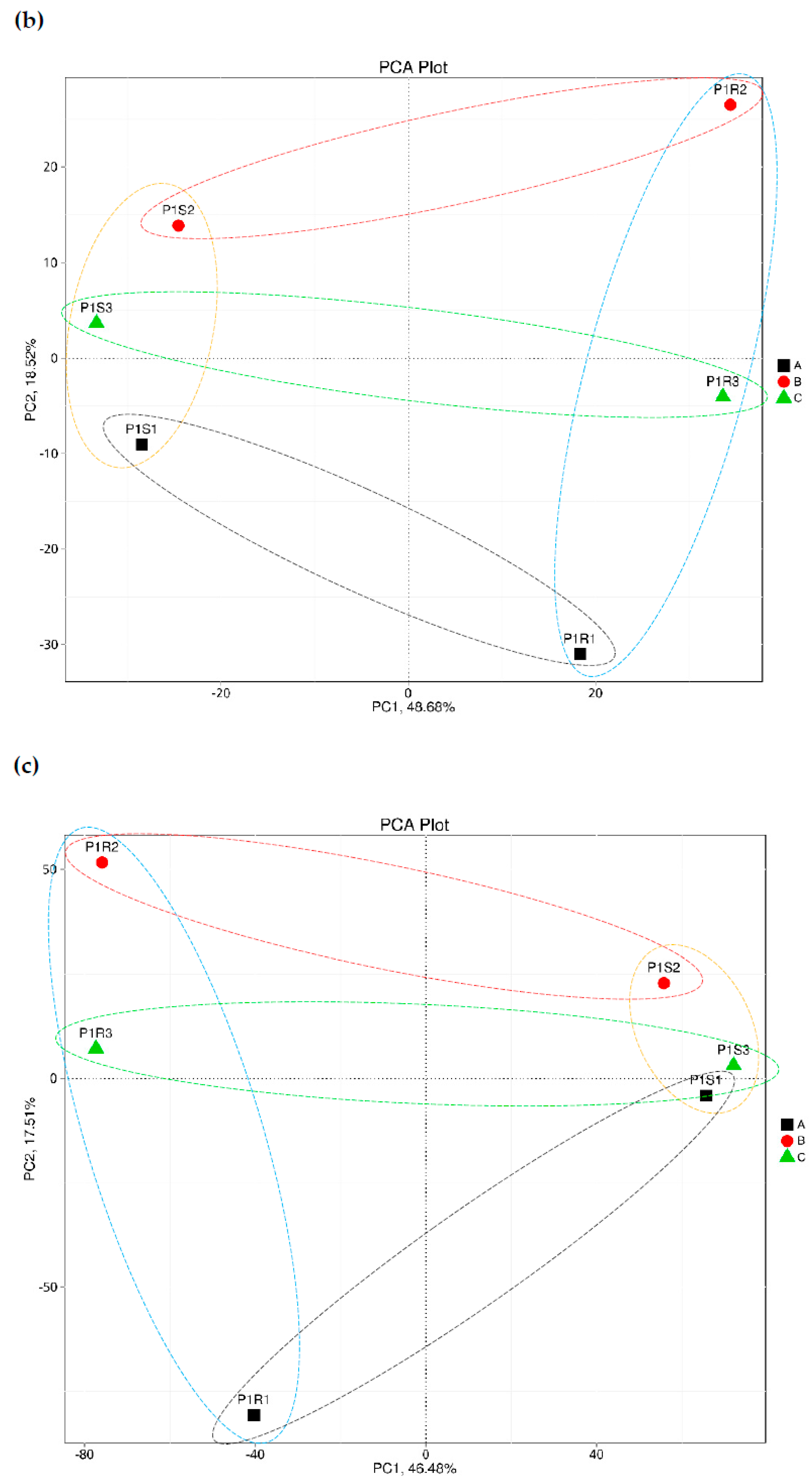
Principal component analyses (PCA) based on the number of genes at phylum, genus and species levels are shown in Figure 1. The results indicate a discernible separation among samples of bulk and rhizosphere soil microbiomes across the three taxonomic levels. Interestingly, the PCA1 (or PC1) axis completely separated samples based on soil type, while the PCA2 (or PC2) axis completely separated samples based on watering time point. At both the phylum and species levels, bulk soil microbiomes are located on the positive side of the PC1 axis, while rhizosphere microbiomes are located on the negative side. At genus level, microbiomes of the two types of soils switched their sides. Across the three taxonomic levels, group A microbiomes were located on the negative side of the PC2 axis, group B microbiomes were located on the positive side of the PC2 axis, while those of group C were located in the middle (or around 0 position of the PC2 axis) (Figure 1).
Overall results of the correlation coefficient and principal component analyses reflect the reliability of the experiment and the rationality of grouping microbiomes based on soil type (e.g., bulk and rhizosphere) and time after watering (e.g., 0, 24 and 48 h).

3.2. Taxonomic Assignment Based on Gene Abundance

As indicated earlier, gene prediction and mapping were carried for the assembled scaftigs (Table S3) and recovered ORFs (Table S4) that accommodate the QC criteria. Then, non-redundant genes were clustered, and catalogues of the initial non-redundant genes (e.g., nrGC) were constructed (Table S5). Gene abundances were then assigned to the four different taxonomic levels, e.g., domain, phylum, genus and species (Tables S6–S9). Then, we studied the relative microbial abundance of the six different metagenomes (Tables S10–S13), as well as of groups of watering time points, e.g., 0 (group A), 24 h (group B) and 48 h (group C) (Tables S14–S17) across type of soil (e.g., S and R) at different taxonomic levels. The data based on type of soil (e.g., groups S and R) and those based on watering time point (groups A, B and C) within each of the four domains are summarized in Tables S18–S21.
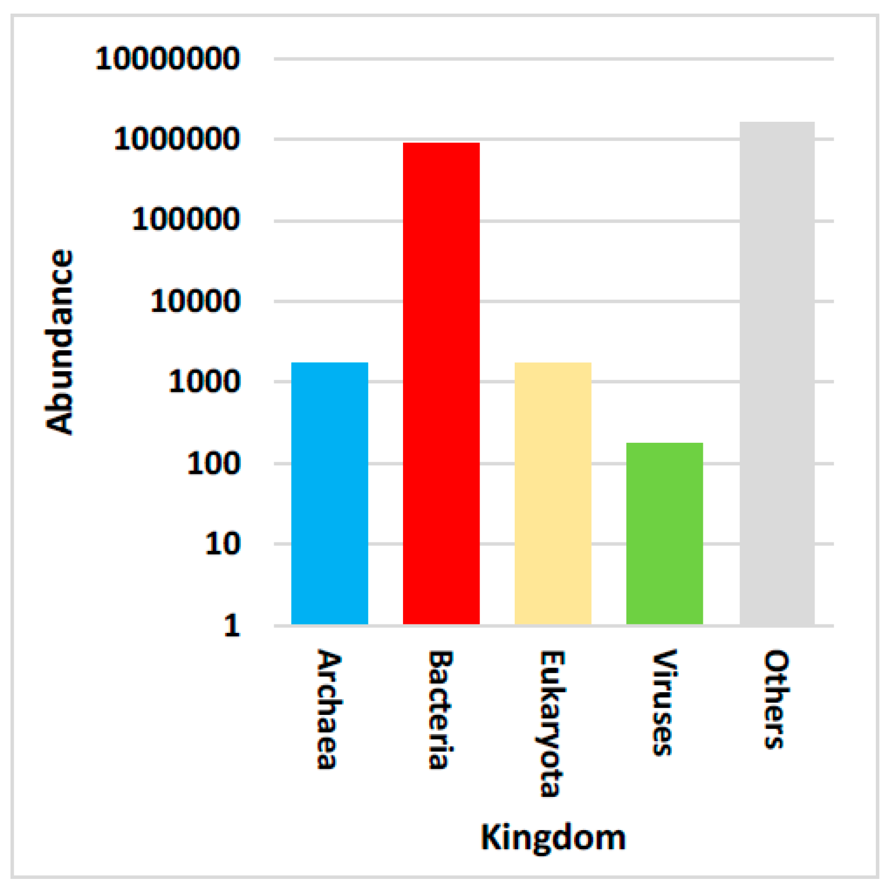
The studied microbial non-redundant genes involved genomes of the following domains: Archaea, Bacteria, Eukaryota and Viruses (virome). As anticipated, the analysis indicated the very high abundance of bacterial genomes, and almost equally followed by Archaeal and Eukaryotic genomes, while abundance of viromes was extremely low (Figure 2 and Figure S6). However, annotation indicated the lack of information for most taxa in the metagenomes, as gene abundance of unknown taxa was high (Figure 2). In addition, it is likely that most of the unknown genetic information reflects the high ration of low abundant unculturable microbes.
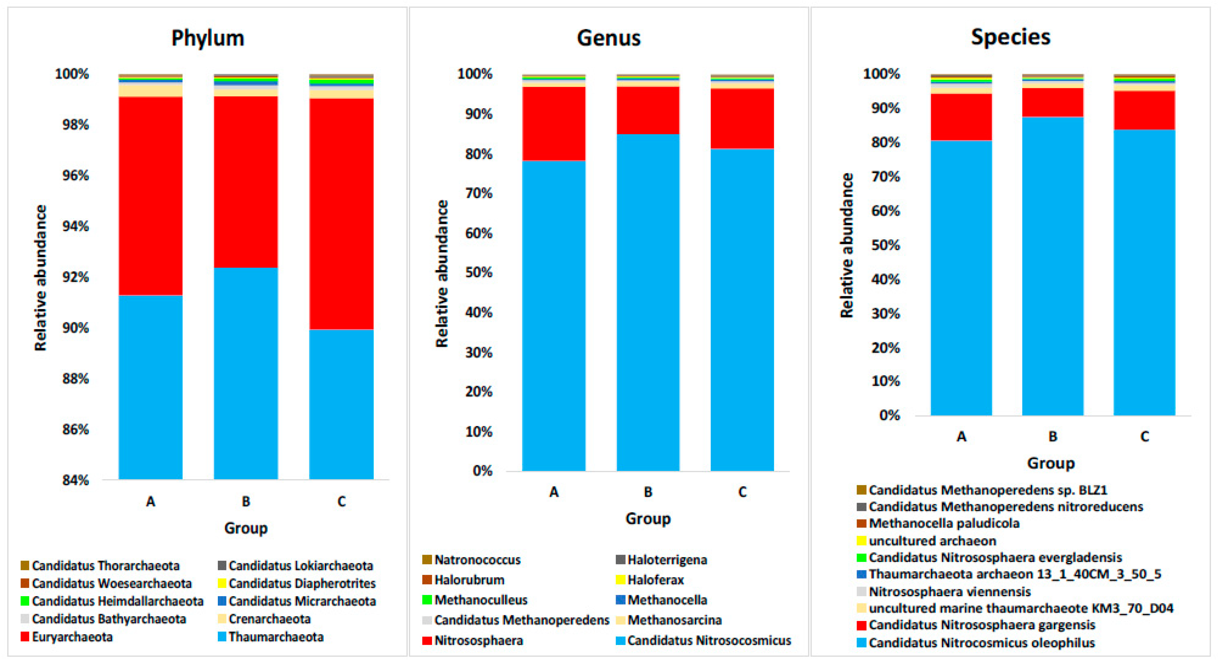
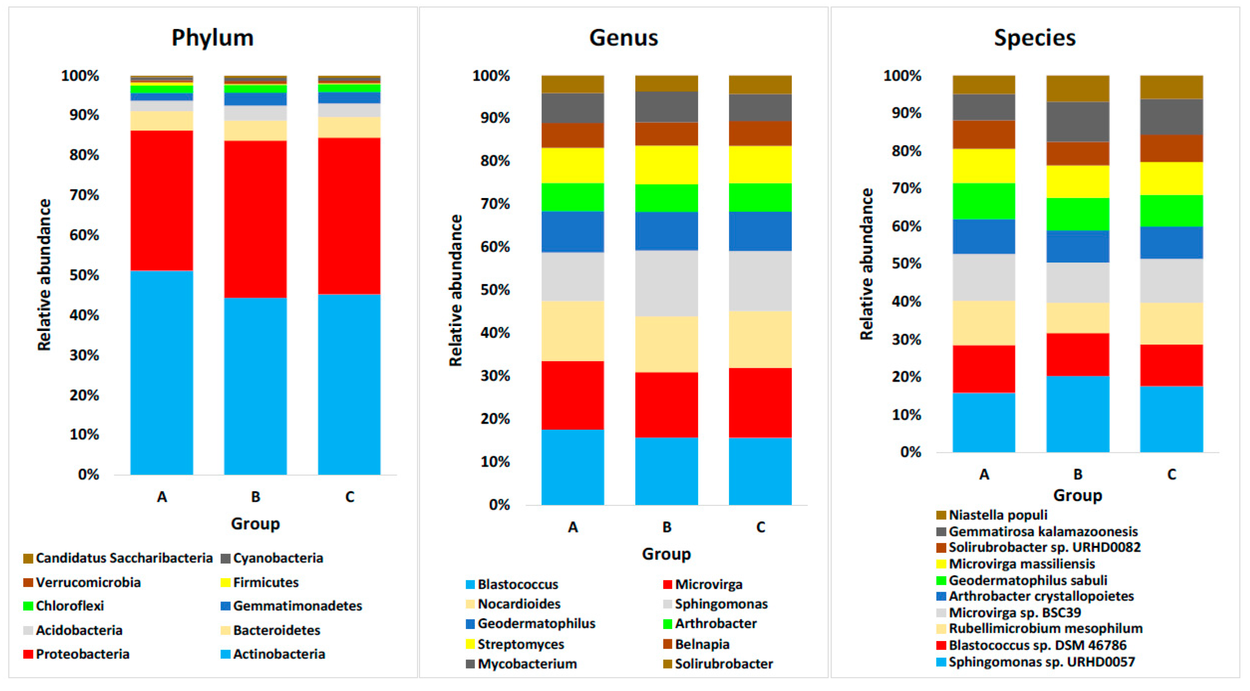
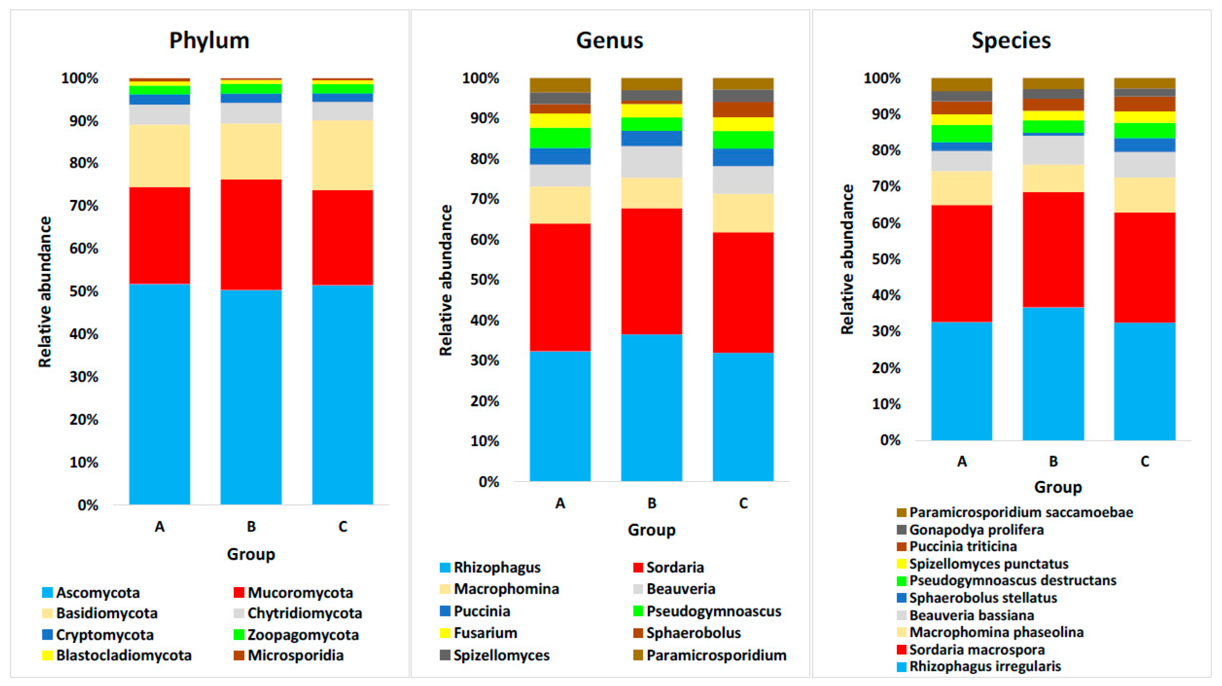
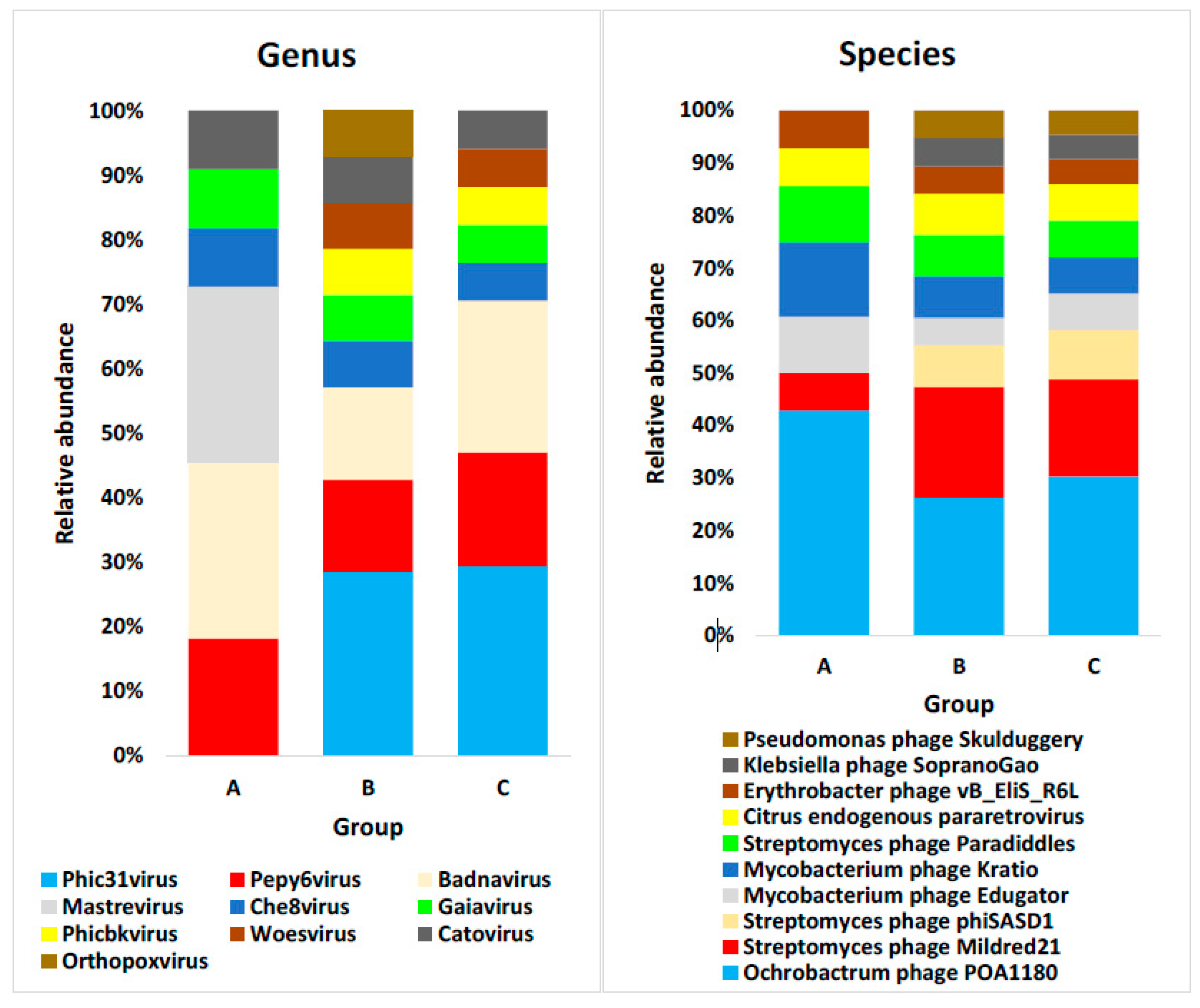
At the three taxonomic levels, e.g., phylum, genus and species, we chose the top 10 most abundant taxa across soil type (e.g., S and R) and time after watering (e.g., 0, 24 and 48 h) for further analysis (Figures S4–S6 and Tables S7–S9, respectively). At the phylum level, the most abundant taxa assigned by non-redundant genes include Thaumarchaeota, Euryarchaeota and Crenarchaeota of the Archaea domain, Actinobacteria, Proteobacteria and Bacteroidetes of the Bacteria domain, and Ascomycota, Mucoromycota and Basidiomycota of the Eukaryota domain. The Viruses domain has no available classification at the phylum level (Figure S4). At genus level, the most abundant taxa assigned by non-redundant genes include Candidatus Nitrosocosmicus, Nitrososphaera and Methanosarcina of the Archaea domain, Blastococcus, Microvirga and Sphingomonas of the Bacteria domain, Rhizophagus, Sordaria and Macrophomina of the Eukaryota domain, and Phic31, Pepy6 and Badnavirus of the Viruses domain (Figure S5 and Table S8). At species level, the most abundant taxa assigned by non-redundant genes include Candidatus Nitrocosmicus oleophilus, Candidatus Nitrososphaera gargensis and marine thaumarchaeote KM3_70_D04 of the Archaea domain, Sphingomonas sp. URHD0057, Rubellimicrobium mesophilum and Blastococcus sp. DSM 46,786 of the Bacteria domain, Rhizophagus irregularis, Sordaria macrospora and Macrophomina phaseolina of the Eukaryota domain, and Ochrobactrum phage POA1180, Streptomyces phage Mildred21 and Streptomyces phage phiSASD1 of the Viruses domain (Figure S6 and Table S9).
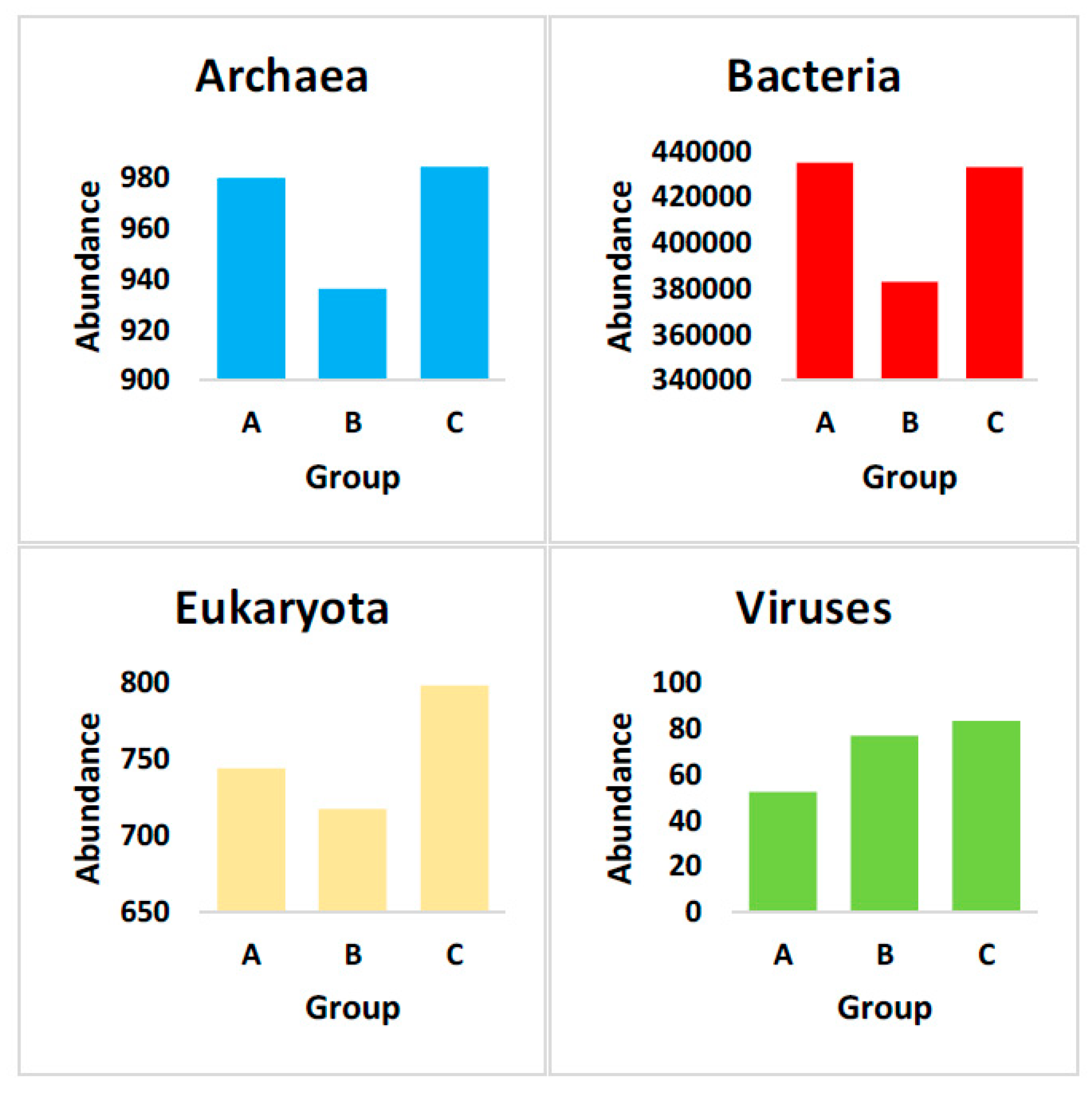
Then, we studied microbial abundance assigned taxonomically by non-redundant genes in metagenomes of the two soil types, e.g., rhizosphere (P1R1–P1R3) and bulk (P1S1-P1S3) across watering time points. This analysis was done among domains (Figure S7 and Table S10) and within the individual domains of Archaea (Figure S8 and Tables S11–S13), Bacteria (Figure S9 and Tables S11–S13), Eukaryota (Figure S10 and Tables S11–S13), and Viruses (Figure S11 and Tables S11–S13). Then, we studied microbial abundance assigned taxonomically by non-redundant genes in metagenomes after 0 (group A), 24 (group B) and 48 h (group C) of watering across soil types (e.g., S and R) among domains (Figure 3 and Table S10) and within the individual domains of Archaea (Figure 4 and Tables S11–S13), Bacteria (Figure 5 and Tables S11–S13), Eukaryota (Figure 6 and Tables S11–S13), and Viruses (Figure 7 and Tables S11–S13). It is important to note that we re-calculated relative abundance within each domain separately for the metagenomic groups A, B and C, as the original relative abundance datasets submitted by the bioinformatics team of the Novogene (Tables S14–S17) were calculated considering abundances of the four domains, which almost hid or vanished the very low abundant genes of the domains other than Bacteria. Abundance and relative abundance in the rhizosphere microbiome of Dipterygium glaucum and surrounding bulk soil microbiomes, as well as influence of watering across taxa of the four domains, are summarized in Tables S18–S21.

4. Discussion

One of the main targets of this study is to use data generated from metagenomic whole genome shotgun sequencing (mWGS) in taxonomic assignment of the Dipterygium glaucum rhizosphere microbiome and surrounding bulk soil microbiome, and shifts due to watering. Although the marker gene sequencing approach, also named the amplicon or metataxonomic sequencing approach, is faster and less expensive than mWGS (metagenomics), especially when the study includes large numbers of samples, mWGS proved to be more powerful in taxonomic assignment, especially when analyzing complex microbial communities such as those in the open environment [25]. During metagenomic classification, reads or assembled contigs are matched against a database of microbial genomes simply to identify microbiomes through a recent tool called metagenomics classifiers [25].
In this study, we detected the available patterns of abundance (AB) and relative abundance (RA) of the rhizosphere microbiome of D. glaucum and surrounding bulk soil microbiome for the top 10 highly abundant phyla, genera and species of the four domains, in a trial to understand the natural growth dynamics of microbes responding differently to the soil type and/or watering. Prior metagenomics research has focused mainly on studying relative abundance only, while almost no prior reports give enough emphasis to further studying the abundance of microbes in a given habitat, especially whether the extreme differential abundance of genomes of different domains is feasible. Therefore, we assume that the picture is not complete if relative abundance referring to beta diversity is studied alone. One other reason for this decision is that there are some pairs of microbes that naturally co-exist at a certain abundance level, regardless of their relative abundance level within a microbiome.
We assume that the growth dynamics of microbes in the present study that showed similar patterns of abundance (AB) and relative abundance (RA), namely AB/RA, e.g., in terms of soil type and/or watering time points, can contribute to all the above-mentioned mechanisms of growth dynamics. An AB/RA pattern refers to a taxon whose AB is higher in a given soil type or watering time point, and also shows a higher RA in the same soil type or watering time point.
The results in Tables S18–S21 indicate that AB/RA patterns of a given taxon based on soil type do not show the AB/RA pattern in watering time point as well, except in two cases. When analyzing these cases, we found that higher AB/RA of a certain taxon in the rhizosphere microbiome is surprisingly accompanied by lower AB/RA in the watering group B (referring to 24 h watering time point) microbiome, compared with those of other soil types or watering time points (first case). In addition, when a higher AB/RA pattern exists in the bulk soil microbiome, a higher AB/RA pattern in the watering group B microbiome also exists (second case). The first case took place for the two archaeal phyla Euryarchaeota and Crenarchaeota and the bacterial species Rubellimicrobium mesophilum, while the second case took place for the eukaryotic genus Rhizophagus and the viral species Streptomyces phage Mildred21. We have no explanation for these cases and speculate that they occurred coincidentally.

4.1. Domain Archaea

In terms of Archaea, the phyla Euryarchaeota and Crenarchaeota showed a pattern of higher AB/RA in the rhizosphere microbiome than that in the bulk soil microbiome, and a lower AB/RA in the watering group B (referring to 24 h watering time point) microbiome than that of group A (referring to 0 h watering time point) (Table S18). Phylum Euryarchaeota is known for its ability to produce methane and survive moderate and extreme environmental conditions, while Crenarchaeota is a thermophilic and acidophilic archaeal phylum that colonizes plant roots and mediates influx of important minerals and has possible roles in root biology [44]. AB of the mycorrhizal population, e.g., genus Rhizophagus, was proven to correlate with those of euryarchaeotal and crenarchaeotal populations, due to their proven direct association (Figure 8) [44]. Nonetheless, archaeal phyla Euryarchaeota and Crenarchaeota showed higher AB/RA in the rhizosphere microbiome in the present study, while eukaryotic genus Rhizophagus showed contrary results (Tables S18 and S20, respectively). There is no clear explanation to the high AB/RA of genus Rhizophagus in the bulk soil, except that the high AB/RA in the bulk soil was diluted by its high AB/RA 24 h after watering. Moreover, members of the genus Rhizophagus not only associate with microbes in the bulk soil, but also with microbes in the rhizosphere microbiome as a bridge between these microbes (or pathogens) and the plant root (Figure 8). As the two highly abundant archaeal phyla can tolerate adverse environmental conditions, it is expected that watering results in higher AB of the other less water stress-tolerant taxa, which, in turn, might reduce the RA of the two phyla at the 24 h watering time point when water is available.
The genus Candidatus Nitrosocosmicus and its species Candidatus Nitrocosmicus oleophilus of the phylum Thaumarchaeota showed higher AB/RA in the plant rhizosphere microbiome than that in the bulk soil (Table S18). The microbe(s) can carry out both ammonia oxidation and nitrogen fixation, but a recent report indicated that the rate of the first action is much lower than that of the other mesophilic ammonia-oxidizing archaea (AOA) [45]. Moreover, ammonia oxidation only takes place upon increase in temperature [46]. This might result in a lower rate of ammonia oxidation than that of nitrogen fixation. In addition, species of the genus Nitrosocosmicus encode beta-1,2-mannosidase, which is a member of the CAZy family involved in the degradation of plant cell wall mannans and the production of sugars. Sugars, in turn, act as substrates for the pyrroloquinoline quinone (PQQ)-dependent glucose/sorbosone dehydrogenases that are further transported into the plant cell to promote central carbon metabolism [47]. This information justifies the high AB/RA of this genus in the rhizosphere microbiome.
The genera Nitrososphaera (along with its two species Candidatus Nitrososphaera gargensis and Candidatus Nitrososphaera evergladensis) and Candidatus Methanoperedens of the phyla Nitrososphaerota and Euryarchaeota, respectively, showed lower AB/RA in the watering group B microbiome (Table S18). The genus Nitrososphaera usually participates in oxidizing ammonia (AOA) in the ocean, while Candidatus Methanoperedens participates in two contradicting actions, e.g., oxidizing ammonia to produce nitrate and anaerobically oxidize methane (AOM) to reduce nitrate level. Again, it seems that the direction of the reaction depends on the culture which these microbes inhabit. As they usually favor salty water (ocean), non-salty water does not promote their growth. In the present study, they seem to instantly respond. Their response, similar to heat shock, to watering is to reduce their growth rate at the 24 h time point; then, they recover their growth rate afterwards, e.g., after 48 h of watering.

4.2. Domain Bacteria

The results of the phylum Actinobacteria in Table S19 indicate higher AB/RA in the rhizosphere microbiome compared to the bulk soil microbiome. This phylum is well-known for the production of several bioactive secondary metabolites to promote plant growth and for the production of several nitrogenous compounds, as well as organic acids, to compensate for any shortage in nitrogen in the plant rhizosphere and to carry out P solubilization [48]. Many other reports support the claim of high AB of Actinobacteria in the rhizosphere microbiome [49].
The phyla Acidobacteria and Gemmatimonadetes showed lower AB/RA at the 0 h watering time point. Both phyla showed higher AB in the plant rhizosphere microbiome, but, interestingly, showed no differential RA in either soil type (Table S19), although the phylum Acidobacteria was reported to have a strong symbiotic association with several plant hosts, as the phylum participates in carbon and sulfur metabolism in plant roots [50]. The phyla Actinobacteria and Gemmatimonadetes were frequently reported as plant growth-promoting bacteria, while other reports indicate that the phylum Acidobacteria shows higher AB in the bulk soil [51]. In terms of the influence of watering, Acidobacteria and Gemmatimonadetes were recently reported to be drought sensitive [52]; therefore, it is expected that their AB/RA pattern increases after watering (Table S19). Interestingly, members of the phylum Actinobacteria (e.g., genus Arthrobacter) were reported to promote plant growth, especially under drought stress, as they were resistant to desiccation and can survive under starvation conditions [53]. We refer these contradicting results to the differential growth dynamics of microbes, diversity of phylum members, and plant rhizosphere that the microbe inhabits. In the present study, the AB/RA pattern of the genus Arthrobacter in the rhizosphere microbiome was higher than that in the bulk soil microbiome (Table S19). These results partially align with those previously mentioned [53].
Seven of the top 10 highly abundant genera, e.g., Blastococcus, Microvirga, Nocardioides, Geodermatophilus, Belnapia, and Solirubrobacter, in addition to Arthrobacter, showed higher AB/RA in the plant rhizosphere microbiome, while Streptomyces showed contrary results (Table S19). None of these genera showed RA in terms of watering time points. No conclusive information is available for the genus Blastococcus and its descendent species Blastococcus sp. DSM 46,786 in terms of association with the plant rhizosphere, except that they survive in marine sediment and in stone interiors, thus, they are resistant to some heavy metals and metalloids [54]. The genus Rubellimicrobium contains only one species, e.g., Rubellimicrobium thermophilum Denner [55]. This species is a photoheterotroph that can grow photoautotrophically [56]. Although this species proved to be highly AB/RA in the rhizosphere microbiome, while showing lower AB/RA in watering group B, no further information is available in terms of its relationship with the plant root or response to watering. The genera Microvirga and Streptomyces were reported to act as endosymbionts [57], although the latter genus unexpectedly showed lower AB/RA in the rhizosphere microbiome in the present study. Members of the genus Microvirga were proven to be endophytes possessing an nodC gene that promotes nodulation in plant roots [58]. In fact, there is no solid support for the morphological characteristics of Dipterygium glaucum; thus, this expectation is required to be proven. The enus Nocardioides was also reported to be an endophyte possessing potent biocontrol activities against the wheat pathogen Rhizoctonia solani. Therefore, it is likely that members of the latter bacterial genus confer resistance against plant pathogens. More recently, Singh and co-authors [59] reported that this oxidative stress-tolerant microbe colonizes the rhizosphere of the Dracaena cochinchinensis plant. Members of the genus Geodermatophilus also colonize the rhizosphere of the Astragalus membranaceus plant [60]. Members of the genus Belnapia were proven to be effective colonizers that promote the growth of several plants, including arabidopsis [61], grave and cacti [62]. The latter microbes are located in the plant phyllosphere in order to influence effectively the growth and health of the host plant. However, prior knowledge about the genus Solirubrobacter, as well as the species Niastella populi and Gemmatirosa kalamazoonensis, is scarce, except that the first is a symbiont of maize [63], the second is a symbiont of Populus [64] and the third exists in organically managed agricultural soil, which seems to exist in the phyllosphere of plants. No information is available to justify the positive response of the third microbe in terms of AB/RA due to watering (Table S19).

4.3. Domain Eukaryota

The taxa that showed higher AB/RA in the bulk soil microbiome include four phyla, e.g., Mucoromycota, Basidiomycota, Zoopagomycota and Blastocladiomycota, and four genera, e.g., Macrophomina, Fusarium, Sphaerobolus, Spizellomyces, in addition to the genus Rhizophagus, compared with those in the rhizosphere microbiome of the Dipterygium glaucum plant. While only three taxa, e.g., the genus Sordaria and its descendent species Sordaria macrospora, as well as the species Pseudogymnoascus destructans, showed contrary results (Table S20). A possible reason for the higher AB of eukaryotic parasites in the bulk soil includes the inhibition of growth of these parasites in the rhizosphere microbiome by plant root exudates. When these pathogens are nearby plant roots, they might indirectly be beneficial by metabolizing complex molecules to simpler useful ones. Indirect relationships of parasite and plant roots that can be mediated by mycorrhiza avoid direct contact and occurrence of several diseases (e.g., rusts, smuts). Such a type of avoidance resistance of wild plants to these parasites might justify their low AB/RA in the rhizosphere microbiome. More explanation of this type of plant–microbe association is shown below. The question that is required to be answered is how this distant indirect contact is mediated.
Members of the phylum Mucoromycota are saprobic Zygospore-forming fungi that live in terrestrial soil or highly polluted water and are historically known to be among the first land colonizers. In addition, Mucoromycota members are considered as powerful factories for biorefinery applications [65]. They act as weak parasites of plants and animals in environments rich in organic matter and are called “sugar fungi” because they grow better on simple sugar substrates. Mucoromycota members also have the ability to assimilate complex organic compounds, e.g., carbon sources, and to accumulate several metabolites, including lipids, chitosan, polyphosphates and proteins [66], that are valuable to plants. Members of Blastocladiomycota are aquatic fungi that live in freshwater, mud and soil [67].
Members of Zoopagomycota are terrestrial obligate parasites of soil animals such as nematodes and amoebae, while some are parasites of plants. They are capable of growing on refractory materials, e.g., pollen, cellulose, chitin, etc. [68], and act as parasites of nematodes, midges and crustaceans, as well as plants, e.g., maize, alfalfa and several angiosperms, to cause several diseases (e.g., leaf brown spot). Meanwhile, members of Basidiomycetes include rusts, a well-known devastating pathogen of staple grain crops, and smuts that cause persistent crop losses. In order to recognize the way by which pathogens or parasites and nearby plants interconnect, several reports indicate that the fungal mycobionts Zoopagomycota and Basidiomycetes are both characterized by the previously described indirect plant associations via plant-based ecologies (e.g., mycorrhiza, root endophytes, etc.) [69]. Members of the genus Macrophomina are soil-borne fungi that cause several plant diseases, e.g., stem and root rot, seedling blight, etc. [70]. Pathogenicity of several fungi, including Macrophomina, is associated with the fungal ability to produce hydrolytic enzymes to degrade polysaccharides and lignocelluloses of the plant cell wall, then penetrate into the host tissue [71]. Again, the low AB of the latter three parasites, Zoopagomycota, Basidiomycetes and Macrophomina, in the plant rhizosphere microbiome indicates the possible release of root exudates that inhibit their propagation in the plant rhizosphere [69]. However, there is no prior supporting information to justify differential AB of the genus Sordaria and its descendent species Sordaria macrospora (Table S20), as they seem to present in animal feces, thus their presence in the microbiomes is coincidental.
The genus Fusarium is among the most important fungal genera that are environmentally ubiquitous and cause severe infections in several economical plants, e.g., cereals, banana, etc. As a soil-borne disease, Fusarium is difficult to control and the severity of infection extends to the production of mycotoxins that are dangerous for human and animal health [72]. It also causes root rot and wilt for plants and a reduced root growth that can increase the severity of the disease. The bacterial genus Burkholderia and eukaryotic genus Trichoderma have antagonistic activities acting synergistically against the eukaryotic genus Fusarium [73]. The two beneficial genera confer improved plant growth, disease resistance and abiotic stress tolerance [74]. Thus, we assume that these two genera represent a sort of natural biological control against Fusarium. To support this claim, our results showed higher AB of the two beneficial genera in the plant rhizosphere microbiome than that of bulk soil as a mechanism to inhibit the growth of the genus Fusarium in the rhizosphere microbiome of the Dipterygium glaucum plant (Figure 9 and Tables S12 and S20).
The eukaryotic genus Spizellomyces was also proven in the present study and in prior reports to be highly abundant in bulk soil [75], while Pseudogymnoascus destructans was shown to be highly AB/RA in the rhizosphere microbiome of Dipterygium glaucum (Table S20). As a saprotrophic fungus, Spizellomyces colonizes decaying plant material that seems to be provided by the nearby plants, while Pseudogymnoascus destructans was proven to live in wood or soil and can cause white-nose syndrome (WNS) in bats [76]. In addition, Sphaerobolus stellatus is known as the shotgun or artillery fungus because it discharges its spores with explosive forces, while Beauveria bassiana grows naturally in the soil and acts as a parasite and biological insecticide to control several pests [77]. None of the latter four taxa seems to have any justified response to either soil type or watering time point.
Recent reports relates less AB of the pathogens in the rhizosphere microbiome to the plant’s ability to resist them [78]. In addition to allelopathy and the indirect connection with the pathogens, explanations to plant resistance involve the action of arbuscular mycorrhizal fungi (AMF) via the previously described complex mutualistic association with plant roots (Figure 8). Other mechanisms include significant physiological and molecular changes in the plant, e.g., reprogramming of plant defense-related gene expression, decreased oxidative damage, superoxidase activity, etc., to reduce the severity of fungal diseases [79]. Plants can also produce secondary metabolites, e.g., alkaloids, saponins, flavonoids, anthraquinones, linoleic acid, etc., with antifungal characteristics [80].

4.4. Domain Viruses

At the virome level, as a part of the rhizosphere microbiome of Dipterygium glaucum, none of the genera showed AB/RA in either soil type or watering time points (Table S21). Rather, three species, e.g., Ochrobactrum phage POA1180, Mycobacterium phage Edugator and Mycobacterium phage Kratio, showed higher AB/RA in the rhizosphere microbiome than in the bulk soil, while two species, Streptomyces phage Mildred21 and Citrus endogenous pararetrovirus, showed contrary results (Table S21).
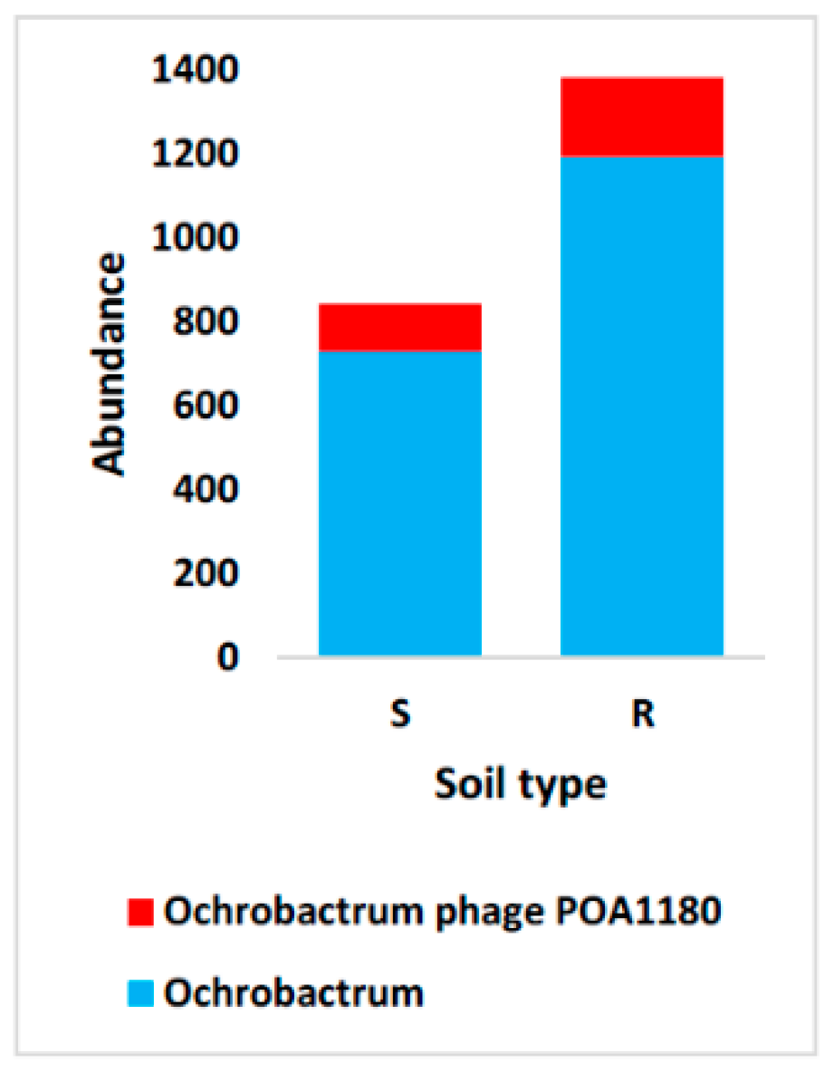
The prophages of Ochrobactrum naturally release phage particles, e.g., phage POA1180 (41,655 bp) with bacterial species O. intermedium as the most known host. However, this action is increased when the prophages are induced by various stress factors. The viral genome contains genes for DNA packaging and virion assembly, and other encoding terminase, transposase, methyltransferases, capsid protein, ATPase, transcriptional regulators and several tail proteins [81]. Moreover, the genome contains two genes that allow the bacteria to resist chromium [82]. In the soil, this phage is beneficial for its bacterial host [82]. For example, it confers resistance against chromate to Ochrobactrum [83] and improves the supply of the host with sulfate [84], characteristics that give Ochrobactrum a selective advantage in its environment. Ochrobactrum further promotes plant growth and an important action among bacteria, quorum sensing [85]. As the phage can either be integrated into the bacterial chromosome or exists as an extrachromosomal entity, it is expected that the AB of the bacteria and of the virus shows the same AB pattern (Figure 10 and Tables S12, S13, S19 and S21). Then, the bacteria have symbiotic activities with the plant, on the one hand, and with the integrated phage, on the other hand, to form a complex symbiotic relationship (Figure 11).
After infection, segments of several endogenous pararetroviruses (EPRVs) are integrated in several plant genomes, including citrus (CitPRV), and their biological effects were studied [86]. Many of these segments were identified in Citrinae genomes and proved to be involved in the occurrence of citrus sudden death (CSD) [86]. This virus is more highly abundant in the bulk soil microbiome than in the rhizosphere microbiome of Dipterygium glaucum (Table S21). Based on the AB level in the rhizosphere microbiome, it is possible that the wild plant possesses a mechanism to prevent integration of this virus in its genome, thus lessening its propagation. However, the existence of the virus in the bulk soil is hard to justify, as it is not propagated in a bacterial host. More research might be required in order to detect the intensity of viral segments integrated in this wild plant genome and to decipher this relationship at the physiological and molecular levels.
The AB/RA of Streptomyces phage Mildred21 aligned with that of the bacterial host Streptomyces, as both taxa showed higher performance in the bulk soil (Tables S19 and S21). The phage also showed higher AB/RA after watering. This phage was highly dynamic in terms of its AB/RA in the soil or due to watering, and we justify its performance in the bulk soil by that of the bacteria that hosts it. In addition, very little is known about both phages of Mycobacterium, i.e., Edugator and Kratio, in terms of their influence on the host range. In the present study, AB of these two phages aligns with that of the bacterium Mycobacterium, as they all showed higher AB in the rhizosphere microbiome than in the bulk soil microbiome (Figure 12 and Tables S12, S13, S19 and S21).
Further studies are required in order to decipher this phage–bacteria, as well as bacteria–plant relationship.
In conclusion, we studied the taxonomic assignment of the Dipterygium glaucum rhizosphere microbiomes and surrounding bulk soil microbiomes using the whole genome shotgun sequencing approach, which is known to be far better than the marker gene (16S rRNA) sequencing approach in terms of data size and accuracy. We also studied shifts in microbial signature due to watering. In the analyses, we considered microbial abundance in addition to relative abundance, as the four domains in the metagenomes extremely differ in their abundance level. We have justified differential abundance and relative abundance for the most highly abundant microbes of each domain at the phylum, genus and species levels. One of the conclusions reached from the study is the complex symbiotic associations of some microbes of different domains, on the one hand, and the plant root, on the other hand. Some types of associations of plants and microbes are mediated by an intermediate microbe, e.g., mycorrhiza. Some of the microbes proved to assist plants to withstand biotic and abiotic stresses, but further analysis is required to justify the latter relationships at the physiological and molecular levels. Thus, we recommend further harnessing the differential microbial dynamics toward the improvement of commercial crop productivity and plant resistance to both biotic and abiotic stresses.

Supplementary Materials

The supplementary materials are available online at https://www.mdpi.com/article/10.3390/su14148764/s1.

Author Contributions

Conceptualization, A.S. and A.A.A.; methodology, R.A.A., H.W.A., L.B., M.Y.R., A.A.B., H.M.B. and M.A.T.; software, R.S.J., H.W.A. and M.A.T.; validation, R.S.J., R.A.A., L.B., S.A. and A.A.A.; formal analysis, A.S., R.A.A., H.W.A. and M.A.T.; investigation, A.S. and L.B.; resources, A.S., R.S.J., R.A.A., H.W.A., L.B., M.Y.R., A.A.B., H.M.B., M.A.T., S.A. and A.A.A.; data curation, A.S., R.S.J., L.B., M.Y.R., H.M.B. and A.A.A.; writing—original draft preparation, A.S., R.S.J., R.A.A., L.B. and A.A.A.; writing—review and editing, H.W.A., M.Y.R., A.A.B., H.M.B., S.A. and A.A.A.; visualization, H.M.B., M.A.T. and S.A.; supervision, A.A.A.; project administration, A.A.A.; funding acquisition, A.S., R.S.J., R.A.A., H.W.A., L.B., M.Y.R., A.A.B., H.M.B., M.A.T., S.A. and A.A.A. All authors have read and agreed to the published version of the manuscript.

Funding

This work was supported by Princess Nourah bint Abdulrahman University Researchers Supporting Project (number PNURSP2022R31), Princess Nourah bint Abdulrahman University, Riyadh, Saudi Arabia.

Acknowledgments

Authors acknowledge receipt of funding via Princess Nourah bint Abdulrahman University Researchers Supporting Project (number PNURSP2022R31), Princess Nourah bint Abdulrahman University, Riyadh, Saudi Arabia.

Conflicts of Interest

The authors declare no conflict of interest.

References

  1. Mehmood, K.; Mehmood, S.; Ramzan, M. Biochemical and phyto chemical analysis of dipterygium glaucum collected from Cholistan desert. J. Sci. Res. 2010, 40, 13–18. [Google Scholar]
  2. Ahmad, S.; Alam, K.; Wariss, H.; Anjum, S.; Mukhtar, M. Ethnobotanical studies of plant resources of Cholistan desert, Pakistan. Int. J. Sci. Res. 2014, 3, 1782–1788. [Google Scholar]
  3. Sherwani, N.; Farooq, S.A. Impact of habitat heterogeneity on growth dynamics and physiological responses of Dipterygium glaucum. Asian J. Plant Sci. 2019, 18, 75–84. [Google Scholar] [CrossRef] [Green Version]
  4. Moussa, S.A.; Taia, W.K.; Al-Ghamdy, F.G. Acclimation of Dipterygium glaucum Decne grown in the Western Coastal part of Saudi Arabia to different water supplies. Int. J. Res. Chem. Environ. 2012, 2, 301–309. [Google Scholar]
  5. Abdel-Mogib, M.; Ezmirly, S.; Basaif, S. Phytochemistry of Dipterygium glaucum and Capparis decidua. J. Saudi Chem. Soc. 2000, 4, 103–108. [Google Scholar]
  6. Hameed, M.; Ashraf, M.; Al-Quriany, F.; Nawaz, T.; Ahmad, M.S.A.; Younis, A.; Naz, N. Medicinal flora of the Cholistan desert: A review. Pak. J. Bot. 2011, 43, 39–50. [Google Scholar]
  7. Gülçin, I. Antioxidant activity of food constituents: An overview. Arch. Toxicol. 2012, 86, 345–391. [Google Scholar] [CrossRef]
  8. Barnard, R.L.; Osborne, C.A.; Firestone, M.K. Responses of soil bacterial and fungal communities to extreme desiccation and rewetting. ISME J. 2013, 7, 2229–2241. [Google Scholar] [CrossRef]
  9. Vogel, T.M.; Simonet, P.; Jansson, J.K.; Hirsch, P.R.; Tiedje, J.M.; Van Elsas, J.D.; Bailey, M.J.; Nalin, R.; Philippot, L. TerraGenome: A consortium for the sequencing of a soil metagenome. Nat. Rev. Microbiol. 2009, 7, 252. [Google Scholar] [CrossRef] [Green Version]
  10. Mendes, R.; Garbeva, P.; Raaijmakers, J.M. The rhizosphere microbiome: Significance of plant beneficial, plant pathogenic, and human pathogenic microorganisms. FEMS Microbiol. Rev. 2013, 37, 634–663. [Google Scholar] [CrossRef]
  11. Liu, K.; McInroy, J.A.; Hu, C.H.; Kloepper, J.W. Mixtures of Plant-Growth-Promoting Rhizobacteria Enhance Biological Control of Multiple Plant Diseases and Plant-Growth Promotion in the Presence of Pathogens. Plant Dis. 2018, 102, 67–72. [Google Scholar] [CrossRef] [PubMed] [Green Version]
  12. Schnitzer, S.A.; Klironomos, J.N.; HilleRisLambers, J.; Kinkel, L.L.; Reich, P.B.; Xiao, K.; Rillig, M.C.; Sikes, B.A.; Callaway, R.M.; Mangan, S.A. Soil microbes drive the classic plant diversity–productivity pattern. Ecology 2011, 92, 296–303. [Google Scholar] [CrossRef] [PubMed]
  13. Philippot, L.; Raaijmakers, J.M.; Lemanceau, P.; van der Putten, W.H. Going back to the roots: The microbial ecology of the rhizosphere. Nat. Rev. Microbiol. 2013, 11, 789–799. [Google Scholar] [CrossRef] [PubMed]
  14. Uroz, S.; Buée, M.; Murat, C.; Frey-Klett, P.; Martin, F. Pyrosequencing reveals a contrasted bacterial diversity between oak rhizosphere and surrounding soil. Environ. Microbiol. Rep. 2010, 2, 281–288. [Google Scholar] [CrossRef]
  15. Duineveld, B.M.; Kowalchuk, G.A.; Keijzer, A.; van Elsas, J.D.; van Veen, J.A. Analysis of bacterial communities in the rhizosphere of chrysanthemum via denaturing gradient gel electrophoresis of PCR-amplified 16S rRNA as well as DNA fragments coding for 16S rRNA. Appl. Environ. Microbiol. 2001, 67, 172–178. [Google Scholar] [CrossRef] [Green Version]
  16. Gonzalez-Franco, A.; Robles-Hernandez, L.; Nuñez-Barrios, A.; Strap, J.; Crawford, D. Molecular and cultural analysis of seasonal actinomycetes in soils from Artemisia tridentata habitat. Phyton 2009, 78, 83. [Google Scholar]
  17. He, S.; Guo, L.; Niu, M.; Miao, F.; Jiao, S.; Hu, T.; Long, M. Ecological diversity and co-occurrence patterns of bacterial community through soil profile in response to long-term switchgrass cultivation. Sci. Rep. 2017, 7, 3608. [Google Scholar] [CrossRef]
  18. Donn, S.; Kirkegaard, J.A.; Perera, G.; Richardson, A.E.; Watt, M. Evolution of bacterial communities in the wheat crop rhizosphere. Environ. Microbiol. 2015, 17, 610–621. [Google Scholar] [CrossRef]
  19. Shi, S.; Nuccio, E.E.; Shi, Z.J.; He, Z.; Zhou, J.; Firestone, M.K. The interconnected rhizosphere: High network complexity dominates rhizosphere assemblages. Ecol. Lett 2016, 19, 926–936. [Google Scholar] [CrossRef] [Green Version]
  20. Walters, W.A.; Jin, Z.; Youngblut, N.; Wallace, J.G.; Sutter, J.; Zhang, W.; Gonzalez-Pena, A.; Peiffer, J.; Koren, O.; Shi, Q.; et al. Large-scale replicated field study of maize rhizosphere identifies heritable microbes. Proc. Natl. Acad. Sci. USA 2018, 115, 7368–7373. [Google Scholar] [CrossRef] [Green Version]
  21. Chaparro, J.M.; Badri, D.V.; Vivanco, J.M. Rhizosphere microbiome assemblage is affected by plant development. ISME J. 2014, 8, 790–803. [Google Scholar] [CrossRef] [PubMed] [Green Version]
  22. Sugiyama, A.; Ueda, Y.; Zushi, T.; Takase, H.; Yazaki, K. Changes in the bacterial community of soybean rhizospheres during growth in the field. PLoS ONE 2014, 9, e100709. [Google Scholar] [CrossRef] [PubMed] [Green Version]
  23. Novello, G.; Gamalero, E.; Bona, E.; Boatti, L.; Mignone, F.; Massa, N.; Cesaro, P.; Lingua, G.; Berta, G. The Rhizosphere Bacterial Microbiota of Vitis vinifera cv. Pinot Noir in an Integrated Pest Management Vineyard. Front. Microbiol. 2017, 8, 1528. [Google Scholar] [CrossRef] [PubMed]
  24. Wang, H.; Zeng, Y.; Guo, C.; Bao, Y.; Lu, G.; Reinfelder, J.R.; Dang, Z. Bacterial, archaeal, and fungal community responses to acid mine drainage-laden pollution in a rice paddy soil ecosystem. Sci. Total Environ. 2018, 616–617, 107–116. [Google Scholar] [CrossRef]
  25. Breitwieser, F.P.; Lu, J.; Salzberg, S.L. A review of methods and databases for metagenomic classification and assembly. Brief Bioinform. 2019, 20, 1125–1136. [Google Scholar] [CrossRef]
  26. Quince, C.; Walker, A.W.; Simpson, J.T.; Loman, N.J.; Segata, N. Shotgun metagenomics, from sampling to analysis. Nat. Biotechnol. 2017, 35, 833–844. [Google Scholar] [CrossRef] [Green Version]
  27. Raes, J.; Foerstner, K.U.; Bork, P. Get the most out of your metagenome: Computational analysis of environmental sequence data. Curr. Opin. Microbiol. 2007, 10, 490–498. [Google Scholar] [CrossRef]
  28. Wilkins, L.G.; Ettinger, C.L.; Jospin, G.; Eisen, J.A. Metagenome-assembled genomes provide new insight into the microbial diversity of two thermal pools in Kamchatka, Russia. Sci. Rep. 2019, 9, 3059. [Google Scholar] [CrossRef] [Green Version]
  29. Al-Eisawi, D.M.; Al-Ruzayza, S. The flora of holy Mecca district, Saudi Arabia. Int. J. Biodivers. Conserv. 2015, 7, 173–189. [Google Scholar]
  30. Geng, L.-L.; Shao, G.-X.; Raymond, B.; Wang, M.-L.; Sun, X.-X.; Shu, C.-L.; Zhang, J. Subterranean infestation by Holotrichia parallela larvae is associated with changes in the peanut (Arachis hypogaea L.) rhizosphere microbiome. Microbiol. Res. 2018, 211, 13–20. [Google Scholar] [CrossRef]
  31. Dai, L.; Zhang, G.; Yu, Z.; Ding, H.; Xu, Y.; Zhang, Z. Effect of drought stress and developmental stages on microbial community structure and diversity in peanut rhizosphere soil. Int. J. Mol. Sci. 2019, 20, 2265. [Google Scholar] [CrossRef] [PubMed] [Green Version]
  32. Nielsen, H.B.; Almeida, M.; Juncker, A.S.; Rasmussen, S.; Li, J.; Sunagawa, S.; Plichta, D.R.; Gautier, L.; Pedersen, A.G.; Le Chatelier, E. Identification and assembly of genomes and genetic elements in complex metagenomic samples without using reference genomes. Nat. Biotechnol. 2014, 32, 822–828. [Google Scholar] [CrossRef] [PubMed]
  33. Qin, N.; Yang, F.; Li, A.; Prifti, E.; Chen, Y.; Shao, L.; Guo, J.; Le Chatelier, E.; Yao, J.; Wu, L.; et al. Alterations of the human gut microbiome in liver cirrhosis. Nature 2014, 513, 59–64. [Google Scholar] [CrossRef] [PubMed]
  34. Qin, J.; Li, R.; Raes, J.; Arumugam, M.; Burgdorf, K.S.; Manichanh, C.; Nielsen, T.; Pons, N.; Levenez, F.; Yamada, T.; et al. A human gut microbial gene catalogue established by metagenomic sequencing. Nature 2010, 464, 59–65. [Google Scholar] [CrossRef] [Green Version]
  35. Zeller, G.; Tap, J.; Voigt, A.Y.; Sunagawa, S.; Kultima, J.R.; Costea, P.I.; Amiot, A.; Bohm, J.; Brunetti, F.; Habermann, N.; et al. Potential of fecal microbiota for early-stage detection of colorectal cancer. Mol. Syst. Biol. 2014, 10, 766. [Google Scholar] [CrossRef]
  36. Sunagawa, S.; Coelho, L.P.; Chaffron, S.; Kultima, J.R.; Labadie, K.; Salazar, G.; Djahanschiri, B.; Zeller, G.; Mende, D.R.; Alberti, A.; et al. Ocean plankton. Structure and function of the global ocean microbiome. Science 2015, 348, 1261359. [Google Scholar] [CrossRef] [Green Version]
  37. Li, W.; Godzik, A. Cd-hit: A fast program for clustering and comparing large sets of protein or nucleotide sequences. Bioinformatics 2006, 22, 1658–1659. [Google Scholar] [CrossRef] [Green Version]
  38. Fu, L.; Niu, B.; Zhu, Z.; Wu, S.; Li, W. CD-HIT: Accelerated for clustering the next-generation sequencing data. Bioinformatics 2012, 28, 3150–3152. [Google Scholar] [CrossRef]
  39. Li, J.; Jia, H.; Cai, X.; Zhong, H.; Feng, Q.; Sunagawa, S.; Arumugam, M.; Kultima, J.R.; Prifti, E.; Nielsen, T.; et al. An integrated catalog of reference genes in the human gut microbiome. Nat. Biotechnol. 2014, 32, 834–841. [Google Scholar] [CrossRef]
  40. Buchfink, B.; Xie, C.; Huson, D.H. Fast and sensitive protein alignment using DIAMOND. Nat. Methods 2015, 12, 59–60. [Google Scholar] [CrossRef]
  41. Huson, D.H.; Beier, S.; Flade, I.; Górska, A.; El-Hadidi, M.; Mitra, S.; Ruscheweyh, H.-J.; Tappu, R. MEGAN community edition-interactive exploration and analysis of large-scale microbiome sequencing data. PLoS Comput. Biol. 2016, 12, e1004957. [Google Scholar] [CrossRef] [PubMed] [Green Version]
  42. Huson, D.H.; Mitra, S.; Ruscheweyh, H.-J.; Weber, N.; Schuster, S.C. Integrative analysis of environmental sequences using MEGAN4. Genome Res. 2011, 21, 1552–1560. [Google Scholar] [CrossRef] [PubMed] [Green Version]
  43. Shaltout, K.H.; Ahmed, M.T.; Alrumman, S.A.; Ahmed, D.A.; Eid, E.M. Evaluation of the carbon sequestration capacity of arid mangroves along nutrient availability and salinity gradients along the Red Sea coastline of Saudi Arabia. Oceanologia 2020, 62, 56–69. [Google Scholar] [CrossRef]
  44. Bomberg, M.; Timonen, S. Distribution of Cren-and Euryarchaeota in Scots pine mycorrhizospheres and boreal forest humus. Microb. Ecol. 2007, 54, 406–416. [Google Scholar] [CrossRef] [PubMed]
  45. Alves, R.J.E.; Kerou, M.; Zappe, A.; Bittner, R.; Abby, S.S.; Schmidt, H.A.; Pfeifer, K.; Schleper, C. Ammonia oxidation by the arctic terrestrial thaumarchaeote Candidatus Nitrosocosmicus arcticus is stimulated by increasing temperatures. Front. Microbiol. 2019, 10, 1571. [Google Scholar] [CrossRef] [PubMed] [Green Version]
  46. Alves, R.J.E.; Minh, B.Q.; Urich, T.; von Haeseler, A.; Schleper, C. Unifying the global phylogeny and environmental distribution of ammonia-oxidising archaea based on amoA genes. Nat. Commun. 2018, 9, 1517. [Google Scholar] [CrossRef] [PubMed] [Green Version]
  47. Nelkner, J.; Henke, C.; Lin, T.W.; Pätzold, W.; Hassa, J.; Jaenicke, S.; Grosch, R.; Pühler, A.; Sczyrba, A.; Schlüter, A. Effect of long-term farming practices on agricultural soil microbiome members represented by metagenomically assembled genomes (MAGs) and their predicted plant-beneficial genes. Genes 2019, 10, 424. [Google Scholar] [CrossRef] [Green Version]
  48. Sathya, A.; Vijayabharathi, R.; Gopalakrishnan, S. Plant growth-promoting actinobacteria: A new strategy for enhancing sustainable production and protection of grain legumes. 3 Biotech 2017, 7, 102. [Google Scholar] [CrossRef] [Green Version]
  49. Lazcano, C.; Boyd, E.; Holmes, G.; Hewavitharana, S.; Pasulka, A.; Ivors, K. The rhizosphere microbiome plays a role in the resistance to soil-borne pathogens and nutrient uptake of strawberry cultivars under field conditions. Sci. Rep. 2021, 11, 3188. [Google Scholar] [CrossRef]
  50. Kalam, S.; Basu, A.; Ahmad, I.; Sayyed, R.; El Enshasy, H.A.; Dailin, D.J.; Suriani, N. Recent understanding of soil Acidobacteria and their ecological significance: A critical review. Front. Microbiol. 2020, 11, 2712. [Google Scholar] [CrossRef]
  51. Walker, L.R.; Wardle, D.A.; Bardgett, R.D.; Clarkson, B.D. The use of chronosequences in studies of ecological succession and soil development. J. Ecol. 2010, 98, 725–736. [Google Scholar] [CrossRef]
  52. Yang, N.; Nesme, J.; Roder, H.L.; Li, X.; Zuo, Z.; Petersen, M.; Burmolle, M.; Sorensen, S.J. Emergent bacterial community properties induce enhanced drought tolerance in Arabidopsis. NPJ Biofilms Microbiomes 2021, 7, 82. [Google Scholar] [CrossRef] [PubMed]
  53. Chukwuneme, C.F.; Babalola, O.O.; Kutu, F.R.; Ojuederie, O.B. Characterization of actinomycetes isolates for plant growth promoting traits and their effects on drought tolerance in maize. J. Plant Interact. 2020, 15, 93–105. [Google Scholar] [CrossRef]
  54. Normand, P.; Daffonchio, D.; Gtari, M. The family geodermatophilaceae. In The Prokaryotes: Actinobacteria; Springer: Berlin/Heidelberg, Germany, 2014; pp. 361–379. [Google Scholar]
  55. Denner, E.B.M.; Kolari, M.; Hoornstra, D.; Tsitko, I.; Kampfer, P.; Busse, H.J.; Salkinoja-Salonen, M. Rubellimicrobium thermophilum gen. nov., sp. nov., a red-pigmented, moderately thermophilic bacterium isolated from coloured slime deposits in paper machines. Int. J. Syst. Evol. Microbiol. 2006, 56, 1355–1362. [Google Scholar] [CrossRef]
  56. Dastager, S.G.; Lee, J.C.; Ju, Y.J.; Park, D.J.; Kim, C.J. Rubellimicrobium mesophilum sp. nov., a mesophilic, pigmented bacterium isolated from soil. Int. J. Syst. Evol. Microbiol. 2008, 58, 1797–1800. [Google Scholar] [CrossRef]
  57. Wang, R.; Zhang, H.; Sun, L.; Qi, G.; Chen, S.; Zhao, X. Microbial community composition is related to soil biological and chemical properties and bacterial wilt outbreak. Sci. Rep. 2017, 7, 343. [Google Scholar] [CrossRef] [Green Version]
  58. Msaddak, A.; Rejili, M.; Duran, D.; Rey, L.; Imperial, J.; Palacios, J.M.; Ruiz-Argueso, T.; Mars, M. Members of Microvirga and Bradyrhizobium genera are native endosymbiotic bacteria nodulating Lupinus luteus in Northern Tunisian soils. FEMS Microbiol. Ecol. 2017, 93, fix068. [Google Scholar] [CrossRef]
  59. Singh, M.; Kumar, A.; Singh, R.; Pandey, K.D. Endophytic bacteria: A new source of bioactive compounds. 3 Biotech 2017, 7, 315. [Google Scholar] [CrossRef]
  60. Zhang, Y.Q.; Chen, J.; Liu, H.Y.; Zhang, Y.Q.; Li, W.J.; Yu, L.Y. Geodermatophilus ruber sp. nov., isolated from rhizosphere soil of a medicinal plant. Int. J. Syst. Evol. Microbiol. 2011, 61, 190–193. [Google Scholar] [CrossRef] [Green Version]
  61. Camarena-Pozos, D.A.; Flores-Núñez, V.M.; López, M.G.; López-Bucio, J.; Partida-Martínez, L.P. Smells from the desert: M icrobial volatiles that affect plant growth and development of native and non-native plant species. Plant Cell Environ. 2019, 42, 1368–1380. [Google Scholar] [CrossRef] [PubMed]
  62. Flores-Núñez, V.M.; Fonseca-García, C.; Desgarennes, D.; Eloe-Fadrosh, E.; Woyke, T.; Partida-Martínez, L.P. Functional signatures of the epiphytic prokaryotic microbiome of agaves and cacti. Front. Microbiol. 2020, 10, 3044. [Google Scholar] [CrossRef]
  63. Zhao, Y.; Fu, W.; Hu, C.; Chen, G.; Xiao, Z.; Chen, Y.; Wang, Z.; Cheng, H. Variation of rhizosphere microbial community in continuous mono-maize seed production. Sci. Rep. 2021, 11, 1544. [Google Scholar] [CrossRef]
  64. Zhang, K.; Wang, Y.; Tang, Y.; Dai, J.; Zhang, L.; An, H.; Luo, G.; Rahman, E.; Fang, C. Niastella populi sp. nov., isolated from soil of Euphrates poplar (Populus euphratica) forest, and emended description of the genus Niastella. Int. J. Syst. Evol. Microbiol. 2010, 60, 542–545. [Google Scholar] [CrossRef]
  65. Dzurendova, S.; Losada, C.B.; Dupuy-Galet, B.X.; Fjær, K.; Shapaval, V. Mucoromycota fungi as powerful cell factories for modern biorefinery. Appl. Microbiol. Biotechnol. 2021, 106, 101–115. [Google Scholar] [CrossRef] [PubMed]
  66. Pawłowska, J.; Okrasińska, A.; Kisło, K.; Aleksandrzak-Piekarczyk, T.; Szatraj, K.; Dolatabadi, S.; Muszewska, A. Carbon assimilation profiles of mucoralean fungi show their metabolic versatility. Sci. Rep. 2019, 9, 11864. [Google Scholar] [CrossRef] [Green Version]
  67. Money, N.P. Fungal diversity. In The Fungi; Elsevier: Amsterdam, The Netherlands, 2016; pp. 1–36. [Google Scholar]
  68. Sparrow, F. Aquatic Phycomycetes; University of Michigan Press: Ann Arbor, MI, USA, 1960. [Google Scholar]
  69. Wu, L.; Chen, J.; Xiao, Z.; Zhu, X.; Wang, J.; Wu, H.; Wu, Y.; Zhang, Z.; Lin, W. Barcoded Pyrosequencing Reveals a Shift in the Bacterial Community in the Rhizosphere and Rhizoplane of Rehmannia glutinosa under Consecutive Monoculture. Int. J. Mol. Sci. 2018, 19, 850. [Google Scholar] [CrossRef] [Green Version]
  70. Ghosh, T.; Biswas, M.; Guin, C.; Roy, P. A review on characterization, therapeutic approaches and pathogenesis of Macrophomina phaseolina. Plant Cell Biotechnol. Mol. Biol. 2018, 19, 72–84. [Google Scholar]
  71. Khan, A.N.; Shair, F.; Malik, K.; Hayat, Z.; Khan, M.A.; Hafeez, F.Y.; Hassan, M.N. Molecular Identification and Genetic Characterization of Macrophomina phaseolina Strains Causing Pathogenicity on Sunflower and Chickpea. Front. Microbiol. 2017, 8, 1309. [Google Scholar] [CrossRef] [Green Version]
  72. Dean, R.; Van Kan, J.A.; Pretorius, Z.A.; Hammond-Kosack, K.E.; Di Pietro, A.; Spanu, P.D.; Rudd, J.J.; Dickman, M.; Kahmann, R.; Ellis, J.; et al. The Top 10 fungal pathogens in molecular plant pathology. Mol. Plant Pathol. 2012, 13, 414–430. [Google Scholar] [CrossRef] [Green Version]
  73. Xu, Z.; Wang, M.; Du, J.; Huang, T.; Liu, J.; Dong, T.; Chen, Y. Isolation of Burkholderia sp. HQB-1, a promising biocontrol bacteria to protect banana against Fusarium wilt through phenazine-1-carboxylic acid secretion. Front. Microbiol. 2020, 11, 3156. [Google Scholar] [CrossRef]
  74. Nieto-Jacobo, M.F.; Steyaert, J.M.; Salazar-Badillo, F.B.; Nguyen, D.V.; Rostas, M.; Braithwaite, M.; De Souza, J.T.; Jimenez-Bremont, J.F.; Ohkura, M.; Stewart, A.; et al. Environmental Growth Conditions of Trichoderma spp. Affects Indole Acetic Acid Derivatives, Volatile Organic Compounds, and Plant Growth Promotion. Front. Plant Sci. 2017, 8, 102. [Google Scholar] [CrossRef] [Green Version]
  75. Russ, C.; Lang, B.F.; Chen, Z.; Gujja, S.; Shea, T.; Zeng, Q.; Young, S.; Cuomo, C.A.; Nusbaum, C. Genome Sequence of Spizellomyces punctatus. Genome Announc. 2016, 4, e00849-00816. [Google Scholar] [CrossRef] [Green Version]
  76. Urbina, J.; Chestnut, T.; Allen, J.M.; Levi, T. Pseudogymnoascus destructans growth in wood, soil and guano substrates. Sci. Rep. 2021, 11, 763. [Google Scholar] [CrossRef]
  77. Barbarin, A.M.; Jenkins, N.E.; Rajotte, E.G.; Thomas, M.B. A preliminary evaluation of the potential of Beauveria bassiana for bed bug control. J. Invertebr. Pathol. 2012, 111, 82–85. [Google Scholar] [CrossRef]
  78. Reznikov, S.; Chiesa, M.A.; Pardo, E.M.; De Lisi, V.; Bogado, N.; Gonzalez, V.; Ledesma, F.; Morandi, E.N.; Ploper, L.D.; Castagnaro, A.P. Soybean-Macrophomina phaseolina-Specific Interactions and Identification of a Novel Source of Resistance. Phytopathology 2019, 109, 63–73. [Google Scholar] [CrossRef] [Green Version]
  79. Marquez, N.; Giachero, M.L.; Gallou, A.; Debat, H.J.; Declerck, S.; Ducasse, D.A. Transcriptome analysis of mycorrhizal and nonmycorrhizal soybean plantlets upon infection with Fusarium virguliforme, one causal agent of sudden death syndrome. Plant Pathol. 2019, 68, 470–480. [Google Scholar] [CrossRef]
  80. Zaynab, M.; Fatima, M.; Abbas, S.; Sharif, Y.; Umair, M.; Zafar, M.H.; Bahadar, K. Role of secondary metabolites in plant defense against pathogens. Microb. Pathog. 2018, 124, 198–202. [Google Scholar] [CrossRef]
  81. Lima-Mendez, G.; Toussaint, A.; Leplae, R. A modular view of the bacteriophage genomic space: Identification of host and lifestyle marker modules. Res. Microbiol. 2011, 162, 737–746. [Google Scholar] [CrossRef]
  82. Jäckel, C.; Hertwig, S.; Scholz, H.C.; Nöckler, K.; Reetz, J.; Hammerl, J.A. Prevalence, host range, and comparative genomic analysis of temperate Ochrobactrum phages. Front. Microbiol. 2017, 8, 1207. [Google Scholar] [CrossRef] [Green Version]
  83. Branco, R.; Morais, P.V. Identification and characterization of the transcriptional regulator ChrB in the chromate resistance determinant of Ochrobactrum tritici 5bvl1. PLoS ONE 2013, 8, e77987. [Google Scholar] [CrossRef] [Green Version]
  84. Piłsyk, S.; Paszewski, A. Sulfate permeasesphylogenetic diversity of sulfate transport. Acta Biochim. Pol. 2009, 56, 375–384. [Google Scholar] [CrossRef] [PubMed]
  85. Imran, A.; Saadalla, M.J.A.; Khan, S.-U.; Mirza, M.S.; Malik, K.A.; Hafeez, F.Y. Ochrobactrum sp. Pv2Z2 exhibits multiple traits of plant growth promotion, biodegradation and N-acyl-homoserine-lactone quorum sensing. Ann. Microbiol. 2014, 64, 1797–1806. [Google Scholar] [CrossRef]
  86. Matsumura, E.E.; Coletta-Filho, H.D.; Nouri, S.; Falk, B.W.; Nerva, L.; Oliveira, T.S.; Dorta, S.O.; Machado, M.A. Deep Sequencing Analysis of RNAs from Citrus Plants Grown in a Citrus Sudden Death-Affected Area Reveals Diverse Known and Putative Novel Viruses. Viruses 2017, 9, 92. [Google Scholar] [CrossRef] [Green Version]
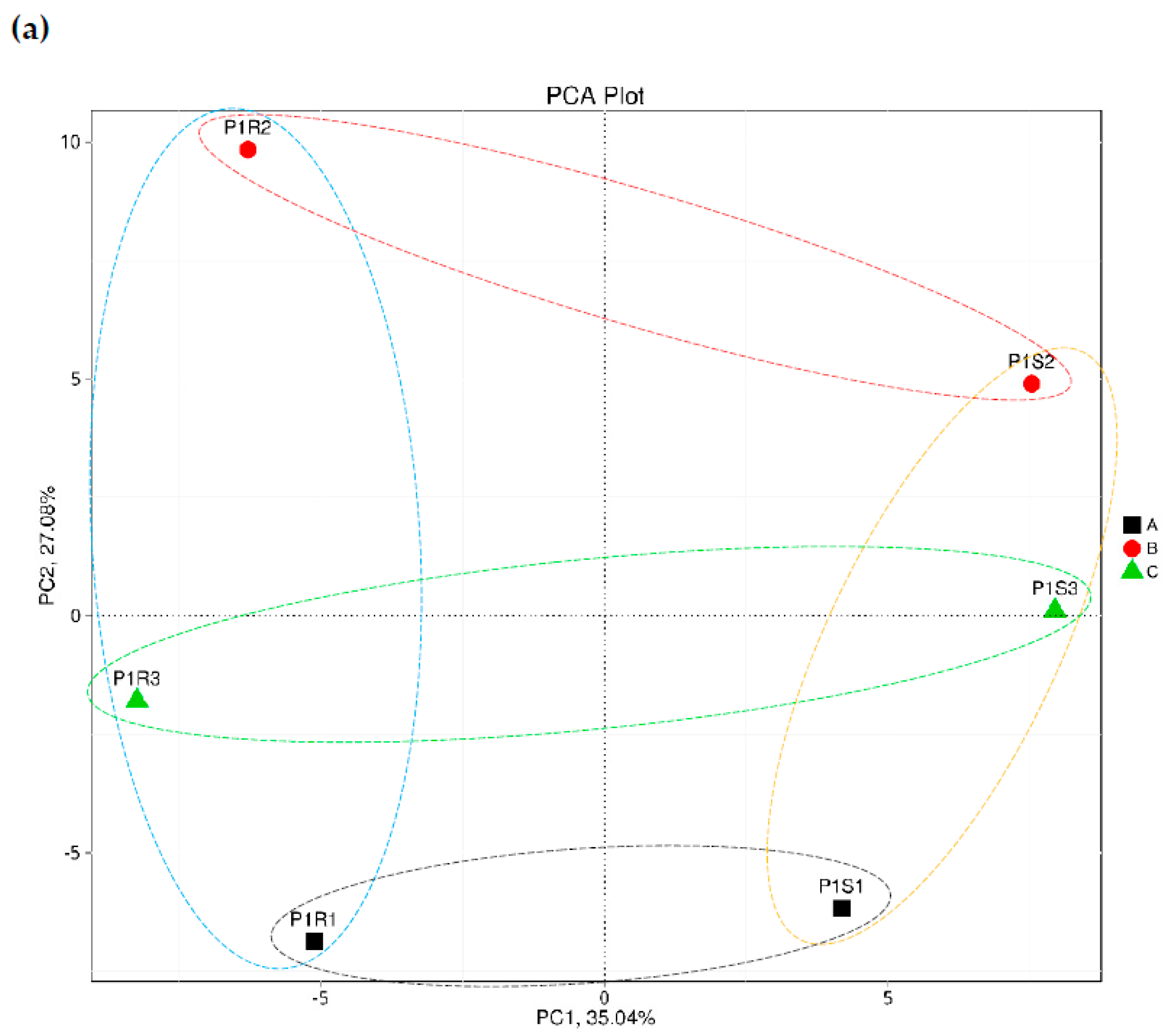
Figure 1. Principal component analysis (PCA) based on the number of genes at phylum (a), genus (b) and species (c) levels of metagenomes collected from surrounding bulk (S) and rhizosphere (R) soils of Dipterygium glaucum after 0 (group A, or P1S1 and P1R1, respectively), 24 (group B, or P1S2 and P1R2, respectively) and 48 h (group C, or P1S3 and P1R3, respectively) of watering. Orange circles = bulk soil microbiomes, blue circles = rhizosphere microbiomes, black circles = group A, red circles = group B, green circles = group C.
Sustainability 14 08764 g001aSustainability 14 08764 g001b
Figure 2. Microbial abundance assigned taxonomically by non-redundant genes at domain level of metagenomes across soil type (e.g., rhizosphere and bulk) of Dipterygium glaucum and time after watering (e.g., 0, 24 and 48 h).
Sustainability 14 08764 g002
Figure 3. Microbial abundance assigned taxonomically by non-redundant genes at domain level of metagenomes across soil (rhizosphere and bulk) type of Dipterygium glaucum after 0 (group A), 24 (group B) and 48 h (group C) of watering.
Sustainability 14 08764 g003
Figure 4. Relative microbial abundance assigned taxonomically by non-redundant genes of the top 10 highly abundant microbes of the Archaea domain at phylum, genus and species levels of metagenomes across soil (rhizosphere and bulk) type of Dipterygium glaucum.
Sustainability 14 08764 g004
Figure 5. Relative microbial abundance assigned taxonomically by non-redundant genes of the top 10 highly abundant microbes of the Bacteria domain at phylum, genus and species levels of metagenomes across soil (rhizosphere and bulk) type of Dipterygium glaucum after 0 (group A), 24 (group B) and 48 h (group C) of watering.
Sustainability 14 08764 g005
Figure 6. Relative microbial abundance assigned taxonomically by non-redundant genes of the top 10 highly abundant microbes of the Eukaryota domain at phylum, genus and species levels of metagenomes across soil (rhizosphere and bulk) type of Dipterygium glaucum after 0 (group A), 24 (group B) and 48 h (group C) of watering after 0 (group A), 24 (group B) and 48 h (group C) of watering.
Sustainability 14 08764 g006
Figure 7. Relative microbial abundance assigned taxonomically by non-redundant genes of the top 10 highly abundant microbes of the Viruses domain at genus and species levels of metagenomes across soil (rhizosphere and bulk) type of Dipterygium glaucum after 0 (group A), 24 (group B) and 48 h (group C) of watering.
Sustainability 14 08764 g007
Figure 8. Complex symbiotic associations and abundance correlation among the plant root, mycorrhiza (Rhizophagus) and eukaryotic microorganisms (Euryarchaeota and Crenarchaeota). AB = abundance, RA = relative abundance.
Sustainability 14 08764 g008
Figure 9. Non-redundant gene abundance of the genera Burkholderia, Trichoderma and Fusarium of the rhizosphere microbiome (R) of Dipterygium glaucum and the surrounding bulk soil microbiome (S) across watering time points.
Sustainability 14 08764 g009
Figure 10. Non-redundant gene abundance of the genus Ochrobactrum and its integrated virus Ochrobactrum phage POA1180 of the rhizosphere microbiome (R) of Dipterygium glaucum and the surrounding bulk soil microbiome (S) across watering time points.
Sustainability 14 08764 g010
Figure 11. Complex symbiotic associations among the plant root, bacteria (Ochrobactrum) and phage (Ochrobactrum phage POA1180).
Sustainability 14 08764 g011
Figure 12. Non-redundant gene abundance of the genus Mycobacterium and its integrated viruses, Mycobacterium phage Edugator and Mycobacterium phage Kratio, of the rhizosphere microbiome (R) of Dipterygium glaucum and the surrounding bulk soil microbiome (S) across watering time points.
Sustainability 14 08764 g012
Publisher’s Note: MDPI stays neutral with regard to jurisdictional claims in published maps and institutional affiliations.