

Seborrheic Keratosis Caused by Human Papillomavirus Type 20 Ameliorated by Zinc Oxide Ointment

 ,
, 
Abstract
1. Introduction
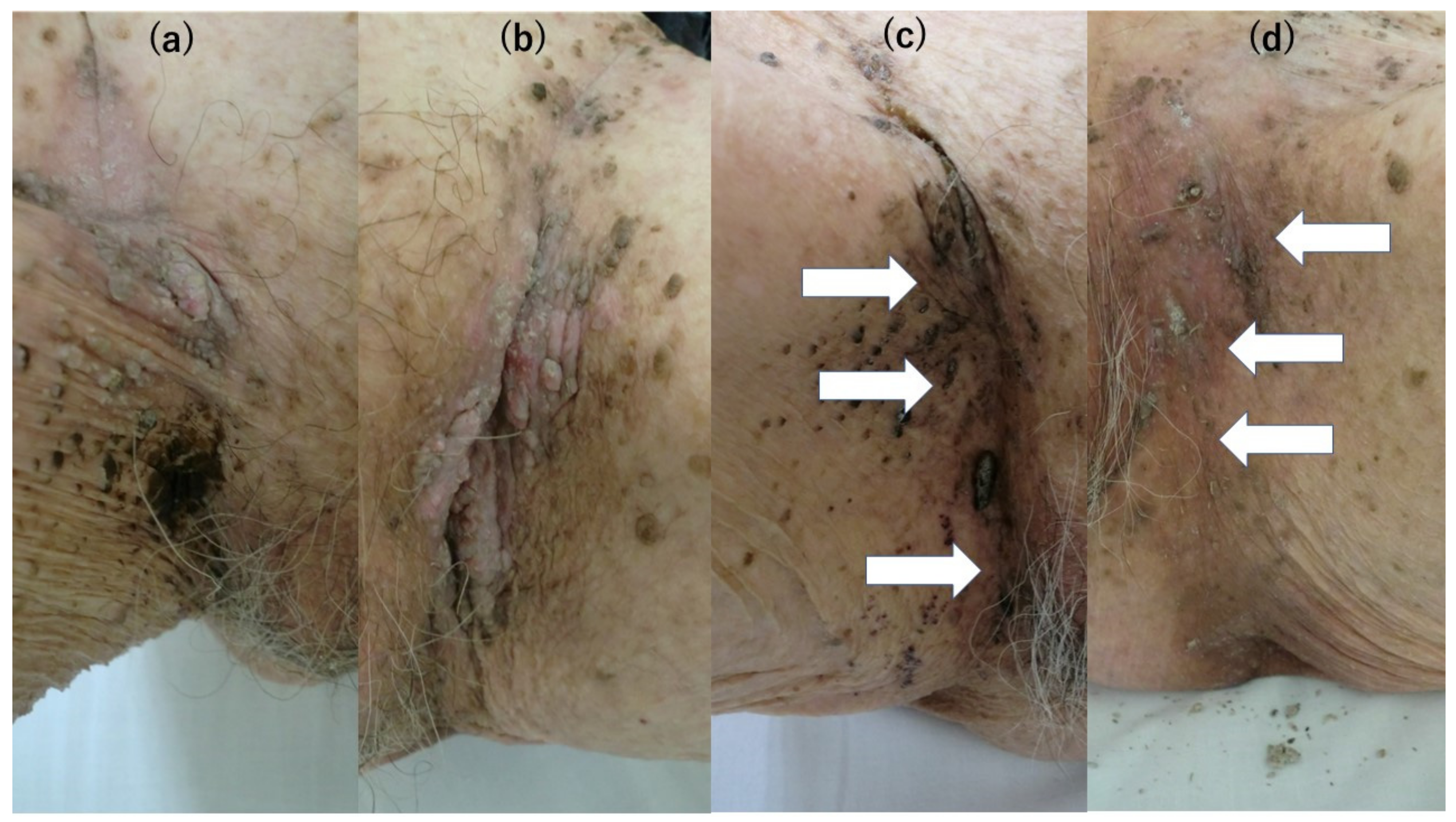
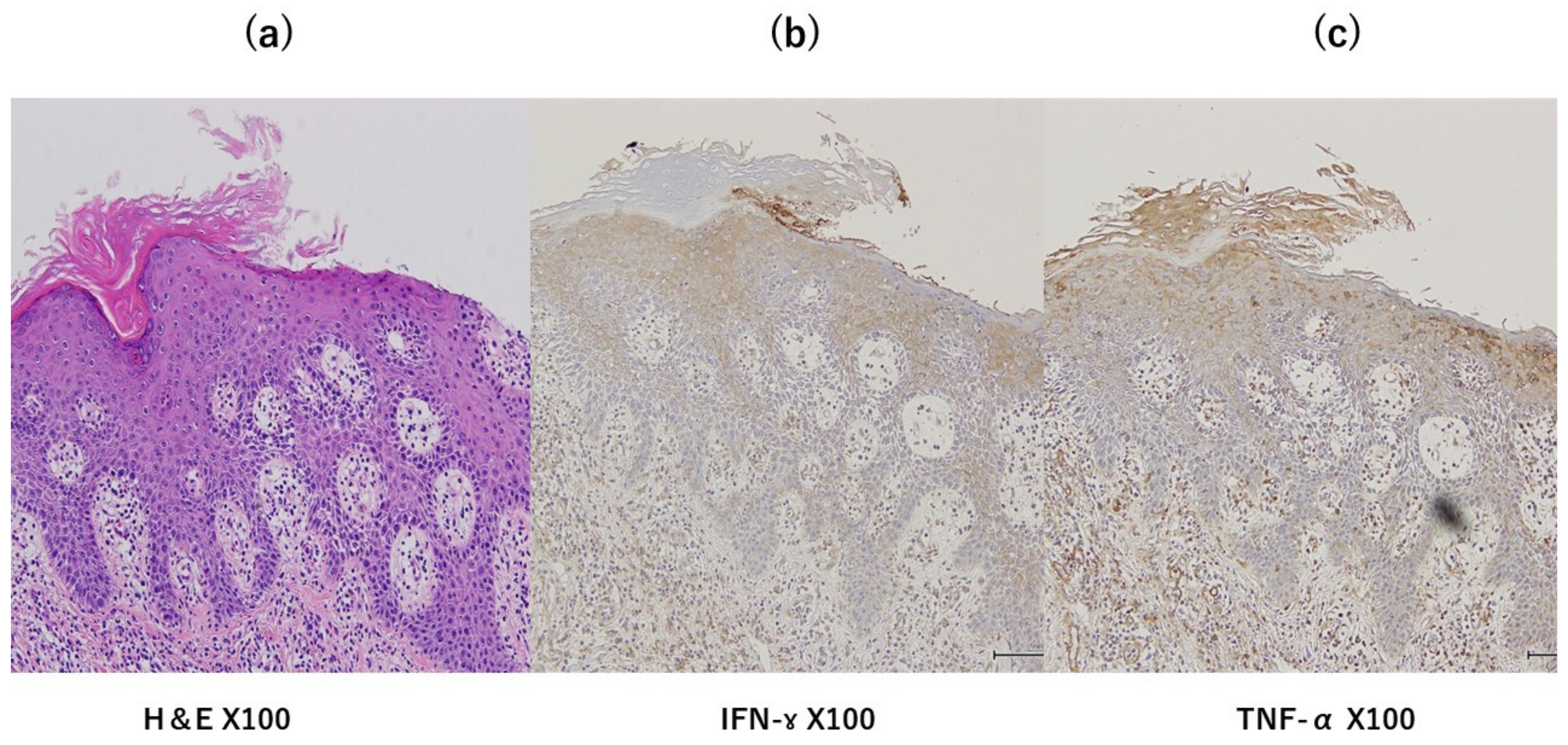
2. Case Report
3. Discussion
4. Conclusions
Author Contributions
Funding
Informed Consent Statement
Data Availability Statement
Conflicts of Interest
References
- Li, Y.-H.; Chen, G.; Dong, X.-P.; Chen, H.-D. Detection of epidermodysplasia verruciformis-associated human papillomavirus DNA in nongenital seborrhoeic keratosis. Br. J. Dermatol. 2004, 151, 1060–1065. [Google Scholar] [CrossRef] [PubMed]
- Berkhout, R.J.; Tieben, L.M.; Smits, H.L.; Bavinck, J.N.; Vermeer, B.J.; Schegget, J.t. Nested PCR approach for detection and typing of epidermodysplasia verruciformis-associated human papillomavirus types in cutaneous cancers from renal transplant recipients. J. Clin. Microbiol. 1995, 33, 690–695. [Google Scholar] [CrossRef] [PubMed]
- Talia, K.L.; Glenn McCluggage, W. Seborrheic Keratosis-like Lesions of the Cervix and Vagina: Report of a New Entity Possibly Related to Low-risk Human Papillomavirus Infection. Am. J. Surg. Pathol. 2017, 41, 517–524. [Google Scholar] [CrossRef] [PubMed]
- Umanoff, N.; Werner, B.; Rady, P.L.; Tyring, S.; Carlson, J.A. Persistent toenail onycholysis associated with Beta-papillomavirus infection of the nail bed. Am. J. Dermatopathol. 2015, 37, 329–333. [Google Scholar] [CrossRef]
- Ayatollahi, H.; Rajabi, E.; Yekta, Z.; Jalali, Z. Efficacy of Oral Zinc Sulfate Supplementation on Clearance of Cervical Human Papillomavirus (HPV); A Randomized Controlled Clinical Trial. Asian Pac. J. Cancer Prev. 2022, 23, 1285–1290. [Google Scholar] [CrossRef] [PubMed]
- Kremsdorf, D.; Favre, M.; Jablonska, S.; Obalek, S.; Rueda, L.A.; Lutzner, M.A.; Blanchet-Bardon, C.; Van Voorst Vader, P.C.; Orth, G. Molecular cloning and characterization of the genomes of nine newly recognized human papillomavirus types associated with epidermodysplasia verruciformis. J. Virol. 2015, 52, 1013–1018. [Google Scholar] [CrossRef] [PubMed]
- Ramoz, N.; Rueda, L.A.; Bouadjar, B.; Montoya, L.-S.; Orth, G.; Favre, M. Mutations in two adjacent novel genes are associated with epidermodysplasia verruciformis. Nat. Genet. 2002, 32, 579–581. [Google Scholar] [CrossRef] [PubMed]
- Lazarczyk, M.; Pons, C.; Mendoza, J.A.; Cassonnet, P.; Jacob, Y.; Favre, M. Regulation of cellular zinc balance as a potential mechanism of EVER-mediated protection against pathogenesis by cutaneous oncogenic human papillomaviruses. J. Exp. Med. 2008, 205, 35–42. [Google Scholar] [CrossRef] [PubMed]
- Allen, J.I.; Perri, R.T.; McClain, C.J.; E Kay, N. Alterations in human natural killer cell activity and monocyte cytotoxicity induced by zinc deficiency. J. Lab. Clin. Med. 1983, 102, 577–589. [Google Scholar] [PubMed]
- Ohkawara, T.; Takeda, H.; Kato, K.; Miyashita, K.; Kato, M.; Iwanaga, T.; Asaka, M. Polaprezinc (N-(3-aminopropionyl)-L-histidinato zinc) ameliorates dextran sulfate sodiuminduced colitis in mice. Scand. J. Gastroenterol. 2005, 40, 1321–1327. [Google Scholar] [CrossRef] [PubMed]
- Tran, C.D.; Ball, J.M.; Sundar, S.; Coyle, P.; Howarth, G.S. The role of zinc and metallothionein in the dextran sulfate sodium-induced colitis mouse model. Dig. Dis. Sci. 2007, 52, 2113–2121. [Google Scholar] [CrossRef] [PubMed]
- Xie, X.; Smart, T.G. Modulation of long-term potentiation in rat hippocampal pyramidal neurons by zinc. Pflug. Arch. 1994, 427, 481–486. [Google Scholar] [CrossRef] [PubMed]
- Li, Y.; Hough, C.J.; Suh, S.W.; Sarvey, J.M.; Frederickson, C.J. Rapid translocation of Zn(2+) from presynaptic terminals into postsynaptic hippocampal neurons after physiological stimulation. J. Neurophysiol. 2001, 86, 2597–2604. [Google Scholar] [CrossRef] [PubMed]


Disclaimer/Publisher’s Note: The statements, opinions and data contained in all publications are solely those of the individual author(s) and contributor(s) and not of MDPI and/or the editor(s). MDPI and/or the editor(s) disclaim responsibility for any injury to people or property resulting from any ideas, methods, instructions or products referred to in the content. |
© 2023 by the authors. Licensee MDPI, Basel, Switzerland. This article is an open access article distributed under the terms and conditions of the Creative Commons Attribution (CC BY) license (https://creativecommons.org/licenses/by/4.0/).
Share and Cite
Kondo, M.; Matsushima, Y.; Nakanishi, T.; Iida, S.; Habe, K.; Yamanaka, K. Seborrheic Keratosis Caused by Human Papillomavirus Type 20 Ameliorated by Zinc Oxide Ointment. Clin. Pract. 2023, 13, 367-371. https://doi.org/10.3390/clinpract13020033
Kondo M, Matsushima Y, Nakanishi T, Iida S, Habe K, Yamanaka K. Seborrheic Keratosis Caused by Human Papillomavirus Type 20 Ameliorated by Zinc Oxide Ointment. Clinics and Practice. 2023; 13(2):367-371. https://doi.org/10.3390/clinpract13020033
Chicago/Turabian StyleKondo, Makoto, Yoshiaki Matsushima, Takehisa Nakanishi, Shohei Iida, Koji Habe, and Keiichi Yamanaka. 2023. "Seborrheic Keratosis Caused by Human Papillomavirus Type 20 Ameliorated by Zinc Oxide Ointment" Clinics and Practice 13, no. 2: 367-371. https://doi.org/10.3390/clinpract13020033
APA StyleKondo, M., Matsushima, Y., Nakanishi, T., Iida, S., Habe, K., & Yamanaka, K. (2023). Seborrheic Keratosis Caused by Human Papillomavirus Type 20 Ameliorated by Zinc Oxide Ointment. Clinics and Practice, 13(2), 367-371. https://doi.org/10.3390/clinpract13020033