Introduction
New classes of silver nanoparticles (AgNPs) with antimicrobial activities are under rapid development for incorporation into consumer products. Important applications are in food packaging materials, odor-resistant textiles, paints, cosmetics, medical devices, water disinfectants,
etc.
1 This increasing use of AgNPs raised the issue of the release of nanosilver from a variety of sources, under different forms, into aquatic environments. Recent studies have confirmed the release of AgNPs from socks and nanotextiles during washing
2,3 or from surfaces coated by nanosilver paints during rain-water runoff events.
4 The environmental effects of the AgNPs discharged into aquatic environments are largely unknown.
1,5 However, laboratory ecotoxicological studies on fishes, crustaceans, algae and bacteria have shown toxic effects on test organisms exposed to AgNPs.
1,5-7 Due to the antimicrobial properties of AgNPs, it is important to understand which factors control their toxicity toward environmental microorganisms since they play important roles in major biogeochemical cycles, in the production of natural organic matter and in the recycling of nutrients. Bacterial sensitivity to silver nanoparticles and ionic silver have been demonstrated on reference bacterial strains of the genera
Bacillus,
Escherichia, Pseudomonas and
Vibrio7-10 as well as on bacterial communities.
6,11,12 Today, there is no consensus on the mechanisms of antimicrobial action of AgNPs. Proposed processes
1,8-10,13 include: i) free silver ion dissolution from AgNPs followed by cellular uptake and disturbance of intracellular reactions; ii) cellular uptake of AgNPs followed by reactive oxygen species (ROS) generation and release of silver ions impacting DNA replication and ATP production; and iii) direct interaction of AgNPs with cell surface altering protein function and membrane permeability. These processes were demonstrated to be dependent on nanoparticle’s size and specific properties (shape, chemical composition, surface charge, coating,
etc.). Moreover, the chemical composition of exposure media (pH, ionic strength, ionic composition, quality and quantity of natural organic matter) is also a relevant factor that would affect the toxicity of AgNPs. The solution chemistry could play an important role in the aggregation and in the fate of AgNPs. If aggregation occurs, the size of AgNPs in solution will change, modifying the sedimentation and the ability of AgNPs to interact with proteins at the cellular surface or to cross-cellular membranes. Thus, aggregation can modulate the toxicity of nanoparticles. Considering that the size of AgNPs can be a significant factor controlling the toxicity of silver nanoparticles, it is important to understand how the exposition media (
e.g., culture broths) modify the size of the nanoparticles used in toxicity assays. The purpose of this study is to assess the stability of AgNPs suspensions with time in different exposure solutions characteristic of natural waters or of media currently used in microbial ecotoxicology studies. We aim to show the importance to control the aging of AgNPs colloidal suspensions during toxicity assays.
Results and Discussion
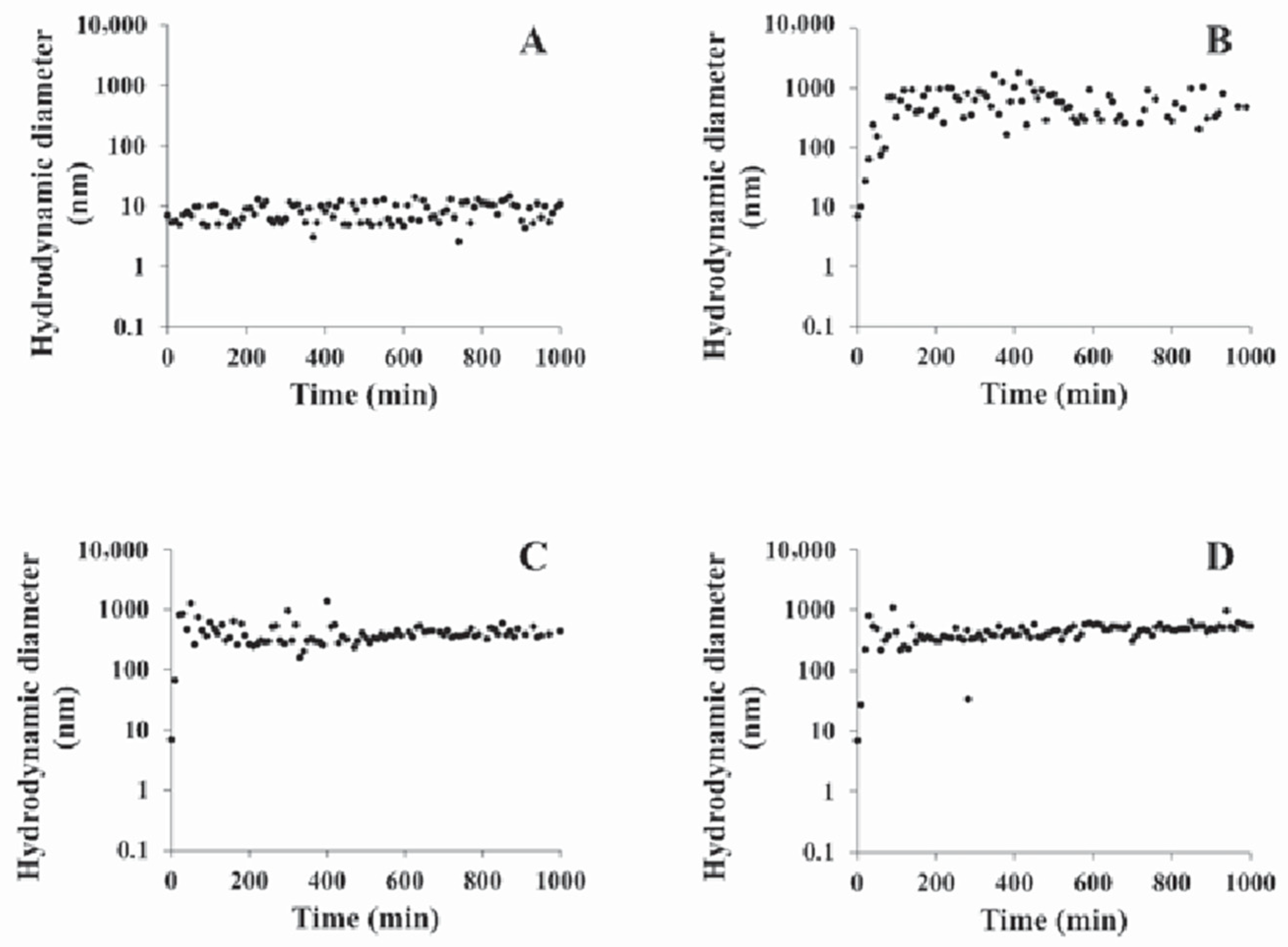
Figure 1 shows the evolution in hydrodynamic diameter (Dh) for AgNPs over 17 h in nanopure water, NaCl, CaCl
2 or MgCl
2 solution. In nanopure water, the Dh of AgNPs remained stable at 9±2 nm during 17 h (
Figure 1A). In presence of NaCl at 25‰, the Dh of AgNPs (9 nm) evolved rapidly (
Figure 1B) to reach 550±280 nm after 100 min. When CaCl
2 was added to nanopure water at 25‰, the Dh of AgNPs increased rapidly during the first 50 min (
Figure 1C) suggesting that the rate of AgNPs aggregation is modulated by the nature of cations in solution. After 50 min, the Dh did not evolved anymore and stabilized at 424±69 nm. In presence of MgCl
2 added to nanopure water at 25‰, the behavior of AgNPs was the same as in the CaCl
2 solution (
Figure 1D). After the rapid increase (50 min), the Dh remained at 548±105 nm. The rapid, but different, increases of Dh observed in NaCl, CaCl
2 and MgCl
2 solutions reflected the aggregation of AgNPs in presence of various cations and showed that divalent cations were more effective in accelerating aggregation than monovalent cations.
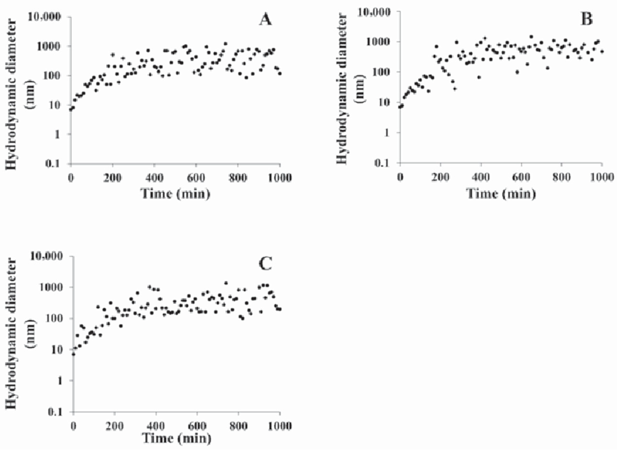
Figure 2 presents the time evolution of AgNPs hydrodynamic diameter in ASW, F/2 and LB media. In ASW, from 0 min to 200 min, the Dh increased rapidly from 9 nm to near 400 nm as shown in
Figure 2A. After 200 min the Dh reached 422±255 nm. In F/2 medium (
Figure 2B), the behavior of AgNPs was similar to that observed in ASW. A rapid increase in Dh was observed during the first 200 min afterward the Dh remained at 635±253 nm. In LB broth, the Dh of AgNPs increased rapidly during the first 250 min. Later on, the diameter stabilized around 511±365 nm. In complex solutions (
Figure 2) slower changes in aggregation rates were observed compared to those observed in simple solutions (
Figure 1) during the first 100 min. This involves that for a complex aqueous solution, the rate of aggregation is modified by the presence of cations but also by other components, such as organic matter (vitamins in F/2 medium and tryptone and yeast extract in LB medium) present in the culture media.
Stability or aggregation of AgNPs could be rationalized, in first approximation, by the Derjarguin, Landau, Verwey and Overbeek (DLVO) theory.
14 Stability is controlled by attractive and repulsive interactions. Attractive interactions are van der Waals forces and repulsive interactions are governed by the electrical double layer.
14 When repulsive interactions are greater than attractive interactions, the conditions are unfavorable for aggregation, but when repulsive interactions are equal or lower than attractive forces, the aggregation is promoted. The transition between these two steps is characterized by the critical coagulation concentration (CCC). For AgNPs coated with citrate, the CCC was generally close to 50 mM for NaCl and near 2 mM for divalent cations
15 present in CaCl
2 or MgCl
2 solution. In this study, the pH solution was near 7 and the salt concentrations were greater than the CCC, thus aggregation occurs. In nanopure water, the citrate moieties of AgNPs were negatively charged. This promotes the electrostatic repulsions between AgNPs, thus avoiding the aggregation of AgNPs.
14,16 Moreover, the presence of salts has increased the ionic strength of the solution decreasing the thickness of the electrical double layer around the silver nanoparticles. This has enhanced the close encounter of AgNPs and therefore the aggregation. However, the presence of salts also affected the charges at the surface of AgNPs. Citrate moieties were negatively charged and can interact with cations. Interactions between citrate and cations decreased the negative charges of AgNPs and lowered the electrostatic repulsion between particles.
14,16 These phenomena enhanced the aggregation of AgNPs
17 and can explain the behavior of AgNPs in nanopure water and salt solutions. Another process that could contribute to the aggregation was the presence of multivalent cations. Those cations could form a bridge between two adjacent AgNPs facilitating the aggregation of nanoparticles
16. The difference observed in the rapid increase in Dh of AgNPs in presence of NaCl and CaCl
2 or MgCl
2 could be explained by this process.
In complex media such as ASW, F/2 or LB, the aggregation rate was lower than in salt solutions. Jin
et al.8 have discussed the effects of synthetic freshwater composition on aggregation and dissolution of AgNPs. When freshwater contained the same salts as those in ASW, the aggregation of AgNPs was modified. This modification was caused by the reaction of Ag
+, released from AgNPs, with anions like Cl
– or HCO
3–. The salts formed during the reaction were insoluble and could interact with the surface of AgNPs to create a new coating.
18 With this new coating, the toxicity of nanoparticles could be different than the toxicity of the native nanoparticles.
In F/2 medium, the trace element concentration could be too low to affect AgNPs aggregation. The behavior of AgNPs in F/2 medium was controlled by the most important cation, Na
+, in this culture broth. The anions, such as Cl
– and HCO
3–, can precipitate with silver ions around the AgNPs as described for ASW. The LB medium has a concentration in NaCl near 171 mM, higher than the CCC. However, the presence of organic matter (OM), like tryptone and yeast extract, could affect the aggregation of AgNPs by increasing the CCC. The addition of OM could create a new coating on AgNPs.
15 This new coating would modify the interactions between AgNPs, OM and ions in solution thereby producing a new equilibrium characterized by a decrease of AgNPs aggregation rate. This hypothesis could explain the difference in the time evolution of AgNPs size observed in this study.
As shown in
Figures 1 and
2, aggregation of nanoparticles is a common phenomenon in complex aqueous environments. The rapid changes in size observed during the first hours of our experiments are related to the charge of the cations, the presence of OM in the culture media, the nature of nanoparticles and the contact time between nanoparticles and the surrounding solution. A steady state in the size of the aggregates has been observed after different time periods, from 50 min in solution of divalent cations to about 200-250 min in culture broth F/2 or LB. In the steady state, for ASW, LB and F/2 respectively, the Dh of the aggregates increased by 46, 57 and 70 fold compared to the nominal Dh of AgNPs. This can have a strong incidence on the toxicity. For instance, the increase in Dh in F/2 medium decreased the number of nanoparticles by about 10,000 between the beginning and the end of the experiment. Such variations in particle numbers, in the size and in the time period where aggregation occur can lead to misrepresentative results or interpretations if toxicological assays are discussed without an in-depth description of conditions used in experiments. The time between the addition of nanoparticles to culture media and the duration of the toxicological assay should be clearly mentioned in the materials and methods of any nanotoxicological study.