
Cumulative and Interactive Effects of Heavy Metal Mixtures Across Neurodegenerative Diseases: A Comparative Review of Alzheimer, Parkinson, Amyotrophic Lateral Sclerosis and Multiple Sclerosis




Abstract
1. Introduction
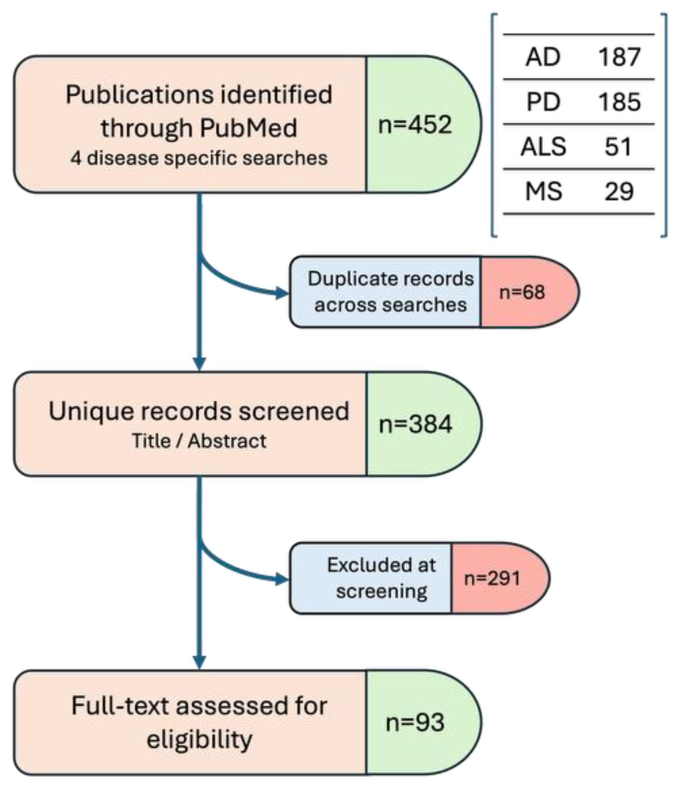
2. Literature Selection Methodology
- (“lead” OR “cadmium” OR “mercury” OR “arsenic” OR “manganese” OR “aluminum” OR “heavy metal”) AND (“metal mixture” OR “metal co-exposure” OR “combined metal exposure” OR “multi-metal” OR “co-exposure to metals”) AND (“Alzheimer’s disease” OR “AD”)
- (“lead” OR “cadmium” OR “mercury” OR “arsenic” OR “manganese” OR “aluminum” OR “heavy metal”) AND (“metal mixture” OR “metal co-exposure” OR “combined metal exposure” OR “multi-metal” OR “co-exposure to metals”) AND (“Parkinson’s disease” OR “PD”)
- (“lead” OR “cadmium” OR “mercury” OR “arsenic” OR “manganese” OR “aluminum” OR “heavy metal”) AND (“metal mixture” OR “metal co-exposure” OR “combined metal exposure” OR “multi-metal” OR “co-exposure to metals”) AND (“Amyotrophic lateral sclerosis” OR “ALS”)
- (“lead” OR “cadmium” OR “mercury” OR “arsenic” OR “manganese” OR “aluminum” OR “heavy metal”) AND (“metal mixture” OR “metal co-exposure” OR “combined metal exposure” OR “multi-metal” OR “co-exposure to metals”) AND (“Multiple Sclerosis”).
3. General Mechanisms of Metal–Metal Interactions
3.1. Competition at Shared Membrane Transporters
3.2. Sequestration and Antioxidant Buffering
3.3. Direct Synergistic and Antagonistic Interactions
4. Disease-by-Disease Comparative Profile
4.1. Alzheimer’s Disease

| Metals/Mixture Studied | Design/Method | Key Findings (Synthesized) |
|---|---|---|
| Pb, Cd, Hg, Mn, Se, Cu, As, Co, W, U, Zn, Ba, Cs, Tl, Al, Li (varies by study) | [Direct] 7 independent human cohorts; 6 use formal mixture statistics (WQS, BKMR, qgcomp) on blood/urine metal panels and 1 uses logistic regression | Metal mixtures—not single metals—consistently predicted worse cognition or higher dementia/MCI risk, dose-dependently; two of the seven studies specifically found that Se attenuated the toxic-metal mixture effect (Cu-Pb-Se; Pb-Cd-Hg-Mn-Se) [21,22,25,26,39,40,41] |
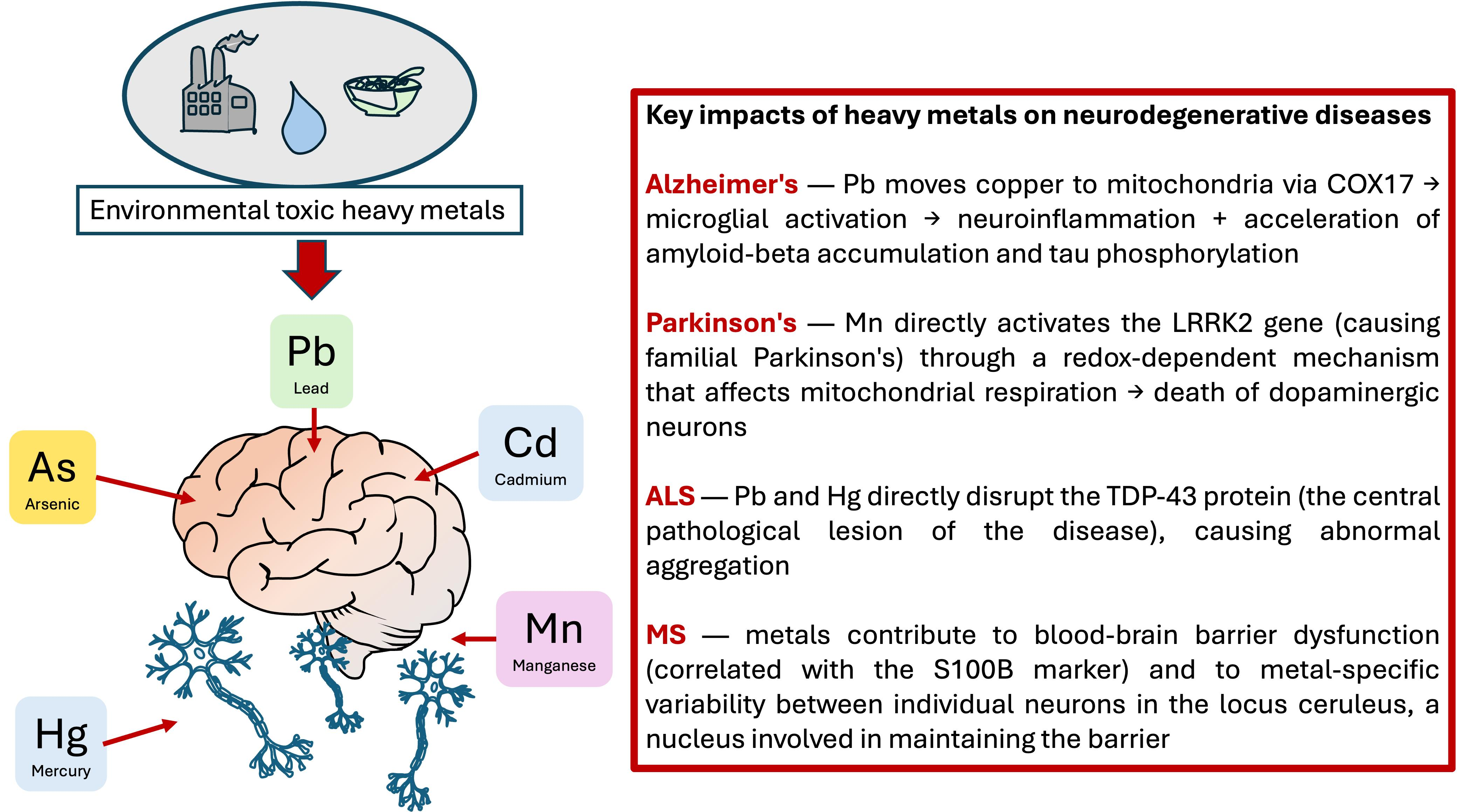
| Al, Pb, Cd, Fe, Cu | [Direct] 4 experimental models (organotypic brain slices; APP/PS1 mice + BV-2 cells; two rat models) exposed to defined metal combinations | Combined metal exposure produced greater Aβ/tau pathology than any single metal in every model tested; two studies identified a specific, reversible mechanism: mitochondrial Cu overload via COX17 (Pb + Cu), and antioxidant rescue by spermine or berberine (Al + Fe; Al + Cd + fluoride) [28,29,42,43] |
| Pb, Cd, As, Hg/MeHg, Au, Cu, Fe, Al | [Direct] 4 in silico toxicogenomic analyses (Comparative Toxicogenomics Database gene-network mining) | All four independent analyses, despite testing different metal combinations, converged on the same core apoptosis/oxidative-stress gene set (BAX, CASP3, BCL2, TNF) as the shared molecular signature of metal-mixture neurotoxicity; one analysis extended this convergence to ALS and PD, identifying SOD2 as a gene mutual to all three diseases [30,44,45,46] |
| Se, Zn vs. Cd | [Direct] 2 in vitro studies, SH-SY5Y neuronal cells | Both Se and Zn, tested independently, attenuated Cd-induced cytotoxicity via antioxidant mechanisms—direct cellular-level confirmation of essential-metal protection against a toxic metal, though Se’s protection did not extend to differentiated cholinergic cells in one study [35,47] |
| Mn, V, Fe, Cu (welding-fume mixture) | [Direct] 2 companion case-control studies in the same welder cohort, diffusion tensor/T1 MRI | Mixed occupational metal exposure altered MRI metrics in both the medial temporal lobe and basal ganglia; Mn and V showed effects individually and jointly as a mixture [31,32] |
| As, Cd, Hg, Ni, Pb, Tl + essential metals (CSF); Fe, Cu, Zn, As, Cd, Mn (postmortem/tissue) | [Multi-measured] CSF/plasma biomarker correlation (n = 193); postmortem brain-vs-fluid comparison; single-neuron synchrotron imaging (n = 7) | Heavy and essential metal levels correlated with CSF markers of AD pathology in living patients, but postmortem data showed brain tissue and ventricular fluid levels are not interchangeable, and single-neuron imaging revealed substantial cell-to-cell variation in metal accumulation—together cautioning against treating any single fluid compartment as a reliable proxy for brain metal burden [33,34,48] |
| Cd, Pb, Hg, As, Se | [Multi-measured] Case-control study (n = 434, propensity-matched) and systematic review/meta-analysis (22 studies, n = 3346) | The case-control study linked As metabolite profile and low Se to elevated AD risk; the meta-analysis, testing four metals across a much larger literature, found only Cd significantly elevated in AD patients versus controls, with Pb, As, and Hg not significantly different [49,50] |
| Cd, Pb (+As, Mn, Hg) | [Multi-measured] Ecological, US geographic correlation (topsoil, sewage sludge, well water, infant blood) | Cd and Pb in sewage sludge, used as an environmental exposure proxy, were significantly associated with neurodegenerative disease prevalence across the United States [51] |
| Pb, As, MeHg | [Multi-measured] In vitro hippocampal cell proteomics | Established a relative potency ranking (Pb < As < MeHg) while identifying mitochondrial dysfunction and oxidative stress as pathways shared across all three metals [52] |
| Pb, Hg, Cd, Mn, As, Cu (+Mg in one) | [Review] 4 narrative reviews, autophagy–lysosomal/mitochondrial mechanism focus | All four reviews converge on the same conclusion: heavy metals impair the autophagy–lysosomal pathway and mitochondrial function (electron transport chain, mtDNA integrity), proposed as a mechanism shared between AD and PD [37,53,54,55] |
| As, Mn, Hg, Al, Pb, Ni, Cd, Cu, Zn, Fe, Co (varies) | [Review] 4 narrative reviews, each centered on a distinct specific mechanism or therapeutic angle | Four reviews each foreground a different, less conventional angle: astrocyte-mediated metal accumulation and iron-induced astrogliopathy, exosomal miRNA transport linking peripheral metal exposure to brain neuroinflammation, ferroptosis as a shared metal-induced cell-death pathway, and nanoparticle-based chelation as a therapeutic strategy; these do not converge on one shared finding but each adds a distinct mechanistic candidate [56,57,58,59] |
| Pb, Al, Hg, Mn, Cd, As (recurring core set) | [Review] 9 narrative reviews, general multi-metal mechanistic overviews | Substantial overlap across all nine: the same core set of metals (Pb, Al, Hg, Mn, Cd, As) is repeatedly implicated via oxidative stress, neuroinflammation, and BBB disruption; individual reviews add specific detail without contradicting this shared core: Pb-BBB/epigenetic effects, Cd-p53/p21/Rb senescence, As-nitric oxide signaling, Mn-glutamate excitotoxicity, and overlap with PD mechanisms [36,44,45,60,61,62,63,64,65] |
| Cu, Cd, As, Pb, Mn (adverse); Se, Fe, Zn, Al, Si (protective or mixed) | [Review] 2 broad reviews synthesizing epidemiological studies (34 studies; 60 of 4784 screened) | Both reviews report the same overall pattern: findings across the underlying literature are inconsistent and metal-specific, with adverse associations most consistent for Cu/Cd/As/Pb/Mn, and protective or mixed evidence for Se/Fe/Zn; moderate evidence also implicates Al in general dementia risk [27,66] |
| Cd, Fe, As, Cu, Li | [Review] 1 narrative review, epigenetics-specific | Proposes that these five metals converge on the same downstream DNA methylation signature at AD-relevant genes despite acting through distinct upstream mechanisms [38] |
| Mn, Zn, Fe, Cu, Ni | [Review] 1 narrative review, aging-specific | Concludes that age-related metal accumulation compounds mitochondrial dysfunction and calcium dyshomeostasis already present in aging neurons [67] |
| 18 metals (evidence-map protocol); Mn (methods chapter) | [Review] 1 published protocol; 1 laboratory methods chapter | No primary findings in either—cited as methodological/procedural context only, not as evidence [68,69] |
4.2. Parkinson’s Disease
| Metals/Mixture Studied | Design/Method | Key Findings (Synthesized) |
|---|---|---|
| Pb, Al, Hg, Mn, Cd, As (recurring core) + Fe, Cu, Zn, Ni in some | [Review] 13 items: 12 narrative reviews + 1 single-neuron synchrotron imaging study | Overlap with the AD literature: oxidative stress, mitochondrial dysfunction, BBB disruption, and autophagy impairment recur as shared mechanisms across the same core metal set; the imaging study adds single-neuron-resolution evidence that individual locus coeruleus neurons vary in toxic/essential metal content [37,45,48,51,54,55,59,60,62,64,65,67,68] |
| Se, Zn vs. Cd | [Direct] 2 in vitro studies, SH-SY5Y neuronal cells | Overlap with the AD literature: direct cellular confirmation of essential-metal protection against a toxic metal [35,47] |
| As, Mn, Hg, Al, Pb, Ni, Cd, Cu, Zn, Fe (astrocytes); As, Co, Cd, Fe, Mg, Mn, Ni, Hg, Zn, Se (ferroptosis) | [Review] 2 narrative reviews, each proposing a distinct specific cell-biology mechanism | Overlap with the AD literature: astrocyte-mediated metal accumulation and iron-induced astrogliopathy; ferroptosis (Fe-dependent cell death via GPX4/Xc-failure, with Se being protective)—the two mechanisms do not converge on a shared finding [56,58] |
| Pb, As, MeHg, Cd | [Multi-measured] In vitro hippocampal proteomics; in silico toxicogenomic gene-network mining; narrative mechanism-of-action review | Three independent approaches converge on shared binding targets for this quaternary mixture (NMDA receptor, Na+-K+ ATPase, Ca2+ signaling, glutamate transmission) and shared downstream pathways (mitochondrial dysfunction, oxidative stress, SOD2); one study established a relative potency ranking of Pb < As < MeHg [52,81,82] |
| Co, Ni, Hg, Cr, Tl; Cu, As, Cd, Fe, Li; Hg, Pb, Cu, Zn, Fe, Mn, Al, As, Cd, Se; Fe, Hg, Mn, Cu, Pb | [Review] 4 narrative reviews specifically on metals and PD mechanisms | All four consistently implicate oxidative stress, mitochondrial dysfunction, and alpha-synuclein aggregation as shared downstream consequences of metal exposure in PD, though each emphasizes a different subset of metals; one explicitly notes that interactions among mixture components may produce synergistic toxicity beyond single-metal effects [5,79,83,84] |
| Mn, V | [Direct] 2 independent mouse studies (same research group), intranasal co-exposure | Ngwa et al. [70]: 4-arm design (control/Mn/V/Mn + V)—genuine metal–metal synergy, co-treatment produced most severe deficits. Kanthasamy et al. [71]: same mixture in WT vs. A53T transgenic mice—genuine gene-environment interaction, but no single-metal arms, so it cannot independently confirm metal–metal synergy. |
| Mn, Zn, Cu | [Multi-measured] In vitro, SH-SY5Y cells, benchmarked against the classic PD toxin 6-OHDA | Zn was the most potent dopaminergic toxin of the three metals tested; Mn and Cu at LC50 produced a response similar to 6-OHDA, suggesting distinct upstream mechanisms converging on a common idiopathic PD-like phenotype [72] |
| As, Cd; Co, Mn | [Direct] 2 in vitro biophysical/structural studies (aggregation kinetics; native mass spectrometry) | Both metal pairs directly bind and alter alpha-synuclein aggregation: As and Cd become incorporated into amyloid fibers and accelerate nucleation while reducing aggregate clearance in yeast cells; Co and Mn bind the C-terminal region and induce structural compaction of the protein [76,77] |
| Mn, Pb, Cr, Ni, Se, Cd (urinary); Ba, Cd, Co, Cs, Mo, Pb, Sb, Tl, U (NHANES) | [Direct] 2 independent human cohort/case-control studies using formal mixture statistics (BKMR, WQS, quantile g-computation) | Metal mixtures elevate PD risk beyond single-metal effects, though the dominant contributors differ by study (Mn 73.7% + Pb 9.3% in one; Mo + Co dominant in the NHANES WQS model) [73,85] |
| Fe, Cu (low); Mn, As (high); Zn (unchanged) | [Multi-measured] Cross-sectional human hair analysis + MPTP-induced mouse model | Lower hair iron (Fe) and copper (Cu) alongside higher manganese (Mn) and arsenic (As) distinguished PD patients from controls; the mouse model linked the iron (Fe) deficit specifically to gut microbiota dysbiosis and impaired intestinal iron (Fe)-transport gene expression (DMT1, FPN), proposing a gut–brain axis mechanism [80] |
| Pb, Cd; Fe, Mn | [Single-metal] Rat model (Pb, Cd, N-acetylcysteine intervention) and SH-SY5Y cells (Fe, +Mn, butyrate, butyrate + nicotine intervention) | Both studies found combined toxic-metal exposure upregulated PD-related genes or toxicity markers (Parkin, Pink1, LRRK2, SNCA for Pb, +Cd), and both identified an antioxidant-based intervention that protected against the combined-metal damage, though via distinct mechanisms [86,87] |
4.3. Amyotrophic Lateral Sclerosis
| Metals/Mixture Studied | Design/Method | Key Findings (Synthesized) |
|---|---|---|
| Pb, Al, Hg, Mn, Cd, As (recurring core) + other metals | [Review] 7 items: narrative reviews, 1 protocol, 1 in silico toxicogenomic analysis, 1 ecological study | Overlap with AD and PD: same recurring core mechanisms (oxidative stress, mitochondrial dysfunction, BBB disruption) across the same core metal set [51,56,59,61,64,68,81] |
| Multi-metal panel in plasma and urine (individual metals significantly associated with risk/survival: copper, selenium, zinc) | [Direct] Case-control study (n = 454 ALS/294 controls), ICP-MS on plasma and urine; ALS and metal polygenic risk scores computed from independent GWAS/literature-selected SNPs | Elevated Cu, Se, and Zn were individually linked to ALS risk and survival, and a combined environmental risk score across all measured metals showed a strong, dose-dependent association with both greater risk (OR ~3) and worse survival (HR ~1.4)—independent of genetic risk, and correlated with known occupational and non-occupational exposure sources [8] |
| Mg, Cu, Se, Fe, Mn, V, Zn, Al, As, Co, Ni, Hg, Pb, Cd, Pd (15 elements); Pb, Cd, Hg | [Multi-measured] 2 case-control/observational studies measuring CSF metal levels directly (ICP-MS) | Mixed evidence. One study found Se and As elevated above reference values in ALS patients, with Cu, Fe, Mn, Zn, Al, Ni, and Pb differing between bulbar and spinal onset subtypes [96]. A methodologically distinct case-control study found higher Pb in ALS, but lower Cd and Hg than controls, with no significant dose–response relationship for any of the three metals—the authors concluded their data did not support a role for these metals in ALS etiology [88]. This null/mixed result should temper strong causal claims about this specific three-metal combination. |
| Hg, Ag, Bi (autometallography) | [Multi-measured] Histological study, spinal cord tissue from 50 individuals without motor neuron disease | Heavy metals detected in spinal interneurons in 33% of individuals aged 61–95, absent at younger ages, suggesting age-related accumulation could predispose inhibitory interneurons to damage relevant to ALS pathogenesis. Note: this study used non-ALS control tissue, not ALS patients directly [97]. |
| Pb, Mn, Se, Cu, Zn (urinary); Pb, Cd, Al, Hg, Mn, Fe, Cu, Zn, Se, Mg, Ca (blood/urine/hair) | [Multi-measured] 2 case-control studies (n = 42 and severity-stratified cohort) | Both identify Pb as a consistent risk-associated metal (elevated urinary Pb in one; Pb as risk factor in blood in the other), while Se showed a protective association in one study; a subtle Mn increase and elevated Cu also appeared in the urinary study [89,98] |
| As, Cd, Pb, Hg, Cr | [Review] Umbrella review of 35 meta-analyses, 103 health outcomes (AMSTAR2-graded) | Pb showed a significant association specifically with ALS (equivalent OR 1.46, 95% CI 1.16–1.83, credibility class III/suggestive) among the 103 health outcomes evaluated for these 5 metals—the only ALS-specific finding within a much broader multi-disease evidence synthesis [99] |
| Pb, Hg, Sn | [Multi-measured] In vitro (cultured cells) + in vivo (mouse cortex) mechanistic study | Pb and meHg directly disrupted TDP-43 homeostasis, triggering nuclear granule accumulation and increased splicing activity; Pb specifically promoted dose-dependent phase separation of TDP-43 in vitro—the clearest direct mechanistic link identified between a specific metal and the primary pathological protein of ALS [90] |
| Heavy metals + organic solvents + diesel exhaust; Ag, Al, Cd, Cr, Cu, Fe, Mn, Pb, Se (ecological) | [Multi-measured] Formal occupational case evaluation (n = 3 workers) + ecological correlation study (n = 62 ALS cases, moss/lichen biomonitoring) | A Korean government committee attributed ALS in three automobile workers to combined 15–33-year occupational exposure to heavy metals (primarily Pb from engine work), organic solvents, and diesel exhaust. Separately, an ecological study in Italy found ALS case density correlated most strongly with Cu air pollution specifically—not with the other 8 metals measured—a metal-specific finding distinct from the Pb-centered evidence elsewhere in this table [94,95]. |
| 15 metals, incl. Al, As, Cd, Cr, Co, Cu, Fe, Pb, Mn, Hg, Ni, Se, U, V, Zn (genotoxicity mapping); Mn, Cu (histone PTM); Cd, Hg + metalwork (DNA methylation) | [Review] Systematic genotoxicity mapping + [Multi-metal] 2 experimental/epidemiological epigenetic studies | A comprehensive mapping against ATSDR’s genotoxicity framework found substantial evidence linking metal exposure to genotoxic damage in both sporadic and familial ALS, though nearly 80% of possible genotoxic endpoints remain unexplored; Mn and Cu directly alter histone H3 modifications in a yeast model relevant to ALS/FTD epigenetics; DNA methylation changes are measurably associated with self-reported Cd, Hg, and metalwork exposure history in a large ALS cohort (n = 855) [91,92,93] |
4.4. Multiple Sclerosis
| Metals/Mixture Studied | Design/Method | Key Findings (Synthesized) |
|---|---|---|
| Pb, Hg, V, Cr (review); Hg, Ag, Bi (spinal interneurons) | [Review] 1 narrative review (shared with AD/PD/ALS tables) + 1 histological aging study (shared with ALS table, non-MS tissue) | The spinal interneuron study used general-population tissue, not MS patients specifically, and is cited here only for relevance to the multifocal CNS pathology pattern proposed to underlie MS as well as ALS [62,97] |
| Hg, Se, Fe, Cu, Pb, Rb | [Multi-measured] Synchrotron X-ray fluorescence, single-neuron resolution, locus coeruleus tissue from 7 actual MS donors | This study used real MS-patient tissue. Individual neurons varied substantially in toxic and essential metal content, with Hg confined to a scattered neuronal subset—proposed as a structural mechanism for the non-random, multifocal destruction pattern characteristic of MS [48] |
| As, Cd (GSTM1 study); Cd, Pb (blood + smoking); As, Cd, Pb (DNA methylation) | [Multi-measured] 3 studies from the same research group, same Tehran cohort (~69 RRMS patients/74 controls), examining genetic polymorphism, blood levels, and epigenetics | As and Cd consistently elevated in MS patients across all three analyses. Pb showed no significant MS-vs-control difference in two of the three (only a sex difference within patients). Cd susceptibility was linked to GSTM1-null genotype and smoking; As-induced hypomethylation of ACKR3 was proposed as a specific epigenetic mechanism [102,103,112]. |
| As, Pb, Cd | [Multi-measured] Cross-sectional, Tehran, blood metals + serum S100B (BBB-disruption marker) | MS patients showed elevated blood As and Cd alongside higher serum S100B, with As showing the strongest correlation (63%) to S100B—proposing metal-induced BBB compromise as a contributing mechanism [110] |
| Cd, Pb (Poland); As, Pb, Hg, Cd (meta-analysis, 16 studies); As, Cd, Pb (Turkey) | [Multi-measured] Cross-sectional cohort (n = 151) + systematic review/meta-analysis (n = 1650) + case-control (n = 50) | Directly conflicting results. The meta-analysis (largest evidence base) found Pb, As, and Cd to be significantly elevated in MS patients (Hg not significant). A Polish cohort found Cd and Pb did not differentiate functional status or disease course (only a non-significant trend at higher Pb). A Turkish case-control found the opposite direction: As, Cd, and Pb were significantly higher in controls than in MS patients. This direct contradiction is a notable feature of the MS literature not seen to the same degree in the other three diseases [100,101,111]. |
| Pb, Hg (+ organic solvents) | [Multi-measured] Case-control, n = 217/496, gene-environment (SNP) interaction analysis | Self-reported Pb (OR = 2.03) and Hg (OR = 2.06) exposure were both significantly associated with MS; potential interactions with SNPs in TNF-a, TNF-b, VDR, MBP, and APOE were noted but flagged as requiring cautious interpretation given limited sample size [113] |
| Co, Cr, Ni, Pb, Zn, Cu (Sardinia); Co, Pb, Cd, Cu, Zn (Isfahan soil); Pb, Cd, Ni, Co, Mn (Isfahan dust/nail); Fe, Al, Mn, Ni, Cu, Cd (Finland) | [Multi-measured] 4 independent geo-environmental/ecological studies across 3 countries (PCA + GLMM; linear regression; dust deposition + nail biomarker; national soil mapping) | No consistent cross-regional signal—each study implicates a different primary metal. Cu was the strongest MS risk factor in Sardinia (OR per 50 ppm = 2.83). Pb and Cd in Isfahan soil showed opposing directions (Pb positively, Cd negatively associated with prevalence). A separate Isfahan study found nail lead to be 18-fold higher in MS patients near steel mills. A Finnish study proposed Fe, Al, and Mn leaching from acid sulphate soils as the geochemical driver of regional MS clustering. This cross-regional heterogeneity is difficult to interpret without individual-level mixture data [104,105,106,107]. |
| As, Ni, Mn, Zn (elevated); Fe, Pb, Ti, Sn (reduced) | [Multi-measured] Stool ICP-MS + 16S rRNA gut microbiome metagenomics | MS patients showed a mixed-direction metal signature in stool—some toxic/essential metals were elevated, while others were reduced—alongside an altered gut microbial community, suggesting gut microbiota (a modifiable target via probiotics/diet) should be evaluated alongside metal exposure itself [108] |
| Mg, Mn, Cu, Fe, Pb, Zn, Ca | [Multi-measured] NMR + biomarker analysis, paired CSF and serum, PPMS vs. SPMS vs. controls | Faster PPMS progression was associated with diminished CNS antioxidative capacity, altered ascorbate retention, and Mg/Cu imbalance; this is among the few MS studies quantifying both toxic and essential metals in the same compartment (descriptive rather than an interaction model) [109] |
5. Cross-Pathology Comparison
| AD | PD | ALS | MS | |
|---|---|---|---|---|
| Best-supported mixture | Al-Pb-Li (synergistic); Cu-Pb-Se (antagonistic) | Mn-V (synergistic, 1 designed 4-arm study) | Multi-metal risk score (Cu-Se-Zn individually significant) | None |
| Formal mixture-statistics evidence | Strong—6 cohorts (WQS/BKMR/qgcomp) | Moderate—2 cohorts (BKMR/WQS/QGC) | Strong but singular—1 cohort using a composite environmental risk score | None |
| Experimental co-exposure studies | Yes—4 models (organotypic slices, APP/PS1 mice, 2 rat models) | Yes—1 confirmed (Mn-V, 4-arm design) | None with proper multi-arm design | None |
| Tested synergy/antagonism | 1 synergy; antagonism best represented (Se vs. Cu-Pb, As-Cd-Pb, Cd) | 2 synergies (Mn-V; Mn-Cr) | None tested—joint association only; 1 null result (Pb-Cd-Hg, CSF) | None |
| Essential-metal modifiers | Se (strong); Zn | Zn (protective); Se (deficiency = risk) | Cu, Se, Zn—elevated, not deficient (opposite direction from AD/PD pattern) | Not systematically tested |
| Main mechanistic pathway(s) | Mitochondrial Cu mishandling (COX17); autophagy–lysosomal impairment; DNA methylation convergence | LRRK2 activation (Mn, redox-dependent); direct α-synuclein binding | TDP-43 disruption (Pb, MeHg) | BBB disruption (As-Cd/S100B); gut–metal interplay |
| Overall consistency of evidence | Moderate–high | Moderate—Mn/Pb recur, but literature notes “some but not all” studies present this correlation | Mixed—strongest population signal of all four, but CSF-level data contradictory | Low—directly conflicting results across cohorts |
| Key research gap | Interaction surfaces rarely reported even when BKMR/WQS used | Broader mixture-interaction testing in PD is still needed | No designed experimental mixture studies; no formal interaction testing despite strong epidemiological signal | Complete absence of mixture-statistics/designed co-exposure approaches |
6. Methodological Considerations for Mixture Research
7. Translational Biomarkers
8. Perspectives and Future Directions
9. Conclusions
Author Contributions
Funding
Institutional Review Board Statement
Informed Consent Statement
Data Availability Statement
Acknowledgments
Conflicts of Interest
Abbreviations
| 8-OHdG | 8-Hydroxy-2′-deoxyguanosine |
| 6-OHDA | 6-Hydroxidopamine |
| Aβ | Amyloid-beta |
| AD | Alzheimer’s Disease |
| Ag | Silver |
| Al | Aluminum |
| ALS | Amyotrophic Lateral Sclerosis |
| AMSTAR | Assessing the Methodological Quality of Systematic Reviews |
| APP | Amyloid Precursor Protein |
| As | Arsenic |
| Au | Gold |
| Ba | Barium |
| BBB | Blood–Brain Barrier |
| Bi | Bismuth |
| BKMR | Bayesian Kernel Machine Regression |
| Ca | Calcium |
| Cd | Cadmium |
| CNS | Central Nervous System |
| Co | Cobalt |
| Cr | Chromium |
| Cs | Cesium |
| CSF | Cerebrospinal Fluid |
| Cu | Copper |
| DMT-1 | Divalent Metal Transporter 1 |
| Fe | Iron |
| FPN | Ferroportin |
| FTD | Frontotemporal Dementia |
| GSSG/GSH | Oxidized Glutathione/Reduced Glutathione Ratio |
| GWAS | Genome-Wide Association Studies |
| Hg | Mercury |
| ICP-MS | Inductively Coupled Plasma–Mass Spectrometry |
| LC50 | Lethal Concentration 50 |
| Li | Lithium |
| LRRK2 | Leucine-Rich Repeat Kinase 2 |
| MCI | Mild Cognitive Impairment |
| MESA | Multi-Ethnic Study of Atherosclerosis |
| MeHg | Methylmercury |
| Mg | Magnesium |
| Mn | Manganese |
| Mo | Molybdenum |
| MPTP | 1-Methyl-4-phenyl-1,2,3,6-tetrahydropyridine |
| MRI | Magnetic Resonance Imaging |
| MS | Multiple Sclerosis |
| NHANES | National Health and Nutrition Examination Survey |
| Ni | Nickel |
| NMR | Nuclear Magnetic Resonance |
| PD | Parkinson’s Disease |
| Pb | Lead |
| Pd | Palladium |
| PPMS | Primary Progressive Multiple Sclerosis |
| Rb | Rubidium |
| ROS | Reactive Oxygen Species |
| Sb | Antimony |
| Se | Selenium |
| Sn | Tin |
| SPMS | Secondary Progressive Multiple Sclerosis |
| Tl | Thallium |
| Ti | Titanium |
| U | Uranium |
| V | Vanadium |
| W | Tungsten |
| WQS | Weighted Quantile Sum Regression |
| Zn | Zinc |
References
- Rehman, K.; Fatima, F.; Waheed, I.; Akash, M.S.H. Prevalence of exposure of heavy metals and their impact on health consequences. J. Cell Biochem. 2018, 119, 157–184. [Google Scholar] [CrossRef] [Scilit]
- Zahoor, S.M.; Ishaq, S.; Ahmed, T. Neurotoxic effects of metals on blood brain barrier impairment and possible therapeutic approaches. Vitam. Horm. 2024, 126, 1–24. [Google Scholar] [CrossRef] [Scilit]
- Bannon, D.I.; Abounader, R.; Lees, P.S.; Bressler, J.P. Effect of DMT1 knockdown on iron, cadmium, and lead uptake in Caco-2 cells. Am. J. Physiol. Cell Physiol. 2003, 284, C44–C50. [Google Scholar] [CrossRef] [Scilit]
- Cheng, Y.; Zhao, Y.; Chen, C.; Zhang, F. Metallothionein and neurodegenerative diseases. Neural Regen. Res. 2026, 21, 3449–3461. [Google Scholar] [CrossRef] [Scilit]
- Coon, S.; Stark, A.; Peterson, E.; Gloi, A.; Kortsha, G.; Pounds, J.; Chettle, D.; Gorell, J. Whole-body lifetime occupational lead exposure and risk of Parkinson’s disease. Environ. Health Perspect. 2006, 114, 1872–1876. [Google Scholar] [CrossRef] [Scilit]
- Lucchini, R.; Tieu, K. Manganese-Induced Parkinsonism: Evidence from Epidemiological and Experimental Studies. Biomolecules 2023, 13, 1190. [Google Scholar] [CrossRef] [Scilit]
- Ashok, A.; Rai, N.K.; Tripathi, S.; Bandyopadhyay, S. Exposure to As-, Cd-, and Pb-mixture induces Aβ, amyloidogenic APP processing and cognitive impairments via oxidative stress-dependent neuroinflammation in young rats. Toxicol. Sci. 2015, 143, 64–80. [Google Scholar] [CrossRef] [Scilit]
- Jang, D.G.; Dou, J.F.; Koubek, E.J.; Teener, S.; Zhou, L.; Bakulski, K.M.; Mukherjee, B.; Batterman, S.A.; Feldman, E.L.; Goutman, S.A. Multiple metal exposures associate with higher amyotrophic lateral sclerosis risk and mortality independent of genetic risk and correlate to self-reported exposures: A case-control study. J. Neurol. Neurosurg. Psychiatry 2025, 96, 329–339. [Google Scholar] [CrossRef] [Scilit]
- Dales, J.P.; Desplat-Jégo, S. Metal Imbalance in Neurodegenerative Diseases with a Specific Concern to the Brain of Multiple Sclerosis Patients. Int. J. Mol. Sci. 2020, 21, 9105. [Google Scholar] [CrossRef] [Scilit]
- Huang, Z.X.; Li, Y.L.; Peng, J.C.; Ho, T.-T.; Huang, H.; Aschner, M.; Jiang, Y.-M. The Effects of Lead, Cadmium and Arsenic Exposure Alone or in Combination on Neurotoxicity Through Neural Signaling Pathways. Biol. Trace Elem. Res. 2026, 204, 3955–3972. [Google Scholar] [CrossRef] [Scilit]
- Tang, M.; Fleming, E.; Gu, J.; Shi, H.; Xu, Y.; Gong, X. Trace Elements Dyshomeostasis and Toxic Metals Neurotoxicity in Neurodegenerative Diseases. Biol. Trace Elem. Res. 2026, 204, 7264–7299, Correction in Biol. Trace Elem. Res. 2026, 204, 7300. [Google Scholar] [CrossRef] [Scilit]
- Skjørringe, T.; Burkhart, A.; Johnsen, K.B.; Moos, T. Divalent metal transporter 1 (DMT1) in the brain: Implications for a role in iron transport at the blood-brain barrier, and neuronal and glial pathology. Front Mol. Neurosci. 2015, 8, 19. [Google Scholar] [CrossRef] [Scilit]
- Wang, X.; Li, G.J.; Zheng, W. Upregulation of DMT1 expression in choroidal epithelia of the blood-CSF barrier following manganese exposure in vitro. Brain Res. 2006, 1097, 1–10. [Google Scholar] [CrossRef] [Scilit]
- Wolff, N.A.; Garrick, M.D.; Zhao, L.; Garrick, L.M.; Ghio, A.J.; Thévenod, F. A role for divalent metal transporter (DMT1) in mitochondrial uptake of iron and manganese. Sci. Rep. 2018, 8, 211. [Google Scholar] [CrossRef] [Scilit]
- Khan, M.A.; Wang, F. Mercury-selenium compounds and their toxicological significance: Toward a molecular understanding of the mercury-selenium antagonism. Environ. Toxicol. Chem. 2009, 28, 1567–1577. [Google Scholar] [CrossRef] [Scilit]
- Ramírez-Acosta, S.; Uhlírová, R.; Navarro, F.; Gómez-Ariza, J.L.; García-Barrera, T. Antagonistic Interaction of Selenium and Cadmium in Human Hepatic Cells Through Selenoproteins. Front Chem. 2022, 10, 891933. [Google Scholar] [CrossRef] [Scilit]
- Day, F.A.; Funk, A.E.; Brady, F.O. In vivo and ex vivo displacement of zinc from metallothionein by cadmium and by mercury. Chem. Biol. Interact. 1984, 50, 159–174. [Google Scholar] [CrossRef] [Scilit]
- Funk, A.E.; Day, F.A.; Brady, F.O. Displacement of zinc and copper from copper-induced metallothionein by cadmium and by mercury: In vivo and ex vivo studies. Comp. Biochem. Physiol. C Comp. Pharmacol. Toxicol. 1987, 86, 1–6. [Google Scholar] [CrossRef] [Scilit]
- Wang, L.; Yang, Y.; Li, J.; Xu, X.; Habibullah-Al-Mamun, M.; Yuan, Y. Mechanistic insights into multi-metal toxicity interactions via plasmid DNA assay. J. Trace Elem. Med. Biol. 2026, 95, 127869. [Google Scholar] [CrossRef] [Scilit]
- Pyatha, S.; Kim, H.; Lee, D.; Kim, K. Co-exposure to lead, mercury, and cadmium induces neurobehavioral impairments in mice by interfering with dopaminergic and serotonergic neurotransmission in the striatum. Front Public Health 2023, 11, 1265864. [Google Scholar] [CrossRef] [Scilit]
- Domingo-Relloso, A.; McGraw, K.E.; Heckbert, S.R.; Luchsinger, J.A.; Schilling, K.; Glabonjat, R.A.; Martinez-Morata, I.; Mayer, M.; Liu, Y.; Wood, A.C.; et al. Urinary Metal Levels, Cognitive Test Performance, and Dementia in the Multi-Ethnic Study of Atherosclerosis. JAMA Netw. Open 2024, 7, e2448286. [Google Scholar] [CrossRef] [Scilit]
- Li, K.; Wu, J.; Mei, Y.; Zhao, J.; Zhou, Q.; Li, Y.; Yang, M.; Xu, J.; Zhao, M.; Xu, Q. Metallomics analysis of metal exposure and cognitive function in older adults: A combined epidemiological and bioinformatics study. Chemosphere 2023, 341, 140049. [Google Scholar] [CrossRef] [Scilit]
- Cheng, B.J.; Sheng, J.; Wang, H.L.; Wang, Y.; Cao, H.-J.; Li, X.-D.; Zhou, T.-T.; Meng, X.-L.; Nie, H.-H.; Wang, S.-F.; et al. Selenium attenuates the association of co-exposure to arsenic, cadmium, and lead with cognitive function among Chinese community-dwelling older adults. Environ. Sci. Pollut. Res. 2023, 30, 36377–36391. [Google Scholar] [CrossRef] [Scilit]
- Sasaki, N.; Carpenter, D.O. Associations between Metal Exposures and Cognitive Function in American Older Adults. Int. J. Environ. Res. Public Health 2022, 19, 2327. [Google Scholar] [CrossRef] [Scilit]
- Guo, X.; Gao, F.; Li, M.; Pan, B.; Gao, F.; Wang, S.; Zhang, J.; Lu, X.; Song, J.; Wang, L.; et al. Association between plasma metal element profiles and cognitive impairment in occupationally aluminum-exposed workers at a large aluminum plant in northern China. J. Prev. Alzheimers Dis. 2026, 13, 100470. [Google Scholar] [CrossRef] [Scilit]
- Li, Y.; Wang, K.; Shi, G.; Qi, A.; Li, Y.; Jiao, Y.; Hu, R.; Hu, G.; Li, Y.; Shen, Z.; et al. Metal exposure is associated with cognitive function and plasma sTrem2 levels in coal miners: A cross-sectional study. Environ. Res. 2025, 279, 121726. [Google Scholar] [CrossRef] [Scilit]
- Rashid, F.; Khan, K.M.; Saiprakash, S.; Ahmed, G.; Sultana, R.; Parvez, F.; Islam, Z.; Rahaman, S. Epidemiological Evidence on the Associations of Metal Exposure with Alzheimer’s Disease and Related Dementias Among Elderly Women. J. Clin. Med. 2025, 14, 3776. [Google Scholar] [CrossRef] [Scilit]
- Korde, D.S.; Humpel, C. A Combination of Heavy Metals and Intracellular Pathway Modulators Induces Alzheimer Disease-like Pathologies in Organotypic Brain Slices. Biomolecules 2024, 14, 165. [Google Scholar] [CrossRef] [Scilit]
- Huang, D.; Chen, L.; Ji, Q.; Xiang, Y.; Zhou, Q.; Chen, K.; Zhang, X.; Zou, F.; Zhang, X.; Zhao, Z.; et al. Lead aggravates Alzheimer’s disease pathology via mitochondrial copper accumulation regulated by COX17. Redox Biol. 2024, 69, 102990. [Google Scholar] [CrossRef] [Scilit]
- Nguyen, H.D.; Kim, M.S. Exposure to a mixture of heavy metals induces cognitive impairment: Genes and microRNAs involved. Toxicology 2022, 471, 153164. [Google Scholar] [CrossRef] [Scilit]
- Lee, E.Y.; Kim, J.; Prado-Rico, J.M.; Du, G.; Lewis, M.M.; Kong, L.; Yanosky, J.D.; Eslinger, P.; Kim, B.-G.; Hong, Y.-S.; et al. Effects of mixed metal exposures on MRI diffusion features in the medial temporal lobe. Neurotoxicology 2024, 105, 196–207. [Google Scholar] [CrossRef] [Scilit]
- Kim, J.; Kim, B.G.; Hong, Y.S.; Lee, E.Y. Effects of mixed metal exposure on MRI metrics in basal ganglia. Toxicol. Sci. 2024, 202, 291–301. [Google Scholar] [CrossRef] [Scilit]
- Szabo, S.T.; Harry, G.J.; Hayden, K.M.; Szabo, D.T.; Birnbaum, L. Comparison of Metal Levels between Postmortem Brain and Ventricular Fluid in Alzheimer’s Disease and Nondemented Elderly Controls. Toxicol. Sci. 2016, 150, 292–300. [Google Scholar] [CrossRef] [Scilit]
- Babić Leko, M.; Mihelčić, M.; Jurasović, J.; Perković, M.N.; Španić, E.; Sekovanić, A.; Orct, T.; Zubčić, K.; Horvat, L.L.; Pleić, N.; et al. Heavy Metals and Essential Metals Are Associated with Cerebrospinal Fluid Biomarkers of Alzheimer’s Disease. Int. J. Mol. Sci. 2022, 24, 467. [Google Scholar] [CrossRef] [Scilit]
- Wang, H.; Sun, S.; Ren, Y.; Yang, R.; Guo, J.; Zong, Y.; Zhang, Q.; Zhao, J.; Zhang, W.; Xu, W.; et al. Selenite Ameliorates Cadmium-induced Cytotoxicity Through Downregulation of ROS Levels and Upregulation of Selenoprotein Thioredoxin Reductase 1 in SH-SY5Y Cells. Biol. Trace Elem. Res. 2023, 201, 139–148. [Google Scholar] [CrossRef] [Scilit]
- Ahmed, G.; Rahaman, M.S.; Perez, E.; Khan, K.M. Associations of Environmental Exposure to Arsenic, Manganese, Lead, and Cadmium with Alzheimer’s Disease: A Review of Recent Evidence from Mechanistic Studies. J. Xenobiotics. 2025, 15, 47. [Google Scholar] [CrossRef] [Scilit]
- Das, S.; Murumulla, L.; Ghosh, P.; Challa, S. Heavy metal-induced disruption of the autophagy-lysosomal pathway: Implications for aging and neurodegenerative disorders. Biometals 2025, 38, 371–417. [Google Scholar] [CrossRef] [Scilit]
- Venkatesan, D.; Muthukumar, S.; Iyer, M.; Babu, H.W.S.; Gopalakrishnan, A.V.; Yadav, M.K.; Vellingiri, B. Heavy metals toxicity on epigenetic modifications in the pathogenesis of Alzheimer’s disease (AD). J. Biochem. Mol. Toxicol. 2024, 38, e23741. [Google Scholar] [CrossRef] [Scilit]
- Zhou, Y.; Wang, Y.; Qiu, C.; Man, Y.; Zhu, X.; Tan, S.; Zeng, H.; Guo, X.; Zhang, Z. Associations among blood heavy metals, neurofilament light chains and cognition function in US adults: NHANES 2013-2014. Ecotoxicol. Environ. Saf. 2025, 299, 118369. [Google Scholar] [CrossRef] [Scilit]
- Wang, X.; Xiao, P.; Wang, R.; Luo, C.; Zhang, Z.; Yu, S.; Wu, Q.; Li, Y.; Zhang, Y.; Zhang, H.; et al. Relationships between urinary metals concentrations and cognitive performance among U.S. older people in NHANES 2011-2014. Front Public Health 2022, 10, 985127. [Google Scholar] [CrossRef] [Scilit]
- Laouali, N.; Benmarhnia, T.; Lanphear, B.P.; Weuve, J.; Mascari, M.; Boutron-Ruault, M.-C.; Oulhote, Y. Association between blood metals mixtures concentrations and cognitive performance, and effect modification by diet in older US adults. Environ. Epidemiol. 2022, 6, e192. [Google Scholar] [CrossRef] [Scilit]
- Raj, K.; Gupta, G.D.; Singh, S. Spermine protects aluminium chloride and iron-induced neurotoxicity in rat model of Alzheimer’s disease via attenuation of tau phosphorylation, Amyloid-β (1-42) and NF-κB pathway. Inflammopharmacology 2021, 29, 1777–1793. [Google Scholar] [CrossRef] [Scilit]
- Hussien, H.M.; Abd-Elmegied, A.; Ghareeb, D.A.; Hafez, H.S.; Ahmed, H.E.A.; El-Moneam, N.A. Neuroprotective effect of berberine against environmental heavy metals-induced neurotoxicity and Alzheimer’s-like disease in rats. Food Chem. Toxicol. 2018, 111, 432–444. [Google Scholar] [CrossRef] [Scilit]
- Bakulski, K.M.; Seo, Y.A.; Hickman, R.C.; Brandt, D.; Vadari, H.S.; Hu, H.; Park, S.K. Heavy Metals Exposure and Alzheimer’s Disease and Related Dementias. J. Alzheimers Dis. 2020, 76, 1215–1242. [Google Scholar] [CrossRef] [Scilit]
- Doroszkiewicz, J.; Farhan, J.A.; Mroczko, J.; Winkel, I.; Perkowski, M.; Mroczko, B. Common and Trace Metals in Alzheimer’s and Parkinson’s Diseases. Int. J. Mol. Sci. 2023, 24, 15721. [Google Scholar] [CrossRef] [Scilit]
- Nguyen, H.D.; Kim, M.S. The Effects of a Mixture of Cadmium, Lead, and Mercury on Metabolic Syndrome and Its Components, as well as Cognitive Impairment: Genes, MicroRNAs, Transcription Factors, and Sponge Relationships. Biol. Trace Elem. Res. 2023, 201, 2200–2221. [Google Scholar] [CrossRef] [Scilit]
- Branca, J.J.V.; Morucci, G.; Maresca, M.; Tenci, B.; Cascella, R.; Paternostro, F.; Ghelardini, C.; Gulisano, M.; Di Cesare Mannelli, L.; Pacini, A. Selenium and zinc: Two key players against cadmium-induced neuronal toxicity. Toxicol. Vitr. 2018, 48, 159–169. [Google Scholar] [CrossRef] [Scilit]
- Pamphlett, R.; Mak, R.; Lee, J.; Buckland, M.E.; Harding, A.J.; Jew, S.K.; Paterson, D.J.; Jones, M.W.M.; Lay, P.A. Concentrations of toxic metals and essential trace elements vary among individual neurons in the human locus ceruleus. PLoS ONE 2020, 15, e0233300. [Google Scholar] [CrossRef] [Scilit]
- Yang, Y.W.; Liou, S.H.; Hsueh, Y.M.; Lyu, W.-S.; Liu, C.-S.; Liu, H.-J.; Chung, M.-C.; Hung, P.-H.; Chung, C.-J. Risk of Alzheimer’s disease with metal concentrations in whole blood and urine: A case-control study using propensity score matching. Toxicol. Appl. Pharmacol. 2018, 356, 8–14. [Google Scholar] [CrossRef] [Scilit]
- Kooshki, A.; Farmani, R.; Mehrpour, O.; Naghizadeh, A.; Amirabadizadeh, A.; Kavoosi, S.; Vohra, V.; Nakhaee, S. Alzheimer’s Disease and Circulatory Imbalance of Toxic Heavy Metals: A Systematic Review and Meta-analysis of Clinical Studies. Biol. Trace Elem. Res. 2025, 203, 1871–1885. [Google Scholar] [CrossRef] [Scilit]
- Newell, M.E.; Babbrah, A.; Aravindan, A.; Rathnam, R.; Kiernan, R.; Driver, E.M.; Bowes, D.A.; Halden, R.U. Prevalence rates of neurodegenerative diseases versus human exposures to heavy metals across the United States. Sci. Total Environ. 2024, 928, 172260. [Google Scholar] [CrossRef] [Scilit]
- Karri, V.; Ramos, D.; Martinez, J.B.; Odena, A.; Oliveira, E.; Coort, S.L.; Evelo, C.T.; Mariman, E.C.; Schuhmacher, M.; Kumar, V. Differential protein expression of hippocampal cells associated with heavy metals (Pb, As, and MeHg) neurotoxicity: Deepening into the molecular mechanism of neurodegenerative diseases. J. Proteom. 2018, 187, 106–125. [Google Scholar] [CrossRef] [Scilit]
- Li, Q.; Feng, Y.; Wang, R.; Liu, R.; Ba, Y.; Huang, H. Recent insights into autophagy and metals/nanoparticles exposure. Toxicol. Res. 2023, 39, 355–372. [Google Scholar] [CrossRef] [Scilit]
- Shah, N.; Saxena, B.; Gupta, R. Mitochondria: Key Mediator for Environmental Toxicant-Induced Neurodegeneration. Int. J. Toxicol. 2025, 44, 507–525. [Google Scholar] [CrossRef] [Scilit]
- Zhang, Z.; Miah, M.; Culbreth, M.; Aschner, M. Autophagy in Neurodegenerative Diseases and Metal Neurotoxicity. Neurochem. Res. 2016, 41, 409–422. [Google Scholar] [CrossRef] [Scilit]
- Li, B.; Xia, M.; Zorec, R.; Parpura, V.; Verkhratsky, A. Astrocytes in heavy metal neurotoxicity and neurodegeneration. Brain Res. 2021, 1752, 147234. [Google Scholar] [CrossRef] [Scilit]
- Dong, Q.; Fu, H.; Jiang, H. The role of exosome-shuttled miRNAs in heavy metal-induced peripheral tissues and neuroinflammation in Alzheimer’s disease. Biomed. Pharmacother. 2024, 176, 116880. [Google Scholar] [CrossRef] [Scilit]
- Sahoo, K.; Sharma, A. Understanding the mechanistic roles of environmental heavy metal stressors in regulating ferroptosis: Adding new paradigms to the links with diseases. Apoptosis 2023, 28, 277–292. [Google Scholar] [CrossRef] [Scilit]
- Dubey, P.R.; Kaur, G.; Shukla, R. Nano-mediated Management of Metal Toxicity-induced Neurodegeneration: A Critical Review. Mol. Neurobiol. 2025, 62, 8400–8419. [Google Scholar] [CrossRef] [Scilit]
- Sharma, S.; Wakode, S.; Sharma, A.; Nair, N.; Dhobi, M.; Wani, M.A.; Pottoo, F.H. Effect of environmental toxicants on neuronal functions. Environ. Sci. Pollut. Res. Int. 2020, 27, 44906–44921. [Google Scholar] [CrossRef] [Scilit]
- Althobaiti, N.A. Heavy metals exposure and Alzheimer’s disease: Underlying mechanisms and advancing therapeutic approaches. Behav. Brain Res. 2025, 476, 115212. [Google Scholar] [CrossRef] [Scilit]
- Wiebe, E.; Olayiwola, Z.; Adebiyi, O. Tracing the threads: How toxic metals contribute to neurodegeneration. IBRO Neurosci. Rep. 2025, 20, 34–48. [Google Scholar] [CrossRef] [Scilit]
- Liu, B.; Ahmad, M.A.; Abbas, G.; Ahmed, U.; Javed, R.; Ali, I.; Ao, Q.; Deng, X. From particulates to pathways: Environmental exposures and their impact on Alzheimer’s disease. Brain Res. 2025, 1865, 149880. [Google Scholar] [CrossRef] [Scilit]
- Jomova, K.; Alomar, S.Y.; Nepovimova, E.; Kuca, K.; Valko, M. Heavy metals: Toxicity and human health effects. Arch. Toxicol. 2025, 99, 153–209. [Google Scholar] [CrossRef] [Scilit]
- Andrade, V.M.; Aschner, M.; Marreilha Dos Santos, A.P. Neurotoxicity of Metal Mixtures. In Neurotoxicity of Metals; Advances in Neurobiology; Springer: Cham, Switzerland, 2017; Volume 18, pp. 227–265. [Google Scholar] [CrossRef] [Scilit]
- Killin, L.O.; Starr, J.M.; Shiue, I.J.; Russ, T.C. Environmental risk factors for dementia: A systematic review. BMC Geriatr. 2016, 16, 175. [Google Scholar] [CrossRef] [Scilit]
- Ijomone, O.M.; Ifenatuoha, C.W.; Aluko, O.M.; Ijomone, O.K.; Aschner, M. The aging brain: Impact of heavy metal neurotoxicity. Crit. Rev. Toxicol. 2020, 50, 801–814. [Google Scholar] [CrossRef] [Scilit]
- Hester, K.; Kirrane, E.; Anderson, T.; Kulikowski, N.; Simmons, J.E.; Lehmann, D.M. Environmental exposure to metals and the development of tauopathies, synucleinopathies, and TDP-43 proteinopathies: A systematic evidence map protocol. Environ. Int. 2022, 169, 107528. [Google Scholar] [CrossRef] [Scilit]
- Biswas, P.; More, S.S. Using Small Molecules for Targeting Heavy Metals in Neurotoxicity and Neuroinflammation. Methods Mol. Biol. 2024, 2761, 135–148. [Google Scholar] [CrossRef] [Scilit]
- Ngwa, H.A.; Bargues-Carot, A.; Jin, H.; Anantharam, V.; Kanthasamy, A.; Kanthasamy, A.G. Manganese and Vanadium Co-Exposure Induces Severe Neurotoxicity in the Olfactory System: Relevance to Metal-Induced Parkinsonism. Int. J. Mol. Sci. 2024, 25, 5285. [Google Scholar] [CrossRef] [Scilit]
- Bargues-Carot, A.; Kondru, N.; Haller, M.; Zenitsky, G.; Jin, H.; Anantharam, V.; Kanthasamy, A.; Kanthasamy, A.G. Neurotoxic Effect of Manganese and Vanadium Co-Exposure in Animal Models of Parkinson’s Disease. Int. J. Mol. Sci. 2026, 27, 1757. [Google Scholar] [CrossRef] [Scilit]
- Pradhan, S.H.; Liu, J.Y.; Sayes, C.M. Evaluating Manganese, Zinc, and Copper Metal Toxicity on SH-SY5Y Cells in Establishing an Idiopathic Parkinson’s Disease Model. Int. J. Mol. Sci. 2023, 24, 16129. [Google Scholar] [CrossRef] [Scilit]
- Shan, C.; Zheng, Q.; Li, M.; Ge, Y.; Bian, J.; Huang, Z.; Guo, Z.; Jiang, Y.; Xu, P.; Lu, S.; et al. Exploring the interactions of urinary metals and the mediating role of oxidative stress in Parkinson’s disease risk: An epidemiological study in the elderly. Environ. Sci. Process. Impacts 2025, 27, 1439–1447. [Google Scholar] [CrossRef] [Scilit]
- Pyatha, S.; Kim, H.; Lee, D.; Kim, K. Association between Heavy Metal Exposure and Parkinson’s Disease: A Review of the Mechanisms Related to Oxidative Stress. Antioxidants 2022, 11, 2467. [Google Scholar] [CrossRef] [Scilit]
- Kentros, M.; Follett, J.; Subrahmanian, N.; Chairmandurai, A.; Melachroinou, K.; Re, D.B.; de Cabo, R.; Chia, R.; Kluss, J.H.; Beilina, A.; et al. Regulation of LRRK2 activity by metabolic stress and heavy metal exposure. Brain Res. 2025, 1863, 149785. [Google Scholar] [CrossRef] [Scilit]
- Wongkongkathep, P.; Han, J.Y.; Choi, T.S.; Yin, S.; Kim, H.I.; Loo, J.A. Native Top-Down Mass Spectrometry and Ion Mobility MS for Characterizing the Cobalt and Manganese Metal Binding of α-Synuclein Protein. J. Am. Soc. Mass Spectrom. 2018, 29, 1870–1880. [Google Scholar] [CrossRef] [Scilit]
- Lorentzon, E.; Horvath, I.; Kumar, R.; Rodrigues, J.I.; Tamás, M.J.; Wittung-Stafshede, P. Effects of the Toxic Metals Arsenite and Cadmium on α-Synuclein Aggregation In Vitro and in Cells. Int. J. Mol. Sci. 2021, 22, 11455. [Google Scholar] [CrossRef] [Scilit]
- Caudle, W.M. Occupational Metal Exposure and Parkinsonism. In Neurotoxicity of Metals; Advances in Neurobiology; Aschner, M., Costa, L., Eds.; Springer: Cham, Switzerland, 2017; Volume 18. [Google Scholar] [CrossRef] [Scilit]
- Raj, K.; Kaur, P.; Gupta, G.D.; Singh, S. Metals associated neurodegeneration in Parkinson’s disease: Insight to physiological, pathological mechanisms and management. Neurosci. Lett. 2021, 753, 135873. [Google Scholar] [CrossRef] [Scilit]
- Li, M.; Li, Y.; Li, Y.; Zhang, C.; Lu, F.; Liu, L.; Wang, H.; Ling, L.; Guo, J. Hair metal dysregulation in Parkinson’s disease: Implications for diagnosis and gut-brain axis involvement. iScience 2026, 29, 114942. [Google Scholar] [CrossRef] [Scilit]
- Živančević, K.; Baralić, K.; Jorgovanović, D.; Djordjević, A.B.; Ćurčić, M.; Miljaković, E.A.; Antonijević, B.; Bulat, Z.; Đukić-Ćosić, D. Elucidating the influence of environmentally relevant toxic metal mixture on molecular mechanisms involved in the development of neurodegenerative diseases: In silico toxicogenomic data-mining. Environ. Res. 2021, 194, 110727. [Google Scholar] [CrossRef] [Scilit]
- Karri, V.; Schuhmacher, M.; Kumar, V. Heavy metals (Pb, Cd, As and MeHg) as risk factors for cognitive dysfunction: A general review of metal mixture mechanism in brain. Environ. Toxicol. Pharmacol. 2016, 48, 203–213. [Google Scholar] [CrossRef] [Scilit]
- Vellingiri, B.; Suriyanarayanan, A.; Abraham, K.S.; Venkatesan, D.; Iyer, M.; Raj, N.; Gopalakrishnan, A.V. Influence of heavy metals in Parkinson’s disease: An overview. J. Neurol. 2022, 269, 5798–5811. [Google Scholar] [CrossRef] [Scilit]
- Vellingiri, B.; Suriyanarayanan, A.; Selvaraj, P.; Abraham, K.S.; Pasha, Y.; Winster, H.; Gopalakrishnan, A.V.; Singaravelu, G.; Reddy, J.K.; Ayyadurai, N.; et al. Role of heavy metals (copper (Cu), arsenic (As), cadmium (Cd), iron (Fe) and lithium (Li)) induced neurotoxicity. Chemosphere 2022, 301, 134625. [Google Scholar] [CrossRef] [Scilit]
- Lv, J.J.; Li, X.Y.; Yang, C.H. Urinary metal levels and their association with Parkinson’s disease risk: Insights from NHANES 2013-2020. Front Public Health 2025, 13, 1439325. [Google Scholar] [CrossRef] [Scilit]
- Jaafarzadeh, M.; Mahjoob Khaligh, R.; Mohsenifar, Z.; Shabani, A.; Gilkalaei, M.R.; Keleshteri, S.R.; Harchegani, A.B. Protecting Effects of N-acetyl Cysteine Supplementation Against Lead and Cadmium-Induced Brain Toxicity in Rat Models. Biol. Trace Elem. Res. 2022, 200, 4395–4403. [Google Scholar] [CrossRef] [Scilit]
- Tizabi, Y.; Getachew, B.; Aschner, M. Butyrate Protects and Synergizes with Nicotine against Iron- and Manganese-induced Toxicities in Cell Culture. Neurotox. Res. 2023, 42, 3. [Google Scholar] [CrossRef] [Scilit]
- Vinceti, M.; Filippini, T.; Mandrioli, J.; Violi, F.; Bargellini, A.; Weuve, J.; Fini, N.; Grill, P.; Michalke, B. Lead, cadmium and mercury in cerebrospinal fluid and risk of amyotrophic lateral sclerosis: A case-control study. J. Trace Elem. Med. Biol. 2017, 43, 121–125. [Google Scholar] [CrossRef] [Scilit]
- Santurtún, A.; Pérez-Soberón, L.; Sedano, M.J.; Riancho, J. Amyotrophic Lateral Sclerosis Patients Show Higher Urinary Levels of Lead and Copper: A Pilot Case-Control Study. Biomedicines 2025, 13, 2385. [Google Scholar] [CrossRef] [Scilit]
- Ash, P.E.A.; Dhawan, U.; Boudeau, S.; Lei, S.; Carlomagno, Y.; Knobel, M.; Al Mohanna, L.F.A.; Boomhower, S.R.; Newland, M.C.; Sherr, D.H.; et al. Heavy Metal Neurotoxicants Induce ALS-Linked TDP-43 Pathology. Toxicol. Sci. 2019, 167, 105–115. [Google Scholar] [CrossRef] [Scilit]
- Villasi, W.; Frederic, R.; Qureshi, S.; Zheng, B.; Torrente, M.P.; Fisher, R.M.A. Histone H3 Post-Translational Modification Changes are Linked to Manganese and Copper Exposure in Saccharomyces cerevisiae. MicroPubl. Biol. 2026, 2026, 10–17912. [Google Scholar] [CrossRef] [Scilit]
- Kim, W.W.; Zarus, G.; Alman, B.; Ruiz, P.; Han, M.; Mehta, P.; Ji, C.; Qureshi, H.; Antonini, J.; Shoeb, M. Metal-Induced Genotoxic Events: Possible Distinction Between Sporadic and Familial ALS. Toxics 2025, 13, 493. [Google Scholar] [CrossRef] [Scilit]
- Freydenzon, A.; Nabais, M.F.; Lin, T.; Williams, K.L.; Wallace, L.; Henders, A.K.; Blair, I.P.; Wray, N.R.; Pamphlett, R.; McRae, A.F. Association between DNA methylation variability and self-reported exposure to heavy metals. Sci. Rep. 2022, 12, 10582. [Google Scholar] [CrossRef] [Scilit]
- Antonioni, A.; Govoni, V.; Brancaleoni, L.; Donà, A.; Granieri, E.; Bergamini, M.; Gerdol, R.; Pugliatti, M. Amyotrophic Lateral Sclerosis and Air Pollutants in the Province of Ferrara, Northern Italy: An Ecological Study. Int. J. Environ. Res. Public Health 2023, 20, 5591. [Google Scholar] [CrossRef] [Scilit]
- Kim, Y.; Jong-Hyeop, O.; Cho, H.; Ye, S. Recognized cases of amyotrophic lateral sclerosis in automobile workers by the Korean Epidemiologic Investigation Evaluation Committee. Ann. Occup. Environ. Med. 2024, 36, e28. [Google Scholar] [CrossRef] [Scilit]
- Patti, F.; Fiore, M.; Chisari, C.G.; D’Amico, E.; Fermo, S.L.; Toscano, S.; Copat, C.; Ferrante, M.; Zappia, M. CSF neurotoxic metals/metalloids levels in amyotrophic lateral sclerosis patients: Comparison between bulbar and spinal onset. Environ. Res. 2020, 188, 109820. [Google Scholar] [CrossRef] [Scilit]
- Pamphlett, R.; Kum Jew, S. Age-Related Uptake of Heavy Metals in Human Spinal Interneurons. PLoS ONE 2016, 11, e0162260. [Google Scholar] [CrossRef] [Scilit]
- Oggiano, R.; Solinas, G.; Forte, G.; Bocca, B.; Farace, C.; Pisano, A.; Sotgiu, M.A.; Clemente, S.; Malaguarnera, M.; Fois, A.G.; et al. Trace elements in ALS patients and their relationships with clinical severity. Chemosphere 2018, 197, 457–466. [Google Scholar] [CrossRef] [Scilit]
- Lee, H.; Jo, Y.; Jung, M.; Lee, J.H.; Kim, T.H.; Lee, J.; Kim, D.J.; Rahmati, M.; Smith, L.; Pizzol, D.; et al. Heavy metal exposure and all health outcomes: An umbrella review of meta-analyses. J. Hazard. Mater. 2026, 503, 141141. [Google Scholar] [CrossRef] [Scilit]
- Keçecioğlu, C.; Sarıkaya, C.; Aydın, A.; Charehsaz, M.; Efendi, H. Investigation of the Relationship Between Heavy Metals (Cadmium, Arsenic, and Lead) and Metallothionein in Multiple Sclerosis. Cureus 2024, 16, e66754. [Google Scholar] [CrossRef] [Scilit]
- Knyszyńska, A.; Skonieczna-Żydecka, K.; Koziarska, D.; Stachowska, L.; Kotwas, A.; Kulaszyńska, M.; Lubkowska, A.; Karakiewicz, B. Searching for the Relationship between the Concentration of Heavy Metals in the Blood and the Clinical Course of Multiple Sclerosis: A Cross-Sectional Study in Poland. Int. J. Environ. Res. Public Health 2022, 19, 6548. [Google Scholar] [CrossRef] [Scilit]
- Aliomrani, M.; Sahraian, M.A.; Shirkhanloo, H.; Sharifzadeh, M.; Khoshayand, M.R.; Ghahremani, M.H. Correlation between heavy metal exposure and GSTM1 polymorphism in Iranian multiple sclerosis patients. Neurol. Sci. 2017, 38, 1271–1278. [Google Scholar] [CrossRef] [Scilit]
- Aliomrani, M.; Sahraian, M.A.; Shirkhanloo, H.; Sharifzadeh, M.; Khoshayand, M.R.; Ghahremani, M.H. Blood Concentrations of Cadmium and Lead in Multiple Sclerosis Patients from Iran. Iran. J. Pharm. Res. 2016, 15, 825–833. [Google Scholar]
- Moradi, A.; Honarjoo, N.; Besalatpour, A.A.; Etemadifar, M. Human exposure to dust and heavy metals in industrial regions and its relationship with the prevalence of multiple sclerosis disease. Environ. Monit. Assess. 2023, 195, 471. [Google Scholar] [CrossRef] [Scilit]
- Etemadifar, M.; Mehrabi, B.; Kiani-Peykani, R.; Abtahi, S.-H.; Nekouie-Isfahani, K.; Ramagopalan, S.V.; Fereidan-Esfahani, M. Soil heavy metals are associated with the distribution of multiple sclerosis in Isfahan, Iran. Acta Neurol. Scand. 2016, 134, 292–299. [Google Scholar] [CrossRef] [Scilit]
- Monti, M.C.; Guido, D.; Montomoli, C.; Sardu, C.; Sanna, A.; Pretti, S.; Lorefice, L.; Marrosu, M.G.; Valera, P.; Cocco, E. Is Geo-Environmental Exposure a Risk Factor for Multiple Sclerosis? A Population-Based Cross-Sectional Study in South-Western Sardinia. PLoS ONE 2016, 11, e0163313. [Google Scholar] [CrossRef] [Scilit]
- Åström, M.E.; Roos, P.M. Geochemistry of multiple sclerosis in Finland. Sci. Total Environ. 2022, 841, 156672. [Google Scholar] [CrossRef] [Scilit]
- Jumaylawee, H.R.H.; Komijani, M.; Shahrjerdi, S.; Sargolzaei, J. The interplay of gut microbiota and heavy metals in multiple sclerosis patients. Microb. Pathog. 2025, 199, 107269. [Google Scholar] [CrossRef] [Scilit]
- Pomary, P.K.; Eichau, S.; Amigó, N.; Barrios, L.; Matesanz, F.; García-Valdecasas, M.; Hrom, I.; Sánchez, M.I.G.; Garcia-Martin, M.L. Multifaceted Analysis of Cerebrospinal Fluid and Serum from Progressive Multiple Sclerosis Patients: Potential Role of Vitamin C and Metal Ion Imbalance in the Divergence of Primary Progressive Multiple Sclerosis and Secondary Progressive Multiple Sclerosis. J. Proteome Res. 2023, 22, 743–757. [Google Scholar] [CrossRef] [Scilit]
- Paknejad, B.; Shirkhanloo, H.; Aliomrani, M. Is There Any Relevance Between Serum Heavy Metal Concentration and BBB Leakage in Multiple Sclerosis Patients? Biol. Trace Elem. Res. 2019, 190, 289–294. [Google Scholar] [CrossRef] [Scilit]
- Sarihi, S.; Niknam, M.; Mahjour, S.; Hosseini-Bensenjan, M.; Moazzen, F.; Soltanabadi, S.; Akbari, H. Toxic heavy metal concentrations in multiple sclerosis patients: A systematic review and meta-analysis. EXCLI J. 2021, 20, 1571–1584. [Google Scholar] [CrossRef] [Scilit]
- Hasani Nourian, Y.; Beh-Pajooh, A.; Aliomrani, M.; Amini, M.; Sahraian, M.A.; Hosseini, R.; Mohammadi, S.; Ghahremani, M.H. Changes in DNA methylation in APOE and ACKR3 genes in multiple sclerosis patients and the relationship with their heavy metal blood levels. Neurotoxicology 2021, 87, 182–187. [Google Scholar] [CrossRef] [Scilit]
- Napier, M.D.; Poole, C.; Satten, G.A.; Ashley-Koch, A.; Marrie, R.A.; Williamson, D.M. Heavy metals, organic solvents, and multiple sclerosis: An exploratory look at gene-environment interactions. Arch. Environ. Occup. Health 2016, 71, 26–34. [Google Scholar] [CrossRef] [Scilit]
- Carrico, C.; Gennings, C.; Wheeler, D.C.; Factor-Litvak, P. Characterization of Weighted Quantile Sum Regression for Highly Correlated Data in a Risk Analysis Setting. J. Agric. Biol. Environ. Stat. 2015, 20, 100–120. [Google Scholar] [CrossRef] [Scilit]
- Bobb, J.F.; Valeri, L.; Claus Henn, B.; Christiani, D.C.; Wright, R.O.; Mazumdar, M.; Godleski, J.J.; Coull, B.A. Bayesian kernel machine regression for estimating the health effects of multi-pollutant mixtures. Biostatistics 2015, 16, 493–508. [Google Scholar] [CrossRef] [Scilit]
- Ameer, S.S.; Xu, Y.; Engström, K.; Li, H.; Tallving, P.; Nermell, B.; Boemo, A.; Parada, L.A.; Peñaloza, L.G.; Concha, G.; et al. Exposure to Inorganic Arsenic Is Associated with Increased Mitochondrial DNA Copy Number and Longer Telomere Length in Peripheral Blood. Front. Cell Dev. Biol. 2016, 4, 87. [Google Scholar] [CrossRef] [Scilit]
- Fu, M.; Wang, C.; Hong, S.; Guan, X.; Meng, H.; Feng, Y.; Xiao, Y.; Zhou, Y.; Liu, C.; Zhong, G.; et al. Multiple metals exposure and blood mitochondrial DNA copy number: A cross-sectional study from the Dongfeng-Tongji cohort. Environ. Res. 2023, 216, 114509. [Google Scholar] [CrossRef] [Scilit]
- Qiu, F.; Zhang, H.; Wang, X.; Jia, Z.; He, Y.; Wu, Y.; Li, Z.; Zheng, T.; Xia, W.; Xu, S.; et al. Prenatal arsenic metabolite exposure is associated with increased newborn mitochondrial DNA copy number: Evidence from a birth cohort study. Environ. Sci. Pollut. Res. Int. 2024, 31, 38142–38152. [Google Scholar] [CrossRef] [Scilit]
- Domingo-Relloso, A.; Grau-Perez, M.; Galan-Chilet, I.; Garrido-Martinez, M.J.; Tormos, C.; Navas-Acien, A.; Gomez-Ariza, J.L.; Monzo-Beltran, L.; Saez-Tormo, G.; Garcia-Barrera, T.; et al. Urinary metals and metal mixtures and oxidative stress biomarkers in an adult population from Spain: The Hortega Study. Environ. Int. 2019, 123, 171–180. [Google Scholar] [CrossRef] [Scilit]
- Buonaurio, F.; Astolfi, M.L.; Pigini, D.; Tranfo, G.; Canepari, S.; Pietroiusti, A.; D’alessandro, I.; Sisto, R. Oxidative Stress Biomarkers in Urine of Metal Carpentry Workers Can Be Diagnostic for Occupational Exposure to Low Level of Welding Fumes from Associated Metals. Cancers 2021, 13, 3167. [Google Scholar] [CrossRef] [Scilit]
- Lehtonen, J.M.; Forsström, S.; Bottani, E.; Viscomi, C.; Baris, O.R.; Isoniemi, H.; Höckerstedt, K.; Österlund, P.; Hurme, M.; Jylhävä, J.; et al. FGF21 is a biomarker for mitochondrial translation and mtDNA maintenance disorders. Neurology 2016, 87, 2290–2299. [Google Scholar] [CrossRef] [Scilit]
- Li, Y.; Li, S.; Qiu, Y.; Zhou, M.; Chen, M.; Hu, Y.; Hong, S.; Jiang, L.; Guo, Y. Circulating FGF21 and GDF15 as Biomarkers for Screening, Diagnosis, and Severity Assessment of Primary Mitochondrial Disorders in Children. Front Pediatr. 2022, 10, 851534. [Google Scholar] [CrossRef] [Scilit]
- Varhegyi, V.; Modos, A.; Trager, D.; Gerszi, D.; Horvath, E.M.; Sipos, M.; Acs, N.; Molnar, M.J.; Varbiro, S.; Gal, A. GDF-15 and mtDNA Deletions Are Useful Biomarkers of Mitochondrial Dysfunction in Insulin Resistance and PCOS. Int. J. Mol. Sci. 2024, 25, 10916. [Google Scholar] [CrossRef] [Scilit]

Disclaimer/Publisher’s Note: The statements, opinions and data contained in all publications are solely those of the individual author(s) and contributor(s) and not of MDPI and/or the editor(s). MDPI and/or the editor(s) disclaim responsibility for any injury to people or property resulting from any ideas, methods, instructions or products referred to in the content. |
© 2026 by the authors. Licensee MDPI, Basel, Switzerland. This article is an open access article distributed under the terms and conditions of the Creative Commons Attribution (CC BY) license.
Share and Cite
Mitu, I.; Caba, I.-C.; Macovei, I. Cumulative and Interactive Effects of Heavy Metal Mixtures Across Neurodegenerative Diseases: A Comparative Review of Alzheimer, Parkinson, Amyotrophic Lateral Sclerosis and Multiple Sclerosis. J. Xenobiotics 2026, 16, 174. https://doi.org/10.3390/jox16050174
Mitu I, Caba I-C, Macovei I. Cumulative and Interactive Effects of Heavy Metal Mixtures Across Neurodegenerative Diseases: A Comparative Review of Alzheimer, Parkinson, Amyotrophic Lateral Sclerosis and Multiple Sclerosis. Journal of Xenobiotics. 2026; 16(5):174. https://doi.org/10.3390/jox16050174
Chicago/Turabian StyleMitu, Ivona, Ioana-Cezara Caba, and Irina Macovei. 2026. "Cumulative and Interactive Effects of Heavy Metal Mixtures Across Neurodegenerative Diseases: A Comparative Review of Alzheimer, Parkinson, Amyotrophic Lateral Sclerosis and Multiple Sclerosis" Journal of Xenobiotics 16, no. 5: 174. https://doi.org/10.3390/jox16050174
APA StyleMitu, I., Caba, I.-C., & Macovei, I. (2026). Cumulative and Interactive Effects of Heavy Metal Mixtures Across Neurodegenerative Diseases: A Comparative Review of Alzheimer, Parkinson, Amyotrophic Lateral Sclerosis and Multiple Sclerosis. Journal of Xenobiotics, 16(5), 174. https://doi.org/10.3390/jox16050174