Review

Cooperation Between Aflatoxin-Induced p53 Aberrations and Hepatitis B Virus in Hepatocellular Carcinoma

by
Carolina Moreno-León
and
Francisco Aguayo
*
Laboratorio de Oncovirología, Departamento de Ciencias Biomédicas, Facultad de Medicina, Universidad de Tarapacá, Arica 1000000, Chile
*
Author to whom correspondence should be addressed.
J. Xenobiot. 2025, 15(4), 96; https://doi.org/10.3390/jox15040096
Submission received: 6 May 2025 / Revised: 4 June 2025 / Accepted: 19 June 2025 / Published: 20 June 2025

Abstract

:
Hepatocellular carcinoma (HCC) imposes a significant burden on global public health. Exposure to aflatoxins, potent mycotoxins produced by Aspergillus fungi contaminating staple foods, and chronic hepatitis B virus (HBV) infection are major etiological factors, especially where they co-exist. This review examines the critical role of the p53 tumor suppressor pathway as a primary target and convergence point for the carcinogenic actions of aflatoxins and HBV. Aflatoxin B1 (AFB1), a Group 1 carcinogen, exerts significant genotoxicity, characteristically inducing a specific hotspot mutation (R249S) in the TP53 gene via DNA adduct formation, thereby compromising p53’s critical tumor suppressor functions. This R249S mutation is considered a molecular fingerprint of aflatoxin exposure. Concurrently, the HBV X protein (HBx) functionally inactivates wild-type p53 through direct binding and by promoting its degradation. The synergistic disruption of the p53 pathway, driven by AFB1-induced mutation and amplified by HBV-mediated functional inhibition, significantly enhances the risk of HCC development. This review addresses how aflatoxin exposure alters key aspects of p53 and how this damage interacts with HBV-mediated p53 suppression, providing crucial insights into hepatocarcinogenesis. The knowledge synthesized here underscores the importance of mitigating aflatoxin exposure alongside HBV control for effective HCC prevention and treatment strategies.

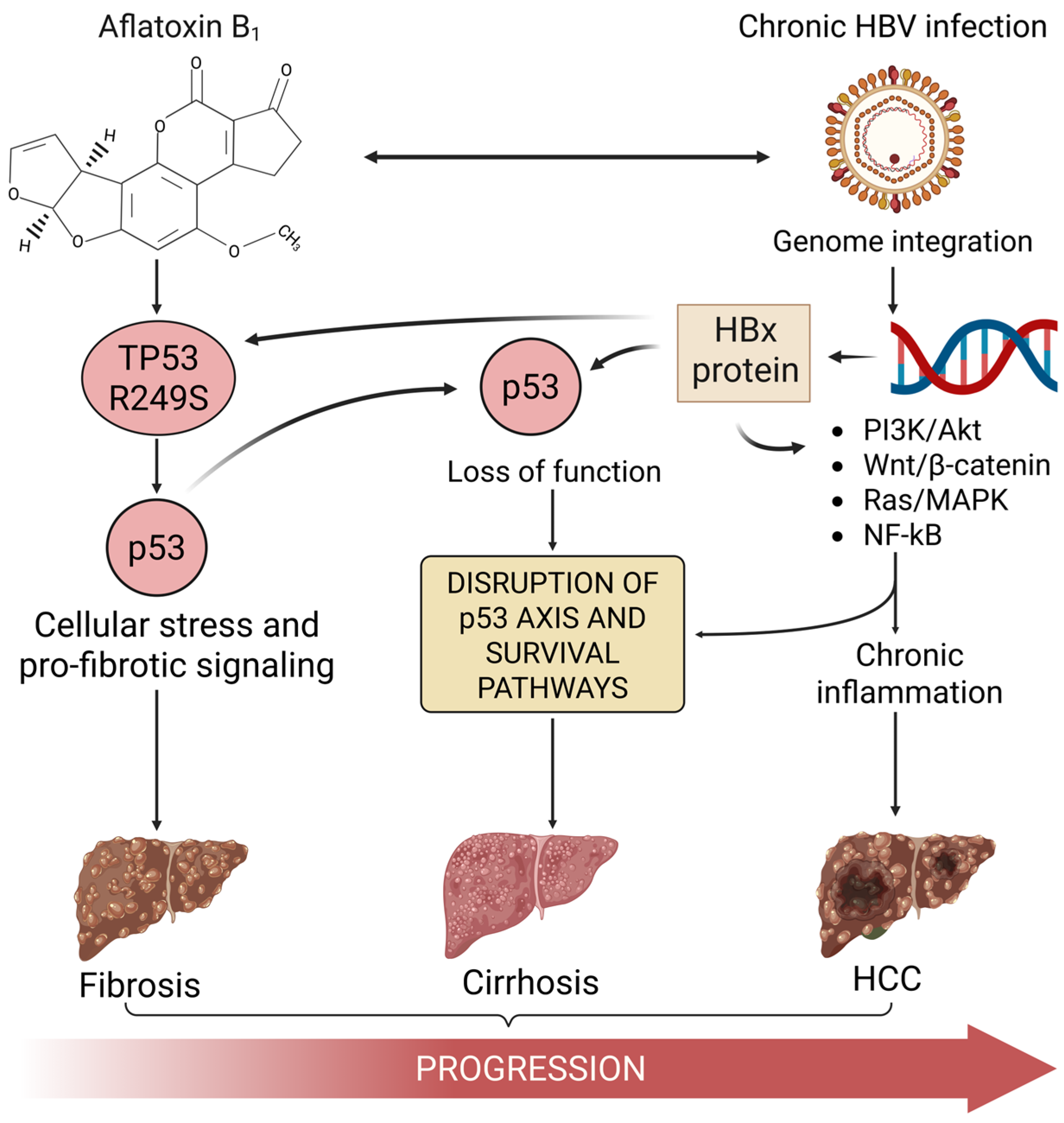
1. The Convergence of HBV, Aflatoxin, and p53 in HCC

Cancer remains a major cause of mortality and reduced life expectancy globally [1]. The development of malignancy is understood as a multistep process involving the acquisition of genetic and epigenetic alterations that drive uncontrolled cell proliferation, evasion of cell death, and eventual metastasis [2,3,4]. Hepatocellular carcinoma (HCC) exemplifies this complexity, ranking as the sixth most common cancer globally and the third leading cause of cancer-related mortality, responsible for hundreds of thousands of deaths annually [5]. In 2022 alone, liver cancer caused approximately 750,000 related deaths worldwide [6]. This highlights HCC as a major global health concern. While HCC etiology is diverse, exposure to dietary aflatoxin B1 (AFB1) and chronic infection with the hepatitis B virus (HBV) are recognized as predominant drivers, particularly in specific geographic regions of East Asia and Sub-Saharan Africa, where exposure to both agents is common [7,8,9]. This review will focus on the critical interplay between these two factors, particularly their convergent impact on the p53 tumor suppressor pathway, a key element in hepatocarcinogenesis. HBV infection alone is the most significant risk factor, accounting for about 50% of HCC incidence, while aflatoxin exposure is estimated to contribute to 4.6–28.2% of all global HCC cases [9]. AFB1, a potent mycotoxin produced by Aspergillus fungi contaminating staple crops like maize and peanuts [10,11,12,13], is classified as a Group 1 human carcinogen by IARC due to its strong association with liver cancer [14]. Chronic HBV infection promotes liver damage through persistent inflammation and viral protein activity, contributing to cirrhosis and creating a pro-carcinogenic environment; HBV can also integrate into the host genome, causing genetic instability and cancer-related gene mutations, further contributing to genetic instability [7,15]. Epidemiological evidence consistently suggests that co-exposure to AFB1 and HBV results in a synergistic increase in HCC risk, often exceeding the additive effects of each factor alone [16,17,18]. A key molecular mechanism underlying this synergy involves the convergence of both agents’ detrimental effects on the p53 tumor suppressor pathway. AFB1’s significant genotoxicity leads characteristically to a specific mutation in the TP53 gene (R249S) [19,20], while the multifunctional HBV X protein (HBx) directly interacts with and functionally inhibits the wild-type p53 protein [21,22,23,24]. This review focuses specifically on the central role of p53 pathway disruption driven by the interplay between potent aflatoxin-induced mutagenesis and HBV infection in the molecular pathogenesis of HCC.
Extensive epidemiological data from diverse populations provide compelling real-world evidence for the critical role of aflatoxin-induced p53 alterations, particularly in conjunction with HBV infection, in driving HCC [18,25,26,27,28]. The frequency of the specific TP53 R249S mutation in HCC cases exhibits significant geographical variation, strongly correlated with regional levels of AFB1 exposure assessed through dietary surveys or biomarker analysis [29,30,31]. This mutation is particularly prevalent in HCC from areas of Sub-Saharan Africa and Southeast Asia, where aflatoxin contamination of staple foods is high, and where HBV infection is often also endemic [19,32,33]. Numerous studies have directly examined the relationship between HBV infection (HBsAg status), quantitative measures of AFB1 exposure (e.g., aflatoxin–albumin adducts in serum), and the presence of the R249S mutation. A consistent finding is the significantly increased HCC risk among individuals with evidence of both HBV infection and significant aflatoxin exposure, demonstrating a synergistic effect where the combined risk is substantially greater than the sum of the individual risks [9,25]. Studies measuring AFB1 exposure biomarkers have directly linked higher adduct levels to increased HCC risk, particularly among chronic HBV carriers [34]. Furthermore, the R249S mutation itself is frequently detected in individuals with dual exposure [30,32,35]. Its detection in circulating DNA from asymptomatic individuals suggests it can be an early event related to exposure [36]. Research in The Gambia associated aflatoxin exposure markers and the plasma R249S mutation with the risk of liver cirrhosis, a major HCC precursor, noting interactions with HBV status [37]. Moreover, the presence of TP53 mutations, heavily influenced by the R249S prevalence in relevant regions, alongside HBV/AFB1 status, independently predicts clinical outcomes such as postoperative tumor recurrence [38]. Conversely, the near absence of the R249S mutation in HCC from populations with low aflatoxin exposure (e.g., Mexico), even if HBV or other risk factors like HCV/alcohol are present [39], strongly reinforces the specific causal link between AFB1 exposure and this p53 alteration. These epidemiological findings collectively highlight aflatoxin’s potent role in mutating p53 and establish this event as a key factor mediating the synergy with HBV in hepatocarcinogenesis.

2. The p53 Tumor Suppressor Pathway

The p53 protein, encoded by the TP53 gene, functions as a central node in the cellular stress response network, acting as a critical barrier against neoplastic transformation. Often termed the “guardian of the genome,” p53 integrates signals from diverse cellular insults, including DNA damage (such as double-strand breaks, adducts, or stalled replication forks), oncogene activation, hypoxia, ribosomal stress, and nutrient deprivation. Its primary role is to safeguard genomic integrity [40]. Under normal, unstressed conditions, p53 protein levels are kept low through continuous degradation, primarily mediated by the E3 ubiquitin ligase MDM2. Upon stress, p53 is stabilized and activated through post-translational modifications, notably phosphorylation by upstream kinases like ATM and ATR, which sense DNA damage. Activated p53 accumulates in the nucleus, where it functions primarily as a transcription factor, regulating a large network of target genes by activating or repressing them [41]. The cellular outcomes of p53 activation are context-dependent but typically involve inducing cell cycle arrest, senescence, or apoptosis. Cell cycle arrest, frequently mediated by transcriptional upregulation of the CDK inhibitor p21 (CDKN1A), occurs mainly at the G1/S or G2/M transitions, providing a window for DNA repair. If the damage is deemed irreparable, p53 promotes apoptosis by inducing the expression of pro-apoptotic proteins, including Bax, Puma, and Noxa, thereby eliminating potentially dangerous cells. This apoptotic function is extensively studied and crucial for resisting transformation and determining cancer therapy efficacy [42]. p53 also directly influences DNA repair pathways and contributes to maintaining overall genomic stability. Beyond these core functions, p53 is closely integrated with major cellular signaling pathways. It often acts antagonistically to pro-survival and pro-proliferative pathways like the PI3K/Akt/mTOR cascade; for instance, p53 can induce the expression of PTEN, a negative regulator of PI3K/Akt signaling. p53 activity is also modulated by these pathways; Akt can phosphorylate MDM2, enhancing its ability to degrade p53, creating complex feedback loops. Furthermore, p53 signaling crosstalk with pathways regulating inflammation (like NF-κB), angiogenesis (e.g., via thrombospondin-1), and cellular metabolism (regulating glycolysis and oxidative phosphorylation). Therefore, the loss or inactivation of p53 removes not only direct tumor-suppressive functions like apoptosis and cell cycle arrest but also disrupts the balance of these critical signaling networks, significantly contributing to cancer development and progression. Mutations affecting p53’s DNA binding domain are common in cancer, leading to the loss of its tumor-suppressor function and promoting tumorigenesis [43].

3. Aflatoxin B1-Induced TP53 R249S Mutation: A Molecular Signature in HCC

Aflatoxins are toxic secondary metabolites from Aspergillus species, posing a significant threat through contamination of major food staples worldwide (Figure 1) [44]. Aflatoxin B1 (AFB1) stands out as the most potent hepatocarcinogen among them [45], causally linked to a substantial proportion of global HCC cases, particularly in high-exposure regions [9,46]. AFB1 exerts its significant carcinogenic effect primarily through its genotoxicity following metabolic activation within the liver. Hepatic cytochrome P450 enzymes convert AFB1 into the highly reactive AFB1-8,9-exo-epoxide [20]. This electrophilic intermediate readily forms covalent adducts with cellular DNA, predominantly at the N7 atom of guanine residues, forming the bulky AFB1-N7-Gua lesion [47]. Although cellular DNA repair mechanisms, such as nucleotide excision repair, attempt to remove these adducts, their persistence or inaccurate processing during DNA replication leads to mutations, characteristically G-to-T transversions [48,49,50,51].
A defining feature of AFB1-induced mutagenesis in human liver cancer is the high frequency of this specific G>T transversion occurring at the third position of codon 249 (AGG → AGT) within the TP53 gene [19]. This mutation, accounting for up to 90% of TP53 mutations in AFB1-related HCC, encodes an arginine residue essential for p53’s DNA binding ability [52]. The resulting R249S amino acid substitution is widely recognized as a molecular signature or fingerprint of significant AFB1 exposure and is frequently detected (sometimes in >50% of tumors) in HCC cases originating from regions with high levels of dietary aflatoxin contamination [30]. This mutation accounts for up to 90% of TP53 mutations in AFB1-related HCC [53]. The R249S mutation represents a critical loss-of-function event, as the altered protein fails to effectively bind to p53 DNA response elements and activate downstream target genes necessary for cell cycle arrest and apoptosis [54]. Notably, evidence indicates that the R249S mutant is not merely inactive but acquires distinct oncogenic gain-of-function properties. These pro-tumorigenic activities may include enhancing the transcription of growth-promoting factors like IGF-II [55], altering interactions with crucial transcription factors such as Sp1 [55], and providing resistance to apoptotic signals, including those triggered by TNF-alpha or the HBV HBx protein [56,57]. Emerging research suggests complex mechanisms for this gain of function, potentially involving interactions with other proteins like CDK4/cyclin D1 and PIN1 or forming complexes with TANK-binding protein kinase 1 to evade immune surveillance [58]. These acquired functions likely confer a selective growth and survival advantage upon hepatocytes harboring the mutation, particularly in the context of chronic liver inflammation typical of high-risk settings [59]. The specific targeting of TP53 codon 249 by AFB1 provides a direct mechanistic link between this significant environmental carcinogen and the disabling of a key tumor suppressor pathway in the liver [60].

4. Hepatitis B Virus and HBx-Mediated Disruption of p53 Function

Hepatitis B virus (HBV) is an enveloped DNA virus, characterized by a small genome of approximately 3.2 kb. While its structure is defined by these features, its replication occurs in the cytoplasm through the reverse transcription of an encapsidated pre-genomic RNA (pgRNA) into a partially double-stranded relaxed circular DNA (RC-DNA) [61]. Chronic HBV infection is a major driver of HCC, involving multiple processes to promote liver cell transformation [62,63]. Besides inducing chronic inflammation and integrating its DNA into the host genome [64], HBV actively interferes with cellular tumor suppressor mechanisms, including the p53 pathway. This interference is largely mediated by the HBV X protein (HBx), a small (154-amino acid) regulatory protein with pleiotropic functions critical for the viral life cycle and implicated in hepatocarcinogenesis [65]. HBx is produced in about 70% of HBV-associated HCC [66]. HBx contributes to the inhibition of wild-type p53 through several distinct mechanisms. Firstly, HBx can physically interact with the p53 protein. This binding, often involving the C-terminal domain of p53, hinders p53’s ability to bind its target DNA sequences, thereby blocking its function as a transcriptional activator of tumor suppressor genes [21]. This direct interaction inhibits p53’s response element-directed transactivation [67]. Secondly, HBx promotes the degradation of the p53 protein. It facilitates this by upregulating or enhancing the activity of MDM2, the primary E3 ubiquitin ligase that targets p53 for destruction via the proteasome [68]. Another proposed mechanism involves HBx sequestering p53 in the cytoplasm, reducing the nuclear concentration available to trigger apoptosis [69]. This effect might be linked to HBx’s ability to activate signaling pathways like NF-κB [70,71]. By reducing the stability and thus the intracellular levels of functional p53, HBx diminishes the cell’s capacity to respond appropriately to stress signals. Thirdly, HBx can suppress p53-mediated apoptosis [21], allowing infected cells potentially harboring DNA damage or viral integration events to survive and proliferate. While HBV integration events can cause genomic instability [62,72], the concurrent functional inactivation of p53 by HBx is likely crucial for preventing the elimination of these genetically compromised cells. Additionally, HBx contributes to a pro-oncogenic cellular environment through the induction of oxidative stress and the activation of various pro-survival signaling pathways (e.g., PI3K/Akt/mTOR, Wnt, and Notch) [64,73,74,75], which can further counteract or bypass p53-dependent tumor suppression. Interestingly, HBx has also been shown to interact with and inactivate TAp63, a p53 homolog that can suppress HCC growth in cells with defective p53 [76].

5. Synergistic Disruption of p53 by Aflatoxin-Induced Mutation and HBV Activity

The significantly increased risk of HCC observed in individuals co-exposed to chronic HBV infection and dietary aflatoxins points strongly toward a synergistic interaction between these two carcinogens [16,37]. This synergy manifests prominently through molecular interactions involving the p53 pathway. The HBV–aflatoxin interaction likely results from the combined and complementary attacks on the p53 function. AFB1 delivers a direct mutagenic hit, frequently causing the function-altering R249S mutation [19], while HBV/HBx works concurrently to inhibit the function and promote the degradation of any remaining wild-type p53 protein [77]. This dual mechanism results in a more significant and sustained loss of p53-mediated tumor suppression than would likely occur with exposure to either agent alone.
Furthermore, HBx might directly enhance the mutagenic impact of AFB1. Experimental studies suggest that HBx expression increases the frequency of the specific AFB1-induced R249S mutation in vitro [78]. This potentiation could arise from HBx impairing the cellular DNA repair pathways responsible for removing the AFB1-DNA adducts before they cause mutations, or by inhibiting the apoptotic elimination of cells that have successfully acquired the TP53 mutation, thereby promoting their survival and potential for clonal expansion [66,79,80]. Additionally, functional cooperation between the R249S mutant p53 protein itself and HBx might contribute to the synergy. Studies suggest that p53-R249S and HBx can form a complex, potentially conferring a growth advantage, particularly in early hepatocarcinogenesis [54]. Both possess anti-apoptotic capabilities and their co-expression could confer a significant survival and proliferative advantage, particularly during the crucial early stages of hepatocarcinogenesis [54,81].
The consequence of this significant p53 pathway disruption, driven by both aflatoxin-induced mutation and HBV/HBx activity, is a compromised cellular response to DNA damage. This failure allows the genomic instability, fueled by both the continuous formation of AFB1 adducts and HBV-related factors (integration, chronic inflammation, and oxidative stress), to accelerate the accumulation of additional genetic and chromosomal alterations [30]. This heightened genomic instability, occurring in cells that have lost their p53 safeguard, contributes to the multistep progression toward malignant HCC [82]. Figure 2 addresses the role of both AFB1 and HBV in promoting p53 downregulation, fibrosis, cirrhosis, and HCC. In addition, it includes the major signaling pathways involved in liver cancer pathogenesis, including Wnt, PI3K-AKT-STAT3, Ras-MAPK1, and NF-κB. These are commonly activated during liver tumor development and are frequently modulated by HBV infection and aflatoxin exposure [14,83].

6. Conclusions: Aflatoxin-Induced p53 Mutation as a Key Driver in HBV-Associated HCC

In conclusion, the p53 tumor suppressor pathway is a central molecular target significantly compromised by both dietary aflatoxin exposure and chronic HBV infection, explaining their strong synergistic interaction in driving hepatocellular carcinoma. Aflatoxin B1, a widespread environmental carcinogen, directly damages DNA and induces a characteristic R249S mutation in the TP53 gene, a key molecular event that impairs p53’s protective functions and may confer oncogenic properties. HBV infection, particularly through the actions of the HBx protein, further undermines the p53 pathway by inhibiting wild-type p53 activity and promoting its degradation. This combined attack—specific mutation by AFB1 coupled with functional inactivation by HBV, leads to a significant disruption of cellular anti-oncogenic mechanisms.
The resulting synergistic loss of p53 function provides a strong molecular explanation for the significantly elevated HCC risk observed in co-exposed populations globally. Understanding this interplay, particularly the important role of aflatoxin in inducing the specific p53 mutation that is then amplified in the context of HBV infection, is crucial for public health. It underscores the critical need for comprehensive prevention strategies targeting both a reduction in aflatoxin exposure in food supplies and widespread HBV vaccination and treatment. The R249S mutation stands as a valuable biomarker of aflatoxin-related genetic damage and HCC risk. While HCC pathogenesis is multifaceted, the convergent disruption of the p53 pathway, initiated significantly by aflatoxin mutagenesis, represents a key mechanism underlying the significant carcinogenicity associated with combined HBV and aflatoxin exposure, demanding continued research and targeted interventions.
Future research should focus on understanding how aflatoxin-induced p53 mutations and HBV infection work together to drive liver cancer. Indeed, more studies are needed to clarify how the HBx protein may increase the mutagenic impact of AFB1, possibly by interfering with DNA repair or by protecting mutated cells from being eliminated. Exploring the combined effects of the TP53 R249S mutation and HBx could also help uncover new mechanisms that give cancer cells a survival advantage and identify potential targets for therapy. From a public health standpoint, efforts should be directed toward using the R249S mutation and HBx expression as early biomarkers for liver cancer risk, especially in regions with high aflatoxin exposure and HBV prevalence. At the same time, reinforcing preventive strategies such as HBV vaccination, antiviral treatment, and strict control of aflatoxin contamination in food can significantly reduce HCC incidence. These combined approaches should be evaluated in real-world settings to guide effective cancer prevention programs.

Author Contributions

Conceptualization, C.M.-L. and F.A.; software, C.M.-L.; validation, C.M.-L. and F.A.; investigation C.M.-L. and F.A.; resources, C.M.-L. and F.A.; writing—original draft preparation, C.M.-L.; writing—review and editing, C.M.-L. and F.A.; supervision, F.A.; project administration, F.A.; funding acquisition, F.A. All authors have read and agreed to the published version of the manuscript.

Funding

This research was funded by Programa Fortalecimiento Grupos de Investigación UTA 7703-24 (F.A.). The APC was funded by the Universidad de Tarapacá.

Institutional Review Board Statement

Not applicable.

Informed Consent Statement

Not applicable.

Data Availability Statement

No new data were created or analyzed in this study.

Conflicts of Interest

The authors declare no conflicts of interest.

References

  1. Ferlay, J.; Colombet, M.; Soerjomataram, I.; Parkin, D.M.; Piñeros, M.; Znaor, A.; Bray, F. Cancer statistics for the year 2020: An overview. Int. J. Cancer 2021, 149, 778–789. [Google Scholar] [CrossRef]
  2. Stratton, M.R.; Campbell, P.J.; Futreal, P.A. The cancer genome. Nature 2009, 458, 719–724. [Google Scholar] [CrossRef] [PubMed]
  3. Vogelstein, B.; Papadopoulos, N.; Velculescu, V.E.; Zhou, S.; Diaz, L.A.; Kinzler, K.W. Cancer genome landscapes. Science 2013, 339, 1546–1558. [Google Scholar] [CrossRef] [PubMed]
  4. Hanahan, D.; Weinberg, R.A. Hallmarks of cancer: The next generation. Cell 2011, 144, 646–674. [Google Scholar] [CrossRef] [PubMed]
  5. Bray, F.; Laversanne, M.; Sung, H.; Ferlay, J.; Siegel, R.L.; Soerjomataram, I.; Jemal, A. Global cancer statistics 2022: GLOBOCAN estimates of incidence and mortality worldwide for 36 cancers in 185 countries. CA Cancer J. Clin. 2024, 74, 229–263. [Google Scholar] [CrossRef]
  6. Li, Q.; Ding, C.; Cao, M.; Yang, F.; Yan, X.; He, S.; Zhang, S.; Teng, Y.; Tan, N.; Wang, J.; et al. Global epidemiology of liver cancer 2022: An emphasis on geographic disparities. Chin. Med. J. 2024, 137, 2334–2342. [Google Scholar] [CrossRef]
  7. Hudu, S.A.; Shinkafi, S.H.; Jimoh, A.O. A critical review of diagnostic and prognostic markers of chronic hepatitis B infection. Med. Rev. (2021) 2024, 4, 225–234. [Google Scholar] [CrossRef]
  8. Wu, H.C.; Santella, R. The role of aflatoxins in hepatocellular carcinoma. Hepat. Mon. 2012, 12, e7238. [Google Scholar] [CrossRef]
  9. Liu, Y.; Wu, F. Global burden of aflatoxin-induced hepatocellular carcinoma: A risk assessment. Environ. Health Perspect. 2010, 118, 818–824. [Google Scholar] [CrossRef]
  10. Shabeer, S.; Asad, S.; Jamal, A.; Ali, A. Aflatoxin Contamination, Its Impact and Management Strategies: An Updated Review. Toxins 2022, 14, 307. [Google Scholar] [CrossRef]
  11. Ostry, V.; Malir, F.; Toman, J.; Grosse, Y. Mycotoxins as human carcinogens-the IARC Monographs classification. Mycotoxin Res. 2017, 33, 65–73. [Google Scholar] [CrossRef]
  12. Wu, F.; Stacy, S.L.; Kensler, T.W. Global risk assessment of aflatoxins in maize and peanuts: Are regulatory standards adequately protective? Toxicol. Sci. 2013, 135, 251–259. [Google Scholar] [CrossRef]
  13. Nugraha, A.; Khotimah, K.; Rietjens, I.M.C.M. Risk assessment of aflatoxin B1 exposure from maize and peanut consumption in Indonesia using the margin of exposure and liver cancer risk estimation approaches. Food Chem. Toxicol. 2018, 113, 134–144. [Google Scholar] [CrossRef] [PubMed]
  14. Marchese, S.; Polo, A.; Ariano, A.; Velotto, S.; Costantini, S.; Severino, L. Aflatoxin B1 and M1: Biological Properties and Their Involvement in Cancer Development. Toxins 2018, 10, 214. [Google Scholar] [CrossRef] [PubMed]
  15. Seeger, C.; Mason, W.S. Molecular biology of hepatitis B virus infection. Virology 2015, 479–480, 672–686. [Google Scholar] [CrossRef] [PubMed]
  16. Ming, L.; Thorgeirsson, S.S.; Gail, M.H.; Lu, P.; Harris, C.C.; Wang, N.; Shao, Y.; Wu, Z.; Liu, G.; Wang, X.; et al. Dominant role of hepatitis B virus and cofactor role of aflatoxin in hepatocarcinogenesis in Qidong, China. Hepatology 2002, 36, 1214–1220. [Google Scholar] [CrossRef]
  17. Chen, C.J.; Hsu, W.L.; Yang, H.I.; Lee, M.H.; Chen, H.C.; Chien, Y.C.; You, S.L. Epidemiology of virus infection and human cancer. Recent Results Cancer Res. 2014, 193, 11–32. [Google Scholar] [CrossRef]
  18. Kew, M.C. Synergistic interaction between aflatoxin B1 and hepatitis B virus in hepatocarcinogenesis. Liver Int. 2003, 23, 405–409. [Google Scholar] [CrossRef]
  19. Hsu, I.C.; Metcalf, R.A.; Sun, T.; Welsh, J.A.; Wang, N.J.; Harris, C.C. Mutational hotspot in the p53 gene in human hepatocellular carcinomas. Nature 1991, 350, 427–428. [Google Scholar] [CrossRef]
  20. Ozturk, M. p53 mutation in hepatocellular carcinoma after aflatoxin exposure. Lancet 1991, 338, 1356–1359. [Google Scholar] [CrossRef]
  21. Iyer, S.; Groopman, J.D. Interaction of mutant hepatitis B X protein with p53 tumor suppressor protein affects both transcription and cell survival. Mol. Carcinog. 2011, 50, 972–980. [Google Scholar] [CrossRef] [PubMed]
  22. Che, L.; Du, Z.B.; Wang, W.H.; Wu, J.S.; Han, T.; Chen, Y.Y.; Han, P.Y.; Lei, Z.; Chen, X.X.; He, Y.; et al. Intracellular antibody targeting HBx suppresses invasion and metastasis in hepatitis B virus-related hepatocarcinogenesis via protein phosphatase 2A-B56γ-mediated dephosphorylation of protein kinase B. Cell Prolif. 2022, 55, e13304. [Google Scholar] [CrossRef]
  23. Kew, M.C. Hepatitis B virus x protein in the pathogenesis of hepatitis B virus-induced hepatocellular carcinoma. J. Gastroenterol. Hepatol. 2011, 26 (Suppl. S1), 144–152. [Google Scholar] [CrossRef] [PubMed]
  24. Kong, G.; Zhang, J.; Zhang, S.; Shan, C.; Ye, L.; Zhang, X. Upregulated microRNA-29a by hepatitis B virus X protein enhances hepatoma cell migration by targeting PTEN in cell culture model. PLoS ONE 2011, 6, e19518. [Google Scholar] [CrossRef]
  25. Wu, H.C.; Wang, Q.; Yang, H.I.; Ahsan, H.; Tsai, W.Y.; Wang, L.Y.; Chen, S.Y.; Chen, C.J.; Santella, R.M. Aflatoxin B1 exposure, hepatitis B virus infection, and hepatocellular carcinoma in Taiwan. Cancer Epidemiol. Biomark. Prev. 2009, 18, 846–853. [Google Scholar] [CrossRef] [PubMed]
  26. Chen, S.Y.; Chen, C.J.; Chou, S.R.; Hsieh, L.L.; Wang, L.Y.; Tsai, W.Y.; Ahsan, H.; Santella, R.M. Association of aflatoxin B(1)-albumin adduct levels with hepatitis B surface antigen status among adolescents in Taiwan. Cancer Epidemiol. Biomark. Prev. 2001, 10, 1223–1226. [Google Scholar]
  27. Bannasch, P.; Khoshkhou, N.I.; Hacker, H.J.; Radaeva, S.; Mrozek, M.; Zillmann, U.; Kopp-Schneider, A.; Haberkorn, U.; Elgas, M.; Tolle, T. Synergistic hepatocarcinogenic effect of hepadnaviral infection and dietary aflatoxin B1 in woodchucks. Cancer Res. 1995, 55, 3318–3330. [Google Scholar] [PubMed]
  28. Kew, M.C. Aflatoxins as a cause of hepatocellular carcinoma. J. Gastrointest. Liver Dis. 2013, 22, 305–310. [Google Scholar]
  29. Schrenk, D.; Bignami, M.; Bodin, L.; Chipman, J.K.; Del Mazo, J.; Grasl-Kraupp, B.; Hogstrand, C.; Hoogenboom, L.R.; Leblanc, J.C.; Nebbia, C.S.; et al. Risk assessment of aflatoxins in food. EFSA J. 2020, 18, e06040. [Google Scholar] [CrossRef]
  30. Han, C.; Yu, T.; Qin, W.; Liao, X.; Huang, J.; Liu, Z.; Yu, L.; Liu, X.; Chen, Z.; Yang, C.; et al. Genome-wide association study of the TP53 R249S mutation in hepatocellular carcinoma with aflatoxin B1 exposure and infection with hepatitis B virus. J. Gastrointest. Oncol. 2020, 11, 1333–1349. [Google Scholar] [CrossRef]
  31. Alvarez, C.S.; Ortiz, J.; Bendfeldt-Avila, G.; Xie, Y.; Wang, M.; Wu, D.; Higson, H.; Lee, E.; Teshome, K.; Barnoya, J.; et al. Analysis of TP53 aflatoxin signature mutation in hepatocellular carcinomas from Guatemala: A cross-sectional study (2016–2017). Health Sci. Rep. 2020, 3, e155. [Google Scholar] [CrossRef] [PubMed]
  32. Gouas, D.A.; Villar, S.; Ortiz-Cuaran, S.; Legros, P.; Ferro, G.; Kirk, G.D.; Lesi, O.A.; Mendy, M.; Bah, E.; Friesen, M.D.; et al. TP53 R249S mutation, genetic variations in HBX and risk of hepatocellular carcinoma in The Gambia. Carcinogenesis 2012, 33, 1219–1224. [Google Scholar] [CrossRef] [PubMed]
  33. Bressac, B.; Kew, M.; Wands, J.; Ozturk, M. Selective G to T mutations of p53 gene in hepatocellular carcinoma from southern Africa. Nature 1991, 350, 429–431. [Google Scholar] [CrossRef]
  34. Chu, Y.J.; Yang, H.I.; Wu, H.C.; Liu, J.; Wang, L.Y.; Lu, S.N.; Lee, M.H.; Jen, C.L.; You, S.L.; Santella, R.M.; et al. Aflatoxin B. Int. J. Cancer 2017, 141, 711–720. [Google Scholar] [CrossRef]
  35. Ortiz-Cuaran, S.; Villar, S.; Gouas, D.; Ferro, G.; Plymoth, A.; Khuhaprema, T.; Kalalak, A.; Sangrajrang, S.; Friesen, M.D.; Groopman, J.D.; et al. Association between HBX status, aflatoxin-induced R249S TP53 mutation and risk of hepatocellular carcinoma in a case-control study from Thailand. Cancer Lett. 2013, 331, 46–51. [Google Scholar] [CrossRef]
  36. Villar, S.; Le Roux-Goglin, E.; Gouas, D.A.; Plymoth, A.; Ferro, G.; Boniol, M.; Lereau, M.; Bah, E.; Hall, A.J.; Wild, C.P.; et al. Seasonal variation in TP53 R249S-mutated serum DNA with aflatoxin exposure and hepatitis B virus infection. Environ. Health Perspect. 2011, 119, 1635–1640. [Google Scholar] [CrossRef] [PubMed]
  37. Kuniholm, M.H.; Lesi, O.A.; Mendy, M.; Akano, A.O.; Sam, O.; Hall, A.J.; Whittle, H.; Bah, E.; Goedert, J.J.; Hainaut, P.; et al. Aflatoxin exposure and viral hepatitis in the etiology of liver cirrhosis in the Gambia, West Africa. Environ. Health Perspect. 2008, 116, 1553–1557. [Google Scholar] [CrossRef]
  38. Qin, W.; Han, C.; Mai, R.; Yu, T.; Shang, L.; Ye, X.; Zhu, G.; Su, H.; Liao, X.; Liu, Z.; et al. Establishment of a prognostic model for predicting short-term disease-free survival in cases of hepatitis B-related hepatocellular carcinoma with the TP53 249Ser mutation in southern China. Transl. Cancer Res. 2020, 9, 4517–4533. [Google Scholar] [CrossRef]
  39. Valdés-Peregrina, E.N.; Sánchez-Hernández, B.E.; Gamboa-Domínguez, A. Metabolic Syndrome Instead of Aflatoxin-Related TP53 R249S Mutation as a Hepatocellular Carcinoma Risk Factor. Rev. Investig. Clin. 2020, 72, 127–134. [Google Scholar] [CrossRef]
  40. Levine, A.J. p53: 800 million years of evolution and 40 years of discovery. Nat. Rev. Cancer 2020, 20, 471–480. [Google Scholar] [CrossRef]
  41. Hernández Borrero, L.J.; El-Deiry, W.S. Tumor suppressor p53: Biology, signaling pathways, and therapeutic targeting. Biochim. Biophys. Acta Rev. Cancer 2021, 1876, 188556. [Google Scholar] [CrossRef] [PubMed]
  42. Aubrey, B.J.; Kelly, G.L.; Janic, A.; Herold, M.J.; Strasser, A. How does p53 induce apoptosis and how does this relate to p53-mediated tumour suppression? Cell Death Differ. 2018, 25, 104–113. [Google Scholar] [CrossRef]
  43. Rivlin, N.; Brosh, R.; Oren, M.; Rotter, V. Mutations in the p53 Tumor Suppressor Gene: Important Milestones at the Various Steps of Tumorigenesis. Genes Cancer 2011, 2, 466–474. [Google Scholar] [CrossRef] [PubMed]
  44. Okechukwu, V.O.; Adelusi, O.A.; Kappo, A.P.; Njobeh, P.B.; Mamo, M.A. Aflatoxins: Occurrence, biosynthesis, mechanism of action and effects, conventional/emerging detection techniques. Food Chem. 2024, 436, 137775. [Google Scholar] [CrossRef] [PubMed]
  45. Hamid, A.S.; Tesfamariam, I.G.; Zhang, Y.; Zhang, Z.G. Aflatoxin B1-induced hepatocellular carcinoma in developing countries: Geographical distribution, mechanism of action and prevention. Oncol. Lett. 2013, 5, 1087–1092. [Google Scholar] [CrossRef]
  46. Hsia, C.C.; Kleiner, D.E.; Axiotis, C.A.; Di Bisceglie, A.; Nomura, A.M.; Stemmermann, G.N.; Tabor, E. Mutations of p53 gene in hepatocellular carcinoma: Roles of hepatitis B virus and aflatoxin contamination in the diet. J. Natl. Cancer Inst. 1992, 84, 1638–1641. [Google Scholar] [CrossRef]
  47. Gramantieri, L.; Gnudi, F.; Vasuri, F.; Mandrioli, D.; Fornari, F.; Tovoli, F.; Suzzi, F.; Vornoli, A.; D’Errico, A.; Piscaglia, F.; et al. Aflatoxin B1 DNA-Adducts in Hepatocellular Carcinoma from a Low Exposure Area. Nutrients 2022, 14, 1652. [Google Scholar] [CrossRef]
  48. Smela, M.E.; Hamm, M.L.; Henderson, P.T.; Harris, C.M.; Harris, T.M.; Essigmann, J.M. The aflatoxin B(1) formamidopyrimidine adduct plays a major role in causing the types of mutations observed in human hepatocellular carcinoma. Proc. Natl. Acad. Sci. USA 2002, 99, 6655–6660. [Google Scholar] [CrossRef]
  49. de Oliveira, C.A.; Germano, P.M. Aflatoxins: Current concepts on mechanisms of toxicity and their involvement in the etiology of hepatocellular carcinoma. Rev. Saude Publica 1997, 31, 417–424. [Google Scholar] [CrossRef]
  50. Coursaget, P.; Depril, N.; Chabaud, M.; Nandi, R.; Mayelo, V.; LeCann, P.; Yvonnet, B. High prevalence of mutations at codon 249 of the p53 gene in hepatocellular carcinomas from Senegal. Br. J. Cancer 1993, 67, 1395–1397. [Google Scholar] [CrossRef]
  51. Pineau, P.; Marchio, A.; Battiston, C.; Cordina, E.; Russo, A.; Terris, B.; Qin, L.X.; Turlin, B.; Tang, Z.Y.; Mazzaferro, V.; et al. Chromosome instability in human hepatocellular carcinoma depends on p53 status and aflatoxin exposure. Mutat. Res. 2008, 653, 6–13. [Google Scholar] [CrossRef] [PubMed]
  52. Hussain, S.P.; Schwank, J.; Staib, F.; Wang, X.W.; Harris, C.C. TP53 mutations and hepatocellular carcinoma: Insights into the etiology and pathogenesis of liver cancer. Oncogene 2007, 26, 2166–2176. [Google Scholar] [CrossRef]
  53. Qi, L.N.; Bai, T.; Chen, Z.S.; Wu, F.X.; Chen, Y.Y.; De Xiang, B.; Peng, T.; Han, Z.G.; Li, L.Q. The p53 mutation spectrum in hepatocellular carcinoma from Guangxi, China: Role of chronic hepatitis B virus infection and aflatoxin B1 exposure. Liver Int. 2015, 35, 999–1009. [Google Scholar] [CrossRef] [PubMed]
  54. Gouas, D.A.; Shi, H.; Hautefeuille, A.H.; Ortiz-Cuaran, S.L.; Legros, P.C.; Szymanska, K.J.; Galy, O.; Egevad, L.A.; Abedi-Ardekani, B.; Wiman, K.G.; et al. Effects of the TP53 p.R249S mutant on proliferation and clonogenic properties in human hepatocellular carcinoma cell lines: Interaction with hepatitis B virus X protein. Carcinogenesis 2010, 31, 1475–1482. [Google Scholar] [CrossRef]
  55. Lee, Y.I.; Lee, S.; Das, G.C.; Park, U.S.; Park, S.M. Activation of the insulin-like growth factor II transcription by aflatoxin B1 induced p53 mutant 249 is caused by activation of transcription complexes; implications for a gain-of-function during the formation of hepatocellular carcinoma. Oncogene 2000, 19, 3717–3726. [Google Scholar] [CrossRef]
  56. Su, F.; Schneider, R.J. Hepatitis B virus HBx protein sensitizes cells to apoptotic killing by tumor necrosis factor alpha. Proc. Natl. Acad. Sci. USA 1997, 94, 8744–8749. [Google Scholar] [CrossRef]
  57. Lam, Y.K.; Yu, J.; Huang, H.; Ding, X.; Wong, A.M.; Leung, H.H.; Chan, A.W.; Ng, K.K.; Xu, M.; Wang, X.; et al. TP53 R249S mutation in hepatic organoids captures the predisposing cancer risk. Hepatology 2023, 78, 727–740. [Google Scholar] [CrossRef] [PubMed]
  58. Liao, P.; Zeng, S.X.; Zhou, X.; Chen, T.; Zhou, F.; Cao, B.; Jung, J.H.; Del Sal, G.; Luo, S.; Lu, H. Mutant p53 Gains Its Function via c-Myc Activation upon CDK4 Phosphorylation at Serine 249 and Consequent PIN1 Binding. Mol. Cell 2017, 68, 1134–1146.e6. [Google Scholar] [CrossRef]
  59. Zhang, W.; Liu, Y.; Jang, H.; Nussinov, R. Cell cycle progression mechanisms: Slower cyclin-D/CDK4 activation and faster cyclin-E/CDK2. bioRxiv 2023. [Google Scholar] [CrossRef]
  60. Fasullo, M. Cellular Responses to Aflatoxin-Associated DNA Adducts. In DNA Repair; Mognato, M., Ed.; IntechOpen: London, UK, 2018. [Google Scholar]
  61. Dias, J.D.; Sarica, N.; Cournac, A.; Koszul, R.; Neuveut, C. Crosstalk between Hepatitis B Virus and the 3D Genome Structure. Viruses 2022, 14, 445. [Google Scholar] [CrossRef]
  62. Yeh, S.H.; Li, C.L.; Lin, Y.Y.; Ho, M.C.; Wang, Y.C.; Tseng, S.T.; Chen, P.J. Hepatitis B Virus DNA Integration Drives Carcinogenesis and Provides a New Biomarker for HBV-related HCC. Cell Mol. Gastroenterol. Hepatol. 2023, 15, 921–929. [Google Scholar] [CrossRef] [PubMed]
  63. Jiang, Y.; Han, Q.; Zhao, H.; Zhang, J. The Mechanisms of HBV-Induced Hepatocellular Carcinoma. J. Hepatocell. Carcinoma 2021, 8, 435–450. [Google Scholar] [CrossRef]
  64. Yen, C.J.; Lin, Y.J.; Yen, C.S.; Tsai, H.W.; Tsai, T.F.; Chang, K.Y.; Huang, W.C.; Lin, P.W.; Chiang, C.W.; Chang, T.T. Hepatitis B virus X protein upregulates mTOR signaling through IKKβ to increase cell proliferation and VEGF production in hepatocellular carcinoma. PLoS ONE 2012, 7, e41931. [Google Scholar] [CrossRef]
  65. Li, D.; Hamadalnil, Y.; Tu, T. Hepatitis B Viral Protein HBx: Roles in Viral Replication and Hepatocarcinogenesis. Viruses 2024, 16, 1361. [Google Scholar] [CrossRef] [PubMed]
  66. Sivasudhan, E.; Blake, N.; Lu, Z.; Meng, J.; Rong, R. Hepatitis B Viral Protein HBx and the Molecular Mechanisms Modulating the Hallmarks of Hepatocellular Carcinoma: A Comprehensive Review. Cells 2022, 11, 741. [Google Scholar] [CrossRef]
  67. Truant, R.; Antunovic, J.; Greenblatt, J.; Prives, C.; Cromlish, J.A. Direct interaction of the hepatitis B virus HBx protein with p53 leads to inhibition by HBx of p53 response element-directed transactivation. J. Virol. 1995, 69, 1851–1859. [Google Scholar] [CrossRef]
  68. Xian, L.; Zhao, J.; Wang, J.; Fang, Z.; Peng, B.; Wang, W.; Ji, X.; Yu, L. p53 Promotes proteasome-dependent degradation of oncogenic protein HBx by transcription of MDM2. Mol. Biol. Rep. 2010, 37, 2935–2940. [Google Scholar] [CrossRef] [PubMed]
  69. Elmore, L.W.; Hancock, A.R.; Chang, S.F.; Wang, X.W.; Chang, S.; Callahan, C.P.; Geller, D.A.; Will, H.; Harris, C.C. Hepatitis B virus X protein and p53 tumor suppressor interactions in the modulation of apoptosis. Proc. Natl. Acad. Sci. USA 1997, 94, 14707–14712. [Google Scholar] [CrossRef]
  70. Lim, K.H.; Choi, H.S.; Park, Y.K.; Park, E.S.; Shin, G.C.; Kim, D.H.; Ahn, S.H.; Kim, K.H. HBx-induced NF-κB signaling in liver cells is potentially mediated by the ternary complex of HBx with p22-FLIP and NEMO. PLoS ONE 2013, 8, e57331. [Google Scholar] [CrossRef]
  71. Chen, L.; Lin, X.; Lei, Y.; Xu, X.; Zhou, Q.; Chen, Y.; Liu, H.; Jiang, J.; Yang, Y.; Zheng, F.; et al. Aerobic glycolysis enhances HBx-initiated hepatocellular carcinogenesis via NF-κBp65/HK2 signalling. J. Exp. Clin. Cancer Res. 2022, 41, 329. [Google Scholar] [CrossRef]
  72. Tu, T.; Budzinska, M.A.; Shackel, N.A.; Urban, S. HBV DNA Integration: Molecular Mechanisms and Clinical Implications. Viruses 2017, 9, 75. [Google Scholar] [CrossRef] [PubMed]
  73. Niu, L.J.; Huang, T.; Wang, L.; Sun, X.F.; Zhang, Y.M. HBX suppresses PTEN to promote the malignant progression of hepatocellular carcinoma through mi-R155 activation. Ann. Hepatol. 2022, 27, 100688. [Google Scholar] [CrossRef]
  74. Sun, Q.; Wang, R.; Wang, Y.; Luo, J.; Wang, P.; Cheng, B. Notch1 is a potential therapeutic target for the treatment of human hepatitis B virus X protein-associated hepatocellular carcinoma. Oncol. Rep. 2014, 31, 933–939. [Google Scholar] [CrossRef]
  75. Gao, W.Y.; Li, D.; Cai, D.E.; Huang, X.Y.; Zheng, B.Y.; Huang, Y.H.; Chen, Z.X.; Wang, X.Z. Hepatitis B virus X protein sensitizes HL-7702 cells to oxidative stress-induced apoptosis through modulation of the mitochondrial permeability transition pore. Oncol. Rep. 2017, 37, 48–56. [Google Scholar] [CrossRef]
  76. Xie, B.; Hao, Q.; Zhou, X.; Chen, D. Inactivation of tumor suppressor TAp63 by hepatitis B virus X protein in hepatocellular carcinoma. Chin. Med. J. 2022, 135, 1728–1733. [Google Scholar] [CrossRef] [PubMed]
  77. Xu, Q.; Gu, S.; Liang, J.; Lin, Z.; Zheng, S.; Yan, J. The Biological Function of Hepatitis B Virus X Protein in Hepatocellular Carcinoma. Oncol. Res. 2019, 27, 509–514. [Google Scholar] [CrossRef]
  78. Madden, C.R.; Finegold, M.J.; Slagle, B.L. Altered DNA mutation spectrum in aflatoxin b1-treated transgenic mice that express the hepatitis B virus x protein. J. Virol. 2002, 76, 11770–11774. [Google Scholar] [CrossRef]
  79. Becker, S.A.; Lee, T.H.; Butel, J.S.; Slagle, B.L. Hepatitis B virus X protein interferes with cellular DNA repair. J. Virol. 1998, 72, 266–272. [Google Scholar] [CrossRef] [PubMed]
  80. Sekiba, K.; Otsuka, M.; Funato, K.; Miyakawa, Y.; Tanaka, E.; Seimiya, T.; Yamagami, M.; Tsutsumi, T.; Okushin, K.; Miyakawa, K.; et al. HBx-induced degradation of Smc5/6 complex impairs homologous recombination-mediated repair of damaged DNA. J. Hepatol. 2022, 76, 53–62. [Google Scholar] [CrossRef]
  81. Choudhary, H.B.; Mandlik, S.K.; Mandlik, D.S. Role of p53 suppression in the pathogenesis of hepatocellular carcinoma. World J. Gastrointest. Pathophysiol. 2023, 14, 46–70. [Google Scholar] [CrossRef]
  82. Farazi, P.A.; DePinho, R.A. Hepatocellular carcinoma pathogenesis: From genes to environment. Nat. Rev. Cancer 2006, 6, 674–687. [Google Scholar] [CrossRef] [PubMed]
  83. Rizzo, G.E.M.; Cabibbo, G.; Craxì, A. Hepatitis B Virus-Associated Hepatocellular Carcinoma. Viruses 2022, 14, 986. [Google Scholar] [CrossRef] [PubMed]
Figure 1. Biotransformation of aflatoxin B1 (AFB1) and DNA adduct formation leading to mutagenesis. The metabolic activation of AFB1 occurs primarily via cytochrome P450 enzymes (notably CYP1A2 and CYP3A4), producing the highly reactive AFB1-8,9-epoxide intermediate. This electrophilic metabolite covalently binds to DNA, preferentially at the N7 position of guanine bases, forming the unstable AFB1-N7-Guanine adduct. Subsequent hydrolysis or ring-opening rearrangement of this adduct yields a more persistent and mutagenic lesion, known as the AFB1-formamidopyrimidine (FAPY) adduct. These bulky DNA lesions, if not repaired, can induce G→T transversions during replication, notably at codon 249 of the TP53 gene, contributing to the characteristic R249S mutation observed in AFB1-related hepatocellular carcinoma.
Jox 15 00096 g001
Figure 2. Cooperative mechanisms by which aflatoxin B1 (AFB1) and chronic hepatitis B virus (HBV) infection promote hepatocellular carcinoma (HCC) through p53 pathway disruption. AFB1 is metabolically activated in the liver and induces the hotspot TP53 R249S mutation, resulting in p53 downregulation. This loss of p53 function contributes to cellular stress and pro-fibrotic signaling, leading to fibrosis. Independently, chronic HBV infection integrates into the host genome and expresses the HBx protein, which functionally suppresses p53 and activates oncogenic signaling pathways including PI3K/Akt, Wnt/β-catenin, Ras/MAPK, and NF-κB. These signals promote chronic inflammation and further impair apoptosis and cell cycle regulation. The combined effects converge on the disruption of the p53 axis and cellular survival pathways, facilitating the pathological progression from fibrosis to cirrhosis and ultimately HCC.
Jox 15 00096 g002
Disclaimer/Publisher’s Note: The statements, opinions and data contained in all publications are solely those of the individual author(s) and contributor(s) and not of MDPI and/or the editor(s). MDPI and/or the editor(s) disclaim responsibility for any injury to people or property resulting from any ideas, methods, instructions or products referred to in the content.

