Review

Environmental Xenobiotics and Epigenetic Modifications: Implications for Human Health and Disease

by
Ana Filipa Sobral
1,2,*,†,
Andrea Cunha
3,†,
Inês Costa
4,5,6,
Mariana Silva-Carvalho
1,7,
Renata Silva
4,5 and
Daniel José Barbosa
1,7,*
1
Associate Laboratory i4HB—Institute for Health and Bioeconomy, University Institute of Health Sciences-CESPU, 4585-116 Gandra, Portugal
2
UCIBIO—Applied Molecular Biosciences Unit, Toxicologic Pathology Research Laboratory, University Institute of Health Sciences (1H-TOXRUN, IUCS-CESPU), 4585-116 Gandra, Portugal
3
UNIPRO—Oral Pathology and Rehabilitation Research Unit, University Institute of Health Sciences (IUCS-CESPU), 4585-116 Gandra, Portugal
4
Associate Laboratory i4HB—Institute for Health and Bioeconomy, Faculty of Pharmacy, University of Porto, 4050-313 Porto, Portugal
5
UCIBIO—Applied Molecular Biosciences Unit, Laboratory of Toxicology, Department of Biological Sciences, Faculty of Pharmacy, University of Porto, 4050-313 Porto, Portugal
6
CIIMAR—Interdisciplinary Centre of Marine and Environmental Research, Terminal de Cruzeiros do Porto de Leixões, 4450-208 Matosinhos, Portugal
7
UCIBIO—Applied Molecular Biosciences Unit, Translational Toxicology Research Laboratory, University Institute of Health Sciences (1H-TOXRUN, IUCS-CESPU), 4585-116 Gandra, Portugal
*
Authors to whom correspondence should be addressed.
These authors contributed equally to this work.
J. Xenobiot. 2025, 15(4), 118; https://doi.org/10.3390/jox15040118
Submission received: 30 April 2025 / Revised: 12 June 2025 / Accepted: 11 July 2025 / Published: 13 July 2025

Abstract

Environmental xenobiotics, including heavy metals, endocrine-disrupting chemicals (EDCs), pesticides, air pollutants, nano- and microplastics, mycotoxins, and phycotoxins, are widespread compounds that pose significant risks to human health. These substances, originating from industrial and agricultural activities, vehicle emissions, and household products, disrupt cellular homeostasis and contribute to a range of diseases, including cancer and neurodegenerative diseases, among others. Emerging evidence indicates that epigenetic alterations, such as abnormal deoxyribonucleic acid (DNA) methylation, aberrant histone modifications, and altered expression of non-coding ribonucleic acids (ncRNAs), may play a central role in mediating the toxic effects of environmental xenobiotics. Furthermore, exposure to these compounds during critical periods, such as embryogenesis and early postnatal stages, can induce long-lasting epigenetic alterations that increase susceptibility to diseases later in life. Moreover, modifications to the gamete epigenome can potentially lead to effects that persist across generations (transgenerational effects). Although these modifications represent significant health risks, many epigenetic alterations may be reversible through the removal of the xenobiotic trigger, offering potential for therapeutic intervention. This review explores the relationship between environmental xenobiotics and alterations in epigenetic signatures, focusing on how these changes impact human health, including their potential for transgenerational inheritance and their potential reversibility.

1. Introduction

The environment plays a critical role in human health, influencing biological processes at both genetic and epigenetic levels [1]. Several environmental xenobiotics have been explored due to their potential to disrupt cellular homeostasis and contribute to disease development. These compounds include a wide range of substances such as heavy metals [e.g., lead (Pb), mercury (Hg), arsenic (As), and cadmium Cd)], endocrine-disrupting chemicals [EDCs; e.g., bisphenol A (BPA), phthalates, and dioxins], pesticides (e.g., organophosphates, glyphosate, and paraquat), various air pollutants like particulate matter [PM; e.g., diesel exhaust particles (DEPs)] and polycyclic aromatic hydrocarbons (PAHs), nano- and microplastics, and natural contaminants like mycotoxins and phycotoxins [2,3,4,5,6]. These compounds have their origin in industrial activities, agricultural practices, vehicle emissions, household products, or natural sources. As a consequence of the ubiquitous presence of these compounds in the environment, chronic exposure to even low concentrations may result in toxic, mutagenic, or teratogenic effects, and has been linked to several health conditions, including cancer [7], neurodevelopmental disorders [8], metabolic diseases [9], and dysfunction of the reproductive system [10].
While the precise mechanisms underlying the toxic effects of chronic low-dose exposure to environmental xenobiotics remain largely uncharacterised, growing evidence suggests that epigenetic alterations may play a key role in mediating these health outcomes. Epigenetics is the study of heritable but reversible modifications in gene activity that do not involve changes in the deoxyribonucleic acid (DNA) sequence, but are essential for proper cellular function [11]. The main epigenetic mechanisms are DNA methylation, histone modifications, and regulation by non-coding ribonucleic acids (ncRNAs) [12]. These epigenetic mechanisms provide a dynamic regulation that allows organisms to adapt to environmental changes. DNA methylation is the most studied epigenetic modification, and abnormal DNA methylation patterns have been implicated in different human diseases [13,14,15,16]. Moreover, histone modifications influence chromatin structure and gene transcription [17], and are implicated in cancer [18,19] and neurodegenerative/neuropsychiatric disorders [20,21,22], among others. Additionally, ncRNAs, including micro ribonucleic acids (miRNAs) and long non-coding ribonucleic acids (lncRNAs), regulate post-transcriptional gene expression and have been implicated in various diseases, including cancer [23] and neurodegeneration [24].
Recent research has demonstrated that environmental xenobiotics can alter epigenetic signatures, leading to long-term effects on gene expression and disease susceptibility. Heavy metals such as Pb [25], Cd [26], and As [27] can disrupt DNA methylation patterns, leading to changes in gene expression. EDCs, including BPA and phthalates, interfere with hormonal signalling pathways by modifying histone acetylation and DNA methylation, thereby increasing the risk of metabolic and reproductive disorders [28,29]. Pesticides and herbicides, such as organophosphates [30,31] and glyphosate [32,33], have been implicated in neurodevelopmental abnormalities and epigenetic dysregulation, particularly in early-life exposures. Additionally, air pollutants, including PM with a diameter of 2.5 µm or less (PM2.5) and PAHs, have been shown to trigger epigenetic modifications that contribute to cardiovascular and respiratory diseases [34,35]. Altogether, these findings highlight the importance of understanding how environmental xenobiotics influence disease outcomes through the modulation of epigenetic signatures.
This review explores the connection between environmental xenobiotics and epigenetics. Additionally, it also examines the consequences of these epigenetic alterations on human health, highlighting their potential transgenerational effects and reversibility.

2. Classes of Environmental Xenobiotics and Human Exposures

Environmental xenobiotics are synthetic or natural chemical compounds that are foreign to a biological system, often introduced into the environment through human activity, and may affect ecosystems and human health. These compounds have different sources, such as industrial processes, agriculture, pharmaceuticals, and consumer products. Therefore, the widespread use of synthetic chemical products and increased industrialization have promoted significant environmental contamination, raising concerns about the toxic long-term effects on human health.
Environmental xenobiotics can be divided into different groups, including heavy metals, EDCs, pesticides, air pollutants, nano- and microplastics (NPs and MPs, respectively), and toxins (e.g., mycotoxins, and phycotoxins), with each one exhibiting different physicochemical properties and, consequently, different outcomes to human health and environment.

2.1. Heavy Metals

Heavy metals are naturally occurring elements in the Earth’s crust that can pose serious risks to human health [36], with industrialization processes significantly increasing human exposure. Consequently, heavy metal contamination has become one of the most important environmental problems [37]. Examples of toxic heavy metals include Pb, Hg, As, and Cd (Figure 1A) [38].
Pb is associated with approximately 540,000 deaths annually, with the highest burden observed in developing countries. The U.S. Environmental Protection Agency (EPA) has set the maximum allowable contaminant level for Pb in drinking water at 15 µg/L [39].
Hg is a naturally occurring heavy metal, with a global production estimated at approximately 2300 tonnes per year [40]. One of its most toxic forms, methylmercury, accumulates in aquatic organisms and biomagnifies up the food chain, resulting in high concentrations in top predators such as seafood species. Individuals who consume large amounts of seafood can accumulate methylmercury in their bodies and experience its harmful health effects [41].
More generally, Hg exposure remains a public health concern. According to the World Health Organization (WHO), an estimated 310,000 to 630,000 children in the United States of America (USA) were born with blood Hg levels exceeding 5.8 µg/L, placing them at risk of adverse developmental outcomes [42]. Similarly, in Europe, it is estimated that around 200,000 children exceed the WHO-recommended limit of 2.5 µg/g for Hg concentration in hair [43].
Compounds containing As have been classified as carcinogenic to humans. Organic As has been placed in Group 2B (possibly carcinogenic to humans), while inorganic As is classified in Group 1 (carcinogenic to humans) by the International Agency for Research on Cancer (IARC) [44]. The WHO has set the maximum allowable As concentration in drinking water at less than 10 µg/L. However, more than 40 countries have reported levels exceeding this threshold, putting approximately 200 million people at risk of health issues [45]. Bangladesh and India (West Bengal) are the most affected regions, with groundwater As concentrations ranging from 0.5 to 4600 µg/L [46].
Cd is a highly reactive heavy metal with no known biological function and is classified as a human carcinogen (Group 1) by the IARC [47]. The EPA and WHO have established that Cd concentrations in drinking water should be below 3–5 µg/L [48]. However, data from Pakistan report Cd concentrations in drinking water ranging from 0.001 to 0.21 mg/L, which are significantly higher than the limits set by the WHO [49]. In Europe, Cd concentrations in drinking water were found to be 5 µg/L in Sweden, while in the Netherlands, values ranged between 0.1 and 0.2 µg/L [50]. Although Cd exposure from drinking water is relatively low, the average total daily intake of Cd from all sources (including food, water, and airborne particles) is estimated to be 30–50 µg, which is associated with a range of health issues [47].

2.2. Endocrine-Disrupting Chemicals

EDCs are a class of environmental xenobiotics that interfere with hormone homeostasis. They are characterised by their ability to disrupt the endocrine system by competition with endogenous steroid hormones for receptor-binding or hormone-transport proteins, or by altering the synthesis and metabolism of endogenous hormones [51,52]. Several compounds belong to this group, including BPA, phthalates, dioxins, and some persistent organic pollutants (POPs; Figure 1B).
BPA is one of the most widely produced and used chemicals worldwide, with an estimated annual production of over 6 billion pounds [53]. This compound is commonly used in the production of polymers, polyvinyl chloride (PVC) plastics, and flame retardants [54]. Food is the primary source of BPA for the general population, with estimated daily consumption levels ranging from 0.48 to 1.6 µg/kg of body weight [55]. A study conducted in Europe detected BPA in urine with a geometric mean concentration of 2040 ng/L in children and 1880 ng/L in mothers. These values were correlated with dietary intake of canned food [56]. In the Lebanese population, a mean urinary BPA level of 3670 ng/L was observed, with higher levels in older women and associated with metabolic syndrome, type 2 diabetes, hypertension, and obesity [57]. In the case of occupational exposure, a study conducted by He and colleagues [58] observed that workers involved in BPA synthesis were more exposed to the compound, with an average personal airborne concentration of 450 µg/m3. Additionally, nearly 90% of workers had higher levels of BPA in their urine after completing their work [58].
Phthalates are hydrophobic compounds widely used in plastic manufacturing as plasticizers to enhance the elasticity and flexibility of plastics [59]. Since they are not covalently bound to the polymer or other plastic materials, they can easily migrate from the plastic matrix into the environment [60]. Several studies monitoring human exposure have concluded that di-(2-ethylhexyl) phthalate (DEHP) metabolites were found in 75–90% of urine samples from randomly selected individuals in the general population [61,62].
POPs are a group of chemical substances defined by the Stockholm Convention (2001) as exhibiting four key characteristics: persistence, bioaccumulation, toxicity, and long-range environmental transport [63]. Many POPs also act as EDCs, interfering with hormonal systems in both wildlife and humans. Importantly, some POPs are pesticides (see Section 2.3. Pesticides), highlighting the considerable chemical and functional overlap among these environmental xenobiotics.
The initial list of twelve POPs, known as the “dirty dozen”, included several organochlorine pesticides [e.g., aldrin, chlordane, dichlorodiphenyltrichloroethane (DDT), dieldrin, endrin, heptachlor, hexachlorobenzene (HCB), mirex, and toxaphene], as well as industrial chemicals and by-products such as polychlorinated biphenyls (PCBs), polychlorinated dibenzo-p-dioxins (PCDDs), and polychlorinated dibenzofurans (PCDFs). In 2009, nine additional substances/categories were added, including alpha- and beta-hexachlorocyclohexane (α-HCH, β-HCH), lindane, chlordecone, hexabromobiphenyl, certain polybrominated diphenyl ethers (PBDEs; e.g., hexabromodiphenyl ether, heptabromodiphenyl ether), pentachlorobenzene, and perfluorooctane sulfonic acid (PFOS), its salts, and its precursor perfluorooctane sulfonyl fluoride (PFOSF) [63].
Due to their lipophilic nature, POPs accumulate in soil, water, and food, and biomagnify through the food chain [64]. This makes them a global public health concern, particularly in low- and middle-income countries with weaker regulatory frameworks (e.g., regions of Africa, Asia, and Latin America). Inadequate waste management practices, such as the open burning of garbage or e-waste, and the insufficient enforcement of environmental policies, further exacerbate human and ecological exposure [65].
Within the broad category of POPs, dioxins (including PCDDs, PCDFs, and certain PCBs) are of particular concern due to their extreme toxicity and endocrine-disrupting potential [66]. These compounds are unintended by-products of various industrial processes, and the most toxic dioxin, 2,3,7,8-tetrachlorodibenzo-p-dioxin (TCDD), has been extensively studied [67]. Dioxins are highly persistent, bind to organic matter in aquatic environments, and accumulate in organisms, ultimately entering the human food chain [66]. In fact, a study conducted in the USA found that food sources of dioxins were significantly higher, by 93%, than other sources [68]. While chronic low-level exposure is more common, notable high-exposure incidents have occurred, such as the poisoning of Ukrainian presidential candidate Viktor Yushchenko with TCDD [69], and an earlier case involving five workers in Vienna, Austria, in 1997, which represents the highest documented human TCDD exposure [70].

2.3. Pesticides

Pesticides are substances used to control or eliminate various types of unwanted organisms, including fungi, rodents, bacteria, insects, and weeds. They are extensively used in agriculture, domestic settings, and public health management. Herbicides, which are primarily used to kill or control unwanted plants, are the most widely used type of pesticide. This group includes compounds such as paraquat, glyphosate, 2,4-cichlorophenoxyacetic acid (2,4-D) (Figure 1C), atrazine, dicamba, trifluralin, metolachlor, and picloram, among others [71].
Glyphosate is an organophosphorus compound extensively used as an herbicide [72]. Although the concentrations of glyphosate and glyphosate-based herbicides (GBHs) found in the environment are relatively low, their widespread use can lead to accumulation and potential risks to animals and humans [73]. In fact, high concentrations of glyphosate have been detected in the urine and organs of farm animals and farmers [74,75,76]. Additionally, a study conducted in Europe found residues of this compound in the urine of 44% of the population, with a maximum concentration of 5 μg/L. In the USA, the results were more concerning, with 60–80% of individuals showing glyphosate residues in their urine, with a maximum concentration of 233 μg/L [77].
Paraquat is one of the most widely used herbicides due to its high efficacy in controlling weed and grass growth [78]. Most cases of paraquat exposure occur through poisoning (typically by accidental ingestion), particularly in developing countries [79]. Paraquat causes multiorgan toxicity in humans. However, it is particularly toxic to the lungs due to its accumulation through putrescine transporters, causing diffuse alveolar collapse, the infiltration of inflammatory cells into the interstitial and alveolar spaces, the proliferation of bronchial epithelial cells, excessive collagen deposition, and vascular congestion [80].
Additionally, insecticides represent another important group of pesticides, which include organochloride and organophosphorus compounds, among others. Organochlorine pesticides include compounds such as DDT, aldrin, dieldrin, endrin, chlordane, heptachlor, mirex, and toxaphene, many of which are classified as POPs (see the previous section) due to their environmental persistence and bioaccumulate effects. Despite their known risks, DDT is still used in certain regions of Africa, Asia, and Latin America for malaria vector control [65].
Organophosphates are a subclass of organophosphorus compounds extensively used as insecticides (e.g., parathion, malathion, and chlorpyrifos). They also have several other applications, including as flame retardants, in building materials, textiles, and as plasticizers [81]. Several studies have found high concentrations of parathion and malathion in water. For example, in India, concentrations of 2.618 μg/L of malathion were found in the surface water from the Ganga River in Kanpur [82]. Likewise, in Spain, extensive sampling in the Llobregat River basin revealed the presence of malathion in 54% of the collected samples, with concentrations reaching up to 320 ng/L. Moreover, out of the 43 pesticides analysed, 24 were found at concentrations exceeding the detection limits of the methods [83]. The toxic potential of these compounds is associated with neurotoxicity (particularly through acetylcholinesterase inhibition), carcinogenicity, kidney, liver and reproductive toxicity, and endocrine disruption [84].

2.4. Air Pollutants

Air pollutants include PM, DEPs, black carbon, and PAHs [e.g., naphthalene, anthracene, and benzo(a)pyrene] (Figure 2A). Other air pollutants include ozone (O3), nitrogen dioxide (NO2), sulphur dioxide (SO2), and carbon monoxide (CO).
PM is a heterogeneous group composed of compounds with varying size, chemical composition, concentration, surface area, and sources [85]. DEPs are a component of air PM and consist of a complex mixture of thousands of compounds, including fine particles, toxic organic materials, and vapours. They are formed by the incomplete combustion of diesel fuel from motor vehicle engines and various industries [86]. DEPs have been classified as a human carcinogen (Group 1: carcinogenic to humans) by the IARC, based on in vivo and human studies [87]. The ability of DEPs to promote adverse health effects depends on particle size and number. It is estimated that 90% of the total particles are ultrafine (a diameter smaller than 100 nm), which are the most hazardous, as they can reach target organs, including the brain [88].
To reduce the toxic effects of DEPs on the environment, renewable alternatives such as biodiesel have been proposed [89]. The chemical composition of these fuels, particularly the presence of oxygen (O2), helps reduce CO and total PM emissions, but increases NO2 emissions, which can also have toxic effects and act as ozone precursors [90], along with aldehydes and other organic compounds [91]. Additionally, although the total mass of PM is reduced, the soluble organic fraction of the emitted particles is greater than that of DEPs, which may influence biological effects and toxicity. Human studies have shown no significant difference in lung and cardiovascular function between exposure to DEPs and biodiesel exhaust particles [92]. Although renewable sources like biodiesel appear promising for environmental protection, studies on their toxicity remain limited. Current findings suggest they may cause effects similar to, or potentially worse than, those of diesel [93].
Black carbon, a component of fine PM, is generated during the partial or inefficient combustion of fuels containing carbon. This compound possesses characteristics that enhance its toxicity to humans and the environment, including high thermal stability, strong light absorption, insolubility in solvents, and weak wavelength dependence of light absorption [94]. Major sources of black carbon include transportation, open biomass burning, residential heating and cooking, and power production [95]. It contributes to various adverse health effects and atmospheric warming by absorbing solar radiation [96]. Several studies have correlated cumulative exposure to black carbon with increased mortality, particularly through respiratory system damage [97].
PAHs are a class of chemical compounds (e.g., benzo(a)pyrene, naphthalene, and anthracene, among others) that are classified as carcinogenic for humans [98]. They are included in the European Union (EU) and EPA priority pollutant lists due to their carcinogenic and mutagenic properties. This group includes over 100 compounds, but 16 are considered priority contaminants due to their toxicity [98,99]. PAHs typically occur as mixtures rather than individual compounds. It is estimated that the primary sources of PAH exposure are the consumption of contaminated food and water, as well as smoking [100]. For example, individuals who smoke are exposed to 15 (±9) ng of benzo(a)pyrene, 119 (±66) ng of phenanthrene, and 37 (±19) ng of pyrene per cigarette [101]. While the WHO has not set limits for all PAHs, it has defined a maximum limit for benzo(a)pyrene in drinking water at 0.7 μg/L [102]. Studies conducted in European and Canadian cities found concentrations ranging from 106.5 to 150.3 ng/L for 16 PAHs [103,104]. In the USA, carcinogenic PAH levels in drinking water ranged from 0.1 to 61.6 ng/L [105].

2.5. Nano- and Microplastics

NPs and MPs are plastic particles smaller than 1 μm and 5 mm, respectively [106,107]. They originate either from the fragmentation of larger plastic debris (secondary particles) or are manufactured intentionally for use in industrial applications, cosmetics, and textiles (primary particles; Figure 2B) [108]. Due to their widespread use and environmental persistence, NPs and MPs have become ubiquitous pollutants, being detected in drinking water [109], food products [110], air [111], and even human tissues, including lungs [112], and blood [113]. As such, they are classified as environmental xenobiotics.
On the other hand, NPs and MPs can adsorb and transport other environmental contaminants, such as heavy metals [114], pesticides [115], and POPs [116], enhancing their mobility and bioavailability. Studies have demonstrated that NPs can disrupt cellular homeostasis, leading to cytotoxic and genotoxic effects [117]. While their health impacts remain under investigation, growing evidence indicates that long-term exposure to NPs/MPs can induce epigenetic alterations [118].

2.6. Mycotoxins and Phycotoxins

Mycotoxins are secondary metabolites produced by fungi, particularly species of Aspergillus, Penicillium, and Fusarium, which contaminate food crops such as maize, cereals, nuts, and spices. Common mycotoxins include aflatoxins, ochratoxin A, fumonisins, deoxynivalenol, and zearalenone (Figure 2C) [119,120].
Aflatoxins are primarily produced by the fungal species Aspergillus flavus and Aspergillus parasiticus. However, other species, such as Aspergillus ochraceus, Aspergillus pseudotamarii, Aspergillus parvisclerotigenus, and Aspergillus bombycis, are also known to produce aflatoxins [121]. More than 20 aflatoxins have been discovered. Among them, aflatoxin B1 is classified as a Group 1 human carcinogen by the IARC due to its toxicity, mutagenicity, immunotoxicity, teratogenicity, and carcinogenicity [122]. Accordingly, the European Commission has established maximum permissible limits for aflatoxins in cereals and cereal-derived products of 2 μg/kg for aflatoxin B1, and 4 μg/kg for the total sum of aflatoxins B1, B2, G1, and G2 [123], below the limit established by the USA Food and Drug Administration (≤20 μg/kg) [124].
Ochratoxin A is naturally produced by species of Penicillium and Aspergillus fungi and is known for its toxicity, including teratogenic effects [125]. It is also classified by the IARC as a Group 2B carcinogen (sufficient evidence of carcinogenicity in animals but limited or inadequate evidence in humans) [126]. Furthermore, its degradation leads to the formation of several carcinogenic by-products, such as 14-(R)-ochratoxin A, ochratoxin-α, and ochratoxin amide, all of which contribute to ochratoxin A toxicity [127].
Fumonisins comprise dozens of compounds, mainly produced by the fungal species Fusarium verticillioides and Fusarium proliferatum. Among them, fumonisin B1 is classified as a Group 2B human carcinogen by the IARC [128].
Zearalenone and deoxynivalenol are produced by various species of Fusarium [5,129]. Zearalenone has been reported to have hepatotoxic, immunotoxin, and carcinogenic effects in animal experiments [130], while the acute effects of deoxynivalenol mainly include nausea, vomiting, and gastroenteritis [131]. However, long-term exposure to contaminated food with deoxynivalenol may cause developmental delays and growth retardation in children [131].
Fusaric acid is a secondary metabolite produced by various Fusarium species, including Fusarium verticillioides and Fusarium oxysporum [132]. Although less toxic than major mycotoxins, it exhibits neurotoxic, immunosuppressive, and phytotoxic properties [132,133,134].
On the other hand, phycotoxins are toxic compounds produced by harmful algal blooms, including dinoflagellates, diatoms, and cyanobacteria. They can be classified in distinct groups: microcystins, azaspiracids, brevetoxins, cyclic imines, okadaic acid and dinophysistoxins, pectenotoxins, yessotoxins, saxitoxins, and domoic acid. Although microcystins are more specifically categorized as cyanotoxins due to their cyanobacterial origin, they are also considered phycotoxins under the broader definition that includes all toxins produced by aquatic photosynthetic organisms. Some phycotoxins can contaminate water bodies and seafood, posing health risks to humans upon ingestion (Figure 2D) [6,135].
Microcystin-LR is one of the most toxic and widely studied variants of microcystins, a family of cyclic heptapeptides produced by freshwater cyanobacteria, including Microcystis, Anabaena, Nostoc, and Planktothrix species, among others [136]. This toxin poses a significant risk to both human and environmental health due to its high stability, bioaccumulation, and potent hepatotoxicity [137]. As such, the WHO has set a provisional guideline value of 1 µg/L for Microcystin-LR in drinking water. Moreover, the IARC has classified Microcystin-LR as possibly carcinogenic to humans (Group 2B) [138].
Okadaic acid is another well-known phycotoxin, produced by marine dinoflagellates such as Dinophysis spp. and Prorocentrum spp. [139]. It acts as a specific inhibitor of serine/threonine protein phosphatases and is the primary agent responsible for diarrhetic shellfish poisoning [139]. The EU recommends a limit of 160 µg of okadaic acid/kg shellfish meat [140]. In addition to its acute gastrointestinal effects, okadaic acid is also recognised as a tumour promoter in human cells and in rodents, although its direct link to human carcinogenesis needs a more exhaustive investigation [139,141]. Nevertheless, despite its tumour-promoting activity, okadaic acid has not been officially classified as a carcinogen by the IARC [139].
Growing evidence indicates that mycotoxins and phycotoxins can induce epigenetic alterations [142,143,144,145,146,147,148,149].

3. Environmental Xenobiotics and Their Impact on Epigenetic Regulation

DNA methylation is a well-established epigenetic mechanism that involves the addition of a methyl group to a cytosine (Figure 3) [150]. This process is strongly linked to gene silencing, genomic imprinting, stem-cell specialization, embryonic growth, X-chromosome inactivation, and inflammatory processes [151,152]. It regulates these cellular processes by modulating gene expression and is tightly controlled by specific enzymatic mechanisms [153]. DNA methylation mainly serves to inhibit transcription, but its effects on transcription vary depending on the cellular and genomic environment [154]. Furthermore, abnormalities in this modification are linked to cancer, neurological conditions, atherosclerosis, and various other diseases [155,156,157].
DNA methylation is mainly carried out by a group of proteins called deoxyribonucleic acid methyltransferases (DNMTs), which include DNMT1 (responsible for maintaining methylation patterns during DNA replication), and DNMT3A and DNMT3B (involved in de novo methylation) [158,159]. Although DNMT2 was initially classified as a DNA methyltransferase, it primarily methylates transfer RNA (tRNA) rather than DNA [160]. DNMT3L, on the other hand, lacks catalytic activity but serves as a regulatory co-factor that enhances the activity of DNMT3A and DNMT3B [158,159]. These enzymes facilitate the transfer of a methyl group from S-adenosyl methionine (SAM) to the 5th carbon of cytosine, generating 5-methylcytosine (5mC; Figure 3) [159]. 5mC is particularly abundant in cytosine–phosphate–guanine (CpG) dinucleotides, which are concentrated in CpG Islands found throughout the genome [161]. In mammalian somatic cells, over 98% of genome methylation occurs at CpG dinucleotides [162]. By contrast, in embryonic stem cells (ESCs), up to 25% of DNA methylation occurs at non-CpG sites [163,164]. Moreover, both ESCs and early embryonic (somatic) cells undergo dynamic DNA methylation changes as part of normal epigenetic reprogramming [165].
Some studies have demonstrated that exposure to environmental xenobiotics induces alterations in DNA methylation patterns that are associated with adverse health effects. Table 1 provides an overview of the effects of environmental xenobiotics on DNA methylation signatures and their associated mechanisms.
In addition to DNA methylation, another fundamental layer of epigenetic regulation involves histone modifications (Figure 4). Histones play a crucial role in preserving chromatin organization and are fundamental to the regulation of gene expression [193]. Histone modifications are extensively explored epigenetic alterations, encompassing processes such as acetylation, methylation, phosphorylation, ubiquitination, SUMOylation, adenosine-5′-diphosphate (ADP)-ribosylation, and deamination [194] (Figure 4). These modifications primarily occur in the N-terminal tails of histones, influencing chromatin structure by altering its compaction and accessibility. As a result, they play a crucial role in regulating gene transcription and overall chromatin function [195].
Histone acetylation mainly takes place at conserved lysine residues and is regulated by histone acetyltransferases (HATs), which add acetyl groups, and histone deacetylases (HDACs), which remove them [196]. HATs catalyse the transfer of an acetyl group to the ε-amino group of lysine side chains, with acetyl coenzyme A serving as the acetyl donor substrate [196]. In contrast, histone deacetylases (HDACs) remove these acetyl groups, restoring the positive charge of lysine (Figure 4). This deacetylation process helps maintain chromatin structure and stability, contributing to transcriptional repression [197].
Histone methylation mainly takes place on the side chains of lysine (K) and arginine (R) residues (Figure 4). Lysine can be modified by the addition of one, two, or three methyl groups, while arginine can be monomethylated or dimethylated in a symmetric or asymmetric manner [198]. The influence of histone methylation on gene expression is complex, as it can either activate or repress gene transcription depending on its specific location and the degree of methylation [199].
Histone phosphorylation commonly occurs on serine, threonine, and tyrosine residues and plays a crucial role in chromatin remodelling during processes such as DNA damage response, transcription, and mitosis. This modification is typically mediated by specific kinases and reversed by phosphatases [200].
Histone ubiquitination involves the covalent attachment of ubiquitin molecules, usually to lysine residues (Figure 4), and can influence transcriptional activation or repression depending on the histone and site involved. Unlike canonical ubiquitination linked to protein degradation, histone ubiquitination often serves non-proteolytic signalling functions [201]. SUMOylation, the attachment of small ubiquitin-like modifier (SUMO) proteins to lysine residues (Figure 4), is generally associated with transcriptional repression and genome stability, often counteracting the effects of acetylation [202]. ADP-ribosylation of histones, mediated by ADP-ribosyltransferases, introduces ADP-ribose units onto amino acid residues (Figure 4) and is involved in chromatin relaxation and DNA repair [203]. Finally, histone deamination, though less frequently studied, involves the conversion of arginine to citrulline by peptidylarginine deiminases (PADs; Figure 4), which may impact gene regulation and immune responses [204]. Collectively, these diverse post-translational modifications form a complex histone code that orchestrates cellular processes by modulating chromatin accessibility and recruiting effector proteins.
Table 2 summarizes the impact of environmental xenobiotics on histone modifications.
In addition to the epigenetic effects mediated by changes in DNA methylation and histone modifications, ncRNAs represent key modulators of gene activity, especially in the cellular response to environmental agents. Although ncRNAs do not encode proteins, they can interact with other ribonucleic acids (RNAs) and molecules, helping to regulate gene expression, transcription control, splicing, and other important cellular functions [217].
The ncRNA landscape comprises a wide array of RNA molecules, which are generally divided into two main categories: housekeeping ncRNAs and regulatory ncRNAs. Within the regulatory group, the most extensively studied are miRNAs, which are short transcripts ranging from 19 to 25 nucleotides, and lncRNAs, which exceed 200 nucleotides in length. miRNAs typically act as negative regulators of gene expression by binding to target messenger ribonucleic acids (mRNAs) in a sequence-specific manner, leading to their degradation or blocking their translation.
In contrast, lncRNAs can either enhance or suppress gene expression at multiple regulatory levels through interactions with DNA, RNA, or proteins, employing diverse molecular mechanisms [218].
Several environmental xenobiotics have been shown to modulate ncRNA profiles (Table 3), which can influence gene expression, cellular processes, and contribute to the development of various diseases.
In summary, exposure to environmental xenobiotics can trigger profound epigenetic changes, involving mechanisms such as DNA methylation, post-translational histone modifications, and the expression of non-coding RNAs. Understanding these processes offers not only new perspectives for prevention and early diagnosis but also potential opportunities for the development of more targeted epigenetic therapies.

4. Xenobiotic-Induced Epigenetic Changes in Disease Pathogenesis

Emerging evidence underscores the pivotal role of epigenetic modifications in the initiation and progression of various diseases, including cancer, neurodegenerative conditions, cardiovascular disorders, and immune dysfunctions [230]. Since several environmental xenobiotics have been consistently associated with epigenetic dysregulation [220,223,231], these mechanisms may play a critical role in the development of chronic diseases, underscoring the need for further research in this field.

4.1. Cancer

Environmental xenobiotics can induce profound epigenetic alterations, particularly affecting DNA methylation patterns, histone modifications, and ncRNA expression, which collectively contribute to carcinogenesis (Figure 5). Long-term exposure to heavy metals has been shown to promote the hypomethylation of CpG sites in key oncogenes like nuclear factor kappa B subunit 1 (NFKB1) [169]. In fact, exposure to Cd, whether occupational or environmental, has been associated with an increased risk of cancers, including those of the lung, breast, prostate, pancreas, urinary bladder, and nasopharynx [232]. A likely mechanism involves the upregulation of lncRNA-ENST00000446135, which targets mRNAs involved in cancer [220]. The upregulation of lncRNA-p21, a regulator of cell-cycle progression, by As [219] also suggests a potential mechanism through which As exposure could contribute to dysregulated cell-cycle control and carcinogenesis.
Similarly, tobacco smoke, a complex environmental mixture, has been linked to DNA methylation alterations in genes implicated in lung cancer susceptibility, such as those coding for Kruppel-like factor 6 (KLF6), serine/threonine kinase 32A (STK32A), telomerase reverse transcriptase (TERT), MutS homologue 5 (MSH5), actin alpha 2 (ACTA2), GATA binding protein 3 (GATA3), vesicle transport through interaction with T-SNAREs homologue 1A (VTI1A), and cholinergic receptor nicotinic alpha 5 subunit (CHRNA5) [175]. These epigenetic disruptions are thought to drive the malignant transformation of normal lung cells.
In human bronchial epithelial cells, environmental carcinogens like benzo(a)pyrene diol epoxide, a metabolite of tobacco smoke and urban air pollution, increased the expression of DNMTs and caused the hypermethylation-dependent downregulation of cadherin 13 (CDH13) [233]. CDH13 is considered an anti-oncogene, and its downregulation through methylation has been implicated in the initiation and progression of different types of cancer [234,235].
While smoking remains a well-established primary risk factor for lung cancer, a significant proportion of cases have also been associated with air pollution and other environmental contaminants [236,237]. Pollutants such as PM, toxic metals, and nitrogen oxides can infiltrate the respiratory system and potentially trigger epigenetic alterations that contribute to the malignant transformation of normal cells [238].
Ochratoxin A has been shown to increase and decrease methylation in various genes associated with the mammalian target of the rapamycin (mTOR) signalling pathway in rat kidney, which may play a role in the development of kidney cancer [183]. Microcystin-LR also altered DNA methylation, leading to the silencing of tumour suppressor genes, thereby promoting carcinogenesis [239].
Exposure to high levels of aflatoxins also disrupted histone modification processes, inducing epigenetic alterations associated with the development of hepatocellular carcinoma [143,225].
Okadaic acid also shows tumour-promoting effects. In fact, in mammalian cells, it induces DNA hypermethylation [192], and alters histone modifications, affecting DNA damage response and cell-cycle regulation [214]. Moreover, it modulates miRNA expression profiles in gastric adenocarcinoma cells [229]. Altogether, this suggests that the tumour-promoting effects of okadaic acid may arise from epigenetic mechanisms, although oxidative DNA damage and other cytotoxicity-related intermediates may also contribute.
p,p’-DDT has been shown to impact the expression of miRNAs that upregulate the expression of tumour protein p53 inducible nuclear protein 1 (TP53INP1) and X-linked inhibitor of apoptosis protein (XIAP) genes, which are frequently upregulated in cancers [222].
Other studies have indicated that aflatoxin B1-induced upregulation of miRNA-34a suppressed the Wnt/β-catenin signalling pathway, favouring liver tumorigenesis [225]. Another key miRNA involved in aflatoxin B1-related hepatocellular carcinoma is miRNA-21, which regulates phosphatase and tensin homologue (PTEN) expression and associated signalling pathways, thereby activating the protein kinase B (PKB/AKT) pathway. miRNA-21 is recognised for its role in stimulating cell growth, inhibiting programmed cell death, and facilitating both tissue invasion and the formation of new blood vessels, characteristics that favour tumorigenesis [144,240].
Prolonged exposure to low levels of microcystin-LR upregulated the expression of oncogenic miRNAs [149]. Similarly, altered miRNA profiles were also observed in the livers of mice treated with microcystin-LR, involving miRNAs directly implicated in the development of liver cancer [241].
In parallel, growing evidence suggests that the epigenetic regulation by lncRNAs is a critical factor in modulating gene expression and the progression of diseases [242]. lncRNAs have been increasingly associated with the advancement of pancreatic cancer, and other types of cancer. Evidence indicates that these molecules influence cancer development through multiple regulatory layers, including transcriptional, post-transcriptional, and epigenetic mechanisms. Particularly, aflatoxin B1 has been shown to upregulate the expression of lncRNA-H19, which enhanced the proliferation and invasiveness of hepatocarcinoma cells in vitro [226]. Similarly, other studies have reported an extensive alteration in lncRNA expression in rat liver cells exposed to aflatoxin B1 [243]. Thus, as research in this field progresses, lncRNAs are emerging as promising biomarkers for prognosis and as potential targets for molecular-based therapeutic strategies in oncology [244].
Overall, these findings highlight that environmental exposures to distinct xenobiotics can drive specific epigenetic changes, including hypermethylation of tumour suppressor genes and hypomethylation of oncogenes, thereby promoting the initiation and progression of cancer. Understanding the role of environmental exposures in driving epigenetic changes may open new avenues for prevention and treatment strategies aimed at mitigating the effects of xenobiotics on cancer development.

4.2. Neurodegenerative Diseases

Neurodegenerative diseases, including Alzheimer’s (AD) and Parkinson’s (PD) diseases, are characterised by progressive alterations in neuronal function and structure, with multifactorial aetiologies involving complex interactions between genetic and environmental factors. Increased evidence has suggested that environmental factors can induce epigenetic modifications that affect gene expression and contribute to the pathogenesis of these diseases (Figure 5) [245,246].
In AD, DNA methylation alterations have been detected in the hippocampus, particularly in regions involved in neural differentiation, correlating with the accumulation of hyperphosphorylated tau protein, a hallmark of the disease [247]. Recent evidence indicates that exposure to particulate air pollution, particularly metal-rich combustion- and friction-derived nanoparticles, reduced the levels of di- and trimethylated histone H3 at lysine 9 (H3K9me2/me3) in the prefrontal white matter of young individuals living in highly polluted environments [248]. Importantly, these individuals also showed elevated levels of hyperphosphorylated tau and amyloid-β (Aβ) plaques, hallmarks of AD [248]. Although a link between histone hypomethylation and tau hyperphosphorylation or Aβ plaque formation has not been established, this raises the possibility that particulate air pollution-induced histone modifications may increase the risk of AD.
On the other hand, manganese (Mn) exposure has been linked to reduced histone acetylation [207] and diminished expression of the glutamate transporter 1 (GLT-1) [249] and the astrocytic glutamate–aspartate transporter (GLAST) [250], processes that facilitate neurotoxic outcomes. Furthermore, in vitro studies demonstrated that Mn can increase DNA methylation of the Parkin and PTEN-induced putative kinase 1 (PINK1) genes [251], implying that Mn may induce epigenetic alterations in key genes implicated in PD pathogenesis.
Other studies have shown that Pb exposure interferes with long interspersed nuclear element-1 (LINE-1) methylation [252]. Since elevated levels of highly active retrotransposition-competent–long interspersed nuclear element-1 (RC-LINE-1) elements have been connected to an increased risk and progression of PD [253], it is possible that this epigenetic alteration serves as a mechanism through which Pb contributes to PD pathogenesis.
The herbicide paraquat, a known inducer of PD [254], promoted the acetylation of histone H3 in dopaminergic neurons and is linked to a decreased HDAC expression [208]. Moreover, 1-methyl-4-phenyl-1,2,3,6-tetrahydropyridine (MPTP), a neurotoxin extensively used to model PD in animals, particularly in primates and rodents, can increase miRNA-380-3p, contributing to neurotoxicity in dopaminergic neurons [255]. This pathway is considered a likely mechanism underlying MPTP-induced neurodegeneration, although further experimental validation is needed. Additionally, the expression levels of miRNA-34a, miRNA-141, and miRNA-9 were altered in PC12 cells following treatment with the MPTP metabolite 1-methyl-4-phenylpyridinium (MPP+) [256].
These findings underscore the critical role of environmental xenobiotics in inducing epigenetic modifications that may influence the onset and progression of neurodegenerative diseases.

4.3. Cardiovascular Diseases

Recent research underscores the significant role of environmental xenobiotics in inducing epigenetic changes that contribute to cardiovascular diseases (CVDs; Figure 5). Exposure to toxins such as As, Pb, and fine PM (PM2.5), has been shown to cause alterations in DNA methylation and histone modifications, which can disrupt gene expression related to cardiovascular health.
A study by Jiang et al. [257] demonstrated that participants with higher urinary As levels had more advanced epigenetic ages, indicating accelerated biological ageing. Furthermore, increased As exposure was associated with a higher risk of CVD incidence, mortality related to CVDs, and all-cause mortality, with approximately 21% to 23% of the CVD incidence risk attributed to accelerated epigenetic ageing [257].
In a related study, Domingo-Relloso et al. [258] investigated whether differentially methylated positions (DMPs) in blood DNA could mediate the relationship between As exposure and CVD incidence and mortality. They identified 20 DMPs as potential mediators of CVD incidence and 13 DMPs associated with CVD mortality. Many of these DMPs were annotated to genes involved in diabetes, suggesting that diabetes may play a significant role in the cardiovascular risks induced by As exposure [258].
PM2.5 is another environmental pollutant, with a well-established correlation with negative human health outcomes, particularly CVDs [259,260]. Studies have shown that PM2.5 can induce the production of reactive oxygen species (ROS) and nitric oxide (NO) in cardiovascular endothelial cells, contributing to oxidative stress [261]. This oxidative stress can lead to epigenetic modifications, potentially linking oxidative damage to the development of atherosclerosis [262]. Other studies have highlighted a strong association between epigenetic regulation and cardiovascular risks following PM2.5 exposure, with mechanisms involving histone acetylation, interferon γ methylation, DNA methylation, m6A RNA methylation, ncRNAs, histone modifications, and chromatin remodelling [263,264]. Notably, m6A RNA methylation has been identified as a dynamic process influenced by PM2.5 exposure, affecting gene transcription. Thus, m6A methylation is emerging as a potential bridge linking PM2.5 exposure to CVDs [263,264].
Collectively, these findings highlight the critical role of environmental xenobiotics in driving epigenetic changes that accelerate CVD risk.

4.4. Immune Disorders

Environmental xenobiotics, including industrial pollutants and heavy metals, can also disrupt immune tolerance by inducing epigenetic modifications (DNA methylation and histone modifications). Such modifications may lead to the development of autoimmune diseases by affecting gene expression in immune cells, especially during critical developmental windows (Figure 5).
Epigenetic changes in immune-related genes, such as those seen in systemic lupus erythematosus (SLE), can interfere with immune regulation, triggering autoantibody production and promoting tissue destruction. [265,266]. DNA hypomethylation, for example, is thought to occur primarily due to a reduced expression or activity of DNMTs [267], which has been linked to the onset of lupus-like diseases in experimental models [268]. Exposure to environmental pollutants like cigarette smoke and heavy metals further exacerbated these changes, enhancing oxidative stress and inflammatory gene expression [269].
Other studies have revealed significant methylation changes in genes associated with various cellular processes, including immune and inflammatory responses. In a study by Stepanyan [169], prolonged exposure to environmental metals resulted in the hypomethylation of CpG sites in the NFKB1 gene, which is crucial for immune response regulation [270].
In parallel, miRNA-146a regulates various immune system processes, including inflammation and the response to infection. Its downregulation by a mixture of pesticides (chlormequat chloride, pirimiphos-methyl, glyphosate, tebuconazole, chlorpyrifos-methyl, and deltamethrin) [221] suggests that these environmental xenobiotics may disrupt the immune system.
These findings underscore the critical role of environmental xenobiotics in disrupting immune tolerance through epigenetic modifications, highlighting their potential contribution for the development of autoimmune diseases.

5. Critical Windows of Susceptibility

The epigenome undergoes dynamic reprogramming during embryonic development, establishing critical gene-expression patterns for cellular differentiation and organogenesis [271]. This stage, as well as the neonatal and infant stages of development, encompassing approximately the first 1000 days post-conception, are periods of rapid cell division and heightened epigenetic remodelling, representing critical windows of susceptibility to xenobiotic-induced epigenetic dysregulation (Figure 6A). Given the high epigenomic plasticity and sensitivity to environmental cues during these developmental periods, even transient exposures can yield long-lasting effects [272]. The association between intrauterine and neonatal exposure to environmental factors and the development of adult diseases has been hypothesized in several studies that date back to the 1930s, though it was more definitively established by Barker and colleagues in the 1990s [273,274,275,276], with the “foetal basis of adult disease” hypothesis (see review [277] for historical background). Since then, the effects of all kinds of environmental xenobiotic exposure during these critical developmental stages have been an object of investigation in different species.
Maternal exposure during gestation directly shapes the foetal epigenome via the placental transfer of xenobiotics, altering DNA methylation and histone patterns in developing tissues and affecting the offspring phenotypes into adulthood (Figure 6B).
Studies in rodents established this direct link using the viable yellow agouti (Avy) mouse model. For instance, prenatal exposure to BPA has been shown to alter DNA methylation patterns, shifting offspring coat colour toward yellow, representing clear evidence of a phenotype alteration due to epigenetic modifications [278]. Similarly, in utero exposure to genistein (a soy-derived phytoestrogen) at doses comparable to those consumed by humans was associated with DNA hypermethylation, leading to a shift in the offspring coat colour of Avy mouse towards brown (pseduoagouti), and was correlated with a reduced risk for their obesity [279]. In humans, maternal exposure to environmental pollutants such as heavy metals, EDCs, air pollutants, and POPs during pregnancy has also been linked to altered epigenetic patterns and, consequently, to different health outcomes in the children (reviewed in [280,281,282]). One specific example is maternal smoking, which represents a source of PAHs and nicotine, and has been shown to induce DNA methylation changes in genes linked to respiratory diseases in the exposed children, with epigenetic alterations persisting at least until late adolescence [283,284,285,286,287]. While DNA methylation is the most readily accessible biomarker for detecting epigenetic alterations, research also indicates that prenatal xenobiotic exposure can lead to post-translational modifications of histones and changes in ncRNAs [280].
Beyond intrauterine exposures during pregnancy, studies in neonates and infants have also linked environmental xenobiotic exposure to epigenetic alterations that are associated with increased risks for diseases later in life (Figure 6B) [288]. Additionally, paternal xenobiotic exposure also impacts offspring epigenetic patterns, although in an indirect manner (see the section on “Transgenerational epigenetic effects of xenobiotics”).
The increased susceptibility to epigenetic dysregulation during early life stages stems from several physiological and behavioural aspects: the rapid cell division, incomplete DNA repair mechanisms, and ongoing epigenetic reprogramming are inherent aspects to embryogenesis and postnatal growth, where minor errors can have serious impacts on the child’s development and health outcomes [289,290,291]; the foetus and newborns solely obtain nutrients from the mother’s sources, being exposed to persistent pollutants in the mothers’ food chain that pass through the physiological barriers [292]; children ingest more food and water per unit of body weight, resulting in higher concentrations of xenobiotics in their bodies compared to adults [293,294]; the metabolism of embryos, newborns, and children is considered immature in terms of detoxification capacity, leading to either reduced or prolonged exposure to xenobiotics within their bodies, depending on the life stage and the xenobiotic [295,296]; infants are particularly prone to increased exposure to environmental pollutants through the ingestion and dermal absorption of contaminated dust particles while playing on the ground [297,298]. All these aspects have the potential to impact epigenetic plasticity in such critical developmental periods, resulting in molecular changes that manifest as phenotypic alterations over time [288,299].

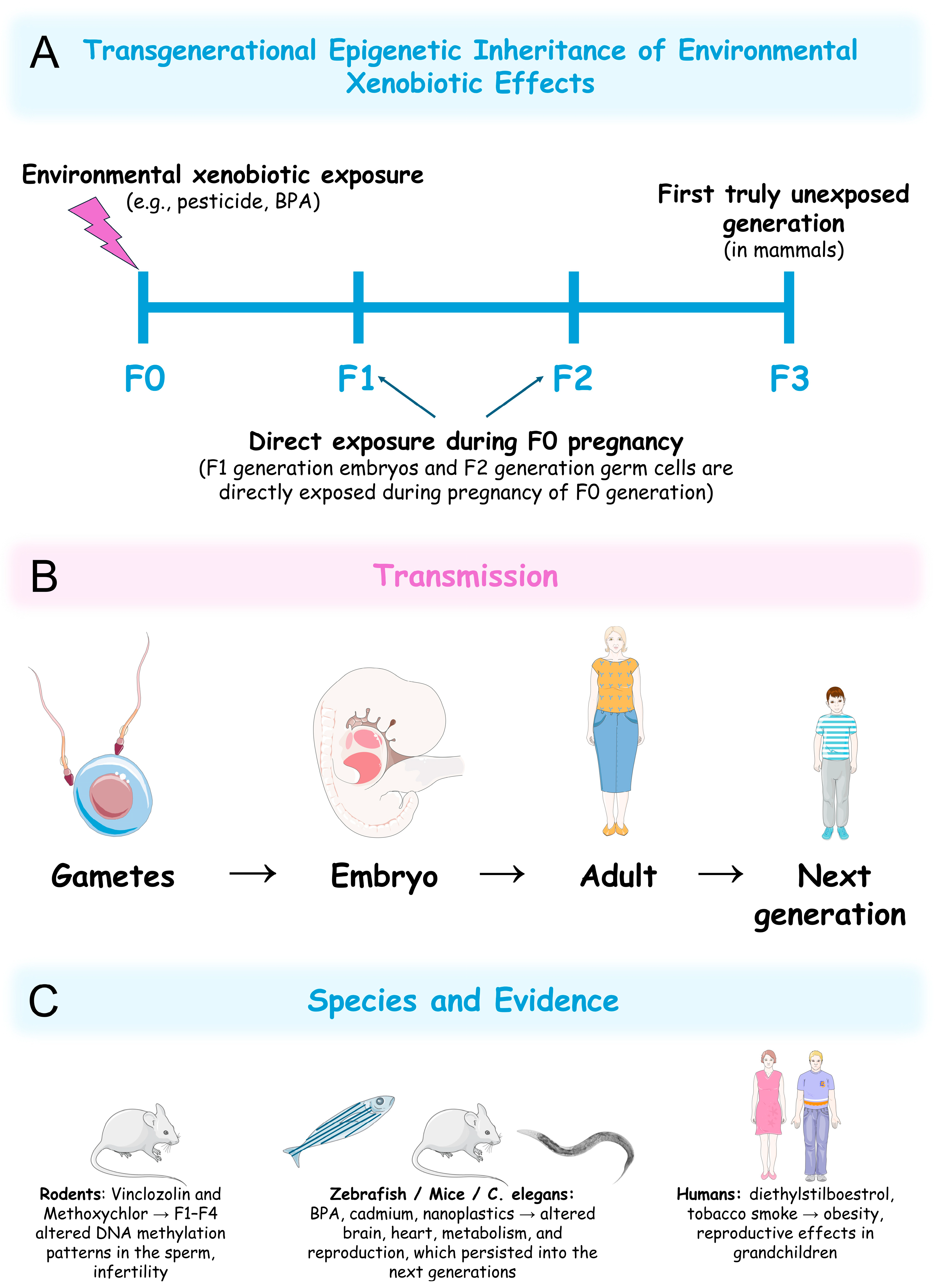
6. Transgenerational Epigenetic Effects of Xenobiotics

Increasing evidence suggests that xenobiotic-induced epigenetic modifications can persist across multiple generations, a phenomenon known as transgenerational epigenetic inheritance (Figure 7A,B) [300]. This refers to the transmission of epigenetic marks to descendants beyond the directly exposed generation, with potential long-term consequences for population health, contributing to disease susceptibility over time. In mammals, true transgenerational effects (observed in completely unexposed generations) require the study of the xenobiotic-induced epigenetic alterations in the third generation of descendants (F3 generation) from an exposed ancestor (F0 generation) (Figure 7A). This is because when exposure occurs during pregnancy, F1-generation embryos and F2-generation germ cells are directly exposed during F0-generation pregnancy [301]. For mammalian paternal exposure or in organisms with external embryonic development, such as zebrafish, transgenerational effects can be studied in the F2 generation [302].
Several studies provide strong evidence of this phenomenon in response to environmental xenobiotic exposure, with effects varying depending on the specific compound, the timing of exposure (e.g., prenatal vs. adult), and the species studied. One significant study exposed pregnant rats (F0 generation) to the endocrine disruptors vinclozolin, a fungicide, or methoxychlor, a pesticide. For both compounds’ exposure, the F1 generation exhibited altered DNA methylation patterns in the sperm that persisted into the F4 generation, accompanied by reproductive defects (Figure 7C) [303]. Exposure to BPA in zebrafish and mice altered histone modifications and gene-expression patterns in reproductive organs, the heart, and the brain that persisted into the next generations (F2 in zebrafish and F3 in mice) (Figure 7C) [304,305,306,307,308]. Similarly, paternal exposure to microcystin-LR led to decreased hatching success, slower heart rates, and lower body weights in both the F1 and F2 generations of zebrafish [142]. Moreover, in C. elegans, exposure to heavy metals (e.g., Cd), NPs, and organic pollutants induced epigenetic changes that resulted in reproductive, behavioural, and metabolic changes transmissible for over four generations (Figure 7C) [309,310,311,312,313,314,315].
While direct transgenerational studies in humans are challenging due to ethical constraints and long generation times, some data indicate that humans are also susceptible to this effect. Prenatal exposure to diethylstilboestrol, a synthetic oestrogen and EDC, has been associated with increased risk of reproductive defects in grandchildren, suggesting epigenetic transmission (Figure 7C) [316]. Additionally, recent studies suggested that the descendants (F1 to F3) of males exposed to tobacco smoke before puberty show increased body fat during childhood, adolescence, and early adulthood (Figure 7C) [317,318]. As this is among the first evidence of xenobiotic-induced transgenerational effects beyond the second generation, it raises the possibility of epigenetic involvement, as the original idea that genetic mutations can fully explain heritability is no longer accepted [319,320]. Moreover, given that obesity is widely accepted to result from a complex interplay of genetic, epigenetic, and environmental influences [321], it is likely that tobacco smoke-induced epigenetic changes may contribute to this outcome, although further investigation is required.
The persistence of epigenetic changes across generations implies that these marks escape the natural epigenetic reprogramming events that occur during gametogenesis and early embryonic development. The proposed mechanisms often involve disrupted DNA methylation patterns transmitted to the subsequent generations via the germline [303,322], histone modifications [304,308], or ncRNAs that alter miRNAs profiles in the germline, which persist in the following generations [323]. The environmental persistence of some xenobiotics may lead to low-dose exposures that also cause epigenetic load accumulation, where successive generations inherit combined epigenetic disruptions from multiple ancestors. Further studies are necessary to clarify the underlying pathways by which these changes may escape erasure during gamete formation.
The transgenerational epigenetic effects of environmental xenobiotics may pose significant challenges for population health and evolution overtime. These effects are linked to a range of disorders, including infertility, obesity, cancer, and neurodevelopmental conditions in animal models [300]. If similar effects occur in humans, populations exposed to pollutants decades ago could exhibit increasing disease burdens today. Additionally, persistent epigenetic changes may alter phenotypic characteristics, potentially changing populations traits over time, as an adaptation to certain pollutants or, conversely, higher sensitivity to their effects, resulting in disease susceptibility. The long-term monitoring of environmental xenobiotic-exposed populations and their descendants is essential for assessing the real impact of those compounds on human health, as current safety assessments often focus on acute or chronic toxicity in exposed individuals, potentially underestimating multigenerational toxicity. Analyses of epigenetic biomarkers in sperm, such as differential methylation at obesity-related genes, could enable the early detection of population-level risks.

7. Reversibility of Xenobiotic-Induced Epigenetic Changes

Unlike genetic mutations, epigenetic modifications are generally considered reversible, as they result from dynamic mechanisms such as DNA methylation and histone modifications [324]. Several studies have demonstrated that xenobiotic-induced epigenetic alterations can, under specific conditions, be reversed, offering promising alternatives for therapeutic intervention.
One study explored the role of 8-oxoguanine deoxyribonucleic acid glycosylase 1 (OGG1) and the sex-specific effects of trichostatin A (TSA), a potent and reversible HDAC inhibitor, in mitigating neurodevelopmental impairments caused by prenatal ethanol exposure [325]. TSA treatment alone was found to influence neurodevelopment and partially reverse ethanol-induced alterations in an OGG1- and sex-dependent manner. Furthermore, TSA improved performance in learning, memory, and cognitive function tasks, supporting the hypothesis that histone acetylation and the resulting upregulation of gene expression are critical for memory formation. TSA also reversed ethanol-induced behavioural alterations such as tolerance, anxiety, and ethanol consumption, primarily through HDAC inhibition and increased histone acetylation [325]. These findings suggest that HDAC inhibition can partially reverse ethanol-induced neurobehavioral alterations, highlighting the involvement of epigenetic mechanisms in these outcomes, rather than proving the reversibility of specific epigenetic changes.
A study investigating the effects of cigarette smoke exposure on bone repair-related genes also reported that the administration of resveratrol, a natural polyphenol with known epigenetic activity [326,327], reversed both epigenetic and transcriptional changes induced by cigarette smoke in rats. Specifically, resveratrol enhanced the transcription of Dnmt3a, Dnmt3b, and sirtuin 1 (Sirt1), while reducing the expression of the receptor activator of nuclear factor κB ligand (Rankl) and tartrate-resistant acid phosphatase (Trap), genes associated with bone resorption [328]. These findings suggest that epigenetic alterations induced by environmental toxins can be pharmacologically reversed, supporting the potential for therapeutic interventions in xenobiotic-induced pathologies.
Clear indications of reversibility of specific epigenetic changes were provided in a longitudinal human study conducted by Richmond et al. [329]. They showed that maternal smoking during pregnancy was associated with altered DNA methylation in offspring at birth, and at ages 7 and 17, in a cohort from the Avon Longitudinal Study of Parents and Children (ALSPAC). While some methylation changes related to development and metabolism persisted into adolescence, others were reversible over time, suggesting a gene- and site-specific pattern of epigenetic plasticity [329].
Further insights into the reversible nature of xenobiotic-induced epigenetic changes were provided by studies on dimethyl sulfoxide (DMSO) exposure [330]. Within 12 to 24 h of exposure, increased expression of Tet and growth arrest and DNA damage-inducible 45 (Gadd45) genes (key regulators of DNA hydroxymethylation and nucleotide excision repair, respectively [331,332,333]) was observed, along with decreased expression of DNA methylation-related genes such as Dnmt1, Dnmt3b, and helicase, lymphoid specific (Hells) [4]. However, by day five, the epigenetic effects of DMSO, including changes in global and promoter-specific methylation and hydroxylation, were diminished or fully reversed [330].
Collectively, these studies underscore the inherent reversibility of many epigenetic changes induced by environmental xenobiotics. Under certain conditions, changes in lifestyle, or the cessation of exposure, the epigenome may return to a baseline state. Recognizing and controlling this flexibility is essential for developing effective strategies to mitigate the long-term health risks associated with environmental xenobiotic exposure.

8. Conclusions

Environmental xenobiotics, including heavy metals, EDCs, pesticides, and air pollutants, represent significant problems to human health by altering gene expression through epigenetic mechanisms. These include DNA methylation changes, a variety of histone modifications, and the altered expression of ncRNAs, each of which plays a critical role in regulating fundamental cellular processes such as gene transcription, and cell-cycle control. Consequently, disruptions in these tightly controlled mechanisms can potentially contribute to the onset and progression of a broad spectrum of diseases, including cancer, neurodegenerative diseases, CVDs, and dysfunction of the immune system.
Importantly, exposure during sensitive developmental windows, including embryogenesis and shortly after birth, has been shown to induce long-lasting epigenetic changes. These alterations may persist throughout an individual’s lifetime, potentially influencing health outcomes and increasing susceptibility to chronic diseases. Moreover, evidence from animal models and limited, but increasing amounts of human data reveal that environmental xenobiotic-induced epigenetic changes can be inherited across multiple generations. This phenomenon, known as transgenerational epigenetic inheritance, suggests that the effects of environmental xenobiotics may extend beyond the directly exposed individual, presenting long-term risks to population health.
At the same time, the reversibility of many epigenetic changes offers a promising opportunity for intervention. Studies have demonstrated that pharmacological agents, such as histone deacetylase inhibitors, and naturally occurring compounds like resveratrol can reverse xenobiotic-induced epigenetic alterations. Additionally, reductions in exposure or lifestyle modifications may also promote the repair of normal epigenetic patterns. This plasticity of the epigenome opens new opportunities for therapeutic development and early prevention strategies.
Overall, these findings highlight the urgent need for a different perspective on how environmental health risks are assessed. Traditional toxicological assessments, which primarily focus on acute or short-term effects in directly exposed individuals, may significantly underestimate the true consequences of environmental xenobiotics in human health. Thus, a more comprehensive, multigenerational approach is needed, accounting for the long-term, heritable, and potentially reversible nature of epigenetic alterations.

9. Future Perspectives

The investigation into how environmental xenobiotics modulate epigenetic mechanisms and influence human health has increased exponentially over the past few years. These modifications can have strong consequences on human health and contribute to a broad spectrum of pathologies, such as cancer and neurodegenerative diseases [7,8]. Despite this increasing evidence, which has guided public health policies, the full impact of environmental xenobiotics on human health through epigenetic mechanisms still requires further investigation.
One of the most critical current limitations is the scarcity of longitudinal studies tracking the long-term effects of environmental xenobiotics on epigenetic markers and their implications for human health [334]. While cross-sectional studies have provided valuable insights into the association between exposure to environmental xenobiotics and epigenetic changes [335,336,337], they do not elucidate the dynamic nature of epigenetic modifications over time. Thus, to fully understand the impact of environmental xenobiotics on epigenetic signatures, future research must focus on prospective longitudinal studies that follow individuals or populations over time. These studies will help identify the timing and duration of exposure to environmental xenobiotics that may lead to epigenetic changes that last for long periods. Additionally, such studies will help characterise whether epigenetic changes caused by environmental xenobiotics are reversible or permanent.
The transgenerational effects of environmental xenobiotics represent another important research question. Many epigenetic modifications can be transmitted to subsequent generations, potentially influencing the health of descendants even in the absence of direct exposure [338]. This phenomenon, known as epigenetic inheritance, has been observed in animal models [303,339,340,341,342], but human studies are fairly limited. Thus, it is crucial to understand how the exposure to environmental xenobiotics, particularly during prenatal or early life, which represent critical developmental periods, can lead to transgenerational epigenetic changes.
Another area that requires detailed investigation is the impact of environmental xenobiotics on the epigenetic background of vulnerable populations, which include children, the elderly, pregnant women, and individuals with comorbidities. Particularly for pregnant women, alterations in the foetal epigenome resulting from prenatal exposure to environmental xenobiotics can result not only in immediate health problems but can also increase susceptibility to diseases later in life. Such knowledge may guide the development of new regulatory guidelines and public health policies that address these particular populations.
On the other hand, since the relationship between environmental xenobiotics, epigenetic modifications, and human health is complex, a more integrated research approach becomes necessary. At this level, multi-omics strategies combining genomics, transcriptomics, epigenomics, metabolomics, and proteomics can help us to understand how exposure to environmental xenobiotics influences health at a molecular level. These strategies have the potential to identify biomarkers of exposure and susceptibility, increasing our understanding of disease mechanisms and possibly predicting outcomes. For example, integrating transcriptomic data, which provides information about changes in gene expression [343], with epigenomic information can potentially reveal direct relationships between exposure to environmental xenobiotics and abnormal gene expression, contributing to diseases involving epigenetic mechanisms.
While identifying the epigenetic changes induced by environmental xenobiotics is important, understanding the precise biological mechanisms behind these modifications is even more crucial. Moreover, it is essential to understand how epigenetic modifications triggered by environmental xenobiotics lead to long-term effects, resulting in diseases such as cancer or neurodegeneration. At this level, advanced technologies such as clustered regularly interspaced short palindromic repeats (CRISPR)-Cas-based epigenome editing and single-cell sequencing show tremendous potential for elucidating the dynamic nature of these epigenetic modifications at both the individual cell level and within complex tissues. Additionally, future research should explore how these epigenetic changes interact with other cellular processes, such as inflammation or oxidative stress, both frequently associated with environmental xenobiotics [344], to promote disease.
Overall, answering these research questions will likely enhance our understanding of the true role that epigenetic mechanisms play in mediating the harmful effects of environmental xenobiotics on human health.

Author Contributions

Conceptualization, D.J.B.; figure preparation, R.S. and D.J.B.; writing—original draft preparation, A.F.S., A.C., I.C., M.S.-C. and D.J.B.; writing—review and editing, A.F.S., A.C., I.C., M.S.-C., R.S. and D.J.B. All authors have read and agreed to the published version of the manuscript.

Funding

This work was supported by FCT/MCTES [projects UIDP/04378/2020 (DOI: 10.54499/UIDP/04378/2020) and UIDB/04378/2020 (DOI:10.54499/UIDB/04378/2020) to UCIBIO, and project LA/P/0140/2020 (DOI: 10.54499/LA/P/0140/2020) to i4HB].

Institutional Review Board Statement

Not applicable.

Informed Consent Statement

Not applicable.

Data Availability Statement

Not applicable.

Acknowledgments

Inês Costa is funded by an FCT PhD fellowship (2023.04980.BD). Figures incorporate elements from Servier Medical Art, a resource of Servier, licensed under a Creative Commons Attribution 4.0 Unported License (https://creativecommons.org/licenses/by/4.0/, accessed on 27 June 2025).

Conflicts of Interest

The authors declare no conflicts of interest.

Abbreviations

The following abbreviations are used in this manuscript:
2,4-D2,4-cichlorophenoxyacetic acid
5mC5-Methylcytosine
Amyloid beta
ACTA2Actin alpha 2
ADAlzheimer’s disease
ADPAdenosine-5′-diphosphate
AKTProtein kinase B (PKB)
ALSPACAvon longitudinal study of parents and children
Ano6Anoctamin 6
Arap2Ankyrin repeat and PH domain 2
ARTsAdenosine-5′-diphosphate-ribosyltransferases
AsArsenic
bdnfBrain-derived neurotrophic factor
BPABisphenol A
CdCadmium
CDH13Cadherin 13
CHRNA5Cholinergic receptor nicotinic alpha 5 subunit
COCarbon monoxide
CpGCytosine–phosphate–guanine
Cpne4Copine 4
CRISPRClustered regularly interspaced short palindromic repeats
Cul2Cullin-2
CVDCardiovascular diseases
DDTDichlorodiphenyltrichloroethane
DEHPDi-(2-ethylhexyl) phthalate
DEPsDiesel exhaust particles
dio3Deiodinase, iodothyronine type III
Dlg2discs large MAGUK scaffold protein 2
DMPsDifferentially methylated positions
DMSODimethyl sulfoxide
DNADeoxyribonucleic acid
DNMTsDeoxyribonucleic acid methyltransferases
Dock3Dedicator of cytokinesis 3
EDCsEndocrine-disrupting chemicals
EPAThe U.S. Environmental Protection Agency
ESCsEmbryonic stem cells
ESCO1Establishment of cohesion 1 homologue 1
EUEuropean Union
G9aEuchromatic histone-lysine N-methyltransferase 2
Gadd45Growth arrest and DNA damage-inducible 45
GATA3GATA binding protein 3
GBHsglyphosate-based herbicides
GLASTAstrocytic glutamate-aspartate transporter
GLT-1Glutamate transporter 1
H2AK119UbMonoubiquitinated histone H2A at lysine 119
H3K4me2Dimethylated histone H3 at lysine 4
H3K4me3Trimethylated histone H3 at lysine 4
H3K9Lysine 9 of histone H3
H3K9acAcetylated histone H3 at lysine 9
H3K9me2Dimethylated histone H3 at lysine 9
H3K9me3Trimethylated histone H3 at lysine 9
H3K14Lysine 14 of histone H3
H3K27Lysine 27 of histone H3
H3K27me3Trimethylated histone H3 at lysine 27
H3K36me3Trimethylated histone H3 at lysine 36
H3R2me2Dimethylated histone H3 at arginine 2
H3S10phPhosphorylated histone H3 at serine 10
H4K5acAcetylated histone H4 at lysine 5
H4K20me2Dimethylated histone H4 at lysine 20
H4K20me3Trimethylated histone H4 at lysine 20
HATsHistone acetyltransferases
HCBHexachlorobenzene
α-HCHAlpha-hexachlorocyclohexane
β-HCHBeta-hexachlorocyclohexane
HDAC2Histone deacetylase 2
HDAC3Histone deacetylase 3
HDACsHistone deacetylases
HellsHelicase, lymphoid specific
HgMercury
IARCInternational Agency for Research on Cancer
KAT2BLysine acetyltransferase 2B
KDM3AHistone H3K9 demethylase lysine-specific demethylase 3A.
KDM5AHistone H3K4 demethylase lysine-specific demethylase 5A
KDM5BHistone H3K4 demethylase lysine-specific demethylase 5B
KLF6Kruppel-like factor 6
LINE-1Long interspersed nuclear element-1
lncRNALong non-coding ribonucleic acid
MBD2Methyl-CpG binding domain protein 2
MnManganese
mRNAMessenger ribonucleic acid
miRNAMicro ribonucleic acid
MPTP1-Methyl-4-phenyl-1,2,3,6-tetrahydropyridine
MPP+1-Methyl-4-phenylpyridinium
MSH5MutS homologue 5
MPsMicroplastics
mTORMammalian target of rapamycin
ncRNANon-coding ribonucleic acid
NFKB1Nuclear factor kappa B subunit 1
NONitric oxide
NO2Nitrogen dioxide
O3Ozone
OgdhOxoglutarate dehydrogenase
OGG18-Oxoguanine deoxyribonucleic acid glycosylase 1
NPsNanoplastics
p21Cyclin-dependent kinase inhibitor 1A
PADsPeptidylarginine deiminases
PAHsPolycyclic aromatic hydrocarbons
PbLead
PBDEsPolybrominated diphenyl ethers
PCBsPolychlorinated biphenyls
PCDDsPolychlorinated dibenzo-p-dioxins
PCDFsPolychlorinated dibenzofurans
Pdpk13-Phosphoinositide-dependent protein kinase-1
PFOSPerfluorooctane sulfonic acid
PFOSFPerfluorooctane sulfonyl fluoride
PDParkinson’s disease
PINK1PTEN-induced putative kinase 1
PMParticulate matter
POPspersistent organic pollutants
PRMT6Protein arginine methyltransferase 6
PTENPhosphatase and tensin homologue
PVCpolyvinyl chloride
RArginine
RanklReceptor activator of nuclear factor κB ligand
RC-LINE-1Retrotransposition-competent long interspersed nuclear element-1
RNARibonucleic acid
ROSReactive oxygen species
RptorRegulatory associated protein of mTOR complex 1
SAMS-adenosyl methionine
Sat2satellite 2 DNA
SETD8SET domain containing 8 or lysine methyltransferase 5A
Sirt1Sirtuin 1
SLESystemic lupus erythematosus
SO2Sulphur dioxide
SpopSpeckle-type POZ protein
STK32ASerine/threonine kinase 32A
SUMOSmall ubiquitin-like modifier
Tbc1d5TBC1 domain family member 5
TCDD2,3,7,8-tetrachlorodibenzo-p-dioxin
TERTTelomerase reverse transcriptase
TETTen-eleven translocation enzymes
TP53INP1Tumour protein p53 inducible nuclear protein 1
TrapTartrate-resistant acid phosphatase
tRNATransfer RNA
TSATrichostatin A
USAUnited States of America
VTI1AVesicle transport through interaction with T-SNAREs homologue 1A
WHOWorld Health Organization
XIAPX-linked inhibitor of apoptosis protein

References

  1. Virolainen, S.J.; VonHandorf, A. Gene-environment interactions and their impact on human health. Genes. Immun. 2023, 24, 1–11. [Google Scholar] [CrossRef]
  2. Fu, W.; Guang, Y. Epidemiological perspectives on emerging contaminants and gout or hyperuricemia. Emerg. Contam. 2025, 11, 100485. [Google Scholar] [CrossRef]
  3. Briffa, J.; Sinagra, E. Heavy metal pollution in the environment and their toxicological effects on humans. Heliyon 2020, 6, e04691. [Google Scholar] [CrossRef]
  4. Metcalfe, C.D.; Bayen, S. An introduction to the sources, fate, occurrence and effects of endocrine disrupting chemicals released into the environment. Environ. Res. 2022, 207, 112658. [Google Scholar] [CrossRef] [PubMed]
  5. Marin, S.; Ramos, A.J. Mycotoxins: Occurrence, toxicology, and exposure assessment. Food Chem. Toxicol. 2013, 60, 218–237. [Google Scholar] [CrossRef] [PubMed]
  6. Pires, E.; Lana, P.D.C. Phycotoxins and marine annelids—A global review. Harmful Algae 2023, 122, 102373. [Google Scholar] [CrossRef]
  7. Pal, S.; Firdous, S.M. Unraveling the role of heavy metals xenobiotics in cancer: A critical review. Discov. Oncol. 2024, 15, 615. [Google Scholar] [CrossRef]
  8. Rock, K.D.; Patisaul, H.B. Environmental mechanisms of neurodevelopmental toxicity. Curr. Environ. Health Rep. 2018, 5, 145–157. [Google Scholar] [CrossRef]
  9. Zhang, J.; Jia, Q. The function of xenobiotic receptors in metabolic diseases. Drug Metab. Dispos. 2023, 51, 237–248. [Google Scholar] [CrossRef]
  10. Hussain, T.; Metwally, E. Redox mechanisms of environmental toxicants on male reproductive function. Front. Cell Dev. Biol. 2024, 12, 1333845. [Google Scholar] [CrossRef]
  11. Gibney, E.R.; Nolan, C.M. Epigenetics and gene expression. Heredity 2010, 105, 4–13. [Google Scholar] [CrossRef] [PubMed]
  12. Bure, I.V.; Nemtsova, M.V. Histone modifications and non-coding RNAs: Mutual epigenetic regulation and role in pathogenesis. Int. J. Mol. Sci. 2022, 23, 5801. [Google Scholar] [CrossRef]
  13. Zhu, S.; Li, Z. VIRMA-mediated the m6A methylation of SCD facilitates wilms’ tumor progression via AMPK pathway. DNA Cell Biol. 2025, 44, 229–237. [Google Scholar] [CrossRef]
  14. Long, Z.Q.; Ding, R. Multi-omics characterization of genome-wide abnormal DNA methylation reveals FGF5 as a diagnosis of nasopharyngeal carcinoma recurrence after radiotherapy. Biomolecules 2025, 15, 283. [Google Scholar] [CrossRef] [PubMed]
  15. Rico-Méndez, M.A.; Trujillo-Rojas, M.A. MLH1 methylation status and microsatellite instability in patients with colorectal cancer. Genes 2025, 16, 182. [Google Scholar] [CrossRef]
  16. Zhang, R.; Nie, Y. A multicenter prospective clinical trial reveals cell free DNA methylation markers for early esophageal cancer. J. Clin. Investig. 2025, 135, e186816. [Google Scholar] [CrossRef] [PubMed]
  17. Bannister, A.J.; Kouzarides, T. Regulation of chromatin by histone modifications. Cell Res. 2011, 21, 381–395. [Google Scholar] [CrossRef]
  18. McCabe, M.T.; Graves, A.P. Mutation of A677 in histone methyltransferase EZH2 in human B-cell lymphoma promotes hypertrimethylation of histone H3 on lysine 27 (H3K27). Proc. Natl. Acad. Sci. USA 2012, 109, 2989–2994. [Google Scholar] [CrossRef]
  19. Dai, Q.; Ye, Y. Development and validation of a novel histone acetylation-related gene signature for predicting the prognosis of ovarian cancer. Front. Cell Dev. Biol. 2022, 10, 793425. [Google Scholar] [CrossRef]
  20. Persico, G.; Casciaro, F. Histone H3 lysine 4 and 27 trimethylation landscape of human Alzheimer’s disease. Cells 2022, 11, 734. [Google Scholar] [CrossRef]
  21. Khazaei, S.; Chen, C.C.L. Single substitution in H3.3G34 alters DNMT3A recruitment to cause progressive neurodegeneration. Cell 2023, 186, 1162–1178.e20. [Google Scholar] [CrossRef]
  22. Shen, T.; Ji, F. Brain-specific deletion of histone variant H2A.z results in cortical neurogenesis defects and neurodevelopmental disorder. Nucleic Acids Res. 2018, 46, 2290–2307. [Google Scholar] [CrossRef]
  23. Lee, K.J.; Ahn, J.H. Non-coding RNA RMRP governs RAB31-dependent MMP secretion, enhancing ovarian cancer invasion. Biochim. Biophys. Acta Mol. Basis Dis. 2025, 1871, 167781. [Google Scholar] [CrossRef]
  24. Massone, S.; Ciarlo, E. NDM29, a RNA polymerase III-dependent non coding RNA, promotes amyloidogenic processing of APP and amyloid β secretion. Biochim. Biophys. Acta 2012, 1823, 1170–1177. [Google Scholar] [CrossRef]
  25. Senut, M.C.; Sen, A. Lead exposure disrupts global DNA methylation in human embryonic stem cells and alters their neuronal differentiation. Toxicol. Sci. 2014, 139, 142–161. [Google Scholar] [CrossRef] [PubMed]
  26. Demanelis, K.; Virani, S. Cadmium exposure and age-associated DNA methylation changes in non-smoking women from northern Thailand. Environ. Epigenet 2017, 3, dvx006. [Google Scholar] [CrossRef] [PubMed]
  27. Bailey, K.A.; Wu, M.C. Arsenic and the epigenome: Interindividual differences in arsenic metabolism related to distinct patterns of DNA methylation. J. Biochem. Mol. Toxicol. 2013, 27, 106–115. [Google Scholar] [CrossRef] [PubMed]
  28. Deb, P.; Bhan, A. Endocrine disrupting chemical, bisphenol-A, induces breast cancer associated gene HOXB9 expression in vitro and in vivo. Gene 2016, 590, 234–243. [Google Scholar] [CrossRef]
  29. Koc, S.; Erdogmus, E. Prepubertal phthalate exposure can cause histopathological alterations, DNA methylation and histone acetylation changes in rat brain. Toxicol. Ind. Health 2025, 41, 163–175. [Google Scholar] [CrossRef]
  30. Schmitt, C.; Peterson, E. Transgenerational effects of developmental exposure to chlorpyrifos-oxon in zebrafish (DANIO RERIO). Toxicol. Appl. Pharmacol. 2020, 408, 115275. [Google Scholar] [CrossRef]
  31. Chiu, K.C.; Sisca, F. Prenatal chlorpyrifos exposure in association with PPARγ H3K4me3 and DNA methylation levels and child development. Environ. Pollut. 2021, 274, 116511. [Google Scholar] [CrossRef]
  32. Rossetti, M.F.; Canesini, G. Epigenetic changes associated with exposure to glyphosate-based herbicides in mammals. Front. Endocrinol. 2021, 12, 671991. [Google Scholar] [CrossRef] [PubMed]
  33. Kubsad, D.; Nilsson, E.E. Assessment of glyphosate induced epigenetic transgenerational inheritance of pathologies and sperm epimutations: Generational toxicology. Sci. Rep. 2019, 9, 6372. [Google Scholar] [CrossRef]
  34. Mukherjee, S.; Dasgupta, S. Air pollution-induced epigenetic changes: Disease development and a possible link with hypersensitivity pneumonitis. Environ. Sci. Pollut. Res. Int. 2021, 28, 55981–56002. [Google Scholar] [CrossRef] [PubMed]
  35. Krolevets, M.; Cate, V.T. DNA methylation and cardiovascular disease in humans: A systematic review and database of known CpG methylation sites. Clin. Epigenetics 2023, 15, 56. [Google Scholar] [CrossRef] [PubMed]
  36. Wu, G.; Kang, H. A critical review on the bio-removal of hazardous heavy metals from contaminated soils: Issues, progress, eco-environmental concerns and opportunities. J. Hazard. Mater. 2010, 174, 1–8. [Google Scholar] [CrossRef]
  37. Pacyna, E.; Pacyna, J. Current and future emissions of selected heavy metals to the atmosphere from anthropogenic sources in Europe. Atmos. Environ. 2007, 41, 8557–8566. [Google Scholar] [CrossRef]
  38. Mitra, S.; Chakraborty, A.J. Impact of heavy metals on the environment and human health: Novel therapeutic insights to counter the toxicity. J. King Saud. Univ. Sci. 2022, 34, 101865. [Google Scholar] [CrossRef]
  39. Perrelli, M.; Wu, R. Heavy metals as risk factors for human diseases—A Bayesian network approach. Eur. Rev. Med. Pharmacol. Sci. 2022, 26, 9275–9310. [Google Scholar]
  40. Xu, J.; Bravo, A.G. Sources and remediation techniques for mercury contaminated soil. Environ. Int. 2015, 74, 42–53. [Google Scholar] [CrossRef]
  41. Gochfeld, M. Cases of mercury exposure, bioavailability, and absorption. Ecotoxicol. Environ. Saf. 2003, 56, 174–179. [Google Scholar] [CrossRef]
  42. Hightower, J.M.; O’Hare, A. Blood mercury reporting in NHANES: Identifying Asian, Pacific Islander, Native American, and multiracial groups. Environ. Health Perspect. 2006, 114, 173–175. [Google Scholar] [CrossRef] [PubMed]
  43. Bellanger, M.; Pichery, C. Economic benefits of methylmercury exposure control in Europe: Monetary value of neurotoxicity prevention. Environ. Health 2013, 12, 3. [Google Scholar] [CrossRef] [PubMed]
  44. IARC Working Group on the Evaluation of Carcinogenic Risks to Humans. Arsenic, metals, fibres, and dusts. IARC Monogr. Eval. Carcinog. Risks Hum. 2012, 100, 11–465. [Google Scholar]
  45. Frisbie, S.H.; Mitchell, E.J. Arsenic in drinking water: An analysis of global drinking water regulations and recommendations for updates to protect public health. PLoS ONE 2022, 17, e0263505. [Google Scholar] [CrossRef]
  46. Chakraborty, M.; Mukherjee, A. A Review of groundwater arsenic in the Bengal basin, Bangladesh and India: From source to sink. Curr. Pollut. Rep. 2015, 1, 220–247. [Google Scholar] [CrossRef]
  47. Genchi, G.; Sinicropi, M.S. The effects of cadmium toxicity. Int. J. Environ. Res. Public Health 2020, 17, 3782. [Google Scholar] [CrossRef]
  48. Rehman, K.; Fatima, F. Prevalence of exposure of heavy metals and their impact on health consequences. J. Cell Biochem. 2018, 119, 157–184. [Google Scholar] [CrossRef]
  49. Lone, I.; Saleem, S. Heavy metal contents of vegetables irrigated by sewage/tubewell water. Int. J. Agric. Biol. 2003, 5, 533–535. [Google Scholar]
  50. World Health Organization. Cadmium in Drinking-Water: Background Document for Development of WHO Guidelines for Drinking-Water Quality (WHO/SDE/WSH/03.04/80/Rev/1). Available online: https://cdn.who.int/media/docs/default-source/wash-documents/wash-chemicals/cadmium.pdf?sfvrsn=4dd545bd_4 (accessed on 15 March 2025).
  51. Casals-Casas, C.; Desvergne, B. Endocrine disruptors: From endocrine to metabolic disruption. Annu. Rev. Physiol. 2011, 73, 135–162. [Google Scholar] [CrossRef]
  52. Diamanti-Kandarakis, E.; Bourguignon, J.P. Endocrine-disrupting chemicals: An Endocrine Society scientific statement. Endocr. Rev. 2009, 30, 293–342. [Google Scholar] [CrossRef]
  53. Vandenberg, L.N.; Maffini, M.V. Bisphenol-A and the great divide: A review of controversies in the field of endocrine disruption. Endocr. Rev. 2009, 30, 75–95. [Google Scholar] [CrossRef] [PubMed]
  54. Geens, T.; Goeyens, L. Are potential sources for human exposure to bisphenol-A overlooked? Int. J. Hyg. Environ. Health 2011, 214, 339–347. [Google Scholar] [CrossRef] [PubMed]
  55. Vandenberg, L.N.; Hauser, R. Human exposure to bisphenol A (BPA). Reprod. Toxicol. 2007, 24, 139–177. [Google Scholar] [CrossRef] [PubMed]
  56. Covaci, A.; Den Hond, E. Urinary BPA measurements in children and mothers from six European member states: Overall results and determinants of exposure. Environ. Res. 2015, 141, 77–85. [Google Scholar] [CrossRef]
  57. Adoamnei, E.; Mendiola, J. Urinary bisphenol A concentrations are associated with reproductive parameters in young men. Environ. Res. 2018, 161, 122–128. [Google Scholar] [CrossRef]
  58. He, Y.; Miao, M. Occupational exposure levels of bisphenol A among Chinese workers. J. Occup. Health 2009, 51, 432–436. [Google Scholar] [CrossRef]
  59. Mariana, M.; Castelo-Branco, M. Phthalates’ exposure leads to an increasing concern on cardiovascular health. J. Hazard. Mater. 2023, 457, 131680. [Google Scholar] [CrossRef]
  60. Koch, H.M.; Preuss, R. Di(2-ethylhexyl)phthalate (DEHP): Human metabolism and internal exposure—An update and latest results. Int. J. Androl. 2006, 29, 155–165. [Google Scholar] [CrossRef]
  61. Hoppin, J.A.; Brock, J.W. Reproducibility of urinary phthalate metabolites in first morning urine samples. Environ. Health Perspect. 2002, 110, 515–518. [Google Scholar] [CrossRef]
  62. Silva, M.J.; Barr, D.B. Urinary levels of seven phthalate metabolites in the U.S. population from the National Health and Nutrition Examination Survey (NHANES) 1999–2000. Environ. Health Perspect. 2004, 112, 331–338. [Google Scholar] [CrossRef] [PubMed]
  63. Karlaganis, G.; Marioni, R. The elaboration of the ‘Stockholm convention’ on persistent organic pollutants (POPs): A negotiation process fraught with obstacles and opportunities. Environ. Sci. Pollut. Res. Int. 2001, 8, 216–221. [Google Scholar] [CrossRef]
  64. Wöhrnschimmel, H.; MacLeod, M. Emissions, fate and transport of persistent organic pollutants to the Arctic in a changing global climate. Environ. Sci. Technol. 2013, 47, 2323–2330. [Google Scholar] [CrossRef]
  65. Brigden, K.; Labunska, I.; Santillo, D.; Allsopp, M. Recycling of Electronic Wastes in China and India: Workplace and Environment Contamination; Greenpeace International: Amsterdam, The Netherlands, 2005. [Google Scholar]
  66. Marinković, N.; Pašalić, D. Dioxins and human toxicity. Arh. Hig. Rada Toksikol. 2010, 61, 445–453. [Google Scholar] [CrossRef] [PubMed]
  67. McKay, G. Dioxin characterisation, formation and minimisation during municipal solid waste (MSW) incineration: Review. Chem. Eng. J. 2002, 86, 343–368. [Google Scholar] [CrossRef]
  68. Lorber, M.; Patterson, D. Evaluation of background exposures of Americans to dioxin-like compounds in the 1990s and the 2000s. Chemosphere 2009, 77, 640–651. [Google Scholar] [CrossRef] [PubMed]
  69. Sorg, O.; Zennegg, M. 2,3,7,8-tetrachlorodibenzo-p-dioxin (TCDD) poisoning in Victor Yushchenko: Identification and measurement of TCDD metabolites. Lancet 2009, 374, 1179–1185. [Google Scholar] [CrossRef]
  70. Geusau, A.; Abraham, K. Severe 2,3,7,8-tetrachlorodibenzo-p-dioxin (TCDD) intoxication: Clinical and laboratory effects. Environ. Health Perspect. 2001, 109, 865–869. [Google Scholar] [CrossRef]
  71. Holt, J.S. Herbicides. In Encyclopedia of Biodiversity, 2nd ed.; Levin, S.A., Ed.; Academic Press: Waltham, MA, USA, 2013; pp. 87–95. [Google Scholar]
  72. Chaufan, G.; Coalova, I. Glyphosate commercial formulation causes cytotoxicity, oxidative effects, and apoptosis on human cells: Differences with its active ingredient. Int. J. Toxicol. 2014, 33, 29–38. [Google Scholar] [CrossRef]
  73. Battaglin, W.A.; Meyer, M.T. Glyphosate and its degradation product AMPA occur frequently and widely in U.S. soils, surface water, groundwater, and precipitation. J. Am. Water Resour. Assoc. 2014, 50, 275–290. [Google Scholar] [CrossRef]
  74. Conrad, A.; Schröter-Kermani, C. Glyphosate in German adults—Time trend (2001 to 2015) of human exposure to a widely used herbicide. Int. J. Hyg. Environ. Health 2017, 220, 8–16. [Google Scholar] [CrossRef]
  75. Krueger, M.; Schrödl, W. Detection of glyphosate in malformed piglets. J. Environ. Anal. Toxicol. 2014, 04, 230. [Google Scholar] [CrossRef]
  76. Acquavella, J.F.; Alexander, B.H. Glyphosate biomonitoring for farmers and their families: Results from the Farm Family Exposure Study. Environ. Health Perspect. 2004, 112, 321–326. [Google Scholar] [CrossRef] [PubMed]
  77. Van Bruggen, A.H.C.; He, M.M. Environmental and health effects of the herbicide glyphosate. Sci. Total Environ. 2018, 616–617, 255–268. [Google Scholar] [CrossRef] [PubMed]
  78. Liu, X.; Yang, H. Signaling pathways involved in paraquat-induced pulmonary toxicity: Molecular mechanisms and potential therapeutic drugs. Int. Immunopharmacol. 2022, 113, 109301. [Google Scholar] [CrossRef]
  79. Buendía, J.A.; Chavarriaga, G.J.R. Burden of paraquat poisoning in the department of Antioquia, Colombia. BMC Pharmacol. Toxicol. 2019, 20, 11. [Google Scholar] [CrossRef]
  80. Dinis-Oliveira, R.J.; Duarte, J.A. Paraquat poisonings: Mechanisms of lung toxicity, clinical features, and treatment. Crit. Rev. Toxicol. 2008, 38, 13–71. [Google Scholar] [CrossRef]
  81. Greaves, A.K.; Letcher, R.J. Comparative body compartment composition and in ovo transfer of organophosphate flame retardants in North American Great Lakes herring gulls. Environ. Sci. Technol. 2014, 48, 7942–7950. [Google Scholar] [CrossRef]
  82. Agarwal, A.; Prajapati, R. Pesticide residue in water—A challenging task in India. Environ. Monit. Assess. 2015, 187, 54. [Google Scholar] [CrossRef]
  83. Masiá, A.; Campo, J. Pesticide monitoring in the basin of Llobregat River (Catalonia, Spain) and comparison with historical data. Sci. Total Environ. 2015, 503–504, 58–68. [Google Scholar] [CrossRef]
  84. Hou, R.; Xu, Y. Review of OPFRs in animals and humans: Absorption, bioaccumulation, metabolism, and internal exposure research. Chemosphere 2016, 153, 78–90. [Google Scholar] [CrossRef] [PubMed]
  85. U.S. EPA. Air Quality Criteria for Particulate Matter; US Environmental Protection Agency, Research Triangle Park: Gaithersburg, MD, USA, 2004.
  86. Ghio, A.J.; Smith, C.B. Diesel exhaust particles and airway inflammation. Curr. Opin. Pulm. Med. 2012, 18, 144–150. [Google Scholar] [CrossRef] [PubMed]
  87. Benbrahim-Tallaa, L.; Baan, R.A. Carcinogenicity of diesel-engine and gasoline-engine exhausts and some nitroarenes. Lancet Oncol. 2012, 13, 663–664. [Google Scholar] [CrossRef] [PubMed]
  88. Kittelson, D.B. Engines and nanoparticles: A review. J. Aerosol Sci. 1998, 29, 575–588. [Google Scholar] [CrossRef]
  89. Hellier, P.; Ladommatos, N. The influence of biodiesel composition on compression ignition combustion and emissions. Proc. Inst. Mech. Eng. Part A J. Power Energy 2015, 229, 714–726. [Google Scholar] [CrossRef]
  90. Hellier, P.; Talibi, M. An overview of the effects of fuel molecular structure on the combustion and emissions characteristics of compression ignition engines. Proc. Inst. Mech. Eng. Part D 2018, 232, 90–105. [Google Scholar] [CrossRef]
  91. Swanson, K.J.; Madden, M.C. Biodiesel exhaust: The need for health effects research. Environ. Health Perspect. 2007, 115, 496–499. [Google Scholar] [CrossRef]
  92. Ogbunuzor, C.; Fransen, L.F.H. Biodiesel exhaust particle airway toxicity and the role of polycyclic aromatic hydrocarbons. Ecotoxicol. Environ. Saf. 2023, 259, 115013. [Google Scholar] [CrossRef]
  93. Hawrot-Paw, M.; Koniuszy, A. Ecotoxicity of soil contaminated with diesel fuel and biodiesel. Sci. Rep. 2020, 10, 16436. [Google Scholar] [CrossRef]
  94. Savadkoohi, M.; Pandolfi, M. Recommendations for reporting equivalent black carbon (eBC) mass concentrations based on long-term pan-European in-situ observations. Environ. Int. 2024, 185, 108553. [Google Scholar] [CrossRef]
  95. Bond, T.C.; Doherty, S.J. Bounding the role of black carbon in the climate system: A scientific assessment. J. Geophys. Res. 2013, 118, 5380–5552. [Google Scholar] [CrossRef]
  96. Zhang, Z.; Cheng, Y. The measurement of atmospheric black carbon: A review. Toxics 2023, 11, 975. [Google Scholar] [CrossRef]
  97. Zhu, X.; Liu, B. Short and long-term association of exposure to ambient black carbon with all-cause and cause-specific mortality: A systematic review and meta-analysis. Environ. Pollut. 2023, 324, 121086. [Google Scholar] [CrossRef]
  98. Patel, A.B.; Shaikh, S. Polycyclic aromatic hydrocarbons: Sources, toxicity, and remediation approaches. Front. Microbiol. 2020, 11, 562813. [Google Scholar] [CrossRef]
  99. Mojiri, A.; Zhou, J.L. Comprehensive review of polycyclic aromatic hydrocarbons in water sources, their effects and treatments. Sci. Total Environ. 2019, 696, 133971. [Google Scholar] [CrossRef] [PubMed]
  100. IARC Working Group on the Evaluation of Carcinogenic Risks to Humans. Tobacco smoke and involuntary smoking. IARC Monogr. Eval. Carcinog. Risks Hum. 2004, 83, 1–1438.
  101. Yershova, K.; Yuan, J.M. Tobacco-specific N-nitrosamines and polycyclic aromatic hydrocarbons in cigarettes smoked by the participants of the Shanghai Cohort Study. Int. J. Cancer 2016, 139, 1261–1269. [Google Scholar] [CrossRef]
  102. Vaz-Ramos, J.; Le Calvé, S. Polycyclic aromatic hydrocarbons in water environments: Impact, legislation, depollution processes and challenges, and magnetic iron oxide/graphene-based nanocomposites as promising adsorbent solutions. J. Hazard. Mater. 2025, 490, 137726. [Google Scholar] [CrossRef]
  103. Manoli, E.; Samara, C. Polycyclic aromatic hydrocarbons in natural waters: Sources, occurrence and analysis. Trac-Trend Anal. Chem. 1999, 18, 417–428. [Google Scholar] [CrossRef]
  104. WHO. WHO Guidelines for Indoor Air Quality: Selected Pollutants; WHO Regional Office for Europe: Copenhagen, Denmark, 2010. [Google Scholar]
  105. Mallah, M.A.; Changxing, L. Polycyclic aromatic hydrocarbon and its effects on human health: An overeview. Chemosphere 2022, 296, 133948. [Google Scholar] [CrossRef]
  106. Ramsperger, A.; Bergamaschi, E. Nano- and microplastics: A comprehensive review on their exposure routes, translocation, and fate in humans. NanoImpact 2023, 29, 100441. [Google Scholar] [CrossRef] [PubMed]
  107. Hartmann, N.B.; Hüffer, T. Are we speaking the same language? Recommendations for a definition and categorization framework for plastic debris. Environ. Sci. Technol. 2019, 53, 1039–1047. [Google Scholar] [CrossRef]
  108. Barnes, D.K.; Galgani, F. Accumulation and fragmentation of plastic debris in global environments. Philos. Trans. R. Soc. Lond. B Biol. Sci. 2009, 364, 1985–1998. [Google Scholar] [CrossRef] [PubMed]
  109. Râpă, M.; Darie-Niță, R.N. Insights into anthropogenic micro- and nanoplastic accumulation in drinking water sources and their potential effects on human health. Polymers 2023, 15, 2425. [Google Scholar] [CrossRef]
  110. EFSA Panel on Contaminants in the Food Chain. Presence of microplastics and nanoplastics in food, with particular focus on seafood. EFSA J. 2016, 14, e04501. [Google Scholar]
  111. Wright, S.L.; Ulke, J. Atmospheric microplastic deposition in an urban environment and an evaluation of transport. Environ. Int. 2020, 136, 105411. [Google Scholar] [CrossRef]
  112. Amato-Lourenço, L.F.; Carvalho-Oliveira, R. Presence of airborne microplastics in human lung tissue. J. Hazard. Mater. 2021, 416, 126124. [Google Scholar] [CrossRef]
  113. Mou, L.; Wu, C. Rapid detection of microplastics/nanoplastics directly exposed to blood during intravenous injections via mie scattering spectra. J. Hazard. Mater. 2024, 480, 136193. [Google Scholar] [CrossRef]
  114. Rezaei Kahkha, M.R.; Piri, J. Investigation of heavy metals adsorbed on microplastics in drinking water and water resources of Zabol. Sci. Rep. 2025, 15, 14378. [Google Scholar] [CrossRef]
  115. Lan, T.; Wang, T. A comparative study on the adsorption behavior of pesticides by pristine and aged microplastics from agricultural polyethylene soil films. Ecotoxicol. Environ. Saf. 2021, 209, 111781. [Google Scholar] [CrossRef]
  116. Llorca, M.; Schirinzi, G. Adsorption of perfluoroalkyl substances on microplastics under environmental conditions. Environ. Pollut. 2018, 235, 680–691. [Google Scholar] [CrossRef]
  117. Berber, A.A.; Akinci Kenanoğlu, N. Genotoxic and cytotoxic effects of polystyrene nanoplastics on human lymphocytes: A comprehensive analysis. Mutat. Res. Genet. Toxicol. Environ. Mutagen. 2025, 902, 503850. [Google Scholar] [CrossRef]
  118. Pontecorvi, P.; Ceccarelli, S. Assessing the impact of polyethylene nano/microplastic exposure on human vaginal keratinocytes. Int. J. Mol. Sci. 2023, 24, 11379. [Google Scholar] [CrossRef]
  119. Niaz, W.; Iqbal, S.Z. Mycotoxins: A comprehensive review of its global trends in major cereals, advancements in chromatographic detections and future prospectives. Food Chem. X 2025, 27, 102350. [Google Scholar] [CrossRef] [PubMed]
  120. Palumbo, R.; Crisci, A. Occurrence and co-occurrence of mycotoxins in cereal-based feed and food. Microorganisms 2020, 8, 74. [Google Scholar] [CrossRef]
  121. Ahmad, M.M.; Ahmad, M. Detection of Aspergillus flavus and Aspergillus parasiticus from aflatoxin-contaminated peanuts and their differentiation using PCR-RFLP. Ann. Microbiol. 2014, 64, 1597–1605. [Google Scholar] [CrossRef]
  122. Marchese, S.; Polo, A. Aflatoxin B1 and M1: Biological properties and their involvement in cancer development. Toxins 2018, 10, 214. [Google Scholar] [CrossRef]
  123. European Commission. Commission regulation (EU) 2023/915 of 25 April 2023 on maximum levels for certain contaminants in food and repealing regulation (EC) no 1881/2006. Off. J. Eur. Union L 2023, 119, 103–157. [Google Scholar]
  124. Kaale, L.D.; Kimanya, M.E. Aflatoxin contamination and recommendations to improve its control: A review. World Mycotoxin J. 2021, 14, 27–40. [Google Scholar] [CrossRef]
  125. Bredenkamp, M.W.; Dillen, J.L.M. Crystal structures and conformational analysis of ochratoxin A and B: Probing the chemical structure causing toxicity. J. Chem. Soc. Perkin Trans 1989, 2, 1835–1839. [Google Scholar] [CrossRef]
  126. Echodu, R.; Maxwell Malinga, G. Prevalence of aflatoxin, ochratoxin and deoxynivalenol in cereal grains in northern Uganda: Implication for food safety and health. Toxicol. Rep. 2019, 6, 1012–1017. [Google Scholar] [CrossRef] [PubMed]
  127. Lee, H.J.; Dahal, S. Reduction of ochratoxin A in oat flakes by twin-screw extrusion processing. J. Food Prot. 2017, 80, 1628–1634. [Google Scholar] [CrossRef]
  128. Domijan, A.M. Fumonisin B(1): A neurotoxic mycotoxin. Arh. Hig. Rada Toksikol. 2012, 63, 531–544. [Google Scholar] [CrossRef] [PubMed]
  129. Azam, M.S.; Ahmed, S. Critical assessment of mycotoxins in beverages and their control measures. Toxins 2021, 13, 323. [Google Scholar] [CrossRef]
  130. Rogowska, A.; Pomastowski, P. Zearalenone and its metabolites: Effect on human health, metabolism and neutralisation methods. Toxicon 2019, 162, 46–56. [Google Scholar] [CrossRef]
  131. Luo, Y.; Liu, X. Complicated interactions between bio-adsorbents and mycotoxins during mycotoxin adsorption: Current research and future prospects. Trends Food Sci. Technol. 2020, 96, 127–134. [Google Scholar] [CrossRef]
  132. López-Díaz, C.; Rahjoo, V. Fusaric acid contributes to virulence of Fusarium oxysporum on plant and mammalian hosts. Mol. Plant Pathol. 2018, 19, 440–453. [Google Scholar] [CrossRef]
  133. Stipanovic, R.D.; Puckhaber, L.S. Phytotoxicity of fusaric acid and analogs to cotton. Toxicon 2011, 57, 176–178. [Google Scholar] [CrossRef]
  134. Porter, J.K.; Bacon, C.W. Fusaric acid in Fusarium moniliforme cultures, corn, and feeds toxic to livestock and the neurochemical effects in the brain and pineal gland of rats. Nat. Toxins 1995, 3, 91–100. [Google Scholar] [CrossRef]
  135. Tang, J.; He, X. Occurrence and distribution of phycotoxins in the Antarctic Ocean. Mar. Pollut. Bull. 2024, 201, 116250. [Google Scholar] [CrossRef]
  136. Salter, C.; VanMensel, D. Investigating the microbial dynamics of microcystin-LR degradation in Lake Erie sand. Chemosphere 2021, 272, 129873. [Google Scholar] [CrossRef] [PubMed]
  137. Krishnan, A.; Mou, X. A brief review of the structure, cytotoxicity, synthesis, and biodegradation of microcystins. Water 2021, 13, 2147. [Google Scholar] [CrossRef]
  138. Chen, L.; Giesy, J.P. The dose makes the poison. Sci. Total Environ. 2018, 621, 649–653. [Google Scholar] [CrossRef]
  139. Valdiglesias, V.; Prego-Faraldo, M.V. Okadaic acid: More than a diarrheic toxin. Mar. Drugs 2013, 11, 4328–4349. [Google Scholar] [CrossRef] [PubMed]
  140. Authority, E.F.S. Marine biotoxins in shellfish—Okadaic acid and analogues—Scientific Opinion of the Panel on Contaminants in the Food chain. EFSA J. 2008, 6, 589. [Google Scholar]
  141. Dietrich, J.; Sommersdorf, C. Okadaic acid activates Wnt/β-catenin-signaling in human HepaRG cells. Arch. Toxicol. 2019, 93, 1927–1939. [Google Scholar] [CrossRef] [PubMed]
  142. Xu, J.; Zhang, W. Microcystin-leucine-arginine affects brain gene expression programs and behaviors of offspring through paternal epigenetic information. Sci. Total Environ. 2023, 857, 159032. [Google Scholar] [CrossRef] [PubMed]
  143. Dai, Y.; Huang, K. Aflatoxin B1-induced epigenetic alterations: An overview. Food Chem. Toxicol. 2017, 109, 683–689. [Google Scholar] [CrossRef]
  144. Ferreira, R.G.; Cardoso, M.V. Epigenetic alterations caused by aflatoxin b1: A public health risk in the induction of hepatocellular carcinoma. Transl. Res. 2019, 204, 51–71. [Google Scholar] [CrossRef]
  145. Karaman, E.F.; Zeybel, M. Evaluation of the epigenetic alterations and gene expression levels of HepG2 cells exposed to zearalenone and α-zearalenol. Toxicol. Lett. 2020, 326, 52–60. [Google Scholar] [CrossRef]
  146. Li, X.; Gao, J. Dynamic changes of global DNA methylation and hypermethylation of cell adhesion-related genes in rat kidneys in response to ochratoxin A. World Mycotoxin J. 2015, 8, 465–476. [Google Scholar] [CrossRef]
  147. Soni, P.; Ghufran, M.S. Epigenetic alterations induced by aflatoxin B(1): An in vitro and in vivo approach with emphasis on enhancer of zeste homologue-2/p21 axis. Sci. Total Environ. 2021, 762, 143175. [Google Scholar] [CrossRef] [PubMed]
  148. Wu, H.C.; Wang, Q. Global DNA methylation in a population with aflatoxin B1 exposure. Epigenetics 2013, 8, 962–969. [Google Scholar] [CrossRef] [PubMed]
  149. Xu, L.; Qin, W. Alterations in microRNA expression linked to microcystin-LR-induced tumorigenicity in human WRL-68 Cells. Mutat. Res. 2012, 743, 75–82. [Google Scholar] [CrossRef]
  150. Zamudio, N.; Barau, J. DNA methylation restrains transposons from adopting a chromatin signature permissive for meiotic recombination. Genes. Dev. 2015, 29, 1256–1270. [Google Scholar] [CrossRef]
  151. Greenberg, M.V.C.; Bourc’his, D. The diverse roles of DNA methylation in mammalian development and disease. Nat. Rev. Mol. Cell Biol. 2019, 20, 590–607. [Google Scholar] [CrossRef] [PubMed]
  152. Sharp, A.J.; Stathaki, E. DNA methylation profiles of human active and inactive X chromosomes. Genome Res. 2011, 21, 1592–1600. [Google Scholar] [CrossRef]
  153. Lee, A.V.; Nestler, K.A. Therapeutic targeting of DNA methylation alterations in cancer. Pharmacol. Ther. 2024, 258, 108640. [Google Scholar] [CrossRef]
  154. Luo, C.; Hajkova, P. Dynamic DNA methylation: In the right place at the right time. Science 2018, 361, 1336–1340. [Google Scholar] [CrossRef]
  155. Ai, T.; Zhang, J. DNA methylation profile is associated with the osteogenic potential of three distinct human odontogenic stem cells. Signal Transduct. Target. Ther. 2018, 3, 1. [Google Scholar] [CrossRef]
  156. Younesian, S.; Yousefi, A.M. The DNA methylation in neurological diseases. Cells 2022, 11, 3439. [Google Scholar] [CrossRef] [PubMed]
  157. Jeong, M.; Sun, D. Large conserved domains of low DNA methylation maintained by Dnmt3a. Nat. Genet. 2014, 46, 17–23. [Google Scholar] [CrossRef] [PubMed]
  158. Jackson, M.; Marks, L. The genetic basis of disease. Essays Biochem. 2018, 62, 643–723. [Google Scholar] [CrossRef] [PubMed]
  159. Moore, L.D.; Le, T. DNA methylation and its basic function. Neuropsychopharmacology 2013, 38, 23–38. [Google Scholar] [CrossRef]
  160. Jeltsch, A.; Ehrenhofer-Murray, A. Mechanism and biological role of Dnmt2 in nucleic acid Methylation. RNA Biol. 2017, 14, 1108–1123. [Google Scholar] [CrossRef]
  161. Weber, M.; Davies, J.J. Chromosome-wide and promoter-specific analyses identify sites of differential DNA methylation in normal and transformed human cells. Nat. Genet. 2005, 37, 853–862. [Google Scholar] [CrossRef]
  162. Sergeeva, A.; Davydova, K. Mechanisms of human DNA methylation, alteration of methylation patterns in physiological processes and oncology. Gene 2023, 875, 147487. [Google Scholar] [CrossRef]
  163. Lister, R.; Pelizzola, M. Human DNA methylomes at base resolution show widespread epigenomic differences. Nature 2009, 462, 315–322. [Google Scholar] [CrossRef]
  164. Lee, J.-H.; Park, S.-J. Differential landscape of non-CpG methylation in embryonic stem cells and neurons caused by DNMT3s. Sci. Rep. 2017, 7, 11295. [Google Scholar] [CrossRef]
  165. Messerschmidt, D.M.; Knowles, B.B. DNA methylation dynamics during epigenetic reprogramming in the germline and preimplantation embryos. Genes. Dev. 2014, 28, 812–828. [Google Scholar] [CrossRef]
  166. Takiguchi, M.; Achanzar, W.E. Effects of cadmium on DNA-(Cytosine-5) methyltransferase activity and DNA methylation status during cadmium-induced cellular transformation. Exp. Cell Res. 2003, 286, 355–365. [Google Scholar] [CrossRef] [PubMed]
  167. Freydenzon, A.; Nabais, M.F. Association between DNA methylation variability and self-reported exposure to heavy metals. Sci. Rep. 2022, 12, 10582. [Google Scholar] [CrossRef] [PubMed]
  168. Bihaqi, S.W. Early life exposure to lead (Pb) and changes in DNA methylation: Relevance to Alzheimer’s disease. Rev. Environ. Health 2019, 34, 187–195. [Google Scholar] [CrossRef]
  169. Stepanyan, A.; Petrackova, A. Long-term environmental metal exposure is associated with hypomethylation of CpG sites in NFKB1 and other genes related to oncogenesis. Clin. Epigenetics 2023, 15, 126. [Google Scholar] [CrossRef]
  170. de Gannes, M.; Ko, C.I. Dioxin disrupts dynamic DNA methylation patterns in genes that govern cardiomyocyte maturation. Toxicol. Sci. 2020, 178, 325–337. [Google Scholar] [CrossRef]
  171. Kelsey, K.T.; Rytel, M. Serum dioxin and DNA methylation in the sperm of operation ranch hand veterans exposed to Agent Orange. Environ. Health 2019, 18, 91. [Google Scholar] [CrossRef]
  172. Khodasevich, D.; Holland, N. Prenatal exposure to environmental phenols and phthalates and altered patterns of DNA methylation in childhood. Environ. Int. 2024, 190, 108862. [Google Scholar] [CrossRef]
  173. Li, W.; Guo, L. Phthalates and phthalate metabolites in urine from Tianjin and implications for platelet mitochondrial DNA methylation. Front. Public Health 2023, 11, 1108555. [Google Scholar] [CrossRef] [PubMed]
  174. Kim, J.H.; Sartor, M.A. Perinatal bisphenol A exposure promotes dose-dependent alterations of the mouse methylome. BMC Genom. 2014, 15, 30. [Google Scholar] [CrossRef]
  175. Gao, X.; Zhang, Y. Tobacco smoking and methylation of genes related to lung cancer development. Oncotarget 2016, 7, 59017–59028. [Google Scholar] [CrossRef]
  176. Stading, R.; Gastelum, G. Molecular mechanisms of pulmonary carcinogenesis by polycyclic aromatic hydrocarbons (PAHs): Implications for human lung cancer. Semin. Cancer Biol. 2021, 76, 3–16. [Google Scholar] [CrossRef]
  177. Ye, F.; Xu, X.C. Benzo[a]pyrene diol epoxide suppresses retinoic acid receptor-beta2 expression by recruiting DNA (cytosine-5-)-methyltransferase 3A. Mol. Cancer 2010, 9, 93. [Google Scholar] [CrossRef] [PubMed]
  178. Lee, Y.H.; Kim, M.S. Nanoplastics induce epigenetic signatures of transgenerational impairments associated with reproduction in copepods under ocean acidification. J. Hazard. Mater. 2023, 449, 131037. [Google Scholar] [CrossRef] [PubMed]
  179. Braham, M.Y.; Franchi, A. Fatal 4-MEC intoxication: Case report and review of literature. Am. J. Forensic Med. Pathol. 2021, 42, 57–61. [Google Scholar] [CrossRef] [PubMed]
  180. Rieswijk, L.; Claessen, S.M. Aflatoxin B1 induces persistent epigenomic effects in primary human hepatocytes associated with hepatocellular carcinoma. Toxicology 2016, 350–352, 31–39. [Google Scholar] [CrossRef]
  181. Liu, J.; Wang, Q.C. Aflatoxin B1 is toxic to porcine oocyte maturation. Mutagenesis 2015, 30, 527–535. [Google Scholar] [CrossRef]
  182. Kharboush, T.G.; Ahmed, I.A. Epigenetic alterations of miR-155 and global DNA methylation as potential mediators of ochratoxin A cytotoxicity and carcinogenicity in human lung fibroblasts. Environ. Sci. Pollut. Res. Int. 2024, 31, 5473–5483. [Google Scholar] [CrossRef]
  183. Ozden, S.; Turgut Kara, N. Assessment of global and gene-specific DNA methylation in rat liver and kidney in response to non-genotoxic carcinogen exposure. Toxicol. Appl. Pharmacol. 2015, 289, 203–212. [Google Scholar] [CrossRef]
  184. Zheng, J.; Zhang, Y. Zinc protects HepG2 cells against the oxidative damage and DNA damage induced by ochratoxin A. Toxicol. Appl. Pharmacol. 2013, 268, 123–131. [Google Scholar] [CrossRef]
  185. Zhu, C.C.; Hou, Y.J. Effect of mycotoxin-containing diets on epigenetic modifications of mouse oocytes by fluorescence microscopy analysis. Microsc. Microanal. 2014, 20, 1158–1166. [Google Scholar] [CrossRef]
  186. Karaman, E.F.; Ozden, S. Alterations in global DNA methylation and metabolism-related genes caused by zearalenone in MCF7 and MCF10F cells. Mycotoxin Res. 2019, 35, 309–320. [Google Scholar] [CrossRef]
  187. So, M.Y.; Tian, Z. Gene expression profile and toxic effects in human bronchial epithelial cells exposed to zearalenone. PLoS ONE 2014, 9, e96404. [Google Scholar] [CrossRef] [PubMed]
  188. Kouadio, J.H.; Dano, S.D. Effects of combinations of Fusarium mycotoxins on the inhibition of macromolecular synthesis, malondialdehyde levels, DNA methylation and fragmentation, and viability in Caco-2 cells. Toxicon 2007, 49, 306–317. [Google Scholar] [CrossRef]
  189. Liu, A.; Hu, S. Dietary exposure of deoxynivalenol affected cytochrome P450 and growth related-gene expression via DNA methylation in piglet liver. Res. Sq. 2020. [Google Scholar]
  190. Ghazi, T.; Nagiah, S. Fusaric acid-induced promoter methylation of DNA methyltransferases triggers DNA hypomethylation in human hepatocellular carcinoma (HepG2) cells. Epigenetics 2019, 14, 804–817. [Google Scholar] [CrossRef] [PubMed]
  191. Chen, H.Q.; Zhao, J. Gene expression network regulated by DNA methylation and microRNA during microcystin-leucine arginine induced malignant transformation in human hepatocyte L02 cells. Toxicol. Lett. 2018, 289, 42–53. [Google Scholar] [CrossRef]
  192. Matias, W.G.; Creppy, E.E. 5-Methyldeoxycytosine as a biological marker of DNA damage induced by okadaic acid in vero cells. Environ. Toxicol. Water Qual. 1998, 13, 83–88. [Google Scholar] [CrossRef]
  193. Rhie, S.K.; Hazelett, D.J. Nucleosome positioning and histone modifications define relationships between regulatory elements and nearby gene expression in breast epithelial cells. BMC Genom. 2014, 15, 331. [Google Scholar] [CrossRef]
  194. Kouzarides, T. Chromatin modifications and their function. Cell 2007, 128, 693–705. [Google Scholar] [CrossRef]
  195. Furukawa, A.; Wakamori, M. Acetylated histone H4 tail enhances histone H3 tail acetylation by altering their mutual dynamics in the nucleosome. Proc. Natl. Acad. Sci. USA 2020, 117, 19661–19663. [Google Scholar] [CrossRef]
  196. Neganova, M.E.; Klochkov, S.G. Histone modifications in epigenetic regulation of cancer: Perspectives and achieved progress. Semin. Cancer Biol. 2022, 83, 452–471. [Google Scholar] [CrossRef] [PubMed]
  197. Brancolini, C.; Gagliano, T. HDACs and the epigenetic plasticity of cancer cells: Target the complexity. Pharmacol. Ther. 2022, 238, 108190. [Google Scholar] [CrossRef]
  198. Huang, H.; Weng, H. Histone H3 trimethylation at lysine 36 guides m(6)A RNA modification co-transcriptionally. Nature 2019, 567, 414–419. [Google Scholar] [CrossRef]
  199. Jambhekar, A.; Dhall, A. Roles and regulation of histone methylation in animal development. Nat. Rev. Mol. Cell Biol. 2019, 20, 625–641. [Google Scholar] [CrossRef] [PubMed]
  200. Banerjee, T.; Chakravarti, D. A peek into the complex realm of histone phosphorylation. Mol. Cell Biol. 2011, 31, 4858–4873. [Google Scholar] [CrossRef]
  201. Cao, J.; Yan, Q. Histone ubiquitination and deubiquitination in transcription, DNA damage response, and cancer. Front. Oncol. 2012, 2, 26. [Google Scholar] [CrossRef]
  202. Rosonina, E.; Akhter, A. Regulation of transcription factors by sumoylation. Transcription 2017, 8, 220–231. [Google Scholar] [CrossRef] [PubMed]
  203. Özdemir, C.; Purkey, L.R. PARticular MARks: Histone ADP-ribosylation and the DNA damage response. DNA Repair 2024, 140, 103711. [Google Scholar] [CrossRef]
  204. Zhai, Q.; Wang, L. Role of citrullination modification catalyzed by peptidylarginine deiminase 4 in gene transcriptional regulation. Acta Biochim. Biophys. Sin. 2017, 49, 567–572. [Google Scholar] [CrossRef]
  205. Xiao, C.; Liu, Y. Cadmium induces histone H3 lysine methylation by inhibiting histone demethylase activity. Toxicol. Sci. 2015, 145, 80–89. [Google Scholar] [CrossRef]
  206. Sun, H.; Zhou, X. Modulation of histone methylation and MLH1 gene silencing by hexavalent chromium. Toxicol. Appl. Pharmacol. 2009, 237, 258–266. [Google Scholar] [CrossRef] [PubMed]
  207. Liu, Y.; Zhao, H. Mechanism by which HDAC3 regulates manganese induced H3K27ac in SH-SY5Y cells and intervention by curcumin. Arch. Biochem. Biophys. 2024, 752, 109878. [Google Scholar] [CrossRef]
  208. Song, C.; Kanthasamy, A. Paraquat induces epigenetic changes by promoting histone acetylation in cell culture models of dopaminergic degeneration. Neurotoxicology 2011, 32, 586–595. [Google Scholar] [CrossRef] [PubMed]
  209. Song, C.; Kanthasamy, A. Environmental neurotoxic pesticide increases histone acetylation to promote apoptosis in dopaminergic neuronal cells: Relevance to epigenetic mechanisms of neurodegeneration. Mol. Pharmacol. 2010, 77, 621–632. [Google Scholar] [CrossRef]
  210. Sundar, I.K.; Nevid, M.Z. Cigarette smoke induces distinct histone modifications in lung cells: Implications for the pathogenesis of COPD and lung cancer. J. Proteome Res. 2014, 13, 982–996. [Google Scholar] [CrossRef] [PubMed]
  211. Marwick, J.A.; Stevenson, C.S. Cigarette smoke exposure alters mSin3a and Mi-2alpha/beta expression; implications in the control of pro-inflammatory gene transcription and glucocorticoid function. J. Inflamm. 2010, 7, 33. [Google Scholar] [CrossRef]
  212. Limbeck, E.; Vanselow, J.T. Linking site-specific loss of histone acetylation to repression of gene expression by the mycotoxin ochratoxin A. Arch. Toxicol. 2018, 92, 995–1014. [Google Scholar] [CrossRef]
  213. Zhang, X.; Zhou, C. Vitamin C protects porcine oocytes from microcystin-LR toxicity during maturation. Front. Cell Dev. Biol. 2020, 8, 582715. [Google Scholar] [CrossRef] [PubMed]
  214. Ajiro, K.; Yoda, K. Alteration of cell cycle-dependent histone phosphorylations by okadaic acid. Induction of mitosis-specific H3 phosphorylation and chromatin condensation in mammalian interphase cells. J. Biol. Chem. 1996, 271, 13197–13201. [Google Scholar] [CrossRef]
  215. Mahadevan, L.C.; Willis, A.C. Rapid histone H3 phosphorylation in response to growth factors, phorbol esters, okadaic acid, and protein synthesis inhibitors. Cell 1991, 65, 775–783. [Google Scholar] [CrossRef]
  216. Zhang, H.; Wang, P. Histone modification is involved in okadaic acid (OA) induced DNA damage response and G2-M transition arrest in maize. PLoS ONE 2016, 11, e0155852. [Google Scholar] [CrossRef] [PubMed]
  217. Sharma, H.; Valentine, M.N.Z. Decryption of sequence, structure, and functional features of SINE repeat elements in SINEUP non-coding RNA-mediated post-transcriptional gene regulation. Nat. Commun. 2024, 15, 1400. [Google Scholar] [CrossRef] [PubMed]
  218. Soni, D.K.; Biswas, R. Role of non-coding RNAs in post-transcriptional regulation of lung diseases. Front. Genet. 2021, 12, 767348. [Google Scholar] [CrossRef]
  219. Tan, J.; Sun, M. Arsenic exposure increased expression of HOTAIR and LincRNA-p21 in vivo and vitro. Environ. Sci. Pollut. Res. Int. 2021, 28, 587–596. [Google Scholar] [CrossRef] [PubMed]
  220. Zhou, Z.; Huang, Z. LncRNA-ENST00000446135 is a novel biomarker of cadmium toxicity in 16HBE cells, rats, and Cd-exposed workers and regulates DNA damage and repair. Toxicol. Res. 2020, 9, 823–834. [Google Scholar] [CrossRef] [PubMed]
  221. Sevim, C.; Tsatsakis, A. Investigation of the miRNA levels changes to acceptable daily intake dose pesticide mixture exposure on rat mesentery and pancreas. Chemosphere 2024, 349, 140712. [Google Scholar] [CrossRef]
  222. Kalinina, T.; Kononchuk, V. Effects of endocrine disruptors o,p’-dichlorodiphenyltrichloroethane, p,p’-dichlorodiphenyltrichloroethane, and endosulfan on the expression of estradiol-, progesterone-, and testosterone-responsive microRNAs and their target genes in MCF-7 cells. Toxics 2022, 10, 25. [Google Scholar] [CrossRef]
  223. Souki, R.; Amosse, J. Small RNA-sequencing reveals the involvement of microRNA-132 in benzo[a]pyrene-induced toxicity in primary human blood cells. Environ. Pollut. 2023, 328, 121653. [Google Scholar] [CrossRef]
  224. Ge, Y.; Gu, P. Benzo[a]pyrene stimulates miR-650 expression to promote the pathogenesis of fatty liver disease and hepatocellular carcinoma via SOCS3/JAK/STAT3 cascades. J. Mol. Cell Biol. 2021, 13, 556–564. [Google Scholar] [CrossRef]
  225. Zhu, L.; Gao, J. miR-34a screened by miRNA profiling negatively regulates Wnt/β-catenin signaling pathway in Aflatoxin B1 induced hepatotoxicity. Sci. Rep. 2015, 5, 16732. [Google Scholar] [CrossRef]
  226. Lv, J.; Yu, Y.Q. Aflatoxin B1 promotes cell growth and invasion in hepatocellular carcinoma HepG2 cells through H19 and E2F1. Asian Pac. J. Cancer Prev. 2014, 15, 2565–2570. [Google Scholar] [CrossRef]
  227. Yang, S.; Chen, L. MicroRNA expression profiling involved in MC-LR-induced hepatotoxicity using high-throughput sequencing analysis. J. Toxicol. Environ. Health A 2018, 81, 89–97. [Google Scholar] [CrossRef]
  228. Ma, J.; Li, Y. Analysis of microRNA expression profiling involved in MC-LR-induced cytotoxicity by high-throughput sequencing. Toxins 2017, 9, 23. [Google Scholar] [CrossRef] [PubMed]
  229. Yan, L.; Lin, P. Exosomal miRNA analysis provides new insights into exposure to nanoplastics and okadaic acid. Sci. Total Environ. 2023, 905, 167010. [Google Scholar] [CrossRef] [PubMed]
  230. Farsetti, A.; Illi, B. How epigenetics impacts on human diseases. Eur. J. Intern. Med. 2023, 114, 15–22. [Google Scholar] [CrossRef]
  231. Costa, C.; Teodoro, M. MicroRNAs alteration as early biomarkers for cancer and neurodegenerative diseases: New challenges in pesticides exposure. Toxicol. Rep. 2020, 7, 759–767. [Google Scholar] [CrossRef] [PubMed]
  232. Mezynska, M.; Brzoska, M.M. Environmental exposure to cadmium-a risk for health of the general population in industrialized countries and preventive strategies. Environ. Sci. Pollut. Res. Int. 2018, 25, 3211–3232. [Google Scholar] [CrossRef]
  233. Damiani, L.A.; Yingling, C.M. Carcinogen-induced gene promoter hypermethylation is mediated by DNMT1 and causal for transformation of immortalized bronchial epithelial cells. Cancer Res. 2008, 68, 9005–9014. [Google Scholar] [CrossRef]
  234. Ye, M.; Huang, T. Role of CDH13 promoter methylation in the carcinogenesis, progression, and prognosis of colorectal cancer: A systematic meta-analysis under PRISMA guidelines. Medicine 2017, 96, e5956. [Google Scholar] [CrossRef]
  235. Sakai, M.; Hibi, K. Frequent promoter methylation and gene silencing of CDH13 in pancreatic cancer. Cancer Sci. 2004, 95, 588–591. [Google Scholar] [CrossRef]
  236. Tseng, C.H.; Tsuang, B.J. The relationship between air pollution and lung cancer in nonsmokers in Taiwan. J. Thorac. Oncol. 2019, 14, 784–792. [Google Scholar] [CrossRef] [PubMed]
  237. Wang, N.; Mengersen, K. Short-term association between ambient air pollution and lung cancer mortality. Environ. Res. 2019, 179, 108748. [Google Scholar] [CrossRef] [PubMed]
  238. Shimizu, K.; Kato, A. Aberrant methylation patterns of the Rassf1a gene in rat lung adenocarcinomas induced by N-nitrosobis(2-hydroxypropyl)amine. Mol. Carcinog. 2006, 45, 112–117. [Google Scholar] [CrossRef] [PubMed]
  239. Rajput, S.; Jain, S. Role of cyanotoxins in the development and promotion of cancer. Toxicol. Rep. 2024, 13, 101798. [Google Scholar] [CrossRef]
  240. Mungamuri, S.K.; Mavuduru, V.A. Role of epigenetic alterations in aflatoxin-induced hepatocellular carcinoma. Liver Cancer Int. 2020, 1, 41–50. [Google Scholar] [CrossRef]
  241. Zhao, Y.; Xie, P. Genomic profiling of microRNAs and proteomics reveals an early molecular alteration associated with tumorigenesis induced by MC-LR in mice. Environ. Sci. Technol. 2012, 46, 34–41. [Google Scholar] [CrossRef]
  242. Qiu, J.; Gu, R. Long noncoding RNA ZFAS1: A novel anti-apoptotic target in Fuchs endothelial corneal dystrophy. Exp. Eye Res. 2024, 241, 109832. [Google Scholar] [CrossRef]
  243. Shi, J.; He, J. Distinct response of the hepatic transcriptome to Aflatoxin B1 induced hepatocellular carcinogenesis and resistance in rats. Sci. Rep. 2016, 6, 31898. [Google Scholar] [CrossRef]
  244. Chen, L.; Zhang, L. Systemic analyses of cuproptosis-related lncRNAs in pancreatic adenocarcinoma, with a focus on the molecular mechanism of LINC00853. Int. J. Mol. Sci. 2023, 24, 7923. [Google Scholar] [CrossRef]
  245. Kovalchuk, O. Epigenetic research sheds new light on the nature of interactions between organisms and their environment. Environ. Mol. Mutagen. 2008, 49, 1–3. [Google Scholar] [CrossRef]
  246. Marques, S.C.; Oliveira, C.R. Epigenetics in neurodegeneration: A new layer of complexity. Prog. Neuropsychopharmacol. Biol. Psychiatry 2011, 35, 348–355. [Google Scholar] [CrossRef] [PubMed]
  247. Altuna, M.; Urdanoz-Casado, A. DNA methylation signature of human hippocampus in Alzheimer’s disease is linked to neurogenesis. Clin. Epigenet. 2019, 11, 91. [Google Scholar] [CrossRef] [PubMed]
  248. Calderón-Garcidueñas, L.; Herrera-Soto, A. Reduced repressive epigenetic marks, increased DNA damage and Alzheimer’s disease hallmarks in the brain of humans and mice exposed to particulate urban air pollution. Environ. Res. 2020, 183, 109226. [Google Scholar] [CrossRef]
  249. Lee, E.; Sidoryk-Wegrzynowicz, M. Transforming growth factor-α mediates estrogen-induced upregulation of glutamate transporter GLT-1 in rat primary astrocytes. Glia 2012, 60, 1024–1036. [Google Scholar] [CrossRef]
  250. Erikson, K.; Aschner, M. Manganese causes differential regulation of glutamate transporter (GLAST) taurine transporter and metallothionein in cultured rat astrocytes. Neurotoxicology 2002, 23, 595–602. [Google Scholar] [CrossRef] [PubMed]
  251. Tarale, P.; Sivanesan, S. Global DNA methylation profiling of manganese-exposed human neuroblastoma SH-SY5Y cells reveals epigenetic alterations in Parkinson’s disease-associated genes. Arch. Toxicol. 2017, 91, 2629–2641. [Google Scholar] [CrossRef]
  252. Issah, I.; Arko-Mensah, J. Global DNA (LINE-1) methylation is associated with lead exposure and certain job tasks performed by electronic waste workers. Int. Arch. Occup. Environ. Health 2021, 94, 1931–1944. [Google Scholar] [CrossRef]
  253. Pfaff, A.L.; Bubb, V.J. An increased burden of highly active retrotransposition competent L1s is associated with Parkinson’s disease risk and progression in the PPMI cohort. Int. J. Mol. Sci. 2020, 21, 6562. [Google Scholar] [CrossRef]
  254. Sharma, P.; Mittal, P. Paraquat (herbicide) as a cause of Parkinson’s Disease. Parkinsonism Relat. Disord. 2024, 119, 105932. [Google Scholar] [CrossRef]
  255. Wang, Q.; Ren, N. Paraquat and MPTP induce neurodegeneration and alteration in the expression profile of microRNAs: The role of transcription factor Nrf2. NPJ Parkinsons Dis. 2017, 3, 31. [Google Scholar] [CrossRef]
  256. Rostamian Delavar, M.; Baghi, M. Differential expression of miR-34a, miR-141, and miR-9 in MPP+-treated differentiated PC12 cells as a model of Parkinson’s disease. Gene 2018, 662, 54–65. [Google Scholar] [CrossRef] [PubMed]
  257. Jiang, E.X.; Domingo-Relloso, A. Arsenic Exposure and Epigenetic Aging: The Association with Cardiovascular Disease and All-Cause Mortality in the Strong Heart Study. Environ. Health Perspect. 2023, 131, 127016. [Google Scholar] [CrossRef]
  258. Domingo-Relloso, A.; Makhani, K. Arsenic exposure, blood DNA methylation, and cardiovascular disease. Circ. Res. 2022, 131, e51–e69. [Google Scholar] [CrossRef] [PubMed]
  259. Wu, X.; Braun, D. Evaluating the impact of long-term exposure to fine particulate matter on mortality among the elderly. Sci. Adv. 2020, 6, eaba5692. [Google Scholar] [CrossRef]
  260. Roth, G.A.; Mensah, G.A. Global Burden of Cardiovascular Diseases and Risk Factors, 1990–2019: Update From the GBD 2019 Study. J. Am. Coll. Cardiol. 2020, 76, 2982–3021. [Google Scholar] [CrossRef] [PubMed]
  261. Montiel-Davalos, A.; Ibarra-Sanchez Mde, J. Oxidative stress and apoptosis are induced in human endothelial cells exposed to urban particulate matter. Toxicol. In Vitro 2010, 24, 135–141. [Google Scholar] [CrossRef]
  262. Traboulsi, H.; Guerrina, N. Inhaled pollutants: The molecular scene behind respiratory and systemic diseases associated with ultrafine particulate matter. Int. J. Mol. Sci. 2017, 18, 243. [Google Scholar] [CrossRef]
  263. Tobaldini, E.; Bollati, V. Acute particulate matter affects cardiovascular autonomic modulation and IFN-gamma methylation in healthy volunteers. Environ. Res. 2018, 161, 97–103. [Google Scholar] [CrossRef]
  264. Wu, X.; Pan, B. In utero exposure to PM2.5 during gestation caused adult cardiac hypertrophy through histone acetylation modification. J. Cell Biochem. 2019, 120, 4375–4384. [Google Scholar] [CrossRef]
  265. Feil, R.; Fraga, M.F. Epigenetics and the environment: Emerging patterns and implications. Nat. Rev. Genet. 2012, 13, 97–109. [Google Scholar] [CrossRef]
  266. Zhang, L.; Lu, Q. Epigenetics in Health and Disease. Adv. Exp. Med. Biol. 2020, 1253, 3–55. [Google Scholar] [PubMed]
  267. Strickland, F.M.; Hewagama, A. Environmental exposure, estrogen and two X chromosomes are required for disease development in an epigenetic model of lupus. J. Autoimmun. 2012, 38, J135–J143. [Google Scholar] [CrossRef] [PubMed]
  268. Gorelik, G.; Richardson, B. Key role of ERK pathway signaling in lupus. Autoimmunity 2010, 43, 17–22. [Google Scholar] [CrossRef]
  269. Perricone, C.; Versini, M. Smoke and autoimmunity: The fire behind the disease. Autoimmun. Rev. 2016, 15, 354–374. [Google Scholar] [CrossRef]
  270. Somma, D.; Kok, F.O. Defining the role of nuclear factor (NF)-κB p105 subunit in human macrophage by transcriptomic analysis of NFKB1 knockout THP1 cells. Front. Immunol. 2021, 12, 669906. [Google Scholar] [CrossRef] [PubMed]
  271. Wilkinson, A.L.; Zorzan, I. Epigenetic regulation of early human embryo development. Cell Stem Cell 2023, 30, 1569–1584. [Google Scholar] [CrossRef]
  272. Gicquel, C.; El-Osta, A. Epigenetic regulation and fetal programming. Best. Pract. Res. Clin. Endocrinol. Metab. 2008, 22, 1–16. [Google Scholar] [CrossRef]
  273. Barker, D.J. The fetal and infant origins of adult disease. BMJ 1990, 301, 1111. [Google Scholar] [CrossRef]
  274. Barker, D.J.; Osmond, C. Growth in utero, blood pressure in childhood and adult life, and mortality from cardiovascular disease. BMJ 1989, 298, 564–567. [Google Scholar] [CrossRef]
  275. Barker, D.J. Developmental origins of adult health and disease. J. Epidemiol. Community Health 2004, 58, 114–115. [Google Scholar] [CrossRef]
  276. Law, C.M.; Barker, D.J. Early growth and abdominal fatness in adult life. J. Epidemiol. Community Health 1992, 46, 184–186. [Google Scholar] [CrossRef]
  277. McMillen, I.C.; Robinson, J.S. Developmental origins of the metabolic syndrome: Prediction, plasticity, and programming. Physiol. Rev. 2005, 85, 571–633. [Google Scholar] [CrossRef] [PubMed]
  278. Dolinoy, D.C.; Huang, D. Maternal nutrient supplementation counteracts bisphenol A-induced DNA hypomethylation in early development. Proc. Natl. Acad. Sci. USA 2007, 104, 13056–13061. [Google Scholar] [CrossRef]
  279. Dolinoy, D.C.; Weidman, J.R. Maternal genistein alters coat color and protects Avy mouse offspring from obesity by modifying the fetal epigenome. Environ. Health Perspect. 2006, 114, 567–572. [Google Scholar] [CrossRef] [PubMed]
  280. Schrott, R.; Song, A. Epigenetics as a biomarker for early-life environmental exposure. Curr. Environ. Health Rep. 2022, 9, 604–624. [Google Scholar] [CrossRef]
  281. Perera, F.; Herbstman, J. Prenatal environmental exposures, epigenetics, and disease. Reprod. Toxicol. 2011, 31, 363–373. [Google Scholar] [CrossRef]
  282. Gómez-Roig, M.D.; Pascal, R. Environmental exposure during pregnancy: Influence on prenatal development and early life: A comprehensive review. Fetal Diagn. Ther. 2021, 48, 245–257. [Google Scholar] [CrossRef] [PubMed]
  283. Breton, C.V.; Byun, H.M. Prenatal tobacco smoke exposure affects global and gene-specific DNA methylation. Am. J. Respir. Crit. Care Med. 2009, 180, 462–467. [Google Scholar] [CrossRef]
  284. Neophytou, A.M.; Oh, S.S. In utero tobacco smoke exposure, DNA methylation, and asthma in Latino children. Environ. Epidemiol. 2019, 3, e048. [Google Scholar] [CrossRef]
  285. Gao, L.; Liu, X. Self-reported prenatal tobacco smoke exposure, AXL gene-body methylation, and childhood asthma phenotypes. Clin. Epigenetics 2018, 10, 98. [Google Scholar] [CrossRef]
  286. Lee, K.W.; Richmond, R. Prenatal exposure to maternal cigarette smoking and DNA methylation: Epigenome-wide association in a discovery sample of adolescents and replication in an independent cohort at birth through 17 years of age. Environ. Health Perspect. 2015, 123, 193–199. [Google Scholar] [CrossRef] [PubMed]
  287. Perera, F.; Tang, W.Y. Relation of DNA methylation of 5’-CpG island of ACSL3 to transplacental exposure to airborne polycyclic aromatic hydrocarbons and childhood asthma. PLoS ONE 2009, 4, e4488. [Google Scholar] [CrossRef]
  288. Grova, N.; Schroeder, H. Epigenetic and neurological impairments associated with early life exposure to persistent organic pollutants. Int. J. Genom. 2019, 2019, 2085496. [Google Scholar] [CrossRef] [PubMed]
  289. Neven, K.Y.; Saenen, N.D. Placental promoter methylation of DNA repair genes and prenatal exposure to particulate air pollution: An ENVIRONAGE cohort study. Lancet Planet. Health 2018, 2, e174–e183. [Google Scholar] [CrossRef]
  290. Wang, X.; Zhong, C. The influence of DNA repair genes and prenatal tobacco exposure on risk of childhood acute lymphoblastic leukemia: A gene-environment interaction study. Cancer Epidemiol. Biomark. Prev. 2025, 34, 100–107. [Google Scholar] [CrossRef] [PubMed]
  291. Khokhlova, E.V.; Fesenko, Z.S. Features of DNA repair in the early stages of mammalian embryonic development. Genes 2020, 11, 1138. [Google Scholar] [CrossRef]
  292. Wu, G.; Bazer, F.W. Maternal nutrition and fetal development. J. Nutr. 2004, 134, 2169–2172. [Google Scholar]
  293. Olsen, A.K.; Li, D. Explore the dosimetric relationship between the intake of chemical contaminants and their occurrence in blood and urine. Environ. Sci. Technol. 2023, 57, 9526–9537. [Google Scholar] [CrossRef]
  294. Sly, P.D.; Flack, F. Susceptibility of children to environmental pollutants. Ann. N. Y Acad. Sci. 2008, 1140, 163–183. [Google Scholar] [CrossRef]
  295. Hines, R.N. The ontogeny of drug metabolism enzymes and implications for adverse drug events. Pharmacol. Ther. 2008, 118, 250–267. [Google Scholar] [CrossRef]
  296. Makri, A.; Goveia, M. Children’s susceptibility to chemicals: A review by developmental stage. J. Toxicol. Environ. Health B Crit. Rev. 2004, 7, 417–435. [Google Scholar] [CrossRef] [PubMed]
  297. Abdallah, M.A.; Harrad, S. Hexabromocyclododecanes and tetrabromobisphenol-A in indoor air and dust in Birmingham, U.K: Implications for human exposure. Environ. Sci. Technol. 2008, 42, 6855–6861. [Google Scholar] [CrossRef]
  298. Abdallah, M.A.; Harrad, S. Personal exposure to HBCDs and its degradation products via ingestion of indoor dust. Environ. Int. 2009, 35, 870–876. [Google Scholar] [CrossRef] [PubMed]
  299. Kundakovic, M.; Jaric, I. The epigenetic link between prenatal adverse environments and neurodevelopmental disorders. Genes 2017, 8, 104. [Google Scholar] [CrossRef]
  300. Xin, F.; Susiarjo, M. Multigenerational and transgenerational effects of endocrine disrupting chemicals: A role for altered epigenetic regulation? Semin. Cell Dev. Biol. 2015, 43, 66–75. [Google Scholar] [CrossRef]
  301. Jiménez-Chillarón, J.C.; Nijland, M.J. Back to the future: Transgenerational transmission of xenobiotic-induced epigenetic remodeling. Epigenetics 2015, 10, 259–273. [Google Scholar] [CrossRef] [PubMed]
  302. Terrazas-Salgado, L.; García-Gasca, A. Epigenetic transgenerational modifications induced by xenobiotic exposure in Zebrafish. Front. Cell Dev. Biol. 2022, 10, 832982. [Google Scholar] [CrossRef]
  303. Anway, M.D.; Cupp, A.S. Epigenetic transgenerational actions of endocrine disruptors and male fertility. Science 2005, 308, 1466–1469. [Google Scholar] [CrossRef]
  304. González-Rojo, S.; Lombó, M. Male exposure to bisphenol a impairs spermatogenesis and triggers histone hyperacetylation in zebrafish testes. Environ. Pollut. 2019, 248, 368–379. [Google Scholar] [CrossRef]
  305. Hao, L.; Ru, S. Transgenerational effects of parental bisphenol S exposure on zebrafish (Danio rerio) reproduction. Food Chem. Toxicol. 2022, 165, 113142. [Google Scholar] [CrossRef]
  306. Lombó, M.; Herráez, M.P. Paternal inheritance of bisphenol A cardiotoxic effects: The implications of sperm epigenome. Int. J. Mol. Sci. 2021, 22, 2125. [Google Scholar] [CrossRef]
  307. Lombó, M.; Fernández-Díez, C. Transgenerational inheritance of heart disorders caused by paternal bisphenol A exposure. Environ. Pollut. 2015, 206, 667–678. [Google Scholar] [CrossRef] [PubMed]
  308. Drobná, Z.; Henriksen, A.D. Transgenerational effects of bisphenol A on gene expression and DNA methylation of imprinted genes in brain. Endocrinology 2018, 159, 132–144. [Google Scholar] [CrossRef]
  309. Yu, C.W.; Luk, T.C. Long-term nanoplastics exposure results in multi and trans-generational reproduction decline associated with germline toxicity and epigenetic regulation in Caenorhabditis elegans. J. Hazard. Mater. 2021, 412, 125173. [Google Scholar] [CrossRef] [PubMed]
  310. Wang, D.; Shen, L. The phenotypic and behavioral defects can be transferred from zinc-exposed nematodes to their progeny. Environ. Toxicol. Pharmacol. 2007, 24, 223–230. [Google Scholar] [CrossRef]
  311. Wang, D.; Wang, Y. Nickel sulfate induces numerous defects in Caenorhabditis elegans that can also be transferred to progeny. Environ. Pollut. 2008, 151, 585–592. [Google Scholar] [CrossRef] [PubMed]
  312. Wang, D.Y.; Yang, P. Multi-biological defects caused by lead exposure exhibit transferable properties from exposed parents to their progeny in Caenorhabditis elegans. J. Environ. Sci. 2007, 19, 1367–1372. [Google Scholar] [CrossRef]
  313. Wang, M.; Nie, Y. Transgenerational effects of diesel particulate matter on Caenorhabditis elegans through maternal and multigenerational exposure. Ecotoxicol. Environ. Saf. 2019, 170, 635–643. [Google Scholar] [CrossRef]
  314. Wang, Y.; Xie, W. Transferable properties of multi-biological toxicity caused by cobalt exposure in Caenorhabditis elegans. Environ. Toxicol. Chem. 2007, 26, 2405–2412. [Google Scholar] [CrossRef]
  315. Zhao, Y.; Chen, J. A review of transgenerational and multigenerational toxicology in the in vivo model animal Caenorhabditis elegans. J. Appl. Toxicol. 2023, 43, 122–145. [Google Scholar] [CrossRef]
  316. Titus, L.; Hatch, E.E. Reproductive and hormone-related outcomes in women whose mothers were exposed in utero to diethylstilbestrol (DES): A report from the US National Cancer Institute DES Third Generation Study. Reprod. Toxicol. 2019, 84, 32–38. [Google Scholar] [CrossRef] [PubMed]
  317. Golding, J.; Gregory, S. Human transgenerational observations of regular smoking before puberty on fat mass in grandchildren and great-grandchildren. Sci. Rep. 2022, 12, 1139. [Google Scholar] [CrossRef] [PubMed]
  318. Svanes, C.; Bertelsen, R.J. Exposures during the prepuberty period and future offspring’s health: Evidence from human cohort studies. Biol. Reprod. 2021, 105, 667–680. [Google Scholar] [CrossRef] [PubMed]
  319. Burton, N.O.; Greer, E.L. Multigenerational epigenetic inheritance: Transmitting information across generations. Semin. Cell Dev. Biol. 2022, 127, 121–132. [Google Scholar] [CrossRef]
  320. Toth, M. Mechanisms of non-genetic inheritance and psychiatric disorders. Neuropsychopharmacology 2015, 40, 129–140. [Google Scholar] [CrossRef]
  321. Ouni, M.; Schürmann, A. Epigenetic contribution to obesity. Mamm. Genome 2020, 31, 134–145. [Google Scholar] [CrossRef]
  322. Guerrero-Bosagna, C.; Settles, M. Epigenetic transgenerational actions of vinclozolin on promoter regions of the sperm epigenome. PLoS ONE 2010, 5, e13100. [Google Scholar] [CrossRef]
  323. Skinner, M.K.; Ben Maamar, M. Alterations in sperm DNA methylation, non-coding RNA and histone retention associate with DDT-induced epigenetic transgenerational inheritance of disease. Epigenetics Chromatin 2018, 11, 8. [Google Scholar] [CrossRef]
  324. Handy, D.E.; Castro, R. Epigenetic modifications: Basic mechanisms and role in cardiovascular disease. Circulation 2011, 123, 2145–2156. [Google Scholar] [CrossRef]
  325. Bhatia, S.; Yan, Y. Sex- and OGG1-dependent reversal of in utero ethanol-initiated changes in postnatal behaviour by neonatal treatment with the histone deacetylase inhibitor trichostatin A (TSA) in oxoguanine glycosylase 1 (Ogg1) knockout mice. Toxicol. Lett. 2022, 356, 121–131. [Google Scholar] [CrossRef]
  326. Hosseini, H.; Teimouri, M. Resveratrol alleviates non-alcoholic fatty liver disease through epigenetic modification of the Nrf2 signaling pathway. Int. J. Biochem. Cell Biol. 2020, 119, 105667. [Google Scholar] [CrossRef]
  327. Chatterjee, B.; Ghosh, K. Resveratrol modulates epigenetic regulators of promoter histone methylation and acetylation that restores BRCA1, p53, p21(CIP1) in human breast cancer cell lines. Biofactors 2019, 45, 818–829. [Google Scholar] [CrossRef]
  328. Lameira, A.G., Jr.; Françoso, B.G.; Absy, S.; Pecorari, V.G.; Casati, M.Z.; Ribeiro, F.V.; Andia, D.C. Resveratrol reverts epigenetic and transcription changes caused by smoke inhalation on bone-related genes in rats. DNA Cell Biol. 2018, 37, 670–679. [Google Scholar] [CrossRef]
  329. Richmond, R.C.; Simpkin, A.J. Prenatal exposure to maternal smoking and offspring DNA methylation across the lifecourse: Findings from the Avon Longitudinal Study of Parents and Children (ALSPAC). Hum. Mol. Genet. 2015, 24, 2201–2217. [Google Scholar] [CrossRef] [PubMed]
  330. Thaler, R.; Spitzer, S. DMSO is a strong inducer of DNA hydroxymethylation in pre-osteoblastic MC3T3-E1 cells. Epigenetics 2012, 7, 635–651. [Google Scholar] [CrossRef] [PubMed]
  331. Zhang, X.; Zhang, Y. TET (Ten-eleven translocation) family proteins: Structure, biological functions and applications. Signal Transduct. Target. Ther. 2023, 8, 297. [Google Scholar] [CrossRef]
  332. Santiago, M.; Antunes, C. TET enzymes and DNA hydroxymethylation in neural development and function—How critical are they? Genomics 2014, 104, 334–340. [Google Scholar] [CrossRef] [PubMed]
  333. Labonté, B.; Jeong, Y.H. Gadd45b mediates depressive-like role through DNA demethylation. Sci. Rep. 2019, 9, 4615. [Google Scholar] [CrossRef]
  334. Ng, J.W.; Barrett, L.M. The role of longitudinal cohort studies in epigenetic epidemiology: Challenges and opportunities. Genome Biol. 2012, 13, 246. [Google Scholar] [CrossRef]
  335. Montrose, L.; Padmanabhan, V. Maternal levels of endocrine disrupting chemicals in the first trimester of pregnancy are associated with infant cord blood DNA methylation. Epigenetics 2018, 13, 301–309. [Google Scholar] [CrossRef]
  336. Curtis, S.W.; Cobb, D.O. Exposure to polybrominated biphenyl (PBB) associates with genome-wide DNA methylation differences in peripheral blood. Epigenetics 2019, 14, 52–66. [Google Scholar] [CrossRef] [PubMed]
  337. Winterbottom, E.F.; Moroishi, Y. Prenatal arsenic exposure alters the placental expression of multiple epigenetic regulators in a sex-dependent manner. Environ. Health 2019, 18, 18. [Google Scholar] [CrossRef]
  338. Klibaner-Schiff, E.; Simonin, E.M. Environmental exposures influence multigenerational epigenetic transmission. Clin. Epigenetics 2024, 16, 145. [Google Scholar] [CrossRef]
  339. Song, J.; Kim, C. Transgenerational effects of polyethylene microplastic fragments containing benzophenone-3 additive in Daphnia magna. J. Hazard. Mater. 2022, 436, 129225. [Google Scholar] [CrossRef] [PubMed]
  340. Harney, E.; Paterson, S. Pollution induces epigenetic effects that are stably transmitted across multiple generations. Evol. Lett. 2022, 6, 118–135. [Google Scholar] [CrossRef]
  341. Bruner-Tran, K.L.; Osteen, K.G. Developmental exposure to TCDD reduces fertility and negatively affects pregnancy outcomes across multiple generations. Reprod. Toxicol. 2011, 31, 344–350. [Google Scholar] [CrossRef]
  342. Wolstenholme, J.T.; Edwards, M. Gestational exposure to bisphenol a produces transgenerational changes in behaviors and gene expression. Endocrinology 2012, 153, 3828–3838. [Google Scholar] [CrossRef] [PubMed]
  343. Lowe, R.; Shirley, N. Transcriptomics technologies. PLoS Comput. Biol. 2017, 13, e1005457. [Google Scholar] [CrossRef]
  344. Zheng, F.; Gonçalves, F.M. Redox toxicology of environmental chemicals causing oxidative stress. Redox Biol. 2020, 34, 101475. [Google Scholar] [CrossRef]
Figure 1. Sources and exposure routes of (A) heavy metals, (B) endocrine-disrupting chemicals, and (C) pesticides. For each group, key examples are listed alongside common routes of human exposure, such as contaminated food, water, air, occupational contact, and consumer products. The illustration incorporates elements from Servier Medical Art, and the chemical structure was generated using the ChemSketch freeware (version 2023.2.4; ACD/Labs, Toronto, ON, Canada).
Jox 15 00118 g001
Figure 2. Sources and exposure routes of (A) air pollutants, (B) nano- and microplastics, (C) mycotoxins, and (D) phycotoxins. For each group, key examples are listed alongside common routes of human exposure, such as contaminated food, water, air, occupational contact, and consumer products. The illustration incorporates elements from Servier Medical Art, and the chemical structures were generated using the ChemSketch freeware (version 2023.2.4; ACD/Labs, Toronto, ON, Canada).
Jox 15 00118 g002
Figure 3. DNA methylation process. Deoxyribonucleic acid methyltransferases (DNMTs) catalyse the addition of a methyl group (CH3) to cytosine, forming 5-methylcytosine (5mC), predominantly abundant in cytosine–phosphate–guanine (CpG) sites. Conversely, ten-eleven translocation (TET) enzymes mediate the removal of the methyl group, returning 5-methylcytosine to unmethylated cytosine. The illustration incorporates elements from Servier Medical Art, and the chemical structures were generated using the ChemSketch freeware (version 2023.2.4; ACD/Labs, Toronto, ON, Canada).
Jox 15 00118 g003
Figure 4. An overview of histone modifications. Histone post-translational modifications primarily occur on the N-terminal tails of histones and include acetylation, methylation, phosphorylation, ubiquitination, SUMOylation, adenosine 5′-diphosphate (ADP)-ribosylation, and deamination. Histone acetylation involves the addition of acetyl groups (Ac) to lysine residues (K) and is catalysed by histone acetyltransferases (HATs). Histone deacetylases (HDACs) are responsible for removing acetyl groups (Ac) from histones. Histone methylation corresponds to the addition of methyl groups (CH3) to lysine (K) or arginine (R) residues. Alternatively, histone phosphorylation (P) can occur on serine, threonine, and tyrosine residues. Ubiquitination attaches ubiquitin molecules (Ub) to histones, usually on lysine residues (K), while SUMOylation involves the addition of SUMO proteins (SUMO) to lysine residues (K). ADP-ribosylation, catalysed by ADP-ribosyltransferases (ARTs), introduces ADP-ribose units (ADP-R) onto amino acid residues. Finally, deamination involves the enzymatic conversion of arginine (R) into citrulline (Cit) by peptidylarginine deiminases (PADs). Altogether, these modifications regulate the accessibility of DNA to transcriptional machinery and play essential roles in epigenetic regulation. The illustration incorporates elements from Servier Medical Art.
Jox 15 00118 g004
Figure 5. The consequences of environmental xenobiotic-induced epigenetic modifications. Even low-level or early exposures to environmental xenobiotics can lead to epigenetic effects that result in a range of adverse health outcomes. These effects can contribute to the global incidence of chronic diseases. The illustration incorporates elements from Servier Medical Art.
Jox 15 00118 g005
Figure 6. Critical windows of susceptibility to epigenetic dysregulation by environmental xenobiotics. (A) The first 1000 days of life (from conception to two years of age) represent a highly sensitive period characterised by high epigenetic plasticity, rapid cell division, epigenetic reprogramming, and immature detoxification systems. (B) Major sources and routes of xenobiotic exposure include food, smoking, and environmental pollutants, affecting both maternal and paternal contributions. Infants are also exposed through dust ingestion, dermal contact, and relatively higher intake of food and water per kg of body weight. These exposures can lead to epigenetic alterations associated with long-term health risks. The illustration incorporates elements from Servier Medical Art.
Jox 15 00118 g006
Figure 7. The transgenerational epigenetic inheritance of xenobiotic effects: mechanisms and evidence. (A) The exposure of the F0 (parental) generation to environmental xenobiotics during pregnancy (e.g., pesticides, BPA) can affect not only the F1 generation (developing embryo) but also the F2 generation (germ cells within the embryo), making the F3 generation the first to be truly unexposed in mammals. (B) Epigenetic marks altered by xenobiotics can be transmitted through the germline, affecting the embryo, adult organism, and potentially the next generation. (C) Experimental evidence from multiple species supports transgenerational epigenetic inheritance. The illustration incorporates elements from Servier Medical Art.
Jox 15 00118 g007
Table 1. Impact of environmental xenobiotics on DNA methylation.
XenobioticEffect on DNA MethylationMechanismReferences
Cadmium (acute exposure—24 h to 1 week)Decreased DNA methylation Inhibition of DNMTs[166]
Cadmium (prolonged exposure—10 weeks)Increased DNA methylation Increased activity of DNMTs[166]
Heavy metals (e.g., arsenic, lead, aluminium)DNA methylation alterations, especially in promoter regionsInterference with DNMTs[167,168]
Heavy metalsHypomethylation of NFKB1 gene-----[169]
DioxinsHypomethylation and alterations in gene expression Interaction with aryl hydrocarbon receptor, affecting DNMT expression[170,171]
PhthalatesAlterations in DNA methylation, particularly in reproductive tissuesDisruption of epigenetic signalling and hormonal pathways[172,173]
Bisphenol AAlterations in methylation of genes involved in hormonal functionInteraction with hormonal receptors and DNMT activity[172,174]
TobaccoDNA hypomethylation-----[175]
Benzo(a)pyreneDNA hypomethylationDNMTs inhibition[176]
Benzo(a)pyrene diol epoxideDNA hypermethylationRecruitment of DNMT3A[177]
Polystyrene nanoplasticsDNA hypomethylation/hypermethylation-----[178]
Aflatoxin B1Hypermethylation of p21 promotor-----[179]
DNA hypermethylation-----[180,181]
DNA hypomethylation -----[180]
Decreased LINE-1 and Sat2 promotor methylation-----[148]
Ochratoxin AGlobal DNA hypermethylation----[182]
Global DNA hypermethylationIncreased DNMT1, and DNMT3B expression, but decreased DNMT3A expression[146]
DNA hypermethylation (Tbc1d5, Arap2, Ano6, Cul2, and Dlg2 gene promoters)-----[183]
DNA hypomethylation (Cpne4, Pdpk1, Spop, Ogdh, Dock3, and Rptor gene promoters) -----
DNA hypomethylation-----[184]
Mixture of aflatoxin, zearalenone, and deoxynivalenolDNA hypermethylation-----[185]
ZearalenoneGlobal DNA hypermethylationIncreased DNMT1 expression[186]
-----[145]
Global DNA hypomethylation-----[187]
Zearalenone, fumonisin B1, and deoxynivalenol, individually or in a mixtureGlobal DNA hypermethylation-----[188]
DeoxynivalenolGlobal DNA hypermethylationIncreased DNMT1 and DNMT3B expression[189]
Fusaric acidDNA hypomethylationDecrease DNMT1, DNMT3A, and DNMT3B expression, and increase MBD2 expression[190]
Microcystin-LRDNA hypermethylation Increased DNMT3A and DNMT3B expression[191]
Global DNA hypermethylation and increased DNA methylation of bdnf gene promoterIncreased expression of DNMTs [142]
DNA hypomethylation (dio3 and gad1 gene promoters) in F1 generation-----[142]
Okadaic acidDNA hypermethylation-----[192]
Ano6, anoctamin 6; Arap2, ankyrin repeat and PH domain 2; bdnf, brain-derived neurotrophic factor; Cpne4, copine 4; Cul2, cullin-2; dio3, deiodinase, iodothyronine type III; Dlg2, discs large MAGUK scaffold protein 2; DNA, deoxyribonucleic acid; DNMT1, DNA methyltransferase 1; DNMT3A, DNA methyltransferase 3A; DNMT3B, DNA methyltransferase 3B; DNMTs, DNA methyltransferases; Dock3, dedicator of cytokinesis 3; LINE-1, long interspersed nuclear element-1; MBD2, methyl-CpG binding domain protein 2; NFKB1, nuclear factor kappa B subunit 1 gene; Ogdh, oxoglutarate dehydrogenase; p21, cyclin-dependent kinase inhibitor 1A; Pdpk1, 3-phosphoinositide-dependent protein kinase-1; Rptor, regulatory associated protein of mTOR complex 1; Sat2, satellite 2 DNA; Spop, speckle-type POZ protein; Tbc1d5, TBC1 domain family member 5.
Table 2. Impact of environmental xenobiotics on histone modifications.
Xenobiotic(s)Effects on HistonesMechanismReferences
CadmiumIncreased H3K4me3 and H3K9me2 levelsReduced activities of H3K4 and H3K9 demethylases; no changes on KDM5A and KDM3A[205]
Hexavalent chromium [Cr(VI)]Increased levels of H3K9me2, H3K9me3, H3K4me2, and H3K4me3Increased expression of G9a histone methyltransferase [206]
Decreased levels of H3K27me3, and H3R2me2-----
ManganeseH3K27 hypoacetylationIncreased expression of HDAC3[207]
ParaquatAcetylation of histone H3Decreased HDAC expression [208]
DieldrinHistones H3 and H4 hyperacetylationReduced proteasomal degradation of HATs[209]
Cigarette smokeIncreased acetylation and methylation of histone H3 and histone H4Alteration of HATs, HDACs, and DNMT activity[210]
Decreased histone acetylationReduced HDAC2 activity[211]
Aflatoxin B1Increased levels of H3K9me3-----[181]
Decreased levels of H3K27me3 and H3K4me2-----
Increased levels of H3K27me3 and H2AK119Ub-----[147]
Ochratoxin ADecreased acetylation at H3K9 and H3K14Inhibition of HATs[212]
Mixture of aflatoxin, zearalenone, and deoxynivalenolIncreased levels of H3K9me3 and H4K20me3, and decreased levels of H3K27me3 and H4K20me2-----[185]
ZearalenoneIncreased levels of H3K9me3, H3K9ac, and H3K27me3Increased expression of HAT1, KAT2B, ESCO1, PRMT6, and SETD8[145]
Microcystin-LRDecreased levels of H3K4me2, H3K4me3, and H3K36me3Increased expression of KDM5B[213]
Okadaic acidIncreased phosphorylation of histones H1 and H3Likely inhibition of phosphatases 1 and 2A[214]
Hyperphosphorylation of histone H3Likely inhibition of phosphatases 1 and 2A[215]
Increased levels of H4K5ac and altered spatial distribution of H3S10ph-----[216]
DNMTs, DNA methyltransferases; ESCO1, establishment of cohesion 1 homologue 1; G9a, euchromatic histone–lysine N-methyltransferase 2 (EHMT2); H2AK119Ub, monoubiquitinated histone H2A at lysine 119; H3K4me2, dimethylated of histone H3 at lysine 4; H3K4me3, trimethylated histone H3 at lysine 4; H3K9, lysine 9 of histone H3; H3K9ac, acetylated histone H3 at lysine 9; H3K9me2, dimethylated histone H3 at lysine 9; H3K9me3; trimethylated histone H3 at lysine 9; H3K14, lysine 14 of histone H3; H3K27, lysine 27 of histone H3; H3K27me3, trimethylated histone H3 at lysine 27; H3K36me3, trimethylated histone H3 at lysine 36; H3R2me2, dimethylated histone H3 at arginine 2; H3S10ph, phosphorylated histone H3 at serine 10; H4K5ac, acetylated histone H4 at lysine 5; H4K20me2, dimethylated histone H4 at lysine 20; H4K20me3, trimethylated histone H4 at lysine 20; HAT1, histone acetyltransferase 1; HATs, histone acetyltransferases; HDAC2, histone deacetylase 2; HDAC3, histone deacetylase 3; HDACs, histone deacetylases; KAT2B, lysine acetyltransferase 2B; KDM3A, histone H3K9 demethylase lysine-specific demethylase 3A; KDM5A, histone H3K4 demethylase lysine-specific demethylase 5A; KDM5B, histone H3K4 demethylase lysine-specific demethylase 5B; PRMT6, protein arginine methyltransferase 6; SETD8, SET domain containing 8 or lysine methyltransferase 5A.
Table 3. Impact of environmental xenobiotics on ncRNA expression.
XenobioticEffects on ncRNAsReferences
ArsenicIncrease expression of lncRNA-p21[219]
CadmiumIncreased expression of lncRNA-ENST00000446135 [220]
Mixture of pesticides (chlormequat chloride, pirimiphos-methyl, glyphosate, tebuconazole, chlorpyrifos-methyl, and deltamethrin)Downregulation of miRNA-146a [221]
p,p’-DDTUpregulation of miRNA-19b, miRNA-27a, miRNA-126, miRNA-190a, miRNA-193b, and miRNA-378 [222]
o,p’-DDTUpregulation of miRNA-126, miRNA-190b, miRNA-193b, miRNA-324, miRNA-342, miRNA-378, and miRNA-423[222]
Downregulation of miRNA-190a
Benzo(a)pyreneUpregulation of miRNA-132[223]
Upregulation of miRNA-650[224]
Aflatoxin B1Upregulation of miRNA-19b, miRNA-19a, miRNA-34a, miRNA-99a, miRNA-190a, and miRNA-16[225]
Downregulation of miRNA-1307, miRNA-99b, and miRNA-100-5p
Aflatoxin B1Upregulation of lncRNA-H19[226]
Ochratoxin AUpregulation of miRNA-155[182]
Microcystin-LRUpregulation of miRNA-15b-3p[227]
Upregulation of miRNA-21, and miRNA-221[149]
Upregulation of miRNA-149-3p, miRNA-449c-5p, and miRNA-454-3p[228]
Downregulation of miRNA-122[149]
Downregulation of miRNA-500a-3p, miRNA-500a-5p, miRNA-500b-5p, and miRNA-4286[228]
Downregulation of miRNA-4521[227]
Okadaic acidDownregulation of miRNA-4492 and miRNA-4497[229]
DDT, dichlorodiphenyltrichloroethane; lncRNA, long non-coding ribonucleic acid; miRNA, micro ribonucleic acid.
Disclaimer/Publisher’s Note: The statements, opinions and data contained in all publications are solely those of the individual author(s) and contributor(s) and not of MDPI and/or the editor(s). MDPI and/or the editor(s) disclaim responsibility for any injury to people or property resulting from any ideas, methods, instructions or products referred to in the content.

