Article

The Extracellular Vesicles Containing Inorganic Polyphosphate of Candida Yeast upon Growth on Hexadecane

by
Anton N. Zvonarev
1,
Ludmila V. Trilisenko
1,
Vasilina V. Farofonova
2,
Ekaterina V. Kulakovskaya
1,
Tatiana N. Abashina
1,*,
Vladimir V. Dmitriev
1 and
Tatiana Kulakovskaya
1
1
Federal Research Center “Pushchino Scientific Center for Biological Research of the Russian Academy of Sciences”, Skryabin Institute of Biochemistry and Physiology of Microorganisms, 142290 Pushchino, Russia
2
Federal Research Center “Pushchino Scientific Center for Biological Research of the Russian Academy of Sciences”, Institute for Biological Instrumentation of the Russian Academy of Sciences, 142290 Pushchino, Russia
*
Author to whom correspondence should be addressed.
J. Xenobiot. 2023, 13(4), 529-543; https://doi.org/10.3390/jox13040034
Submission received: 31 July 2023 / Revised: 16 September 2023 / Accepted: 21 September 2023 / Published: 23 September 2023

Abstract

:
The cell wall of Candida yeast grown on presence of hexadecane as a sole carbon source undergoes structural and functional changes including the formation of specific supramolecular complexes—canals. The canals contain specific polysaccharides and enzymes that provide primary oxidization of alkanes. In addition, inorganic polyphosphate (polyP) was identified in Candida maltosa canals. The aim of the work was a comparative study of the features of cell walls and extracellular structures in yeast C. maltosa, C. albicans and C. tropicalis with special attention to inorganic polyphosphates as possible part of these structures when grown on the widely used xenobiotic hexadecane (diesel fuel). Fluorescence microscopy with DAPI has shown an unusual localization of polyP on the cell surface and in the exovesicles in the three yeast species, when growing on hexadecane. Electron-scanning microscopy showed that the exovesicles were associated with the cell wall and also presented in the external environment probably as biofilm components. Treatment of hexadecane-grown cells with purified Ppx1 polyphosphatase led to the release of phosphate into the incubation medium and the disappearance of polyP in vesicles and cell wall observed using microscopic methods. The results indicate the important role of polyP in the formation of extracellular structures in the Candida yeast when consuming hexadecane and are important for the design of xenobiotic destructors based on yeast or mixed cultures.

1. Introduction

Studying the microorganisms capable of degradation and consumption of petroleum hydrocarbons is one of the most important directions for the creation of new biotechnologies for bioremediation of soils, wastes and natural reservoirs exposed to oil pollution. The Candida yeasts [1,2,3] and Yarrowia lipolytica [4] are the most studied eukaryotic microorganisms capable of degradation of hydrocarbons. The metabolic pathways of hydrocarbons and their derivatives assimilation by nonconventional yeasts still remain in researchers’ attention [4,5,6,7]. Multiple genes participate in the assimilation of n-alkanes in yeasts [8]. Although the details of metabolic pathways have their own characteristics in yeast of different species, researchers agree that the initial stage of alkane metabolism is their oxidation, carried out by specific cytochromes. The expression of genes belonging to the cytochrome P450 family was increased in Candida maltosa grown in the medium with hydrocarbons as sole carbon source [2,9]. It was believed that the alkane-induced cytochrome P450alk, which belongs to the CYP52 family, was responsible for the oxidation of n-alkanes to fatty acids [3,10,11,12]. In C. maltosa, eight genes (ALK1-ALK8) belonging to the above protein family were overexpressed when cultivated on n-alkanes as a carbon source [3,13,14]. The yeast Y. lipolytica has 12 genes encoding cytochromes of the CYP52 family. Some of them hydroxylate n-alkanes, and the others hydroxylate an ω-terminal end of dodecanoic acid [15]. However, the localization of enzymes catalyzing the primary oxidation of n-alkanes in yeast cells needs further study and clarification. It has been suggested that n-alkanes were transported across plasma membrane of yeast cell and their primary oxidation take place in intracellular compartments, because the cytochrome P 450 was found in the microsomal fraction [16,17] quite a long time ago. The proliferation of the endoplasmic reticulum and an increase in the level of cytochrome P 450alk has been observed in alkane-grown yeast for long time ago [17,18]. For Y. lipolytica, alkanes are hydroxylated to the corresponding fatty alcohols and then the alcohols are oxidized to fatty acids in microsomes, mitochondria and peroxisomes, respectively. Degradation of fatty acids to acetyl-CoA via the β-oxidation pathways occurred in peroxisomes, acetyl-CoA, produced in peroxisomes, is converted to C4-compounds by the cooperative action of peroxisomes and mitochondria [3,4]. The lipid transfer proteins are involved in the utilization of n-alkanes and the regulation of cell morphology in response to n-alkanes in Y. lipolytica [4].
The question arises how hydrocarbons or their derivatives enter the cell since the role of peroxisomes in the consumption of hydrocarbons has been proven [3,4]. Recently, the transmembrane transport of n-hexadecane by Candida tropicalis was revealed [19]. Yeast cells were cultivated with n-hexadecane, and the content of intracellular n-hexadecane and transport dynamics were assayed [19]. The intracellular concentration of n-hexadecane has increased within 15 min, and uptake was inhibited by NaN3, an ATPase inhibitor. The uptake kinetics of n-hexadecane corresponds to the Michaelis–Menten model indicating that n-hexadecane transport in the yeast cells is energy-dependent and carried out by a specific transport system [19]. The differentially expressed membrane proteins induced by n-hexadecane are identified and quantified by tandem mass tag labeling coupled with liquid chromatography tandem mass spectrometry analysis. The proteome analysis results demonstrated that proteins responsible for energy production and conversion were among differentially expressed proteins. Protein functional analysis also has suggested that differentially expressed proteins associated with transmembrane transport processes are clearly enriched in endocytosis and phagosome pathways, and the analysis has supported the notion that the underlying transmembrane transport mechanism might be associated with endocytosis and phagosome pathways [19].
Our research develops the concept of a significant role of the cell wall in the processes of consumption of alkanes by yeast cells. It has been shown that a number of ascomycete yeasts, when growing on petroleum hydrocarbons, form modified zones in the cell wall—canals in which oxidative enzymes and cytochrome P-450 are localized, participating in the primary oxidation of these substrates. In addition, it has been found that similar structures are also formed in the yeast cell wall during carbon starvation. Electron microscopic and biochemical studies revealed for the first time the formation of canals in the cell wall during the growth on hydrocarbons [20,21,22,23]. The unique properties of canals—substrate dependence, hydrophobicity, high concentration of oxidizing enzymes and strong chemical and mechanical connection with other components of the cell wall—indicate that these structures are the key components of metabolic interaction with the environment. The high difference in protein profiles of culture liquids and cell wall extracts of C. maltosa grown on glucose and hexadecane are revealed [23]. Mass spectrometry has shown that a protein belonging to the cytochrome C family in the NaCl has been extracted from the whole cells grown on hexadecane [23]. Summing up, we assume that the yeast growth on alkanes is accompanied by significant changes in the protein and carbohydrate profile of the cell wall, and the primary oxidation and further transporting of oxidized derivatives into the cell are concentrated in specific molecular machines of the cell wall, called canals. We have obtained a number of cytochemical and microscopic evidence that these structures of C. maltosa contain oxidative enzymes related to cytochromes, as well as specific structural polysaccharides and inorganic polyphosphates, which apparently play the role of structural organization and regulation of activity for cytochromes [20,21,22,23]. This concept does not contradict the idea of the important role of endoplasmic reticulum and peroxisomes in the metabolism of alkanes. It is obvious that the formation of canals requires both energy and proliferation of the endoplasmic reticulum and the membrane traffic of proteins, and these processes importance for the consumption of alkanes has been proven [4,19].
Currently, the hypothesis of the relationship between the ability of yeast species to consume petroleum hydrocarbons and their pathogenicity has become widespread. This hypothesis is based on the fact that in both cases there are profound changes in the cell wall, including the formation of biofilms. The universality of channels is indicated by their structural similarity with modified zones of the cell wall of pathogenic yeast of the genus Candida under the invasion in a living organism [24,25]. This structural relationship is also expressed in the hydrophobicity of the cell surface and the formation of a biofilm during the transition to a pathogenic state [26,27]. The pathogenicity of the genus Candida is due to certain virulence factors, such as the ability to avoid host protection, adhesion, biofilm formation (on host tissue and on medical devices) and the production of tissue-damaging hydrolytic enzymes such as proteases, phospholipases and hemolysin [28]. The formation of biofilms creates clinical problems of concern, since they increase resistance to antifungal drugs, and the mechanism of resistance of biofilms to them is not fully known. This ability can currently be regarded as a pathogenicity factor: microbes inside biofilms acquire pronounced resistance to immune defense effectors and antimicrobial drugs. Therefore, we assume that canals are structures through which these processes can be implemented.
It is known that the cell wall of pathogenic Candida species plays a key role in the fungi–host interaction, biofilm formation and drug resistance [29,30,31,32]. There is evidence of a correlation between the ability to consume oil components and pathogenicity for yeast-like fungi of other systematic groups. A collection of black yeast-like fungi from the CBS Fungal Biodiversity Center (Utrecht, The Netherlands) was analyzed, for the ability to grow on hexadecane, toluene and polychlorinated biphenyl as the sole carbon and energy source. Among them, Exophiala mesophila and Cladophialophora immunda isolated from a patient with chronic sinusitis and a polluted soil sample, respectively, showed the ability to grow on toluene as the sole carbon and energy source, and the capacity to grow on alkylbenzenes has been demonstrated for clinical isolates [33]. The neurotropic black yeast Exophiala dermatitidis which inhabits diverse indoor environments was shown to be able to assimilate toluene and hexadecane [34].
It is important to note another aspect that characterizes changes in extracellular structures formed by microorganisms during pathogenesis. It is well known that the composition of biofilms and capsules in pathogenic bacteria such as Ps. auruginosa, N. meningitides, and others includes polyP (inorganic polyphosphates, linear polymers of orthophosphoric acid), and mutants in the genes encoding polyphosphate kinases are deprived of capsules, the ability to form biofilms, mobility and, as a result, lose pathogenicity [35].
A detailed review is devoted to the problem of the relationship between phosphoric metabolism and pathogenicity in yeast [36]. Phosphorus-rich structures in Cryptococcus neoformans were analyzed by combining fluorescence microscopy, biochemical extraction, scanning electron microscopy, electron probe X-ray microanalysis and 3D reconstruction of high-pressure frozen and freeze-substituted cells by focused ion beam-scanning electron microscopy. Intracellular and surface phosphorus-enriched structures were identified. PolyP was essential for capsule assembly, as demonstrated with mutants with defects in the synthesis of this polymer. The demonstration of intracellular and cell wall-associated polyP in C. neoformans may lead to future studies involving their participation in both physiologic and pathogenic processes [37]. The impact of a PHO (Phosphate-responsive signal transduction) pathway activation-defective C. neoformans mutant (pho81Δ) and a constitutively activated PHO pathway mutant (pho80Δ) on fungal virulence. Irrespective of phosphate availability, the PHO pathway was derepressed in pho80Δ with all phosphate acquisition pathways upregulated and much of the excess phosphate stored as polyP. Elevated phosphate in pho80Δ coincided with elevated metal ions, metal stress sensitivity and a muted calcineurin response, all of which were ameliorated by phosphate depletion. In the pho81Δ mutant, phosphate (Pi), polyP, ATP and energy metabolism were reduced. Blocking the PHO pathway reduced fungal virulence in mouse infection models to a greater extent, and this is most likely attributable to depleted phosphate stores and ATP and compromised cellular bioenergetics [38].
As for Candida albicans, a number of data have also been obtained for this pathogen indicating the involvement of PHO pathway components in the processes of cell wall rearrangements and pathogenesis. For example, the protein CaPhm7 in Candida albicans localized in the plasma membrane participates in drug resistance and filamentous growth [39]. In S. cerevisiae its ortholog is responsible for polyphosphate accumulation [40].
The C. albicans high-affinity phosphate transporter Pho84 is required for normal Target of Rapamycin (TOR) signaling, oxidative stress resistance and virulence of this fungal pathogen. It also contributes to C. albicans’ tolerance of two antifungal drug classes, polyenes and echinocandins. Echinocandins inhibit biosynthesis of a major cell wall component, beta-1,3-glucan. Pho84 is not present in humans, and targeting of Pho84 and cell wall-constructing enzymes was proposed to be a new strategy for antifungal therapy [41]
C. maltosa yeast, which was the main experimental model in our studies of structural and functional adaptations of the cell wall when growing on hexadecane, are not pathogenic. However, back in 1993 [42], the evolutionary position was clarified of C. maltosa, an n-alkane-assimilating yeast, by sequencing the nucleotides of the small-subunit ribosomal RNA gene. Phylogenetic analyses showed the close evolutionary relationships of C. maltosa with C. tropicaiis, C. viswanathii, C. aibicans, C. parapsiiosis and C. guiiiiermondii, forming a subgroup within this genus [42]. Biofilms are also used as a structure-forming matrix for the food industry and sorption of heavy metals in bioremediation [43,44,45,46]. These materials have good stability and biocompatibility [47] and have significant potential for various biomedical and biotechnological applications [48,49]. Immobilization of microorganisms or enzymes in the matrix leads to stabilization of their catalytic activity, providing the possibility of repeated or continuous use as biocatalysts [44,50].
Overall, the data discussed above suggest the possibility of some similarities between the adaptation of yeast cells to hydrocarbon consumption and the process of interaction with host cells, such as changes in cell wall structure, biofilm formation and specific changes in phosphorus metabolism.
In this regard, the aim of the work was a comparative study of the features of cell walls and extracellular structures in yeast C. maltosa, C. albicans and C. tropicalis, with special attention to inorganic polyphosphates as possible part of these structures when grown on the widely used xenobiotic hexadecane (diesel fuel).

2. Materials and Methods

2.1. Strains and Growth Conditions

Yeast Candida albicans Y-2994, Candida tropicalis Y-2771, Candida maltosa Y1506 were obtained from All-Russian Collection of Microorganisms “https://vkm.ru/index.htm (accessed on 20 September 2023)”. The cells were cultured to the stationary growth stage at t 28 °C with stirring (150 rpm) in a YNB medium (Difco, Sparks, MD, USA) containing 1% glucose or 1% hexadecane as a carbon source and 1.5 g/L yeast extract (Difco, Sparks, MD, USA). The culture reached the stationary growth phase in the case of glucose for 48 h, and in the case of hexadecane for 96 h.

2.2. Fluorescent Microscopy

To stain the polyP, the cells were incubated with 0.05 mg/mL DAPI (1,4′,6′-diamino-2-phenylindole 2HCl) (Sigma, St. Loui, MI, USA) at room temperature for 15 min. The cells were examined using phase contrast and fluorescence microscopy in AXIO Imager A1 (Carl Zeiss AG, Oberkochen, Germany) with a set of filters 56HE and a set of filters 49 () at a wavelength of 365 nm (excitation) and 512 + 630 nm (emission). An Axiocam 506 (Carl Zeiss AG, Oberkochen, Germany) camera was used to obtain the images.

2.3. Scanning Electron Microscopy

The surface morphology of the cells was examined with scanning electron microscopy (SEM). Samples of the cells placed on membrane filters were fixed at 4 °C for 24 h in glutaraldehyde vapor and post fixed at 20 °C for 3 h in OsO4 vapor. After dehydration in propylene oxide vapor, the samples were coated with gold (Fine Coat Ion Sputter JFC-1100, Tokyo, Japan) and examined under a scanning microscope JSM-6510LV (JEOL, Tokyo, Japan).

2.4. Phosphate and Polyphosphate Extraction and Assay

The extraction and assay of Pi and acid-soluble and acid-insoluble polyP in the biomass were carried out according to the method described earlier [51]. Biomass samples were separated from the culture medium using centrifugation at 5000× g for 15 min and washed twice with distilled water using centrifugation at 5000× g for 15 min. Acid-soluble polyP and Pi were extracted from biomass with 0.5 M HClO4 at 0 °C as described earlier [51,52]. The Pi in extracts was assayed with a colorimetric method [53]. Then, the extracts were treated with 0.5 M HClO4 at 90 °C for 20 min and polyP content was estimated from the amount of additional phosphate released [52]. The amount of acid-insoluble polyP was estimated in biomass samples remained after above treatment. The samples were treated with 0.5 M HClO4 at 100 °C for 20 min [51]. The released phosphate was measured with a colorimetric method [53]. The amount of polyP was calculated as released phosphate amount per g of wet biomass. All biomass samples were obtained with standard centrifugation conditions applied in our previous studies, 5000× g for 15 min [51].

2.5. The Treatment of Biomass with Polyphosphatase Ppx1

The yeast strains Y-2994, Y-1516 and Y-2771 were cultured in YNB medium containing hexadecane for 96 h. Then samples of 1 mL of culture were taken and the cells were separated using centrifugation at 7000× g for 5 min. The cell samples were washed 2 times with 0.5 mL of 50 mM Tris HCl, pH 7.0 and precipitated at the same conditions. The resulting cell sediments were used for treatment with purified polyphosphatase Ppx1. The enzyme preparation was purified as described earlier [54] and stored at −20 °C in 50 mM Tris HCl, pH 7.0. Its activity was 3.46 ± 0.065 U per 1 mL. The incubation medium contained 0.01 mL of 200 mM Tris HCl, pH 7.0, 0.005 mL of 100 mM MgSO4, 0.01 mL of 2M NH4Cl and 0.05 mL of Ppx1 preparation. In the control samples, 0.05 mL of 50 mM tris HCl, pH 7.0 was added instead of the enzyme preparation. The samples were incubated in a thermostat at 30 °C with stirring for 2 h. After that, cells were precipitated with centrifugation at 7000× g for 5 min and used for polyP and Pi extraction and assay. Supernatants were used to measure the amount of released Pi.
Extraction and assay of Pi and acid-soluble and acid-insoluble polyP from biomass samples was carried out according as described in Section 2.4. Pi was assayed using malachite green colorimetric method [54].

2.6. Amino Acid Sequences Alignments

The data were obtained through Basic Local Alignment Search Tool for proteins “https://blast.ncbi.nlm.nih.gov/Blast.cgi?PAGE=Proteins (accessed on 20 September 2023)”. Validated AA sequences from the Saccharomyces Genome Database (SGD, strain S288C): VTC4 (SGD:S000003549), PPX1 (SGD:S000001244), PPN1 (SGD:S000002860) and PPN2 (SGD:S000005161), were used as a reference sequence for searching homologous protein sequences in Candida species through a blastp algorithm (protein–protein BLAST) with default settings. Comparative analysis of protein sequences was also performed using a blastp algorithm. All Candida proteins accession numbers are valid for NCBI database. Candida albicans proteins belong to four different strains (VTC4—SC5314 strain, PPX1—P37039, PPN2—P75016, PPN1—P76067), Candida tropicalis—strain Candida tropicalis MYA-3404 and Candida maltosaCandida maltosa Xu316.

2.7. Statistical Evaluation

The results are shown as the mean and standard deviation (SD) of at least three separate experiments, and we present the p-values generated by this data. The standard Excel program was used for this analysis.

3. Results and Discussion

3.1. Fluorescent and Scanning Electron Microscopy of Yeasts Cells

DAPI staining is a convenient and specific fluorescence method to detect not only DNA (0.1–1 μg/mL DAPI), but also polyP (3–50 μg/mL DAPI) [55,56]. Long-chained polyP forms with DAPI complex with orange fluorescence, short-chained polyP-DAPI complex has green fluorescence, while DNA–DAPI complex has blue fluorescence [55,56]. Earlier, using DAPI staining and treatment of the cells with polyphosphatase, we obtained evidence of the presence of polyP in the canals of an another C. maltosa strain cultivated on hexadecane [22]. Fluorescence microscopy with DAPI showed unusual localization of polyP on the cell wall surface Candida yeast under study (Figure 1). In the case of glucose grown cells, a part of the population had an orange fluorescence of the cell walls indicating the presence of long-chained polyP. The cells cultivated in hexadecane containing medium had a discrete arrangement of short-chained polyP in the cell wall, revealed by green fluorescence (Figure 1a–c). However, a large number of spherical exovesicles were also found in the external environment with an orange fluorescence indicating the presence of a long-chained polyP (Figure 1d–f). Treatment of hexadecane-grown cells with yeast polyphosphatase Ppx1, an enzyme highly specific to polyP degradation [57], completely removed the orange fluorescence of cell walls and exovesicles, which confirmed the presence of long-chained polyP in these structures (Figure 1g–i). It is unlikely that polyphosphatase Ppx1, an enzyme with a molecular mass of 40 kD, could penetrate into the cell through the cytoplasmic membrane. Microscopy (Figure 1) shows that after Ppx1 treatment, the cells retain their intactness, while the DAPI-stained compounds of extracellular vesicles and cell wall disappeared. Therefore, Ppx1 in our experimental conditions most likely destroyed polyP both in the vesicles and in the cell wall.
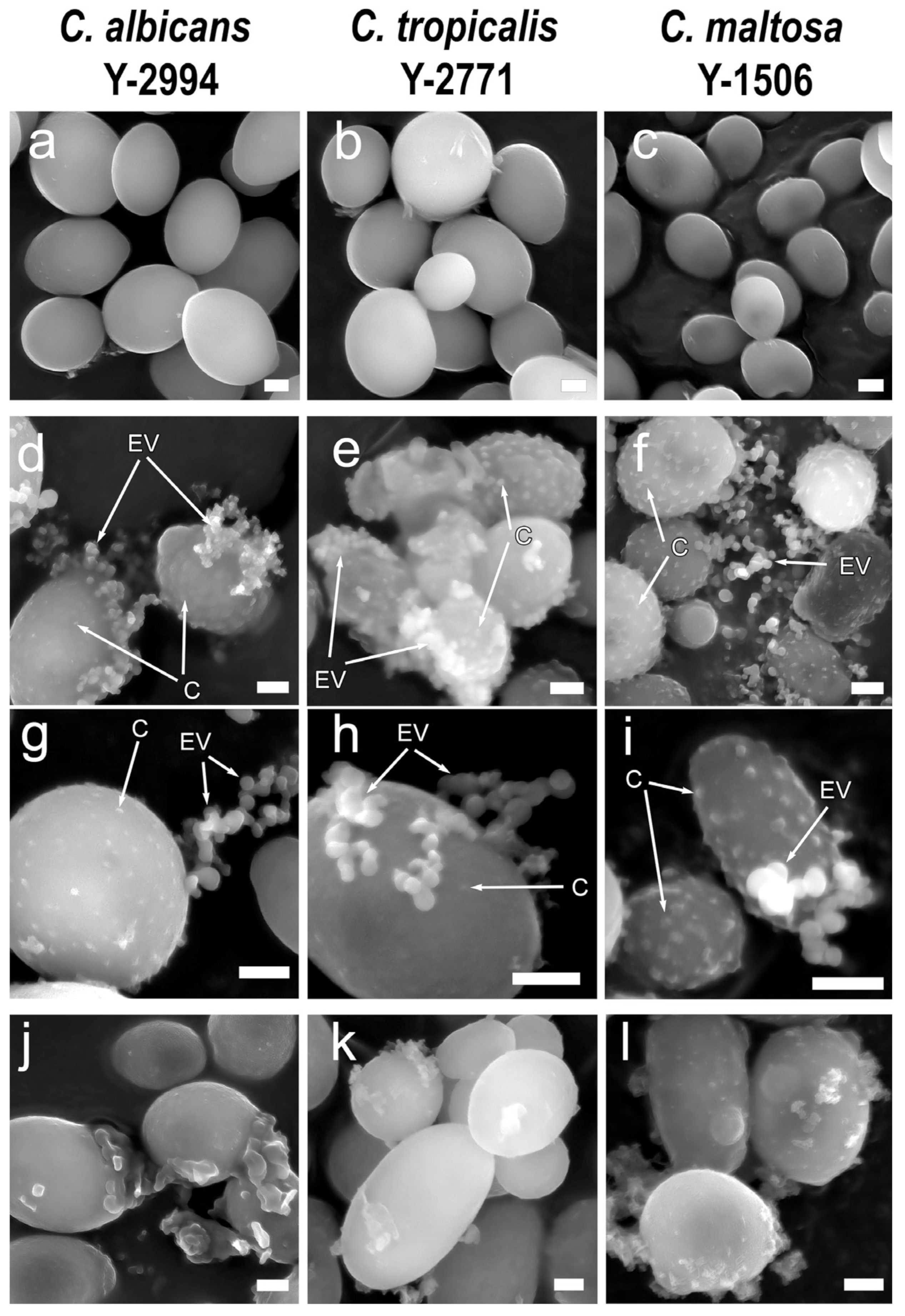
Electron microscopic analysis of the cell surface and their environment during growth on glucose revealed as expected nothing unusual (Figure 2a–c). The cells utilizing hexadecane had modified cell wall zones on the surface (canals) similar to observed earlier in hexadecane-grown C. maltosa [20,21,22,23]. The presence of extracellular vesicles was also confirmed, as well as with fluorescence microscopy. The exovesicles of all three species had approximately the same size and shape (Figure 2d–f). Treatment of cells with Ppx1 polyphosphatase also showed destruction of canals and exovesicles. At the same time, the detachment of the outer layer of the cell wall was detected (Figure 2g–i). Thus, scanning microscopy in combination with treatment with polyphosphatase confirmed the necessity of the presence of polyP in the canals and extracellular vesicles of three yeast species under study.

3.2. Polyphosphate Content in the Cells Cultivated on Glucose or Hexadecane

We determined the content of Pi, acid-soluble polyP and acid-insoluble polyP in C. maltosa, C. albicans and C. tropicalis cells grown on both glucose and hexadecane. We used an extraction method which allows the differentiation of acid-soluble polyP with an average chain length of 15 to 25 phosphate residues and acid-insoluble polyphosphates with an average chain length of 45 to 200 or more phosphate residues [22,52]. There is evidence that acid-insoluble polyPs, which can be obtained in several fractions with yeast cell treatment with saline and alkaline solutions, are associated with subcellular fractions, including vacuoles and cell walls [52]. This was believed to be the reason for their insolubility in acidic solutions [52].
It should be noted that the biomass yield on the two media differed significantly. The yield of wet biomass on glucose for all three species was 22–23 g/L, while on hexadecane this yield was significantly lower and did not exceed 4–6 g/L. As for the content of polyP, it was not possible to find any regularity characteristic of the three studied species. The total content of polyP increased by 53% in C. maltosa and by 60% in C. tropicalis but decreased by 30% in C. albicans. In C. albicans, the content of acid-insoluble polyP decreased, but the content of acid-soluble polyP increased. In C. maltosa and C. tropicalis, the main contribution to the increase in the content of polyP was made by the acid-insoluble fraction (Table 1). An increase in the content of the acid-insoluble fraction was observed earlier for another strain of C. maltosa cultivated on hexadecane [23].

3.3. The Treatment with Polyphosphatase

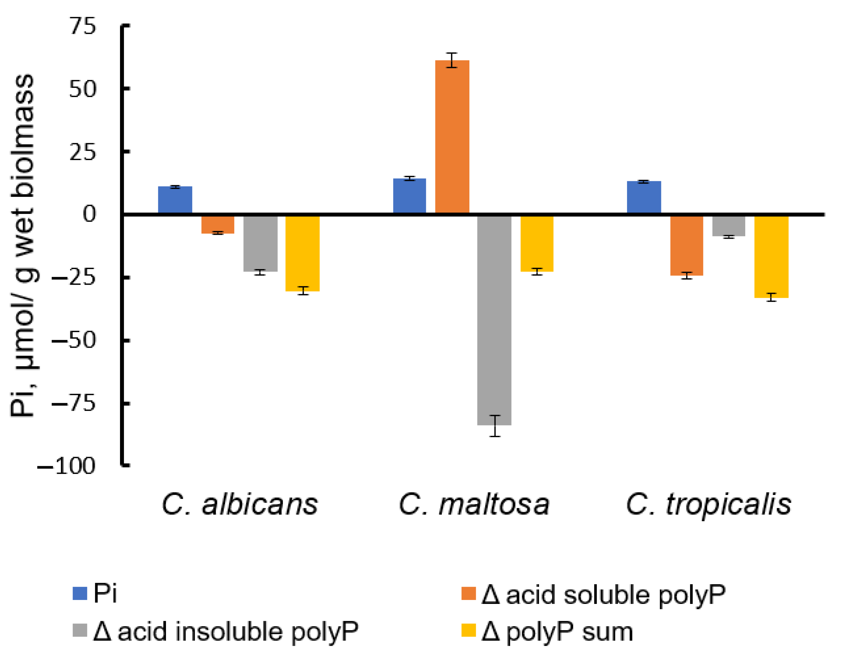
In order to assess the content of extracellular polyP in the biomass, we applied the treatment of biomass samples with a polyphosphatase Ppx1 from S. cerevisiae. This enzyme does not hydrolyze nucleoside phosphates and pyrophosphate, but it is capable of cleaving polyP with different chain lengths from three to hundreds of phosphate residues [54]. Note, that according to microscopy data (Figure 1), the cells remain intact under this treatment, and the likelihood of penetration of a 40 kDa Ppx1 protein through the cell wall is unlikely. Incubation of biomass samples at neutral pH and 30 °C resulted in a small release of phosphate into the medium. When treated under the same conditions with Ppx1, the release of phosphate increased almost fourfold (Figure 3). At the same time, in the biomass of all three species, the total content of polyP was lower in the samples treated with Ppx1 compared to the control ones (Figure 4).
In all three species, the reduction affected the fraction of acid-insoluble polyP. This suggests that the extracellular polyP is protected from acid extraction either by lipids and/proteins of vesicles or by binding to proteins and polysaccharides in the cell wall. At the same time, this polyP was available for enzymatic hydrolysis by Ppx1.

4. Discussion

This work, using fluorescence and microscopy methods in combination with the enzymatic assay of polyP, shows that the cells three Candida species cultured in a medium with hexadecane form extracellular vesicles containing PolyP. Apparently, such structures are involved in the formation of biofilms necessary for interaction with such insoluble hydrophobic substrate as hexadecane which presents in the medium as microdrops of the hydrophobic phase. In addition, polyP seems to be part of canals, a specific structure of the cell wall. In this study we revealed such structures not only in C. maltosa, but also in other Candida species.
The origin of the extracellular polyP is of special interest, since no similar structures have been found in the yeast S. cerevisiae, where the polyP metabolism and cellular localization has been studied to the greatest extent. According to modern concepts, polyP in yeast is synthesized by the Vtc4 polyphosphate synthase, which is part of the VTC (vacuolar transporter chaperone) complex [58,59]. There are two forms of VTC complex present in the yeast cells: Vtc4/Vtc3/Vtc1 and Vtc4/Vtc2/Vtc1 [60]. The former is typical of the vacuolar membrane and the latter is typical of the endoplasmic reticulum membrane and nuclear envelope [60]. This complex synthesizes long-chain polyP [58], which can then split into short-chain through hydrolysis by endopolyphosphatases Ppn1 and Ppn2 [53]. Apparently, this complex in the membrane of the endoplasmic reticulum should be responsible for the synthesis of polymers localized in the cell wall and extracellular structures. In this case, the traffic of these polymers into the cell wall and extracellular space should be carried out by means of transport vesicles of various types.
We compared the sequences of VTC4, PPN1 and PPN2 genes in three studied species using the sequences of S. cerevisiae as a comparative sample (Table 2). Vtc4 sequences have significant similarities, while the sequences of three polyphosphatases, both exopolyphosphatase PPX1 and both endopolyphosphatases show a lower level of similarity with the sequences of S. cerevisiae. The similarity of above sequences between Candida species used in this study was more pronounced (Table 3). Currently, there are no data in the literature on the properties and features of the regulation of enzymes of polyP metabolism in Candida yeasts. Therefore, we have no convincing hypothesis on the structural features of these proteins associated with the ability of Candida cells to form a significant pool of extracellular polyPs. The analysis of such features is of interest for future research in order to understand the interaction of these yeasts with the environment. It is possible that the observed differences may be related to differences in the regulatory regions of these enzymes, which causes a decrease in the polyP chains fragmentation and increase in content of extracellular long-chain polyP when growing on hexadecane.
Extracellular vesicles (EVs) are membranous, rounded vesicles formed by prokaryotic and eukaryotic cells both at normal growth and at growth in unfavorable environments [61]. These vesicles are a part of a system of communication between cells and are involved in cell wall formation and functioning, supporting cell integrity, isolation and resistance against the environment. In the case of pathogenic strains, they might take part in the interactions with the host and affect the infection outcomes [61]. In Candida albicans and Candida haemulonii, fungal extracellular vesicles (EVs) play a critical role in the pathogenicity and virulence and perform specific functions during infections, such as host cells interaction, transport of virulence factors and influence on cell survival in host organism [62,63].
Under interaction of Candida cells and mammalian cells, multiple changes in gene expression and in cellular morphology appeared, for example the expression of virulence factors (adhesins, invasins and extracellular hydrolytic enzymes), the transition to filamentous hyphae form and the formation of biofilm [64,65,66]. Researchers are increasingly interested in the role of PHO regulon [67] and the extracellular matrix [68] in these processes.
The study of the role of polyP in the organization of extracellular structures in yeast is of importance in the search for new mechanisms of adaptation to adverse conditions. We studied extracellular polyphosphates during the growth of three yeast species on hexadecane. Considering that C. maltosa is not a pathogenic strain, we assume that the observed effects in our case are associated with the need to consume a hydrophobic substrate, hexadecane. We have no reason to assert that the observed effects are related to pathogenicity. However, the formation of extracellular vesicles and biofilms is characteristic of pathogenic yeast, and we believe that in further studies in other experimental models, including models of interaction of yeast cells with mammalian cells, it is of interest to pay special attention to the presence of inorganic polyphosphates in the cell wall and extracellular structures, including biofilms.

5. Conclusions

This study shows that Candida yeast, when cultured on hexadecane, forms not only special cell wall structures but also extracellular vesicles containing inorganic polyphosphates. These polyphosphates are stained with DAPI and are available for enzymatic hydrolysis by phosphatase. Microscopic studies have shown that treatment with polyphosphatase leads to the destruction of these structures, which indicates the important role of polyP in their formation and functioning. The data obtained in this work that long-chain polyP were included in the extracellular vesicles under hexadecane consumption indicate these polymers as a component of vesicles and biofilms formed by yeasts. Such extracellular structures participate in pathogenic processes caused by yeast, and the future studies of polyP role in fungal pathogenicity may open new ways in the development of new drugs against fungi pathogens. For example, yeast polyphosphate synthetase, responsible for polyP biosynthesis, is absent in humans and may be of promising interest as a target of new drugs against candidiasis. The use of hexadecane as a model xenobiotic expands the understanding of its destruction by microorganisms in the natural environment and can also help in future medical research.

Author Contributions

Conceptualization, T.K. and A.N.Z.; methodology, L.V.T.; validation, A.N.Z. and V.V.F.; formal analysis, V.V.F.; investigation, E.V.K.; writing—original draft preparation, T.K., writing—review and editing, A.N.Z. and T.N.A.; visualization, A.N.Z.; supervision, T.K.; project administration, V.V.D. All authors have read and agreed to the published version of the manuscript.

Funding

This research received no external funding.

Institutional Review Board Statement

Not applicable.

Informed Consent Statement

Not applicable.

Data Availability Statement

Not applicable.

Acknowledgments

Electron microscopy characterization was performed in the Core Shared Research Facility of the Pushchino Center of Biological Research of the Russian Academy of Sciences. “https://www.pbcras.ru/services/tskp/ (accessed on 20 September 2023)”.

Conflicts of Interest

The authors declare no conflict of interest.

References

  1. Fukui, S.; Tanaka, A. Metabolism of Alkanes by Yeasts. In Proceedings of the Reactors and Reactions; Springer: Berlin/Heidelberg, Germany, 1981; pp. 217–237. [Google Scholar]
  2. Kogure, T.; Takagi, M.; Ohta, A. N-Alkane and Clofibrate, a Peroxisome Proliferator, Activate Transcription of ALK2 Gene Encoding Cytochrome P450alk2 through Distinct Cis-Acting Promoter Elements in Candida Maltosa. Biochem. Biophys. Res. Commun. 2005, 329, 78–86. [Google Scholar] [CrossRef] [PubMed]
  3. Fukuda, R.; Ohta, A. Genetic Features and Regulation of N-Alkane Metabolism in Yeasts. In Aerobic Utilization of Hydrocarbons, Oils and Lipids; Rojo, F., Ed.; Handbook of Hydrocarbon and Lipid Microbiology; Springer International Publishing: Cham, Switzerland, 2017; pp. 1–13. ISBN 978-3-319-39782-5. [Google Scholar]
  4. Fukuda, R. Utilization of N-Alkane and Roles of Lipid Transfer Proteins in Yarrowia lipolytica. World J. Microbiol. Biotechnol. 2023, 39, 97. [Google Scholar] [CrossRef] [PubMed]
  5. Fukuda, R. Metabolism of Hydrophobic Carbon Sources and Regulation of It in N-Alkane-Assimilating Yeast Yarrowia Lipolytica. Biosci. Biotechnol. Biochem. 2013, 77, 1149–1154. [Google Scholar] [CrossRef] [PubMed]
  6. Finogenova, T.; Morgunov, I.; Kamzolova, S.; Chernyavskaya, O. Organic Acid Production by the Yeast Yarrowia Lipolytica: A Review of Prospects. Appl. Biochem. Microbiol. 2005, 41, 418–425. [Google Scholar] [CrossRef]
  7. Beier, A.; Hahn, V.; Bornscheuer, U.T.; Schauer, F. Metabolism of Alkenes and Ketones by Candida Maltosa and Related Yeasts. AMB Express 2014, 4, 75. [Google Scholar] [CrossRef]
  8. Mori, K.; Iwama, R.; Kobayashi, S.; Horiuchi, H.; Fukuda, R.; Ohta, A. Transcriptional Repression by Glycerol of Genes Involved in the Assimilation of N-Alkanes and Fatty Acids in Yeast Yarrowia Lipolytica. FEMS Yeast Res. 2013, 13, 233–240. [Google Scholar] [CrossRef]
  9. Kogure, T.; Horiuchi, H.; Matsuda, H.; Arie, M.; Takagi, M.; Ohta, A. Enhanced Induction of Cytochromes P450alk That Oxidize Methyl-Ends of n-Alkanes and Fatty Acids in the Long-Chain Dicarboxylic Acid-Hyperproducing Mutant of Candida Maltosa. FEMS Microbiol. Lett. 2007, 271, 106–111. [Google Scholar] [CrossRef]
  10. Takagi, M.; Ohkuma, M.; Kobayashi, N.; Watanabe, M.; Yano, K. Purification of Cytochrome P-450«/A from n-Alkane-Grown Cells of Candida Maltosa, and Cloning and Nucleotide Sequencing of the Encoding Gene. Agric. Biol. Chem. 1989, 53, 2217–2226. [Google Scholar] [CrossRef]
  11. Schunck, W.H.; Kärgel, E.; Gross, B.; Wiedmann, B.; Mauersberger, S.; Köpke, K.; Kiessling, U.; Strauss, M.; Gaestel, M.; Müller, H.G. Molecular Cloning and Characterization of the Primary Structure of the Alkane Hydroxylating Cytochrome P-450 from the Yeast Candida Maltosa. Biochem. Biophys Res. Commun. 1989, 161, 843–850. [Google Scholar] [CrossRef]
  12. Mauersberger, S.; Ohkuma, M.; Schunck, W.-H.; Takagi, M. Candida Maltosa. In Nonconventional Yeasts in Biotechnology: A Handbook; Wolf, K., Ed.; Springer: Berlin/Heidelberg, Germany, 1996; pp. 411–580. ISBN 978-3-642-79856-6. [Google Scholar]
  13. Ohkuma, M.; Tanimoto, T.; Yano, K.; Takagi, M. CYP52 (Cytochrome P450alk) Multigene Family in Candida Maltosa: Molecular Cloning and Nucleotide Sequence of the Two Tandemly Arranged Genes. DNA Cell Biol. 1991, 10, 271–282. [Google Scholar] [CrossRef]
  14. Ohkuma, M.; Muraoka, S.; Tanimoto, T.; Fujii, M.; Ohta, A.; Takagi, M. CYP52 (Cytochrome P450alk) Multigene Family in Candida Maltosa: Identification and Characterization of Eight Members. DNA Cell Biol. 1995, 14, 163–173. [Google Scholar] [CrossRef] [PubMed]
  15. Iwama, R.; Kobayashi, S.; Ishimaru, C.; Ohta, A.; Horiuchi, H.; Fukuda, R. Functional Roles and Substrate Specificities of Twelve Cytochromes P450 Belonging to CYP52 Family in N-Alkane Assimilating Yeast Yarrowia Lipolytica. Fungal. Genet. Biol. 2016, 91, 43–54. [Google Scholar] [CrossRef] [PubMed]
  16. Kawamoto, S.; Tanaka, A.; Yamamura, M.; Teranishi, Y.; Fukui, S. Microbody of N-Alkane-Grown Yeast. Arch. Microbiol. 1977, 112, 1–8. [Google Scholar] [CrossRef] [PubMed]
  17. Mauersberger, S.; Kärgel, E.; Matyashova, R.N.; Müller, H.-G. Subcellular Organization of Alkane Oxidation in the Yeast Candida Maltosa. J. Basic Microbiol. 1987, 27, 565–582. [Google Scholar] [CrossRef]
  18. Vogel, F.; Kärgel, E.; Schunck, W.H. In Situ Localization of Cytochrome P-450, the First Enzyme Involved in Aliphatic Hydrocarbon Degradation in the Yeast Candida Maltosa. Prog. Histochem. Cytochem. 1991, 23, 383–389. [Google Scholar] [CrossRef] [PubMed]
  19. Li, J.; Xu, Y.; Song, Q.; Zhang, S.; Xie, L.; Yang, J. Transmembrane Transport Mechanism of N-Hexadecane by Candida Tropicalis: Kinetic Study and Proteomic Analysis. Ecotoxicol. Environ. Saf. 2021, 209, 111789. [Google Scholar] [CrossRef] [PubMed]
  20. Dmitriev, V.V.; Crowley, D.; Rogachevsky, V.V.; Negri, C.M.; Rusakova, T.G.; Kolesnikova, S.A.; Akhmetov, L.I. Microorganisms Form Exocellular Structures, Trophosomes, to Facilitate Biodegradation of Oil in Aqueous Media. FEMS Microbiol. Lett. 2011, 315, 134–140. [Google Scholar] [CrossRef] [PubMed]
  21. Dmitriev, V.V.; Crowley, D.E.; Zvonarev, A.N.; Rusakova, T.G.; Negri, M.C.; Kolesnikova, S.A. Modifications of the Cell Wall of Yeasts Grown on Hexadecane and under Starvation Conditions. Yeast 2016, 33, 55–62. [Google Scholar] [CrossRef]
  22. Zvonarev, A.N.; Crowley, D.E.; Ryazanova, L.P.; Lichko, L.P.; Rusakova, T.G.; Kulakovskaya, T.V.; Dmitriev, V.V. Cell Wall Canals Formed upon Growth of Candida Maltosa in the Presence of Hexadecane Are Associated with Polyphosphates. FEMS Yeast Res. 2017, 17, fox026. [Google Scholar] [CrossRef]
  23. Zvonarev, A.; Farofonova, V.; Kulakovskaya, E.; Kulakovskaya, T.; Machulin, A.; Sokolov, S.; Dmitriev, V. Changes in Cell Wall Structure and Protein Set in Candida Maltosa Grown on Hexadecane. Folia Microbiol. 2021, 66, 247–253. [Google Scholar] [CrossRef]
  24. Anderson, J.; Mihalik, R.; Soll, D.R. Ultrastructure and Antigenicity of the Unique Cell Wall Pimple of the Candida Opaque Phenotype. J. Bacteriol. 1990, 172, 224–235. [Google Scholar] [CrossRef] [PubMed]
  25. Staniszewska, M.; Bondaryk, M.; Swoboda-Kopec, E.; Siennicka, K.; Sygitowicz, G.; Kurzatkowski, W. Candida Albicans Morphologies Revealed by Scanning Electron Microscopy Analysis. Braz. J. Microbiol. 2013, 44, 813–821. [Google Scholar] [CrossRef] [PubMed]
  26. Nett, J.E.; Sanchez, H.; Cain, M.T.; Ross, K.M.; Andes, D.R. Interface of Candida Albicans Biofilm Matrix-Associated Drug Resistance and Cell Wall Integrity Regulation. Eukaryot. Cell 2011, 10, 1660–1669. [Google Scholar] [CrossRef] [PubMed]
  27. Silva-Dias, A.; Miranda, I.M.; Branco, J.; Monteiro-Soares, M.; Pina-Vaz, C.; Rodrigues, A.G. Adhesion, Biofilm Formation, Cell Surface Hydrophobicity, and Antifungal Planktonic Susceptibility: Relationship among Candida spp. Front. Microbiol. 2015, 6, 205. [Google Scholar] [CrossRef] [PubMed]
  28. Silva, S.; Negri, M.; Henriques, M.; Oliveira, R.; Williams, D.W.; Azeredo, J. Candida Glabrata, Candida Parapsilosis and Candida Tropicalis: Biology, Epidemiology, Pathogenicity and Antifungal Resistance. FEMS Microbiol. Rev. 2012, 36, 288–305. [Google Scholar] [CrossRef] [PubMed]
  29. Dižová, S.; Černáková, L.; Bujdáková, H. The Impact of Farnesol in Combination with Fluconazole on Candida Albicans Biofilm: Regulation of ERG20, ERG9, and ERG11 Genes. Folia Microbiol. 2018, 63, 363–371. [Google Scholar] [CrossRef] [PubMed]
  30. Suchodolski, J.; Derkacz, D.; Muraszko, J.; Panek, J.J.; Jezierska, A.; Łukaszewicz, M.; Krasowska, A. Fluconazole and Lipopeptide Surfactin Interplay During Candida Albicans Plasma Membrane and Cell Wall Remodeling Increases Fungal Immune System Exposure. Pharmaceutics 2020, 12, 314. [Google Scholar] [CrossRef]
  31. Yeh, Y.-C.; Wang, H.-Y.; Lan, C.-Y. Candida Albicans Aro1 Affects Cell Wall Integrity, Biofilm Formation and Virulence. J. Microbiol. Immunol. Infect. 2020, 53, 115–124. [Google Scholar] [CrossRef]
  32. Garcia-Rubio, R.; de Oliveira, H.C.; Rivera, J.; Trevijano-Contador, N. The Fungal Cell Wall: Candida, Cryptococcus, and Aspergillus Species. Front. Microbiol. 2019, 10, 2993. [Google Scholar] [CrossRef]
  33. Blasi, B.; Poyntner, C.; Rudavsky, T.; Prenafeta-Boldú, F.X.; Hoog, S.D.; Tafer, H.; Sterflinger, K. Pathogenic Yet Environmentally Friendly? Black Fungal Candidates for Bioremediation of Pollutants. Geomicrobiol. J. 2016, 33, 308–317. [Google Scholar] [CrossRef]
  34. Lavrin, T.; Konte, T.; Kostanjšek, R.; Sitar, S.; Sepčič, K.; Prpar Mihevc, S.; Žagar, E.; Župunski, V.; Lenassi, M.; Rogelj, B.; et al. The Neurotropic Black Yeast Exophiala Dermatitidis Induces Neurocytotoxicity in Neuroblastoma Cells and Progressive Cell Death. Cells 2020, 9, 963. [Google Scholar] [CrossRef] [PubMed]
  35. Rao, N.N.; Gómez-García, M.R.; Kornberg, A. Inorganic Polyphosphate: Essential for Growth and Survival. Annu. Rev. Biochem. 2009, 78, 605–647. [Google Scholar] [CrossRef] [PubMed]
  36. Ikeh, M.; Ahmed, Y.; Quinn, J. Phosphate Acquisition and Virulence in Human Fungal Pathogens. Microorganisms 2017, 5, 48. [Google Scholar] [CrossRef] [PubMed]
  37. Ramos, C.L.; Gomes, F.M.; Girard-Dias, W.; Almeida, F.P.; Albuquerque, P.C.; Kretschmer, M.; Kronstad, J.W.; Frases, S.; de Souza, W.; Rodrigues, M.L.; et al. Phosphorus-Rich Structures and Capsular Architecture in Cryptococcus Neoformans. Future Microbiol. 2017, 12, 227–238. [Google Scholar] [CrossRef] [PubMed]
  38. Bowring, B.G.; Sethiya, P.; Desmarini, D.; Lev, S.; Tran Le, L.; Bahn, Y.-S.; Lee, S.-H.; Toh-e, A.; Proschogo, N.; Savage, T.; et al. Dysregulating PHO Signaling via the CDK Machinery Differentially Impacts Energy Metabolism, Calcineurin Signaling, and Virulence in Cryptococcus Neoformans. mBio 2023, 14, e0355122. [Google Scholar] [CrossRef] [PubMed]
  39. Jiang, L.; Pan, H. Functions of CaPhm7 in the Regulation of Ion Homeostasis, Drug Tolerance, Filamentation and Virulence in Candida Albicans. BMC Microbiol. 2018, 18, 49. [Google Scholar] [CrossRef] [PubMed]
  40. Kulakovskaya, E.; Zvonarev, A.; Kulakovskaya, T. PHM6 and PHM7 Genes Are Essential for Phosphate Surplus in the Cells of Saccharomyces Cerevisiae. Arch. Microbiol. 2023, 205, 47. [Google Scholar] [CrossRef]
  41. Liu, N.-N.; Acosta-Zaldívar, M.; Qi, W.; Diray-Arce, J.; Walker, L.A.; Kottom, T.J.; Kelly, R.; Yuan, M.; Asara, J.M.; Lasky-Su, J.A.; et al. Phosphoric Metabolites Link Phosphate Import and Polysaccharide Biosynthesis for Candida Albicans Cell Wall Maintenance. mBio 2020, 11, e03225-19. [Google Scholar] [CrossRef]
  42. Ohkuma, M.; Hwang, C.W.; Masuda, Y.; Nishida, H.; Sugiyama, J.; Ohta, A.; Takagi, M. Evolutionary Position of N-Alkane-Assimilating Yeast Candida Maltosa Shown by Nucleotide Sequence of Small-Subunit Ribosomal RNA Gene. Biosci. Biotechnol. Biochem. 1993, 57, 1793–1794. [Google Scholar] [CrossRef]
  43. Olanbiwoninu, A.A.; Popoola, B.M. Biofilms and Their Impact on the Food Industry. Saudi. J. Biol. Sci. 2023, 30, 103523. [Google Scholar] [CrossRef]
  44. Li, X.; Zhang, K.; Xue, Y. Continuous Immobilization of Lead by Biomineralization in a Fluidized-Bed Biofilm Reactor. J. Clean. Prod. 2022, 379, 134765. [Google Scholar] [CrossRef]
  45. An, H.; Tian, T.; Wang, Z.; Jin, R.; Zhou, J. Role of Extracellular Polymeric Substances in the Immobilization of Hexavalent Chromium by Shewanella Putrefaciens CN32 Unsaturated Biofilms. Sci. Total Environ. 2022, 810, 151184. [Google Scholar] [CrossRef] [PubMed]
  46. Yunda, E.; Gutensohn, M.; Ramstedt, M.; Björn, E. Methylmercury Formation in Biofilms of Geobacter Sulfurreducens. Front. Microbiol. 2023, 14, 1079000. [Google Scholar] [CrossRef] [PubMed]
  47. Hermida, L.; Agustian, J. The Application of Conventional or Magnetic Materials to Support Immobilization of Amylolytic Enzymes for Batch and Continuous Operation of Starch Hydrolysis Processes. Rev. Chem. Eng. 2022. [Google Scholar] [CrossRef]
  48. Kim, J.-H.; Lee, E.-S.; Song, K.-J.; Kim, B.-M.; Ham, J.-S.; Oh, M.-H. Development of Desiccation-Tolerant Probiotic Biofilms Inhibitory for Growth of Foodborne Pathogens on Stainless Steel Surfaces. Foods 2022, 11, 831. [Google Scholar] [CrossRef] [PubMed]
  49. Bouaziz, A.; Houfani, A.A.; Baoune, H. Enzymology of Microbial Biofilms. In Ecological Interplays in Microbial Enzymology; Maddela, N.R., Abiodun, A.S., Prasad, R., Eds.; Environmental and Microbial Biotechnology; Springer Nature: Singapore, 2022; pp. 117–140. ISBN 978-981-19015-5-3. [Google Scholar]
  50. Sheng, Y.; Chen, Z.; Wu, W.; Lu, Y. Engineered Organic Nanoparticles to Combat Biofilms. Drug Discov. Today 2023, 28, 103455. [Google Scholar] [CrossRef] [PubMed]
  51. Eldarov, M.A.; Baranov, M.V.; Dumina, M.V.; Shgun, A.A.; Andreeva, N.A.; Trilisenko, L.V.; Kulakovskaya, T.V.; Ryasanova, L.P.; Kulaev, I.S. Polyphosphates and Exopolyphosphatase Activities in the Yeast Saccharomyces Cerevisiae under Overexpression of Homologous and Heterologous PPN1 Genes. Biochem. Mosc. 2013, 78, 946–953. [Google Scholar] [CrossRef] [PubMed]
  52. Vagabov, V.M.; Trilisenko, L.V.; Kulaev, I.S. Dependence of Inorganic Polyphosphate Chain Length on the Orthophosphate Content in the Culture Medium of the Yeast Saccharomyces Cerevisiae. Biochemistry 2000, 65, 349–354. [Google Scholar]
  53. Heinonen, J.K.; Lahti, R.J. A New and Convenient Colorimetric Determination of Inorganic Orthophosphate and Its Application to the Assay of Inorganic Pyrophosphatase. Anal. Biochem. 1981, 113, 313–317. [Google Scholar] [CrossRef]
  54. Andreeva, N.; Ledova, L.; Ryazanova, L.; Tomashevsky, A.; Kulakovskaya, T.; Eldarov, M. Ppn2 Endopolyphosphatase Overexpressed in Saccharomyces Cerevisiae: Comparison with Ppn1, Ppx1, and Ddp1 Polyphosphatases. Biochimie 2019, 163, 101–107. [Google Scholar] [CrossRef]
  55. Serafim, L.S.; Lemos, P.C.; Levantesi, C.; Tandoi, V.; Santos, H.; Reis, M.A.M. Methods for Detection and Visualization of Intracellular Polymers Stored by Polyphosphate-Accumulating Microorganisms. J. Microbiol. Methods 2002, 51, 1–18. [Google Scholar] [CrossRef] [PubMed]
  56. Andreeva, N.; Ryazanova, L.; Dmitriev, V.; Kulakovskaya, T.; Kulaev, I. Adaptation of Saccharomyces Cerevisiae to Toxic Manganese Concentration Triggers Changes in Inorganic Polyphosphates. FEMS Yeast Res. 2013, 13, 463–470. [Google Scholar] [CrossRef] [PubMed]
  57. Lichko, L.; Kulakovskaya, T. Polyphosphatase PPX1 of Saccharomyces Cerevisiae as a Tool for Polyphosphate Assay. Adv. Enzym. Res. 2015, 3, 93. [Google Scholar] [CrossRef]
  58. Hothorn, M.; Neumann, H.; Lenherr, E.D.; Wehner, M.; Rybin, V.; Hassa, P.O.; Uttenweiler, A.; Reinhardt, M.; Schmidt, A.; Seiler, J.; et al. Catalytic Core of a Membrane-Associated Eukaryotic Polyphosphate Polymerase. Science 2009, 324, 513–516. [Google Scholar] [CrossRef] [PubMed]
  59. Guan, Z.; Chen, J.; Liu, R.; Chen, Y.; Xing, Q.; Du, Z.; Cheng, M.; Hu, J.; Zhang, W.; Mei, W.; et al. The Cytoplasmic Synthesis and Coupled Membrane Translocation of Eukaryotic Polyphosphate by Signal-Activated VTC Complex. Nat. Commun. 2023, 14, 718. [Google Scholar] [CrossRef] [PubMed]
  60. Gerasimaitė, R.; Mayer, A. Enzymes of Yeast Polyphosphate Metabolism: Structure, Enzymology and Biological Roles. Biochem. Soc. Trans. 2016, 44, 234–239. [Google Scholar] [CrossRef] [PubMed]
  61. Liebana-Jordan, M.; Brotons, B.; Falcon-Perez, J.M.; Gonzalez, E. Extracellular Vesicles in the Fungi Kingdom. Int. J. Mol. Sci. 2021, 22, 7221. [Google Scholar] [CrossRef]
  62. Oliveira, B.T.M.; Dourado, T.M.H.; Santos, P.W.S.; Bitencourt, T.A.; Tirapelli, C.R.; Colombo, A.L.; Almeida, F. Extracellular Vesicles from Candida Haemulonii Var. Vulnera Modulate Macrophage Oxidative Burst. J. Fungi 2023, 9, 562. [Google Scholar] [CrossRef]
  63. Wei, Y.; Wang, Z.; Liu, Y.; Liao, B.; Zong, Y.; Shi, Y.; Liao, M.; Wang, J.; Zhou, X.; Cheng, L.; et al. Extracellular Vesicles of Candida Albicans Regulate Its Own Growth through the L-Arginine/Nitric Oxide Pathway. Appl. Microbiol. Biotechnol. 2023, 107, 355–367. [Google Scholar] [CrossRef]
  64. Macias-Paz, I.U.; Pérez-Hernández, S.; Tavera-Tapia, A.; Luna-Arias, J.P.; Guerra-Cárdenas, J.E.; Reyna-Beltrán, E. Candida albicans the main opportunistic pathogenic fungus in humans. Rev. Argent Microbiol. 2023, 55, 189–198. [Google Scholar] [CrossRef]
  65. Branco, J.; Miranda, I.M.; Rodrigues, A.G. Candida parapsilosis Virulence and Antifungal Resistance Mechanisms: A Comprehensive Review of Key Determinants. J. Fungi 2023, 9, 80. [Google Scholar] [CrossRef]
  66. Dos Santos, M.M.; Ishida, K. We need to talk about Candida tropicalis: Virulence factors and survival mechanisms. Med. Mycol. 2023, 61, myad075. [Google Scholar] [CrossRef]
  67. Martín, J.F. Interaction of calcium responsive proteins and transcriptional factors with the PHO regulon in yeasts and fungi. Front. Cell Dev. Biol. 2023, 11, 1225774. [Google Scholar] [CrossRef]
  68. Lucas, C.; Silva, C. The Extracellular Matrix of Yeasts: A Key Player in the Microbial Biology Change of Paradigm. Front. Biosci.-Elite 2023, 15, 13. [Google Scholar] [CrossRef]
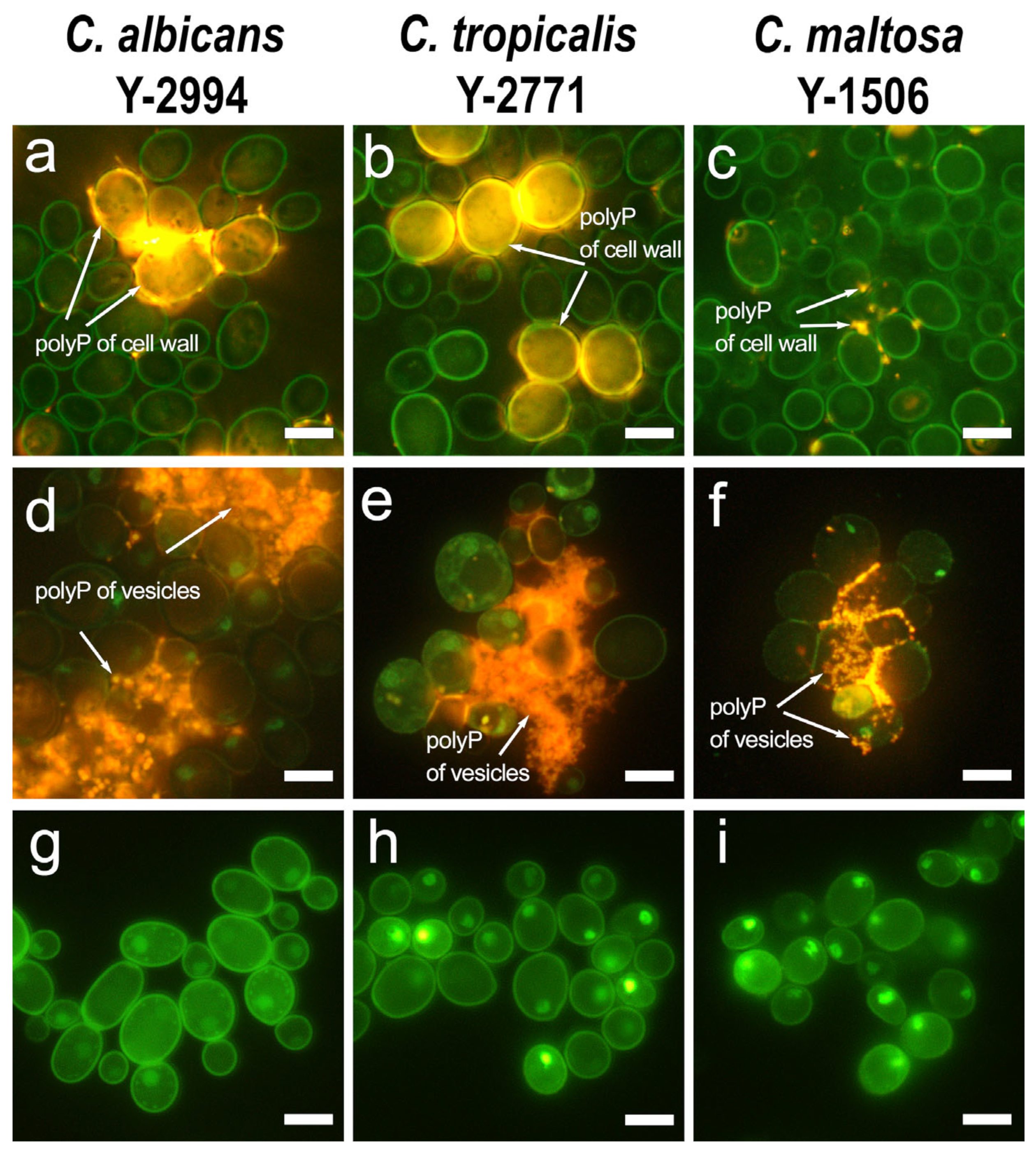
Figure 1. Fluorescent microscopy with DAPI staining for polyphosphates: (a) (C. albicans), (b) (C. tropicalis), (c) (C. maltosa)—cells grown on a medium with glucose; (d) (C. albicans), (e) (C. tropicalis), (f) (C. maltosa) —cells grown on a medium with hexadecane; (g) (C. albicans), (h) (C. tropicalis), (i) (C. maltosa)—treatment with Ppx1 polyphosphatase. Scale bar 5 µm.
Jox 13 00034 g001
Figure 2. Scanning electron microscopy: (a) (C. albicans), (b) (C. tropicalis), (c) (C. maltosa)—cells grown on a medium with glucose; (d) (C. albicans), (e) (C. tropicalis), (f) (C. maltosa)—cells grown on a medium with hexadecane; (g) (C. albicans), (h) (C. tropicalis), (i) (C. maltosa)—cells grown on the medium with hexadecane (higher magnification then on (df)); (j) (C. albicans), (k) (C. tropicalis), (l) (C. maltosa)—treatment with Ppx1 polyphosphatase. C—canals of cell wall, EV—extracellular vesicles. Scale bar 1 µm.
Jox 13 00034 g002
Figure 3. Pi release to the medium after incubation of biomass of three yeast species under control conditions and in the presence of Ppx1 polyphosphatase. The cells were grown in a medium with hexadecane. The experiments were performed in 3 replicates; the values denote mean and the error bars denote s.d. p < 10−4 in all cases, significance was assessed with the one-tailed Student’s t-test performed for Ppx1 treatment versus control incubation of biomass samples.
Jox 13 00034 g003
Figure 4. The amount of Pi released into the medium from the biomass, and changes in the amount of acid-soluble polyP and acid-insoluble polyP in biomass of three yeast species after incubation in the presence of Ppx1 polyphosphatase. The cells were cultivated in the medium with hexadecane. The experiments were performed in 3 replicates; the values denote mean and the error bars denote s.d. p < 10−4 in all cases, significance was assessed with the one-tailed Student’s t-test performed for Ppx1 treatment versus control incubation of biomass samples.
Jox 13 00034 g004
Table 1. The content of Pi, acid-soluble and acid insoluble polyP in yeast cells grown on glucose or hexadecane, μmol P/g wet biomass.
GlucoseHexadecane
Yeast SpeciesPiAcid Soluble polyP Acid Insoluble polyP PiAcid Soluble polyP Acid Insoluble polyP
C albicans10.6 ± 0.2022.0 ± 1.93115 ± 4.127.6 ± 1.62534.0 ± 1.2659.1 ± 13.4
C. tropicalis10.3 ± 0.253.93 ± 0.6630.5 ± 1.014.9 ± 0.2310.9 ± 0.9144.8 ± 4.97
C. maltosa15.8 ± 2.48.02 ± 2.0479.6 ± 1.116.3 ± 1.211.5 ± 4.24124 ± 9.9
Table 2. The AA sequence alignment of VTC4, PPX1, PPN1 and PPN2 for C. albicans, C. tropicalis and C. maltosa by BLAST with sequences of S. cerevisiae as a reference.
SpeciesC. albicansC. tropicalisC. maltosa
VTC4
(SGD:S000003549)
Accession numberXP_711583.1XP_002545726.1EMG48228.1
Query Cover, %999999
Percent identity60.8260.2760.95
PPX1
(SGD:S000001244)
Accession numberKHC58625.1XP_002547193.1EMG50694.1
Query Cover, %979095
Percent identity32.7834.3634.2
PPN2
(SGD:S000005161)
Accession numberKHC62723.1XP_002550886.1EMG47562.1
Query Cover, %959596
Percent identity29.013029.14
PPN1
(SGD:S000002860)
Accession numberKHC31142.1XP_002550589.1EMG47375.1
Query Cover, %949387
Percent identity41.0140.5942.95
Table 3. The AA sequence alignment of VTC4, PPX1, PPN1 and PPN2 for C. tropicalis and C. maltosa by BLAST with sequences of C. albicans as a reference.
SpeciesC. tropicalisC. maltosa
VTC4
(XP_711583.1)
Accession numberXP_002545726.1EMG48228.1
Query Cover, %9999
Percent identity82.7186.85
PPX1
(KHC58625.1)
Accession numberXP_002547193.1EMG50694.1
Query Cover, %100100
Percent identity61.9463.03
PPN2
(KHC62723.1)
Accession numberXP_002550886.1EMG47562.1
Query Cover, %100100
Percent identity59.9559.44
PPN1
(KHC31142.1)
Accession numberXP_002550589.1EMG47375.1
Query Cover, %9799
Percent identity65.164.05
Disclaimer/Publisher’s Note: The statements, opinions and data contained in all publications are solely those of the individual author(s) and contributor(s) and not of MDPI and/or the editor(s). MDPI and/or the editor(s) disclaim responsibility for any injury to people or property resulting from any ideas, methods, instructions or products referred to in the content.