Article

Exploring the Potential of Biocontrol Agent Against Root and Stem Rot Disease in Durian (Durio zibethinus)

Department of Agricultural Technology, Faculty of Science and Technology, Thammasat University, Khlong Nueng 12120, Pathum Thani, Thailand
*
Author to whom correspondence should be addressed.
Int. J. Plant Biol. 2025, 16(3), 75; https://doi.org/10.3390/ijpb16030075
Submission received: 22 May 2025 / Revised: 27 June 2025 / Accepted: 30 June 2025 / Published: 6 July 2025
(This article belongs to the Section Plant–Microorganisms Interactions)

Abstract

The study of antagonistic bacterial strains isolated from the soil around durian tree roots demonstrated their ability to inhibit the growth of Phytophthora palmivora. The pathogens were screened from 30 samples collected around durian trees (leaves, soil around the roots, and debris under the tree) showing symptoms of root and stem rot disease. A total of 17 pathogen strains were isolated and grouped into 3 groups, TNP05, MNP13, and KNP21, originating from Chanthaburi province, Thailand. When P. palmivora isolates were tested for pathogenicity on leaves and durian trees, it was found that the strain MNP13 had the highest capacity to cause root and stem rot disease. A total of 196 beneficial bacteria isolates were collected from several samples around durian trees. The samples included leaves, soil surrounding the roots, and organic debris beneath the trees. Based on their colony characteristics on nutrient glucose agar (NGA), these isolates were divided into 8 groups. The efficacy of the beneficial bacteria against root and stem rot disease was tested using the Dual culture method and arranged in a Completely Randomized Design (CRD) with 5 replications. The experiment showed that bacterial isolates NJTU05, NJTU10, and NJTU13 effectively inhibited the growth of P. palmivora isolate MNP13, with inhibition rates of 76.66, 67.59, and 69.07%, respectively, compared to chemical control using metalaxyl 80% WP. Among the tested strains, NJTU05 was identified as the most effective bacterial strain for controlling major durian diseases. Biochemical identification and 16S rRNA sequencing revealed that bacterial strain NJTU05 was closely related to Brevibacillus formosus with a 99.70% identity.

1. Introduction

Durian (Durio zibethinus Murr.), often referred to as the “King of Fruits”, plays a vital role in Thailand’s agricultural economy. Over 200 varieties of durian are cultivated in Thailand, with 5 main varieties being grown commercially: Chanee, Kan Yao, Puangmanee, Kradum, and Monthong. Among these, Monthong is the most favored by consumers [1,2]. As the world’s largest producer and exporter of durian, Thailand cultivates over one million rai of durian plantations, primarily concentrated in the eastern and southern regions, including Chanthaburi, Rayong, and Chumphon provinces [3]. The expansion of export markets, particularly to China, has significantly driven the increase in cultivation area and production volume over the past decade.
According to the Ministry of Agriculture and Cooperatives [4], Thailand produced approximately 1.44 million tons of durian in 2022 from 1.12 million rai of farmland. The harvest season typically peaks between May and July, aligning with export demands. Eastern provinces contribute over 70% of the national production due to favorable climate, infrastructure, and access to export channels. As domestic consumption remains stable, international demand, especially from China, continues to be the primary growth driver.
Thailand has maintained its position as the world’s leading exporter of fresh durian. In 2021 alone, the country exported over 800,000 tons of fresh durian, generating more than 110 billion baht in revenue. The majority of these exports up to 95% were destined for the Chinese market [5]. The Chinese market favors Thai durian due to its consistent quality, established logistics infrastructure, and strategic trade agreements. Other notable importers include Hong Kong, Malaysia, and Vietnam. To meet international phytosanitary and food safety standards, Thai exporters have implemented good agricultural practices (GAP) and enhanced post-harvest technologies to ensure the quality and shelf life of exported fruits.
A major factor contributing to the reduction of cultivated durian yield in Thailand is the severe outbreak of diseases caused by pathogens such as Phytophthora palmivora, Fusarium spp., Pythium spp., Sclerotium spp., and Rhizoctonia spp. Therefore, farmers use chemicals in the prevention and eradication of disease to solve the problem. This causes higher production costs, lower yields, and pesticide residues in production; consequently, farmers suffer a financial loss [6]. Durian disease is the most prevalent disease affecting all trees, such as anthracnose, leaf blight, pink disease, and particularly root rot and stem rot, which pose the greatest threat to durian farmers. Root rot and stem rot are caused by P. palmivora, the primary disease affecting durian. This disease can infect all parts of a durian tree, including roots, stems, leaves, and fruit, often leading to the death of trees during growth and fruit production. The epidemic of this disease causes significant damage to durian crops across all Thai growing regions, leading to reduced yields and poor-quality production [7]. To combat root and stem rot, most farmers resort to using chemical agents, resulting in chemical residues that affect consumers, the environment, and durian export of durian. Therefore, controlling and eliminating plant diseases through biological methods, like antagonistic microorganisms to control durian root and stem rot, has become increasingly important [8]. According to Pengnoo et al. [9], Bacillus velezensis SM1 exhibited strong antagonistic activity against P. palmivora, the causal agent of durian root rot. The strain produced significant inhibition zones in dual culture assays, and genome analysis revealed biosynthetic gene clusters responsible for antimicrobial compounds such as surfactin and fengycin. These findings support the potential of B. velezensis SM1 as a promising biocontrol agent for managing root and stem rot in durian. Similarly, Nor Danial et al. [10] reported the effectiveness of Bacillus spp. in reducing disease incidence through various application methods at the nursery stage. Soil drenching with Bacillus alone reduced disease severity by 62%, while co-application with Streptomyces spp. increased efficacy to 74%. This suggests a synergistic effect and highlights the importance of application strategy in field conditions. Together, these studies demonstrate that Bacillus spp., particularly B. velezensis, offers an effective and environmentally friendly alternative to chemical fungicides. Their integration into nursery-stage disease management can support sustainable durian production and enhance the resilience of young plants against root and stem rot pathogens.
However, there are currently only a few biological products available in Thailand for the treatment of durian root and stem rot. Therefore, the development of effective biocontrol agents presents a promising alternative to chemical fungicides. This research aimed to investigate antagonistic microorganisms with biocontrol potential for managing durian root and stem rot, thereby supporting sustainable disease management under field conditions.

2. Materials and Methods

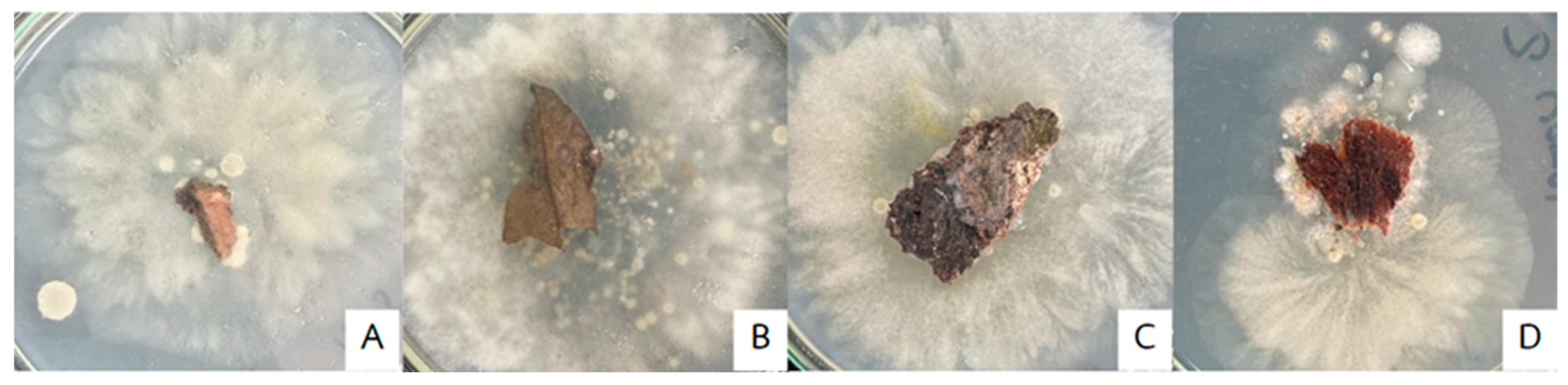
2.1. Sampling and Pathogen Isolation

2.1.1. Plant Tissue Samples

Thirty samples were collected from different parts of durian trees exhibiting disease symptoms and were cultured on potato dextrose agar (PDA), then incubated at room temperature (28 ± 3 °C) [11]. Once Phytophthora palmivora growth was observed on the plant tissues, the pathogen was isolated using the hyphal tip method to obtain a pure culture. Morphological characteristics of P. palmivora were subsequently examined.

2.1.2. Soil Sample

Pathogens were isolated from the soil by mixing 1 g of soil with 9 mL of distilled water, followed by shaking at 120 rpm for 10 min. The soil suspension was then diluted using the ten-fold serial dilution method [12]. An aliquot of the diluted suspension was spread evenly onto PDA plates and incubated at room temperature for 24–48 h. Colonies showing P. palmivora like growth were subcultured for further study [13].

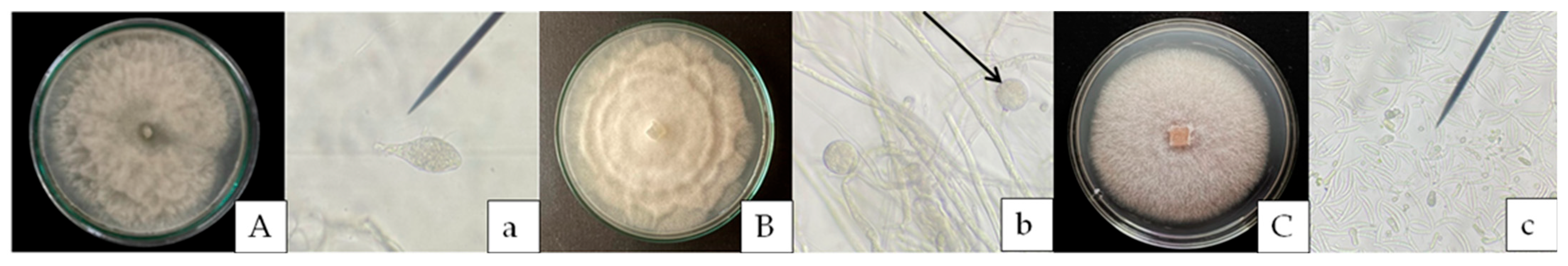
2.1.3. Morphological Investigation

The morphology of the isolated pathogens was examined through both macroscopic and microscopic observations. Macroscopic characteristics including colony color, texture, and growth pattern were observed on PDA medium after incubation. Microscopic features such as hyphae, sporangia, and zoospores were examined under a light microscope, which aided in the preliminary identification compared previous study [14].

2.2. Pathogenicity Test

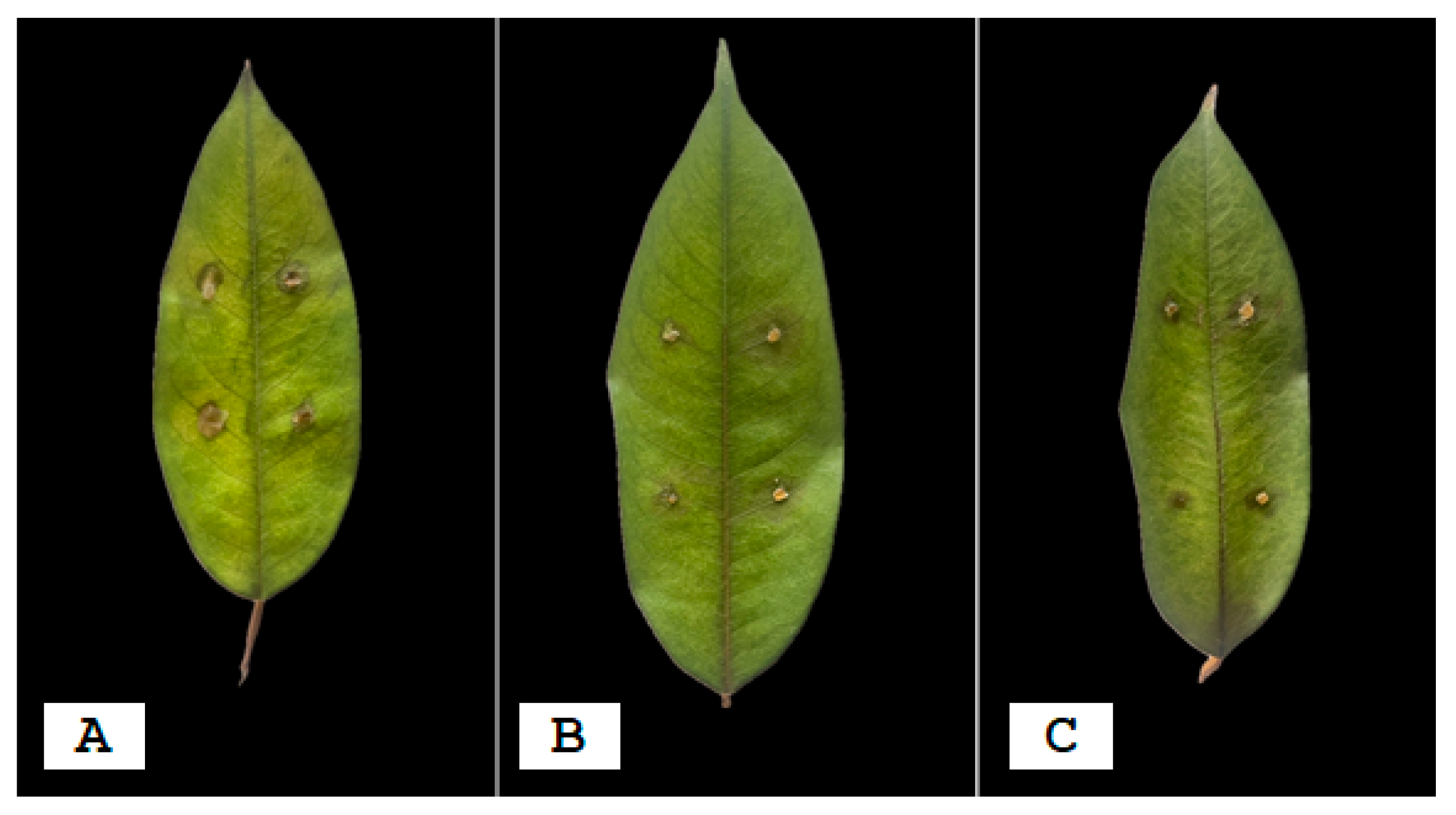
2.2.1. Detached Leaf Technique

To confirm the pathogenicity of the isolated pathogen (fungal and oomycetes strains), a detached leaf technique was used. The pathogens cultures on PDA were incubated at room temperature (28 ± 3 °C). Then, 3 strains with different morphologies were chosen for pathogenicity testing. The pathogens were selected by using the detached leaf technique, inoculating on 5 leaves/plant [15]. The leaves were washed with control treatments and dried. Then, a cork borer was used to cut 0.5 × 0.5 cm of the mycelium of pathogen cultured on PDA and placed on the leaves. The leaves were incubated in a controlled chamber at 100% relative humidity (%RH). The leaves were checked daily until disease symptoms were observed.

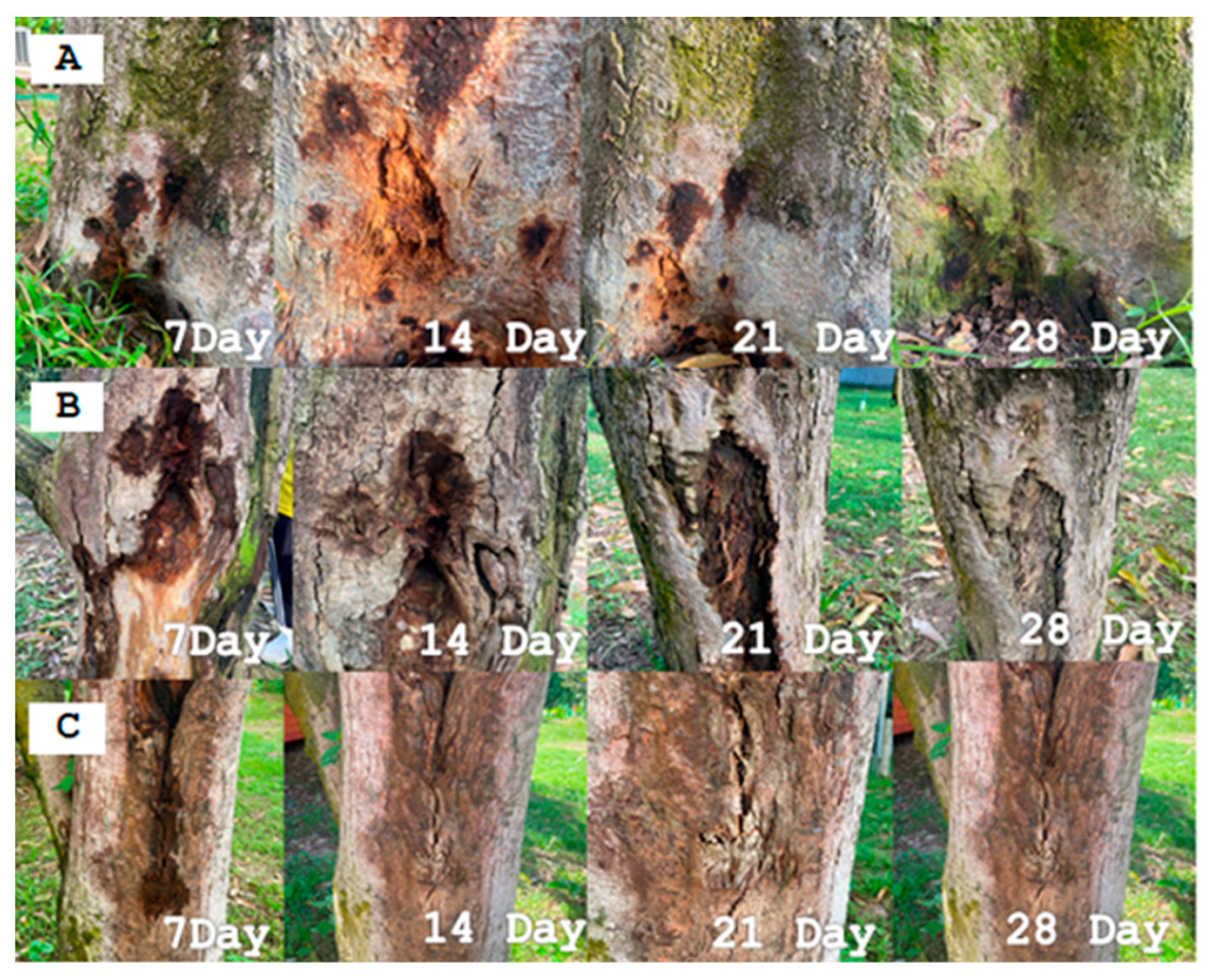
2.2.2. Test on Durian Trees

This experiment design was arranged with a CRD, and each treatment had three replications. The plants tested were 1-year-old durian trees. The first 3-pathogenic strain on the leaves from Section 2.2.1 were used in this experiment. The pathogens, including P. palmivora, Fusarium sp., and Pythium sp. were cultured on PDA for 7 d. The inoculum suspension of pathogens prepared at a concentration of 3.1 × 10⁸ spore/mL was inoculated, 20 mL/pot (pot size 12 inches), into the durian planting soil [16] compared with distilled water (Control treatment).

2.3. Isolation of Bacteria

The bacteria isolated from soil samples were obtained from the rhizosphere of durian plantations showing no apparent symptoms of root or stem rot. The 10 g of the soil was mixed in 90 mL of distilled water and then shaken at 120 rpm for 10 min to expose bacteria to the water; this was used as a stock suspension to dilute the concentration. The tenfold serial dilution method involved 100 μL of bacteria suspension spread over the surface of NGA [17] and incubated at room temperature (28 ± 3 °C) for 24–48 h. The colonies of bacteria grown on the NGA were selected and purified for further experiments.

2.4. Testing the Efficacy of Isolated Bacteria in Inhibiting the Growth of the Mycelium of 3 Pathogens

The ability of beneficial bacteria to inhibit the growth of P. palmivora was tested using the Dual culture method [18]. The experiment was designed using a CRD with 3 replications. The procedure was as follows: P. palmivora strain MNP13, Fusarium sp. strain FS4, and Pythium sp. strain PY2 was used, and hyphal tips were cut using a cork borer. The hyphal tips of pathogens were placed at the center of a PDA plate (9 cm in diameter). Beneficial bacteria were cultured on NGA for 24–48 h, streaked on the PDA plate opposite the pathogen plug, maintaining a 2 cm distance from the edge of the plate for 7 d (28 ± 3 °C). The efficiency of the beneficial bacteria was compared with two high quality bacterial strains, B. megaterium strain KP14 is capable of controlling Fusarium sp., the causal agent of yellow leaf spot disease in cannabis [19], Bacillus subtilis strain TU-089 exhibited plant growth-promoting and disease controlling properties [18], ddH2O, and metalaxyl.
The efficacy of biological control agents was evaluated by measuring the radial growth of pathogen colonies, including P. palmivora, Fusarium sp., and Pythium sp. The percentage inhibition of radial growth (PIRG) was calculated using the following formula [20]:
PIRG (%) = [(R1 − R2)/R1 × 100]
  • R1 = Radius of the fungal and oomycetes colony on the control plate
  • R2 = Radius of the fungal and oomycetes colony on the test plate

2.5. Physiological, Biochemical, and Genetic Characterization of the Bacterial Isolates

To comprehensively identify and characterize the bacterial isolates obtained from cultivation soil, leaf, and root of durian, a series of physiological, biochemical, and molecular analyses were conducted.

2.5.1. Physiological

A Gram reaction test was conducted using the potassium hydroxide (KOH) method. A clean microscope slide was prepared with a drop of 3% KOH solution. A sterile inoculating loop was used to transfer a bacterial colony grown on nutrient glucose agar (NGA) incubated at room temperature for 24 h. The bacterial sample was mixed thoroughly with the KOH solution on the slide to observe for viscosity or mucoid string formation, indicating a Gram-negative reaction [21].

2.5.2. Biochemical

The biochemical properties test of the beneficial bacteria was studied and analyzed to classify and identify microbial strains. The Vitek 2 Compact System was utilized for biochemical testing to identify microbial species. Testing conditions were conducted under controlled conditions at a temperature of 37 °C. The biochemical profiles obtained through this method were used to classify and identify the microorganisms [22] accurately.

2.5.3. Molecular Analyses

The genetic identification of beneficial microorganisms was performed using colony PCR to amplify the 16S rRNA gene from each bacterial isolate. Universal primers (Sigma-Aldrich Chemie, St. Louis, MO, USA) were used for amplification [23]. PCR products were separated by agarose gel electrophoresis (1.5% agarose), stained with ethidium bromide, and visualized under a UV transilluminator. The expected band sizes were approximately 1000 base pairs (F27 and F984′ primers) and 500 base pairs (F984 and R1492 primers). The complete 16S rRNA gene sequences of all samples were submitted for sequencing at Macrogen Inc. (Seoul, Republic of Korea). The obtained sequences were compared to reference sequences in the GenBank database using the BLASTN version 267.0 program provided by the National Center for Biotechnology Information (NCBI) for species-level identification [23].

2.6. Testing the Efficacy of 3-Selected Bacterial Strains Against P. palmivora in the Field

The selection of effective bacteria for controlling root and stem rot disease was tested in a field experiment using a Randomized Complete Block Design (RCBD). Beneficial bacteria grown in NGB medium (at a concentration of approximately 108 CFU/mL) were applied to durian trees showing symptoms of root and stem rot in The Mai District, Chanthaburi province, Thailand. Each treatment was replicated 5 times as follows: T1 = Sprayed with ddH2O (Control treatment), T2 = Sprayed with fresh cell NJTU05, T3 = Sprayed with fresh cell NJTU10, T4 = Sprayed with fresh cell NJTU13, T5 = Sprayed with metalaxyl 80% WP (recommended dose). The beneficial bacteria were applied by spraying around the canopy area of each durian tree at a rate of 100 L per tree, calculated based on the soil volume (width × length × depth: 8 × 8 × 0.15 = 9.6 m3). Applications were performed at 7-d intervals, 5 times (Day 0, 7, 14, 21, and 28). The development of lesions on the test trees was monitored and compared before and after the application of the bioproduct. Disease severity was assessed using [24] with modifications suitable for this experiment. Disease severity (%) levels; Level 0: No symptoms of root and stem rot (Resistant), Level 1: Rot present on 1–25% of the lesion area, with small, closed lesions and no further lesion development (moderately resistant), Level 2: Rot present on 25–50% of the lesion area, with lesion development not exceeding 1× the original size (Moderately susceptible) and Level 3: Rot present on >50% of the lesion area, with open lesions, visible spore powder and continuous spreading across the tree (Susceptible).

3. Results and Discussion

3.1. Isolation Pathogens Causing Root and Stem Rot in Durian

A total of 30 durian tree samples exhibiting symptoms of root and stem rot were collected. From these, 17 isolates of P. palmivora were successfully obtained. The samples were collected from durian orchards in Chanthaburi province, specifically from three districts. Makham District had 4 isolates: MNP11, MNP13, MNP19, and MNP20. Tha Mai District had 6 isolates: TNP01, TNP02, TNP03, TNP04, TNP17, and TNP18. Khao Khitchakut District had 7 isolates: KNP05, KNP06, KNP07, KNP09, KNP10, KNP16, and KNP21. Based on spore morphology, 26 isolates were assigned to P. palmivora, Pythium sp., and Fusarium sp., with 17, 2, and 7 strains, respectively. In addition, a total of 17 strains of P. palmivora were isolated based on colony characteristics and were classified into 3 groups: (1) Radial morphology groups had 4 isolates, (2) Chrysanthemum morphology groups had 5 isolates, and (3) Rosaceous morphology groups had 8 isolates (Figure 1 and Table 1). Colony growth rates on PDA were also measured by recording the diameter of pathogen colonies. Growth rates varied among the isolates: MNP13 showed the fastest growth on PDA within 3 d, KNP21 followed with optimal growth in 7 d, and TNP05 exhibited significant growth in 9 d. Microscopic examination of P. palmivora isolate MNP13 under 40× magnification revealed ovoid-shaped sporangia with an average size of 6.23 × 10.41 µm (Figure 2A) [14]. In comparison with previous studies, This finding aligns with research conducted by Kongcharoensunthorn et al. [23], which characterized P. palmivora isolated from 100 samples collected from durian plantations across 5 provinces and identified 57 isolates of P. palmivora, which were categorized into 5 colony types: uniform, radial, stellate, chrysanthemum, and rosette. The pathogenicity tests of P. palmivora 18 isolates revealed varying degrees of disease severity, demonstrated by water-soaked lesions on inoculated durian leaves. The colony Pythium sp. was flower shaped with white fibers. Similar to oospore powder and has a thin round wall (Figure 2B). Growth rates. The isolate PY2 grew on PDA within 3 d, whereas PY1 died. The colony Fusarium sp. was initially creamy-white and then changed to yellow-white as it aged. Macroconidia are short-curved-clear with 3–4 septum, while microconidia are oval (Figure 2C). Growth rates varied among the isolate FS4 showed the fastest growth on PDA within 3 d, FS1 and FS2 followed with optimal growth in 5 d, and FS7 exhibited significant growth in 6 d, whereas Fusarium sp. FS3, FS5, and FS6 did not grow on the PDA media [14].

3.1.1. Plate Assay with Detached Leaf Technique

The ability of P. palmivora isolates MNP13, KNP21 and TNP04, Pythium sp. isolates PY1 and PY2, Fusarium sp. isolates FS1, FS2, and FS4 to cause disease on durian leaves was tested. The results showed that isolates MNP13, PY2, and FS2 caused infection the fastest, with symptoms appearing within 3 d. Isolates FS1, KNP21, FS4, and TNP05 caused infection within 5, 7, 9 and 14 d, respectively. When comparing infection with and without leaf wounds (Figure 3), leaves with wounds developed disease more rapidly than those without. This is because pathogen can more easily penetrate and infect durian leaves through wounds. Among the isolates tested for pathogenicity, MNP13 demonstrated the highest ability to cause root and stem rot disease, with the fastest infection observed within 3 d. For the pathogenicity of Pythium sp. PY2 and Fusarium sp. FS4 demonstrated the highest ability to cause disease. This finding aligns with the research conducted by [25], which compared the infection of P. palmivora on durian leaves with and without wounds. The study found that durian leaves with wounds exhibited a faster rate of disease development compared to unwounded leaves. This was because P. palmivora could more easily penetrate and infect the leaves through wounds research highlights that durian trees with wounds caused by harvesting or natural damage are more susceptible to infection by pathogens, allowing diseases to develop more easily and rapidly [13].

3.1.2. Plant Assay Test on Durian Trees

The severity of P. palmivora strains TNP04, KNP21, and MNP13, along with Pythium sp. isolates PY2 and Fusarium sp. isolates FS1, FS2, and FS4 on 1-year-old durian trees, demonstrated significant pathogenicity. Specifically, P. palmivora showed 56%, 73%, and 87% of the trees exhibiting severe symptoms within 14 d, while Pythium sp. affected 43%, and Fusarium sp. affected 51%, 54%, and 49%, respectively, compared to the control treatment with ddH2O. The affected trees showed symptoms of severe root rot accompanied by a slimy texture, a foul odor, and yellowing leaves that eventually fell off. After 14 d of inoculation, trees infected with MNP13 displayed 87% symptom severity. Symptoms included yellowing and dropping of leaves, and the trunk bark exhibited dark brown discoloration with a foul odor (Figure 4). The soil exhibited high humidity (73%RH). Disease symptoms on root and trunk sections were further tested on PDA at room temperature for 7 d, confirming the pathogen as P. palmivora strain MNP13. Among the 3 tested strains of P. palmivora, strain MNP13 exhibited the highest ability to cause root and stem rot disease in durian trees. The disease symptoms observed in this study aligned with the report from the Agricultural Technology Promotion Center for Plant Protection, Suphan Buri Province, Thailand (2024), which identified P. palmivora as the causative agent of root and stem rot in durian. According to the report, the characteristic symptoms of root and stem rot caused by P. palmivora include the following. When excavated, fibrous roots exhibit rot symptoms, with the bark peeling off and appearing soft and brown. Branches and leaves: some branches displayed yellowing leaves, while the bark of the trunk and branches showed water soaked spots resembling water stains. This consistency in symptoms further supported the identification of P. palmivora as the primary pathogen responsible for the observed disease in durian trees [25].

3.2. Isolation of Beneficial Bacteria

Isolation from various samples around durian trees, including the leaves, soil surrounding the roots, and organic debris beneath the trees, revealed a diverse population structure of beneficial bacteria. A total of 196 isolates were obtained, categorized into eight groups based on their morphological characteristics, Groups A to H (Table 2). All bacterial groups were Gram-positive except for Group B, which was the only Gram-negative group.

3.3. The Efficacy of Beneficial Bacteria Inhibited the Mycelium Pathogen Caused Durian Root and Stem Rot

The efficiency testing of beneficial bacteria in inhibiting the growth of P. palmivora strain MNP13 causal agent of root and stem rot is NJTU05 showed the highest inhibition at 77.96 ± 1.39% (1.56 cm diameter of colony), followed by NJTU13 at 76.48 ± 1.83% (1.63 cm), and NJTU10 at 63.70 ± 2.50% (3.4 cm). Additionally, NJTU05 effectively inhibited Fusarium sp. strain FS2 and Pythium sp. strain PY2 with 76.67% (2.1 cm) and 53.70% (4.16 cm), respectively (Table 3). These results were similar to Agricultural Research Development Agency and Swiatczak et al. [26,27]. The antagonistic effects of bacteria like Brevibacillus laterosporus in controlling plant pathogens, including Phytophthora capsica, Rhizoctonia solani, and Fusarium oxysporum, were reported.

3.4. Classification of Beneficial Bacteria

3.4.1. The Beneficial Bacteria Was Identified and Classified

The test and identification of the beneficial bacterial strain NJTU05 were conducted using the Vitek 2 Compact biochemical system (bioMérieux SA, Marcy-l’Étoile, France) at 37 °C. The results revealed that NJTU05 was a Gram-positive (+ve) bacterium capable of producing various enzymes, including leucine arylamidase, β-N-acetyl-glucosaminidase, Ala-Phe-Pro arylamidase, Ellman, and glycine arylamidase, etc. (Table 4). When compared with the database, it was found to be similar to Brevibacillus formosus with a 99.70% identity.

3.4.2. Identification of Microorganisms Using Genetic Information

The identification and analysis of the beneficial microorganism strain NJTU05 were performed using 16S rRNA gene sequencing with specific primers Forward: 5′-GCTATCACTGGGAGATGGGC-3′, Reverse: 5′-CCGGGCTTTAACACCAGACT-3′ (Tm = 59.96 °C). A DNA band of approximately 1465 base pairs was observed under a UV transilluminator. The 16S rRNA sequence of B. formosus strain NJTU05 (1465 bp) was compared with the NCBI database, revealing 99.70% similarity to B. formosus and 96, 97, and 98% identity to B. formosus strain SARR8, Brevibacillus sp. strain RJ-7-4, and Brevibacillus porteri strain HB 1.2, respectively, using the BLAST program (Figure 5). The 16S rRNA gene sequence of the isolated Brevibacillus brevis strain NJTU05 was deposited in the GenBank database under the accession number PV809671.

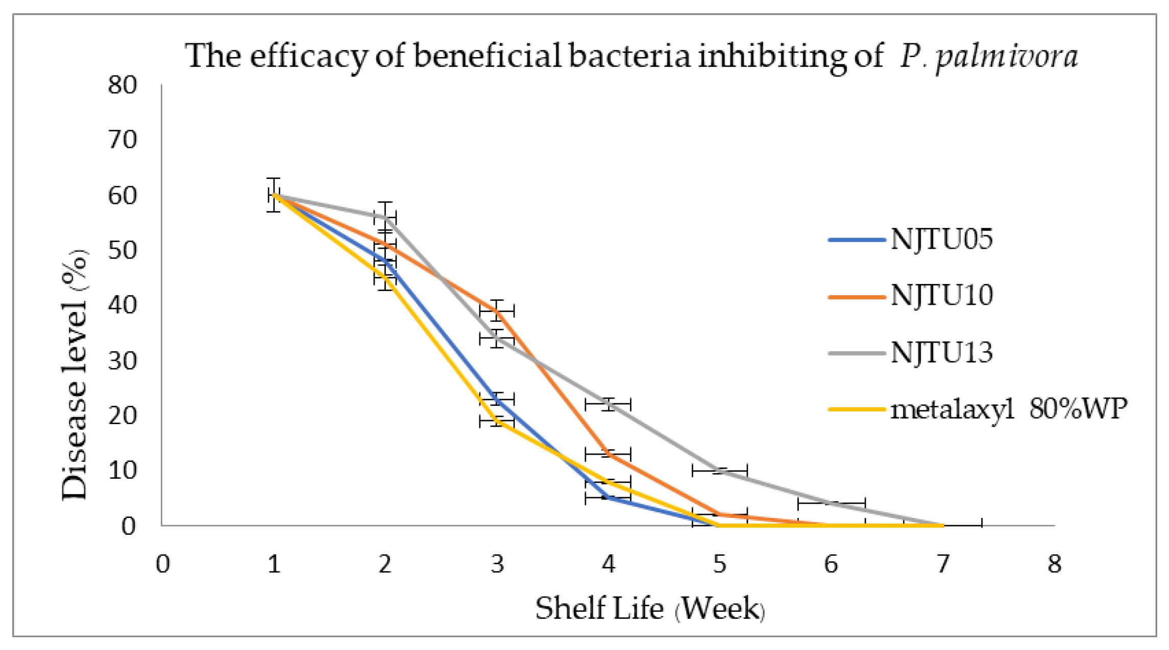
3.5. Efficacy of Three Selected Bacterial Strains Against P. palmivora in the Field

The efficacy of beneficial bacterial strains NJTU05, NJTU10, and NJTU13 was compared with metalaxyl 80% WP (chemical treatment) and ddH2O as the control treatment. NJTU05 and NJTU10 showed the highest efficiency, with inhibition percentages of 73.33% and 53.33%, respectively, followed by NJTU13 with 46.66% inhibitory efficiency p ≤ 0.05). This finding aligned with research conducted by Zhao et al. [28], which found that bacteria from the Brevibacillus laterosporus group, isolated from hot springs, could inhibit plant pathogenic fungi such as P. capsici, R. solani, and F. oxysporum by 96.55%, 80.17%, and 26%, respectively.
The observations before and after applying NJTU05 showed the following: Day 0 (control): The durian trees exhibited a severity level of 3 (susceptible) (disease 60%), with symptoms including watery, brownish necrosis at the base of the trunk and a foul odor. Day 7: After applying the beneficial treatment by spraying the wounds and applying it to the soil, the wounds still had a watery appearance and a foul odor and exhibited severity level 2 (moderately susceptible) (disease 48%). Day 14: The condition of the wounds improved, with less waterlogging and no foul odor. Exhibited to severity level 2 (moderately susceptible) (disease 23%). Day 21: The wounds were drier, with new wood formation and no foul odor exhibited, to a severity level 1 (moderately resistant) (disease 5%). Day 28: The wounds were dry with slight cracks from the original injury and new wood formation. The weather conditions during the test ranged from a minimum humidity of 58% RH to a maximum of 71% RH, with an average temperature of 32–35 °C. The durian tree was healthy with green leaves, and no signs of root and crown rot were exhibited, to a severity level 0 (resistant) (disease 0%) (Figure 6). Comparison of B. formosus strain NJT05 effectively reduced the disease severity from susceptible to resistant within weeks 4, faster than NJTU10 and NJTU13, results in weeks 5 and 6, respectively (Figure 7). This finding aligns with research conducted by Sivakorn et al. [29], which investigated the use of antagonistic microorganisms to control root and stem rot in durian using B. subtilis WD20. They found that treatments with B. subtilis WD20, applied 4 times and once by injection, resulted in the recovery of the diseased tissues to regular wood, with improved tree health and vibrant green leaves.

4. Conclusions

The causative agent of root and stem rot disease was isolated into multiple isolates, which were grouped into three main groups: P. palmivora isolates TNP05, MNP13, and KNP21, all collected from Chanthaburi province. Examination of spore morphology under a compound microscope (40× magnification) revealed distinct spore shapes for each isolate TNP05 (Obpyriform), MNP13 (Limoniform), and KNP21 (Ovoid-obpyriform). Additionally, 2 and 7 isolates were suspected to be Pythium sp. and Fusarium sp., respectively. The pathogenicity tested on durian leaves and trees demonstrated that P.palmivora strain MNP13 had the highest ability to cause root and stem rot disease, making it the most virulent strain among the tested isolates. This information is critical for understanding the disease dynamics and guiding future control strategies, Pythium sp. strain PY2 and Fusarium sp. strain FS4 also have the highest ability to cause root and stem rot disease.
A total of 196 strains of beneficial bacteria were isolated and classified into eight groups based on colony characteristics. Among these, the three most effective strains, NJTU05, NJTU10, and NJTU13, were identified for their high inhibitory percentages against root and stem rot disease in durian. These strains demonstrated efficacy comparable to the chemical control process using metalaxyl under laboratory and field conditions. The results demonstrated that beneficial bacterial strains NJTU05, NJTU10, and NJTU13 effectively inhibited the growth of P. palmivora isolates MNP13, the causative agent of root and stem rot in durian. Among these, NJTU05 was identified as the most effective strain with the highest inhibitory percentage (76.66%) in laboratory testing, comparable to the chemical control metalaxyl 80% WP). Additionally, NJTU05 effectively inhibited Fusarium sp. and Pythium sp. at 76.67 and 53.70%, respectively. Further analysis using biochemical tests and 16S rRNA sequencing confirmed that the beneficial bacterial strain NJTU05 was closely related to Brevibacillus formosus with 99.70% identity. The efficacy of beneficial bacteria under field trials in Chanthaburi province revealed that strain NJTU05 reduced disease severity and transformed durian plants from susceptibility to resistance within four weeks, outperforming NJTU10 (5 weeks) and NJTU13 (6 weeks). The inhibition percentages under field trials were NJTU05: 73.33%, NJTU10: 53.33%, and NJTU13: 46.66% (p ≤ 0.05).
These findings establish B. formosus strain NJTU05 as a promising biological control agent for managing root and stem rot disease in durian, offering an effective, and sustainable alternative to chemical fungicides.

Author Contributions

Conceptualization, W.C.; methodology, D.A. and N.J.; writing—original draft preparation, P.D. All authors have read and agreed to the published version of the manuscript.

Funding

The Thammasat University Research Fund, Contract No. TUGR 2/14/2019 and Thailand Science Research and Innovation Fundamental Fund fiscal year 2024, Thammasat University.

Data Availability Statement

All available data are contained within the article.

Acknowledgments

This study was supported by Thammasat University Research Fund, Contract No. TUGR 2/14/2019 and Thailand Science Research and Innovation Fundamental Fund fiscal year 2024, Thammasat University.

Conflicts of Interest

The authors declare that this study received funding from the Thailand Science Research and Innovation Fundamental Fund and the Thammasat University Research Fund. The funder was not involved in the study design, collection, analysis, interpretation of data, the writing of this article, or the decision to submit it for publication.

References

  1. Durian, the King of Thai Fruits, Is Loved by Foreigners. Available online: https://tpso.go.th/en (accessed on 13 February 2025).
  2. Angiosperm Phylogeny Group. An update of the angiosperm phylogeny group classification for the orders and families of flowering plants: APG IV. Bot. J. Linn. Soc. 2016, 181, 1–20. [Google Scholar] [CrossRef]
  3. Department of Agricultural Extension. Durian Production Statistics 2022. Ministry of Agriculture and Cooperatives. 2023. Available online: https://www.example.com (accessed on 16 June 2025).
  4. Ministry of Agriculture and Cooperatives. Durian Development Strategic Plan (2023–2027). 2023. Available online: https://www.moac.go.th (accessed on 16 June 2025).
  5. Saradee Story. Export Statistics of Thai Durian in 2021 and 2022. 2022. Available online: https://saradeestory.com/story/durian-exports64vs65/ (accessed on 29 June 2025).
  6. Suksiri, S.; Laipasu, P.; Soytong, K.; Poeaim, S. Isolation and identification of Phytophthora sp. and Pythium sp. from durian orchard in Chumphon province, Thailand. Int. J. Agric. Technol. 2018, 14, 389–402. [Google Scholar]
  7. Root Rot and Foot Rot. Available online: https://esc.doae.go.th (accessed on 18 January 2025).
  8. Jitfour, N.; Athinuwat, D.; Chuaboon, W. Brevibacillus formosusstrain NJTU05 powder formulation bioproduct to control root and stem rotdisease of durian. KKU Res. J. 2025, 53, 479–490. [Google Scholar]
  9. Pengnoo, A.; Lohlaeh, U.; Maduerehand, F.; Kaewmano, C.; Krualee, S.; Wongpisal, P.; Homhaul, W.; Boonyapipat, P.; Saeng-ngam, S.; Äkbärjan, A.; et al. Bacillus velezensis SM1: A promising biocontrol solution for Phytophthora durian root rot. Appl. Microbiol. 2025, 5, 21. [Google Scholar] [CrossRef]
  10. Danial, N.D.; Matthews, S.; Amaran, N.F.; Abd Kharim, N.A. Assessment of Streptomyces sp. and Bacillus sp. against Phytophthora palmivora, the causal agent of durian canker disease through different application methods at the nursery stage. J. Agrobiotechnol. 2024, 15, 18–24. [Google Scholar] [CrossRef]
  11. Detmanee, P.; Athinuwat, D.; Chueaboon, W. Characteristics and Efficacy of Antagonistic Microorganisms in Controlling Phytophthora palmivora, the Causal Agent of Root and Stem Rot in Durian. In Proceedings of the 15th National Plant Protection Conference, Rama Gardens Hotel, Bangkok, Thailand, 22–24 November 2022. [Google Scholar]
  12. Chueboon, W. Characteristics and Effectiveness Testing of Beneficial Bacteria for Controlling Erwinia carotovora subsp. Carotovora, the Cause of Soft Rot. Master’s Thesis, Kasetsart University, Bangkok, Thailand, 2008. [Google Scholar]
  13. Raza, W.; Ghazanfar, M.U.; Ullah, Z. Isolation and morphological characterization of Phytophthora palmivora on PDA medium. Plant Pathol. J. 2022, 38, 215–223. [Google Scholar]
  14. Salvamani, S.; Nawawi, N.M. Macroscopic and Microscopic Approaches for Identification of Fungi from Plant Soil of Cameron Highlands. Bioremediat. Sci. Technol. Res. 2014, 2, 14–18. [Google Scholar] [CrossRef]
  15. Vawdrey, L.; Martin, T.M.; De Faveri, J. A detached leaf bioassay to screen durian cultivars for susceptibility to Phytophthora palmivora. Australas. Plant Pathol. 2005, 34, 251–253. [Google Scholar] [CrossRef]
  16. Chatchaisiri, L.; Pongpisutta, R.; Rattanakreetakul, C.; Bincader, S. Pythium sp. Causing root of durian and efficacy of chemical fungicides to control. Agric. Sci. J. 2018, 49, 171–174. [Google Scholar]
  17. Datmanee, P.; Suknant, T.; Srisuwan, K.; Chuaboon, W. Isolation beneficial bacteria degrade heavy metal (Arsenic). In Proceedings of the 8th Academic Science and Technology Conference, Patumthani, Thailand, 26 March 2021. [Google Scholar]
  18. Somjinda, S.; Athinuwat, D.; Chueboon, W. Effects of Beneficial Bacteria in Controlling Colletotrichum capsici the Cause of Anthracnose in Chili Peppers. In Proceedings of the 10th RMUT Surin National Conference on Research and Innovation for Sustainable Development, Surin, Thailand, 19–20 September 2019. [Google Scholar]
  19. Jaiyen, K.; Laitalb, A.; Athinuwat, D.; Chueaboon, W. Selection and Testing of Beneficial Microorganisms for Controlling Major Diseases in Cannabis. In Proceedings of the 1st National Conference on Cannabis, Hemp, and Kratom, Nakhon Pathom, Thailand, 20 April 2023. [Google Scholar]
  20. Rahman, M.A.; Begum, M.F.; Alam, M.F. Screening of Trichoderma isolates as a biological control agent against Ceratocystis paradoxa causing pineapple disease of sugarcane. Mycol. Res. 2009, 37, 277–285. [Google Scholar]
  21. Farmer, K.; James, A.; Naraine, R.; Dolphin, G.; Sylvester, W.; Amadi, V.; Kotelnikova, S.V. Urinary tract infection Escherichia coli is related to the environmental Escherichia coli in their DNA barcoding and antibiotic resistance patterns in Grenada. Adv. Microbiol. 2016, 6, 33–46. [Google Scholar] [CrossRef]
  22. Valenza, G.; Ruoff, C.; Vogel, U.; Frosch, M.; Horn, M.A. Microbiological evaluation of the new VITEK 2 Neisseria-Hemophilus identification card. J. Clin. Microbiol. 2007, 45, 3493–3497. [Google Scholar] [CrossRef] [PubMed]
  23. Kongcharoensunthorn, W.; Treyasinha, R. PCR for screening of multidrug resistant Acinetobacter baumannii by MexA, MexB like gene, and 16–23S rRNA intergenic spacer region (ISR). BUU Sci. J. 2021, 21, 10–16. [Google Scholar]
  24. Guidelinrs for Surveillance for Plant Pests in Asia and the Pacific. Available online: https://www.aciar.gov.au/sites/default/files/legacy/node/2311/mn119_guidelines_for_surveillance_for_plant_pests__16233.pdf (accessed on 13 February 2025).
  25. Phongsuthat, N.; Phankamolsil, N.; Moonjuntha, P.; Amawan, S.; Intanoo, W.; Unartgam, J. Pathogenicity of cassava rot associated fungi and genetic variation of fungi. Thai Agric. Res. J. 2022, 40, 238–250. [Google Scholar]
  26. Agricultural Research Development Agency (ARDA). “Remove Eastern Durian Lessons 2022 for the Future Opportunities,” and New Challenges” and Training “Management and Control of Dry Twig and Root Rot Disease to Quality Durian Product”. Available online: https://en.arda.or.th/newlist-detail.php?id=990 (accessed on 13 February 2025).
  27. Swiatczak, J.; Kalwasinska, A.; Wojciechowska, A.; Brzezinska, M.S. Physiological properties and genomic insights into the plant growth promoting rhizobacterium Brevibacillus laterosporus K75 isolated from maize rhizosphere. J. Sci. Food Agric. 2023, 103, 1432–1444. [Google Scholar] [CrossRef] [PubMed]
  28. Zhao, J.; Guo, L.; Zeng, H.; Yang, X.; Yuan, J.; Shi, H.; Xiong, Y.; Chen, M.; Han, L.; Qiu, D. Purification and characterization of a novel antimicrobial peptide from Brevibacillus laterosporus A60. J. Pept. Sci. 2012, 33, 206–211. [Google Scholar] [CrossRef] [PubMed]
  29. Sivakorn, N.; Trakul-Sukrat, P.; Songsaeng, P.; Worakuldamrongchai, S. Control of Durian Root and Crown Rot Diseases Using Bioagents Derived from Bacillus Subtilis; Plant Protection Research and Development Office: Bangkok, Thailand, 2011. [Google Scholar]
Figure 1. Pathogens isolated around durian trees showing symptoms of root and stem rot disease from Chanthaburi province include the stem (A), leaves (B), bark (C), and the succulent area of the durian trunk (D).
Ijpb 16 00075 g001
Figure 2. Colony morphology of P. palmivora MNP13 (A), Pythium sp. PY2 (B), and Fusarium sp. FS2 (C) on PDA medium at 7 ds and spore morphology under a microscope at 40× magnification (ac).
Ijpb 16 00075 g002
Figure 3. Pathogen ability caused disease on leaves was P. palmivora isolates MNP13 (A), KNP21 (B), and TNP04 (C) caused lesions on durian leaves.
Ijpb 16 00075 g003
Figure 4. Show soft rot symptoms of durian inoculated with P. palmivora MNP13 after 14 d root (A), stem (B), and root in soil (C).
Ijpb 16 00075 g004
Figure 5. The 16S rRNA gene sequence of Brevibacillus formosus strain NJTU05 consists of 1465 base pairs. A phylogenetic tree based on 16S rRNA gene sequences was constructed to show the relationship between the bacterial isolate and related Brevibacillus species. The bacterial strain sequenced in this study, Brevibacillus formosus strain NJTU05, is indicated with a red star (★). The tree was constructed using the neighbor-joining method with 1000 bootstrap replications. Bootstrap values are shown at the branch nodes.
Ijpb 16 00075 g005
Figure 6. Efficacy after using beneficial bacteria strains of NJTU05 (A), NJTU10 (B), and NJTU13 (C) within 28 d.
Ijpb 16 00075 g006
Figure 7. The efficacy of beneficial bacteria inhibiting P. palmivora, shows disease levels: Level 0: Resistant (0%), Level 1: moderately resistant (1–25%), Level 2: moderately susceptible (25–50%), and Level 3: Susceptible (>50%).
Ijpb 16 00075 g007
Table 1. Characteristics of P. palmivora on PDA Medium Incubated at Room Temperature (28 ± 3 °C).
IsolatesSampleLocation of IsolationCharacteristics
TNP01soilTha Mai, ChanthaburiRosaceous
TNP02leafTha Mai, ChanthaburiRosaceous
TNP03soilTha Mai, ChanthaburiRosaceous
TNP04rootTha Mai, ChanthaburiChrysanthemum
TNP17soilTha Mai, ChanthaburiChrysanthemum
TNP18soilTha Mai, ChanthaburiRadial
MNP11rootMakham, ChanthaburiChrysanthemum
MNP13soilMakham, ChanthaburiRadial
MNP19leafMakham, ChanthaburiRosaceous
MNP20rootMakham, ChanthaburiRosaceous
KNP05leafKhao Khitchakut, ChanthaburiChrysanthemum
KNP06soilKhao Khitchakut, ChanthaburiRadial
KNP07soilKhao Khitchakut, ChanthaburiRosaceous
KNP09rootKhao Khitchakut, ChanthaburiRosaceous
KNP10rootKhao Khitchakut, ChanthaburiChrysanthemum
KNP16leafKhao Khitchakut, ChanthaburiRosaceous
KNP21soilKhao Khitchakut, ChanthaburiRadial
Table 2. Beneficial bacteria isolated from various parts of durian plants.
GroupNameColonyGram Reaction
ANJTU01Round, convex, wavy edges, and milky white+
BNJTU02Round, convex, smooth edges, and dull yellow
CNJTU03Round, smooth surface, smooth edges, and opaque color+
DNJTU04Round, shiny, small smooth edges, and clear green+
ENJTU05Round, shiny, small smooth edges, and clear tan+
FNJTU10Round, smooth, and large wavy edges+
GNJTU13Round, with serrated edges, rough surface, and dark opaque color+
HNJTU14Cylinder, slightly wavy edge, and cloudy white+
+ = positive, − = negative.
Table 3. The efficacy of beneficial bacteria inhibited pathogen caused durian root and stem rot with the Dual culture method.
TreatmentInhibition of Pathogen (%)
P. palmivoraFusarium sp.Pythium sp.
Control (ddH2O)0.00 ± 0.00 d0.00 ± 0.00 d0.00 ± 0.00 d
B. subtilis strain TU-08968.52 ± 2.14 b57.22 ± 1.98 c22.59 ± 5.64 c
B. megaterium strain KP1471.30 ± 3.02 ab69.26 ± 5.26 b26.67 ± 5.11 c
NJTU0577.96 ± 1.39 a76.67 ± 0.45 a53.70 ± 1.89 b
NJTU1063.70 ± 2.50 c67.59 ± 0.94 b47.22 ± 3.93 b
NJTU1376.48 ± 1.83 a69.07 ± 4.72 b24.44 ± 7.63 c
metalaxyl75.19 ± 5.37 a73.89 ± 2.77 a77.22 ± 1.20 a
F-test***
C.V. (%)22.2723.4619.69
* Column mean ± standard deviation followed by different lowercase letters are significantly different at 95% confidence level by DMRT. Mean of * = significantly.
Table 4. Biochemical test results of beneficial bacteria strain NJTU05.
CharacteristicsReactions 1CharacteristicsReactions 1
Gram reaction+veD-mannitol
β-xylosidaseD-mannose
L-lysine arylamidaseD-melezitose
L-aspartate arylamidaseN-acetyl-D-glucosamine
Leucine arylamidase+Palatinose
Phenylalanine arylamidase+L-rhamnose
L-proline arylamidase+β-glucosidase
β-galactosidase+β-mannosidase
L-pyrrolidonyl arylamidase+Phosphoryl choline
α-galactosidasePyruvate
Alanine arylamidaseα-glucosidase
Tyrosine arylamidase+D-tagatose
β-N-acetly-glucosaminidase+D-trehalose
Ala-Phe-Pro arylamidase+Inulin
CyclodextrineD-glucose
D-galactoseD-ribose
GlycogenePutrescine assimilation
Myo-inositolGrowth in 6.5% NaCl
Methyl-α-D-glucopyranoside acidificationKanamycin resistance
Ellman+Oleandomycin resistance
Methyl-D-xylosideEsculin hydrolyse
α-monnosidaseTetrazolium red
MaltotriosePolymixin_B resistance
Glycine arylamidase+Glycine arylamidase+
1+ve=Gram positive bacteriaID Message Confidence level%Probability
+=Positive reactionExcellent96 to 99
=Negative reactionVery Good93 to 95
Disclaimer/Publisher’s Note: The statements, opinions and data contained in all publications are solely those of the individual author(s) and contributor(s) and not of MDPI and/or the editor(s). MDPI and/or the editor(s) disclaim responsibility for any injury to people or property resulting from any ideas, methods, instructions or products referred to in the content.

