Article

Evaluating Growth and Physiological Responses of a Medicinal Plant Phyla nodiflora to Salinity

1
Faculty of Biology and Environmental Science, The University of Danang—University of Science and Education, Da Nang 550000, Vietnam
2
Division of Integrative Bioscience and Biotechnology, College of Life Sciences, Sejong University, Seoul 05006, Republic of Korea
*
Author to whom correspondence should be addressed.
Int. J. Plant Biol. 2024, 15(1), 187-197; https://doi.org/10.3390/ijpb15010015
Submission received: 2 December 2023 / Revised: 17 December 2023 / Accepted: 18 December 2023 / Published: 12 March 2024
(This article belongs to the Topic Tolerance to Drought and Salt Stress in Plants)

Abstract

:
Phyla nodiflora is a valuable medicinal plant growing in coastal areas, hypothesizing its adaptability to salinity; however, it has not been investigated. This study, for the first time, elucidated responses in the growth of the shoots and its physiology to different soil salinity of 50–400 mM NaCl. The data showed that the shoot’s dry biomass was not affected by the salinity levels up to 100 mM, and it only decreased 33.50–56.33% compared to the control under 200–400 mM NaCl, indicating that P. nodiflora is a salt-tolerant plant that could survive under high salinity. In addition, the plant also had physiological responses which indicated its salt-induced injuries and adaptation to the salt stress. The chlorophyll a content was increased while the chlorophyll b remained unchanged under the salt stress. The proline and salt accumulation increased under the salinity, but the K+ and NO3 accumulation decreased. Moreover, increases in malondialdehyde and electrolyte leakage were observed, indicating salt-induced membrane damages. These responses suggested that the plant might evolve adaptive mechanisms to salinity. Our findings are useful information for further research in order to elucidate the salt-tolerant mechanisms and develop this plant for saline agriculture.

1. Introduction

Soil salinity, mainly caused by an abundant accumulation of NaCl in soils, is one of the environmental problems that adversely affect the growth and development of plants. Since most crops (>95%) are sensitive to salinity, and the global area of salt-affected lands tends to increase by climate changes and human activities, the agricultural productivity and sustainability in agriculture production are being threatened in the future [1,2]. Thus, both improving the salt tolerance of crops and developing alternative plants as new crops are considered to be potentially effective solutions for a sustainable agriculture on salt-affected areas (also referred as saline agriculture) [2]. To achieve this goal, a comprehensive understanding of salt tolerance mechanisms of salt-tolerant plants that can grow under high saline conditions (equivalent to above 80 mM NaCl) is required [3,4]. It is evidenced that salinity can inhibit the growth of plants by imposing two primary detrimental effects such as osmotic and ionic stress, in addition to disadvantageous changes in soil characteristics to plants [5,6]. High salt concentrations will reduce the water potential of a soil solution, leading to inhibition of the root’s water uptake that induces osmotic stress in plants. In addition, salinity will promote the entry of Na+ and Cl ions into cells, of which a high accumulation of these ions is harmful to cytosolic metabolisms. The osmotic and ionic stress also produce reactive oxygen species (ROS), causing oxidative effects on the integrity of membranes via lipid peroxidation and emembrane leakage [6,7]. Together, these effects of salinity interrupt physiological processes, such as respiration, photosynthesis, membrane transport, nutrient and water absorption, and homeostasis, thereby inhibiting cell elongation and division that result in a reduction in plant growth. Plants have different responses and tolerance mechanisms to salinity, depending on the plant species/varieties [5,8,9]. Various tolerance mechanisms, at cellular and tissue levels, are evolved in salt-tolerant plants for their survival under salt stress [1,5,10]. Shoots have a major contribution in plant’s biomass by its important functions in plant growth, particularly vegetables; thus, the effects of salinity on plants can be examined via changes in shoot’s traits in comparison to that under non-saline conditions.
Phylla nodiflora (syn. Lippia nodiflora) belongs to the Verbenaceae family and is a creeping herbaceous plant distributed in many countries around the world, such as Asia, Australia, Central-South America, the Mediterranean, and Africa [11,12]. The whole plant is used in traditional medicine to treat various diseases such as asthma, bronchitis, knee pain, gonorrhea, hemorrhoids, heart disease, hepatitis, and fever [13]. The plant extracts are also reported to have antibacterial, anticancer, anti-inflammatory, antidiabetic, hepatoprotective, and antioxidant activities [13,14]. In Taiwan, the aerial parts of P. nodiflora are also a common ingredient in herbal teas used for the prevention of inflammation, menstrual disorders, and infectious diseases [12]. In addition, it is also popularly used in landscaping and livestock feed [15]. It was found that P. nodiflora can grow on various ranges of soil types such as garden, field, wetland, and sandy soil [12,15]. Also, the plant commonly distributes in coastal saline areas, suggesting that it may have an adaptability to salinity; however, there has not been any studies on its capability and adaptive mechanisms to salinity.
Thus, the purpose of this study is to understand responses of P. nodiflora shoots to salinity in term of growth and physiological traits such as photosynthesis, cell-membrane integrity, ion homeostasis, and osmotic adjustment. These responses were examined based on changes in growth and physiological parameters of shoots such as biomass; contents of proline, chlorophylls (a and b), ions (Na+, K+, NO3, and Cl), and malondialdehyde (MDA); and electrolyte leakage (EL) of the shoots grown under different salt concentrations (50, 100, 200, and 400 mM NaCl). The findings will be useful information for further studies to understand salt tolerance mechanisms in the plant and for its application in saline agriculture.

2. Materials and Methods

2.1. Plant Material and Growth Conditions

P. nodiflora seeds were collected from the plants (Figure S1) that grow on a coastal sandy area (location: 15°59′13.6″ N; 108°16′27.2″ E) in Da Nang city (Vietnam) during spring-summer 2022. The seed was sown in a plastic tray filled up with a mixture of coco-peat, vermiculite, and perlite (ratio 2:1:1). Two-week-old seedlings were transplanted into a 0.5 L pot filled with the same mixture. The pots were placed in a growth chamber (CMP6010-Conviron, Winnipeg, MB, Canada) with growth conditions such as a temperature/photoperiod regime with 28 °C for 14 h light and 25 °C for 10 h dark; a relative humidity for 75%; and a light intensity for 10,000 lux. Irrigation with half-strength Hoagland nutrient solution (No. 2) was applied to the seedlings for 4–6 weeks prior to the salt treatments [16].

2.2. Salt Treatment

For the salt treatments, the young plants with four true-leaf pairs and uniform appearance were irrigated with the half-strength Hoagland nutrient solutions contained with 50, 100, 200, and 400 mM NaCl. The irrigation was carried out according to a procedure as described by [16]. In brief, the plants were irrigated with the salt solutions of increasing concentrations before the treatment of the designated level to avoid osmotic shocks. The treated plants were under the saline conditions for a total of 14 days after the onset of the 50 mM NaCl treatment. In each irrigation, the salt solutions were applied continuously until an out-flow occurred from the bottom with a minimum amount that is approximate to the pot’s volume, to maintain the soil salinity. The irrigation was repeated each day during the treatment period. For control, the plants were irrigated with the nutrient solution without NaCl. For the purpose of the study, plant growth was observed on day 14 of the treatment, which showed obvious effects of the salinity, and the physiological parameters were analyzed on day 7.

2.3. Biomass Measurement

The fresh weight (FW) of shoots (aerial part) of the plant were measured after removal from the treatments. Then, the samples were dried at 70 °C for 48 h in a drying oven prior to measuring the dry weight (DW) [16].

2.4. Determination of Chlorophyll Content

The content of chlorophyll a (chl a) and b (chl b) of fresh young leaves from the treatments were determined as described by a previous study [17]. The leaf samples (0.1 g) were homogenized with 10 mL of 80% acetone. The mixture was centrifuged at 5000 rpm for 10 min, and the supernatant extract was measured with an absorbance (A) at a 645 and 663 nm wavelength, using a spectrometer (Jasco V730 UV-VIS, Tokyo, Japan) for determining the chlorophyll contents. The content of chlorophylls was calculated with the equation below and expressed on the basis of FW (µg mg−1 FW).
Chl a (µg mL−1) = 12.25 × A663 − 2.79 × A645;
Chl b (µg mL−1) = 21.5 × A645 − 5.11 × A663;

2.5. Determination of Proline and MDA Contents

The proline content in the fresh young leaves was determined according to the method described by [18]. In brief, the leaf sample (50 mg) was homogenized in 2 mL of 3% sulfosalicylic acid and then centrifuged at 12,000× g, at 4 °C for 15 min. A reaction mixture consisting of the supernatant, ninhydrin acid, and acetic acid was incubated at 100 °C for 60 min. After quickly stopping the reaction by cooling on an ice box, the reaction mixture was mixed with 4 mL of toluene. The solvent fraction was measured with an absorbance at a 520 nm wavelength, and the proline content was calculated based on a prepared standard curve.
The MDA content of the fresh young leaves was determined according to the method described by [19]. In brief, the leaf sample (50 mg) was homogenized with 2 mL of 0.1% trichloracetic acid (TCA) and centrifuged at 12,000× g for 10 min. The supernatant was mixed with 0.5% thiobarbituric acid and incubated at 95 °C for 25 min. The reaction was quickly interrupted by cooling. After the centrifugation, absorbance of the supernatant was measured at a 532 and 600 nm wavelength. The MDA content was determined with an extinction coefficient of 155 mmol L−1 cm−1.

2.6. Determination of EL and Ion Content

The EL of young leaves was measured according to the method described by [16]. In brief, the leaf discs were detached from the plants and washed with 30 mL of deionized water in a 50 mL tube. Then, the samples were incubated with 30 mL of freshly deionized water, in darkness for 24 h at room temperature, before the electric conductance of incubating solution (EC1) was determined. Then, the solution’s electric conductance (EC2) was also determined after heating the sample in a water bath at 95 °C for 20 min. The EL was calculated with the ratio of EC1/EC2.
To determine the ion content, the dried leaf sample (ca. 50 mg) was finely ground using a mortar and pestle and soaked with a total 10 mL volume of deionized water at room temperature for at least 24 h [20]. The extract was centrifuged at 5000 rpm for 10 min and the supernatant was subjected to the ion content determination. For cations Na+ and K+, a suitable dilution of the supernatant using a 0.1% HNO3 solution was conducted and these ions were analyzed via the flame emission spectrophotometry method using an atomic absorption spectrophotometer (ZEEnit 700 P, Analytik Jena, Jena, Thüringen, Germany) [21]. For anions, the NO3 concentration was determined using the Brucine-sulfanilic acid method and the Cl concentration was estimated using the chloride titration method with a 0.025 N AgNO3 standard titrate. The ion concentration in the sample was calculated using a standard curve of prepared ion solutions, and the ion content was expressed on the basic of DW (mg g−1 DW).

2.7. Experimental Design and Statistical Analysis

Three plants were grown in a pot and each the treatment was repeated randomly with five pots (replicates). The data were expressed as mean values and standard deviations with α = 0.05. The statistically significant difference of parameters between the treatments was determined according to Tukey’s test with p-value ≤ 0.05. The statistical analyses were conducted using the R software (Rstudio Cloud version).

3. Results and Discussion

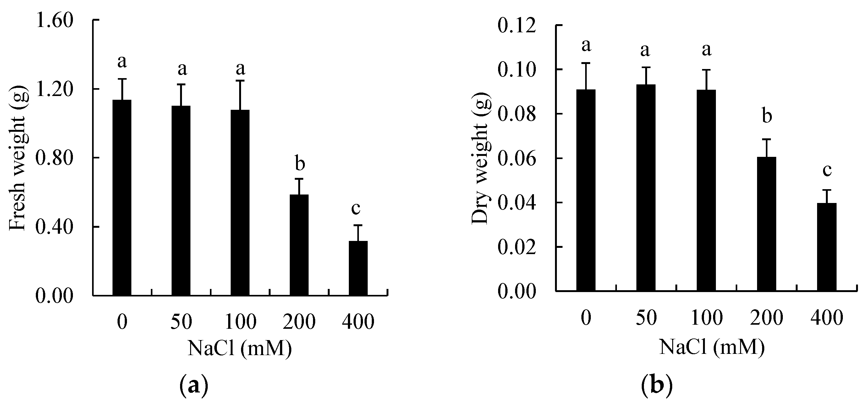
3.1. Effects of Salinity on Biomass of Shoots

The data showed that salinity had effects on the growth of P. nodiflora shoots depending on the salt concentration, which was expressed by the changes in biomass (Figure 1). Both FW and DW of the shoots were primarily unchanged under a salt concentration up to 100 mM, but declined with the higher salinity levels. The FW of shoots treated with 200 and 400 mM NaCl were 0.586 and 0.317 g, respectively, which were 48.39 and 72.11% lower than that of the control (1.135 g) (Figure 1a). Meanwhile, the decreases of 33.50 and 56.33% in the DW were observed for the shoots treated with 200 and 400 mM NaCl, of which the shoot DW were 0.060 and 0.040 g, respectively (Figure 1b). Although the shoot biomass was sharply decreased by 200 and 400 mM NaCl, the shoots still grew with the formation of new shoots and leaves (Figure S2). This result indicated that P. nodiflora had a tolerance to high salinity and could be considered as a salt-tolerant plant that could become a useful plant material for studies on salt tolerance mechanisms [4].
In addition, it is proposed that salt-tolerant plants with potential uses for medicine, forage, essential oils, and biofuels can be developed for saline agriculture because the biomass reduction is not always proportional to their quality [22,23]. P. nodiflora aerial parts are mainly used for medicinal purposes in folk medicine and the pharmaceutical industry [14]. Thus, its biomass maintenance at a concentration of 100 mM NaCl suggested that the plant has potential to be cultivated in salt-affected lands. It was also noticed that there were different responses between the fresh and dry biomass of shoots, of which the dry mater decreased less significantly than that of the fresh biomass (Figure 1). This observation suggested that high salinity reduced the leaf water content of P. nodiflora shoots [5,24].

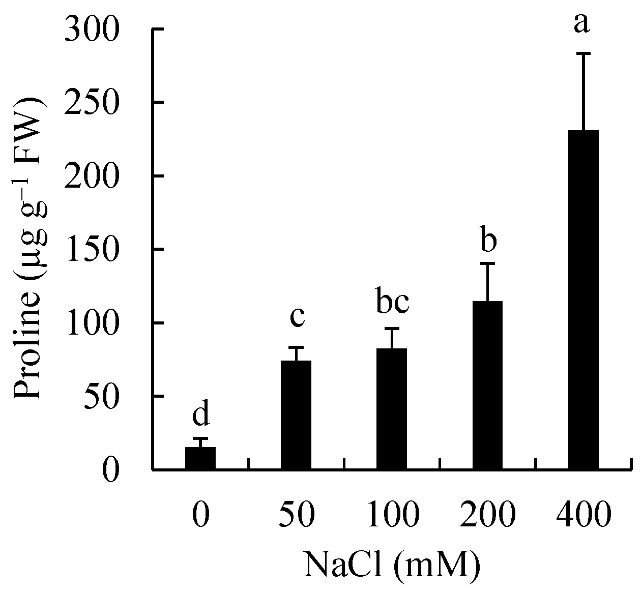
3.2. Effects of Salinity on Proline Accumulation

The data showed that the accumulation of proline in the salt-treated shoots was also gradually increased with the increasing concentrations of 50–400 mM NaCl. The proline content varied from 74.073–230.894 µg g−1 FW in the salt-treated shoots, which was 4.89–15.25 times higher than that of the control (15.144 µg g−1 FW) (Figure 2). The maximum accumulation of proline was 230.894 µg g−1 FW (15.25 times) in the 400 mM treated shoots. Proline plays a role as a compatible osmolyte that contributes to osmotic adjustment in the cytosol of plant cells. Previous reports showed that proline was highly accumulated in various plants for the osmotic adjustment, which retains the water uptake of plants cells under a lowered external water potential due to salt [25]. Our results suggested that P. nodiflora triggered an osmotic adjustment to maintain its water uptake and that proline might be involved in this response. In addition to being an effective osmolyte for osmotic adjustment in plants under saline conditions, proline is also considered to be an osmoprotectant that reduces damages of salt-induced ROS [26]. Due to its roles, the proline accumulation is considered to have a positive correlation to the salt tolerance capacity of plants. In case of P. nodiflora, there was an opposing trend, but in a similar pattern of change, between the shoot’s biomass (Figure 1) and the proline content under salinity (Figure 2), it suggests a positive correlation between the proline accumulation and the plant’s salt tolerance.

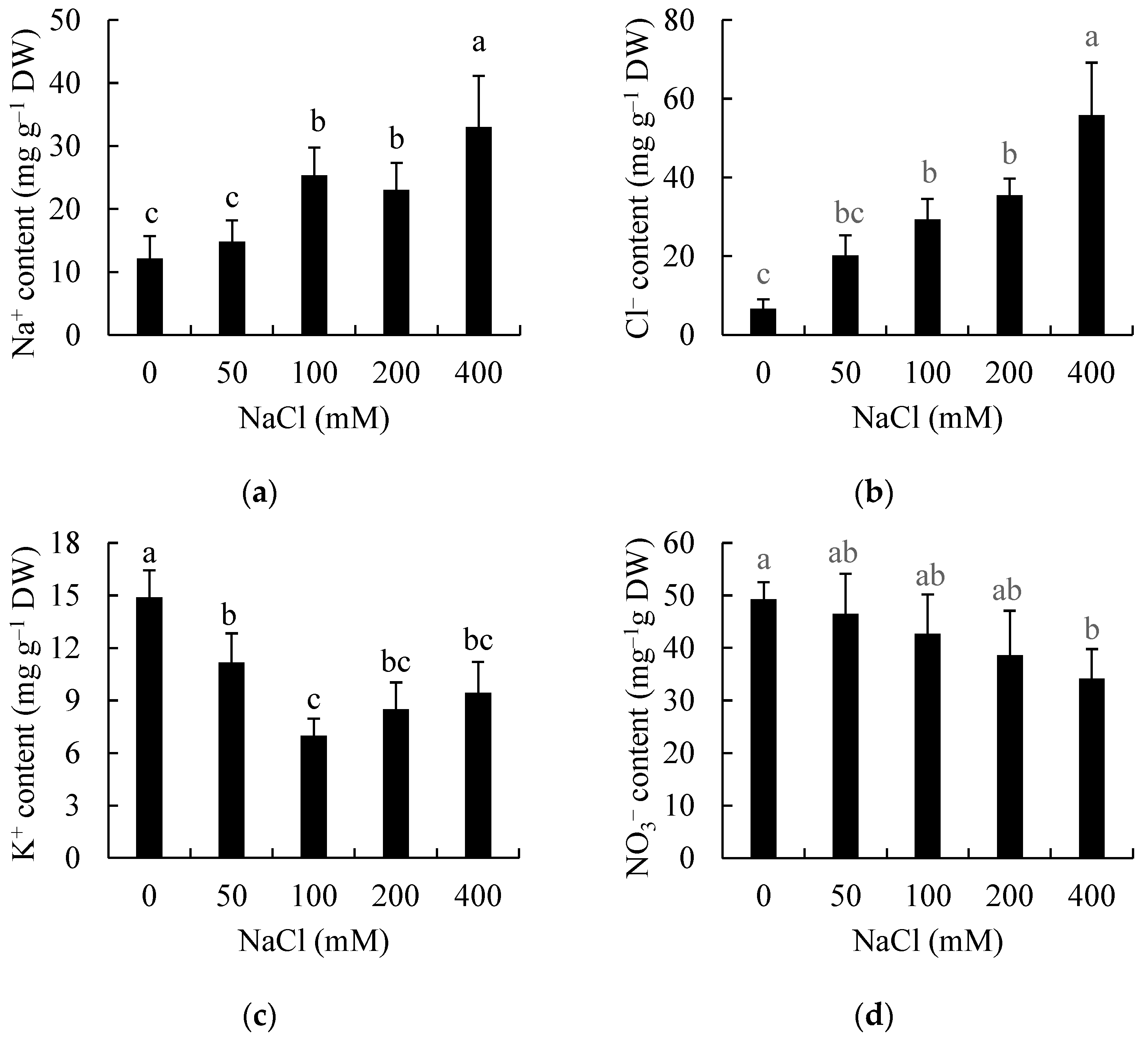
3.3. Effects of Salinity on Ion Accumulation

The data showed that the accumulation of all four ions Na+, K+, NO3, and Cl in P. nodiflora leaves varied between the salt treatments (Figure 3). An increasing trend of both the Na+ and Cl content in the plants treated with increasing salt concentrations was noticed, although the statistical significance was not obtained with 50 mM NaCl (Figure 3a,b). The Na+ content was 1.22 times (14.794 mg−1 DW) higher in the 50 mM NaCl-treated shoots than the control (12.148 mg g−1 DW), but it increased by 2.0 times (23.028–25.392 mg g−1 DW) in the shoots treated with 100–200 mM NaCl. Despite the lower Na+ content in the 200 mM NaCl-treated shoots than that in the 100 mM NaCl-treated shoots, there was no statistically significant difference between those. But, the Na+ content reached a maximum increase (2.72 times) in the 400 mM NaCl-treated shoots (Figure 3a). Similarly, the Cl content was increased, but with a higher level than that of the Na+ content, which was 3.05 times (20.228 mg g−1 DW) higher in the 50 mM NaCl-treated shoots than the control (6.635 mg g−1 DW), but it was increased 4.42, 5.34, and 8.41 times (29.332, 35.461, and 55.786 mg g−1 DW) in the shoots treated with 100, 200, and 400 mM NaCl, respectively (Figure 3b). The leaf accumulation of Na+ and Cl indicated that these ions was absorbed by the roots and transported to the shoots through vascular systems. Although the shoot’s growth did not become reduced with the salt concentrations of up to 100 mM (Figure 1), the increased accumulation was observed in the salt-treated plants (Figure 3a,b), suggesting an effective control to these ions of P. nodiflora shoots for their growth. When the salt concentration increased to the higher levels (200–400 mM NaCl), salt was still accumulated in the shoots for maintaining their minimum growth (Figure 1 and Figure 3a,b). It is reported that high salinity can induce passive influxes of Na+ and Cl through voltage-dependent/independent ion channels or cation/anion transporters in the plasma membrane [27]. Plants are different in their capacity of salt accumulation in cells that are associated with their salt tolerance [3]. An example for halophytic species that have a high tolerance to salinity can sequestrate, with extremely high salt amounts, into their vacuoles to remove the detrimental effects of salt [20,28,29]. From the salt accumulation in the P. nodiflora shoots (Figure 3a,b), we proposed that a similar mechanism of ion control might be involved in its salt tolerance. In addition, it was observed that the oldest leaf pair of the plant treated with 400 mM NaCl became yellow and fell after 7 days of the treatment (Figure S2), suggesting a tissue tolerance mechanism where salt was sequestrated into senescent tissue/organs to eliminate the salinity effects on shoots. It was also remarkable that the Cl content was increased with higher levels than that of the Na+ content (Figure 3a,b), suggesting that Cl may have significance in the salt tolerance of P. nodiflora.
In addition, our data showed that both the K+ and NO3 contents tended to be declined with the increasing salt concentrations (Figure 3c,d). The K+ content significantly decreased to 11.1676 mg g−1 DW in the 50 mM NaCl-treated shoots, which was 0.25 times lower than the control (14.894 mg g−1 DW), and it was decreased to 6.990–9.438 mg g−1 DW with higher salinity levels, which was 0.27–0.53 times lower than the control (Figure 3c). Moreover, among the shoots treated with 100–400 mM NaCl, a maximum reduction in the K+ content (0.53 times) was observed in the 100 mM NaCl-treated shoots, although there was no statistically significant difference in the other treatments (Figure 3c). The shoots tended to slightly increase the K+ content with 200–400 mM NaCl, whereas the Na+ content under these salinity conditions was dramatically increased (Figure 3a,c). On the other hand, the data showed the NO3 content in the salt-treated shoots tended to decrease to 46.538–34.173 mg g−1 DW while increasing the salt concentrations, which was 0.06–0.31 times compared to the control (Figure 3d). The statistically significant reduction (0.31 times) was observed for the 400 mM NaCl treatment. It is well known that K+ and NO3 are important nutrient sources for the growth and development of plants. Previous studies reported that salinity can interfere with the K+ and NO3 uptake by plant cells due to antagonistic competition of salt ions for membrane channels/transporters [5]. It may cause a salt-induced efflux of K+ and NO3 due to the requirement of a membrane potential balance [20,27]. As a result, plants may fail to meet its nutrient requirement. A decrease in K+ and NO3 accumulation in many salt-tolerant plants grown under salinity was reported in previous studies [7,28,30]. In our study, the results also indicated the reduced accumulation of these ions in P. nodiflora shoots. The reduction in K+ and NO3 accumulation in the shoots treated with 200–400 mM NaCl might be involved in the growth inhibition of the shoots due to the nutrient deficiencies. However, the accumulation of these ions also decreased in the shoots treated with 50–100 mM NaCl, although their biomass did not change compared to the control (Figure 1 and Figure 3c,d). In addition to playing roles as nutrient elements, K+ and NO3 are also used as osmolytes by plant cells. Thus, we proposed that their accumulation under 50–100 mM NaCl conditions might be sufficient to the plant’s nutrient needs, and their role in osmotic adjustment might be replaced by salt (Figure 3a,b) and compatible osmolytes such as proline (Figure 2). Previous studies reported similar responses in salt-tolerant plants such as Potulace oleracea [29], Chenopodium quinoa Willd. [26], and Salicornia herbacea [28]. In addition, the shoots tended to slightly increase their K+ content with 200–400 mM NaCl in comparison to that in the 100 NaCl-treated shoots, whereas their Na+ content retained the increase (Figure 3a,c). There might be a mechanism by which the plant enhanced the K+ uptake under these saline conditions to enhance its osmotic adjustment for tolerating salt-induced osmotic stress. Moreover, due to the increase of Na+ content and the decrease of K+ content, the ratio of K+/Na+ decreased in the salt-treated shoots, of which the ratio values reduced by 0.43–0.72 times compared to the control (Figure S3). A maximum reduction in the ratio was observed in all the plants treated with 100–400 mM NaCl. It is clear that plants need to maintain an optimal ratio of K+/Na+ for its normal growth and development. Thus, our result suggested the plant might lose its ion balance under salinity due to salt accumulation. It was reported that the retention of a certain threshold ratio of K+/Na+ for minimum growth is a trait associated with the salt tolerance of salt-tolerant plants [29]. In case of P. nodiflora, the K+/Na+ ratio in the plant treated with 100–400 mM NaCl might be a necessary threshold for maintaining its growth under the salt stress. Together, the ion accumulation suggested that the plant might have ion homeostasis mechanisms for its salt tolerance.

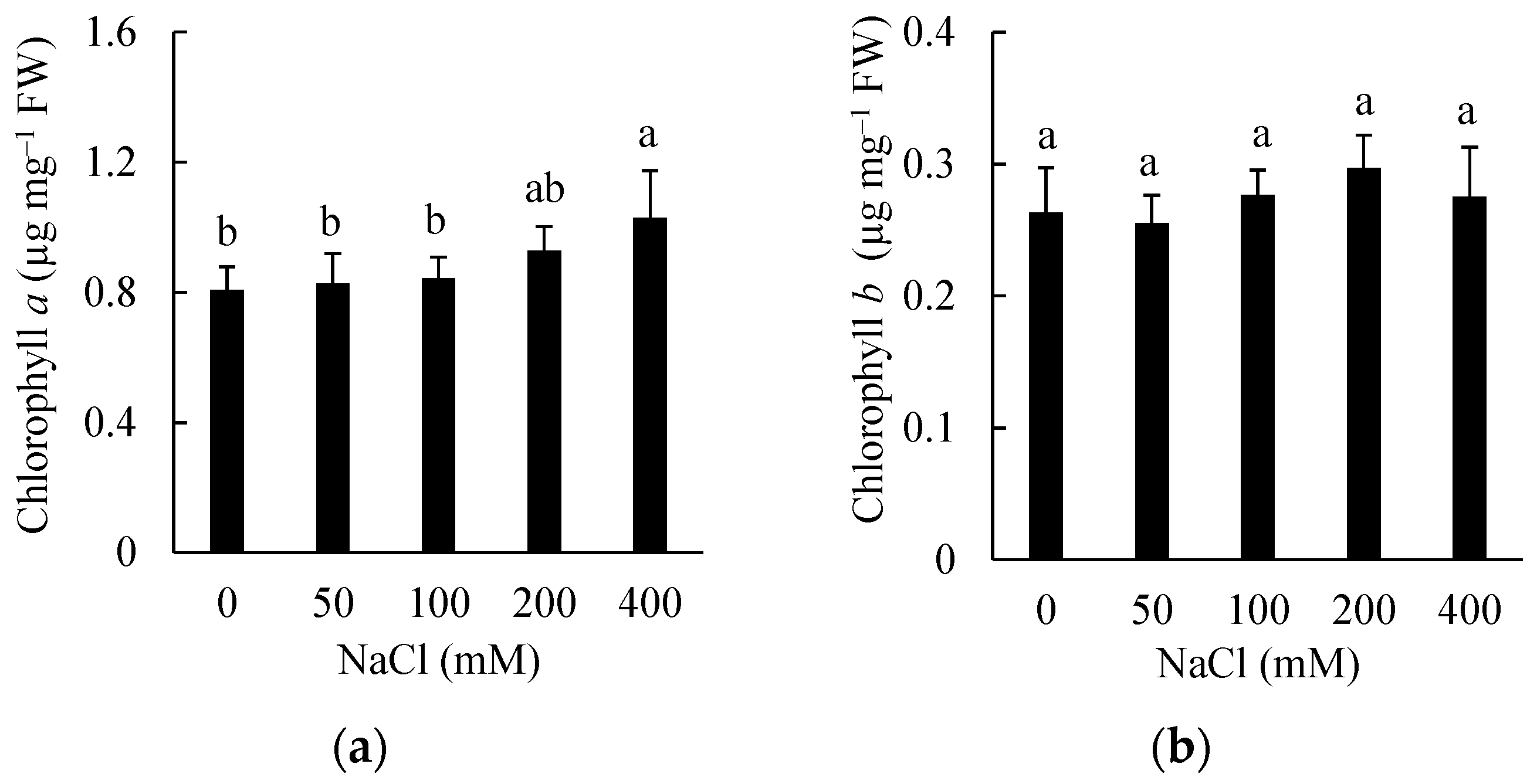
3.4. Effects of Salinity on Chlorophyll Content

In our previous observation, it was shown that the content of total chlorophylls (chl a + chl b) retained in the salt-treated shoots, in fact, tended to increase under high salinity (Figure S4). To examine details of how different types of chlorophylls were affected by salinity, here, the content of these pigments was analyzed. The data showed that the salinity affected on the accumulation of chl a in the same pattern to the total chlorophyll content, but it did not change chl b (Figure 4). The chl a content was maintained in salt concentrations of 50–100 mM, but it tended to increase with the shoot treated with 200–400 mM NaCl, which was reached 1.15 times (1224 µg mg−1 FW)–1.27 times (1.419 µg mg−1 FW) compared to the control (0.806 µg mg−1 FW), respectively (Figure 4a). The significant increase was obtained at 400 mM NaCl compared to the control. However, there was no significant changes in the chl b content in all salt treatments (Figure 4b). Chl a and b are two primary photosynthetic pigments that absorb energy from sunlight for plant photosynthesis and metabolisms [31]. As a result, the accumulation of these pigments is considered to be related to photosynthetic functions of leaves. Many previous studies reported different effects of salt stress on chlorophylls, depending on plant species and the salinity level [7,30,31,32]. In our study, the results showed that the chl a and chl b accumulations were maintained under the salinity, even enhanced (as for chl a) under the extreme saline condition (400 mM NaCl) (Figure 4). The similar pattern of change in the accumulation of chlorophylls was reported for a halophyte Kalidium foliatum under high salinity [31]. It is proposed that salt stress increases the chlorophyllase activity that promotes the degradation of chlorophylls and reduces the chlorophyll content in plants, and the effect degree depends on the salt tolerance of plants [31,32]. However, the accumulation of chlorophylls was maintained, and even the salt-induced increase in the chl a accumulation occurred in the P. nodiflora shoots under the 400 mM NaCl treatment (Figure 4). Possibly, the chlorophyll enhancement was required for the tolerance of P. nodiflora to the salt stress that affects the plant’s growth (Figure 1), although the increase in the chlorophyll content may not be positively related to the photosynthetic efficiency of plants. We propose that an investigation on the photosynthesis activity of the salt-treated shoots is necessary to elucidate this question. Although both chl a and chl b absorb light, chl a plays a unique and crucial role in converting light energy to chemical energy. As a result, this pigment could be enhanced instead of chl b.

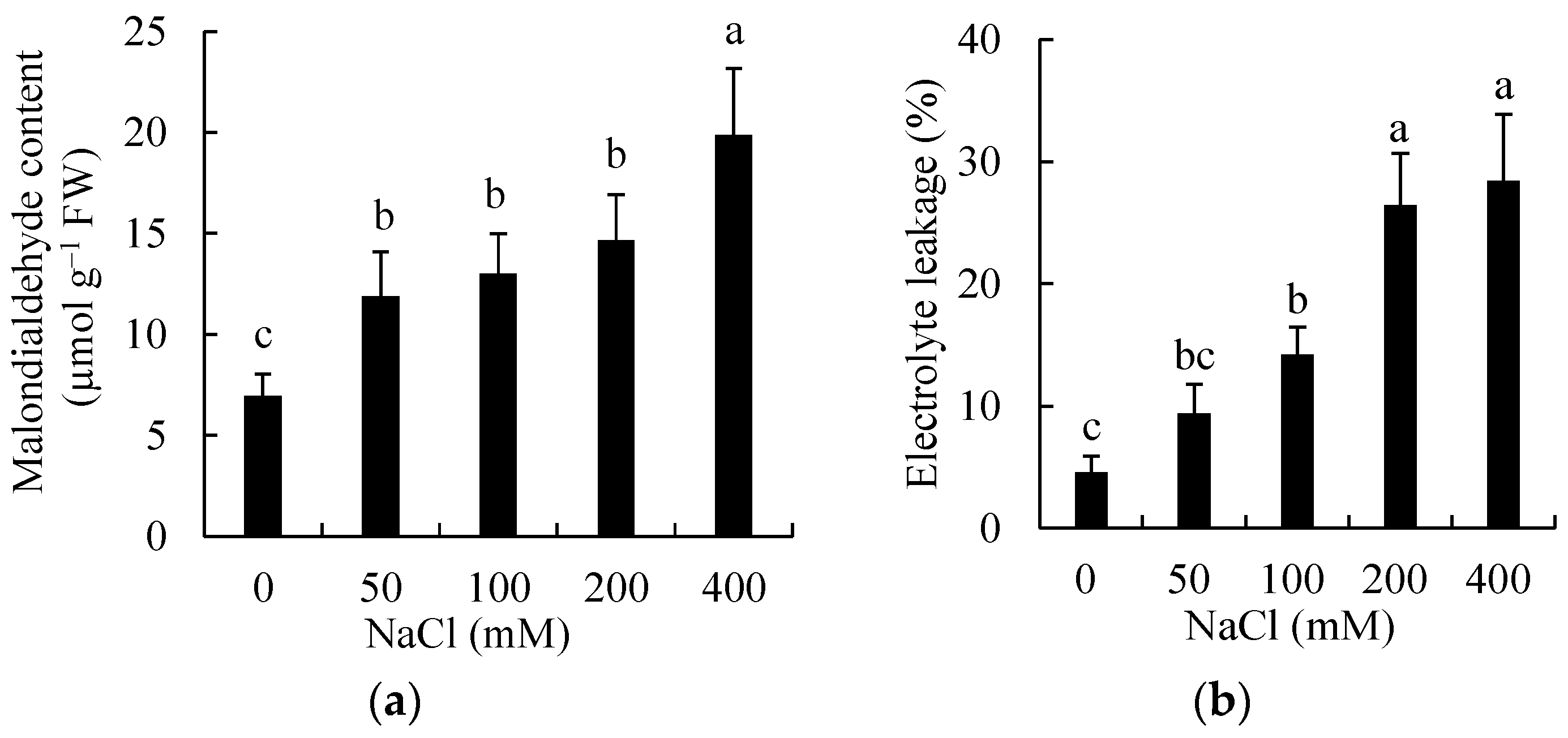
3.5. Effects of Salinity on Lipid Peroxidation and Membrane Damage

Our data showed that the leaf MDA content of P. nodiflora shoots under the salt treatments was gradually increased with the increasing salt concentrations (Figure 5a). The MDA content was increased to 11.881–19.870 µmol g−1 FW, which is equivalent to 1.70–2.85 times in the salt-treated shoots compared to the control (6.977 µmol g−1 FW). The significant difference was not obtained between the treatments of salt concentrations from 50 to 200 mM, but the significant and highest values of the MDA content was established in the 400 mM NaCl-treated shoots (Figure 5a). The similar pattern was also observed in EL, in which the value was 2.06–6.25 times (9.38–28.41%) in the salt-treated shoots compared to the control (Figure 5b). In plants, EL and MDA accumulation in plant tissues under salt stress are considered useful indicators for assessing the injury of the cell membrane, which may be caused by both salt and salt-induced ROS [29,33]. A significant increase in the contents of indicators is associated with a loss of membrane integrity [7]. In our study, the increase in MDA accumulation and EL suggested that salinity had negative effects on the membrane integrity of leaf cells. The growth of shoots was not affected by salinity levels up to 100 mM, but the contents of MDA and EL increased (Figure 1 and Figure 5). These results suggested that the rate of damages caused by 50 and 100 mM NaCl might be insignificant to the growth of shoots. However, it was the serious impacts under higher salinity that could attributed to the biomass reduction in shoots. Similar responses were reported for salt-tolerant plants such as Tetragonia tetratgoinodes and P. oleracea was under salinity. It was stated that the EL increase in salt-stressed plants, in some cases, may be related to the K+ efflux due to the abundant accumulation of this ion in stressed cells [29].

4. Conclusions

In this study, the results showed that salinity had effects on the growth of P. nodiflora shoots. The shoot’s survival under 400 mM NaCl salinity suggested that the plant could be salt tolerant, which equals to the adaptability to high salinity. The shoot biomass only decreased with the salt concentrations more than 100 mM. Also, the salinity affected the accumulation of proline, MDA, and chl a; the uptake of salt and nutrient ions; and EL in the shoots in a concentration-dependent manner. The changes in physiological responses might be related to the growth responses of shoots and it might indicate salt tolerance mechanisms that evolved in the plant. Further works are necessary to understand these mechanisms.

Supplementary Materials

The following supporting information can be downloaded at: https://www.mdpi.com/article/10.3390/ijpb15010015/s1, Figure S1: P. nodiflora plants growing in natural distribution area. Photos was taken in June 2022; Figure S2: The appearance of P. nodiflora plants at 14 days after the onset of salt treatments; Figure S3: The K+/Na+ ratio of P. nodiflora shoots at 7 days after the onset of NaCl treatments; Figure S4: The content of leaf total chlorophylls (chl a + chl b) of P. nodiflora shoots at 7 days after the onset of NaCl treatments.

Author Contributions

Conceptualization by D.Q.T. and T.C.V.; methodology by A.C.P. and D.Q.T.; experiments and data curation by A.C.P., T.D.B., T.-T.H.V. and D.Q.T.; writing—original draft preparation by D.Q.T. and A.C.P.; writing—review and editing by D.Q.T. All authors have read and agreed to the published version of the manuscript.

Funding

This study was financially supported by the Ministry of Education and Training (MOET-Vietnam), code: B2021-DNA-10.

Institutional Review Board Statement

Not applicable.

Informed Consent Statement

Not applicable.

Data Availability Statement

The data are contained within the article.

Acknowledgments

The authors also give thanks to significant supports from The University of Danang (UD) and The University of Danang-University of Science and Education, as well as to those who gave us useful helps during the experimental works and the manuscript preparation.

Conflicts of Interest

The authors declare no conflict of interest.

References

  1. Abobatta, W. Some physiological mechanisms of salt tolerance in the glycophytes plant: Overview. Acta Sci. Agric. 2018, 2, 154–156. [Google Scholar]
  2. Panta, S.; Flowers, T.; Lane, P.; Doyle, R.; Haros, G.; Shabala, S. Halophyte agriculture: Success stories. Environ. Exp. Bot. 2014, 107, 71–83. [Google Scholar] [CrossRef]
  3. Gupta, B.; Huang, B. Mechanism of salinity tolerance in plants: Physiological, biochemical, and molecular characterization. Int. J. Genom. 2014, 2014, 701596. [Google Scholar] [CrossRef] [PubMed]
  4. Garcia-Caparros, P.; Al-Azzawi, M.J.; Flowers, T.J. Economic uses of salt-tolerant plants. Plants 2023, 12, 2669. [Google Scholar] [CrossRef] [PubMed]
  5. Zhao, C.; Zhang, H.; Song, C.; Zhu, J.-K.; Shabala, S. Mechanisms of plant responses and adaptation to soil salinity. Innovation 2020, 1, 100017. [Google Scholar] [CrossRef] [PubMed]
  6. Safdar, H.; Amin, A.; Shafiq, Y.; Ali, A.; Yasin, R.; Shoukat, A.; Hussan, M.U.; Sarwar, M.I. A review: Impact of salinity on plant growth. Nat. Sci. 2019, 17, 34–40. [Google Scholar]
  7. Taïbi, K.; Taïbi, F.; Abderrahim, L.A.; Ennajah, A.; Belkhodja, M.; Mulet, J.M. Effect of salt stress on growth, chlorophyll content, lipid peroxidation and antioxidant defence systems in Phaseolus vulgaris L. S. Afr. J. Bot. 2016, 105, 306–312. [Google Scholar] [CrossRef]
  8. Kotagiri, D.; Kolluru, V.C. Effect of salinity stress on the morphology and physiology of five different Coleus species. Biomed. Pharmacol. J. 2017, 10, 1639–1649. [Google Scholar] [CrossRef]
  9. Khan, M.O.; Irfan, M.; Muhammad, A.; Ullah, I.; Nawaz, S.; Khalil, M.K.; Ahmad, M. A practical and economical strategy to mitigate salinity stress through seed priming. Front. Environ. Sci. 2022, 10, 991977. [Google Scholar] [CrossRef]
  10. Meng, X.; Zhou, J.; Sui, N. Mechanisms of salt tolerance in halophytes: Current understanding and recent advances. Open Life Sci. 2018, 13, 149–154. [Google Scholar] [CrossRef]
  11. Gross, C.L.; Fatemi, M.; Julien, M.; McPherson, H.; Van Klinken, R. The phylogeny and biogeography of Phyla nodiflora (Verbenaceae) reveals native and invasive lineages throughout the world. Diversity 2017, 9, 20. [Google Scholar] [CrossRef]
  12. Yang, Y.; Lu, S.; Chen, T. Verbenaceae. Fl. Taiwan 1998, 127, 421. [Google Scholar]
  13. Jabeen, M.; Jillani, U.; Chaudhary, B.A.; Uzair, M. Phytochemical and pharmacological studies of Phyla nodiflora (Verbenaceae): A review. Pak. J. Pharm. Res. 2016, 2, 49–54. [Google Scholar]
  14. Parmar, G.R.; Baile, S.B.; Gohel, K.; Shah, A.; Patel, S.; Seth, A.K. An ethnobotanical and pharmacological review on Phyla nodiflora. Int. J. Pharm. Res. 2020, 12, 3667–3673. [Google Scholar]
  15. Abbas, T.; Ahmad, I.; Khan, Z.I.; Okla, M.K.; Saleh, I.A.; AbdElgawad, H. Comparative physiological adaptations to industrial pollution stress mediated by melatonin in riparian vegetation and Phyla nodiflora an ornamental plant. Sci. Hortic. 2023, 321, 112367. [Google Scholar] [CrossRef]
  16. Sahin, U.; Ekinci, M.; Ors, S.; Turan, M.; Yildiz, S.; Yildirim, E. Effects of individual and combined effects of salinity and drought on physiological, nutritional and biochemical properties of cabbage (Brassica oleracea var. capitata). Sci. Hortic. 2018, 240, 196–204. [Google Scholar] [CrossRef]
  17. Wellburn, A.R. The spectral determination of chlorophylls a and b, as well as total carotenoids, using various solvents with spectrophotometers of different resolution. J. Plant Physiol. 1994, 144, 307–313. [Google Scholar] [CrossRef]
  18. Bates, L.S.; Waldren, R.; Teare, I. Rapid determination of free proline for water-stress studies. Plant Soil. 1973, 39, 205–207. [Google Scholar] [CrossRef]
  19. Senthilkumar, M.; Amaresan, N.; Sankaranarayanan, A.; Senthilkumar, M.; Amaresan, N.; Sankaranarayanan, A. Estimation of malondialdehyde (MDA) by thiobarbituric acid (TBA) assay. In Plant-Microbe Interactions; Springer Protocols Handbooks; Springer: New York, NY, USA, 2021; pp. 103–105. [Google Scholar]
  20. Tran, D.Q.; Konishi, A.; Cushman, J.C.; Morokuma, M.; Toyota, M.; Agarie, S. Ion accumulation and expression of ion homeostasis-related genes associated with halophilism, NaCl-promoted growth in a halophyte Mesembryanthemum crystallinum L. Plant Prod. Sci. 2020, 23, 91–102. [Google Scholar] [CrossRef]
  21. Gulzar, S.; Khan, M.A.; Ungar, I.A. Salt tolerance of a coastal salt marsh grass. Commun. Soil Sci. Plant Anal. 2003, 34, 2595–2605. [Google Scholar] [CrossRef]
  22. Said-Al Ahl, H.; Omer, E. Medicinal and aromatic plants production under salt stress. A review. Herba Pol. 2011, 57, 72–87. [Google Scholar]
  23. De Almeida Cartaxo, P.H.; da Silva, D.G.; Araújo, J.R.E.S.; da Silva, J.H.B.; Targino, V.A.; da Silva Xavier, L.M.; Neto, F.P.; de Oliveira, A.B.; da Silva, A.M. Salinity and medicinal plants: Challenges and strategies for production. Sci. Electron. Arch. 2022, 15, 8–15. [Google Scholar]
  24. González, L.; González-Vilar, M. Determination of relative water content. In Handbook of Plant Ecophysiology Techniques; Springer: Berlin/Heidelberg, Germany, 2001; pp. 207–212. [Google Scholar]
  25. Munns, R.; Passioura, J.B.; Colmer, T.D.; Byrt, C.S. Osmotic adjustment and energy limitations to plant growth in saline soil. New Phytol. 2020, 225, 1091–1096. [Google Scholar] [CrossRef]
  26. Adolf, V.I.; Jacobsen, S.-E.; Shabala, S. Salt tolerance mechanisms in quinoa (Chenopodium quinoa Willd.). Environ. Exp. Bot. 2013, 92, 43–54. [Google Scholar] [CrossRef]
  27. Flowers, T.J.; Munns, R.; Colmer, T.D. Sodium chloride toxicity and the cellular basis of salt tolerance in halophytes. Ann. Bot. 2015, 115, 419–431. [Google Scholar] [CrossRef] [PubMed]
  28. Amiri, B.; Assareh, M.; Rasouli, B.; Jafari, M.; Arzani, H.; Jafari, A. Effect of salinity on growth, ion content and water status of glasswort (Salicornia herbacea L.). Casp. J. Environ. Sci. 2010, 8, 79–87. [Google Scholar]
  29. Hniličková, H.; Hnilička, F.; Orsák, M.; Hejnák, V. Effect of salt stress on growth, electrolyte leakage, Na+ and K+ content in selected plant species. Plant Soil Environ. 2019, 65, 90–96. [Google Scholar] [CrossRef]
  30. Kumar, S.; Li, G.; Yang, J.; Huang, X.; Ji, Q.; Liu, Z.; Ke, W.; Hou, H. Effect of salt stress on growth, physiological parameters, and ionic concentration of water dropwort (Oenanthe javanica) cultivars. Front. Plant Sci. 2021, 12, 660409. [Google Scholar] [CrossRef]
  31. Gong, D.; Wang, G.; Si, W.; Zhou, Y.; Liu, Z.; Jia, J. Effects of salt stress on photosynthetic pigments and activity of ribulose-1, 5-bisphosphate carboxylase/oxygenase in Kalidium foliatum. Russ. J. Plant Physiol. 2018, 65, 98–103. [Google Scholar] [CrossRef]
  32. Yang, J.; Zheng, W.; Tian, Y.; Wu, Y.; Zhou, D. Effects of various mixed salt-alkaline stresses on growth, photosynthesis, and photosynthetic pigment concentrations of Medicago ruthenica seedlings. Photosynthetica 2011, 49, 275–284. [Google Scholar] [CrossRef]
  33. Sarker, U.; Oba, S. The response of salinity stress-induced A. tricolor to growth, anatomy, physiology, non-enzymatic and enzymatic antioxidants. Front. Plant Sci. 2020, 11, 559876. [Google Scholar] [CrossRef] [PubMed]
Figure 1. The biomass of P. nodiflora shoots at 14 days after the onset of NaCl treatments. (a) fresh shoot weight; (b) dry shoot weight. Error bars indicate standard deviations. Different letters represent significant differences between the treatments with p-value ≤ 0.05.
Ijpb 15 00015 g001
Figure 2. The leaf proline content of P. nodiflora shoots at 7 days after the onset of treatments. Error bars indicate standard deviations. Different letters represent significant differences between the treatments with p-value ≤ 0.05.
Ijpb 15 00015 g002
Figure 3. The leaf ion content of P. nodiflora shoots at 7 days after the onset of NaCl treatments. (a) Na+, (b) Cl, (c) K+, and (d) NO3. Error bars indicate standard deviations. Different letters represent significant differences between the treatments with p-value ≤ 0.05.
Ijpb 15 00015 g003
Figure 4. The leaf chlorophyll content of P. nodiflora shoots at 7 days after the onset of NaCl treatments. (a) chl a; (b) chl b. Error bars indicate standard deviations. Different letters represent significant differences between the treatments with p-value ≤ 0.05.
Ijpb 15 00015 g004
Figure 5. The leaf MDA content and EL of P. nodiflora shoots at 7 days after the onset of NaCl treatments. (a) MDA content; (b) EL. Error bars indicate standard deviations. Different letters represent significant differences between the treatments with p-value ≤ 0.05.
Ijpb 15 00015 g005
Disclaimer/Publisher’s Note: The statements, opinions and data contained in all publications are solely those of the individual author(s) and contributor(s) and not of MDPI and/or the editor(s). MDPI and/or the editor(s) disclaim responsibility for any injury to people or property resulting from any ideas, methods, instructions or products referred to in the content.

