Article

Screening of the Most Effective Media for Bioprospecting Three Indigenous Freshwater Microalgae Species

by
Kushi Yadav
1,
Ganesh Chandrakant Nikalje
2,
Dibyajyoti Pramanik
3,
Penna Suprasanna
4 and
Monika Prakash Rai
1,*
1
Amity Institute of Biotechnology, Amity University Uttar Pradesh, Sector 125, Noida 201313, India
2
Seva Sadan’s R.K. Talreja College of Arts, Science and Commerce, University of Mumbai, Ulhasnagar 421003, India
3
BK21 Four Program, Division of Applied Life Science, Plant Molecular Biology and Biotechnology Research Center, Gyeongsang National University, Jinju 52828, Republic of Korea
4
Amity Centre for Nuclear Biotechnology & Amity Institute of Biotechnology, Amity University, Mumbai 201313, India
*
Author to whom correspondence should be addressed.
Int. J. Plant Biol. 2023, 14(3), 558-570; https://doi.org/10.3390/ijpb14030044
Submission received: 25 May 2023 / Revised: 19 June 2023 / Accepted: 21 June 2023 / Published: 24 June 2023

Abstract

:
Microalgae are a natural source of lipids, carotenoids, and other value-added compounds. The combination of nutrients and their precise ratio have a significant impact on the productivity of microalgae-mediated compounds. The biomass, lipid, chlorophyll, and carotenoid production of three microalgae species, namely, Monoraphidium sp., Chlorella sorokiniana, and Scenedesmus obliquus, were investigated by employing standard photoautotrophic media, namely, BG-11, BBM, and HS CHU-10. BBM was found as the most effective medium since it obtained higher biomass, lipids, and carotenoids from microalgae. The lipids and carotenoids were identified using chromatographic and microscopy techniques. The findings showed that although the Monoraphidium sp. biomass productivity was the lowest, it emerged as a substantial producer of astaxanthin, whereas the Chlorella sorokiniana culture could grow in a variety of media and produced β-carotene as a major carotenoid. On the other hand, Scenedesmus obliquus was found to be a considerable source of lipids and β-carotene. This study provided a comprehensive understanding of the appropriate medium selection in order to extract an assortment of value-added compounds from freshwater microalgae species.

1. Introduction

The booming population has triggered a food, feed, and power dilemma, redirecting the emphasis of researchers to alternative sources. The scarcity of nonrenewable resources has switched biotechnologists’ attention to certain renewable resources, among which photosynthetic autotrophs have received significant attention. Plants appear to be a prospective feedstock at first glance, but there are several limitations, including food sufficiency, huge arable area consumption, time-demanding production, lower yield, and seedling toxicity. Algae progressively replaced plants owing to a multitude of advantages, such as inexpensive cultivation costs, increased yield, utilization of non-arable land, nutrient recovery from wastewater, and effective carbon capture. Microalgae have emerged as a key contributor to the food, feed, pharmaceutical, and energy industries as researchers seek an economically and sustainably viable alternative [1]. Aside from biofuels, microalgae are a possible feedstock for pharmaceutical, nutraceutical, and cosmeceutical products. Microalgae from the Chlorophyceae family contain 20–70% lipids in addition to a variety of valuable biomolecules, such as proteins, carbohydrates, and pigments [2]. Only a few species, including plants, algae (Chlorophyceae, Cyanophyceae, and Eustigmatophyceae), marine bacteria (Agrobacterium aurantiacum), and yeast (Xanthophyllomyces dendrorhous), can produce carotenoids with pharmaceutical significance [3,4]. Microalgae have received recognition from several sectors as the preferred alternative for producing a wide range of value-added chemicals, including biofuels, medications, cosmetics, food, and feed [5]. However, researchers have concentrated on a few strains that were considered to be commercially useful amid the large range of microalgae species. Individual modification is necessary among this wide range of microalgae in order to achieve a good yield through careful medium selection [6].
Microalgae can potentially thrive in freshwater, ocean, and wastewater; however, due to cellular toxicity, wastewater-cultivated algae are the least frequently explored for nutraceutical and medicinal applications [7]. As the growth of microalgae is greatly influenced by the macronutrients and micronutrients of the culture medium, optimization of the culture medium is a necessity for their commercialization [8]. Numerous studies investigated marine microalgae, while there have been fewer studies on freshwater microalgae. In this study, three commercially recognized freshwater microalgae strains were used in order to evaluate the impact of various culture media on their biomass, lipid, chlorophyll, and carotenoid outputs. Three recognized culture media (BG-11, BBM, and HS-CHU 10) were used for the cultivation of these freshwater microalgae. This study allowed for the selection of the most appropriate medium for a strain-specific culture. The media were also evaluated for the yields of lipids and carotenoids.

2. Materials and Methods

2.1. Cultivation and Morphometric Analysis

Three freshwater green microalgae, namely, Monoraphidium sp. (NCIM 5585), Chlorella sorokiniana (NCIM 5561), and Scenedesmus obliquus (NCIM 5586), were acquired from the National Chemical Laboratory (NCL) Pune, India. Microalgae were selected due to their fast growth and high carotenoid production ability, along with other lipids. To begin the microalgae growth process, 100 mL Erlenmeyer flasks were filled with 50 mL of sterilized BG-11 media and inoculated with exponentially growing cells till the OD reached 0.1. The flasks were placed in an incubator chamber that was held at a constant temperature of 28 °C with regular illumination of 100 μmol·m−2·s−1 (24 h, 40 W, white light) and intermittent shaking. Light microscopy (Nikon A1, Tokyo, Japan) was used to determine the morphology of the axenic microalgal cells.

2.2. Screening of Cultivation Media for Maximum Biomass, Lipids, and Carotenoids

The composition of the cultivation medium has a considerable influence on carotenoid, lipid, and biomass yields in microalgae. Three media were employed for photoautotrophic cultivation of microalgae species: Bold’s basal medium (BBM), BG-11, and half-strength CHU-10 (HS CHU-10) medium and their compositions were shown in Table 1. All species were grown in 500 mL Erlenmeyer flasks with 250 mL medium by keeping other culture conditions the same as before. The media compositions of the three growth media used are shown below.

2.3. Biomass Estimation

Microalgae growth was monitored after 48 h at 680 nm using a Microplate Reader Synergy H1 (BioTek, Winooski, VT, USA). On alternate days, the growth was measured using a 200 µL culture loaded on 96-well plates (Nuncmaxisorp, Waltham, MA, USA) [9]. Equations (1)–(3) were used to determine the biomass concentration, productivity, and specific growth rate (3).
B i o m a s s   c o n c e n t r a t i o n   g / L = 0.675 × A 680 0.0841
B i o m a s s   p r o d u c t i v i t y   g / L / d = B i o m a s s   c o n c e n t r a t i o n   g / L C u l t i v a t i o n   d u r a t i o n   d
S p e c i f i c   g r o w t h   r a t e   µ p e r   d a y = l n   X 2 X 1 T 2 T 1
T stands for the time in days; X2 and X1 represent the specific final and initial biomass concentrations, respectively; and T2 and T1 stand for the specific final and initial times, respectively.

2.4. Quantification of Lipids

Total lipids were extracted from 1 g of dried microalgal biomass using the Bligh and Dyer method and quantified using the gravimetric method [10]. Equations (4)–(6) were used to calculate the total lipid production, productivity, and content
T o t a l   l i p i d   p r o d u c t i o n   mg / L = M a s s   o f   l i p i d   mg V o l u m e   L
L i p i d   p r o d u c t i v i t y   mg / L / D = M a s s   o f   l i p i d   mg V o l u m e L × C u l t i v a t i o n   d u r a t i o n   d
L i p i d   c o n t e n t   % = M a s s   o f   l i p i d   g M a s s   o f   c u l t u r e   g × 100

2.5. Pigment Estimation

Every other day, 200 µL of microalgal culture was placed in 96-well plates (Nuncmaxisorp, Waltham, MA, USA), and microalgae pigment was measured using a Synergy H1 microplate reader (BioTek, Winooski, VT, USA). The pigment concentrations were calculated for chlorophyll a (Chl a), chlorophyll b (Chl b), and total carotenoids (Car) using Equations (7)–(9), respectively [11].
C h l   a   µ g / mL = 13.36   A 664 5.19 A 648
C h l   b   µ g / mL = 27.43   A 648 8.12 A 664
C a r   µ g / mL = 1000   A 470 2.13 C h l   a 97.64   C h l   b 209

2.6. Microscopic Examination of Chlorophyll, Lipid, and Lipophilic Compounds

Confocal laser scanning microscopy was used to examine the chlorophyll and lipid fluorescence of cells. Cell suspensions were collected and centrifuged for 5 min at 8000 rpm. Pellets were rinsed twice with PBS buffer before being exposed to 1 mL (20%) dimethyl sulfoxide (DMSO, Merck, Darmstadt, Germany, 99.9%) for 10 min. Following centrifugation, 100 µL PBS was added to each tube, which was then incubated at room temperature for 10 min. Confocal microscopy (Nikon A1, Tokyo, Japan) was used to measure the chlorophyll fluorescence in the microalgae cells [12]. The presence of neutral lipids and lipophilic compounds in the microalgae was evaluated using Nile red staining and confocal microscopy by employing laser light with excitation and emission wavelengths of 552 and 636 nm, respectively [13].

2.7. Pigments Quantification

A total of 1 g of biomass was crushed with a mortar and pestle, and the pigment was extracted by adding 10 mL of DCM to the disrupted cells. The extraction was carried out three more times until the cell debris became colorless. The pigment-rich DCM extract was evaporated using a rotary evaporator (Buchi, R-210) and saponified for 3 h at room temperature in the dark. The mixture of 2.25 mL acetone, 0.25 mL methanol, and 0.5 mL of 0.05 M NaOH in methanol was poured into the obtained pigments. Then, 3 mL petroleum ether was poured. The mixture was centrifuged for two minutes at 5500 rpm and cleaned using 3 mL of 10% dilute NaCl solution. The lowest phase was then discarded, and the remaining solution was washed twice using NaCl solution [14].

2.8. TLC Analysis

Silica-gel-coated plates (Merck 20 × 20 cm) were employed for the TLC of the extracted carotenoids content and a 4:1 mixture of the solvent system (n-hexane/acetone). Initially, the solvent was employed to pre-saturate chromatography sheets. The sheets were then lightly covered with 5 µL of the carotenoid samples before drying. Once the sheets were loaded, they were carefully positioned in a pre-saturated tank so that the applied samples did not accidentally fall further into the solvent system. The system was left undisturbed, and the solvent was permitted to move up to a height of 5 cm. The sheets were then removed from the tank and the spots were promptly noted [12].

2.9. Statistics

The acquired data were analyzed using one-way ANOVA in OriginPro 2021 (OriginLab, Northampton, MA, USA). Tukey’s honestly significant difference test at p < 0.05 was used to distinguish the mean differences. All data were collected in triplicate, as mentioned in the figures and tables [13].

3. Results and Discussion

Screening for adequate media for microalgae cultivation is the fundamental methodological step to attain ideal control conditions. A culture medium is described as a medium that provides an adequate amount of nutrients to microalgae for optimal growth and development, and it is necessary to optimize to find a suitable medium that may encourage the development of a wide range of microalgae species [14].

3.1. Biomass Analysis

Green freshwater microalgae Scenedesmus obliquus, Monoraphidium sp., and Chlorella sorokiniana were grown in different media for 25 days by utilizing light and atmospheric CO2. The growth profiles of the Scenedesmus obliquus, Monoraphidium sp., and Chlorella sorokiniana cultures evaluated in BG-11, BBM, and HS CHU-10 media are shown in Figure 1.
Among the media studied, BBM supported high-biomass yields, followed by HS CHU-10 and BG11. The growth curve of Scenedesmus obliquus in BG-11, BBM, and HS CHU-10 media, shown in Figure 1, revealed that in the stationary phase, the maximum biomass productions of 881.2 mg/L, 1002.7 mg/L, and 918.98 mg/L were obtained, respectively. Figure 1 depicts the growth curves of Monoraphidium sp. and Chlorella sorokiniana in the three media, resulting in the highest biomass productions of 567.6 mg/L (BG-11), 724.3 mg/L (BBM), and 586.4 mg/L (HS CHU-10) and 974.3 mg/L (BG-11), 1004.7 mg/L (BBM), and 904.5 mg/L (HS CHU-10), respectively. Chankhong et al. (2018) reported that when cultivating Chlorella sp. T12 in BBM, the maximum biomass yield was obtained, i.e., 0.20 ± 0.05 g/L/d [15].
Scenedesmus obliquus, Monoraphidium sp., and Chlorella sorokiniana showed significant differences in growth with p-values of 0.042 and 0.02 for BBM and HS CHU-10, respectively, as shown in Figure 2. In this scenario, the p-values were smaller than the generally accepted significance level of 0.05, indicating that the differences identified in the growth of microalgal species were statistically significant in different media.
Therefore, we could deduce that there were species-specific differences in the impacts of BBM and HS CHU-10 media compositions on biomass productivity. In contrast, the p-value for BG-11 was 0.10, which is more than 0.05, meaning that the composition of BG-11 had no significant influence on the biomass production of the aforementioned species. Therefore, the investigation of media selection demonstrated that different media compositions had substantial effects on the growth of freshwater microalgal species. According to George et al. 2014, when grown on BG-11 or CHU-10 medium, the Ankistrodesmus falcatus biomass production was almost identical. Nevertheless, the effects of different media compositions on the morphology of microalgal cells varied greatly [16]. This indicates that the media compositions significantly affect the microalgal species being studied. Among modified Bold’s basal medium (BBM), modified CFTRI medium, modified BG11 medium, and modified CHU medium, Sangapillai et al. 2019 found that modified BBM is the optimal medium for freshwater Asterarcys quadricellulare. A maximal biomass of 1.44±0.015 g/L was produced using modified BBM, which was the best result from the four experimental media [17].

3.2. Lipids Analysis

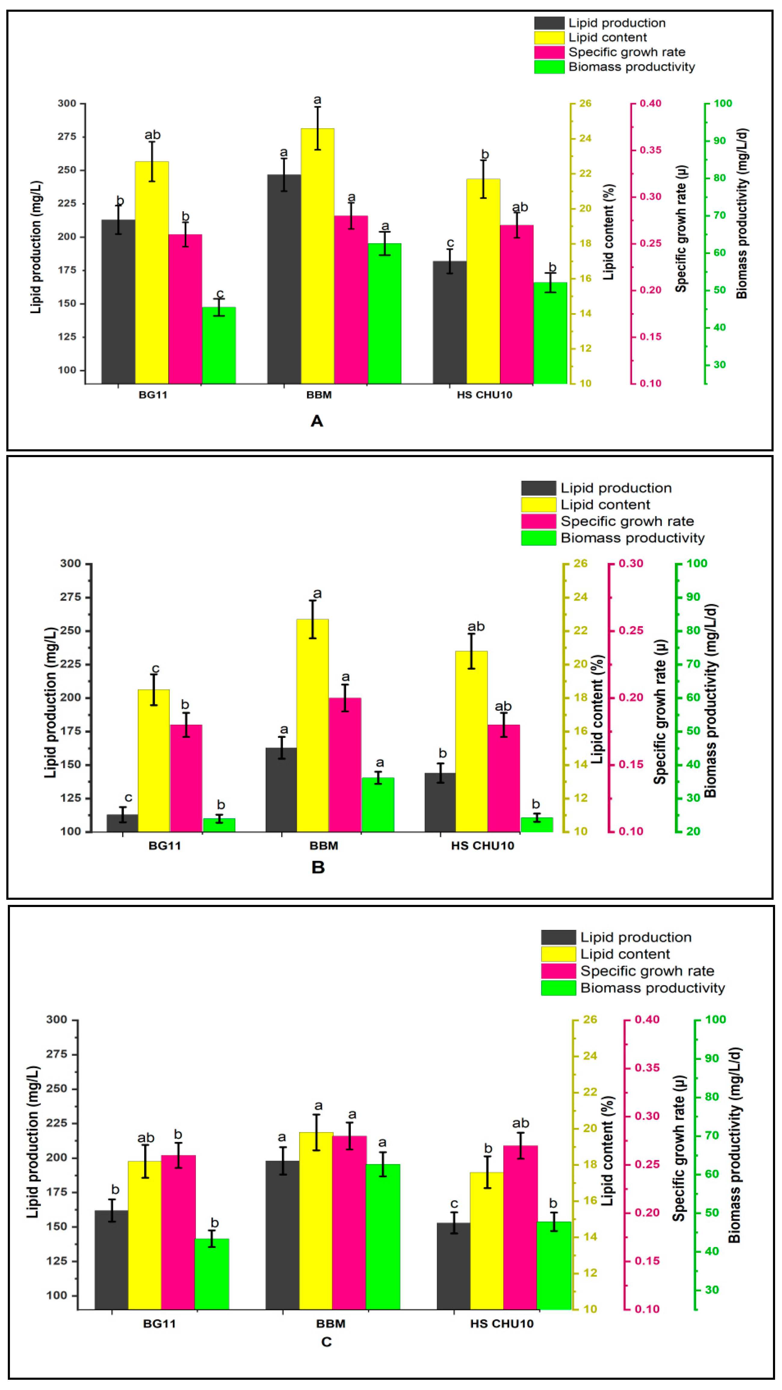
The freshwater microalgae species used in this study had significant lipid contents and might be explored for industrial applications. Alongside lipids and carotenoids, cell density is an essential feature that defines microalga viability for use as a biofuel or medicinal feedstock [18]. As a result, the species was grown in three distinct culture media (BG-11, BBM, and HS CHU-10).
For Scenedesmus obliquus, the lipid production was highest in BBM, i.e., 246.8 mg/L, with the highest lipid accumulation of 24.6%, whereas the lipid production was nearly equivalent in the other two media, accruing nearly 15.7% and 17.8% lipid contents with the highest productions of 138.7 mg/L and 163.3 mg/L in BG-11 and HS CHU-10, respectively. Nonetheless, statistical analysis was carried out, as shown in Figure 3, to ascertain the significance of the lipid production and lipid content by Scenedesmus obliquus on the 16th day of inoculation in different media. BBM had significantly greater lipid productivity than the BG-11 medium with 15.4 mg/L/d. The lowest lipid productivity of 11.3 mg/L/d was found in HS CHU-10. The results show that the medium composition had a direct influence on the productivity of the value-added compounds in microalgae. Conversely, in Monoraphidium sp. and Chlorella sorokiniana, the maximum lipid productions were obtained using BBM, i.e., 162.9 mg/L and 198.8 mg/L, respectively, with the highest lipid accumulations of 22.7% and 19.8%, respectively. Furthermore, as indicated in Figure 3, the substantial lipid productions in both Monoraphidium sp. and Chlorella sorokiniana were recorded in BBM, followed by HS CHU-10 and BG-11 media. The lipid production in the case of Monoraphidium sp. was at a maximum on the 18th day after inoculation in BBM. Similarly, the highest lipid content was obtained on the 16th day after inoculation of Chlorella sorokiniana in BBM. In order to ascertain the statistical significance of the impacts of various media on lipid production, the aforementioned days were considered. It is evident that BBM had the highest biomass and lipid concentration. A. falcatus accumulated the highest amount of lipids in ZM, although the biochemical makeups of BG-11 and CHU-10 media are fairly comparable [19]. Kirrolia et al. 2011 determined that BBM was the best-suited cultivation medium for Chlorococcum sp. lipid accumulation, with 19.25% lipid content in comparison to HS CHU-10, Allen, BG-11, and CHU-10 media [20]. In the present study, Scenedesmus obliquus emerged as a prospective strain for the biofuel business due to the greatest lipid content of the three freshwater microalgal species. For promoting microalgae toward the production of lipids, BBM was a more efficient medium compared with BG-11 and HS CHU-10.

3.3. Pigment Analysis

Chlorophyll and carotenoids are the most substantial pigments found in freshwater green microalgae. The intriguing family of natural pigments known as microalgae carotenoids has several potential uses [21]. Although they have been employed as coloring agents in food for many years, prospective uses for them are fast evolving as further research has allowed for utilizing their characteristics and advantages. The antioxidant and anti-inflammatory properties of microalgae carotenoids, as well as their potential application in dietary supplements and capacity to replace artificial coloring in food items, are all being explored [22].
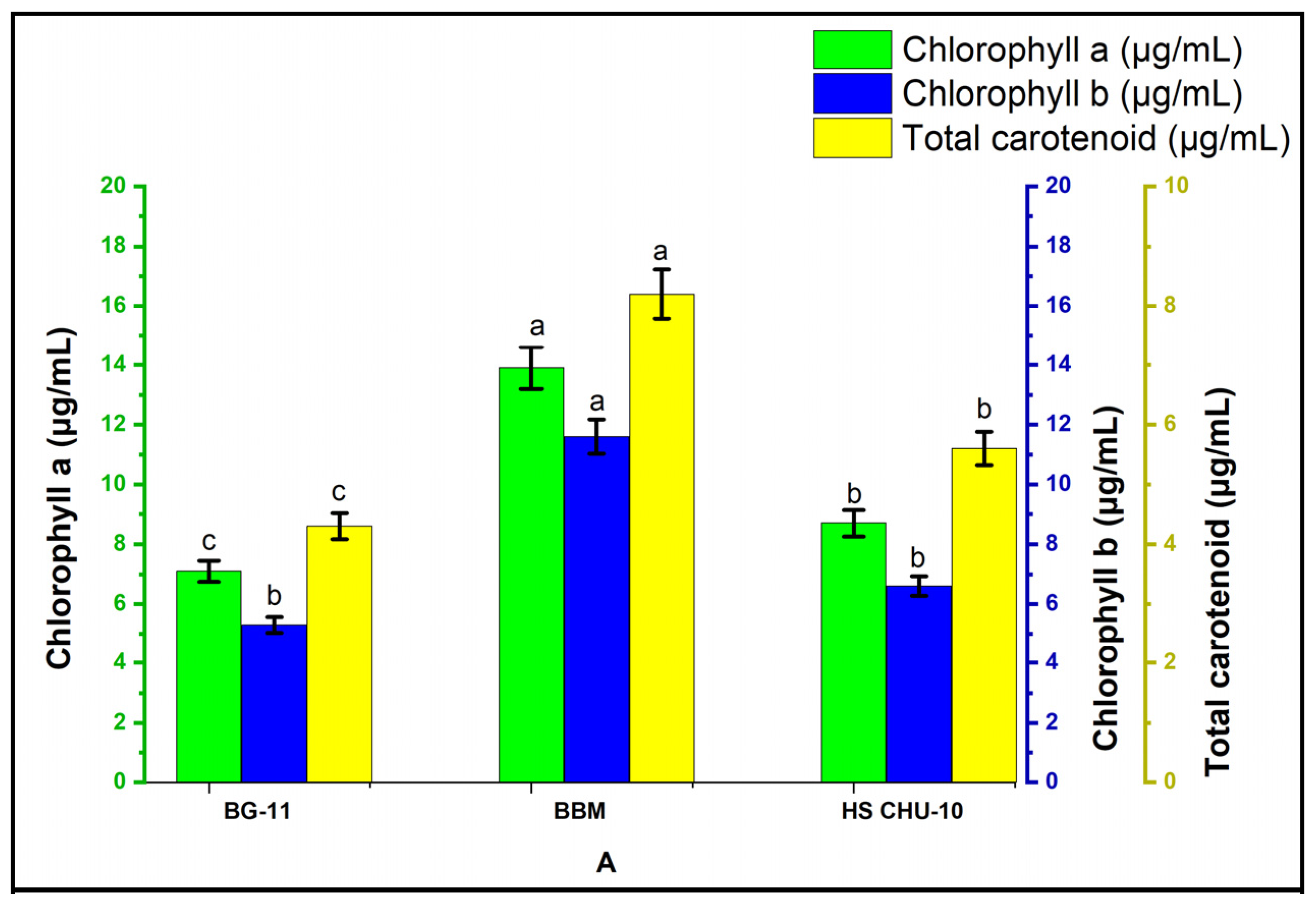
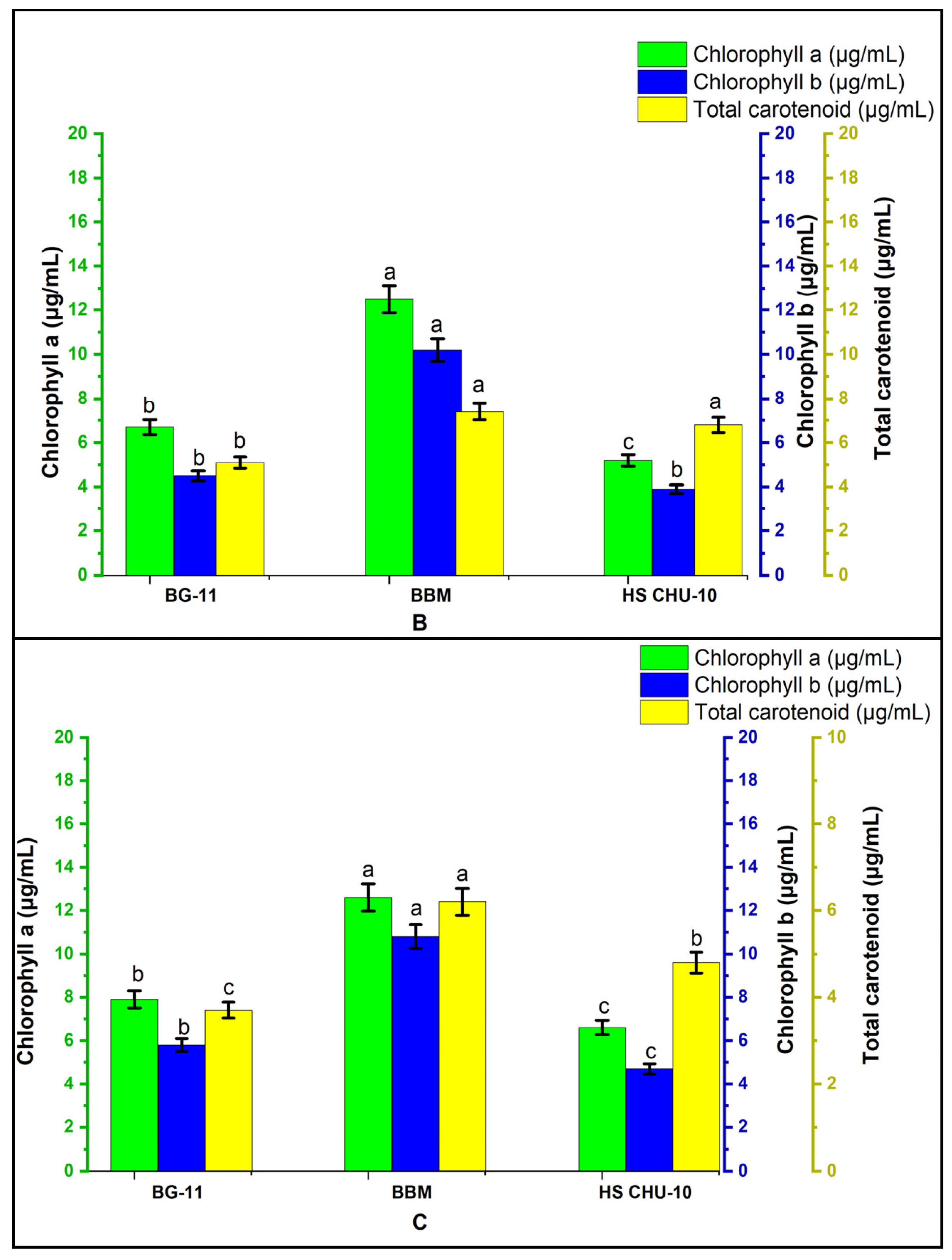
Carotenoids have been linked to the reduced risk of certain diseases, as well as improved vision and skin health [21]. The total pigment accumulations in Scenedesmus obliquus, Monoraphidium sp., and Chlorella sorokiniana under three different culture media at specific time intervals are shown in Figure 4. This result reveals the significant difference between the pigment productivity of the aforementioned microalgal species cultivated under distinct media compositions at specific time intervals. The maximum quantities of total chlorophyll and carotenoid produced by these microalgae for various media and independent of the time interval are revealed in Table 2. Time-dependent pigment analysis was done to compare the significant difference in quantities of pigments obtained at specific time intervals. However, we found that the best yield of microalgae among all selected media compositions was obtained using time-independent pigment analysis, which confirmed the maximum efficacy of all selected media on distinct microalgal species. From the time-independent pigment analysis, we could find the highest efficacy of a particular medium among all the selected media. The results obtained in the current investigation from the time-independent pigment analysis showed less variation in chlorophyll and total carotenoids production. Nevertheless, the number of days required to obtain that yield varied drastically. However, from both the time-dependent and time-independent pigment analysis, BBM was found to be an efficient medium for freshwater microalgae. In Asterarcys quadricellulare cultivated under four different growth media, namely, modified Bold’s basal medium (BBM), modified CFTRI medium, BG11 medium and CHU medium, the highest chlorophyll a (23.07 ± 0.049 mg/L), chlorophyll b (16.76 ± 0.010 mg/L) and total carotenoids (8.92 ± 0.031 mg/L) was obtained with the modified Bold’s basal medium (BBM) [17].
This indicates that while the growth continued, the level of chlorophyll rose to a specific level and then decreased, indicating the end of the stationary phase. However, during the stationary phase, the concentration of carotenoids increased [23].

3.4. Confocal Imaging for the Visualization of Lipophilic Compounds

Scenedesmus obliquus, Monoraphidium sp., and Chlorella sorokiniana were grown on photoautotrophic optimal BBM and studied morphologically under confocal microscopy. Figure 5 shows confocal images of Scenedesmus obliquus, Monoraphidium sp., and Chlorella sorokiniana cultures taken in the exponential phase.
Carotenoids are lipophilic molecules that are present both within and outside of chloroplasts; consequently, lipids and carotenoids may be detected as red globular forms using confocal microscopy and the Nile Red strain, as seen in Figure 5. Primary and secondary carotenoids are the two categories into which carotenoids are often divided. Primary carotenoids are photosynthetic pigments that are structurally and functionally linked to the mechanism of photosynthesis. The photosynthetic apparatus is not connected to secondary carotenoids, and their concentration is not restricted by stoichiometry with other cellular components [11]. Chlorophyll was identified as green globules present in both the light micrograph and fluorescence images of different microalgae species.

3.5. Carotenoid Chromatography

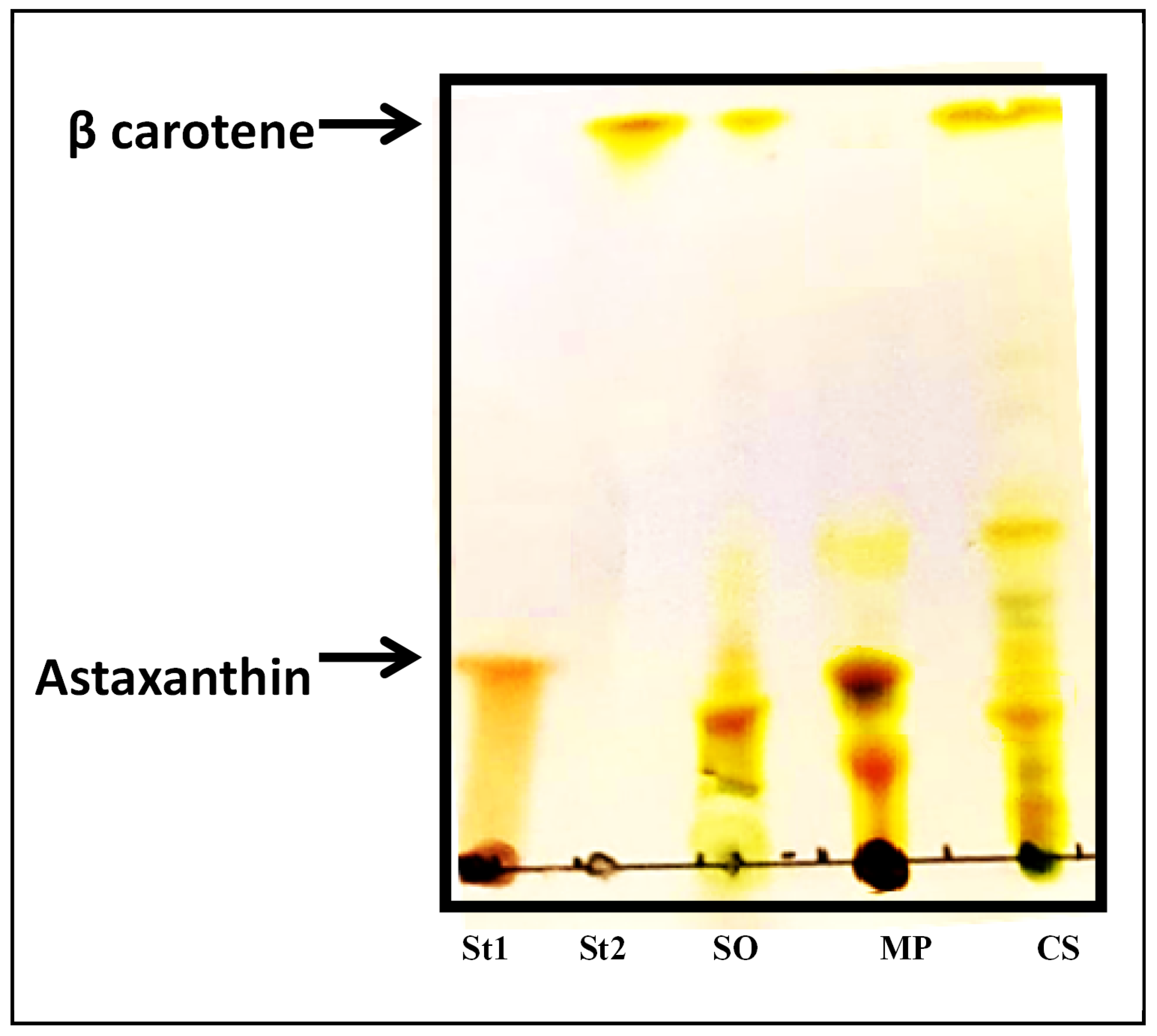
The thin layer chromatography technique was used with the astaxanthin and beta carotene standards, along with carotenoids extracted from Scenedesmus obliquus, Monoraphidium sp., and Chlorella sorokiniana. The Rf values for the standard astaxanthin and beta carotene were 0.45 and 0.98, respectively, and this matched the retention time for the extracted samples.
TLC was used to detect the presence of two economically relevant carotenoids in the extracts of Scenedesmus obliquus, Monoraphidium sp., and Chlorella sorokiniana. As shown in Figure 6, the chromatogram revealed that astaxanthin was found in the Monoraphidium sp. and Scenedesmus obliquus carotenoid extracts. Despite this, Scenedesmus obliquus and Chlorella sorokiniana are recognized for the presence of beta-carotene. While optimizing the culture conditions for the development and carotenoid synthesis, Rajput et al. 2022 discovered the occurrence of both carotene and astaxanthin in Scenedesmus sp. [24]. A study done by Kaha et al. 2021 on the effect of black light on astaxanthin production validated the presence of astaxanthin in Monoraphidium sp. [25]. In Roth et al. 2017, beta-carotene pigment was isolated from the green algae Chlorella vulgaris and Scendesmus regularizes [26]. Rajput et al. 2021 stated that in Scenedesmus quadricauda PUMCC 4.1.40 under ideal growing conditions, there were presences of 23.8, 19.0, 6.5, and 4.0 μg astaxanthin, β-carotene, lutein, and canthaxanthin/mg dry biomass, respectively [24]. To obtain these carotenoid profiles, high-performance thin-layer chromatography (HPTLC) separation followed by flash chromatography purification and HPLC quantification was employed [24]. The carotenoid profile of Monoraphidium sp. grown under optimal conditions produces a significant amount of astaxanthin, as reported by Yadav et al. 2023 [11]. The current study revealed the presence of commercially significant carotenoids in two freshwater microalgal species. Consequently, freshwater microalgal species cultivated in suitable media have immense potential of becoming commercially viable microalgal strains.

4. Conclusions

The current research examined the finest media for cultivating freshwater microalgae in order to maximize biomass, lipid, and carotenoid production. Green freshwater microalgae have enormous potential in the energy, medicinal, cosmeceutical, and nutraceutical industries. These microalgae were investigated to generate a variety of industrially important carotenoids. This investigation was carried out to screen for the appropriate media among BG-11, BBM, and HS CHU-10 for photoautotrophic cultures of these axenic species. It was found that BBM was the best medium for cultivating these three freshwater microalgae species. These findings suggest that in addition to biodiesel, Scenedesmus obliquus, Monoraphidium sp., and Chlorella sorokiniana have the potential to grow in a variety of environments and generate astaxanthin and β-carotene as main carotenoids. Monoraphidium sp. could be a potential resource of astaxanthin, whereas Scenedesmus obliquus and Chlorella sorokiniana are excellent sources of beta carotene to be used in nutraceutical and pharmaceutical applications.

Author Contributions

K.Y. is the first author and conducted all the experiments, recorded the research observations, was responsible for the data curation, analyzed the research data, and wrote the original draft. G.C.N. data analysis; reviewed and edited the draft and acquired funding. D.P. reviewed and edited the draft. P.S. reviewed and edited the draft. M.P.R. conceptualized this study, methodology, and data analysis; supervised and validated the research work; reviewed and edited the manuscript; and acquired resources and funding. All authors have read and agreed to the published version of the manuscript.

Funding

M.P.R. expresses her gratitude to the Mission Innovation India Unit, Department of Biotechnology (MI-DBT), Ministry of Science and Technology, New Delhi (India), for their financial support (file no. BT/PR31218/PBD/26/771/2019).

Institutional Review Board Statement

Not applicable.

Informed Consent Statement

Not applicable.

Data Availability Statement

The data are contained within this article.

Acknowledgments

K.Y. and M.P.R. show their gratitude to the Amity Institute of Biotechnology, AUUP Noida, for providing the necessary infrastructure and facilities for the research. The authors also acknowledge the Amity Institute of Microbial Technology, AUUP Noida, for providing confocal microscopy.

Conflicts of Interest

The authors declare no conflict of interest.

References

  1. Putri, R.D.; Pradana, Y.S.; Koerniawan, M.D.; Suwanti, L.T.; Siregar, U.J.; Budiman, A.; Suyono, E.A. Effect of distinct nitrate concentrations on pigment content of mixed culture of Chlorella vulgaris and Dunaliella sp. Asia-Pac. J. Mol. Biol. Biotechnol. 2022, 30, 15–23. [Google Scholar] [CrossRef]
  2. Singh, B.; Guldhe, A.; Singh, P.; Singh, A.; Rawat, I.; Bux, F. Sustainable production of biofuels from microalgae using a biorefinery approach. In Applied Environmental Biotechnology: Present Scenario and Future Trends; Springer: New Delhi, Inida, 2015; pp. 115–128. [Google Scholar]
  3. Sun, T.; Rao, S.; Zhou, X.; Li, L. Plant carotenoids: Recent advances and future perspectives. Mol. Hortic. 2022, 2, 3. [Google Scholar] [CrossRef]
  4. Watcharawipas, A.; Runguphan, W. Red yeasts and their carotenogenic enzymes for microbial carotenoid production. FEMS Yeast Res. 2023, foac063. [Google Scholar] [CrossRef] [PubMed]
  5. Siddiki, S.Y.A.; Mofijur, M.; Kumar, P.S.; Ahmed, S.F.; Inayat, A.; Kusumo, F.; Badruddin, I.A.; Khan, T.Y.; Nghiem, L.D.; Ong, H.C.; et al. Microalgae biomass as a sustainable source for biofuel, biochemical and biobased value-added products: An integrated biorefinery concept. Fuel 2022, 307, 121782. [Google Scholar] [CrossRef]
  6. Debnath, C.; Bandyopadhyay, T.K.; Bhunia, B.; Mishra, U.; Narayanasamy, S.; Muthuraj, M. Microalgae: Sustainable resource of carbohydrates in third-generation biofuel production. Renew. Sustain. Energy Rev. 2021, 150, 111464. [Google Scholar] [CrossRef]
  7. Timotius, V.; Suyono, E.A.; Suwanti, L.T.; Koerniawan, M.D.; Budiman, A.; Siregar, U.J. The content of lipid, chlorophyll, and carotenoid of Euglena sp. under various salinities. Asia-Pac. J. Mol. Biol. Biotechnol. 2022, 30, 114–122. [Google Scholar] [CrossRef]
  8. de Carvalho, J.C.; Sydney, E.B.; Tessari, L.F.; Soccol, C.R. Culture media for mass production of microalgae. In Biofuels from Algae; Elsevier: Amsterdam, The Netherlands, 2019; pp. 33–50. [Google Scholar]
  9. Zhang, L.; Pei, H.; Chen, S.; Jiang, L.; Hou, Q.; Yang, Z.; Yu, Z. Salinity-induced cellular cross-talk in carbon partitioning reveals starch-to-lipid biosynthesis switching in low-starch freshwater algae. Bioresour. Technol. 2018, 250, 449–456. [Google Scholar] [CrossRef]
  10. Bligh, E.G.; Dyer, W.J. A rapid method of total lipid extraction and purification. Can. J. Biochem. Physiol. 1959, 37, 911–917. [Google Scholar] [CrossRef]
  11. Yadav, K.; Kumar, S.; Nikalje, G.C.; Rai, M.P. Combinatorial Effect of Multiple Variables on Up-Regulation of Carotenoids and Lipids in Monoraphidium sp. for Pharmacological and Nutraceutical Applications. Appl. Sci. 2023, 13, 6107. [Google Scholar] [CrossRef]
  12. Agarwal, A.; Jeevanandham, S.; Kar, C.; Rai, M.P.; Kumar, V.; Bohidar, H.B.; Biswas, S.; Mukherjee, M. Crystalline Domains Nested on Two-Dimensional Nanosheets as Heterogeneous Nanomachineries for the Sustainable Production of Bioactive Compounds from Chlorella sorokiniana. ACS Sustain. Chem. Eng. 2022, 10, 9732–9748. [Google Scholar] [CrossRef]
  13. Zohir, W.F.; Kapase, V.U.; Kumar, S. Identification and Characterization of a New Microalga Dysmorphococcusglobosus-HI from the Himalayan Region as a Potential Source of Natural Astaxanthin. Biology 2022, 11, 884. [Google Scholar] [CrossRef] [PubMed]
  14. Radzun, K.A.; Wolf, J.; Jakob, G.; Zhang, E.; Stephens, E.; Ross, I.; Hankamer, B. Automated nutrient screening system enables high-throughput optimisation of microalgae production conditions. Biotechnol. Biofuel. 2015, 8, 65. [Google Scholar] [CrossRef] [PubMed] [Green Version]
  15. Chankhong, K.; Chotigeat, W.; Iewkittayakorn, J. Effects of culture medium on growth kinetics and fatty acid composition of Chlorella sp. T12. Songklanakarin J. Sci. Technol. 2018, 40, 1098–1104. [Google Scholar]
  16. George, B.; Pancha, I.; Desai, C.; Chokshi, K.; Paliwal, C.; Ghosh, T.; Mishra, S. Effects of different media composition, light intensity and photoperiod on morphology and physiology of freshwater microalgae Ankistrodesmusfalcatus–A potential strain for bio-fuel production. Bioresour. Technol. 2014, 171, 367–374. [Google Scholar] [CrossRef] [PubMed]
  17. Sangapillai, K.; Marimuthu, T. Isolation and selection of growth medium for freshwater microalgae Asterarcys quadricellulare for maximum biomass production. Water Sci. Technol. 2019, 80, 2027–2036. [Google Scholar] [CrossRef] [PubMed]
  18. Babu, S.S.; Gondi, R.; Vincent, G.S.; JohnSamuel, G.C.; Jeyakumar, R.B. Microalgae Biomass and Lipids as Feedstock for Biofuels: Sustainable Biotechnology Strategies. Sustainability 2022, 14, 15070. [Google Scholar] [CrossRef]
  19. Yee, W. Microalgae from the Selenastraceae as emerging candidates for biodiesel production: A mini review. World J. Microbiol. Biotechnol. 2016, 32, 64. [Google Scholar] [CrossRef]
  20. Kirrolia, A.; Bishnoi, N.R.; Singh, R. Effect of shaking, incubation temperature, salinity and media composition on growth traits of green microalgae Chlorococcum sp. J. Algal Biomass Util. 2012, 3, 46–53. [Google Scholar]
  21. Yadav, K.; Vasistha, S.; Nawkarkar, P.; Kumar, S.; Rai, M.P. Algal biorefinery culminating multiple value-added products: Recent advances, emerging trends, opportunities, and challenges. 3 Biotech 2022, 12, 244. [Google Scholar] [CrossRef]
  22. Mapelli-Brahm, P.; Gómez-Villegas, P.; Gonda, M.L.; León-Vaz, A.; León, R.; Mildenberger, J.; Rebours, C.; Saravia, V.; Vero, S.; Vila, E.; et al. Microalgae, Seaweeds and Aquatic Bacteria, Archaea, and Yeasts: Sources of Carotenoids with Potential Antioxidant and Anti-Inflammatory Health-Promoting Actions in the Sustainability Era. Mar. Drugs 2023, 21, 340. [Google Scholar] [CrossRef]
  23. Japar, A.S.; Takriff, M.S.; Yasin, N.H. Microalgae acclimatization in industrial wastewater and its effect on growth and primary metabolite composition. Algal Res. 2021, 53, 102163. [Google Scholar] [CrossRef]
  24. Rajput, A.; Singh, D.P.; Khattar, J.S.; Swatch, G.K.; Singh, Y. Evaluation of growth and carotenoid production by a green microalga Scenedesmus quadricauda PUMCC 4.1. 40. under optimized culture conditions. J. Basic Microbiol. 2022, 62, 1156–1166. [Google Scholar] [CrossRef] [PubMed]
  25. Kaha, M.; Iwamoto, K.; Yahya, N.A.; Suhaimi, N.; Sugiura, N.; Hara, H.; Othman, N.A.; Zakaria, Z.; Suzuki, K. Enhancement of astaxanthin accumulation using black light in Coelastrum and Monoraphidium isolated from Malaysia. Sci. Rep. 2021, 11, 11708. [Google Scholar] [CrossRef] [PubMed]
  26. Roth, M.S.; Cokus, S.J.; Gallaher, S.D.; Walter, A.; Lopez, D.; Erickson, E.; Endelman, B.; Westcott, D.; Larabell, C.A.; Merchant, S.S.; et al. Chromosome-level genome assembly and transcriptome of the green alga Chromochloriszofingiensis illuminates astaxanthin production. Proc. Natl. Acad. Sci. USA 2017, 114, E4296–E4305. [Google Scholar] [CrossRef] [Green Version]
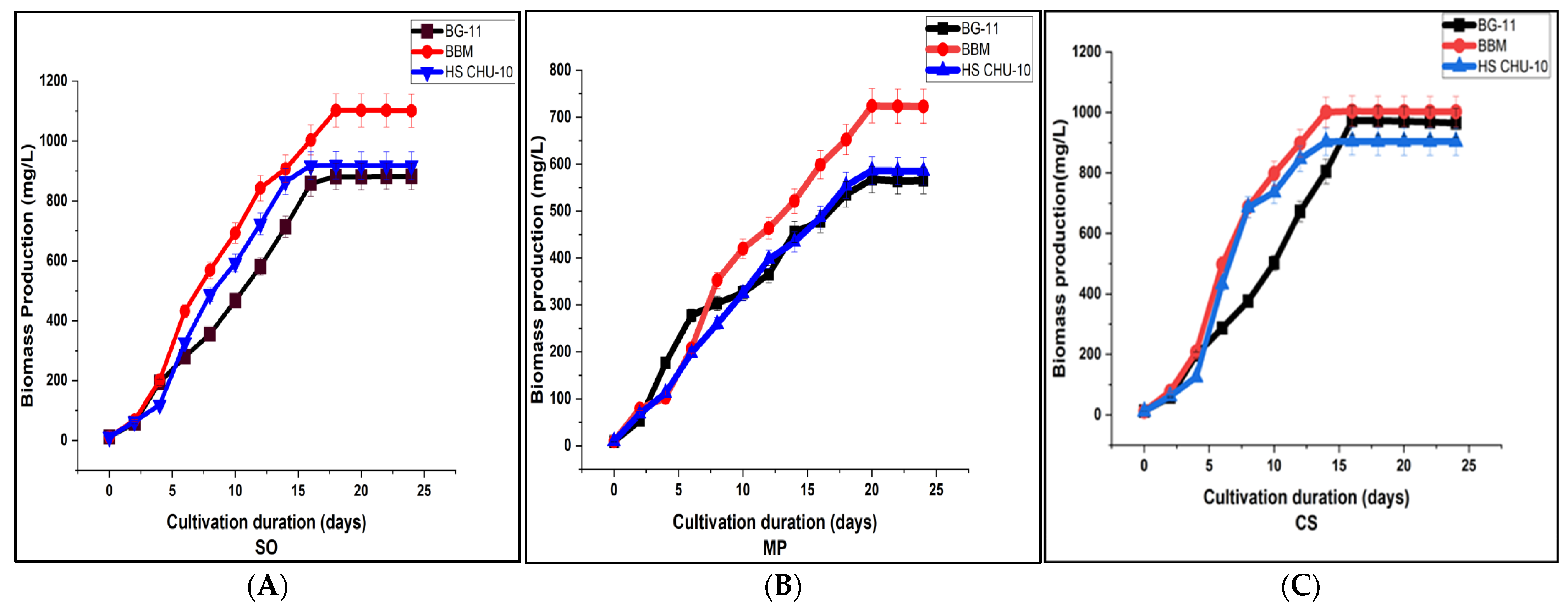
Figure 1. Graphical illustrations of a comparative growth examination of Scenedesmus obliquus (SO), Monoraphidium sp. (MP), and Chlorella sorokiniana (CS) in three different growth media: BG-11, BBM, and HS CHU-10. Where, (AC) represents growth curves of Scenedesmus obliquus, Monoraphidium sp., and Chlorella sorokiniana correspondingly.
Ijpb 14 00044 g001
Figure 2. Statistical analysis of the impacts of media compositions on the biomass productivity of different species of freshwater microalgae displayed by grouped mean graphical representation. Scenedesmus obliquus (SO), Monoraphidium sp. (MP), and Chlorella sorokiniana (CS) cultivated in three different growth media: BG-11, BBM, and HS CHU-10. Data are represented as mean ± SD, n = 3. * represents p < 0.05, and NS signifies non-significantly different.
Ijpb 14 00044 g002
Figure 3. Scenedesmus obliquus, Monoraphidium sp., and Chlorella sorokiniana development and lipid analyses in three different media. The microalgae biomass and lipids determined where (AC) corresponds to Scenedesmus obliquus (SO), Monoraphidium sp. (MP), and Chlorella sorokiniana (CS) respectively. Bars with the same letters were non-significantly different at p ≤ 0.05.
Ijpb 14 00044 g003
Figure 4. Pigments obtained from microalgae under three different media at specific time intervals. The microalgae pigments investigated where (AC) corresponds to Scenedesmus obliquus (SO), Monoraphidium sp. (MP), and Chlorella sorokiniana (CS) respectively. Bars with the same letters were non-significantly different at p ≤ 0.05.
Ijpb 14 00044 g004aIjpb 14 00044 g004b
Figure 5. Light micrographs and microalgae fluorescence images treated with Nile Red and viewed at 60× magnification, where (AC) represents Scenedesmus obliquus, Monoraphidium sp., and Chlorella sorokiniana, respectively. Dynamic duplex filters were used to differentiate between the presence of lipids (shown in red) and the presence of chlorophyll (shown in green) (scale bar 10 μm).
Ijpb 14 00044 g005
Figure 6. Thin-layer chromatography profile of carotenoids obtained from Scenedesmus obliquus (SO), Monoraphidium sp. (MP), and Chlorella sorokiniana (CS), along with astaxanthin standard (St1) and β-carotene (St2).
Ijpb 14 00044 g006
Table 1. Freshwater algal culture media compositions.
BG-11g/LBBMg/LHS-CHU10g/L
NaNO31.5NaNO30.25Ca(NO3)2.4H2O2
K2HPO40.04K2HPO40.075K2HPO40.25
MgSO4.7H2O0.075MgSO4.7H2O0.075MgSO4.7H2O1.25
CaCl2.2H2O0.036CaCl2.2H2O0.025Na2CO31
Na2CO30.02KH2PO40.175Na2SiO31.25
Na2EDTA0.001NaCl0.025FeCl30.04
Ferric ammonium citrate0.006H3BO30.011
Citric acid0.006FeSO4.7H2O0.004
Na2 EDTA0.05
KOH0.031
H2SO4 conc.1 µL
Micronutrients
g/L mg/L mg/L
H3BO32.86ZnSO4.7H2O8.82H3BO30.24
MnCl2.4H2O1.81MnCl2.4H2O0.44MnSO4.H2O0.147
ZnSO4.7H2O0.22MoO30.71ZnSO4.7H2O0.023
Na2MoO4.2H2O0.039CuSO4.5H2O1.57CuSO4.5H2O0.010
CuSO4.5H2O0.079Co(NO3)2.6H2O0.49(NH4)6Mo7O24.4H2O0.007
Co(NO3)2.6H2O0.049Vit B110 µgCo(NO3)2.6H2O0.014
Vit B1210 µgVit B15
Vit B72.5
Vit B122.5
Table 2. Time-independent pigment analysis showing the maximum concentrations of chlorophyll a, chlorophyll b, chlorophyll a + b, chlorophyll a/b, and total carotenoids obtained in three microalgal strains cultured in different media (BG-11, BBM, and HS CHU-10). Each statistic is the average of three replicates.
SpeciesPigment CompositionsBG-11BBMHS CHU10
Scenedesmus obliquusChlorophyll-a (µg/mL)12.8 ± 0.6413.9 ± 0.6913.2 ± 0.66
Chlorophyll-b (µg/mL)10.9 ± 0.5411.6 ± 0.5811.3 ± 0.56
Chlorophyll a + b (µg/mL)23.7 ± 1.1825.5 ± 1.2724.5 ± 1.22
Chlorophyll a/b1.17 ± 0.051.19 ± 0.051.16 ± 0.05
Carotenoid (µg/mL)7.6 ± 0.078.2 ± 0.087.9 ± 0.07
Monoraphidium sp.Chlorophyll-a (µg/mL)10.7 ± 0.5312.5 ± 0.6211.2 ± 0.56
Chlorophyll-b (µg/mL)9.5 ± 0.4710.2 ± 0.519.9 ± 0.49
Chlorophyll a + b (µg/mL)20.2 ± 1.0122.7 ± 1.1321.1 ± 1.05
Chlorophyll a/b1.12 ± 0.051.22 ± 0.061.13 ± 0.05
Carotenoid (µg/mL)6.8 ± 0.347.4 ± 0.378.9 ± 0.44
Chlorella sorokinianaChlorophyll-a (µg/mL)11.6 ± 0.5812.6 ± 0.6312.3 ± 0.61
Chlorophyll-b (µg/mL)9.7 ± 0.4810.8 ± 0.5410.2 ± 0.51
Chlorophyll a + b (µg/mL)21.3 ± 1.0623.4 ± 1.1722.5 ± 1.12
Chlorophyll a/b1.19 ± 0.051.16 ± 0.051.2 ± 0.06
Carotenoid (µg/mL)5.7 ± 0.286.2 ± 0.317.36 ± 0.36
Disclaimer/Publisher’s Note: The statements, opinions and data contained in all publications are solely those of the individual author(s) and contributor(s) and not of MDPI and/or the editor(s). MDPI and/or the editor(s) disclaim responsibility for any injury to people or property resulting from any ideas, methods, instructions or products referred to in the content.