Article

Root ABA Accumulation Delays Lateral Root Emergence in Osmotically Stressed Barley Plants by Decreasing Root Primordial IAA Accumulation

1
Ufa Institute of Biology, Ufa Federal Research Center, Russian Academy of Sciences, pr. Oktyabrya 69, 450054 Ufa, Russia
2
The Lancaster Environment Centre, Lancaster University, Lancaster LA1 4YQ, UK
*
Author to whom correspondence should be addressed.
Int. J. Plant Biol. 2023, 14(1), 77-90; https://doi.org/10.3390/ijpb14010007
Submission received: 28 November 2022 / Revised: 21 December 2022 / Accepted: 3 January 2023 / Published: 9 January 2023

Abstract

:
Increased auxin levels in root primordia are important in controlling root branching, while their interaction with abscisic acid (ABA) likely regulates lateral root development in water-deficient plants. The role of ABA accumulation in regulating root branching was investigated using immunolocalization to detect auxin (indoleacetic acid, IAA) and ABA (abscisic acid) in root primordia of the ABA-deficient barley mutant Az34 and its parental genotype (cv. Steptoe) barley plants. Osmotic stress strongly inhibited lateral root branching in Steptoe plants, but hardly affected Az34. Root primordial cells of Steptoe plants had increased immunostaining for ABA but diminished staining for IAA. ABA did not accumulate in root primordia of the Az34, and IAA levels and distribution were unaltered. Treating Az34 plants with exogenous ABA decreased root IAA concentration, while increasing root primordial ABA accumulation and decreasing root primordial IAA concentration. Although ABA treatment of Az34 plants increased the root primordial number, it decreased the number of visible emerged lateral roots. These effects were qualitatively similar to that of osmotic stress on the number of lateral root primordia and emerged lateral roots in Steptoe. Thus ABA accumulation (and its crosstalk with auxin) in root primordia seems important in regulating lateral root branching in response to water stress.

1. Introduction

Lateral root development is genetically and environmentally regulated, with the inhibition of lateral root development by low soil water availability considered an important adaptive response [1,2]. Nevertheless, extensive branching in the upper soil layers may allow root exploitation of light rainfalls that fail to infiltrate the soil to great depths [3]. Roots position new lateral branches according to the spatial distribution of available water in the substrate [4] and not in dry patches [5]. This adaptive response allows continued water uptake even as the soil dries.
Various growth hormones are involved in regulating root growth through complex signaling networks, with abscisic acid (ABA)–auxin crosstalk in lateral roots providing an adaptive strategy under drought stress conditions [6,7]. ABA likely exerts its repressive effects on lateral root formation through auxin [8]. The promotive effects of auxin on root branching might be inhibited by the repressive actions of ABA during lateral root emergence and initiation [9,10,11]. Auxin-sensitive reporter constructs such as DR5::GUS [12] and DR5::YFP [13] or immunohistochemical localization [14] have demonstrated auxin accumulation in root primordial cells. However, to the best of our knowledge, immunolocalization was not used to study the accumulation of ABA in the cells of lateral root primordia, even though immunohistochemistry determined ABA accumulation in the stele of Arabidopsis primary root tips [15] and the beginning of the root hair zone of primary barley roots [16]. While immunohistochemical approaches allow the spatial accumulation of ABA and IAA in water-stressed plants to be determined, they have not yet been applied together in studies aiming to determine the regulation of lateral root branching.
Mutants with an impaired ability to synthesize hormones under stress conditions provide a useful resource to investigate the importance of phytohormonal regulation of lateral root branching. For example, the ABA-deficient tomato mutants flacca and notabilis had more and longer lateral roots when grown in vitro compared to their wild-type (WT), even though all genotypes had the same primary root length [17]. The ABA-deficient Arabidopsis mutant nced3 had more and longer lateral roots (LRs) [18], while aba2-3, aba2-4 and aba3-2 also had more LR growth [19]. Disrupting genes responsible for the control of auxin synthesis also affects lateral root development in mutants [20]. Still, most of these studies were conducted under optimal conditions that may not adequately reflect the role of ABA (or indoleacetic acid (IAA) under water deficit.
Lateral root branching is important in regulating the responses of cereal crops to water deficit [21]. Moreover, ABA maintains primary root elongation [22] and inhibits root branching [23] of maize plants grown at low soil water availability. Nevertheless, lateral root growth of ABA-deficient cereals has attracted little attention. When grown in split-pots with distinct dry and wet soil compartments, both Steptoe plants and the ABA-deficient barley mutant Az34 showed a similar root biomass distribution between the two pots [24]. Nevertheless, Az34 had lower root ABA concentrations than Steptoe plants under optimal conditions [16] and failed to accumulate ABA in the roots when an atmospheric water deficit was imposed [25]. However, ABA and auxin concentrations in the cells of root primordia, and lateral root branching phenotypes of these genotypes have not been studied, irrespective of soil water availability. To test the importance of stress-induced changes in root hormone concentration, and their possible relationship to root branching, a histochemical technique enabled simultaneous immunolocalization of IAA and ABA in root sections. To determine the role of water-stress induced root ABA accumulation in these phenotypic responses, the ABA-deficient barley mutant Az34 and its Steptoe were grown hydroponically with an osmotic stress applied to simulate decreased soil water availability. Moreover, exogenous ABA was applied to the mutant in attempting to revert its phenotype. We hypothesized that root ABA accumulation was necessary to inhibit lateral root branching in response to water deficit, either independently or via its interaction with IAA.

2. Materials and Methods

2.1. Plant Material and Growth Conditions

Seeds of barley Hordeum vulgare L. (ABA deficient mutant Az34 and its wild-type cv. Steptoe) were germinated for 3 d in darkness at 21–24 °C on rafts made from sealed glass tubes tied together, which were then suspended over 0.1 strength Hoagland–Arnon nutrient medium (0.5 mM KNO3, 0.5 mM Ca(NO3)2, 0.1 mM KH2PO4, 0.1 mM MgSO4, 0.5 mM CaSO4) in 3-litre containers and grown at an irradiance of 400 µmol m–2 s–1 and a 14-h photoperiod. When seedlings were 3 days old, they were placed into containers with either polyethylene glycol (PEG 6000, 0.15 MPa) or 0.1 mg/L ABA in the nutrient solution. Three days after PEG addition, roots were fixed in a mixture of ethanol and glacial acetic acid (3:1), stained with acetocarmine and emerged lateral roots and their primordia (detected inside the primary roots) were counted under the microscope as described [26].

2.2. Exogenous ABA Treatment

Two days after adding 0.1 mg/L of exogenous ABA to plants grown under control conditions, roots of Az34 were harvested for hormone immunoassay, as previously described [25,27]. Hormones were extracted from homogenized roots of barley plants with 80% ethanol overnight at 4 °C. ABA, and the auxin IAA were partitioned with diethyl ether from the aqueous residue after its dilution with distilled water and acidification with HCl to pH 2.5. Then, the hormones were transferred from the organic phase into a solution of NaHCO3, re-extracted from the acidified aqueous phase with diethyl ether and immunoassayed after methylation using antibodies to ABA and IAA [26]. Reducing the amount of extractant at each stage of extraction, re-extraction increased the selectivity of hormone recovery [28]. Together with the approaches mentioned below, the specificity of immunoassay for ABA was confirmed in previous experiments, with fluridone-treated plants (which inhibits ABA biosynthesis) having lower ABA concentrations [29].

2.3. Immunolocalization of Hormones

Two days after adding PEG to the nutrient medium, 5 mm long root sections were collected at different positions (located from 10 to 45 mm) from the root tip. Root segments were fixed in 4% carbodiimide (1-ethyl-3-(3-dimethylaminopropyl) carbodiimide, (Merck, Darmstadt, Germany) for 4 h as described earlier [16,30] to prevent the washing out of hormones from tissues during dehydration. For better structural preservation, this procedure was followed by fixation with 4 % paraformaldehyde (Riedel de Haens, Seelze, Germany) for 10 h. During this process, carboxylic groups of ABA and IAA were linked with amino groups of proteins in the tissue. After dehydration with ethanol solutions of increasing grades (up to 96%), samples were embedded in the methacrylate resin (JB-4, Hatfield, PA, USA) as recommended by manufacturers. Histological sections (1.5 μm thick) were cut with a rotation microtome (HM 325, MICROM Laborgerate GmbH, Walldorf, Germany) and placed on slides. Sections were then incubated in 0.1 M Na-phosphate buffer (pH 7.3) containing 0.05% Tween 20 and 0.2% gelatin for 30 min and called below as PGT. Specific rabbit antisera against ABA [16,25,31,32] and IAA [26,30,33] were used for their immunolocalization. Anti-ABA or anti-IAA sera (20 μL) in PGT (1:80 and 1:40, respectively), were poured over some sections and incubated in a moist chamber at room temperature for 2 h. Some sections were incubated with the non-immune serum to check specificity of immunostaining. To visualize serum binding with ABA and IAA, sections were treated for 1 h at room temperature with the goat antibodies against rabbit immunoglobulin, which were labeled with colloidal gold (Aurion, Wageiningen, Netherlands). After three washes with 0.1 M Na-phosphate buffer containing 0.05% Tween 20, sections were treated with silver enhancer (Aurion, Wageiningen, Netherlands) for 15–20 min and examined under a light microscope. Excess silver was removed with distilled water. Preparations were visualized under an Axio Imager.A1 light microscope (Carl Zeiss, Jena, Germany) supplied with an AxioCam MRc5 digital camera (Carl Zeiss, Jena, Germany).
Intensity of immunostaining for IAA and ABA was estimated as described [34]. Staining values were averaged for the areas of primordia. Images were taken from 10 independent sections per treatment. Intensity of staining was expressed in arbitrary units: maximal staining was taken as 100%, and minimal staining was 0%.
Earlier specificity and reliability of immunostaining was confirmed in experiments, where increased immunostaining was detected in the plants treated with exogenous hormones (positive control), as well as by decreased immunostaining of the sections of primary roots of ABA-deficient mutant (negative control) [16]. In the present study, immunostaining for the two hormones of the identical sections, fixed in a similar way, confirmed that differences in staining were due to specificity of the antibodies.

2.4. Staining of Nuclei by Schiff Reagent

Sections were hydrolyzed with hydrochloric acid at 24 °C (1 M HCl, 5 min; 5 M HCl, 30 min; 1 M HCl, 5 min), slides were stained with Schiff’s reagent (Feulgen reaction for a period of 12 h and then washed in SO2-water (3 × 5 min) and H2O [35].

2.5. Statistics

Data were expressed as means ± S.E., which were calculated in all treatments using MS Excel. Significant differences between means were analyzed by a t-test.

3. Results

In the absence of osmotic stress, the Az34 mutant and its parental cultivar Steptoe had similar numbers of lateral roots and primordia (Figure 1). No lateral roots had emerged before PEG was added to the nutrient medium, and later changes in the total number of lateral roots were due to the osmotic stress treatment. Three days of osmotic stress decreased the number of lateral roots of Steptoe plants by 40% compared to the control plants, but had no effect on the ABA-deficient mutant (Figure 1). Osmotic stress increased the number of primordia in the Steptoe, but this response was attenuated in the mutant. Primary root length was similar in both genotypes, and not affected by the osmotic stress, at least over 3 days (Table 1).
In consecutive slides reflecting a developmental gradient from small primordia to lateral root emergence, when plants were grown under normal conditions the two genotypes did not differ in the level and distribution of IAA (Figure 2) and ABA (Figure 3) between the root cells (Figure 2). The subcellular localization of ABA and IAA clearly differed, the latter concentrating in the nuclei (nuclear localization was confirmed with the help of the Schiff reagent—Figure S1B). Interestingly, the preferential staining of nuclei for IAA was detected in primordia with elongating cells (Figure 2B,C,E,F) and was not observed at earlier stages of primordial development (Figure 2A,D). In primordia ready to emerge from the primary root, the distribution of immunostaining for the two hormones between the central cylinder and the cortex also differed (compare Figure 2B,C,E,F with Figure 3B,C,E,F). Closer to the base of these primordia, nuclei staining for IAA was greater in the cells of the central cylinder, while that of ABA was similar in intensity either in the central cylinder or the cortex. Interestingly, staining with the Schiff reagent revealed more stained nuclei in the emerging central cylinder or the cortex of the primordia (Figure S1B), while many of the cellular nuclei remained unstained in the case of IAA immunolocalization (Figure S1A). Thus, not all the nuclei were labeled with anti-IAA serum. Despite the difference in the subcellular localization of IAA and ABA, and their distribution between root zones during development of root primordia, there were no differences between the genotypes in staining for each of the hormones in the absence of osmotic stress.
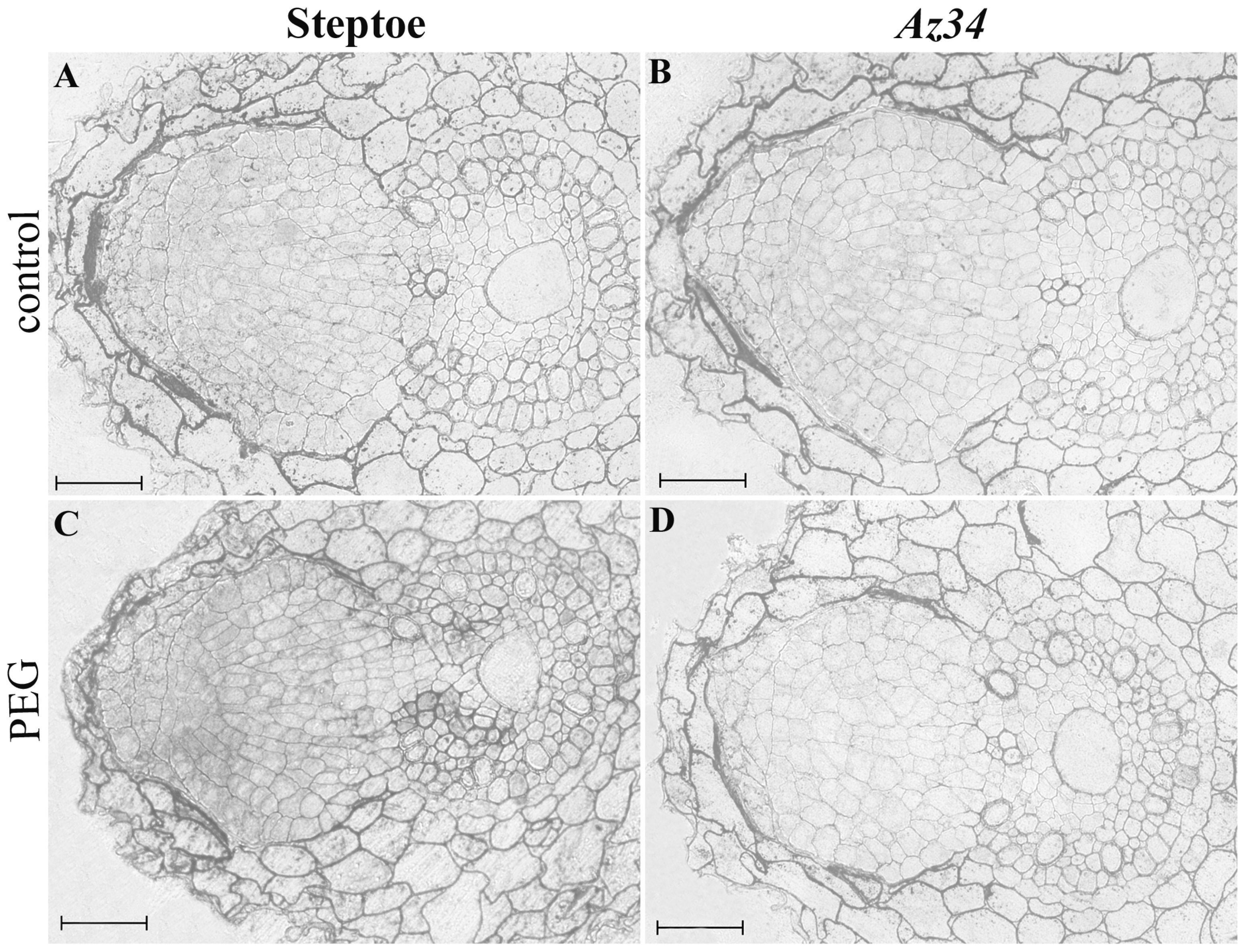
Although there were no genotypic differences in the intensity of staining of developing primordia for either hormone when grown under optimal conditions, under osmotic stress the subcellular localization and distribution of ABA and IAA between the cells of the stele and cortex differed between genotypes (Figure 4 and Figure 5). Osmotic stress increased staining of primordial cells for ABA in Steptoe plants, but did not change this in Az34 (Figure 4). For IAA a different pattern was detected, with weaker immunostaining in the stressed Steptoe than the mutant (Figure 5). Increased staining of root primordial cells for ABA in the stressed Steptoe plants, and their weakened staining for IAA, was statistically significant as shown by semi-quantitative analysis of the staining with the help of the ImageJ program (Figure 6). The difference in root staining between stressed plants of the two genotypes was not only in root cell IAA levels, but also in the subcellular distribution of the hormone. Thus increased nuclei staining for IAA disappeared in the root cells of stressed Steptoe plants (Figure 5C), but remained in Az34 (Figure 5D). Thus the capacity of Steptoe plants to accumulate ABA in primordial cells under stress conditions was accompanied by decreased IAA level in those cells, while the absence of ABA changes in the mutant may be related to the absence of changes in IAA.
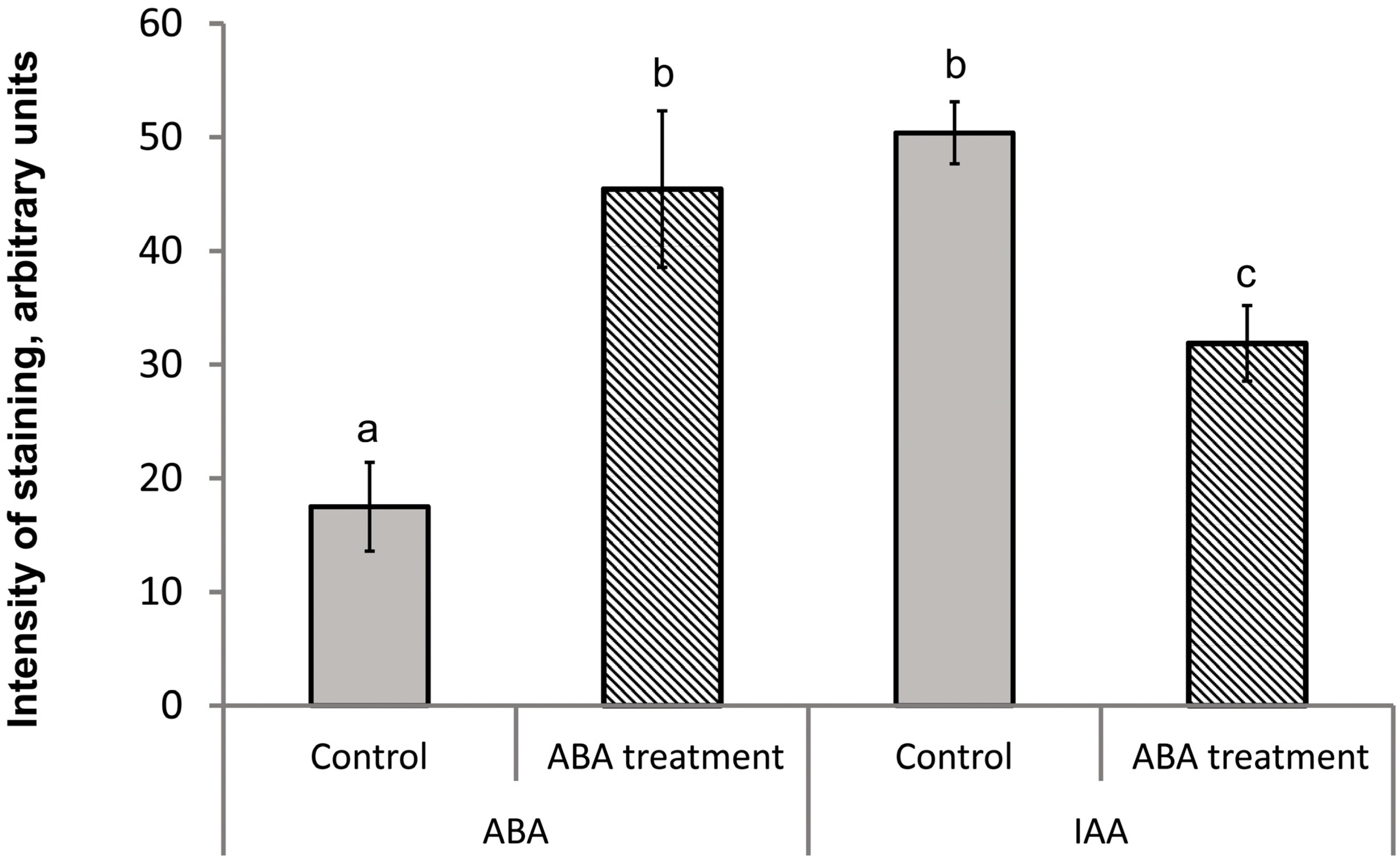
To confirm whether root ABA status affected the pattern of root IAA concentration and lateral branching, ABA was added to the nutrient solution of Az34 plants grown under optimal conditions (Figure 7). This treatment decreased bulk root IAA concentration by 34% and the number of lateral roots by 56%, but increased the number of root primordia by 55% as compared to plants untreated with ABA. ABA treatment had no effect on the primary root length in this experiment, but increased immunostaining for ABA. Such ABA accumulation was accompanied by weakened immunostaining for IAA (Figure 8 and Figure 9), while nuclear staining for IAA disappeared as in PEG-treated Steptoe plants (Figure 5). Thus exogenous ABA treatment was able to phenotypically revert lateral root branching and IAA response of the ABA-deficient mutant, making them similar to Steptoe plants under stress conditions.

4. Discussion

Both auxins and ABA can influence lateral root development, which is likely important for regulating growth responses to water deficit. Since ABA’s effects on root branching may be due to its cross-talk with IAA, we simultaneously determined localization of these hormones in the cells of root primordia under normal conditions and moderate PEG-induced water deficit. Differential ABA and IAA accumulation in root primordia of Steptoe plants and the ABA-deficient barley mutant Az34 under osmotic stress was correlated with inhibition of lateral root branching in the parental cultivar plants, but no such effect in the mutant. Exogenous ABA treatment of Az34 restored a parental cultivar response, further supporting the hypothesis that root ABA accumulation inhibits lateral root emergence at low soil water availability.
Root primordial nuclei of both genotypes were immuno-labelled for IAA, but not for ABA (Figure 2, Figure 3 and Figure S1). The presence of IAA in the nuclei is not surprising as the auxin TIR1 receptor is localized in the nuclei and auxin perception is known to be a nuclear effect [36]. Although the PYR/PYL/RCAR family of ABA receptors is also nuclear localized [37,38], the absence of nuclei immunostaining for ABA suggests that ABA signaling in root primordia uses other receptors, such as the GCR1 receptor located in the plasma membrane [39]. Previous studies indicate more intensive staining for ABA in the central cylinder of Arabidopsis [15] and barley [16] primary roots, with the maximum level in a single cell layer surrounding the root stele [15]. This pattern was not detected in barley root primordia, although the root central cylinder was expected to show increased staining for ABA when emerged lateral roots are studied.
Increased immunostaining for ABA of primordial cells of ABA-treated roots agrees with data obtained for primary root tips [16] and confirmed the reliability and specificity of immunostaining for this hormone (positive control). Interestingly, immunostaining for IAA followed an opposite pattern to ABA. In parental cultivar plants, more intensive staining for ABA in the primordia of stressed roots contrasted with weaker immunostaining for IAA in control (unstressed) roots. Exogenous ABA treatment also decreased staining for IAA of primordial cells of Az34 plants. Lower immunostaining for IAA of stressed Steptoe primordia as well as ABA-treated Az34 plants agrees with data showing that drought and ABA treatments may decrease auxin level by up-regulating GH3 genes that encode enzymes that conjugate amino acids to IAA, particularly in the lateral root primordia [6]. Thus, ABA may modulate auxin-signaling by inducing GH3 enzyme genes, ultimately reducing lateral root formation. Alternatively, changes in auxin transporter levels and localization may reduce root auxin concentrations in an ABA-dependent [11,40] and osmotic stress dependent manner [7].
When osmotically stressed, the ABA-deficient Az34 barley mutant had more lateral roots than its parental cultivar Steptoe (Figure 1). Thus, cereals and Arabidopsis seem to regulate lateral root growth similarly, since ABA-deficient mutants (such as the biosynthetic mutant aba2-1) have greater lateral root development than Steptoe plants under osmotic stress [1]. Both osmotic stress and ABA-treatment abolished preferential staining for IAA of the root cell nuclei of Steptoe plants, correlating with the decreased branching. Since this differential subcellular staining was absent at early stages of primordial development, increased IAA levels in root cell nuclei are likely important for lateral root emergence. Under osmotic stress, this increased staining of the root cell nuclei was retained in Az34 plants, and the lateral root number was maintained. These results suggest ABA accumulation prevents IAA accumulation in the nuclei of stressed plants, thereby delaying lateral emergence.
For the first time, immunohistochemistry demonstrated greater staining for ABA in lateral roots of stressed Steptoe plants but not in the ABA deficient mutant. Stressed Steptoe plants had fewer lateral roots but more root primordia than the control Steptoe plants. In both PEG-treated or control plants, the sum of primordia and emerged lateral roots remained the same, suggesting that stress-induced ABA accumulation inhibited root emergence, as previously reported ([10] and references therein). Although ABA accumulation maintains primary root elongation in stressed plants [41], an opposing effect on lateral root emergence has been detected [1,40,42]. Further experiments are needed to explain this discrepancy. Although osmotic stress increases bulk root ABA accumulation ([43] and references therein), ABA accumulation in primordial cells (Figure 4) likely inhibited lateral root emergence. Despite osmotic stress, the lack of ABA accumulation in root primordia of the ABA-deficient mutant Az34 allowed normal lateral root emergence.
This comparative study of immunolocalization of ABA and IAA in the ABA-deficient barley mutant Az34 and its parental cultivar Steptoe revealed root primordial hormone accumulation. Stress-induced ABA accumulation in Steptoe plants correlated with decreased IAA concentrations and changes in the intracellular distribution of IAA. The Az34 mutant did not accumulate ABA under osmotic stress, but exogenous ABA treatment decreased root IAA concentration of this genotype (Figure 7A and Figure 8) and decreased lateral root number while increasing the number of primordia. These effects mimicked the effects of stress-induced ABA accumulation in Steptoe primordia. Thus, stress-induced changes in hormone levels and their cross-talk in the cells of root primordia likely regulate root branching.

Supplementary Materials

The following supporting information can be downloaded at: https://www.mdpi.com/article/10.3390/ijpb14010007/s1, Figure S1. Immunolocalization of IAA (A) and staining with the Schiff reagent (B) of developing lateral root Steptoe primordia. Scale bar is 50 μm.

Author Contributions

Conceptualization, G.S., G.K. and I.C.D.; methodology, D.V., G.A., R.I. and G.S.; software, I.I.; formal analysis, D.V., G.A., R.I. and G.S.; investigation, D.V., G.A., R.I. and G.S.; resources, D.V.; data curation, G.S; writing—original draft preparation, G.K.; writing—review and editing, I.C.D. and G.S.; visualization, G.A. and G.S.; supervision, G.K.; project administration, G.K.; funding acquisition, G.S. All authors have read and agreed to the published version of the manuscript.

Funding

This research was funded by Ministry of Science and Higher Education of Russia, grant number AAAA-A18-118022190099-6 and Russian Foundation for Basic Research, grant number 20-04-00305.

Institutional Review Board Statement

Not applicable.

Informed Consent Statement

Not applicable.

Data Availability Statement

Not applicable.

Conflicts of Interest

The authors declare no conflict of interest.

References

  1. Xiong, L.; Wang, R.G.; Mao, G.; Koczan, J.M. Identification of drought tolerance determinants by genetic analysis of root response to drought stress and abscisic acid. Plant Physiol. 2006, 142, 1065–1074. [Google Scholar] [CrossRef] [PubMed] [Green Version]
  2. Feng, W.; Lindner, H.; Robbins II, N.E.; Dinneny, J.R. Growing out of stress: The role of cell- and organ-scale growth control in plant water-stress responses. Plant Cell 2016, 28, 1769–1782. [Google Scholar] [CrossRef] [PubMed] [Green Version]
  3. Hodge, A. Roots: The acquisition of water and nutrients from the heterogeneous soil environment. Prog. Bot. 2010, 71, 307–337. [Google Scholar] [CrossRef]
  4. Robbins II, N.E.; Dinneny, J.R. The divining root: Moisture-driven responses of roots at the micro- and macro-scale. J. Exp. Bot. 2015, 66, 2145–2154. [Google Scholar] [CrossRef] [PubMed] [Green Version]
  5. Orman-Ligeza, B.; Morris, E.C.; Parizot, B.; Lavigne, T.; Babé, A.; Ligeza, A.; Klein, S.; Sturrock, C.; Xuan, W.; Novák, O.; et al. The xerobranching response represses lateral root formation when roots are not in contact with water. Curr Biol. 2018, 28, 3165–3173. [Google Scholar] [CrossRef] [Green Version]
  6. Seo, P.J.; Xiang, F.; Qiao, M.; Park, J.Y.; Lee, Y.N.; Kim, S.G.; Lee, Y.H.; Park, W.J.; Park, C.M. The MYB96 transcription factor mediates abscisic acid signaling during drought stress response in Arabidopsis. Plant Physiol. 2009, 151, 275–289. [Google Scholar] [CrossRef] [PubMed] [Green Version]
  7. Rowe, J.H.; Topping, J.F.; Liu, J.; Lindsey, K. Abscisic acid regulates root growth under osmotic stress conditions via an interacting hormonal network with cytokinin, ethylene and auxin. New Phytol. 2016, 211, 225–239. [Google Scholar] [CrossRef] [Green Version]
  8. Emenecker, R.J.; Strader, L.C. Auxin-abscisic acid interactions in plant growth and development. Biomolecules 2020, 10, 281. [Google Scholar] [CrossRef] [Green Version]
  9. De Smet, I.; Signora, L.; Beeckman, T.; Inzé, D.; Foyer, C.H.; Zhang, H. An abscisic acid-sensitive checkpoint in lateral root development of Arabidopsis. Plant J. 2003, 33, 543–555. [Google Scholar] [CrossRef] [Green Version]
  10. Harris, J.M. Abscisic acid: Hidden architect of root system structure. Plants 2015, 4, 548–572. [Google Scholar] [CrossRef]
  11. Lu, C.; Chen, M.-X.; Liu, R.; Zhang, L.; Hou, X.; Liu, S.; Ding, X.; Jiang, Y.; Xu, J.; Zhang, J.; et al. Abscisic acid regulates auxin distribution to mediate maize lateral root development under salt stress. Front. Plant Sci. 2019, 10, 716. [Google Scholar] [CrossRef] [PubMed]
  12. Pérez-Torres, C.A.; López-Bucio, J.; Cruz-Ramírez, A.; Ibarra-Laclette, E.; Dharmasiri, S.; Estelle, M.; Herrera-Estrella, L. Phosphate availability alters lateral root development in Arabidopsis by modulating auxin sensitivity via a mechanism involving the TIR1 auxin receptor. Plant Cell 2008, 20, 3258–3272. [Google Scholar] [CrossRef] [PubMed] [Green Version]
  13. Yang, J.; Yuan, Z.; Meng, Q.; Huang, G.; Périn, C.; Bureau, C.; Meunier, A.C.; Ingouff, M.; Bennett, M.J.; Liang, W.; et al. Dynamic regulation of auxin response during rice development revealed by newly established hormone biosensor markers. Front Plant Sci. 2017, 7, 256. [Google Scholar] [CrossRef] [PubMed] [Green Version]
  14. Benková, E.; Michniewicz, M.; Sauer, M.; Teichmann, T.; Seifertová, D.; Jürgens, G.; Friml, J. Local, efflux-dependent auxin gradients as a common module for plant organ formation. Cell 2003, 115, 591–602. [Google Scholar] [CrossRef] [PubMed] [Green Version]
  15. Ondzighi-Assoume, C.A.; Chakraborty, S.; Harris, J.M. Environmental nitrate stimulates abscisic acid accumulation in Arabidopsis root tips by releasing it from inactive stores. Plant Cell 2016, 3, 729–745. [Google Scholar] [CrossRef] [PubMed] [Green Version]
  16. Sharipova, G.; Veselov, D.; Kudsoyarova, G.; Fricke, W.; Dodd, I.C.; Katsuhara, M.; Furuichi, T.; Ivanov, I.; Veselov, S. Exogenous application of abscisic acid (ABA) increases root and cell hydraulic conductivity and abundance of some aquaporin isoforms in the ABA deficient barley mutant Az34. Ann. Bot. 2016, 118, 777–785. [Google Scholar] [CrossRef] [Green Version]
  17. Belimov, A.A.; Dodd, I.C.; Safronova, V.I.; Dumova, V.A.; Shaposhnikov, A.I.; Ladatko, A.G.; Davies, W.J. Abscisic acid metabolizing rhizobacteria decrease ABA concentrations in planta and alter plant growth. Plant Physiol. Biochem. 2014, 74, 84–91. [Google Scholar] [CrossRef]
  18. Guo, D.; Liang, J.; Li, L. Abscisic acid (ABA) inhibition of lateral root formation involves endogenous ABA biosynthesis in Arachis hypogaea L. Plant Growth Regul. 2009, 58, 173–179. [Google Scholar] [CrossRef]
  19. Signora, L.; De Smet, I.; Foyer, C.H.; Zhang, H. ABA plays a central role in mediating the regulatory effects of nitrate on root branching in Arabidopsis. Plant J. 2001, 28, 655–662. [Google Scholar] [CrossRef] [Green Version]
  20. Olatunji, D.; Geelen, D.; Verstraeten, I. Control of endogenous auxin levels in plant root development. Int. J. Mol. Sci. 2017, 18, 2587. [Google Scholar] [CrossRef]
  21. Comas, L.H.; Becker, S.R.; Cruz, V.M.; Byrne, P.F.; Dierig, D.A. Root traits contributing to plant productivity under drought. Front. Plant Sci. 2013, 4, 442. [Google Scholar] [CrossRef] [PubMed] [Green Version]
  22. Saab, I.N.; Sharp, R.E.; Pritchard, J. Effect of inhibition of ABA accumulation on the spatial distribution of elongation in the primary root and mesocotyl of maize at low water potentials. Plant Physiol. 1992, 99, 26–33. [Google Scholar] [CrossRef] [PubMed]
  23. Friero, I.; Alarcón, M.V.; Gordillo, L.; Salguero, J. Abscisic acid is involved in several processes associated with root system architecture in maize. Acta Physiol. Plant. 2022, 44, 28. [Google Scholar] [CrossRef]
  24. Martin-Vertedor, A.I.; Dodd, I.C. Root-to-shoot signalling when soil moisture is heterogeneous: Increasing the proportion of root biomass in drying soil inhibits leaf growth and increases leaf abscisic acid concentration. Plant Cell Environ. 2011, 34, 1164–1175. [Google Scholar] [CrossRef] [PubMed]
  25. Veselov, D.S.; Sharipova, G.V.; Veselov, S.Y.; Dodd, I.C.; Ivanov, I.; Kudoyarova, G.R. Rapid changes in root HvPIP2;2 aquaporins abundance and ABA concentration are required to enhance root hydraulic conductivity and maintain leaf water potential in response to increased evaporative demand. Funct. Plant Biol. 2018, 45, 143–149. [Google Scholar] [CrossRef] [PubMed] [Green Version]
  26. Vysotskaya, L.B.; Korobova, A.V.; Kudoyarova, G.R. Abscisic acid accumulation in the roots of nutrient-limited plants: Its impact on the differential growth of roots and shoots. J. Plant Physiol. 2008, 165, 1274–1279. [Google Scholar] [CrossRef]
  27. Veselov, D.S.; Sharipova, G.V.; Veselov, S.U.; Kudoyarova, G.R. The effects of NaCl treatment on water relations, growth and ABA content in barley cultivars differing in drought tolerance. J. Plant Growth Regul. 2008, 27, 380–386. [Google Scholar] [CrossRef]
  28. Veselov, S.U.; Kudoyarova, G.R.; Egutkin, N.L.; Gyuli-Zade, V.G.; Mustafina, A.R.; Kof, E.K. Modified solvent partitioning scheme providing increased specificity and rapidity of immunoassay for indole-3-acetic acid. Physiol. Plant 1992, 86, 93–96. [Google Scholar] [CrossRef]
  29. Vysotskaya, L.B.; Korobova, A.V.; Veselov, S.Y.; Dodd, I.C.; Kudoyarova, G.R. ABA mediation of shoot cytokinin oxidase activity: Assessing its impacts on cytokinin status and biomass allocation of nutrient deprived durum wheat. Funct. Plant Biol. 2009, 36, 66–72. [Google Scholar] [CrossRef]
  30. Seldimirova, O.A.; Kudoyarova, G.R.; Kruglova, N.N.; Zaytsev, D.Y.; Veselov, S.Y. Changes in distribution of zeatin and indole-3-acetic acid in cells during callus induction and organogenesis in vitro in immature embryo culture of wheat. In Vitro Cell Dev. Biol.-Plant 2016, 52, 251. [Google Scholar] [CrossRef]
  31. Vysotskaya, L.B.; Veselov, S.Y.; Kudoyarova, G.R. Effect on shoot water relations, and cytokinin and abscisic acid levels of inducing expression of a gene coding for isopentenyltransferase in roots of transgenic tobacco plants. J. Exp. Bot. 2010, 61, 3709–3717. [Google Scholar] [CrossRef] [PubMed] [Green Version]
  32. Kudoyarova, G.; Veselova, S.; Hartung, W.; Farhutdinov, R.; Veselov, D.; Sharipova, G. Involvement of root ABA and hydraulic conductivity in the control of water relations in wheat plants exposed to increased evaporation demand. Planta 2011, 233, 87–94. [Google Scholar] [CrossRef] [PubMed]
  33. Vysotskaya, L.B.; Veselov, S.Y.; Veselov, D.S.; Filippenko, V.N.; Ivanov, E.A.; Ivanov, I.I.; Kudoyarova, G.R. Immunohistological localization and quantification of IAA in studies of root growth regulation. Russ. J. Plant Physiol. 2007, 54, 827–832. [Google Scholar] [CrossRef]
  34. Veselov, S.Y.; Timergalina, L.N.; Akhiyarova, G.R.; Kudoyarova, G.R.; Korobova, A.V.; Ivanov, I.I.; Arkhipova, T.N.; Prinsen, E. Study of cytokinin transport from shoots to roots of wheat plants is informed by a novel method of differential localization of free cytokinin bases or their ribosylated forms by means of their specific fixation. Protoplasma 2018, 255, 1581–1594. [Google Scholar] [CrossRef]
  35. Ozerov, I.A.; Zhinkina, N.A.; Efimov, A.M.; Machs, E.M.; Rodionov, A.V. Feulgen-positive staining of the cell nuclei in fossilized leaf and fruit tissues of the Lower Eocene Myrtaceae. Bot. J. Linn 2006, 150, 315–321. [Google Scholar] [CrossRef]
  36. McSteen, P.; Zhao, Y. Plant hormones and signaling: Common themes and new developments. Dev. Cell 2008, 14, 467–473. [Google Scholar] [CrossRef] [Green Version]
  37. Ma, Y.; Szostkiewicz, I.; Korte, A.; Moes, D.; Yang, Y.; Christmann, A.; Grill, E. Regulators of PP2C phosphatase activity function as abscisic acid sensors. Science 2009, 324, 1064–1068. [Google Scholar] [CrossRef]
  38. Park, S.Y.; Fung, P.; Nishimura, N.; Jensen, D.R.; Fujii, H.; Zhao, Y.; Lumba, S.; Santiago, J.; Rodrigues, A.; Chow, T.F.; et al. Abscisic acid inhibits type 2C protein phosphatases via the PYR/PYL family of START proteins. Science 2009, 324, 1068–1071. [Google Scholar] [CrossRef] [Green Version]
  39. Wang, X.F.; Zhang, D.P. Abscisic acid receptors: Multiple signal perception sites. Ann. Bot. 2008, 101, 311–317. [Google Scholar] [CrossRef] [Green Version]
  40. Shkolnik-Inbar, D.; Bar-Zvi, D. ABI4 mediates abscisic acid and cytokinin inhibition of lateral root formation by reducing polar auxin transport in Arabidopsis. Plant Cell 2010, 22, 3560–3573. [Google Scholar] [CrossRef]
  41. Sharp, R.E.; Wu, Y.; Voetberg, G.S.; Saab, I.N.; LeNoble, M.E. Confirmation that abscisic acid accumulation is required for maize primary root elongation at low water potentials. J. Exp. Bot. 1994, 45, 1743–1751. [Google Scholar] [CrossRef]
  42. Duan, L.; Dietrich, D.; Ng, C.H.; Chan, P.M.Y.; Bhalerao, R.; Bennett, M.L.; Dinneny, J.R. Endodermal ABA signaling promotes lateral root quiescence during salt stress in Arabidopsis seedlings. Plant Cell 2013, 25, 324–341. [Google Scholar] [CrossRef] [PubMed] [Green Version]
  43. Kudoyarova, G.; Dodd, I.C.; Veselov, D.S.; Rothwell, S.A.; Veselov, S.U. Common and specific responses to availability of mineral nutrients and water. J. Exp. Bot. 2015, 66, 2133–2144. [Google Scholar] [CrossRef] [PubMed]
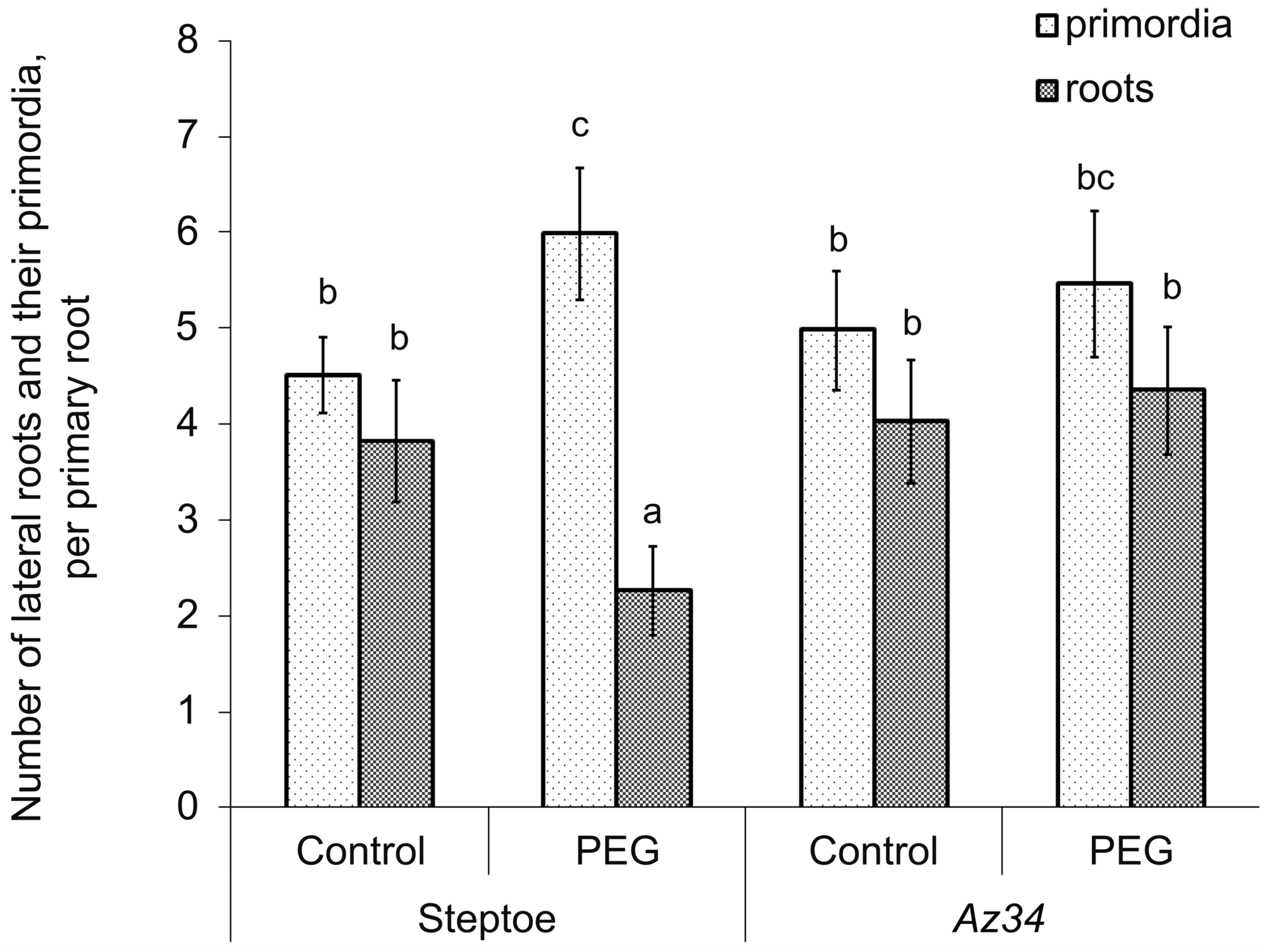
Figure 1. Number of lateral roots and their primordia in 6-day-old ABA-deficient Az34 barley plants and their parental cultivar Steptoe grown under control conditions, or exposed for 3 days to 6 % polyethylene glycol (PEG). Significant differences are marked with different letters. Values are means ± S.E. (n = 25) (p ≤ 0.05, t-test).
Ijpb 14 00007 g001
Figure 2. Immunostaining of IAA in root primordia of Steptoe (AC) and Az34 (DF) at successive stages of their development. Scale bar is 50 μm.
Ijpb 14 00007 g002
Figure 3. Immunostaining of ABA in root primordia of Steptoe (AC) and Az34 (DF) at successive stages of their development. Scale bar is 50 μm.
Ijpb 14 00007 g003
Figure 4. Effect of 6% polyethylene glycol (PEG) treatment on immunolocalization of ABA in the cells of root primordia of Steptoe (A,C) and Az34 (B,D) plants; (A,B)—plants grown under normal conditions; (C,D)—stressed plants. Scale bar is 50 μm.
Ijpb 14 00007 g004
Figure 5. Effect of 6% polyethylene glycol (PEG) treatment on immunolocalization of IAA in the cells of root primordia of Steptoe (A,C) and Az34 (B,D) plants; (A,B)—plants grown under normal conditions; (C,D)—stressed plants. Scale bar is 50 μm.
Ijpb 14 00007 g005
Figure 6. Intensity of immunostaining for ABA (A) and IAA (B) of root primordia of Az34 and Steptoe plants grown under normal or stress conditions (6% polyethylene glycol (PEG)). Images were taken from the sections presented in Figure 2, Figure 3, Figure 5 and Figure 6. Means (arbitrary units, maximal staining taken as 100%, minimal—as 0%). Significant differences are marked with different letters. Values are means ± S.E. (n = 25) (p ≤ 0.05, t-test).
Ijpb 14 00007 g006
Figure 7. ABA and IAA content in roots (A) and numbers of primordia and lateral roots (B) in ABA-deficient Az34 barley plants grown under control conditions or exposed for 3 days to 0.1 mg/L ABA. Significantly different means from control are marked with asterisk. Values are means ± S.E. (n = 25) (p ≤ 0.05, t-test).
Ijpb 14 00007 g007
Figure 8. Effect of 0.1 mg/L ABA treatment on immunolocalization of ABA (A,B) and IAA (C,D) in the cells of root primordia of ABA–deficient Az34 barley plants. (A,C)—plants grown under normal conditions; (B,D)—plants grown under ABA treatment for 3d. Scale bar is 50 μm.
Ijpb 14 00007 g008
Figure 9. Intensity of immunostaining for ABA and IAA of root primordia of Az34 grown under normal conditions or under 0.1 mg/L ABA treatment. Means (arbitrary units, maximal staining taken as 100%, minimal—as 0%). Significant differences are marked with different letters. Values are means ± S.E. (n = 25) (p ≤ 0.05, t-test).
Ijpb 14 00007 g009
Table 1. The length of all seminal roots (mm) of Steptoe and Az34 grown under normal conditions and polyethylene glycol (PEG) treatment. Data are means ± SE of 50 replicates. No statistical differences were detected in these experiments.
GenotypesNormal ConditionsPEG Treatment
Steptoe199 ± 6206 ± 11
Az34184 ± 9201 ± 8
Disclaimer/Publisher’s Note: The statements, opinions and data contained in all publications are solely those of the individual author(s) and contributor(s) and not of MDPI and/or the editor(s). MDPI and/or the editor(s) disclaim responsibility for any injury to people or property resulting from any ideas, methods, instructions or products referred to in the content.