1. Introduction
Alport syndrome (AS) is characterized by progressive renal disease, sensorineural hearing loss, and ocular abnormalities [
1]. AS has three forms: X-linked AS (the most common, about 80%), autosomal recessive AS (about 15%), and autosomal dominant AS (about <5%) [
2].
X-linked AS is caused by variants of COL4A5 on chromosome X, autosomal recessive AS by homozygous or compound heterozygous rare variants of COL4A3 or COL4A4, and autosomal dominant AS (ADAS) by heterozygous rare variants of COL4A3 and/or COL4A4 genes, all encoding type 4 collagen.
AS is frequently identified through familial hematuria. Previously, if asymptomatic hematuria was familial and renal failure was absent, benign familial hematuria, such as thin basement membrane nephropathy, was considered. Benign familial hematuria was historically regarded as a harmless condition in which invasive diagnostic procedures such as renal biopsy were considered unnecessary. However, this descriptive term is now being replaced by genetically defined diagnoses, particularly COL4A3- and COL4A4-associated disease, as these conditions are increasingly recognized to have potential for renal progression. In the present case, we consider discontinuation of follow-up once the proband reached adulthood; just before doing so, a diagnosis of ADAS was established.
2. Case Report
The proband, a 7-year-old girl, first presented to our department with microhematuria detected through school urinary screening. Her microhematuria had been detected through urinary screening for 3-year-old children in Japan. Urinalysis demonstrated microhematuria (20–29 red blood cells/high-power field) without kidney function impairment. Her mother similarly had microhematuria with normal renal function without prior hearing loss or ocular abnormalities. Although the mother was estranged from her parents, her family history was unremarkable for renal disease, and we confirmed the diagnosis of benign familial microhematuria. The proband developed normally with no hearing loss or ocular abnormalities and was followed up until age 20 as having benign familial hematuria. Once the proband reached 20 years of age, we considered the discontinuation of follow-up.
Around that time, her mother developed sensorineural hearing loss at 42 years old. She also reconnected with her father (the proband’s grandfather) after a long period of estrangement, and found that he was undergoing dialysis for end-stage renal failure of unknown etiology. Based on the clinical suspicion of AS, genetic testing was conducted sequentially—first in the mother and then in the proband—after obtaining informed consent. Genetic testing was performed by the Kazusa DNA Research Institute. Both were found to carry a heterozygous variant (c.2317_2318del; p.Arg773GlyfsTer14) of
COL4A4 (NM_000092.5) [
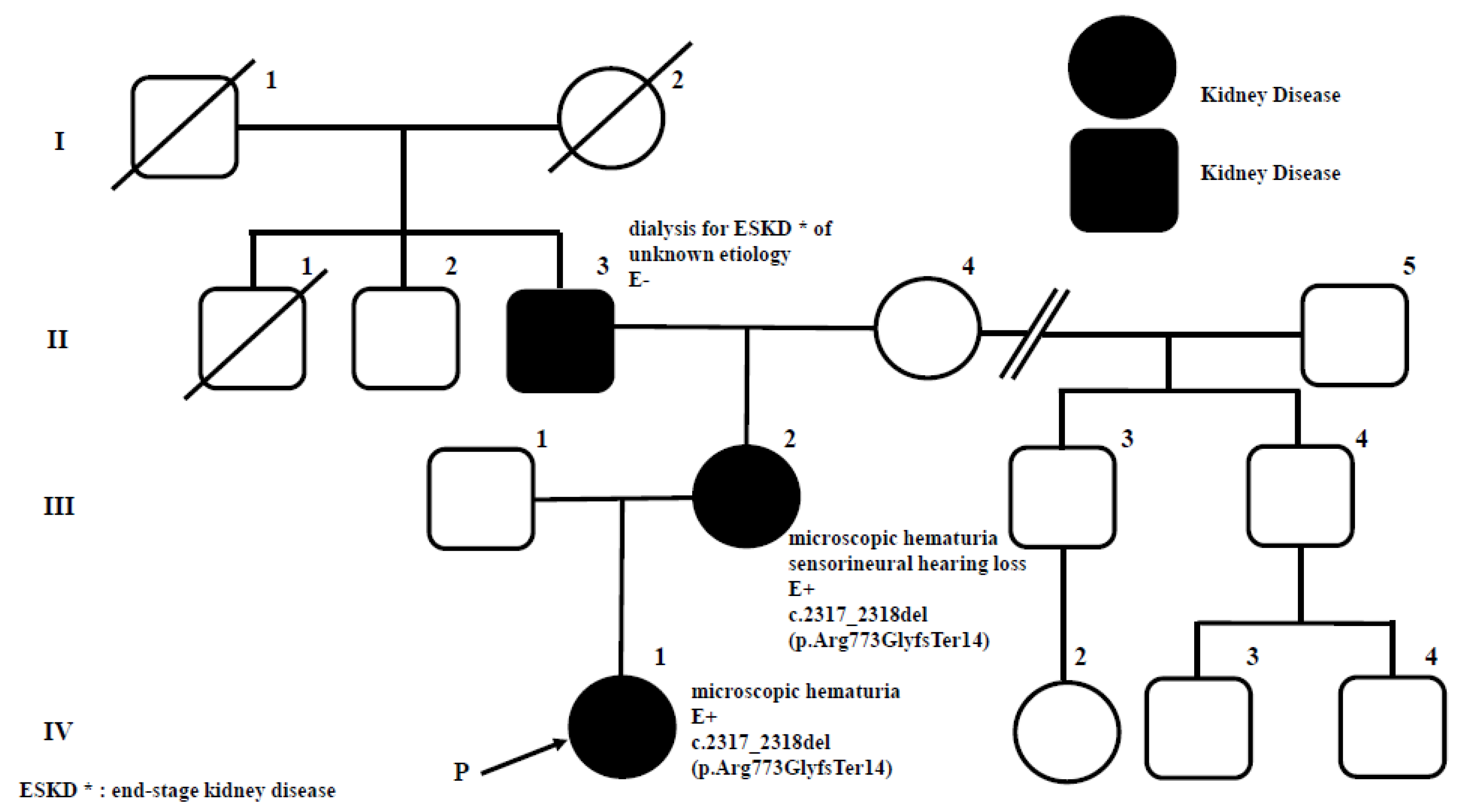
Supplementary Materials]. The pedigree chart is shown in
Figure 1. Because the proband’s mother had long been estranged from her parents, detailed medical histories of other family members could not be obtained. Thus, only the information shown in
Figure 1 was available for constructing the pedigree.
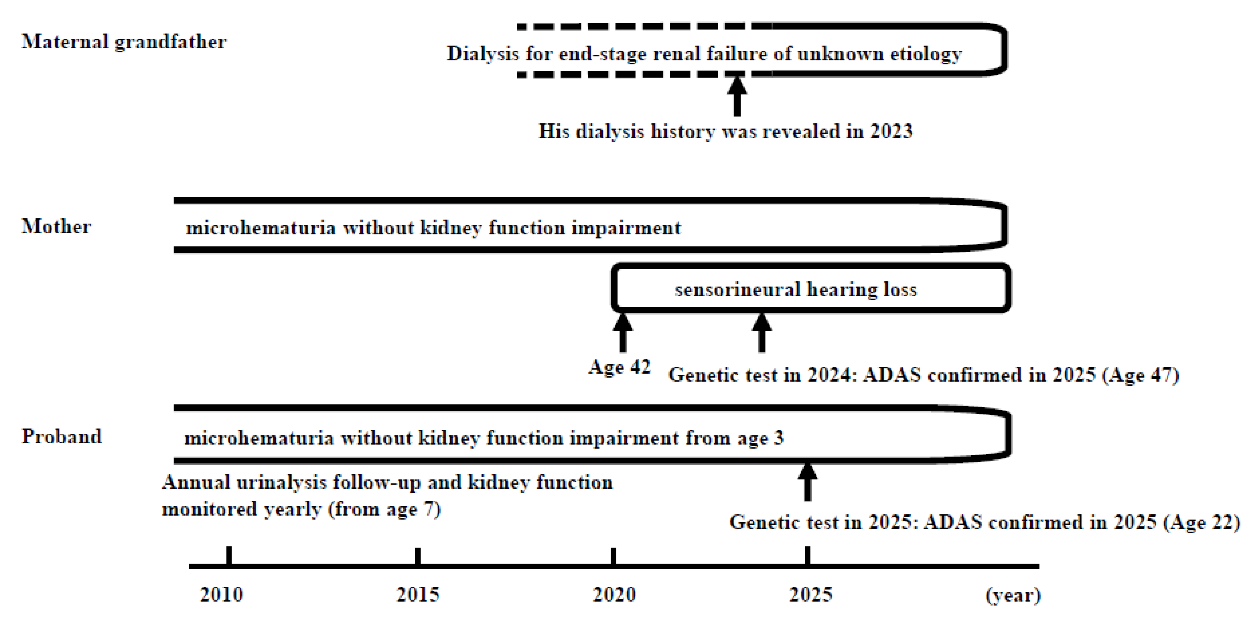
Genetic analysis confirmed the diagnosis of ADAS in the proband and her mother. The timeline of the clinical course is demonstrated in
Figure 2.
3. Discussion
In the present case, we confirmed the diagnosis of benign familial microhematuria and followed up until age 22 over a long period. Microhematuria is common in children and is present in 4.1% of school-age children [
3]. Persistent microscopic hematuria has long been regarded as a benign condition. However, recent evidence indicates that a substantial proportion of patients actually have underlying glomerular diseases, including collagen IV-related nephropathies. In young individuals with microscopic hematuria, up to approximately 30% may miss opportunities for early kidney-protective interventions [
4]. Familial microscopic hematuria represents a genetically heterogeneous group of disorders, encompassing
COL4A3/A4/A5-associated nephropathies, heritable C3/CFHR5 nephropathy, and glomerulopathy with fibronectin deposits [
5]. Notably, heterozygous
COL4A3/A4 variants, traditionally associated with thin basement membrane nephropathy (TBMN), are now recognized to predispose to progressive renal impairment, indicating that this condition is not always benign [
5]. Therefore, in patients presenting with glomerular microhematuria, molecular genetic testing should be considered to enable early diagnosis and appropriate follow-up.
We summarized the differential diagnoses for persistent glomerular hematuria in this patient, including benign familial hematuria/TBMN, IgA nephropathy, and ADAS. For each condition, clinical features supporting or arguing against the diagnosis are organized in comparative
Table 1. The presence of a pathogenic
COL4A4 frameshift variant, the family history of end-stage kidney disease, and the mother’s late-onset sensorineural hearing loss strongly favored ADAS over other conditions.
The concept of ADAS has evolved substantially in recent years. With the widespread availability of genetic testing, many more individuals with
COL4A3/COL4A4-associated disease are now being identified, leading to major advances in understanding its clinical spectrum and genotype–phenotype correlations. ADAS should be considered in individuals presenting with persistent glomerular hematuria, proteinuria, or chronic kidney disease. Importantly, ADAS is sufficiently prevalent that
COL4A3/A4 variants may coexist with other renal diseases, including IgA nephropathy, complicating clinical interpretation. These recent insights indicate that ADAS is no longer a condition with “uncertain clinical, genetic, or pathological features,” but rather a well-characterized spectrum of collagen IV-related nephropathies with increasingly clear diagnostic and therapeutic implications [
6].
Extrarenal manifestations of AS were also considered. Ocular abnormalities such as anterior lenticonus, perimacular flecks, and temporal retinal thinning are well-described features of X-linked and autosomal recessive AS, but are much less common in ADAS. In this family, no ocular findings were observed in either the proband or the mother. The mother developed sensorineural hearing loss in her forties, which is consistent with the reported but relatively infrequent occurrence of hearing impairment in ADAS. In Japan, a retrospective analysis of 25 patients with genetically confirmed ADAS and their family members (a total of 72 individuals from 16 unrelated families) was conducted [
7], and only 1 patient was found to have hearing loss. Hearing loss in ADAS patients is a rare finding in the previous literature [
7,
8]. The absence of ocular involvement and the late-onset, mild extrarenal phenotype align with the known clinical spectrum of
COL4A4-related ADAS.
The proband’s mother had been estranged from her parents for many years, and therefore, the grandfather’s history of end-stage renal disease remained unknown throughout the patient’s childhood and adolescence. This missing information contributed to the long-standing assumption of benign familial hematuria. However, in the context of current diagnostic paradigms, the delay in diagnosis cannot be attributed solely to the lack of accessible family history. Contemporary practice increasingly recognizes that persistent glomerular hematuria warrants a low threshold for molecular testing of
COL4A3, COL4A4, and
COL4A5, regardless of the availability of family history. Recent literature emphasizes that early genetic evaluation enables timely diagnosis and appropriate follow-up in patients with glomerular microhematuria, so genetic evaluation is the gold standard [
5]. In retrospect, earlier consideration of collagen IV-related nephropathy based on the patient’s persistent hematuria alone might have shortened the diagnostic delay.
In genetic evaluation, a heterozygous frameshift variant in COL4A4 (c.2317_2318del, p.Arg773GlyfsTer14) was identified. This variant is not listed in ClinVar and is absent from gnomAD v3.1, with an extremely low allele frequency in the Japanese population (ToMMo 0.000013). It has been reported as a pathogenic loss-of-function variant in COL4A4 in a recent publication. According to ACMG/AMP criteria, the variant meets PVS1 (null variant in a gene where loss of function is a known mechanism), PM2 (absent from population databases), and PP4 (phenotype highly specific for COL4A4-related disease), and is therefore classified as Pathogenic.
The family pedigree demonstrates notable intrafamilial phenotypic variability associated with the heterozygous COL4A4 frameshift variant. The maternal grandfather developed end-stage kidney disease requiring dialysis, whereas the proband’s mother had long-standing microscopic hematuria and developed sensorineural hearing loss in her forties, without progression to kidney failure to date. The proband presented with isolated glomerular hematuria and preserved renal function. Such variability in renal progression and extrarenal manifestations is consistent with the known phenotypic spectrum of ADAS.
In current clinical practice, earlier genetic testing could be considered in children with persistent microscopic hematuria when family history is incomplete or unreliable, even in the absence of overt clinical features. Advances in genetic testing accessibility and increased recognition of ADAS support a lower threshold for genetic evaluation in such cases. Furthermore, long-term follow-up remains essential, as clinical manifestations such as hearing loss may appear only in adulthood.
Persistent glomerular hematuria warrants long-term follow-up and a low threshold for COL4A3–COL4A5 genetic testing, especially when family history is incomplete. Early diagnosis enables appropriate monitoring and cascade testing, and under current standards, this case would likely have been identified earlier.