Article

Evaluation of an SNP-Based Diagnostic Assay for Enteric Fever Detection in Resource-Limited Settings


1
Infectious Diseases Division, International Centre for Diarrhoeal Disease Research Bangladesh (ICDDR,B), 68 Shaheed Tajuddin Ahmed Sarani, Mohakhali, Dhaka 1212, Bangladesh
2
Department of Medicine, University of Cambridge, Cambridge CB2 0AW, UK
3
Infection, Immunity and Inflammation Department, Great Ormond Street Institute of Child Health, University College London, London WC1N 1EH, UK
4
London School of Hygiene and Tropical Medicine, London WC1E 7HT, UK
5
PATH HQ, 437 N 34th St, Seattle, WA 98103, USA
*
Author to whom correspondence should be addressed.
Microbiol. Res. 2026, 17(6), 104; https://doi.org/10.3390/microbiolres17060104
Submission received: 26 March 2026 / Revised: 27 April 2026 / Accepted: 29 April 2026 / Published: 28 May 2026
(This article belongs to the Section Medical and Veterinary Microbiology)

Abstract

The diagnosis of enteric fever has become difficult due to the nonspecific and overlapping clinical syndrome of typhoid and paratyphoid infections with other febrile illnesses. Moreover, the rapid emergence of fluoroquinolone-resistant typhoidal Salmonella and the lack of robust diagnostic methods highlight the urgent need for highly sensitive molecular techniques. Here, we evaluated the performance of a rapid, reliable, and cost-effective molecular diagnostic approach for detecting Salmonella Typhi, including the globally dominant haplotype H58 lineage (H58), and Salmonella Paratyphi A. An in-house-built conventional polymerase chain reaction (PCR) was performed on a collection of blood-culture-positive strains, and the sensitivity and specificity were compared with those of the standard blood culture results. H58 and non-H58 Typhi lineages with distinct resistance patterns were confirmed from the previously reported sequencing data. Our PCR result showed that target genes SSPA2308, STY2513, and STY0307 demonstrated 100% sensitivity and specificity for Salmonella Paratyphi A, Salmonella Typhi, and H58 Salmonella Typhi, respectively. The PCR assay reliably detected bacterial DNA at 5.2 × 10⁴ colony-forming units (CFUs), with consistent amplification observed up to 10−1 dilution. This single-nucleotide polymorphism (SNP)-based diagnostic approach has added a new dimension to designing unique markers for multidrug-resistant (MDR)-associated H58 lineage detection and has the potential to inform local treatment algorithms.

1. Introduction

Enteric fever caused by Salmonella enterica serovars Typhi (Salmonella Typhi) and Paratyphi A (Salmonella Paratyphi A) resulted in an estimated 9.3 million cases and 107,500 deaths globally in 2021, with Salmonella Typhi constituting 76.8% of enteric fever cases [1]. In an urban slum population in Dhaka, the incidence of typhoid and paratyphoid was 161 and 42 per 100,000 person-years, respectively [2]. Preventing enteric fever through improvements in water, sanitation, and hygiene (WaSH) remains a major challenge in resource-constrained settings, leading to widespread reliance on antimicrobial therapy. MDR Salmonella Typhi, defined as resistance to ampicillin, chloramphenicol, and trimethoprim–sulfamethoxazole, first emerged in the late 1980s, driven by the global spread of the H58 lineage (known as genotype 4.3.1) [3,4,5]. H58 is a highly clonal MDR haplotype and dominant lineage of Salmonella Typhi that emerged throughout Asia and Africa over the last 30 years [6,7]. Fluoroquinolones (e.g., ciprofloxacin) became the preferred treatment, but since the early 2000s, reduced susceptibility has been increasingly reported [8,9]. In Bangladesh, around 99% of all Salmonella Typhi and Salmonella Paratyphi A strains exhibit decreased susceptibility to fluoroquinolones due to the acquisition of nonsynonymous mutations in the quinolone resistance-determining region (QRDR) of gyrA and gyrB, parC, and parE genes [4,10,11]. This has led to the use of third-generation cephalosporins (e.g., ceftriaxone and cefixime) and azithromycin for treatment. However, resistance to these antibiotics in Salmonella Typhi and Salmonella Paratyphi A is still relatively rare in Bangladesh, except for a few sporadic reports [4,12,13].
Enteric fever typically presents overlapping clinical symptoms, including prolonged fever, abdominal discomfort, and generalized malaise, and is often indistinguishable from other febrile illnesses, which makes accurate diagnosis challenging [14,15]. Blood culture remains the gold standard for diagnosis, but its sensitivity is limited (30–70%) due to prior antibiotic exposure and the low bacterial concentration in peripheral blood (0.1–1.0 colony-forming units (CFUs)/mL) [16]. Moreover, bacterial isolation is time-consuming and often impractical for routine use in endemic areas lacking adequate microbiology laboratory infrastructure. However, rapid serological tests like Widal, Tubex, and Typhidot are widely used in low-resource settings but often yield false positives due to cross-reactivity with other Salmonella serovars [17,18,19]. Another immunodiagnostic assay named TPTest can detect Salmonella-specific IgA in lymphocyte culture supernatant but cannot distinguish between typhoidal serovars [20]. Clinicians commonly manage enteric infections caused by typhoidal serovars using similar antimicrobial regimens because of their clinically indistinguishable presentation. Thus, the emergence of MDR-associated H58 Salmonella Typhi lineage threatens the effectiveness of these standard practices. However, there is a critical need for a rapid, reliable, cost-effective, and early diagnostic method that not only distinguishes typhoidal serovars but also identifies high-AMR-risk lineages in resource-limited settings where comprehensive antimicrobial susceptibility testing is not readily available. Such an approach can complement resistance profiling and support timely and informed treatment decisions for strengthening public health surveillance.
Recent advances in whole-genome sequencing (WGS) have contributed to identifying serovar-specific unique conserved regions specific to Salmonella serovars Typhi and Paratyphi A, which are not found in other Salmonella serovars [21,22]. In 2016, a comprehensive, robust SNP-based genotyping scheme named “GenoTyphi” was developed to define phylogenetically informative lineages for Salmonella Typhi by utilizing a set of 68 phylogenetically informative SNPs for the detection of epidemiologically important and geographically clustered Salmonella Typhi subpopulations [7,23]. Under this GenoTyphi nomenclature, the globally disseminated MDR H58 Salmonella Typhi was classified as genotype 4.3.1 [7]. Similarly, the SNP-based genotyping scheme “Paratype” was developed to classify Salmonella Paratyphi A lineages and enable high-resolution genomic tracking [24]. Building on the genotyping framework, Khokhar et al. (2022) reported in silico-designed primers for the detection of Salmonella Typhi, including the globally dominant H58 lineage [25]. In this current study, we evaluated the performance of previously in silico-designed primer sets [25] on the blood-culture-confirmed Bangladeshi participants. In addition to typhoid lineage detection, we included a primer set targeting the SSPA2308 region of Salmonella Paratyphi A for distinguishing typhoidal serovars. This assay could offer a simple, rapid, and cost-effective point-of-care solution, with demonstrated performance against blood culture and strong potential for routine use in molecular surveillance and treatment guidance in resource-limited settings, including Bangladesh.

2. Materials and Methods

2.1. Patient Enrolment and Bacterial Isolation

We utilized a stored strain collection of blood-culture-confirmed Salmonella Typhi and Paratyphi A isolated between 2004 and 2016 from the Typhoid Immunization Surveillance Study, which was conducted within three urban areas in Dhaka city (Mirpur field site, Kamalapur field site, and icddr,b Hospital) [20,26]. Suspected enteric fever patients were enrolled from three sites based on the criteria of fever of at least 38 °C with a minimum duration of three days. Blood samples (3 mL for children <5 years of age and 5 mL for others) were cultured using the automated BacT/ALERT system (BioMérieux SA, Marcy-IEtoile, France), with any positive flagged samples subsequently sub-cultured on MacConkey agar plates. Identification of Salmonella Typhi and Salmonella Paratyphi A from bacterial colonies was confirmed using standard biochemical tests and serotyping with Salmonella-specific antisera (Denka Seiken Co., Ltd., Tokyo, Japan) [27,28].

2.2. WGS Analysis and Strain Selection

All blood-culture-confirmed bacterial isolates were previously subjected to Illumina whole-genome sequencing, and the genomic findings have been reported in our earlier studies [4,10]. Briefly, WGS analysis combined with the GenoTyphi script assigned the Salmonella Typhi genomes to previously defined SNP-based genotypes, including the dominant H58 sublineages, and also reported the resistance gene profile [6,7]. Based on the blood culture and genotype result, 62 representative strains, including non-H58 Salmonella Typhi (n = 20), H58 Salmonella Typhi (n = 20), and Salmonella Paratyphi A (n = 22), were selected to evaluate the performance of the in silico-designed primers.

2.3. Primer Design

Reference genome sequences for Salmonella Typhi CT18 (accession number: NC_003198.1) and Salmonella Paratyphi A AKU_12601 (accession number: FM200053) were used for primer design targeting a highly conserved region and an intergenic region using Basic Local Alignment Search Tool for Nucleotide sequences (BLASTn) [29] and Primer-BLAST [30]. The Salmonella Typhi serovar-specific primers (targeting STY0307) and the H58 Salmonella Typhi lineage-specific primers (targeting STY2513) were previously developed and validated by Khokhar et al. [30], by applying the GenoTyphi framework to enable precise distinction of genotype 4.3.1 from other Salmonella Typhi lineages. The H58 Salmonella Typhi-specific SNP was defined by the synonymous mutation T349T in the STY2513 gene at nucleotide position 2348902 in Salmonella Typhi CT18, encoding the anaerobic glycerol-3-phosphate dehydrogenase A (glpA) gene. A Salmonella Paratyphi A-specific primer targeting a conserved region within the SSPA2308 gene was included in this study to detect Salmonella Paratyphi A-positive patients. All three primers were designed at the University of Cambridge and synthesized by Integrated DNA Technologies (IDT, USA). A summary of the primer targets and sequences is shown in Table 1.

2.4. Optimization of an In-House Conventional PCR Assay

For DNA extraction, one loop of bacteria containing multiple discrete colonies was taken from MacConkey agar plate and suspended in 300 μL of nuclease-free water (NFW). The bacterial suspension was heated at 95 °C in a water bath for 10 min, followed by centrifugation at 14,000 rpm for 5 min, with the resulting supernatant then transferred to a new 1.5 mL microcentrifuge tube for use as the DNA template for the PCR reaction. A single-plex PCR assay was carried out in a total reaction volume of 25 μL containing 2 μL of template DNA, 12.5 μL of 2× PCR master mix (Thermo Fisher Scientific Inc., Waltham, MA, USA), 0.5 μL of each set of primers, and 9.5 μL of NFW for a total of 30 cycles. The PCR thermal cycling conditions were as follows: an initial preheating step at 95 °C for 2 min, followed by 30 cycles of denaturation at 95 °C for 30 s, annealing at 60 °C for 45 s, and extension at 68 °C for 60 s. This was followed by a final extension at 68 °C for 10 min and a final hold at 4 °C indefinitely. The amplified PCR products were analyzed by agarose gel electrophoresis on 2.0% (w/v) agarose gel containing 4 µL of fluorescent nucleic acid dye GelRed (Biotium Inc., Fremont, CA, USA). A total of 4.5 µL of amplified PCR product was mixed with 3 µL of loading dye to load into the gel and run on the electrophoresis at 150 volts for 30 min. The band was visible on an automated gel documentation system (Gel Doc, Bio-Rad Laboratories Inc., Hercules, CA, USA) under UV light [31].

2.5. Determination of Limit of Detection (LOD) for PCR Assay

The LOD was defined as the lowest load of bacteria measured in CFU at which a visible amplification band was consistently observed [32]. A bacterial suspension was prepared from 9 individual colonies from MacConkey agar plates and dissolved in 300 µL of nuclease-free water. This crude bacterial suspension was then serially diluted tenfold (from 100 to 105), and bacterial DNA was extracted from each dilution of the suspension. Each diluted DNA sample was subjected to PCR amplification under optimized assay conditions. Negative control containing only NFW was included in each run to confirm assay specificity and rule out contamination. To quantify bacterial load, CFU was measured from each diluted suspension by plating 100 µL volumes on MacConkey agar plates and counting colonies after overnight incubation.

3. Results

3.1. Selection of Study Samples for PCR Testing

A total collection of 241 stored blood-culture-positive bacterial strains (Salmonella Typhi =202, Salmonella Paratyphi A = 39) from three different urban areas of Dhaka between 2004 and 2016 was previously subjected to WGS [4,10]. Of these, 62 strains (Salmonella Typhi = 40, Salmonella Paratyphi A = 22) were purposively selected to evaluate the performance of the PCR-based diagnostic assay. Of the 62 enteric fever patients, 34 (54.8%) were male, and 28 (45.1%) were female. Age information was available for 54 individuals; of these, 23 (42.6%) were young children aged 0–5 years, 20 (37.0%) were older children aged 6–17 years, and 9 (16.7%) were adults aged ≥18 years. All the metadata, including demographic information (study site, year, age, gender), sequence accession number of each isolate with the genotype, and AMR gene profile, were included in Table S1. As reported in an earlier study, patients presented with high-grade fever (median temperature: 39.1 °C), accompanied by headache, abdominal pain, constipation, coated tongue, diarrhea, vomiting, rose spots, and rash [33].

3.2. WGS Analysis and AMR Pattern

Genomic data were analyzed previously to determine lineage distribution and the presence of AMR genes (Table S1). Our SNP-based genotypic analysis of 202 Salmonella Typhi isolates revealed that the majority (n = 83; 41.1%) belonged to the globally dominant H58 lineage (genotype 4.3.1), while the remaining strains (n = 119; 58.9%) represented seven diverse ranges of non-H58 genotypes [4]. Based on genomic findings, 20 H58 (genotypes 4.3.1.1, 4.3.1.2, 4.3.1.3) and 20 non-H58 (remaining genotypes) Salmonella Typhi isolates were selected to validate lineage-specific primers targeting the STY2513 and STY0307 regions. In our previous genomic analysis, we also reported the resistance pattern of Salmonella Typhi, including H58 lineage and Salmonella Paratyphi A isolates. Many of the H58 Typhi isolates (n = 57, 68.7%) carried MDR genes (catA1, dfrA7, sul1, sul2, blaTEM-1) associated with resistance to first-line drugs chloramphenicol, trimethoprim–sulfamethoxazole, and ampicillin, whereas non-H58 Typhi and Salmonella Paratyphi A isolates did not carry any MDR genes. However, QRDR mutation associated with reduced fluoroquinolone susceptibility was commonly seen amongst H58 (n = 83; 100%), non-H58 (n = 102; 85.8%) Typhi and Salmonella Paratyphi A (n = 39; 100%) due to acquisition of gyrA genes [4,10].

3.3. Optimization and Diagnostic Performance of an In-House PCR Assay

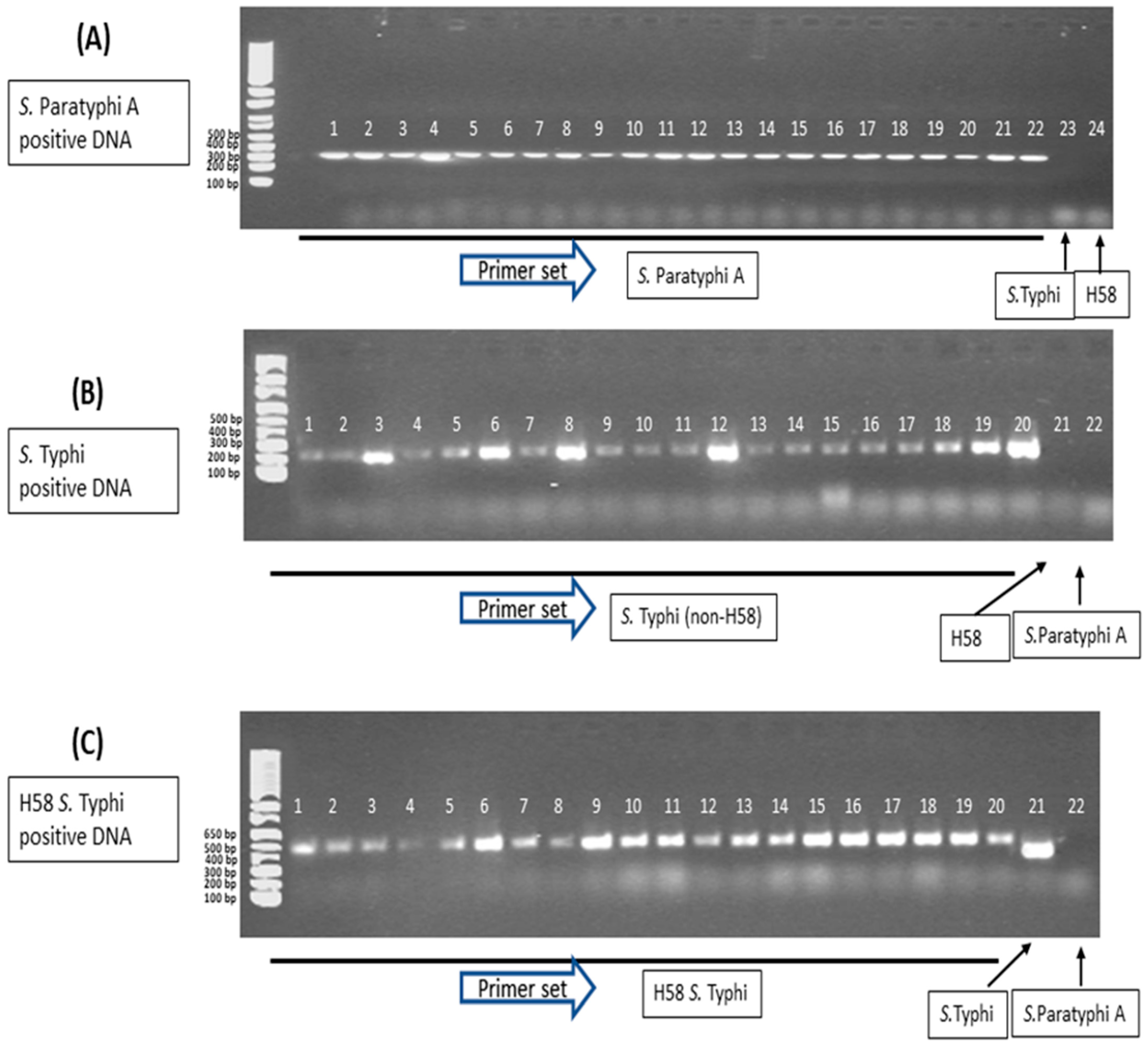
An in-house conventional PCR assay was optimized on these three groups of blood-culture-confirmed bacterial strains (Group A: 22 Salmonella Paratyphi A, Group B: 20 non-H58 Salmonella Typhi, and Group C: 20 H58 Salmonella Typhi). The optimized single-plex PCR assays correctly amplified SSPA2308, STY2513, and STY0307 target genes for Salmonella Paratyphi A (22/22, 100%)-, non-H58 Salmonella Typhi (20/20, 100%)-, and H58 Salmonella Typhi (20/20, 100%)-positive DNA samples, respectively (Figure 1A lanes 1–22, Figure 1B lanes 1–20, Figure 1C lanes 1–20). We also performed cross-reactivity tests to assess specificity. The Salmonella Paratyphi A-positive DNA sample did not amplify for Salmonella Typhi- and H58 Typhi-specific primer sets (Figure 1A, lanes 23 and 24). Similarly, a non-H58 Salmonella Typhi-positive DNA sample showed no amplification with H58 Typhi- and Salmonella Paratyphi A-specific primers (Figure 1B, lanes 21 and 22). As H58 Typhi is a defined lineage of Salmonella Typhi, H58 Typhi-positive DNA also amplified for the STY2513 target gene of Typhi (Figure 1C, lane 21), but not the Salmonella Paratyphi A target gene (Figure 1C, lane 22).

3.4. PCR Sensitivity Evaluation and Bacterial Load Detection

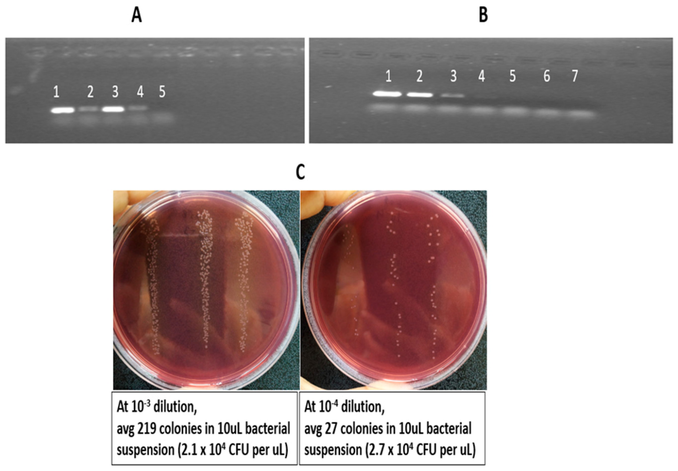
To assess the sensitivity of the PCR assay in relation to bacterial load, DNA extracted from nine individual colonies consistently produced clear positive bands (Figure 2A, lane 3). However, lower colony counts or dilutions from the suspension of nine colonies resulted in faint or absent amplification (Figure 2A, lanes 2, 4, 5). Based on these observations, the nine-colony/300 µL bacterial suspension was selected for further 10-fold serial dilution, and it was found that PCR amplification was detectable at dilutions ≥10−1 (Figure 2B, lanes 2, 3). Beyond this dilution point, no visible bands were observed (Figure 2B, lanes 4–7).
Bacterial load was calculated based on colony counts obtained from serial dilutions (neat to 10−6 dilution) (bacterial counts at only 10−3 and 10−4 dilutions are shown in Figure 2C). At the 10−3 dilution, an average of 219 colonies was observed from 10 µL of plated bacterial suspension, corresponding to approximately 2.1 × 10⁴ CFU/µL. Similarly, at the 10−4 dilution, an average of 27 colonies from 10 µL corresponded to approximately 2.7 × 10⁴ CFU/µL after adjusting for the dilution factor. The CFU/µL values derived from these two dilutions were then averaged to obtain a more reliable estimate of bacterial concentration, yielding approximately 2.6 × 10⁴ CFU/µL. Based on this average concentration, the total bacterial load present in the 2 µL template DNA used for PCR was calculated to be approximately 5.2 × 10⁴ CFU.

4. Discussion

In enteric fever-endemic areas such as Bangladesh, the persistent burden and its nonspecific clinical presentation underscore the urgent need for rapid and reliable diagnostic tools. In this study, we evaluated the SNP-based diagnostic method, which offers a cost-effective solution and can be routinely implemented in basic molecular laboratories. Our study demonstrates the successful development and validation of a conventional PCR assay for not only the specific detection of typhoidal Salmonella serovars but also addressing the globally dominant MDR-associated H58 lineage of Salmonella Typhi. The performance of this assay was assessed by comparing it with the blood culture result as a reference standard. The integration of an SNP-based genotyping framework enabled the design of in silico primer sets with enhanced discriminatory power and allowed precise detection to track clinically significant H58 strains.
The widespread dissemination of the H58 lineage in multiple countries is of great concern due to its strong association with MDR genes [6]. Leveraging our previously reported WGS data of 241 blood-culture-confirmed isolates collected in Dhaka between 2004 and 2016 enabled us to confirm the H58 and non-H58 Typhi isolates and to understand their diverse resistance patterns [4,10]. Our genomics analysis reported H58 dominance before 2011, followed by a decline in MDR H58 strains and a rise in non-H58 genotypes carrying QRDR mutations. These patterns suggested a shift of current treatment practices towards third-generation cephalosporin and azithromycin [4]. Moreover, the widespread occurrence of reduced fluoroquinolone susceptibility associated with QRDR mutations among both Salmonella Typhi lineages and all Salmonella Paratyphi A isolates was the result of an increase in the over-the-counter sale of ciprofloxacin over the last decade for treatment [3,4,10]. The implementation of these genomic findings strengthens the accuracy of this diagnostic assay and ensures its relevance to current circulating strains, guiding appropriate antimicrobial therapy. However, while WGS provides high-resolution insights into lineage and AMR diversity, it remains expensive and not feasible for monitoring AMR risk in typhoid lineages in highly typhoid-endemic settings. In contrast, a PCR-based assay offers a more practical and cost-effective alternative for routine use in large-scale surveillance studies. Integration of this assay into routine local surveillance frameworks could enhance early detection of emerging AMR-associated lineages to inform empirical treatment decisions, guide antimicrobial stewardship, and strengthen public health responses. The use of PCR-based diagnostics for enteric fever detection is increasing as an alternative to the gold standard blood culture method because of the low sensitivity and specificity of culture. Previous studies report PCR sensitivities ranging from 70 to 95%, although performance declines when applied directly to blood samples [34]. However, a multiplex PCR assay developed previouslydemonstrated 100% specificity for high-risk lineages of Salmonella Typhi isolated from culture. While the previous study used an intergenic region (SSPA1732a-SSPA1724) for Salmonella Paratyphi A detection [25], we targeted the conserved and serovar-specific SSPA2308 gene region for Salmonella Paratyphi A detection to minimize potential variability associated with intergenic regions. Consistent with findings of previous studies, our in-house single-plex PCR assays demonstrated high diagnostic accuracy for Bangladeshi blood-culture-positive isolates, with 100% amplification success for each target gene (SSPA2308, STY2513, STY0307). Cross-reactivity testing confirmed the specificity of each primer set, with no off-target amplification observed between serovars or lineages. Notably, H58 Salmonella Typhi strains amplified both the lineage-specific (STY0307) and species-specific (STY2513) targets, reflecting their dual identity and validating the assay’s discriminatory power.
Establishment of LOD is critical for assessing the effectiveness of diagnostics in clinical and surveillance settings. Sensitivity testing of this PCR assay demonstrated consistent amplification from DNA extracted by suspending a minimum of nine colonies in 300 µL of nuclease-free water, with LOD at ≥10−1 dilution, equivalent to approximately 5.2 × 10⁴ CFU per 2 µL template. This relatively high LOD reflected that DNA was derived from enriched bacterial populations following culture rather than direct clinical samples, where bacterial load is typically much lower (less than 1 CFU/mL). Furthermore, we used an in-house built crude DNA extraction method using the heat-lysis method, which demonstrates the practical utility of direct testing without a commercial DNA extraction kit and extensive DNA purification steps. It reinforces its suitability for routine diagnostics, particularly in resource-limited laboratories where rapid, low-cost testing is essential.
Our study has some limitations. First, we evaluated this PCR assay on blood-culture-positive strains, not directly from raw clinical samples (blood or stool). As a result, the LOD of this assay was expressed in terms of CFU equivalents derived from DNA extracted from pure bacterial cultures to reduce the complexity of raw clinical samples containing mixed microbial and host DNA. Our current optimized PCR-based approach does not reduce the initial diagnostic turnaround time of culture, as low bacterial counts in blood fall below the detection limit of PCR. Future work will focus on optimizing this PCR assay directly from the blood sample by enriching the bacterial population and depleting host DNA contamination to improve the practical implementation of this assay for routine surveillance [35]. Second, the current work was limited to a single-plex format. Further optimization into a multiplex format is necessary to allow simultaneous detection of multiple targets, enhance workflow efficiency, and reduce reagent costs. The multiplex optimization work is underway to ensure robust performance for each target. Despite these limitations, the strength of this study is that this assay bridges the gap between high-resolution genomic surveillance and practical diagnostic application. While WGS and SNP typing remain the gold standard for lineage and resistance profiling, their cost and infrastructure demands limit routine use in low- and middle-income countries [23,36]. The SNP-based PCR assay could be an alternative to WGS typing for serovar- and lineage-specific identification, thereby informing local treatment algorithms, enhancing early detection, guiding empirical therapy, and supporting targeted public health interventions.

5. Conclusions

This study provides a proof-of-concept for a reliable, affordable alternative to the gold standard blood culture method. The implementation of SNP-based diagnostic assays in routine surveillance could play a crucial role in outbreak monitoring, enabling the timely identification of circulating strains. These findings bridge the gap between genomic insights and molecular assays for strengthening the capacity to monitor the emergence and spread of enteric fever in endemic regions.

Supplementary Materials

The following supporting information can be downloaded at https://www.mdpi.com/article/10.3390/microbiolres17060104/s1, Table S1: Metadata of 62 Typhoidal Salmonella strains isolated from the patients.

Author Contributions

A.M., G.D., F.Q. and S.I.A.R. contributed to the design of the study and A.M., F.Q., G.D. and F.K. (Farhana Khanam) supervised the study. Z.D., F.K. (Fahad Khokhar), D.J.P. and S.I.A.R. performed the laboratory work and data analysis. S.I.A.R. wrote the initial draft and all authors contributed to the interpretation of results and editing of the manuscript and approved the final version. All authors have read and agreed to the published version of the manuscript.

Funding

This work was supported by the University of Cambridge, UK, and the International Centre for Diarrhoeal Disease Research, Bangladesh (icddr,b). This study was supported by the grants from Forgarty International Center Training Grant in Vaccine Development and Public Health (TW005572) and Swedish International Development Cooperation Agency (54100020, 51060029). Primers and PCR reagents were kindly donated by Ankur Mutreja’s Lab, Cambridge, to evaluate the performance of this assay.

Institutional Review Board Statement

Ethical approval (protocol number 2007-007, ethics approval date: 17 May 2007) was obtained from the Research Review Committee (RRC) and the Ethical Review Committee (ERC) of the International Centre for Diarrhoeal Disease Research, Bangladesh (icddr,b).

Informed Consent Statement

Informed written consent and clinical information were taken from legal guardians of child participants and adult participants.

Data Availability Statement

The data presented in this study are openly available in Pubmed at https://pubmed.ncbi.nlm.nih.gov/32106221/ (accessed on 14 March 2026), reference number [4252889].

Acknowledgments

We acknowledge the support of dedicated field and laboratory workers at the icddr,b involved in this study. icddr,b is grateful to the Governments of Bangladesh and Canada for providing core support.

Conflicts of Interest

The authors declare no conflicts of interest.

Abbreviations

The following abbreviations are used in this manuscript:
PCRPolymerase chain reaction
WaSHWater, sanitation, and hygiene
MDRMultidrug-resistant
H58Haplotype 58 lineage
QRDRQuinolone resistance-determining region
CFUColony-forming unit
WGSWhole-genome sequencing
SNPSingle-nucleotide polymorphism
BLASTnBasic Local Alignment Search Tool for Nucleotide sequences
glpAGlycerol-3-phosphate dehydrogenase A
NFWNuclease-free water
LODLimit of detection

References

  1. Piovani, D.; Figlioli, G.; Nikolopoulos, G.K.; Bonovas, S. The global burden of enteric fever, 2017–2021: A systematic analysis from the global burden of disease study 2021. EClinicalMedicine 2024, 77, 102883. [Google Scholar] [CrossRef]
  2. Meiring, J.E.; Shakya, M.; Khanam, F.; Voysey, M.; Phillips, M.T.; Tonks, S.; Thindwa, D.; Darton, T.C.; Dongol, S.; Karkey, A.; et al. Burden of enteric fever at three urban sites in Africa and Asia: A multicentre population-based study. Lancet Glob. Health 2021, 9, e1688–e1696. [Google Scholar] [CrossRef] [PubMed]
  3. Britto, C.D.; Dyson, Z.A.; Duchene, S.; Carter, M.J.; Gurung, M.; Kelly, D.F.; Murdoch, D.R.; Ansari, I.; Thorson, S.; Shrestha, S.; et al. Laboratory and molecular surveillance of paediatric typhoidal Salmonella in Nepal: Antimicrobial resistance and implications for vaccine policy. PLoS Negl. Trop. Dis. 2018, 12, e0006408. [Google Scholar] [CrossRef]
  4. Rahman, S.I.A.; Dyson, Z.A.; Klemm, E.J.; Khanam, F.; Holt, K.E.; Chowdhury, E.K.; Dougan, G.; Qadri, F. Population structure and antimicrobial resistance patterns of Salmonella Typhi isolates in urban Dhaka, Bangladesh from 2004 to 2016. PLoS Negl. Trop. Dis. 2020, 14, e0008036. [Google Scholar] [CrossRef]
  5. Mirza, S.H.; Beeching, N.J.; Hart, C.A. Multi-drug resistant typhoid: A global problem. J. Med. Microbiol. 1996, 44, 317–319. [Google Scholar] [CrossRef] [PubMed]
  6. Wong, V.K.; Baker, S.; Pickard, D.J.; Parkhill, J.; Page, A.J.; Feasey, N.A.; Kingsley, R.A.; Thomson, N.R.; Keane, J.A.; Weill, F.X.; et al. Phylogeographical analysis of the dominant multidrug-resistant H58 clade of Salmonella Typhi identifies inter- and intracontinental transmission events. Nat. Genet. 2015, 47, 632–639. [Google Scholar] [CrossRef]
  7. Wong, V.K.; Baker, S.; Connor, T.R.; Pickard, D.; Page, A.J.; Dave, J.; Murphy, N.; Holliman, R.; Sefton, A.; Millar, M.; et al. An extended genotyping framework for Salmonella enterica serovar Typhi, the cause of human typhoid. Nat. Commun. 2016, 7, 12827. [Google Scholar] [CrossRef] [PubMed]
  8. Pham Thanh, D.; Karkey, A.; Dongol, S.; Ho Thi, N.; Thompson, C.N.; Rabaa, M.A.; Arjyal, A.; Holt, K.E.; Wong, V.; Tran Vu Thieu, N.; et al. A novel ciprofloxacin-resistant subclade of H58 Salmonella Typhi is associated with fluoroquinolone treatment failure. elife 2016, 5, e14003. [Google Scholar] [CrossRef]
  9. Turner, A.K.; Nair, S.; Wain, J. The acquisition of full fluoroquinolone resistance in Salmonella Typhi by accumulation of point mutations in the topoisomerase targets. J. Antimicrob. Chemother. 2006, 58, 733–740. [Google Scholar] [CrossRef][Green Version]
  10. Rahman, S.I.A.; Nguyen, T.N.T.; Khanam, F.; Thomson, N.R.; Dyson, Z.A.; Taylor-Brown, A.; Chowdhury, E.K.; Dougan, G.; Baker, S.; Qadri, F. Genetic diversity of Salmonella Paratyphi A isolated from enteric fever patients in Bangladesh from 2008 to 2018. PLoS Negl. Trop. Dis. 2021, 15, e0009748. [Google Scholar] [CrossRef]
  11. Rahman, M.; Siddique, A.K.; Shoma, S.; Rashid, H.; Salam, M.A.; Ahmed, Q.S.; Nair, G.B.; Breiman, R.F. Emergence of multidrug-resistant Salmonella enterica serotype Typhi with decreased ciprofloxacin susceptibility in Bangladesh. Epidemiol. Infect. 2006, 134, 433–438. [Google Scholar] [CrossRef]
  12. Hooda, Y.; Sajib, M.S.; Rahman, H.; Luby, S.P.; Bondy-Denomy, J.; Santosham, M.; Andrews, J.R.; Saha, S.K.; Saha, S. Molecular mechanism of azithromycin resistance among typhoidal Salmonella strains in Bangladesh identified through passive pediatric surveillance. PLoS Negl. Trop. Dis. 2019, 13, e0007868. [Google Scholar] [CrossRef]
  13. Djeghout, B.; Saha, S.; Sajib, M.S.I.; Tanmoy, A.M.; Islam, M.; Kay, G.L.; Langridge, G.C.; Endtz, H.P.; Wain, J.; Saha, S.K. Ceftriaxone-resistant Salmonella Typhi carries an IncI1-ST31 plasmid encoding CTX-M-15. J. Med. Microbiol. 2018, 67, 620–627. [Google Scholar] [CrossRef]
  14. Parry, C.M.; Hien, T.T.; Dougan, G.; White, N.J.; Farrar, J.J. Typhoid fever. N. Engl. J. Med. 2002, 347, 1770–1782. [Google Scholar] [CrossRef]
  15. Rahman, T.; Hosen, I.; Chakraborty, S. A Rapid Glimpse on Typhoid Fever: An Updated Mini Review. J. Life Med. 2013, 1, 83–92. [Google Scholar] [CrossRef]
  16. Gasem, M.H.; Dolmans, W.M.; Isbandrio, B.B.; Wahyono, H.; Keuter, M.; Djokomoeljanto, R. Culture of Salmonella typhi and Salmonella paratyphi from blood and bone marrow in suspected typhoid fever. Trop. Geogr. Med. 1995, 47, 164–167. [Google Scholar] [PubMed]
  17. Jesudason, M.; Esther, E.; Mathai, E. Typhidot test to detect IgG & IgM antibodies in typhoid fever. Indian J. Med. Res. 2002, 116, 70–72. [Google Scholar] [PubMed]
  18. Saha, S.K.; Ruhulamin, M.; Hanif, M.; Islam, M.; Khan, W.A. Interpretation of the Widal test in the diagnosis of typhoid fever in Bangladeshi children. Ann. Trop. Paediatr. 1996, 16, 75–78. [Google Scholar] [CrossRef] [PubMed]
  19. Naheed, A.; Ram, P.K.; Brooks, W.A.; Mintz, E.D.; Hossain, M.A.; Parsons, M.M.; Luby, S.P.; Breiman, R.F. Clinical value of Tubex and Typhidot rapid diagnostic tests for typhoid fever in an urban community clinic in Bangladesh. Diagn. Microbiol. Infect. Dis. 2008, 61, 381–386. [Google Scholar] [CrossRef]
  20. Khanam, F.; Sheikh, A.; Sayeed, M.A.; Bhuiyan, M.S.; Choudhury, F.K.; Salma, U.; Pervin, S.; Sultana, T.; Ahmed, D.; Goswami, D.; et al. Evaluation of a typhoid/paratyphoid diagnostic assay (TPTest) detecting anti-Salmonella IgA in secretions of peripheral blood lymphocytes in patients in Dhaka, Bangladesh. PLoS Negl. Trop. Dis. 2013, 7, e2316. [Google Scholar] [CrossRef]
  21. Parkhill, J.; Dougan, G.; James, K.D.; Thomson, N.R.; Pickard, D.; Wain, J.; Churcher, C.; Mungall, K.L.; Bentley, S.D.; Holden, M.T.G.; et al. Complete genome sequence of a multiple drug resistant Salmonella enterica serovar Typhi CT18. Nature 2001, 413, 848–852. [Google Scholar] [CrossRef] [PubMed]
  22. McClelland, M.; Sanderson, K.E.; Clifton, S.W.; Latreille, P.; Porwollik, S.; Sabo, A.; Meyer, R.; Bieri, T.; Ozersky, P.; McLellan, M.D.; et al. Salmonella enterica subsp. enterica Serovar Paratyphi A Str. ATCC 9150, Complete Genome; NCBI: Bethesda, MD, USA, 2004.
  23. Dyson, Z.A.; Holt, K.E. Five Years of GenoTyphi: Updates to the Global Salmonella Typhi Genotyping Framework. J. Infect. Dis. 2021, 224, S775–S780. [Google Scholar] [CrossRef]
  24. Tanmoy, A.M.; Hooda, Y.; Sajib, M.S.; Da Silva, K.E.; Iqbal, J.; Qamar, F.N.; Luby, S.P.; Dougan, G.; Dyson, Z.A.; Baker, S.; et al. Paratype: A genotyping tool for Salmonella Paratyphi A reveals its global genomic diversity. Nat. Commun. 2022, 13, 7912. [Google Scholar] [CrossRef]
  25. Khokhar, F.; Pickard, D.; Dyson, Z.; Iqbal, J.; Pragasam, A.; John, J.J.; Veeraraghavan, B.; Qamar, F.; Dougan, G.; MacQueen, H.; et al. Multiplex PCR assay to detect high risk lineages of Salmonella Typhi and Paratyphi A. PLoS ONE 2022, 17, e0267805. [Google Scholar] [CrossRef]
  26. Sheikh, A.; Bhuiyan, M.S.; Khanam, F.; Chowdhury, F.; Saha, A.; Ahmed, D.; Jamil, K.M.A.; LaRocque, R.C.; Harris, J.B.; Ahmad, M.M.; et al. Salmonella enterica serovar Typhi-specific immunoglobulin A antibody responses in plasma and antibody in lymphocyte supernatant specimens in Bangladeshi patients with suspected typhoid fever. Clin. Vaccine Immunol. 2009, 16, 1587–1594. [Google Scholar] [CrossRef] [PubMed]
  27. Thorpe, T.C.; Wilson, M.L.; Turner, J.E.; DiGuiseppi, J.L.; Willert, M.; Mirrett, S.; Reller, L. BacT/Alert: An automated colorimetric microbial detection system. J. Clin. Microbiol. 1990, 28, 1608–1612. [Google Scholar] [CrossRef]
  28. Schrader, K.N.; Fernandez-Castro, A.; Cheung, W.K.; Crandall, C.M.; Abbott, S.L. Evaluation of commercial antisera for Salmonella serotyping. J. Clin. Microbiol. 2008, 46, 685–688. [Google Scholar] [CrossRef]
  29. Chen, Y.; Ye, W.; Zhang, Y.; Xu, Y. High speed BLASTN: An accelerated MegaBLAST search tool. Nucleic Acids Res. 2015, 43, 7762–7768. [Google Scholar] [CrossRef]
  30. Ye, J.; Coulouris, G.; Zaretskaya, I.; Cutcutache, I.; Rozen, S.; Madden, T.L. Primer-BLAST: A tool to design target-specific primers for polymerase chain reaction. BMC Bioinform. 2012, 13, 134. [Google Scholar] [CrossRef] [PubMed]
  31. Lee, P.Y.; Costumbrado, J.; Hsu, C.Y.; Kim, Y.H. Agarose gel electrophoresis for the separation of DNA fragments. J. Vis. Exp. 2012, 62, 3923. [Google Scholar]
  32. Forootan, A.; Sjöback, R.; Björkman, J.; Sjögreen, B.; Linz, L.; Kubista, M. Methods to determine limit of detection and limit of quantification in quantitative real-time PCR (qPCR). Biomol. Detect. Quantif. 2017, 12, 1–6. [Google Scholar] [CrossRef] [PubMed]
  33. Khanam, F.; Sayeed, M.A.; Choudhury, F.K.; Sheikh, A.; Ahmed, D.; Goswami, D.; Hossain, M.L.; Brooks, A.; Calderwood, S.B.; Charles, R.C.; et al. Typhoid fever in young children in Bangladesh: Clinical findings, antibiotic susceptibility pattern and immune responses. PLoS Negl. Trop. Dis. 2015, 9, e0003619. [Google Scholar] [CrossRef] [PubMed]
  34. Nga, T.V.T.; Karkey, A.; Dongol, S.; Thuy, H.N.; Dunstan, S.; Holt, K.; Tu, L.T.P.; Campbell, J.I.; Chau, T.T.; Chau, N.V.V.; et al. The sensitivity of real-time PCR amplification targeting invasive Salmonella serovars in biological specimens. BMC Infect. Dis. 2010, 10, 125. [Google Scholar] [CrossRef] [PubMed]
  35. Zhou, L.; Pollard, A.J. A novel method of selective removal of human DNA improves PCR sensitivity for detection of Salmonella Typhi in blood samples. BMC Infect. Dis. 2012, 12, 164. [Google Scholar] [CrossRef]
  36. Ibrahim, G.M.; Morin, P.M. Salmonella Serotyping Using Whole Genome Sequencing. Front. Microbiol. 2018, 9, 2993. [Google Scholar] [CrossRef]
Figure 1. Single-plex PCR for the diagnosis of typhoidal pathogens. Agarose gel electrophoresis results showed positive and negative bands for the respective target genes separately. I Kb plus DNA ladder (Invitrogen, Thermofisher) was used as a molecular size marker. (A) Blood-culture-confirmed Salmonella Paratyphi A-positive strains: lanes 1–22—positive bands on SSPA2308 primer targeting Salmonella Paratyphi A, lane 23—negative band on STY0307 primer targeting Salmonella Typhi, lane 24—negative band on STY2513 primer targeting H58 Salmonella Typhi; (B) blood-culture-confirmed non-H58 Salmonella Typhi-positive strains: lanes 1–20—positive bands on STY0307 primer targeting Salmonella Typhi, lane 21—negative band on STY2513 primer targeting H58 Salmonella Typhi, lane 22—negative band on SSPA2308 primer targeting Salmonella Paratyphi A; (C) WGS-confirmed H58 Salmonella Typhi-positive strains: lanes 1–20—positive bands on STY2513 primer targeting H58 Salmonella Typhi, lane 21—positive band on STY0307 primer targeting Salmonella Typhi, lane 22—negative band on SSPA2308 primer targeting Salmonella Paratyphi A.
Microbiolres 17 00104 g001
Figure 2. Limit of detection (LOD) of the PCR assay. Lane 1 in panels (A,B) was used as a positive control (H58-positive DNA template from single-plex PCR). (A) Lane 2: DNA extracted from three colonies suspended in 300 µL NFW, lane 3: DNA extracted from nine colonies suspended in 300 µL NFW, lane 4: 1:10 serial dilution from lane 3 preparation, and lane 5: 1:105 serial dilution from lane 3 preparation. (B) Lane 2: DNA extracted from nine colonies, lanes 3–7: tenfold serial dilutions of the lane 2 preparation, ranging from 10−1 to 10−5, used to determine the assay’s detection threshold. (C) Count bacterial load by plating 100 µL bacterial suspension on the MacConkey agar plates at 10−3 and 10−4.
Microbiolres 17 00104 g002
Table 1. List of primers targeting Salmonella Typhi, Salmonella Paratyphi A and H58 Typhi genes.
Target GeneOrganismPrimer NamePrimer Sequence (5′-3′)Amplicon Length (bp)Source
STY0307Salmonella
Typhi
ST_227FGGCAGATATACTTTCGCAGGCA227Khokhar et al. [25]
ST_227RCCCAGAACCAAATTTGCTTACA
SSPA2308Salmonella
Paratyphi A
SPA_305FAGGGATGAGAATTTTCAGACGT305This study
SPA_305RACCCCAGCTCTGAGAGATATCT
STY2513H58 Salmonella
Typhi
H58_509FGGGCTTGATGGCTTCATTAGT509Khokhar et al. [25]
H58_509RACAGGTTGTACGCCTTTCCA
Disclaimer/Publisher’s Note: The statements, opinions and data contained in all publications are solely those of the individual author(s) and contributor(s) and not of MDPI and/or the editor(s). MDPI and/or the editor(s) disclaim responsibility for any injury to people or property resulting from any ideas, methods, instructions or products referred to in the content.