Article

Production of Surface-Active Metabolites by Bacillus sp. from Vegetable Oil-Impacted Soil: Ecological Implications and Screening Limitations


by
Eugenia Guadalupe Ortiz-Lechuga
1,
Verónica Almaguer-Cantú
1,
Hiram Herrera-Barquín
2,
Karla Katiushka Solís-Arévalo
1,
Ramón Alberto Batista-García
3 and
Katiushka Arévalo-Niño
1,*
1
Instituto de Biotecnología, Facultad de Ciencias Biológicas, Universidad Autónoma de Nuevo León, Ciudad Universitaria, Av. Pedro de Alba S/N, San Nicolás de los Garza 66455, Nuevo León, Mexico
2
Laboratorio de Ictiología, Facultad de Ciencias Biológicas, Universidad Autónoma de Nuevo León, Ciudad Universitaria, Av. Pedro de Alba S/N, San Nicolás de los Garza 66455, Nuevo León, Mexico
3
Cellular Dynamics Research Center, Autonomous University of the State of Morelos, Av. Universidad 10001, Col Chamilpa, Cuernavaca 62209, Morelos, Mexico
*
Author to whom correspondence should be addressed.
Microbiol. Res. 2026, 17(4), 76; https://doi.org/10.3390/microbiolres17040076

Submission received: 1 March 2026 / Revised: 26 March 2026 / Accepted: 26 March 2026 / Published: 8 April 2026

Abstract

Biosurfactant-producing microorganisms play an important ecological role in soils impacted by hydrophobic contaminants by enhancing substrate bioavailability and influencing microbial interactions. In this study, we critically evaluated the reliability of commonly used screening methods for biosurfactant detection. A total of 71 microbial isolates (16 bacteria and 55 fungi) were obtained from vegetable oil-contaminated soil and screened using a multi-step approach combining enzymatic assays (lipolytic and hemolytic activity) and physicochemical methods, including drop-collapse, oil spreading, emulsification index (E24), and surface tension reduction. Although 21 isolates exhibited lipolytic activity and 9 showed hemolysis, inconsistent responses among assays revealed significant limitations of individual screening methods. Only two bacterial isolates consistently tested positive across all criteria. When cultivated in mineral salt medium supplemented with hydrophobic substrates, both isolates produced stable emulsions and significantly reduced surface tension (from 54.26 mN/m to 31.46 mN/m). Substrate-dependent variation was observed for isolate C3, which showed reduced surface tension (39.63 mN/m) when grown with biodiesel. These findings highlight the risk of relying on single assays and emphasize the need for integrated screening strategies to ensure reliable detection of biosurfactant-producing microorganisms.

1. Introduction

Biosurfactants are surface-active metabolites produced by microorganisms that act specifically at interfaces [1,2], thereby influencing substrate accessibility and microbial competitiveness in complex environments. Their amphipathic structure enables a reduction in surface and interfacial tension, facilitating substrate mobilization.
Despite their ecological and biotechnological relevance, the detection of biosurfactant-producing microorganisms remains methodologically challenging. While the assays usually employed focus on particular characteristics and evaluate distinct aspects, on occasion, they can yield inconsistent results when applied independently [2]. These limitations can lead to overestimation or misinterpretation of biosurfactant production, particularly in ecological studies where functional inference is critical.
Biosurfactants play a fundamental ecological role in soil ecosystems, particularly in hydrocarbon-impacted environments, due to their broad substrate selectivity, low toxicity and tolerance to physicochemical stress [3,4].
Classified as glycolipids, lipopeptides, phospholipids, polymeric biosurfactans, and fatty acids [5,6] are typically growth-associated and have been reported across a wide diversity of bacterial and fungal genera, including Bacillus, Pseudomonas, Nocardia Acinetobacter, Flavobacterium, Arthrobacter, Rhodococcus, Mycobacterium, Corynebacterium, Streptomyces, Serratia, and Candida, among others [2,7,8,9]. Members of the genus Bacillus are frequently associated with impacted environments due to their metabolic versatility, tolerance and ability to produce structurally diverse metabolites [2,4]. In oil-impacted soils, these traits may confer adaptive advantages by enhancing hydrocarbon bioavailability. Thus, their production should not be interpreted only as a biochemical trait but as a functional strategy often linked to pollution-induced pressure [6,10,11,12,13].
The isolation and characterization of biosurfactant-producing microorganisms capable of forming stable emulsions is increasingly recognized not only as a technological resource but also as an indicator of diversity in ecological adaptation, nutrient cycling and contamination attenuation [14,15,16]. In this context, an integrated screening strategy becomes essential to link microbial function with environmental adaptation. By combining enzymatic and physicochemical assays and pollutant-induced culture conditions, biosurfactant production can be evaluated as a functional response.
Conventionally employed screening assays—such as lipolytic and hemolytic activity, drop-collapse, oil spreading and emulsification assays, are reported to produce these methodological limitations [2,16,17,18,19,20,21] that can lead to overestimation or misinterpretation of biosurfactant production, particularly in ecological studies where functional inference is central to the response to environmental pressure.
In this context, this work aims to critically assess the coherence and limitation of commonly applied screening methods while exploring surface-active metabolite production from microbial isolates from vegetable oil-impacted soil recovered from an outdoor food market, through an integrated enzymatic and physicochemical approach.

2. Materials and Methods

2.1. Soil Sampling and Site Description

The soil samples were collected along a 10 km transect from an outdoor food market found in the locality of Ocho de Enero, located in the municipality of Frontera in Coahuila, México. The soil in this region is primarily classified as Xerosol and Regosol, with an average annual temperature of 18–22 °C with wide fluctuations in summer and winter due to the arid nature of the region. Five sampling points were established at 2 km intervals. At each point, 20 g of superficial soil (5 cm depth) was collected using a sterile spatula, placed in polyethylene bags, transported to the laboratory, and stored at 4 °C until further processing. The concentration of contaminants in the soil samples was not quantified prior to analysis; however, sampling sites were selected based on visible contamination with vegetable oil residues.

2.2. Microorganism Isolation and Preservation

Samples were processed by serial dilution with 0.85% saline solution (10−1 to 10−5). Under aseptic conditions, 100 µL aliquots from the last two dilutions were plated onto general isolation media: nutrient agar (NA; 3 g/L beef extract, 5 g/L peptone, 15 g/L agar) and potato dextrose agar (PDA; 4 g/L potato extract, 20 g/L dextrose, 15 g/L agar) [22]. After growth, individual colonies were sub-cultured and preserved by transferring 1 cm × 1 cm agar blocks into sterile containers with 10% glycerol and preserved at −20 °C. All isolated microorganisms were assigned a letter and consecutive number according to the order of recovery from isolation. The nomenclature was established as C for bacteria and G for fungi.

2.3. Primary Screening for Lipolytic and Hemolytic Activity

2.3.1. Lipolytic Activity Detection on Rhodamine B Agar

Lipolytic activity was assessed using Rhodamine B Agar (0.02%) prepared with a lipid emulsion solution (30 mL olive oil, 250 μL Tween 80 and 50 mL distilled water), a rhodamine B solution (20 mg rhodamine B in 20 mL sterile water) and a base medium (4.5 g nutrient broth, 1.25 g yeast extract, 10 g agar, 450 mL distilled water) following the method described by Alken-Murray [23]. Plates were incubated at 28 °C (fungi) and 37 °C (bacteria) for 96 h, with fluorescence monitored every 24 h under 350 nm UV light. Positive samples were identified by orange fluorescent halos. Selected isolates were re-inoculated using a single-streak method, incubated for 96 h and photographed using a Nikon d3100 camera (Nikon, Tokyo, Japan) with an 18–55 mm lens. Images were analyzed using ImageJ software (version 1.54f) [24] and classified as + (low), ++ (moderate) or +++ (high) based on pixel intensity.

2.3.2. Hemolytic Activity Assay

Hemolysis was evaluated using 1.5% blood agar prepared using 5 g of yeast extract, 5 g of sodium chloride (NaCl), 15 g of agar, 13 g of casein peptone and 15 mL of fresh sheep blood per liter.
Isolates were spot-inoculated at the center of the plate and incubated at 28 °C for 168 h (fungi) or 37 °C for 72 h (bacteria). Hemolysis was classified as α, β or γ according to standard criteria [25].

2.3.3. Production of Cell-Free Supernatants

Positive lipolytic isolates were pre-incubated for 24 h in nutrient broth (NB; 3 g/L beef extract, 5 g/L peptone) for bacteria and yeast potato dextrose (YPD; 10 g/L yeast extract, 20 g/L bactopeptone, 20 g/L dextrose) for fungi. Inoculum density was adjusted to 3 × 10⁸ CFU/mL using the McFarland scale for bacteria and 5 × 10⁶ spores/mL for fungi using a Neubauer chamber.
Cultures were transferred to minimal salt medium (MSM; 1.4 g KH2PO4, 2.2 g Na2HPO4, 3 g (NH4)2SO4, 0.6 g MgSO47H2O, 0.05 g NaCl, 1 g yeast extract, 0.01 g FeSO4 7H2O and 0.02 g CaCl2 7H2O per liter) supplemented with 2% (v/v) olive oil. Incubation was at 28 °C (fungi) and 37 °C (bacteria) for 72 h on an orbital shaker. Aliquots collected every 24 h were centrifuged at 6000 rpm at 4 °C for 15 min. Supernatants were filtered through 0.2 μm membranes for further analysis [26,27].

2.4. Secondary Physicochemical Screening for Biosurfactant Activity

2.4.1. Drop-Collapse Assay (Microplate Method)

For the drop-collapse assay (DC), 100 μL of crude oil was dispensed into each well of a 96-well microplate and allowed to stabilize at room temperature (25 °C) for 20 min. Subsequently, 5 μL of cell-free supernatant was carefully added and the reaction observed after 1 min. The diameter of the observed zones was measured manually using a ruler. Measurements were performed in triplicate by placing the plates on a white background to improve contrast and ensure consistent visualization of the zones.
Positive controls: Pseudomonas aeruginosa (L1-IB strain collection); Dodecyl sodium sulfate (SDS) 0.6%; Cetyltrimethylammonium bromide (CTAB) 0.06%.
Negative control: Distilled water.
Dispersion > 0.5 mm was considered a positive result [28,29,30]. A crescent moon-shaped drop or undisturbed oil indicated a negative outcome.

2.4.2. Oil Spreading Test

The oil spreading test was carried out by adding 50 mL of distilled water to a plastic Petri dish (100 mm), followed by the gentle addition of 100 μL of crude oil onto the water surface. Afterward, 10 μL of cell-free supernatant was dispensed onto the oil layer using a 20–200 μL micropipette [31,32].
SDS (0.6%) was used as an anionic detergent and CTAB (0.06%) as a cationic detergent, and a cell-free supernatant of Pseudomonas aeruginosa served as a positive control; distilled water was used as a negative control. The displacement of the oil layer (clear zone) was considered positive result; the diameter of the displaced zone was measured and compared with the negative control. For photographic documentation, the assay was additionally conducted using 50 mL centrifuge tubes (30 × 150 mm) to enhance visualization.

2.5. Biosurfactant Stimulation Under Pollutant-Induced Conditions and Recovery for Solvent Precipitation

Cultures were grown in 40 mL MSM supplemented with 2% (v/v) biodiesel (BD), glycerol (G), kerosene (K), diesel (D) or motor oil (M) as sole carbon sources. As a reference strain for biosurfactant production, Pseudomonas aeruginosa was included under three conditions: MSM without an additional carbon source (C−), MSM supplemented with 2% glucose (C+ Glu) and nutrient broth (C+ NB) to evaluate growth-associated surface activity. The bacterial inoculum was adjusted to 3 × 10⁸ CFU/mL.
Cultures were incubated at 30 °C, 150 rpm for 72 h. Aliquots were collected every 24 h, centrifuged at 6000 rpm and 4 °C for 15 min, and filtered through 0.2 μm membranes prior to the following assays [33,34]. For biosurfactant recovery, three volumes of cold acetone were added to one volume of supernatant followed by incubation at 4 °C for 10 h. Precipitated biosurfactant was considered to be crude biosurfactant extract and was recovered by centrifugation (5000 rpm for 15 min at 4 °C), dried to remove acetone, weighed and resuspended in 1 mL of sterile water for storage at 4 °C [35].

2.6. Quantitative Evaluation of Biosurfactant Activity

2.6.1. Emulsification Index (Ei24)

The emulsification capacity of the cell-free supernatant was evaluated using the emulsification index (Ei24) [36,37,38]. The assay was performed by adding 2 mL of stimulated supernatant (BD, K, G, D or M) and mixing with 2 mL of motor oil, diesel, biodiesel or kerosene in 20 mL screw-capped glass tubes. The mixture was vortexed for 2 min to ensure uniform dispersion and allowed to stand undisturbed at room temperature for 24 h. The height of the resulting emulsion layer was measured, and the emulsification index (Ei24) was calculated using the following equation:
E i 24 = H 1 H 2 × 100 %
H1 = height of the emulsion layer; H2 = height of the mixture.

2.6.2. Surface Tension Reduction (Pendant Drop Method)

Biosurfactant activity was evaluated on cell-free supernatant from two selected treatments using the pendant drop method with a Drop Shape Analyzer DSA30S (Krüss, Hamburg, Germany). Uninoculated minimal salt medium (MSM) served as a negative control, while 0.6% sodium dodecyl sulfate (SDS) and unstimulated cell-free supernatant of Pseudomonas aeruginosa were used as positive controls. Surface tension was determined by analyzing three independent drops per sample. All measurements were performed in triplicate at 20 °C, and the average values were reported. The instrumental error was considered according to the manufacturer’s specifications (0.01 mN/m).

2.7. Molecular Identification

Genomic DNA was extracted from overnight cultures of isolate C2. Cells were harvested by centrifugation (10,000 rpm for 10 min at 4 °C), and pellets were ground in liquid nitrogen before standard DNA extraction. PCR amplification targeted the 16S rRNA gene using primers 27F (5′-AGAGTTTGATCMTGGCTCAG-3′) and 1492R (5′-TACGGYTACCTTGTTACGACTT-3′). Sequences were analyzed using BLAST (version 2.14) [39] and RDP (Ribosomal Database Project) [40] provided by GenBank. Phylogenetic trees were constructed using the Neighbor-Joining method in MEGA12 for phylogenetic tree construction [41].

2.8. Statistical Analyses

All experiments were performed in triplicate. Data is presented as mean ± standard deviation. Statistical significance was evaluated using one-way analysis of variance (ANOVA) followed by Tukey’s post hoc test, with a significance threshold set at p < 0.05. Statistical analyses were performed using SPSS v21 (IBM Corp., Amork, NY, USA).

3. Results

3.1. Primary Screening Based on Lipolytic Activity

From soil sampling and microbial isolation, a total of 71 microbial isolates were obtained, comprising 55 fungal and 16 bacterial strains. All isolates were morphologically characterized at macroscopic and microscopic levels and subsequently preserved. Lipolytic activity was employed as the first screening criterion for biosurfactant production. Among the 71 isolates, 21 (29.6%) exhibited positive lipolytic activity, as evidenced by orange fluorescence on Rhodamine B agar under UV light. Fluorescence intensity was quantified using ImageJ software and classified into three categories: low (+), medium (++) and high (+++), supported by pixel density values. Based on this classification, a total of 15 (21.1%) isolates—7 bacterial (43.8%) and 8 fungal (14.54%)—showing high (+++) lipolytic activity were selected for subsequent biosurfactant screening assays.

3.2. Hemolytic Activity as Complementary Primary Screening

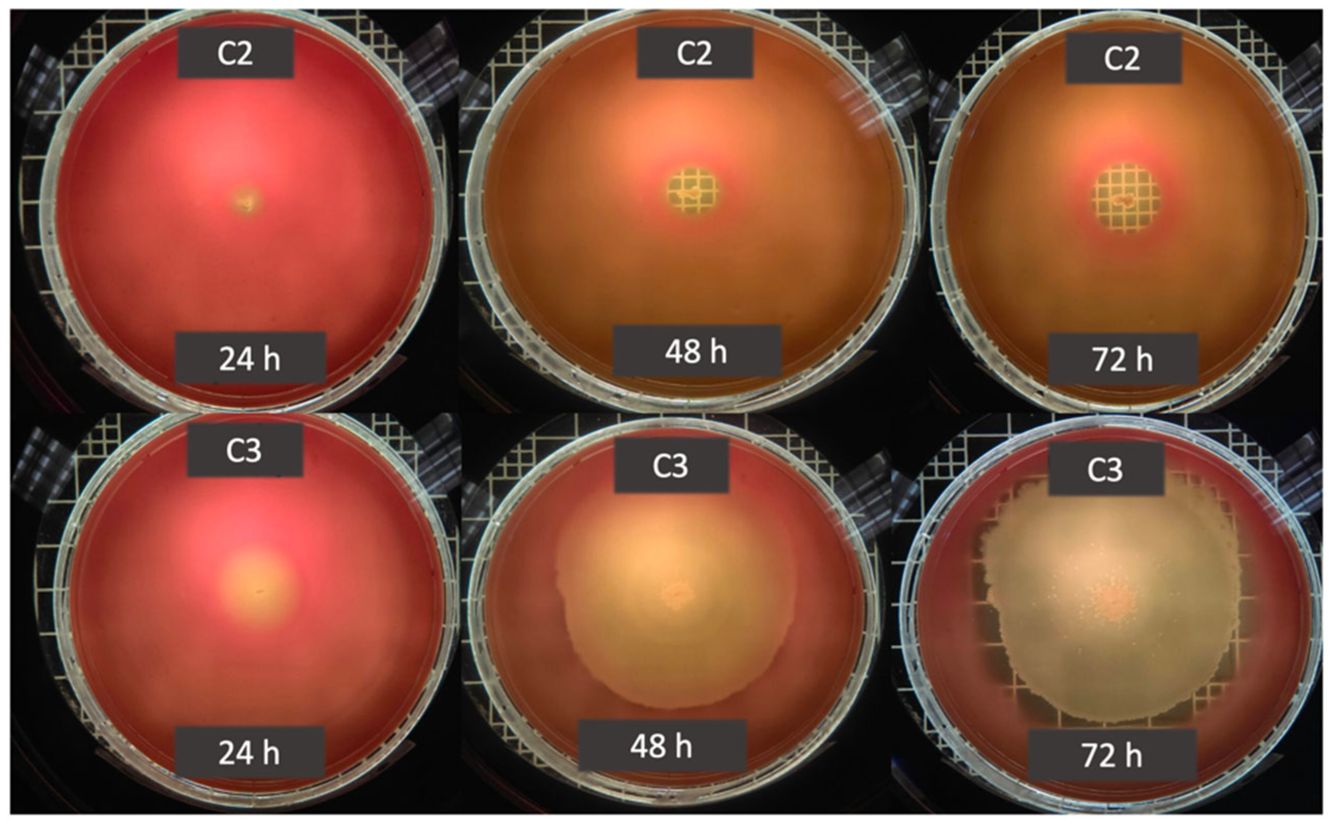
Hemolytic activity was detected in 66% of the bacterial isolates and 50% of the fungal isolates. Two hemolysis patterns were observed: alpha (α) and beta (β). Beta hemolysis, indicative of complete erythrocyte lysis, was the most prevalent response among bacterial isolates and was characterized by the formation of well-defined translucent halos surrounding the colonies (Figure 1). Alpha hemolysis, associated with partial hemoglobin degradation, was also observed but produced diffuse and poorly defined halos, limiting reliable activity quantification [42].
Although lipolytic and hemolytic activities have been widely used as preliminary indicators of biosurfactant production, our results revealed substantial variability among isolates and limited discriminatory power when these assays were applied independently. In particular, hemolytic responses were frequently diffuse and time-dependent, especially among fungal isolates, reducing their reliability as a standalone screening criterion. From an ecological perspective, enzymatic traits such as lipolytic or hemolytic activity reflect potential metabolic capabilities but do not necessarily translate into effective surface modification. Therefore, to better capture the functional expression of biosurfactant production, selected positive isolates were subjected to physicochemical assays that measure interfacial activity.

3.3. Surface Tension Tests

Techniques that assess changes in physical properties are commonly used to detect or confirm biosurfactant production. However, it is important to note that these assays detect different physicochemical properties of biosurfactants, which can lead to variability in their responses. The drop-collapse method is based on the ability of biosurfactant molecules to reduce surface tension, leading to deformation or collapse of a liquid droplet placed on a hydrophobic surface. In the absence of surface-active compounds, droplets remain stable due to stronger cohesive interactions [8,26,28,43]. In this study, four bacterial and six fungal isolates showed a clear positive drop-collapse response within one minute of contact. In contrast, five bacterial isolates yielded inconclusive results, characterized by a “crescent moon” pattern in which the droplet migrated toward the edge of the well rather than fully collapsing.
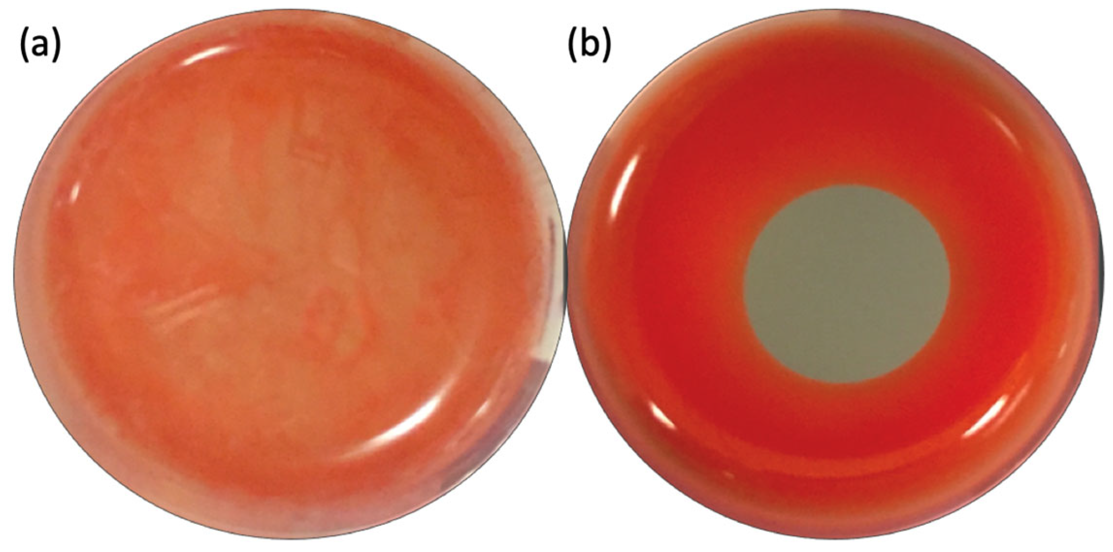
The oil spreading assay, originally developed by Morikawa et al., 2000 [31], was employed as a complementary approach to further evaluate biosurfactant activity. This method is based on the displacement of oil by surface-active compounds and reflects a different aspect of interfacial activity compared to the drop-collapse assay. In contrast to the drop-collapse results, all fungal isolates and eight of the nine bacterial isolates produced a positive oil displacement response (Table 1). Notably, isolate C5 (previously classified as inconclusive in the drop-collapse assay), exhibited a clear and measurable oil displacement zone in this test (Figure 2), supporting its potential biosurfactant-producing capacity and illustrating assay-dependent variability. Comparative analysis of the drop-collapse and oil spreading assays revealed partial overlap among positive isolates but also notable discrepancies. While several isolates exhibited clear surface-active behavior in both tests, others showed assay-dependent responses or intermediate patterns (Table 1).
Despite the high proportion of positive responses observed across individual assays, only two bacterial isolates (C2 and C3) displayed consistent and reproducible activity throughout the initial screening battery. Based on this integrated assessment, these isolates were selected for subsequent characterization.

3.4. Adaptive Biosurfactant Production Under Carbon Source Stimulation

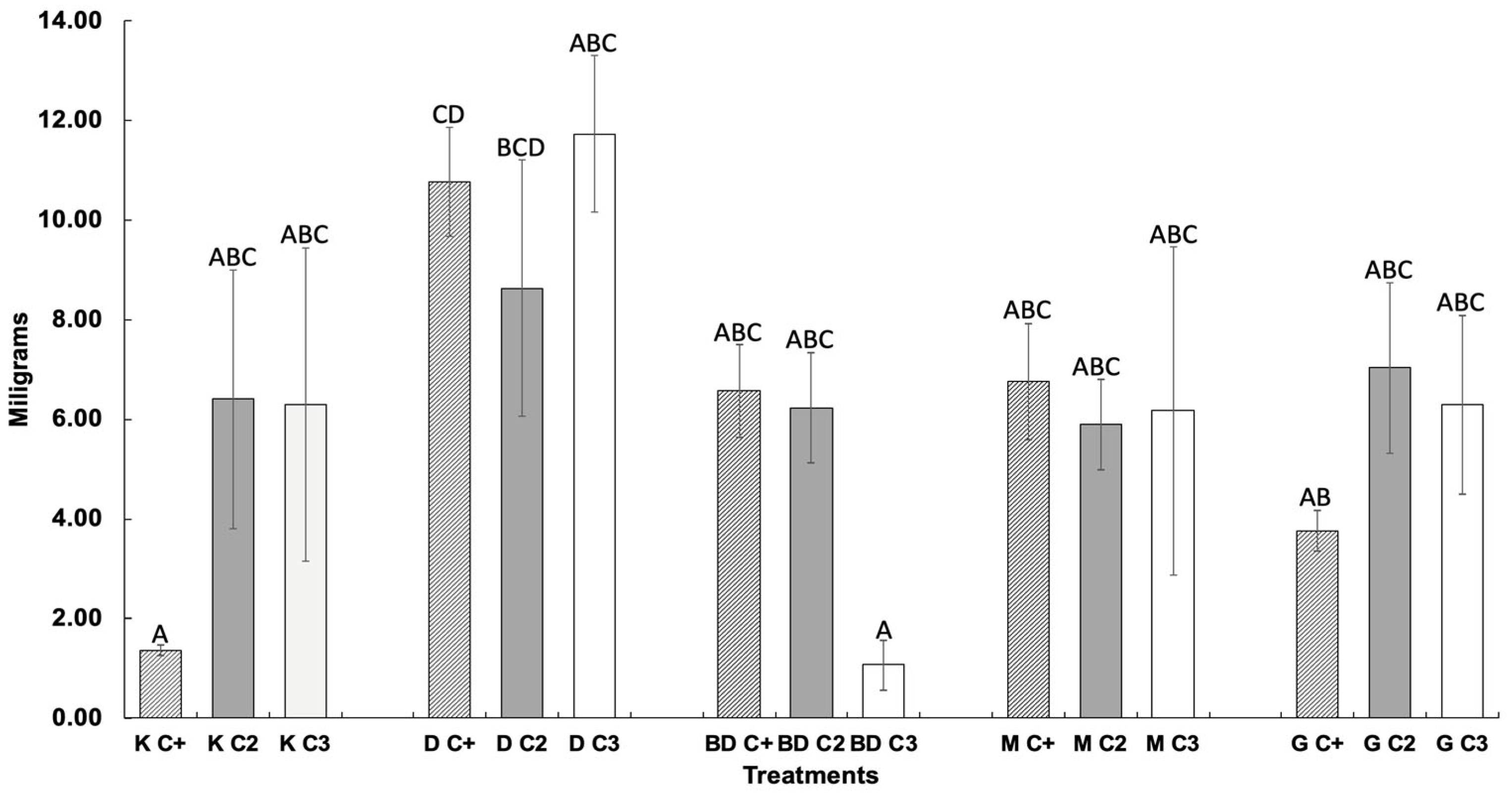
To evaluate biosurfactant production under different stimulatory conditions, precipitates obtained after fermentation were quantified. Consistent with previous assays, isolates C2 and C3 exhibited differences in biosurfactant yields across treatments. The reference strain, Pseudomonas aeruginosa, showed the highest biosurfactant yield when stimulated with diesel (10.77 ± 1.10 mg) and the lowest with kerosene (1.37 ± 0.12 mg). This stimulation-based approach enabled the identification of relationships between carbon source type and biosurfactant production (Figure 3).

3.5. Emulsification Index

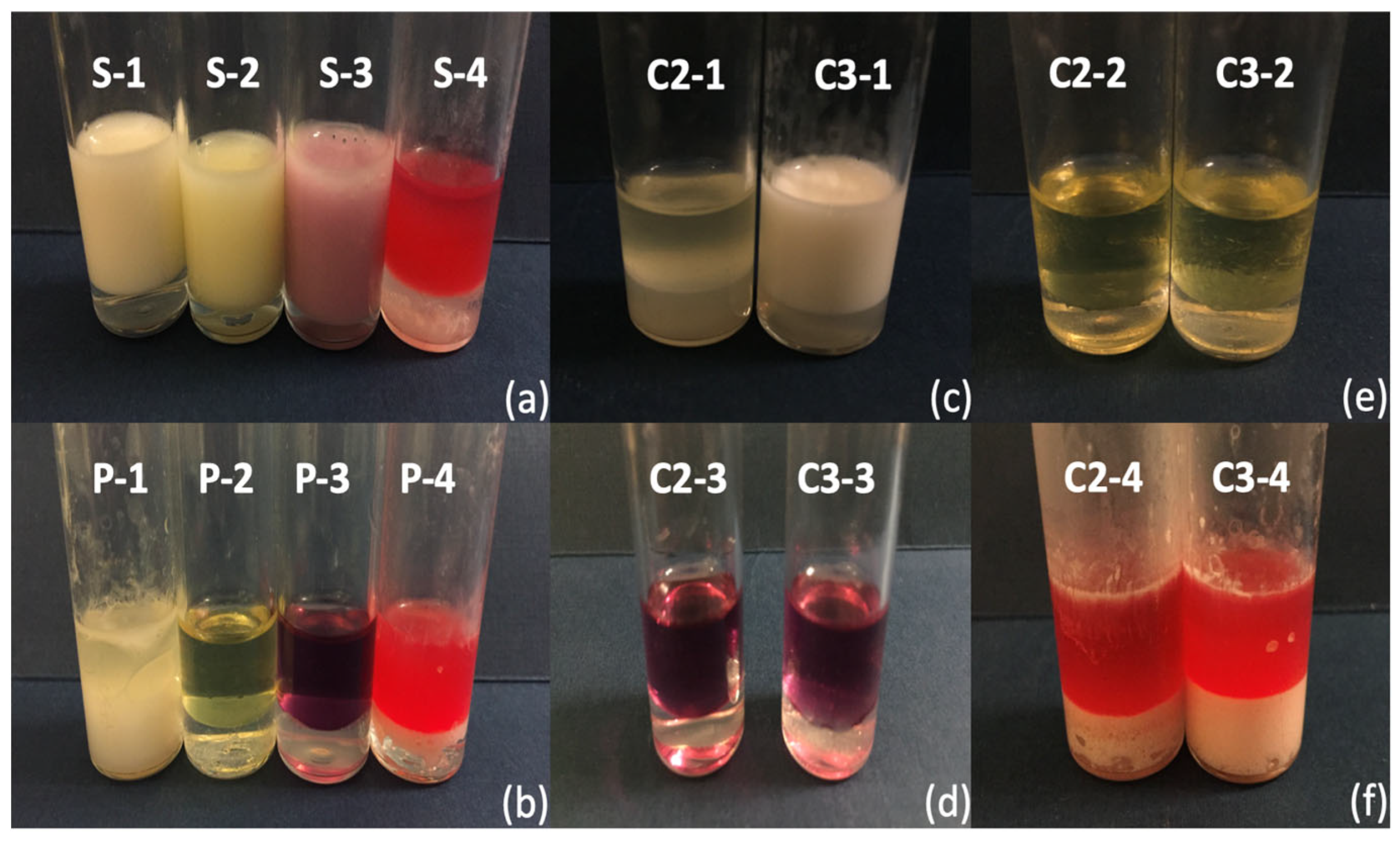
The emulsion stability of the biosurfactants produced by isolates C2 and C3 was evaluated using the emulsification index after 24 h (Ei24). An emulsion was considered stable when at least 30–50% of the initial emulsion height was maintained after 24 h [30,44,45]. Both isolates formed stable emulsions with biodiesel (BD) and motor oil (M) (Figure 4). For biodiesel, C2-1 achieved 40.39% ± 14.87, while C3-1 exhibited a higher emulsification capacity of 55.79% ± 7.31. In the case of motor oil, C2-4 achieved 43.96% ± 1.55 and C3-4 reached 32.20% ± 2.54. The reference strain, Pseudomonas aeruginosa, showed an Ei24 of 50.00% ± 3.40 for biodiesel but only 15.96% ± 4.35 for motor oil, in agreement with previously reported values [8]. As expected, the chemical surfactant SDS (0.6%) produced high emulsification indices across all substrates, including biodiesel (85% ± 4.60), motor oil (50% ± 3.40), diesel (85% ± 3.95) and kerosene (90% ± 4.61). Neither isolates C2 and C3 nor P. aeruginosa produced stable emulsions with diesel or kerosene. Notably, emulsion type and spatial distribution varied depending on the substrate. Biodiesel promoted the formation of water-in-oil (W/O) emulsions localized in the upper hydrophobic layer, whereas motor oil resulted in oil-in-water (O/W) emulsions accumulating in the lower aqueous phase, indicating substrate-dependent emulsification behavior.

3.6. Surface Tension Reduction

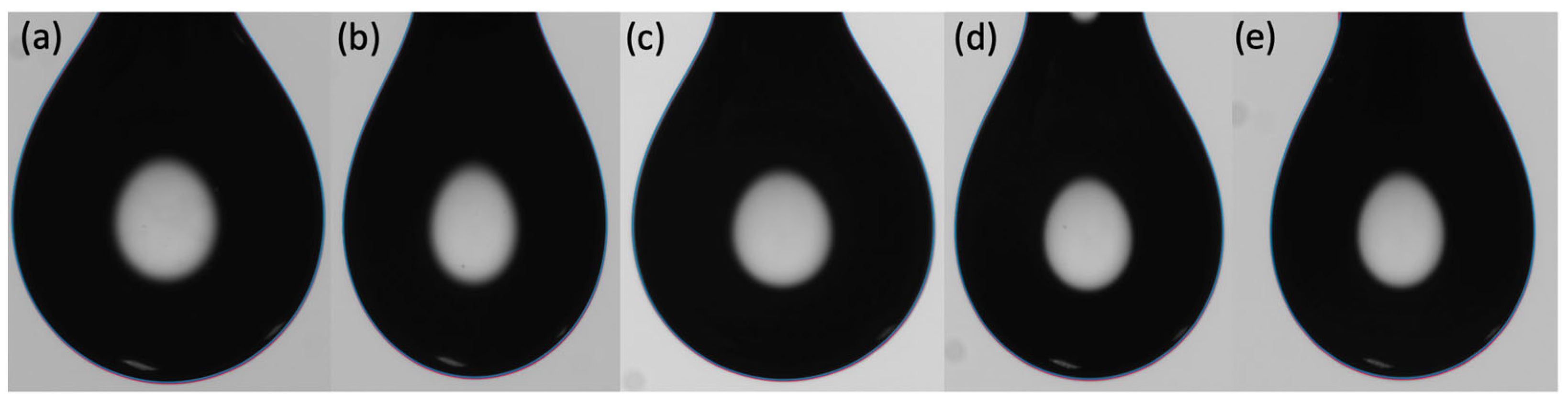
The surface tension of uninoculated MSM was measured at 54.26 mN/m. Among the controls, the cell-free supernatant of Pseudomonas aeruginosa reduced surface tension to 48.31 mN/m, whereas SDS (0.6%) produced a further decrease to 36.94 mN/m. Notably, the cell-free supernatants from isolates C2-4 and C3-1 achieved even greater reductions, reaching 31.46 mN/m and 39.63 mN/m, respectively (Figure 5). According to Agarwal and Sharma (2010) [46] and Decesaro et al. (2021) [47], microbial strains capable of lowering surface tension below 40 mN/m are considered strong biosurfactant producers. Based on the overall performance across assays, isolate C2 was selected for molecular identification, given that strain C3-4 did not show surface tension reduction. The isolate exhibited 98.82% sequence identity with Bacillus paramycoides strain MCCC 1A04098.
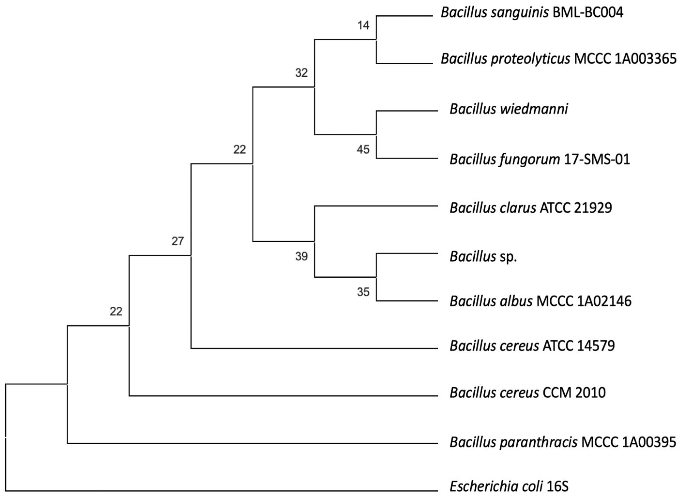
This result indicates affiliation within the genus Bacillus and suggests close relatedness to members of the Bacillus cereus group. However, given the limited discriminatory power of the 16S rRNA gene within this complex, species-level assignment cannot be conclusively established without additional genomic or multilocus sequence analysis. Therefore, the isolate was designated as Bacillus sp. (Figure 6).

4. Discussion

The search for novel microbial isolates capable of producing biosurfactants has expanded across multiple sectors, ranging from emerging applications in food processing, agriculture and cosmetics to more established fields such as bioremediation and enhanced oil recovery. However, the chemical diversity and functional variability of biosurfactants often complicate their detection and characterization. Consequently, the use of efficient, rapid, and reliable screening strategies is essential for evaluating large numbers of isolates and identifying candidates with the functional performance required under specific environmental or industrial conditions. Such approaches not only accelerate strain selection but also contribute to cost reduction by prioritizing microorganisms with high biosurfactant yields and strong adaptive potential [47]. Lipolytic activity was selected as the initial screening criterion based on the well-documented functional linkage between lipase production and biosurfactant activity. Both processes play complementary roles in the metabolism of hydrophobic substrates and frequently operate as coordinated systems that facilitate emulsification and substrate accessibility [2,22,48,49,50]. This interaction is particularly relevant in oil-impacted soils, where microbial survival depends on efficient strategies to access poorly soluble carbon sources.
Rhodamine B agar provides a rapid and widely used method for detecting lipolytic activity through the formation of a fluorescent complex between the cationic dye and free fatty acids released from lipid substrates. Despite its advantages, this assay presents notable limitations. Certain microorganisms naturally produce fluorescent metabolites that may interfere with fluorescence-based interpretation, leading to false positive results. For instance, Pseudomonas aeruginosa, a well-known biosurfactant producer, synthesizes the fluorescent siderophore pyoverdine, which can mask or mimic lipase-derived fluorescence. In addition, biosurfactants themselves may participate in alternative metabolic pathways that generate fluorescence unrelated to lipolytic activity. These limitations prompted methodological modifications aimed at minimizing assay interference [51,52,53]. However, while lipolytic activity represents a useful preliminary indicator of biosurfactant potential, its interpretation is not always straightforward. The possibility of intrinsic pigment interference and pathway overlap underscores the necessity of integrating complementary physicochemical assays to reliably confirm biosurfactant production.
While lipolytic activity represents an initial adaptive response facilitating substrate hydrolysis, it does not fully capture the range of surface-active strategies involved in microbial survival and competition. Therefore, hemolytic activity was evaluated as an additional functional indicator of membrane-interacting compounds. The ability to disrupt erythrocyte membranes reflects the production of amphipathic molecules capable of altering lipid bilayers [54,55], a property that may confer ecological advantages such as improved nutrient acquisition, microbial antagonism, or enhanced persistence in contaminated environments.

4.1. Hemolytic Activity

The detection of hemolytic activity as an indicator of biosurfactant production was first reported by Bernheimer and Avigad (1970) [56], who demonstrated that surfactin was capable of lysing erythrocytes. Based on this observation, blood agar assays have become one of the most widely adopted preliminary screening methods for identifying biosurfactant-producing microorganisms [25,26,32,57,58]. In this assay, α-hemolysis is associated with partial hemoglobin degradation and appears as a greenish halo, whereas β-hemolysis results from complete erythrocyte lysis and is generally more pronounced [42,59]. However, the visual interpretation of hemolytic zones is inherently subjective and may vary between observers, limiting its reproducibility [25]. Consequently, although blood agar is simple and cost-effective, its correlation with actual biosurfactant production remains inconsistent and should be considered only when complemented by additional screening techniques [28].
From an ecological and metabolic perspective, both bacterial and fungal isolates that are known to produce hemolysins often produce them as a part of a broader survival strategy, including iron acquisition and stress response, particularly in nutrient-limited or contaminated environments [60,61,62]. Such activities may generate hemolytic zones on blood agar that are unrelated to biosurfactant production, as hemolysis can also result from cytotoxins or other virulence factors [63]. Conversely, false negatives may arise due to the limited diffusion of biosurfactant molecules within the agar matrix [64,65,66,67].

4.2. Surface Tension Tests

To aid in the interpretation of drop-collapse results, several methodological modifications have been proposed, including staining the supernatant or placing the droplet over a high-contrast grid to enhance visual discrimination, as described by Maczek et al. (2007) [68].
Nevertheless, the overall sensitivity of this assay to low biosurfactant concentrations remains limited [52]. This constrains likely explains the inconclusive responses observed for some isolates, as the amount of biosurfactant present in the cell-free supernatant may have been insufficient to induce complete droplet deformation or collapse. Despite these limitations, the drop-collapse method remains a simple, rapid, and reliable qualitative screening tool, particularly when applied to crude extracts [65]. Furthermore, when used with purified biosurfactants, droplet diameter has been shown to provide semi-quantitative information correlated with surface activity [29]. The oil spreading assay complements the drop-collapse method by offering higher sensitivity at low surfactant concentrations. This technique detects reductions in interfacial tension through the formation of a clear oil displacement zone on the water surface [65]. In this context, the positive response observed for isolate C5 in the oil spreading assay, despite its inconclusive drop-collapse result, suggests biosurfactant production at concentrations below the threshold of the latter method. Such contrasting outcomes highlightshighlight the importance of combining complementary assays to improve detection accuracy and reduce false negatives [12,18,25,32,69].

4.3. Stimulation and Precipitation Assay

Biosurfactant yield is strongly influenced by the metabolic pathway activated during growth and by the specific molecular fractions synthesized under different nutritional conditions. Water-soluble substrates such as glucose and glycerol generally promote biomass accumulation and favor the formation of the hydrophilic portion of the biosurfactant molecules, as previously reported by Haferburg et al. (1986) [70], Roongaswang et al. (2002) [71], Silva et al. (2010) [72], and Archana et al. (2016) [73]. In contrast, hydrophobic substrates (e.g., hydrocarbons of varying chain lengths) tend to stimulate the production of the lipidic moiety, which is directly involved in interfacial activity. Under these conditions, biosurfactant synthesis enhances substrate solubility and carbon bioavailability, facilitating microbial uptake and metabolism. Depending on the carbon source supplied, central metabolic routes such as glycolysis (favored by hydrophilic substrates) or gluconeogenesis (favored by hydrophobic substrates) may be differentially activated, ultimately shaping biosurfactant yield and composition [74].
At the same time, variability complicates direct comparisons among treatments, as differences in pathway activation may explain the inconsistencies in yield. Notably, isolates C2 and C3 exhibited a strong recovery rate in response to kerosene stimulation, despite the comparatively low yield observed for the reference strain. This pattern suggests strain-specific metabolic adaptability and highlights the potential of these isolates for biosurfactant production using low-cost hydrophobic substrates; however, it is very important to note that this technique does not discriminate between the nature of the molecule recovered, which can include biosurfactants, bio-emulsifiers or even high-molecular-weight polymers [75], a behavior that was later confirmed by the emulsification index. Such methodological limitations underscore the importance of distinguishing between functional surface activity and crude extract recovery, particularly when interpreting substrate-induced responses.

4.4. Emulsification Index

The ability of isolates C2 and C3 to form stable emulsions with biodiesel and motor oil but not with kerosene or diesel indicated that the metabolites they produced functioned as effective emulsifying agents, meeting the minimum stability threshold after 24 h; this differential behavior indicates the substrate-dependent selectivity of the produced compound rather than uniform emulsifying capacity, reflecting differences in physicochemical compatibility between the compound produced and tested substrate. This pattern suggests affinity to complex hydrocarbon mixtures, which could reflect ecological adaptation to the contaminated soil environment from which the microorganisms were obtained.
Beyond emulsion stability, the formation of different emulsion types (water in oil, W/O versus oil in water, O/W) provided insight into their functional properties. In general, W/O emulsions are associated with more hydrophobic molecular structures, whereas O/W emulsions suggest a predominance of hydrophilic moieties [19,76]. This functional variability is likely influenced by the carbon source supplied during cultivation, as substrate composition can shape biosurfactant structure and interfacial behavior [77,78]. Ivanova et al. (2021) [79] similarly reported that hydrophobic carbon sources tended to induce the production of surface-active biosurfactants, while hydrophilic substrates favored the synthesis of emulsifiers. Although synthetic surfactants like SDS exhibited higher emulsification efficiency, the environmental drawbacks—(including persistence, bioaccumulation, and toxicity) restrict their suitability for environmental applications, while microbial biosurfactants combine functional effectiveness with ecological compatibility, offering advantages such as biodegradability, low toxicity, and integration into natural attenuation and bioremediation processes, as well as enhanced oil recovery strategies [8,9,25,80,81,82,83].

4.5. Surface Tension Reduction

Several species within the Bacillus genus, particularly members of the Bacillus subtilis and Bacillus cereus groups, are well known for producing bioactive surface-active metabolites, mainly lipopeptides and less commonly rhamnolipids. Bacillus paramycoides has been specifically reported to synthesize lipopeptides belonging to three major families: surfactin, fengycin, and iturin. Among these, surfactin is regarded as one of the most potent biosurfactants, capable of reducing surface tension. Its activity is strongly influenced by molecular features such as fatty-acid chain length, branching patterns, and cultivation conditions [84,85,86,87,88,89]. Yousaf et al. (2011) [90] suggested that microorganisms isolated from polluted environments tend to develop enhanced biodegradation capabilities due to selective environmental pressure. This ecological adaptation is supported by recent studies identifying B. paramycoides as a metabolically versatile species with broad environmental relevance, including applications in bioremediation, environmental restoration, and soil conditioning [82,91,92], as well as biopolymer (PHB) production [93]. Although surface tension reduction and emulsification capacity are not always directly correlated, high-molecular-weight biosurfactants, particularly those enriched in lipid fractions or forming lipid–protein complexes, are known to exert stronger surface-active effects [84]. In this context, the ability of isolate C2-4 to reduce surface tension below 40 mN/m, while simultaneously forming stable emulsions, contrary to isolate C3-1, provides strong evidence of its functional potential as an effective biosurfactant producer adapted to oil-impacted environments.

4.6. Strengths, Limitations, and Complementarity of Biosurfactant Screening Methods

A comparative analysis of the screening methods employed in this study highlights relevant differences in sensitivity, specificity, and detection principles.
Lipolytic activity and hemolysis assays serve as rapid preliminary indicators; however, both are indirect and may yield false positives due to the production of other metabolites unrelated to biosurfactant activity. In contrast, physicochemical methods such as drop-collapse and oil spreading directly assess surface and interfacial activity, offering functional relevance, although their qualitative nature may lead to ambiguous interpretations. The emulsification index (E24) provides insight into emulsion stability but does not necessarily correlate with surface tension reduction, reflecting a different functional property of biosurfactants. Surface tension measurement, as determined by the pendant drop method, represents a more quantitative and reliable indicator of biosurfactant activity; however, it requires specialized instrumentation and is less suitable for high-throughput screening.
Collectively, these observations demonstrate that the use of complementary assays allows for a more robust and reliable evaluation.

5. Conclusions

This study evaluated several of the most widely adopted techniques for screening of biosurfactant-producing microorganisms isolated from vegetable oil-impacted soil. Initial selection based on lipolytic and hemolytic activities reduced the candidate pool, while subsequent evaluation using physicochemical assays (drop-collapse and oil spreading) enabled the detection of surface-active properties. Although some isolates exhibited clear surface activity and were selected for further evaluation, variability in their performance across assays revealed that different methods capture distinct functional properties, which may lead to discrepancies in interpretation. In particular, the lack of direct correlation between emulsion stability and surface tension reduction underscores the complexity of biosurfactant behavior and reinforces the need for integrated screening strategies. Taken together, these results highlight not only the potential of environmental isolates as biosurfactant producers but also the persistent challenges in drawing definitive conclusions when relying on commonly used screening methods. Based on the consistency of responses across all four assays, two bacterial isolates (C2 and C3) were selected for further assays. Cell-free supernatants obtained from stimulation assays achieved a marked reduction in surface tension from 54.26 mN/m (uninoculated MSM) to 31.46 mN/m (C2-4) and 39.63 mN/m (C3-1), representing a maximum reduction of 42.02% relative to the control. However, isolate C3-4 exhibited high emulsion stability despite limited surface tension reduction, reinforcing the need for an integrated evaluation strategy. Although further characterization is required to elucidate the chemical structure of the biosurfactants produced, the observed activity patterns, together with previous reports on related strains, may indicate the production of lipopeptide-type biosurfactants, potentially related to the surfactin family; however, this remains a tentative inference in the absence of direct chemical characterization (e.g., LC-MS, HPLC). Overall, these findings emphasize that traditional screening methods, when used in isolation, may lead to false positives, false negatives, or non-specific responses and highlight the importance of combining complementary approaches for a more reliable and functionally meaningful evaluation.

Author Contributions

All authors contributed to the study conception and design. Material preparation and data collection were performed by E.G.O.-L. with supervision from K.A.-N. The first draft of the manuscript was written by E.G.O.-L. with supervision from K.A.-N. and R.A.B.-G. with critical review from V.A.-C., H.H.-B. and K.K.S.-A. All authors provided critical feedback and helped shape the research. All authors have read and agreed to the published version of the manuscript.

Funding

This project was supported by Programa para el Desarrollo Profesional Docente, para el Tipo Superior (PRODEP). Resource assignment: 511-6/18-334.

Institutional Review Board Statement

Not applicable.

Informed Consent Statement

Not applicable.

Data Availability Statement

The original contributions presented in this study are included in the article. Further inquiries can be directed to the corresponding author.

Conflicts of Interest

The authors declare no conflicts of interest.

Abbreviations

The following abbreviations are used in this manuscript:
NANutrient Agar
PDAPotato Dextrose Agar
YPDYeast Potato Dextrose
NBNutrient Broth
DCDrop-collapse
OSOil Spreading
HAHemolytic activity
LALipolytic activity
Ei24Emulsification index
H1Height of emulsion layer
H2Height of mixture
MSMMinimal salt medium
C−Negative carbon source
C+Positive carbon source
GluGlucose

References

  1. Rubina, S.; Khanna, S. Biodegradation studies with reference to Indian isolates. Ind. J. Microbiol. 1995, 35, 165–184. [Google Scholar]
  2. Sekhon, K.K.; Khanna, S.; Cameotra, S.S. Biosurfactant Production and Potential Correlation with Esterase Activity. J. Pet. Environ. Biotechnol. 2012, 3, 133. [Google Scholar] [CrossRef]
  3. Donio, M.B.S.; Ronica, S.F.A.; Thanga Viji, V.; Velmurugan, S.; Adlin, J.J.; Michaelbabu, M.; Citarasu, T. Isolation and Characterization of Halophilic Bacillus sp. BS3 Able to Produce Pharmacologically Important Biosurfactants. Asian Pac. J. Trop. Med. 2013, 6, 873–886. [Google Scholar] [CrossRef] [PubMed]
  4. Fenibo, E.O.; Ijoma, G.N.; Selvarajan, R.; Chikere, C.B. Microbial Surfactants: The next Generation Multifunctional Biomolecules for Applications in the Petroleum Industry and Its Associated Environmental Remediation. Microorganisms 2019, 7, 581. [Google Scholar] [CrossRef] [PubMed]
  5. Biermann, M.; Lange, F.; Piorr, R.; Ploog, U.; Rutzen, H.; Schindler, J.; Schmid, R. Synthesis of Surfactants. In Surfactants in Consumer Products, Theory, Technology and Application; Falbe, J., Ed.; Springer: Heidelberg, Germany, 1987; pp. 23–132. [Google Scholar]
  6. Gayathiri, E.; Prakash, P.; Karmegam, N.; Varjani, S.; Awasthi, M.K.; Ravindran, B. Biosurfactants: Potential and Eco-Friendly Material for Sustainable Agriculture and Environmental Safety—A Review. Agronomy 2022, 12, 662. [Google Scholar] [CrossRef]
  7. Sharma, R.; Lamsal, B.P. Understanding Bio-Based Surfactants, Their Production Strategies, Techno-Economic Viability, and Future Prospects of Producing Them on Sugar-Rich Renewable Resources. Processes 2025, 13, 2811. [Google Scholar] [CrossRef]
  8. Al-Otibi, F.; Al-Zahrani, R.M.; Marraiki, N. The Crude Oil Biodegradation Activity of Candida Strains Isolated from Oil-Reservoirs Soils in Saudi Arabia. Sci. Rep. 2022, 12, 10708. [Google Scholar] [CrossRef]
  9. Silva, N.A.; Luna, M.; Santiago, A.; Franco, L.; Silva, G.; De Souza, P.; Okada, K.; Albuquerque, C.; Silva, C.; Campos-Takaki, G. Biosurfactant-and-Bioemulsifier Produced by a Promising Cunninghamella echinulata Isolated from Caatinga Soil in the Northeast of Brazil. Int. J. Mol. Sci. 2014, 15, 15377–15395. [Google Scholar] [CrossRef]
  10. Louhasakul, Y.; Cheirsilp, B.; Intasit, R.; Maneerat, S.; Saimmai, A. Enhanced Valorization of Industrial Wastes for Biodiesel Feedstocks and Biocatalyst by Lipolytic Oleaginous Yeast and Biosurfactant-Producing Bacteria. Int. Biodeter. Biodegr. 2020, 148, 104911. [Google Scholar] [CrossRef]
  11. Kitamoto, D.; Isoda, H.; Nakahara, T. Functions and Potential Applications of Glycolipid Biosurfactants from Energy-Saving Materials to Gene Delivery Carriers. J. Biosci. Bioeng. 2002, 94, 187–201. [Google Scholar] [CrossRef]
  12. Sarwar, A.; Brader, G.; Corretto, E.; Aleti, G.; Ullah, M.A.; Sessitsch, A.; Hafeez, F.Y. Qualitative analysis of biosurfactants from Bacillus species exhibiting antifungal activity. PLoS ONE 2018, 13, e0198107, Erratum in PLoS ONE 2018, 13, e0201624. https://doi.org/10.1371/journal.pone.0201624. [Google Scholar] [CrossRef]
  13. Purwasena, I.A.; Astuti, D.I.; Syukron, M.; Amaniyah, M.; Sugai, Y. Stability test of biosurfactant produced by bacillus licheniformis DS1 using experimental design and its application for MEOR. J. Pet. Sci. Eng. 2019, 183, 106383. [Google Scholar] [CrossRef]
  14. Elayaperumal, S.; Sivamani, Y.; Bhattacharya, D.; Lahiri, D.; Nag, M. Eco-friendly biosurfactant solutions for petroleum hydrocarbon cleanup in aquatic ecosystems. Sustain. Chem. Environ. 2025, 9, 100207. [Google Scholar] [CrossRef]
  15. Urum, K.; Pekdemir, T. Evaluation of Biosurfactants for Crude Oil Contaminated Soil Washing. Chemosphere 2004, 57, 1139–1150. [Google Scholar] [CrossRef]
  16. Bachmann, R.T.; Johnson, A.C.; Edyvean, R.G.J. Biotechnology in the Petroleum Industry: An Overview. Int. Biodeterior. Biodegrad. 2014, 86, 225–237. [Google Scholar] [CrossRef]
  17. Patowary, K.; Patowary, R.; Kalita, M.C.; Deka, S. Characterization of Biosurfactant Produced during Degradation of Hydrocarbons Using Crude Oil as Sole Source of Carbon. Front. Microbiol. 2017, 8, 279. [Google Scholar] [CrossRef]
  18. Rodrigues, L.R.; Teixeira, J.A.; Van der Mei, H.C.; Oliveira, R. Physicochemical and Functional Characterization of a Biosurfactant Produced by Lactococcus Lactis 53. Colloids Surf. B Biointerfaces 2006, 49, 79–86. [Google Scholar] [CrossRef]
  19. Colla, L.M.; Rizzardi, J.; Pinto, M.H.; Reinehr, C.O.; Bertolin, T.E.; Costa, J.A.V. Simultaneous Production of Lipases and Biosurfactants by Submerged and Solid-State Bioprocesses. Bioresour. Technol. 2010, 101, 8308–8314. [Google Scholar] [CrossRef]
  20. Jadhav, V.V.; Yadav, A.; Shouche, Y.S.; Aphale, S.; Moghe, A.; Pillai, S.; Arora, A.; Bhadekar, R.K. Studies on Biosurfactant from Oceanobacillus Sp. BRI 10 Isolated from Antarctic Sea Water. Desalination 2013, 318, 64–71. [Google Scholar] [CrossRef]
  21. Phan, C.W.; Abu Bakar, N.F.; Hamzah, A. A Comparative Study on Biosurfactant Activity of Crude Oil–Degrading Bacteria and Its Correlation to Total Petroleum Hydrocarbon Degradation. Bioremediat. J. 2013, 17, 240–251. [Google Scholar] [CrossRef]
  22. De Carvalho-Gonçalves, L.C.T.; Gorlach-Lira, K. Lipases and biosurfactants production by the newly isolated Burkholderia sp. Braz. J. Biol. Sci. 2018, 5, e289. [Google Scholar] [CrossRef]
  23. Alken-Murray Corporation (n.d.). Rhodamine B Agar Preparation Procedure. New Hyde Park, NY, USA. Available online: https://www.alken-murray.com/QC99.pdf (accessed on 5 December 2024).
  24. Rasband, W.S. ImageJ. U.S. National Institutes of Health, Bethesda, MD, USA, 1997–2008. Available online: https://imagej.net/ij/ (accessed on 13 December 2024).
  25. Kalvandi, S.; Garousin, H.; Pourbabaee, A.A.; Farahbakhsh, M. The release of petroleum hydrocarbons from a saline-sodic soil by the new biosurfactant-producing strain of Bacillus sp. Sci. Rep. 2022, 12, 19770. [Google Scholar] [CrossRef]
  26. Thavasi, R.; Sharma, S.; Jayalakshmi, S. Evaluation of screening methods for the isolation of biosurfactant producing marine bacteria. J. Pet. Environ. Biotechnol. 2011, S1, 2–6. [Google Scholar] [CrossRef]
  27. Park, T.; Joo, H.W.; Kim, G.Y.; Kim, S.; Yoon, S.; Kwon, T.H. Biosurfactant as an enhancer of geologic carbon storage: Microbial modification of interfacial tension and contact angle in carbon dioxide/water/quartz systems. Front. Microbiol. 2017, 8, 1285. [Google Scholar] [CrossRef] [PubMed]
  28. Jain, D.K.; Collins-Thompson, D.L.; Lee, H.; Trevors, J.T. A drop-collapsing test for screening surfactant-producing microorganisms. J. Microbiol. Methods 1991, 13, 271–279. [Google Scholar] [CrossRef]
  29. Bodour, A.A.; Miller-Maier, R.M. Application of a modified drop-collapse technique for surfactant quantitation and screening of biosurfactant-producing microorganisms. J. Microbiol. Methods 1998, 32, 273–280. [Google Scholar] [CrossRef]
  30. Ariech, M.; Guechi, A. Assessment of four different methods for selecting biosurfactant producing extremely halophilic bacteria. Afr. J. Biotechnol. 2015, 14, 1764–1772. [Google Scholar] [CrossRef]
  31. Morikawa, M.; Hirata, Y.; Imanaka, T. A study on the structure-function relationship of lipopeptide biosurfactants. Biochim. Biophys. Acta 2000, 1488, 211–218. [Google Scholar] [CrossRef]
  32. Youssef, N.H.; Duncan, K.E.; Nagle, D.P.; Savage, K.N.; Knapp, R.M.; McInerney, M.J. Comparison of methods to detect biosurfactant production by diverse microorganisms. J. Microbiol. Methods 2004, 56, 339–347. [Google Scholar] [CrossRef]
  33. Joshi, S.J.; Al-Wahaibi, Y.M.; Al-Bahry, S.N.; Elshafie, A.E.; Al-Bemani, A.S.; Al-Bahri, A.; Al-Mandhari, M.S. Production, characterization, and application of Bacillus licheniformis W16 biosurfactant in enhancing oil recovery. Front. Microbiol. 2016, 7, 1853. [Google Scholar] [CrossRef]
  34. Rani, M.; Weadge, J.T.; Jabaji, S. Isolation and characterization of biosurfactant-producing bacteria from oil well batteries with antimicrobial activities against food-borne and plant pathogens. Front. Microbiol. 2020, 11, 64. [Google Scholar] [CrossRef] [PubMed]
  35. Pruthi, V.; Singh, C.S. Rapid method for monitoring maximum biosurfactant production obtained by acetone precipitation. Biotechnol. Tech. 1995, 9, 271–276. [Google Scholar] [CrossRef]
  36. Nitschke, M.; Pastore, G.M. Production and properties of a surfactant obtained from Bacillus subtilis grown on cassava wastewater. Bioresour. Technol. 2006, 97, 336–341. [Google Scholar] [CrossRef]
  37. Cooper, D.G.; Goldenberg, B.G. Surface-active agents from two bacillus species. Appl. Environ. Microbiol. 1987, 53, 224–229. [Google Scholar] [CrossRef] [PubMed]
  38. Liu, J.; Chen, Y.; Xu, R.; Jia, Y. Screening and Evaluation of Biosurfactant-Producing Strains Isolated from Oilfield Wastewater. Indian J. Microbiol. 2013, 53, 168–174. [Google Scholar] [CrossRef]
  39. Altschul, S.F.; Gish, W.; Miller, W.; Myers, E.W.; Lipman, D.J. Basic local alignment search tool. J. Mol. Biol. 1990, 215, 403–410. [Google Scholar] [CrossRef]
  40. Cole, J.R.; Wang, Q.; Fish, J.A.; Chai, B.; McGarrell, D.M.; Sun, Y.; Brown, C.T.; Porras-Alfaro, A.; Kuske, C.R.; Tiedje, J.M. Ribosomal Database Project: Data and tools for high-throughput rRNA analysis. Nucleic Acids Res. 2013, 42, D633–D642. [Google Scholar] [CrossRef]
  41. Stecher, G.; Suleski, M.; Tao, Q.; Tamura, K.; Kumar, S. MEGA 12.1: Cross-Platform release for MacOS and Linux operating systems. J. Mol. Evol. 2025, 94, 14–18. [Google Scholar] [CrossRef]
  42. Ogunshe, A.A.O.; Falode, O.A. The intriguing extrapolations of haemolysis assay as screening criterion for selecting biosurfactant-producing microorganisms in petroleum industries process-conditions. J. Pet. Environ. Biotechnol. 2021, 12, 431. [Google Scholar]
  43. De Cássia Miranda, R.; De Souza, C.S.; De Barros Gomes, E.; Lovaglio, R.B.; Lopes, C.E.; De Fátima Vieira De Queiroz Sousa, M. Biodegradation of diesel oil by yeasts isolated from the vicinity of suape port in the state of Pernambuco - Brazil. Braz. Arch. Biol. Technol. 2007, 50, 147–152. [Google Scholar] [CrossRef]
  44. Willumsen, P.A.; Karlson, U. Screening of bacteria, isolated from PAH-contaminated soils, for production of biosurfactants and bioemulsifiers. Biodegradation 1997, 7, 415–423. [Google Scholar] [CrossRef]
  45. Shoeb, E.; Ahmed, N.; Akhter, J.; Badar, U.; Siddiqui, K.; Ansari, F.; Waqar, M.; Imtiaz, S.; Akhtar, N.; Shaikh, Q.U.; et al. Screening and characterization of biosurfactant-producing bacteria isolated from the Arabian Sea coast of Karachi. Turk. J. Biol. 2015, 39, 210–216. [Google Scholar] [CrossRef]
  46. Agarwal, P.; Sharma, D. Comparative studies on the bio-desulfurization of crude oil with other desulfurization techniques and deep desulfurization through integrated processes. Energy Fuels 2010, 24, 518–552. [Google Scholar] [CrossRef]
  47. Decesaro, A.; Rempel, A.; Machado, T.S.; Cappellaro, Â.C.; Machado, B.S.; Cechin, I.; Thomé, A.; Colla, L.M. Bacterial Biosurfactant Increases Ex Situ Biodiesel Bioremediation in Clayey Soil. Biodegradation 2021, 32, 389–401. [Google Scholar] [CrossRef]
  48. Becerra, L.; Horna, M. Isolation of Biosurfactant Producing Microorganisms and Lipases from Wastewaters from Slaughterhouses and Soils Contaminated with Hydrocarbons. Sci. Agropecu. 2016, 7, 23–31. [Google Scholar] [CrossRef]
  49. Thies, S.; Rausch, S.C.; Kovacic, F.; Schmidt-Thaler, A.; Wilhelm, S.; Rosenaum, F.; Daniel, R.; Streit, W.; Pietruszka, J.; Jaeger, K.E. Metagenomic discovery of novel enzymes and biosurfactants in a slaughterhouse biofilm microbial community. Sci. Rep. 2016, 6, 27035. [Google Scholar] [CrossRef]
  50. Machado, T.S.; Decesaro, A.; Cappellaro, A.C.; Machado, B.S.; Van Schaik Reginato, K.; Reinehr, C.O.; Thomé, A.; Colla, L.M. Effects of Homemade Biosurfactant from Bacillus Methylotrophicus on Bioremediation Efficiency of a Clay Soil Contaminated with Diesel Oil. Ecotoxicol. Environ. Saf. 2020, 201, 110798. [Google Scholar] [CrossRef]
  51. Kouker, G.; Jaeger, K.E. Specific and sensitive plate assay for bacterial lipases. Appl. Environ. Microbiol. 1987, 53, 211–213. [Google Scholar] [CrossRef]
  52. Walter, V.; Syldatk, C.; Hausmann, R. Screening concepts for the isolation of biosurfactant producing microorganisms. In Biosurfactants; Landes Bioscience: Austin, TX, USA, 2010. Available online: https://www.ncbi.nlm.nih.gov/books/NBK6189/ (accessed on 10 February 2024).
  53. Molitor, R.; Bollinger, A.; Kubicki, S.; Loeschcke, A.; Jaeger, K.E.; Thies, S. Agar plate-based screening methods for the identification of polyester hydrolysis by Pseudomonas species. Microb. Biotechnol. 2019, 13, 274–284. [Google Scholar] [CrossRef]
  54. Vallejo, C.M.; Restrepo, M.A.F.; Duque, F.L.G.; Díaz, J.C.Q. Production, characterization and kinetic model of biosurfactant produced by lactic acid bacteria. Electron. J. Biotechnol. 2021, 53, 14–22. [Google Scholar] [CrossRef]
  55. Bamba, T.; Aoki, R.; Hori, Y.; Ishikawa, S.; Yoshida, K.I.; Taoka, N.; Kobayashi, S.; Yasueda, H.; Kondo, A.; Hasunuma, T. High-throughput evaluation of hemolytic activity through precise measurement of colony and hemolytic zone sizes of engineered Bacillus subtilis on blood agar. Biol. Methods Protoc. 2024, 9, bpae044. [Google Scholar] [CrossRef] [PubMed]
  56. Bernheimer, A.W.; Avigad, L.S. Nature and properties of a cytolytic agent produced by Bacillus subtilis. J. Gen. Microbiol. 1970, 61, 361–369. [Google Scholar] [CrossRef] [PubMed]
  57. Mulligan, C.; Cooper, D.; Neufeld, R. Selection of microbial surfactant production in continuous culture. J. Ferment. Technol. 1984, 62, 311–314. [Google Scholar]
  58. Olmedo, C.; Koch, A.; Izquierdo, A. Antifungal potential of biosurfactants produced by strains of Bacillus spp. (Bacillales:Bacillaceae) selected by molecular screening. Rev. Biol. Trop. 2022, 70, 636–646. [Google Scholar] [CrossRef]
  59. Mogrovejo, D.C.; Perini, L.; Gostinčar, C.; Sepčić, K.; Turk, M.; Ambrožič-Avguštin, J.; Brill, F.H.H.; Gunde-Cimerman, N. Prevalence of Antimicrobial Resistance and Hemolytic Phenotypes in Culturable Arctic Bacteria. Front. Microbiol. 2020, 11, 570. [Google Scholar] [CrossRef]
  60. Bairwa, G.; Jung, W.H.; Kronstad, J.W. Iron Acquisition in Fungal Pathogens of Humans. Metallomics 2017, 9, 215–227. [Google Scholar] [CrossRef]
  61. Furlaneto, M.C.; Góes, H.P.; Perini, H.F.; Dos Santos, R.C.; Furlaneto-Maia, L. How Much Do We Know about Hemolytic Capability of Pathogenic Candida Species? Folia Microbiol. 2018, 63, 405–412. [Google Scholar] [CrossRef]
  62. Kalidasan, V.; Joseph, N.; Kumar, S.; Hamat, R.A.; Neela, V.K. Iron and Virulence in Stenotrophomonas Maltophilia: All We Know so Far. Front. Cell. Infect. Microbiol. 2018, 8, 401. [Google Scholar] [CrossRef]
  63. Balashova, N.V.; Crosby, J.A.; Al Ghofaily, L.; Kachlany, S.C. Leukotoxin confers beta-hemolytic activity to Actinobacillus actinomycetemcomitans. Infect. Immun. 2006, 74, 2015–2021. [Google Scholar] [CrossRef]
  64. Lin, S.C.; Lin, K.G.; Lo, C.C.; Lin, Y.M. Enhanced Biosurfactant Production by a Bacillus Licheniformis Mutant. Enzym. Microb. Technol. 1998, 23, 267–273. [Google Scholar] [CrossRef]
  65. Plaza, G.A.; Zjawiony, I.; Banat, I.M. Use of Different Methods for Detection of Thermophilic Biosurfactant-Producing Bacteria from Hydrocarbon-Contaminated and Bioremediated Soils. J. Pet. Sci. Eng. 2006, 50, 71–77. [Google Scholar] [CrossRef]
  66. Satpute, S.K.; Bhawsar, B.D.; Dhakephalkar, P.K.; Chopade, B.A. Assessment of different screening methods for selecting biosurfactant producing marine bacteria. Indian J. Mar. Sci. 2008, 37, 243–250. [Google Scholar]
  67. Méndez-Trujillo, V.; Carrillo-Beltrán, M.; Valdez-Salas, B.; Gonzalez-Mendoza, D. Bacteria with capacities of production of biosurfactants isolated from native plants of Baja California, México. Phyton-Int. J. Exp. Bot. 2016, 85, 225–230. [Google Scholar]
  68. Maczek, J.; Junne, S.; Götz, P. Examining biosurfactant producing bacteria—An example for an automated search for natural compounds. In CyBio Application Note; CyBio AG: Jena, Germany, 2007. [Google Scholar]
  69. Garg, M.; Priyanka, R.; Chatterjee, M. Isolation, characterization and antibacterial effect of biosurfactant from Candida parapsilosis. Biotechnol. Rep. 2018, 18, e00251. [Google Scholar] [CrossRef]
  70. Haferburg, D.; Hommel, R.; Claus, R.; Kleber, H.P. Extracelullar microbial lipids as biosurfactants. Adv. Biochem. Eng. 1986, 33, 53–93. [Google Scholar]
  71. Roongaswang, N.; Thaniyavarn, J.; Thaniyavarn, S.; Kameyama, T.; Haruki, M.; Imanaka, T.; Morikawa, M.; Kanaya, S. Isolation and characterization of a halotolerant Bacillus subtilis BBK-1 which produces three kinds of lipopeptides: Bacillomycin L, plipastatin, and surfactin. Extremophiles 2002, 6, 499–506. [Google Scholar] [CrossRef]
  72. Silva, S.N.R.L.; Farias, C.B.B.; Rufino, R.D.; Luna, J.M.; Sarubbo, L.A. Glycerol as substrate for the production of biosurfactant by Pseudomonas aeruginosa UCP0992. Colloids Surf. B 2010, 79, 174–183. [Google Scholar] [CrossRef]
  73. Archana, S.; Tomar, G.S.; Srinikethan, G. Studies on production of biosurfactant from Pseudomonas aeruginosa (MTCC7815) & its application in microbial enhanced oil recovery. Res. J. Chem. Environ. Sci. 2016, 4, 84–91. [Google Scholar]
  74. Nurfarahin, A.H.; Mohamed, M.S.; Phang, L.Y. Culture Medium Development for Microbial-Derived Surfactants Production—An Overview. Molecules 2018, 23, 1049. [Google Scholar] [CrossRef]
  75. Ferreira, I.N.S.; Rodríguez, D.M.; Campos-Takaki, G.M.; da Silva Andrade, R.F. Biosurfactant and bioemulsifier as promising molecules produced by Mucor hiemalis isolated from Caatinga soil. Electron. J. Biotechnol. 2020, 47, 51–58. [Google Scholar] [CrossRef]
  76. Fennema, O.R. Fennema’s Food Chemistry, 4th ed.; CRC Press: Boca Raton, FL, USA, 2007. [Google Scholar]
  77. Mouafo, T.H.; Mbawala, A.; Ndjouenkeu, R. Effect of Different Carbon Sources on Biosurfactants’ Production by Three Strains of Lactobacillus spp. Biomed. Res. Int. 2018, 2018, 5034783. [Google Scholar] [CrossRef] [PubMed]
  78. Silva, T.P.; Paixão, S.M.; Tavares, J.; Gil, C.V.; Torres, C.A.V.; Freitas, F.; Alves, L.A. A New Biosurfactant/Bioemulsifier from Gordonia alkanivorans Strain 1B: Production and Characterization. Processes 2022, 10, 845. [Google Scholar] [CrossRef]
  79. Ivanova, A.E.; Borzenkov, I.A.; Sokolova, D.S. Catabolic Potential and Surfactant Activity of Halotolerant Hydrocarbon-Oxidizing Bacteria. Microbiology 2021, 90, 405–415. [Google Scholar] [CrossRef]
  80. Banat, I.M.; Makkar, R.S.; Cameotra, S.S. Potential commercial applications of microbial surfactants. Appl. Microbiol. Biotechnol. 2000, 53, 495–508. [Google Scholar] [CrossRef]
  81. Farias, C.B.B.; Almeida, F.C.; Silva, I.A.; Souza, T.C.; Meira, H.M.; Luna, J.M.; Santos, V.A.; Converti, A.; Banat, I.M.; Sarubbo, L.A. Production of green surfactants: Market prospects. Electron. J. Biotechnol. 2021, 51, 28–39. [Google Scholar] [CrossRef]
  82. Olukunle, O.F.O.; Olowoyeye, B.R.; Fadipe, T.O. Biosurfactant Producing Bacteria Associated with Oil Polluted Soils of some Auto Mechanic Workshops in Akure, Ondo State, (Southwest) Nigeria. Adv. Life Sci. 2023, 10, 356–361. [Google Scholar] [CrossRef]
  83. Elhamrouni, I.; Alhammadi, E.; Ishak, M.Y. Study on biodegradation of used engine oil in a stirred batch bioreactor by Ochrobactrum intermedium and Bacillus paramycoides isolates. Sci. Rep. 2025, 15, 36707. [Google Scholar] [CrossRef]
  84. Rosenberg, E.; Ron, E.Z. High- and low-molecular-mass microbial surfactants. Appl. Microbiol. Biotechnol. 1999, 52, 154–162. [Google Scholar] [CrossRef]
  85. Razafindralambo, H.; Thonart, P.; Paquox, M. Dynamic and equilibrium surface tensions of surfactin aqueous solutions. J. Surfactants Deterg. 2004, 7, 41–46. [Google Scholar] [CrossRef]
  86. Rahman, P.K.; Gakpe, E. Production, Characterisation and Applications of Biosurfactants—Review. Biotechnology 2008, 7, 360–370. [Google Scholar] [CrossRef]
  87. Liu, Y.; Du, J.; Lai, Q.; Zeng, R.; Ye, D.; Xu, J.; Shao, Z. Proposal of nine novel species of the Bacillus cereus group. Int. J. Syst. Evol. Microbiol. 2017, 67, 2499–2508, Erratum in Int. J. Syst. Evol. Microbiol. 2018, 68, 2706. https://doi.org/10.1099/ijsem.0.002902. [Google Scholar] [CrossRef]
  88. Nehal, N.; Singh, P. Role of nanotechnology for improving properties of biosurfactant from newly isolated bacterial strains from Rajasthan. Mater. Today Proc. 2021, 50, 2555–2561. [Google Scholar] [CrossRef]
  89. Théatre, A.; Hoste, A.C.; Rigolet, A.; Benneceur, I.; Bechet, M.; Ongena, M.; Deleu, M.; Jacques, P. Bacillus sp.: A Remarkable Source of Bioactive Lipopeptides. In Biosurfactants for the Biobased Economy; Advances in Biochemical Engineering/Biotechnology; Hausmann, R., Henkel, M., Eds.; Springer: Cham, Switzerland, 2022; Volume 181. [Google Scholar] [CrossRef]
  90. Yousaf, S.; Afzal, M.; Reichenauer, T.G.; Brady, C.L.; Sessitsch, A. Hydrocarbon degradation, plant colonization and gene expression of alkane degradation genes by endophytic Enterobacter ludwigii strains. Environ. Pollut. 2011, 159, 2675–2683. [Google Scholar] [CrossRef]
  91. Sandhu, M.; Paul, A.T.; Procków, J.; de la Lastra, J.M.P.; Jha, P.N. PCB-77 biodegradation potential of biosurfactant producing bacterial isolates recovered from contaminated soil. Front. Microbiol. 2022, 13, 952374. [Google Scholar] [CrossRef]
  92. Yang, Z.; Kang, J.; Ye, Z.; Qiu, W.; Liu, J.; Cao, X.; Ge, J.; Ping, W. Synergistic benefits of Funneliformis mosseae and Bacillus paramycoides: Enhancing soil health and soybean tolerance to root rot disease. Environ. Res. 2023, 238, 117219. [Google Scholar] [CrossRef]
  93. Djerrab, L.; Chekroud, Z.; Rouabhia, A.; Dems, M.A.; Attailia, I.; Garcia, L.I.R.; Smadi, M.A. Potential use of Bacillus paramycoides for the production of the biopolymer polyhydroxybutyrate from leftover carob fruit agro-waste. AIMS Microbiol. 2022, 8, 318–337. [Google Scholar] [CrossRef]
Figure 1. Representative hemolytic patterns of bacterial isolates C2 and C3 evaluated over time on blood agar (24, 48, and 72 h). Isolate C2 exhibited stable β-hemolysis with clearly defined halos, while isolate C3 showed a time-dependent expansion of hemolytic zones, highlighting variability in hemolysis expression among biosurfactant-producing candidates.
Microbiolres 17 00076 g001
Figure 2. Oil spreading: (a) negative control, (b) positive result from cell-free supernatant from isolate C5.
Microbiolres 17 00076 g002
Figure 3. Effect of carbon source on biosurfactant precipitation yield. Reference strain (C+) (Pseudomonas aeruginosa). Treatments: kerosene (K), diesel (D), biodiesel (BD), motor oil (M), and glycerol (G). Bars represent mean values ± standard deviation. Different letters above the bars indicate statistically significant differences among treatments according to one-way ANOVA followed by Tukey’s post hoc test (p < 0.05). The reported values correspond to acetone-precipitated crude extracts obtained from cell-free supernatants.
Microbiolres 17 00076 g003
Figure 4. Representative emulsification patterns after 24 h (Ei24) obtained with chemical surfactants, biological controls, and bacterial isolates. (a) SDS (0.6%); (b) Pseudomonas aeruginosa cell-free supernatant used as biological positive control; (c) water-in-oil (W/O) emulsions formed by isolates C2 and C3 with biodiesel; (d) lack of stable emulsification with kerosene; (e) absence of emulsification with diesel; and (f) oil-in-water (O/W) emulsions formed by isolates C2 and C3 with motor oil. Numeric labels indicate the hydrocarbon used: 1 = biodiesel, 2 = diesel, 3 = kerosene, and 4 = motor oil.
Microbiolres 17 00076 g004
Figure 5. Pendant drop assay on MSM: (a) uninoculated MSM, (b) SDS (0.6%), (c) cell-free supernatant from Pseudomonas aeruginosa, (d) cell-free supernatant from C2, (e) cell-free supernatant from C3.
Microbiolres 17 00076 g005
Figure 6. Phylogenetic tree based on partial 16S rRNA gene sequences illustrating the evolutionary relationship between the isolate and representative species of the Bacillus cereus group. The tree was constructed using the Maximum Likelihood method in MEGA 12. Bootstrap values (500 replicates) are shown at the nodes. Escherichia coli was used as an outgroup.
Microbiolres 17 00076 g006
Table 1. Integrated screening results of bacterial (C) and fungal (G) isolates for biosurfactant-related activity using enzymatic and physicochemical assays *.
IsolateHADCOSLAIsolateHADCOSLA
C1---Microbiolres 17 00076 i001---+++G1---+++++
C2β+++++G2---+++++
C3β+++++G3---+++++
C4αMicrobiolres 17 00076 i001---+++G4---+++++
C5---Microbiolres 17 00076 i001++++G5α------+++
C6β+++G6β------+++
C7βMicrobiolres 17 00076 i001---+++G7---+++++
C8βMicrobiolres 17 00076 i001---+++G8---+++++
C9---+++
* = Hemolytic activity (HA) was classified as α or β hemolysis; drop-collapse (DC) and oil spreading (OS) assays scored as positive (+), inconclusive (Microbiolres 17 00076 i001) or negative (---); lipolytic activity (LA) classified as low (+) or high (+++).
Disclaimer/Publisher’s Note: The statements, opinions and data contained in all publications are solely those of the individual author(s) and contributor(s) and not of MDPI and/or the editor(s). MDPI and/or the editor(s) disclaim responsibility for any injury to people or property resulting from any ideas, methods, instructions or products referred to in the content.