Article

Identification and Characterization of Two Antibacterial Compounds Extracted from Thuja arborvitae

1
Center for Cancer Research and Therapeutic Development, Department of Biological Sciences, Clark Atlanta University, 223 James P. Brawley Drive, S.W., Atlanta, GA 30314, USA
2
Corteva Agriscience, 9330 Zionsville Road, Indianapolis, IN 46268, USA
*
Author to whom correspondence should be addressed.
Microbiol. Res. 2026, 17(2), 38; https://doi.org/10.3390/microbiolres17020038
Submission received: 10 December 2025 / Revised: 27 January 2026 / Accepted: 6 February 2026 / Published: 10 February 2026

Abstract

Thuja arborvitae are widely grown in North America and East Asia for their ornamental value, and their leaf oil extracts have been used to treat bacterial infections. This study aimed to identify antibacterial compounds from Thuja leaves. The methanol extract of Thuja leaves exhibited strong antibacterial activity against Gram-positive (Staphylococcus aureus and Streptococcus mutans) and Gram-negative (Acinetobacter baumannii, Pseudomonas aeruginosa) bacteria. The major compounds in the active fractions were isolated and identified as apigenin-7-di-p-coumarylglucoside and eicosapentaenoic acid. The identified compounds showed potent antibacterial activity against the four tested microorganisms with IC50 values of 10 to 50 µg/mL. More importantly, these compounds showed potent inhibitory activity (IC50: 10 μg/mL) against the multidrug-resistant bacterial strain Acinetobacter baumannii. Two antibacterial compounds are now being reported for the first time in Thuja arborvitae, and they may have potential for the treatment of bacterial infections.

1. Introduction

Bacterial infections are among the leading causes of health problems and have a large impact on public health [1]. Antibacterial agents are considered the most promising chemotherapeutic agents that have been used to cure infectious diseases. By killing or reducing the metabolic activity of bacteria, their pathogenic effect in biological environments will be minimized. Today, the use of biomaterials with antibacterial effects in medical treatment is rapidly progressing. Antibacterial agents can be classified based on their types action: bactericidal and bacteriostatic [2]. Bactericidal action destroys bacteria by targeting the cell wall or cell membrane of the bacteria, while bacteriostatic action slows down or inhibits the growth of bacteria. Antibacterial agents can also be classified based on how a drug works or its mode of action. The major processes or functions responsible for bacterial growth are cell wall synthesis, cell membrane function, protein synthesis, nucleic acid synthesis, etc. Antibacterial agents interfering with or disturbing such processes in different ways can be subdivided into four groups: cell wall synthesis inhibitors [3,4], inhibitors of membrane function [3], inhibitors of protein synthesis [5], and inhibitors of nucleic acid synthesis [6].
Although plenty of antibacterial agents have been discovered and clinically used, diseases caused by bacterial pathogens are still challenging in public health due to the emergence of multidrug-resistant bacteria [7,8,9,10,11,12]. To overcome multidrug-resistant bacteria, it is important to develop novel and more effective antibacterial agents, especially those with different mechanisms of action [13]. An enormous increase in the number and types of the newly added antibacterial agents (e.g., structurally different agents and those with a slightly different pattern of activity) has been observed [14]. In addition, novel strategies have been developed to combat drug-resistant bacterial infections. By attaching drugs to siderophores, bacteria are tricked into importing the “Trojan horse” package via their own uptake systems, targeting a specific bacterial ultra-structure from within, leading to cell death or metabolic disruption [15,16].
Although researchers have primarily used synthetic compounds, natural materials are a key source of novel antibacterial agents that help in the management of bacterial infectious diseases [17,18,19]. Antimicrobials from different plants have enormous therapeutic potential and fewer side effects than synthetic antibiotics [20]. For example, Thuja leaf oil extracts possess antimicrobial properties and have demonstrated antibacterial activity against various pathogens, showing promise as natural antibacterial agents [21]. Thus, it is desirable and essential to develop an effective, safe, and natural antibiotic product to control multiple drug-resistant pathogens. The random collection of plant samples from certain habitats with high species diversity can be very useful for the identification of novel chemical entities [22].
In the present study, methanol extracts of eight plants were used to screen for antibacterial activity. From our screening, three plants were found that contained antibacterial activity. Two chemical compounds were purified and identified from Thuja arborvitae. The spectrum and action of these antibacterial compounds were investigated.

2. Materials and Methods

2.1. Collection of Plant Samples and Preparation of Plant Extracts

Seeds of Foeniculum vulgare, Ginkgo biloba, Pimpinella anisum, and Zanthoxylum americanum were purchased from local grocery stores and ground with a blender. The plants used were purchased from a local Home Depot store and sold as garden plants. Leaves of Magnolia grandiflora, Toxicodendron radicans, and Thuja arborvitae, and berries of Lycium barbarum were dried at room temperature and ground with a blender. Ten grams of each powdered sample was mixed with 20 mL of the solvent (water, methanol, or ethyl acetate) in a 50 mL plastic tube, and extraction was performed at room temperature for 24 h on a rotator. The extract was filtered and dried under vacuum. The dried powders were stored at −20 °C.

2.2. Bacterial Strains and Maintenance of Bacteria

The bacterial strains Staphylococcus aureus (ATCC 49775; ATCC 12600), Acinetobacter baumannii (ATCC 19606), Pseudomonas aeruginosa (ATCC 10145), and Streptococcus mutans (ATCC 25175) were purchased from ATCC (American Type Culture Collection, Manassas, VA, USA). Bacterial cultures were stored in 20% glycerol at −80 °C. The bacterial strains were revived by streaking the stored culture onto LB agar plates and incubating them overnight at 37 °C. Individual colonies were selected, transferred to the LB broth, and incubated overnight at 37 °C before use.

2.3. Antibiotics and Chemicals

Ampicillin (A9518), chloramphenicol (C0378), eicosapentaenoic acid (EPA) (44864), oleic acid (O1008), abietic acid (00010), apigenin (10798), apigenin-7-glucoside (44692), and apigenin 7-O-neohesperidoside (A8906) were purchased from Sigma-Aldrich (Saint Louis, MO, USA). Apigenin 7-(2″,6″-di-p-coumarylglucoside) (BCN9608) was sourced from BioCrick (Chengdu, China). These compounds were dissolved in solvents to achieve a stock concentration of 10 mg/mL, aliquoted, and stored at −20 °C.

2.4. Zone of Inhibition Assay

Bacteria were seeded onto LB plates (1 × 10⁵ CFU/plate), and wells (7 mm in diameter and 5 mm deep) were made on the solid medium. The wells were then filled with 50 µL of the tested samples. The solvent (methanol) used to dissolve the sample was used as a negative control. Ampicillin (100 µg/mL) and chloramphenicol (25 µg/mL) were used as positive controls. The plates were incubated at 37 °C for 18 h and then zones of inhibition were measured with a ruler. Samples with zones of inhibition greater than or equal to 10 mm diameter were considered positive.

2.5. Minimum Inhibitory Concentration

The Minimum Inhibitory Concentration (MIC) was performed as described by the European Committee on Antimicrobial Susceptibility Testing (EUCAST) (www.eucast.org, accessed on 20 July 2021). Bacteria were cultured in the LB medium for 16 h and centrifuged for 5 min at 4000 rpm (850× g) at room temperature to collect the bacterial pellet, which was resuspended in the fresh LB medium at 1 × 10⁵ CFU/mL. The diluted bacterial culture (0.1 mL) was distributed to each well of a 96-well plate. A series of 2× dilutions of the test samples were prepared in sterile 96-well microplates, starting with the highest concentration and diluting further to achieve a range of concentrations needed. Control wells containing only the solvent (methanol) used to dissolve the sample (negative) and ampicillin or chloramphenicol (positive) were included. The microplates were incubated at 37 °C for 18 h and bacteria were resuspended. The absorbance values at 600 nm were detected with an automatic microplate reader (Synergy H1, Agilent BioTek, Santa Clara, CA, USA). The minimum concentration (µg/mL) of the test sample that completely inhibited the growth of a given strain of bacteria was determined.

2.6. Isolation of Antibacterial Compounds from Thuja arborvitae

Dried leaves of Thuja arborvitae (800 g) were extracted with methanol (MeOH) (3 L) at room temperature for 24 h. The mixture was then filtered by a porcelain funnel with filter paper. After removal of the majority of MeOH with a rotavapor, the residue was lyophilized under vacuum to yield crude MeOH extract (140 g). The crude extract was dissolved in methanol (14 mL) and loaded onto an alumina column (2.4 × 40 cm). The column was eluted with a fixed ratio of ethyl acetate and methanol (1:1), and fractions (10 mL each) were collected using a fraction collector (LKB 2111 MultiRac, LKAB, Lulea, Sweden). Fifty microliters of each fraction was dried via SpeedVac (Thermo Savant, Waltham, MA, USA) and submitted for the zone inhibition assay to detect the antibacterial activity. The fractions with antibacterial activity were pooled and dried under vacuum. The dried material was then dissolved in methanol (10 mL) and loaded onto a silica column (1.25 × 27.5 cm). The column was eluted with ethyl acetate (800 mL) followed by methanol. The fractions were dried and underwent the zone inhibition assay. The fractions with the antibacterial activity were pooled and dried. The dried material was dissolved in methanol and loaded onto a HPLC C18 column (19 × 100 mm, Xterra, Waters, Milford, MA, USA). The column was eluted with a linear gradient of 10 to 100% acetonitrile in water. The peaks were detected with the absorbance at 280 nm, collected, dried, and investigated with the zone inhibition assay. Four peaks, P1, P2, P3, and P4, showed antibacterial activity with a retention time at 45, 46, 47, and 71 min, respectively. P4 was further purified by HPLC C18 (7.8 × 50 mm, Symmetry, Waters) reverse-phase chromatography with a linear gradient of 50% methanol in water–ethyl acetate from 10 to 100%. Amounts of 0.015 mg P1, 0.021 mg P2, 0.0096 mg P3, and 0.051 mg P4 were obtained per gram of dry weight.

2.7. Bacterial Colony-Forming Assay

The bacterial culture (1 × 10⁶ CFU) was inoculated in 3 mL of the LB medium for 1 h at 37 °C. Then, 50 µL of the tested compound was added into the culture (0.1 mL) and then plated on LB agar plates (35 mm). The plates were incubated for 16 h at 37 °C, and the number of colonies was counted.

2.8. Viability Analysis of Bacteria

The LIVE/DEAD® BacLight™ Bacterial Viability Kit (Life Technologies, Carlsbad, CA, USA) was used to detect viable and dead counts of bacteria. The 100 µL aliquots of S. aureus (1 × 10⁶ CFU/mL) in LB broth, in the absence or presence of the tested compound, were dispensed into each well of the 96-well plates and incubated at 37 °C for 18 h. Subsequent reactions with dyes were performed following the manufacturer’s protocol. The fluorescence intensity of the stained samples at 530 nm and 630 nm was measured using a fluorophotometer (Gemini XPS: Molecular Devices, Sunnyvale, CA, USA) at an excitation wavelength of 485 nm. The ratio of fluorescence observed at 530 nm to 630 nm was measured to determine the ratio of the number of viable cells to the number of dead cells. For microscopic analysis, S. aureus (1 × 10⁹ CFU/mL) in LB medium was incubated at 37 °C for 1 h in the absence and presence of the tested compound. The stained samples were observed under a fluorescence microscope (AX10, ZEISS, Hebron, KY, USA) with a 100× lens.

2.9. Immunochemical Detection of BrdU in Genomic DNA by Dot Blotting

A single colony of S. aureus was inoculated in 3 mL of LB medium and grown at 37 °C for 16 h in a shaker. The overnight culture (0.3 mL) was diluted into fresh LB medium (3 mL) with and without BrdU (20 µM) in the presence or absence of the test compound. The culture was incubated for one doubling of optical density at 600 nm (OD600) (about 1 h). The bacterial genomic DNA was isolated according to the guanidium isothiocyanate protocol [23]. The DNA preparation was treated with RNase A and the DNA concentration was determined with ethidium bromide-stained agarose gel and according to the absorbance at 260 nm (OD260) using a NANODROP 2000C (Thermo Scientific, Waltham, MA, USA). Twenty microliters (1.0 µg) of genomic DNA from each culture was denatured at 95 °C with the addition of 2.2 µL of 4 N NaOH, renatured by the addition of 2.4 µL of 4 N HCl and then spotted onto a Zeta probe hybridization membrane. The DNA was cross-linked to the membrane by the GS Gene Linker (Bio-Rad, Hercules, CA, USA) and baked for 2 h at 80 °C. The membrane was washed three times with TBS-0.5% Tween 20 and blocked by 3% non-fat milk in TBS-0.5% Tween 20 for 30 min. The membrane was incubated with the anti-BrdU antibody (BD Biosciences, Franklin Lakes, NJ, USA; 1:1000) for 2 h and washed with TBS-0.5% Tween 20 three times. The membrane was then incubated with the second antibody (peroxidase-conjugated goat anti-mouse IgG1, BD Biosciences, 1:5000) for 90 min. The membrane was washed three times with TBS-0.5% Tween and antibody binding was visualized with ChemiDoc Imaging System (Bio-Rad) using a Western blot chemiluminescence detection system (Cytiva, Amersham, UK).

2.10. In Vitro Translation Assay

The E. coli S30 Extract System (Promega, Madison, WI, USA) was used for in vitro translation analysis since the purified compounds also inhibited growth of E. coli. The pBESTluc vector (1 µg) was linearized by XhoI digestion and the linearized template was transcribed by T7 RNA polymerase to generate the mRNA of the luciferase gene. The luciferase mRNA was then translated to the luciferase protein using 25 µL of E. coli S30 extract by following the manufacturer’s instructions. A 10 µL aliquot of each reaction was placed in a well of a 96-well white plate and 50 µL of the luciferase assay reagent was added. The plate was immediately placed in the ChemiDoc Imaging System to take the image. The luciferase activity was also measured by the luminometer (BioTek H1 Hybrid Reader, Santa Clara, CA, USA).

3. Results and Conclusions

3.1. Screening for the Antibacterial Activity from Plants

Seeds of Foeniculum vulgare, Ginkgo biloba, Pimpinella anisum, and Zanthoxylum americanum, leaves of Magnolia grandiflora, Toxicodendron radicans, and Thuja arborvitae, and berries from Lycium barbarum were collected. Four species of bacteria (2 Gram-positive and 2 Gram-negative) commercially available from ATCC and frequently found in common infections were used to screen for the antibacterial activity. Methanol was used to prepare the crude extracts of different plant species for screening.
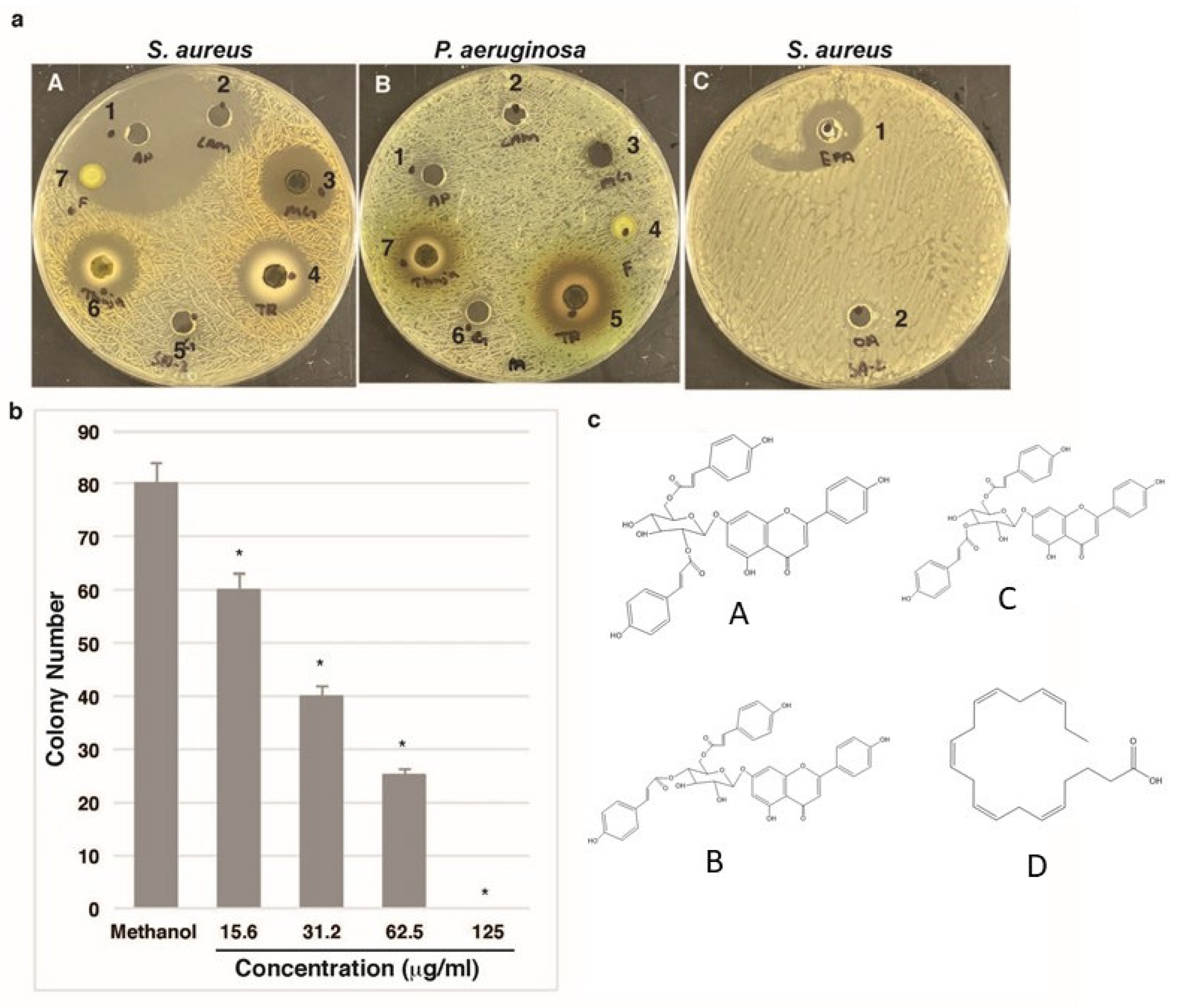
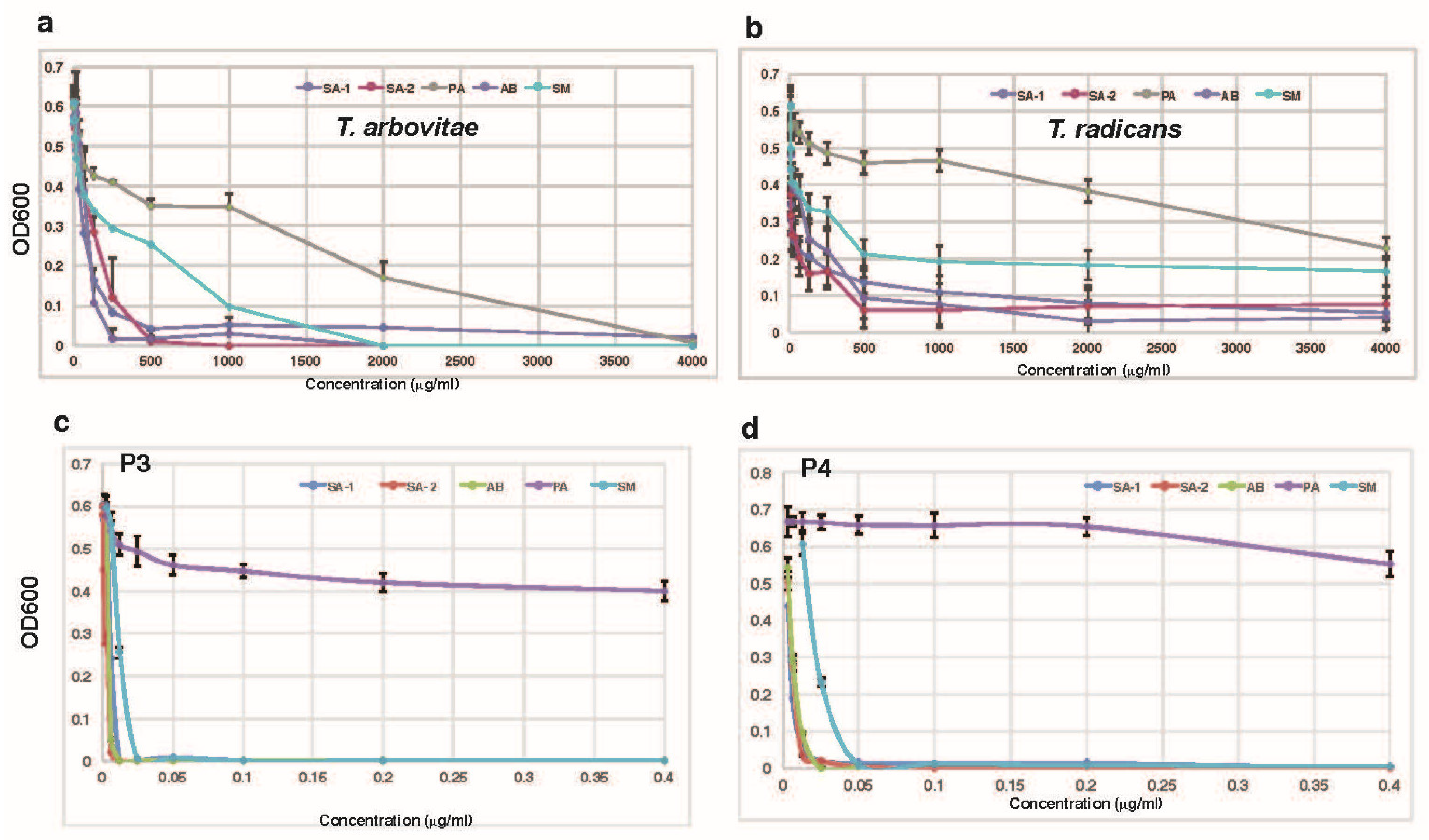
The methanol extracts from M. grandiflora (#3), T. arborvitae (#4), and T. radicans (#6) were active against the bacterial strains of S. aureus. (Figure 1a, Table 1). A. baumannii exhibits inherent resistance to certain antibiotics, including penicillin and chloramphenicol, owing to its unique structural and biochemical characteristics [13]. We found that the T. arborvitae (#5) and T. radicans (#7) extracts also inhibited the bacterial strain P. aeruginosa (Figure 1b). The extract of T. arborvitae (Figure 2a) was more potent than that of T. radicans (Figure 2b) in inhibiting the growth of the four bacterial strains tested. Both extracts strongly inhibited the growth of bacterial strains of S. aureus (SA-1 and SA-2), A. baumannii (AB), and S. mutans (SM) but had less effect on the growth of the P. aeruginosa (PA) bacteria. Under a high concentration (4000 µg/mL), the extract of T. arborvitae could completely suppress the growth of the P. aeruginosa. The leaf extract of T. arborvitae inhibited the colony formation ability of S. aureus in a dosage-dependent manner (Figure 1b). At the concentration of 125 µg/mL, colony formation was completely abolished. Consistent with our observations, antibacterial activity was previously reported in Toxicodendron and Thuja [21,24], but the antibacterial compounds that are responsible for the observed antibacterial activity have not been identified from these plants. Several antibacterial compounds were identified from M. grandiflora [25]. Since leaves of T. arborvitae are largely available and have the most significant antibacterial activity, we decided to use them as the material with which to purify and identify the chemical compounds that contribute to the observed antibacterial activity.

3.2. Purification of Antibacterial Chemical Compounds from T. arborvitae

Dried leaves of T. arborvitae were extracted with water, methanol, or ethyl acetate at room temperature overnight. Ethyl acetate and methanol but not water extract showed antibacterial activity against S. aureus, suggesting that the antibacterial constituents are not water-soluble. The ethanol extract was separated on an alumina column with the mobile phase of ethyl acetate and methanol (1:1). The zone inhibition assay was used to detect the antibacterial activity of column fractions. Fractions containing the antibacterial activity were pooled, condensed, and freeze-dried under a vacuum. The dried materials were dissolved in ethyl acetate and loaded onto a Silica column. The unbound materials were eluted from the column with ethyl acetate and the bound materials were eluted with methanol. The antibacterial activity was detected only in the methanol fraction. The materials in the methanol fraction were further separated by HPLC C18 reverse phase chromatography. Twenty-three major peaks were collected and dried under a vacuum. The zone inhibition assay indicated that four peaks, designated as P1, P2, P3, and P4 with retention times of 45, 46, 47, and 71 min, had antibacterial activity, suggesting that T. arborvitae contains multiple antibacterial constituents. P4 was further purified by another round of HPLC C18 reverse phase chromatography. From 800 g of dry leaves of T. arborvitae, we obtained about 12.7 mg of P1, 16.8 mg of P2, 7.7 mg of P3, and 41.4 mg of P4. The purified compounds showed an MIC of 10 to 50 µg/mL against the bacterial strain S. aureus (Table 2).

3.3. Identification of Isolated Compounds

The three purified samples (P2, P3, and P4) were subjected to HPLC-HRMS (high-resolution mass spectrometry) analysis. The TICs (both positive and negative modes) and UV chromatograms show that P3 is a nearly pure compound with the MS/MS spectrum matching 3″,4″-di-O-p-coumaroylafzelin (Figure S1A). In silico fragment interpretation also supported the structure identification (Figure S1B). P2 is a mixture of a m/z 288.290, P3, and a P3 isomer. P4 mainly contains a compound with m/z 303.2320 (Figure S2A). An MS/MS library search suggested that it is likely eicosapentaenoic acid. An early in silico fragmentation analysis suggested that eicosapentaenoic acid fits well with the MS/MS spectrum of m/z 303.2320 in P4 (Figure S2B).
It was reported that EPA has antibacterial activity [26], but the antibacterial activity of apigenin-di-p-coumaryglucoside has not been documented. The commercial apigenin 7-(3″,6″-di-p-coumarylglucoside) and EPA showed an extent of antibacterial activity similar to that of the purified P3 and P4 preparations (Table 2). The apigenin 7-di-p-coumarylglucoside has three isomers that exist in nature, i.e., apigenin-7-(2″,6″-di-p-coumarylglucoside), apigenin-7-(3″,6″-di-p-coumarylglucoside), and apigenin-7-(4″,6″-di-p-coumarylglucoside) (Figure 1c). Based on their similar chromatographic behavior, we speculate that P1, P2, and P3 are three isomers of apigenin 7-di-p-coumarylglucoside. Abietic acid is an isomer of eicosapentaenoic and was identified as an antibacterial molecule [26]. EPA is an attractive new antibacterial agent, particularly due to its potency and perceived safety. We also observed its antibacterial activity (Table 2), indicating that EPA might exist in multiple plants as an important antibacterial agent.

3.4. Effects of the Purified Compounds on Bacterial Growth and Viability

Apigenin and its derivative compounds were purchased and tested for antibacterial activity against the bacteria S. aureus. Apigenin, apigenin-7-glucoside, and apigenin-7-O-neosperidoside showed no detectable antibacterial activity (Table 2), suggesting that apigenin-di-p-coumarylglucoside might be the unique compound that has antibacterial activity. Although EPA and abietic acid belong to the fatty acid family and have antibacterial activity, fatty acid oleic acid (OA) did not show antibacterial activity in the same assay (Figure 1a(C)).
The purified P3 and P4 compounds completely inhibited the growth of bacterial strains of S. aureus, A. baumannii, and S. mutans at concentrations below 0.05 mg/mL (Figure 2c,d). P3 had less effect on the growth of the bacterial strain P. aeruginosa. Similar effects of the purchased compounds [apigenin 7-(3″,6″-di-p-coumarylglucoside) and EPA] on the growth of these bacterial strains were observed.
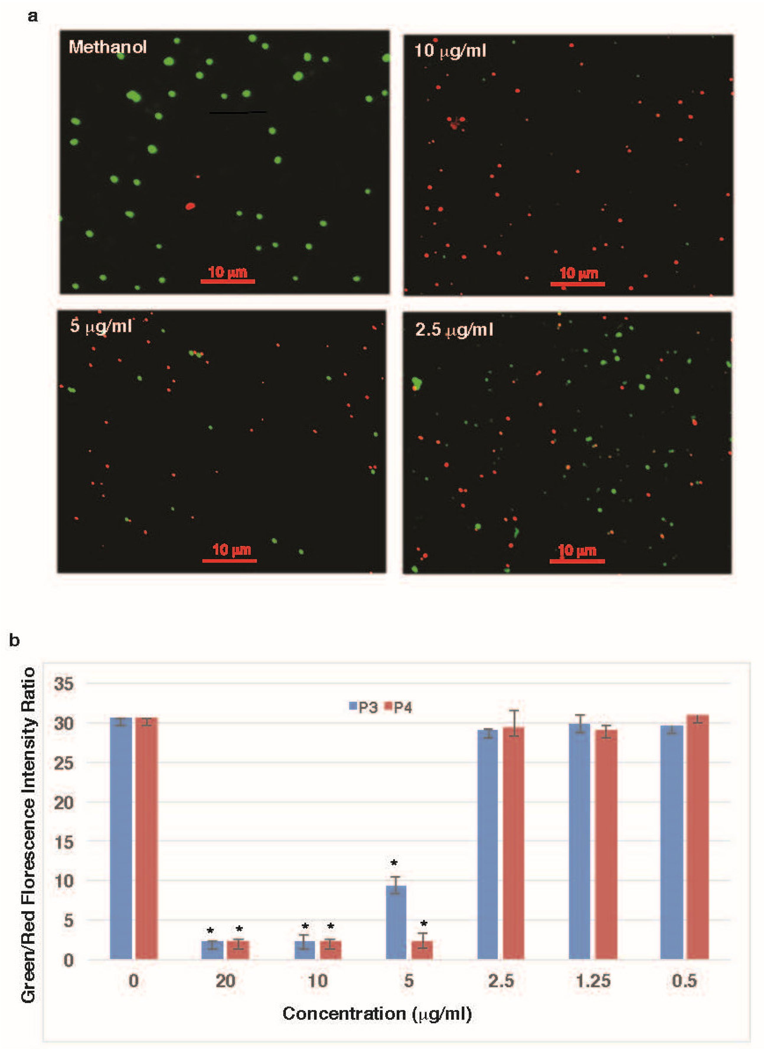
The LIVE/DEAD viability kit, which employs two nucleic acid-binding dyes (SYTO 9™, and propidium iodide), was used to determine the effect of purified compounds on bacterial cells’ viability and membrane integrity. The purified compounds P3 and P4 significantly induced cell death of the bacterial strain S. aureus at concentrations at or above 5 µg/mL (Figure 3a). By quantifying the ratio of green to red fluorescence, the effectiveness of the purified compounds is shown in Figure 3b. In the presence of 2.5 µg/mL or more of P3 or P4, the intensity of the red fluorescence was significantly high, indicating that the bacterial membrane was disrupted. In contrast, little red fluorescence was observed in the presence of 2.5 µg/mL or lower P3 or P4 (Figure 3b). The results showed that the purified antibacterial compounds effectively induced cell death or damage to the bacterial membrane of S. aureus.

3.5. Effects of the Purified Compounds on the Permeability of the Bacterial Membrane

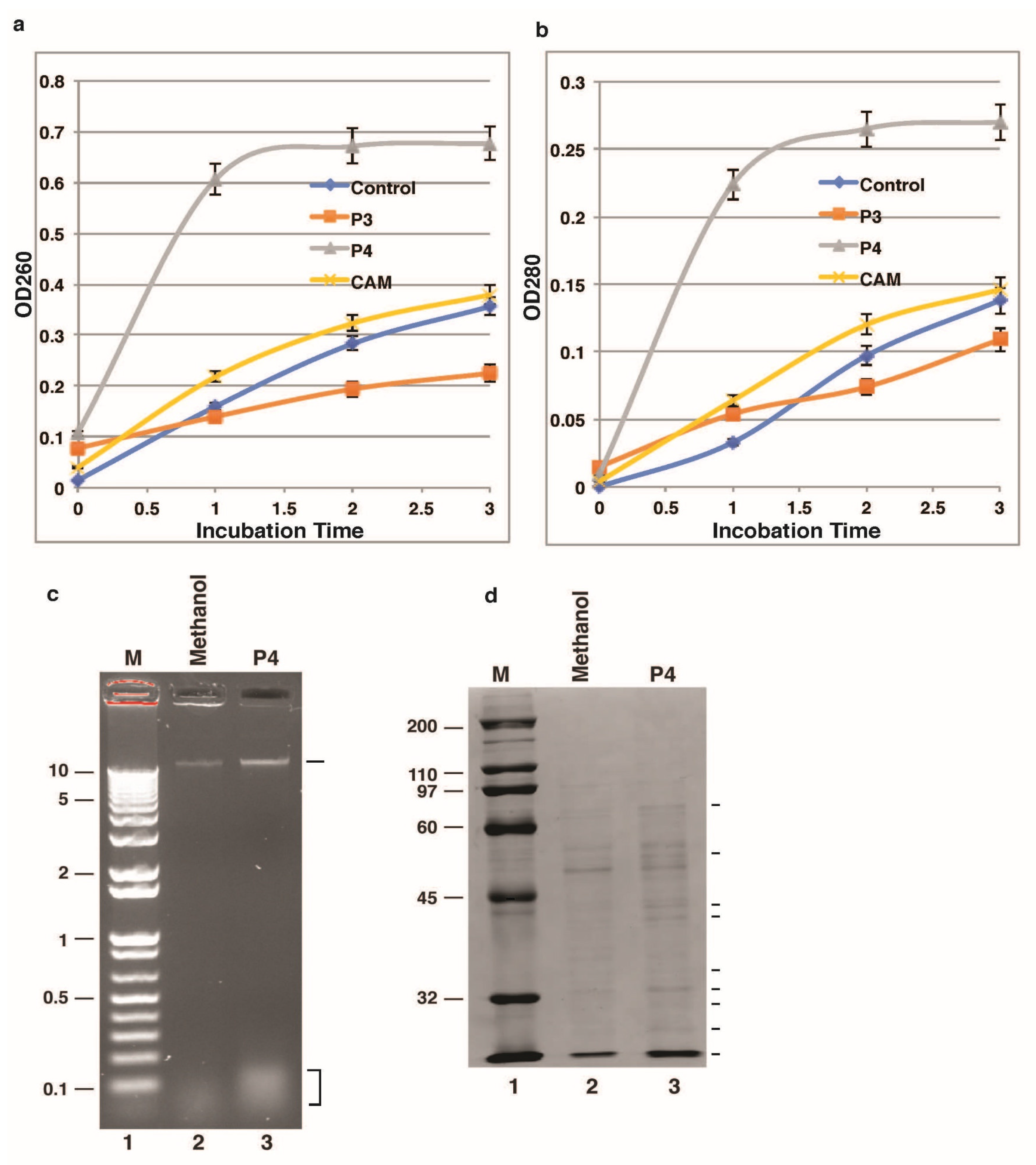
The leakage of nucleic acid and protein into the supernatant of the bacterial culture in the presence of the purified compounds was determined to evaluate its effects on the cell permeability of the bacterial membrane. The leakage of nucleic acid (Figure 4a) and protein (Figure 4b) was significantly higher than that in the control group at 1, 2, and 3 h post-treatment with P4. The nucleic acid and protein in the supernatant of the cultured bacterial cells were also analyzed by agarose electrophoresis (Figure 4c) and SDS-PAGE (Figure 4d). The treatment with P4 significantly increased the levels of nucleic acid and protein in the bacterial culture. The results indicated that the P4 compounds led to the leakage of intracellular nucleic acid and protein in the bacteria. Under the same conditions, P3 and chloramphenicol did not affect the permeability of the bacteria (Figure 4a,b).

3.6. The Effects of P3 on Translation and Transcription of the Bacterial

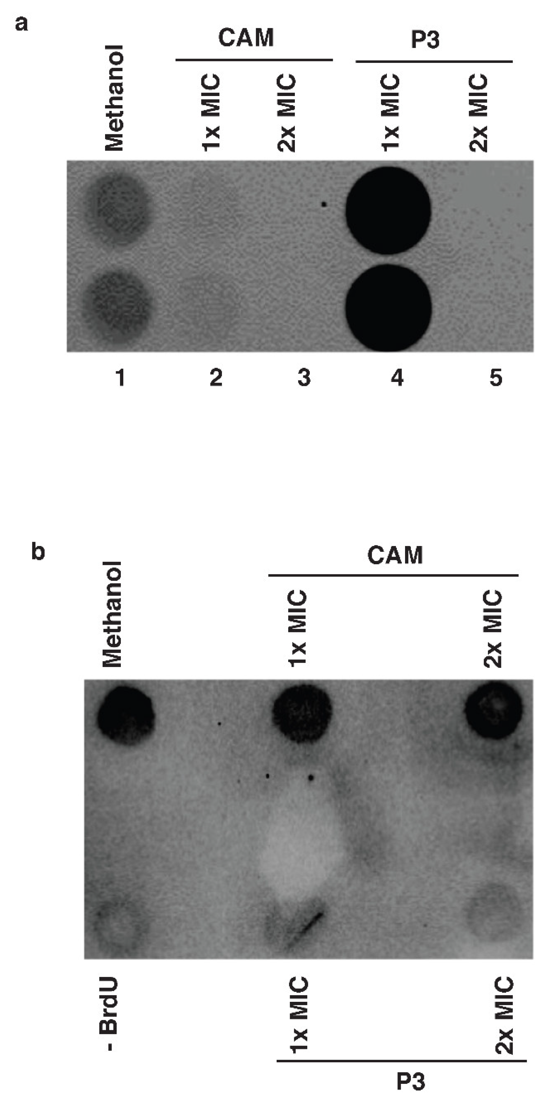
The in vitro cell-free translation system was used to determine the effect of the purified compounds on bacterial protein synthesis. Chloramphenicol, a well-known inhibitor of bacterial translation, was used as a positive control. As expected, chloramphenicol strongly inhibited the bacterial translation at the concentration of 1× MIC (0.8 µg/mL) and 2× MIC (1.6 µg/mL) (Figure 5a). P3 dramatically enhanced the bacterial translation at the concentration of 1× MIC. However, it blocked the bacterial translation at the concentration of 2× MIC. Therefore, P3 might inhibit bacterial translation at a higher concentration.
We next investigate the effect of P3 on bacterial DNA synthesis. The bacterial DNA was isolated from the bacterial cells cultured in the presence of BrdU as well as in the absence or presence of P3. The DNA synthesis (BrdU-incorporation) was monitored by immunoblotting with the anti-BrdU antibody. P3 strongly inhibited the bacterial DNA synthesis at the concentrations of 1× MIC and 2× MIC (Figure 5b). Under the same conditions, chloramphenicol did not affect the bacterial DNA synthesis (Figure 5b).

4. Discussion

The emergence of new diseases resistant to current antibiotics has been one of the challenging problems healthcare providers encounter. This study described the purification and identification of two antibacterial compounds from the Thuja leaf extract. Their antibacterial activity was observed against two Gram-positive and two Gram-negative bacterial strains.
The oil extract of Thuja leaves has been used to prevent and treat infectious diseases [27], and antibacterial activity has been detected in the crude Thuja extract [28,29,30]. However, these studies did not determine the compounds responsible for the observed antibacterial activity in the crude Thuja extract. In this study, two chemical compounds were successfully isolated and purified from Thuja leaves. One compound, EPA, was previously reported to exhibit antibacterial activity [26]. The other compound was identified as apigenin-di-p-coumarylglucoside, for which antibacterial activity had not been previously documented, and it represents a novel antibacterial agent within this chemical class. The two compounds show strong antibacterial activity against the three bacterial strains tested but have less effect on the bacterial strain P. aeruginosa. More importantly, these compounds can inhibit the growth of the bacterial strain (A. baumannii) with multiple drug-resistant phenotypes. A. baumannii exhibits a multifaceted antibiotic resistance profile, often being dubbed a superbug [31]. This includes resistance to a broad spectrum of antibiotics, including carbapenems, aminoglycosides, fluoroquinolones, and various beta-lactam antibiotics. This extensive antibiotic resistance in A. baumannii poses a significant clinical challenge, necessitating a refined and tailored approach in the selection of antimicrobial agents for effective treatment. Mechanistic insights into how the identified compounds inhibited growth of the A. baumannii will require further investigation.
The cytoplasmic membrane serves as a selective barrier and controls the cell’s internal composition. Whenever these functional roles of the cytoplasmic membrane become disturbed, macromolecules and ions will outflow, which will result in cell destruction or death. Polymyxins are active antibacterial agents that are cyclic peptides, having a long hydrophobic tail [32,33]. Polymyxins show their specificity for polysaccharide molecules, which are present in the outer membrane of many Gram-negative bacteria. After association with the lipopolysaccharide substrate in the outer membrane of Gram-negative bacteria, polymyxins change the membrane structure so that its permeability increases, which results in disruption of the osmotic balance. Since Gram-positive bacteria have a thick cell wall, which prohibits the access of these molecules to the Gram-positive bacterial cell membrane, polymyxins have less or even no effect on Gram-positives. We found that EPA could change the membrane permeability to enhance the outflow of macromolecules (protein and DNA). This molecule has bulky hydrophobic tails that can bind the cell membrane and disrupt its structure, leading to leakage of the cell membrane. Unlike polymyxins, these two antibiotics inhibited Gram-positives as well as Gram-negatives.
Apigenin is a flavone that is abundantly present in plants and can confer various health benefits such as antioxidant [34], anti-inflammatory [35], and chemopreventative effects [36]. Apigenin-di-p-coumarylglucoside is one of the apigenin derivatives. In our analyses, apigenin and some derivatives had no detectable antibacterial activity against the four tested bacterial strains. In contrast, apigenin-di-p-coumarylglucoside showed strong antibacterial activity against the three bacterial strains tested. Apigenin-di-p-coumarylglucoside has some structural similarities to aminoglycoside, which contains two amino sugars joined by a glycosidic bond to an aminocyclitol [37]. Some commonly used aminoglycosides are streptomycin, gentamicin, sisomicin, netilmicin, kanamycin, amikacin, neomycin, tobramycin, spectinomycin, and paromomycin, which function as protein synthesis inhibitors. Apigenin-di-p-coumarylglucoside at a high concentration (2× MIC) also inhibited protein synthesis but it promoted protein synthesis at a low concentration (1× MIC). Under the same conditions, we also observed that ampicillin enhanced in vitro translation. The reason for the observed enhancement of protein synthesis by these two antibacterial compounds is unclear.
One of the most important targets for antibiotics to cure infectious diseases is nucleic acid synthesis. A large difference in the enzymes that carry out DNA and RNA synthesis between eukaryotic and prokaryotic cells is that some achieve selective toxicity. A well-known example is the rifamycin family, which binds to DNA-dependent RNA polymerase, thereby inhibiting the elongation of RNA [38]. Quinolones bind to DNA gyrase, inhibiting their function, which results in inhibition of the DNA replication that ultimately results in cell death [39]. In this report, the BrdU incorporation assay indicated that Apigenin-di-p-coumarylglucoside strongly inhibited the DNA synthesis of the tested bacteria, suggesting that it targets DNA synthesis. This is also consistent with its lack of effect on cell membrane permeability. Future investigation is needed to find the target site of Apigenin-di-p-coumarylglucoside in the DNA synthesis machinery.

5. Conclusions

Two antibacterial compounds were purified from the T. arborvitae plant and showed strong antibacterial activity against four bacterial strains. One antibacterial compound induced damage to the cell membrane, leading to leakage of DNA and protein. The other identified compound inhibited DNA synthesis. The identified antibacterial compounds could provide a useful tool for controlling bacterial pathogens and infectious diseases by inhibiting bacterial DNA synthesis or inducing cell membrane permeability.

Supplementary Materials

The following supporting information can be downloaded at https://www.mdpi.com/article/10.3390/microbiolres17020038/s1, Figure S1: Identification of the compound in P3 as Apigenin-di-p-courmarylglucoside. A. MS/MS spectra of P3. B. Partial MS2 fragments interpretation.; Figure S2: Identification of the compound in P4 as eicosapentanoic acid. A. MS/MS spectra of P4. B. Partial MS2 fragments interpretation.

Author Contributions

T.M.J. performed experiments, analyzed the data, and wrote the manuscript; Q.X. performed experiments and analyzed the data; Z.W. performed data analysis and wrote the manuscript. All authors have read and agreed to the published version of the manuscript.

Funding

The U.S. Department of Education—Title III Part B: Strengthening Historically Black Colleges and Universities Program, Award Number: P031K19000, Grant Recipient: Clark Atlanta University.

Institutional Review Board Statement

Not applicable.

Informed Consent Statement

Not applicable.

Data Availability Statement

The original contributions presented in this study are included in the article/Supplementary Materials. Further inquiries can be directed to the corresponding author.

Acknowledgments

We thank Nathan Bowen and D’Netria Bledsoe for reading and comments on the manuscript.

Conflicts of Interest

Author Quanbo Xiong was employed by the company Corteva Agriscience. The remaining authors declare that the research was conducted in the absence of any commercial or financial relationships that could be construed as a potential conflict of interest.

Abbreviations

EPAEicosapentaenoic acid
OAOleanolic acid
MICMinimum inhibition concentration
CFUColony-forming unit

References

  1. Ferri, M.; Ranucci, E.; Romagnoli, P.; Giaccone, V. Antimicrobial resistance: A global emerging threat to public health systems. Crit. Rev. Food Sci. Nutr. 2017, 57, 2857–2876. [Google Scholar] [CrossRef] [Scilit]
  2. Chen, L.; Kumar, S.; Wu, H. A review of current antibiotic resistance and promising antibiotics with novel modes of action to combat antibiotic resistance. Arch. Microbiol. 2023, 205, 356. [Google Scholar] [CrossRef] [Scilit]
  3. Schafer, A.B.; Sidarta, M.; Abdelmesseh Nekhala, I.; Marinho Righetto, G.; Arshad, A.; Wenzel, M. Dissecting antibiotic effects on the cell envelope using bacterial cytological profiling: A phenotypic analysis starter kit. Microbiol. Spectr. 2024, 12, e0327523. [Google Scholar] [CrossRef] [Scilit] [PubMed]
  4. Gupta, R.; Singh, M.; Pathania, R. Chemical genetic approaches for the discovery of bacterial cell wall inhibitors. RSC Med. Chem. 2023, 14, 2125–2154. [Google Scholar] [CrossRef] [Scilit]
  5. Vazquez-Laslop, N.; Mankin, A.S. Context-Specific Action of Ribosomal Antibiotics. Annu. Rev. Microbiol. 2018, 72, 185–207. [Google Scholar] [CrossRef] [Scilit]
  6. Kirsch, S.H.; Haeckl, F.P.J.; Muller, R. Beyond the approved: Target sites and inhibitors of bacterial RNA polymerase from bacteria and fungi. Nat. Prod. Rep. 2022, 39, 1226–1263. [Google Scholar] [CrossRef] [Scilit] [PubMed]
  7. Sofianos, G.; Samaras, A.; Karaoglanidis, G. Multiple and multidrug resistance in Botrytis cinerea: Molecular mechanisms of MLR/MDR strains in Greece and effects of co-existence of different resistance mechanisms on fungicide sensitivity. Front. Plant Sci. 2023, 14, 1273193. [Google Scholar] [CrossRef] [Scilit]
  8. Wang, Z.; Zhu, M.; Wang, M.; Gao, Y.; Zhang, C.; Liu, S.; Qu, S.; Liu, Z.; Zhang, C. Integrated Multiomic Analysis Reveals the High-Fat Diet Induced Activation of the MAPK Signaling and Inflammation Associated Metabolic Cascades via Histone Modification in Adipose Tissues. Front. Genet. 2021, 12, 650863. [Google Scholar] [CrossRef] [Scilit] [PubMed]
  9. Van Boeckel, T.P.; Pires, J.; Silvester, R.; Zhao, C.; Song, J.; Criscuolo, N.G.; Gilbert, M.; Bonhoeffer, S.; Laxminarayan, R. Global trends in antimicrobial resistance in animals in low- and middle-income countries. Science 2019, 365, eaaw1944. [Google Scholar] [CrossRef] [Scilit]
  10. Holmes, A.H.; Moore, L.S.; Sundsfjord, A.; Steinbakk, M.; Regmi, S.; Karkey, A.; Guerin, P.J.; Piddock, L.J. Understanding the mechanisms and drivers of antimicrobial resistance. Lancet 2016, 387, 176–187. [Google Scholar] [CrossRef] [Scilit]
  11. Antimicrobial Resistance, C. Global burden of bacterial antimicrobial resistance in 2019: A systematic analysis. Lancet 2022, 399, 629–655. [Google Scholar] [CrossRef] [Scilit]
  12. Aslam, B.; Wang, W.; Arshad, M.I.; Khurshid, M.; Muzammil, S.; Rasool, M.H.; Nisar, M.A.; Alvi, R.F.; Aslam, M.A.; Qamar, M.U.; et al. Antibiotic resistance: A rundown of a global crisis. Infect. Drug Resist. 2018, 11, 1645–1658. [Google Scholar] [CrossRef] [Scilit]
  13. Munita, J.M.; Arias, C.A. Mechanisms of Antibiotic Resistance. Microbiol. Spectr. 2016, 4, 481–511. [Google Scholar] [CrossRef] [Scilit]
  14. Jacobs, L.M.C.; Consol, P.; Chen, Y. Drug Discovery in the Field of beta-Lactams: An Academic Perspective. Antibiotics 2024, 13, 59. [Google Scholar]
  15. Ezzeddine, Z.; Ghssein, G. Towards new antibiotics classes targeting bacterial metallophores. Microb. Pathog. 2023, 182, 106221. [Google Scholar] [CrossRef] [Scilit]
  16. Tillotson, G.S. Trojan Horse Antibiotics-A Novel Way to Circumvent Gram-Negative Bacterial Resistance? Infect. Dis. 2016, 9, 45–52. [Google Scholar]
  17. Clardy, J.; Walsh, C. Lessons from natural molecules. Nature 2004, 432, 829–837. [Google Scholar] [CrossRef] [Scilit] [PubMed]
  18. Abdallah, E.M.; Alhatlani, B.Y.; de Paula Menezes, R.; Martins, C.H.G. Back to Nature: Medicinal Plants as Promising Sources for Antibacterial Drugs in the Post-Antibiotic Era. Plants 2023, 12, 3077. [Google Scholar] [CrossRef] [Scilit]
  19. Ruggieri, F.; Compagne, N.; Antraygues, K.; Eveque, M.; Flipo, M.; Willand, N. Antibiotics with novel mode of action as new weapons to fight antimicrobial resistance. Eur. J. Med. Chem. 2023, 256, 115413. [Google Scholar] [CrossRef] [Scilit] [PubMed]
  20. Geng, J.; Liu, X.; Wang, J.; Li, S. Accumulation and risk assessment of antibiotics in edible plants grown in contaminated farmlands: A review. Sci. Total Environ. 2022, 853, 158616. [Google Scholar]
  21. Puskarova, A.; Buckova, M.; Krakova, L.; Pangallo, D.; Kozics, K. The antibacterial and antifungal activity of six essential oils and their cyto/genotoxicity to human HEL 12469 cells. Sci. Rep. 2017, 7, 8211. [Google Scholar] [CrossRef] [Scilit]
  22. Vuorelaa, P.; Leinonenb, M.; Saikkuc, P.; Tammelaa, P.; Rauhad, J.P.; Wennberge, T.; Vuorela, H. Natural products in the process of finding new drug candidates. Curr. Med. Chem. 2004, 11, 1375–1389. [Google Scholar] [CrossRef] [Scilit]
  23. McOrist, A.L.; Jackson, M.; Bird, A.R. A comparison of five methods for extraction of bacterial DNA from human faecal samples. J. Microbiol. Methods 2002, 50, 131–139. [Google Scholar] [CrossRef] [Scilit]
  24. Saravanakumar, K.; Chelliah, R.; Hu, X.; Oh, D.H.; Kathiresan, K.; Wang, M.H. Antioxidant, Anti-Lung Cancer, and Anti-Bacterial Activities of Toxicodendron vernicifluum. Biomolecules 2019, 9, 127. [Google Scholar] [CrossRef] [Scilit] [PubMed]
  25. Zeutsop, J.F.; Nono, R.N.; Frese, M.; Chouna, J.R.; Lenta, B.N.; Nkeng-Efouet-Alango, P.; Sewald, N. Phytochemical, antibacterial, antioxidant and cytoxicity investigation of Tarenna grandiflora. Z. Für Naturforschung C 2021, 76, 285–290. [Google Scholar] [CrossRef] [Scilit]
  26. Wei, M.; Wang, P.; Li, T.; Wang, Q.; Su, M.; Gu, L.; Wang, S. Antimicrobial and antibiofilm effects of essential fatty acids against clinically isolated vancomycin-resistant Enterococcus faecium. Front. Cell. Infect. Microbiol. 2023, 13, 1266674. [Google Scholar] [CrossRef] [Scilit] [PubMed]
  27. Fu, C.; Lan, X.; Yuan, J.; Li, C.; Li, L.; Yu, Z.; Tan, T.; Yuan, M.; Du, F. Research on the optimization, key chemical constituents and antibacterial activity of the essential oil extraction process of Thuja koraiensis Nakai. J. Microbiol. Methods 2022, 194, 106435. [Google Scholar] [CrossRef] [Scilit]
  28. Thakur, M.; Guleria, P.; Sobti, R.C.; Gautam, A.; Kaur, T. Comparative analysis of the antibacterial efficacy and bioactive components of Thuja occidentalis obtained from four different geographical sites. Mol. Cell. Biochem. 2023, 479, 283–296. [Google Scholar] [CrossRef] [Scilit] [PubMed]
  29. Bakht, J.; Zafar, Z.; Ahmad, J.; Khan, S. Antibacterial activity of the crude extracts from medicinally important Thuja occidentalis. Pak. J. Pharm. Sci. 2020, 33, 627–630. [Google Scholar]
  30. Wang, M.; Zhao, L.; Chen, K.; Shang, Y.; Wu, J.; Guo, X.; Chen, Y.; Liu, H.; Tan, H.; Qiu, S.X. Antibacterial sesquiterpenes from the stems and roots of Thuja sutchuenensis. Bioorg. Chem. 2020, 96, 103645. [Google Scholar] [CrossRef] [Scilit]
  31. Mukhopadhyay, H.; Bairagi, A.; Mukherjee, A.; Prasad, A.K.; Roy, A.D.; Nayak, A. Multidrug resistant Acinetobacter baumannii: A study on its pathogenesis and therapeutics. Curr. Res. Microb. Sci. 2025, 8, 100331. [Google Scholar] [CrossRef] [Scilit] [PubMed]
  32. Nang, S.C.; Azad, M.A.K.; Velkov, T.; Zhou, Q.T.; Li, J. Rescuing the Last-Line Polymyxins: Achievements and Challenges. Pharmacol. Rev. 2021, 73, 679–728. [Google Scholar] [CrossRef] [Scilit] [PubMed]
  33. Mohapatra, S.S.; Dwibedy, S.K.; Padhy, I. Polymyxins, the last-resort antibiotics: Mode of action, resistance emergence, and potential solutions. J. Biosci. 2021, 46, 85. [Google Scholar] [CrossRef] [Scilit]
  34. Muhammed, T.M.; Jalil, A.T.; Taher, W.M.; Aminov, Z.; Alsaikhan, F.; Ramirez-Coronel, A.A.; Ramaiah, P.; Farhood, B. The Effects of Apigenin in the Treatment of Diabetic Nephropathy: A Systematic Review of Non-clinical Studies. Mini Rev. Med. Chem. 2024, 24, 341–354. [Google Scholar] [CrossRef] [Scilit]
  35. Wang, F.; Xiao, L.; Zhang, H.; Wang, Y.; Zhong, L.; Feng, X.; Liu, J.; Gong, X. Systemic meta-analysis: Apigenin’s effects on lung inflammation and oxidative stress. J. Asthma 2023, 61, 271–281. [Google Scholar] [CrossRef] [Scilit]
  36. Fossatelli, L.; Maroccia, Z.; Fiorentini, C.; Bonucci, M. Resources for Human Health from the Plant Kingdom: The Potential Role of the Flavonoid Apigenin in Cancer Counteraction. Int. J. Mol. Sci. 2023, 25, 251. [Google Scholar] [CrossRef] [Scilit]
  37. Tevyashova, A.N.; Shapovalova, K.S. Potential for the Development of a New Generation of Aminoglycoside Antibiotics. Pharm. Chem. J. 2021, 55, 860–875. [Google Scholar] [CrossRef] [Scilit] [PubMed]
  38. Gugnani, J.S.; Abhishek, F.; Agarwal, Y.; Damera, A.R.; Kaur, H.; Taleb, B.; Mane, R.; Soni, U.; Nayar, K.D. Effectiveness of Rifabutin-Based Regimens in Treating Helicobacter pylori Infections. Cureus 2023, 15, e50541. [Google Scholar] [CrossRef] [Scilit]
  39. Chen, J.P.; Battini, N.; Ansari, M.F.; Zhou, C.H. Membrane active 7-thiazoxime quinolones as novel DNA binding agents to decrease the genes expression and exert potent anti-methicillin-resistant Staphylococcus aureus activity. Eur. J. Med. Chem. 2021, 217, 113340. [Google Scholar] [CrossRef] [Scilit]
Figure 1. (a) The zone of inhibition assay performed to detect antibacterial activity. The zone of inhibition assay was performed with plant extracts against S. aureus (ATCC 49775) (A,C) and A. baumannii (B). 1, ampicillin (5 µg); 2, chloramphenicol (1.25 µg); 3, M. grandiflora extract (5 mg); 4, T. radicans extract (5 mg); 5, G. biloba extract, (5 mg); 6, T. arborvitae extract (5 mg); and 7, F. vulgare extract (5 mg). The zone of inhibition assay was performed with EPA (0.5 mg) and Oleic acid (OA) (0.5 mg) against S. aureus (ATCC 49775) (C). (b) Antibacterial effects of T. arborvitae extract on colony-forming ability. The y-axis shows the colony numbers of S. aureus (ATCC 49775). The x-axis shows the concentrations of T. arborvitae extract. Methanol was used as a solvent to dissolve the extract. Data are represented as the mean ± SD of three independent tests. * Significantly different (p < 0.05) compared to the methanol control. (c) Chemical structures of identified antibacterial compounds. (A): apigenin-7-(2″,6″-di-p-coumarylglucoside); (B): apigenin-7-(4″,6″-di-p-coumarylglucoside); (C): apigenin-7-(3″,6″-di-p-coumarylglucoside), (D): EPA.
Microbiolres 17 00038 g001
Figure 2. Effects of T. arborvitae and T. Radicans extracts (a,b) and purified P3 and P4 compounds (c,d) on bacterial growth. Growth curves were determined by culturing bacteria for 18 h in the presence of various concentrations of plant extracts. The y-axis shows the turbidity of the bacterial culture measured by spectrometry. OD600: absorbance at 600 nm. The x-axis shows the concentration of the extract. SA-1 (ATCC 49775) and SA-2 (ATCC 12600): S. aureus; PA: P. aeruginosa: (ATCC 10145); AB: A. baumannii (ATCC 19606); SM: S. mutans (ATCC 25175). The values are plotted as the mean ± SD obtained from the experiment performed in triplicate.
Microbiolres 17 00038 g002
Figure 3. Antibacterial effects of purified P3 and P4 on bacterial viability. (a) Fluorescence microscopic analysis was performed using the bacteria S. aureus incubated in P3 or P4 for 1 h and stained with SYTO 9 (green: viable cells) and propidium iodide (red: dead cells). (b) Fluorophotometric measurement was performed using S. aureus treated with several concentrations of P3 or P4 for 18 h. The y-axis shows the ratio of green/red fluorescence as indicated by the intensity at the wavelength of 530 nm divided by that of 630 nm, and the horizontal axis shows the concentration of the P3 or P4 compound. The data represent the mean ± SD of three independent tests. * Significantly different (p < 0.05) compared to the untreated control.
Microbiolres 17 00038 g003
Figure 4. Effects of purified P3 and P4 on intracellular nucleic acid and protein leakage in S. aureus. The y-axis shows the absorbance at 260 nm (OD260) (a) or 280 nm (OD280) (b) of the supernatant of the bacterial culture in the presence of methanol (control), chloramphenicol (CAM) (25 µg/mL), P3 (100 µg/mL), and P4 (20 µg/mL) dissolved in methanol. The x-axis represents the incubation time. The values are plotted as the mean ± SD obtained from experiments performed in triplicate. (c) DNA analysis by agarose gel electrophoresis of the supernatant of the bacteria culture in the presence of methanol and P4 (20 µg/mL) in the methanol with ethidium bromide staining. Ten micrograms of DNA samples were loaded on each lane. Lane 1 shows the 1 kb plus DNA marker (Promega). The gel was stained with ethidium bromide (0.5 µg/mL). (d) Protein analysis by SDS-polyacrylamide gel electrophoresis (SDS-PAGE) of the bacterial culture in the presence of methanol or P4 in the methanol (20 µg/mL). Ten microliters of each sample were loaded into each lane. Lane 1 shows standard protein markers (Bio-Rad). Coomassie Blue R250 was used to stain proteins (distinct bands in lanes 2 and 3).
Microbiolres 17 00038 g004
Figure 5. Effects of P3 on protein and DNA synthesis. (a) Photographic luciferase assays. In vitro translation assay with E. coli S30 extracts in the presence of methanol, chloramphenicol (CAM, 25 µg/mL), and P3 at the concentrations of lx MIC (50 µg/mL) and 2× MIC (100 µg/mL). Briefly, 50 µL of the luciferase reagent was added into each well of a white 96-well plate and 50 µL of the luciferase assay reagent was added into each well. The image was taken by a digital camera with a 6 min exposure. (b) Immunochemical detection of BrdU in DNA by dot blotting. DNA was from cultured bacteria grown in the absence of BrdU and in the presence of BrdU plus methanol, CAM, or P3 at concentrations of l× MIC and 2× MIC. The DNA sample (1 µg) was spotted onto a Zeta probe hybridization membrane and the membrane was probed with an anti-BrdU antibody and peroxidase-conjugated secondary antibody.
Microbiolres 17 00038 g005
Table 1. Zone of inhibition assay of different plant extracts (NA, Not Detected).
PlantS. aureasP. aeruginosa
F. vulgareNANA
G. bilobaNANA
L. barbarumNANA
M. grandiflora20 ± 2NA
P. anisumNANA
T. arborvitae19 ± 111 ± 1
T. radicans17 ± 117 ± 2
Z. americamumNANA
Table 2. Minimal inhibition concentrations of purified compounds.
CompoundsMIC (µg/mL)
Crude Extract500 ± 20
P120 ± 0.8
P210 ± 0.4
P350 ± 2
P410 ± 0.4
Apigenin>2000
Apigenin-7-Glucoside>2000
Apigenin-7,3″,6″-di-p-Coumarylglucoside50 ± 2
Apigenin-7-o-Neosperidoside>2000
Abietic Acid20 ± 0.8
Eicosapentanoic Acid10 ± 0.8
Disclaimer/Publisher’s Note: The statements, opinions and data contained in all publications are solely those of the individual author(s) and contributor(s) and not of MDPI and/or the editor(s). MDPI and/or the editor(s) disclaim responsibility for any injury to people or property resulting from any ideas, methods, instructions or products referred to in the content.