Article

Isolation of Lactic Acid Bacteria from Raw Camel Milk in Saudi Arabia and Evaluation of Their Probiotic Potential

1
Department of Food Science and Nutrition, Taif University, Taif 21974, Saudi Arabia
2
Clinical Laboratory Sciences Department, College of Applied Medical Science, University of Hafr Al Batin, Hafr Al Batin 31991, Saudi Arabia
3
Department of Biology, University of Hafr Al Batin, Hafr Al Batin 31991, Saudi Arabia
4
Dairy Science Department, Faculty of Agriculture, Sohag University, Sohag 82524, Egypt
5
Agricultural Microbiology Department, Faculty of Agriculture, Sohag University, Sohag 82524, Egypt
*
Author to whom correspondence should be addressed.
Microbiol. Res. 2025, 16(12), 248; https://doi.org/10.3390/microbiolres16120248
Submission received: 6 October 2025 / Revised: 1 November 2025 / Accepted: 3 November 2025 / Published: 26 November 2025

Abstract

Milk contains wide microbial diversity, composed mainly of lactic acid bacteria (LAB), which are used as probiotics for both humans and livestock. We isolated, characterized, and evaluated LAB from indigenous Saudi Arabian camel milk to assess its probiotic potential, including antagonistic activity (against Methicillin-Resistant Staphylococcus aureus (MRSA) and Klebsiella pneumoniae), survivability in simulated gastric juice, tolerance to bile salts, cell surface hydrophobicity, auto- and co-aggregation, and antibiotic susceptibility tests. The two most promising LAB strains showed probiotic potential and were identified as Leuconostoc mesenteroides based on 16S rRNA gene sequences. These strains inhibited all pathogens tested to varying degrees and were resistant to kanamycin and vancomycin. None of the LAB cultures demonstrated hemolytic or gelatinase activity. Overall, the current data suggests that camel milk has substantial potential for introducing probiotics/LAB strains into the human food chain, making camel milk a potentially sustainable food.

1. Introduction

The Arabian camel, or Camelus dromedarius, is a large, even-toed ungulate with one hump on its back. Camel milk is a nutritionally beneficial substrate for the formation of lactic acid bacteria because it includes roughly 3.0–3.5% protein, 3.0–3.8% fat, and 4.0–4.5% lactose. It is also high in vitamin C, calcium, and iron [1]. There are roughly 23.9 million camels worldwide [2], with 485,926 in Saudi Arabia [3]. Camel milk is well known for its considerable benefits, including its therapeutic properties against a variety of disorders [4,5], and is considered a nutritious replacement for cow’s milk that contains more functional dietary qualities [6]. Lactoferrin, peptidoglycan, antibodies, immunoglobulins, and enzymes (lysozyme and lactoperoxidase) make up the majority of its protein composition and are beneficial in the treatment of various diseases [7]. Consuming camel milk daily may enhance host immune defenses. When compared to other ruminant milks, camel milk is preferable as it contains all of these critical components, as well as minimal levels of unhealthy fat (i.e., cholesterol) and sugar. It also has a significant amount of vitamin C and insulin, both of which are helpful to human health. Probiotics derive their name from the words pro (for) and bios (life). The Food and agriculture organization (FAO) defines probiotics as “live microorganisms that, when administered in adequate amounts, confer a health benefit on the host” [6]. Regardless of its physiochemical makeup, camel milk contains beneficial microorganisms, the majority of which are LAB. Camel milk is in high demand due to its nutritional value. Many previous attempts have been made to examine LAB in camel milk and its products [8,9].
Camel milk has antibacterial properties and is beneficial for conservation. Camel milk suppresses some pathogenic germs due to various protective proteins found in it, including lysozymes, lactoperoxidase, lactoferrin, immunoglobulin, and vitamin C [10,11]. For these reasons, Konuspayeva and Faye [12] argue that pasteurization is unnecessary for camel milk if the camels are healthy. The composition of camel milk is less stable than milk from other animals. Many factors contribute to these variances, including lactation stage, dietary conditions, and the camel’s breed and age [13]. LAB from goat and cow milk have been extensively researched for their antibacterial activity and bacteriocin production [14,15]. However, few studies have been undertaken on the isolation and characterization of LAB from camel milk [16,17] or on its antibacterial activity [18,19]. LAB are a functionally defined group of Gram-positive, low-G+C, acid-tolerant, and generally fermentative bacteria that belong mainly to the order Lactobacillales within the phylum Firmicutes. Genera commonly included among LAB are Lactobacillus (recently reclassified into multiple genera), Lactococcus, Leuconostoc, Pediococcus, Enterococcus, Streptococcus, Weissella, Carnobacterium, Oenococcus, Tetragenococcus, Vagococcus, and Aerococcus [20,21,22]. These bacteria are usually utilized as starters in fermented food items, where they can create certain organoleptic features and extend shelf life [23].
LAB occur naturally in the gastrointestinal tract, as well as in various plant-derived environments, and strains from both sources have been shown to exhibit probiotic properties [24,25]. Furthermore, mixed strain cultures may result in microbial interactions that are either advantageous (cooperative) or negative (inhibitive). These mixed cultures are widely employed as starter cultures in dairy production due to their acid production, growth rate, proteolytic activity, bacteriocin production and sensitivity, fragrance, and phage sensitivity [26]. Nevertheless, due to drawbacks or low response repeatability, these technologies may not be suitable for widespread commercial use [27]. For these reasons, the ability of LAB to withstand gastrointestinal conditions, such as the low pH, presence of proteolytic enzymes, and bile salt concentrations, should be taken into consideration when characterizing them as probiotic cultures [28]. Among multidrug-resistant organisms, MRSA and K. pneumoniae are particularly concerned due to their association with mastitis in the milk of certain ruminant species being linked to the high frequency of multidrug-resistant bacteria [29,30,31]. Probiotic bacteria with the ability to inhibit or resist colonization by such pathogens represent a promising complementary strategy to reduce their burden and mitigate the global threat of antimicrobial resistance.
The purpose of this study was to evaluate the probiotic potential of two strains of lactic acid bacteria isolated from camel milk in the Saudi Arabian region of El Sharqia. To demonstrate their probiotic potential, in vitro tests were performed on their antibiotic susceptibility, tolerance to gastrointestinal disorders, and antagonistic behavior toward human pathogenic microbes.

2. Materials and Methods

2.1. Sampling and Isolation of LAB

2.1.1. Raw Camel Milk Sampling

Nine raw camel milk samples were selected at random from camels that appeared to be in good health and belonged to nomads residing in various parts of the Saudi Arabian province of El Sharqia. Milk samples were aseptically collected directly from the udder. Before collecting, the udder was cleansed using distilled water and patted dry using a single-use towel. The first three streams of milk were discarded. Following appropriate preparation, the samples were collected immediately into sterile containers, kept in an icebox, and sent to the laboratory. In February 2024, samples were taken from camels (Camelus dromedarius) with varying colors. The first samples were taken from black camels from Nairyah, while the second were from brown camels in the Hafr Al Batin desert. The camels’ native diet consisted of Saharan plants such as camel thorns (Alhagi graecorum).

2.1.2. Isolation

The milk was allowed to ferment spontaneously at 30 °C until coagulation, and then serial dilutions were prepared from it using sterile physiological water (0.85%) up to 10−6. For each dilution, 0.1 mL was spread onto de Man, Rogosa and Sharpe (MRS) agar (Hangzhou Reagents, China). Inoculated plates were incubated anaerobically at 30 °C for 48 h (Thermo Scientific Heracell 150i, Thermo Fisher Scientific, Waltham, MA, USA). Plates containing 10–300 Colony-Forming Units (CFUs) were used to randomly select three to five single colonies [32]. Three successive subcultures on MRS agar were used to purify the isolates. Twenty-two pure bacterial isolates were obtained and subjected to initial screening. Gram stains and the catalase test were used to initially characterize the isolates. Only Gram-positive, catalase-negative isolates were preserved in reconstituted skim milk with 30% (w/v) glycerol at −80 °C (Thermo Scientific Forma 89000 Series, Thermo Fisher Scientific, Waltham, MA, USA).

2.2. Screening of Selected Bacterial Isolates for Probiotic Attributes

To investigate the probiotic qualities of twelve isolates that were chosen (based on the results of the initial screening), each isolate underwent a second test. This screening entailed carrying out a number of in vitro tests, which are detailed in the sections that follow. Three separate biological replicates were used for each assay (n = 3).

2.2.1. Antagonistic Activity

An agar diffusion experiment was used to measure the antagonistic activity. Briefly, an optical density of 1 (OD 600 = 1) (UV-1800, Shimadzu Corporation, Kyoto, Japan) was achieved by adjusting the turbidity of the overnight culture of MRSA and K. pneumoniae (obtained from our previous study) [33,34]. The tested bacteria suspension was then seeded into molten MRS soft agar (0.7%) maintained at 45 °C. Ten milliliters were then poured onto an MRS agar plate to create an overlay. Once the plate had cooled, wells were cut on the top covered test plate using cork borer, and five microliters of the bacterial suspension from every isolate was placed into it. After the plates were incubated for 48 h at 37 °C, the inhibition zones were assessed. As a control, Lactobacillus rueckii ASO100 was used from our previous study [35].

2.2.2. Resistance to Lysozyme

An agar diffusion experiment was used to evaluate the lysozyme tolerance of six isolates (which exhibited antagonistic action) [36]. MRS soft agar (25 mL 0.7% agar) was used to seed the overnight LAB (McFarland = 0.5) cultures before being transferred onto Petri dishes. Next, 25 μL of lysozyme (Sigma-Aldrich, St. Louis, MO, USA) (concentrations: 0.2, 0.4, 0.6, 0.8, 1, and 2 mg mL−1) was added to each of the 4 mm diameter wells created by slicing these agar plates using a corkborer. The plates were incubated at 37 °C for a whole day. The inhibition zones were evaluated following incubation. As a control, Lactobacillus rueckii ASO100-seeded MRS agar plates were employed.

2.2.3. Acid pH and Bile Salt Tolerance

Kobierecka’s approach was used to conduct the bile and acid resistance tests [37]. MRS broth (7 mL) was infected with bacterial solutions (1 mL, 1 × 10⁸ cfu mL−1) and incubated for the entire night at 37 °C. Ten milliliters of the following test solutions were injected with 100 μL of overnight cultures that had been diluted to an optical density (OD) OD660 nm of 1.0: MRS broth control and MRS broth adjusted to pH 2, pH 3, pH 4, and pH 5 with 1 M HCl. On the other hand, 10 mL of the following test solutions were inoculated with 100 μL from overnight cultures: MRS broth that included 0.1, 0.2, 0.3, 0.4, and 0.5% bovine bile salts (Sigma-Aldrich, St. Louis, MO, USA). Broth that had not been inoculated served as a control. Samples were collected at 7 h and 100 μL were plated on MRS agar following the proper dilution to test for survival under various conditions. Following a 48 h incubation period at 37 °C, colonies were counted. The log10 value of CFU/mL was used to compute the survival rate.

2.2.4. Cell Surface Hydrophobicity

The primary requirement for the adhesion process is an organism’s capacity to cling to hydrocarbons, as established by the protocol of Vinderola and Reinheimer [38]. To obtain an OD of roughly 1.0, the LAB culture cultured in MRS broth was collected by centrifugation at 6000× g for 10 min (Eppendorf 5810R, Eppendorf AG, Hamburg, Germany), thrice washed in 0.05 M K2HPO4, and suspended in the same buffer. After rotating on a vortex for two minutes, 3 mL of the bacterial suspension were mixed with 0.6 mL of three distinct hydrocarbons (n-hexadecane, toluene, and xylene). The aqueous phase was decanted into a sterile test tube after the phases were allowed to separate by decantation at 37 °C for one hour. Optical density (OD560) of the eliminated aqueous phase was determined. The reduction in the aqueous phase absorbance value corresponded to the cell surface hydrophobicity (H%), which was calculated using the following formula: H% = [(A0 − A)/A0] × 100, where A0 and A represent the absorbance prior to and following hydrocarbon extraction.

2.2.5. LAB Isolates’ Aggregate Abilities

To screen for possible probiotic strains, features relating to each LAB isolate’s capacity to cling to intestinal epithelial cells (auto-aggregation and co-aggregation abilities) were assessed. To ascertain auto-aggregation, Reuben et al.’s method [39] was used, which involved vortexing precisely 5 mL of bacterial suspension (10−8), measuring the absorbance at 600 nm (initial optical density, or ODi) using a spectrophotometer (UV-1800, Shimadzu Corporation, Kyoto, Japan), and then incubating the suspension for two hours at 37 °C. After two hours of incubation, the absorbance of the supernatant was measured (OD2h), and the auto-aggregation percentage was calculated as [1 − (OD2h/ODi)] × 100. Two milliliters of each pathogen culture and LAB isolate were combined, vortexed, and incubated for two hours at 37 °C for the co-aggregation experiment [40]. Each control tube contained 4 mL of each bacterial suspension (i.e., the LAB strain and the pathogen). After two hours of incubation, the absorbance of each mixed suspension was measured at 600 nm (ODmix) and compared to that of the control tubes that contained the pathogen (ODpathogen) and the LAB isolate (ODisolate). The formula for co-aggregation (%) was [1 − ODmix/(ODisolate + ODpathogen)/2] × 100.

2.3. Characterization and Identification of Bacterial Isolates

2.3.1. Phenotypic Identification

The criteria outlined by Axelsson [41] were used to determine the genus of a selection of probiotics. These criteria included shape, cultural traits, CO2 from glucose, growth at different temperatures (10 °C for 7 days and 45 °C for 48 h), growth at different concentrations of NaCl (6.5% and 18% for 48 h), and growth at pH 4.5 and 9.5.

2.3.2. Genotypic Identification

DNA Extraction and PCR
The genomic DNA of the bacterial isolates HFR5 (AUMC B-561) and NAR14 (AUMC B-562) were extracted using the genomic DNA Prep kit (SolGent, Daejeon, Republic of Korea) according to the manufacturer’s instructions after glass bead beating to disrupt the cell walls. The extracted DNA was then used as a template for PCR to amplify the 16S rRNA gene. A universal primer set of 27F (5′-AGA GTT TGA TCC TGG CTC AG-3′) and 1492R (5′-GGT TAC CTT GTT ACG ACT T-3′) was used to amplify the near-complete 16S rRNA gene [42]. The PCR product was then purified using a SolGent PCR purification kit (SolGent, Daejeon, Republic of Korea) according to the manufacturer’s instructions [43]. The amplified 16S rRNA gene was sequenced using an ABI Big Dye Terminator (v 3.1) cycle sequencing kit (Applied Biosystems, Foster City, CA, USA) and an ABI 373 0XL DNA analyzer (Applied Biosystems, Foster City, CA, USA).
Phylogenetic Analysis
A contiguous sequence for each isolate was produced using the DNASTAR computer package (version 5.05). The most similar sequences to those in this study were downloaded from GenBank (https://www.ncbi.nlm.nih.gov/nuccore/PV124336 and https://www.ncbi.nlm.nih.gov/nuccore/PV124337, accessed on 4 August 2025). Weissella cibaria II-I-59 served as an outgroup in this analysis. All sequences in this analysis were aligned using MAFFT [44] with the default options. Alignment gaps and parsimony uninformative characters were optimized using BMGE [45]. Maximum likelihood (ML) and maximum parsimony (MP) phylogenetic analyses were performed using MEGA X (version 10.2.6) [46]. The robustness of the most parsimonious trees was evaluated using 1000 bootstrap replications [47]. The optimal model of nucleotide substitution for the ML analyses was determined using the Akaike Information Criterion (AIC) as implemented in Modeltest 3.7 [48]. The phylogenetic tree was visualized using MEGA X (version 10.2.6), and the resulting tree was edited and saved as a TIF file.

2.4. LAB Safety Assessment

All safety assessments were carried out for only two isolates of L. mesenteroides (HFR5 and NAR14). Three separate biological replicates were used for each assay (n = 3).

2.4.1. Antibiotic Susceptibility

Discs (6 mm) were used to assess the antibiotic susceptibility of the two isolates (HFR5 and NAR14) against seven different antibiotics. Isolates of L. mesenteroides were cultivated for 18 h at 30 °C in MRS broth. After that, they were adjusted to the 0.5 McFarland standard and spread onto MRS agar plates. After the antibiotic discs were placed on the plates, they were incubated at 37 °C for 24 h. The antibiotics used in this study included kanamycin (K, 30 µg), cefotaxime (30 µg), penicillin (6 µg), vancomycin (30 µg), moxifloxacin (5 µg), chloramphenicol (30 µg), and erythromycin (15 µg). After 24 h of incubation, the diameters of the inhibition zones were measured. As previously reported by Liasi et al. [49] and in accordance with the National Committee for Clinical Laboratory Standards (NCCLS) [50], activity was evaluated as sensitive (≥21 mm), intermediate (16–20 mm), and resistant (≤15 mm).

2.4.2. Hemolytic Activity Test

The method used by Wang et al. [51] tested the production of hemolysin exotoxins, which can degrade hemoglobin and red blood cells (RBCs). The LAB strains that had grown overnight were streaked onto blood agar plates and then incubated at 37 °C. The plates were incubated for two to three days, and the colonies’ surrounding areas were examined for the development of any β-hemolysis (clean), α-hemolysis (greenish), or ɤ-hemolysis (no such hemolytic zones).

2.4.3. Gelatinase Activity

Circular streaks measuring approximately one centimeter in diameter were inoculated on plates containing gelatine agar (30 g L−1 gelatine, 5 g L−1 peptone, 3 g L−1 yeast extract, and 17 g L−1 agar) derived from overnight cultures. Following a 24–48 h incubation period at 37 °C, a saturated ammonium sulfate solution was added. Gelatinase synthesis is indicated by the formation of halo zones [52]. Staphylococcus aureus was used as positive control.

2.5. Statistical Analysis

When appropriate, data were subjected to One-Way ANOVA using IBM SPSS Statistics 25, and means were compared according to Duncan’s multiple-range test at p ≤ 0.05.

3. Results

3.1. Isolation

Only 12 of the 22 bacterial isolates were selected as LAB isolates that were non-endospore-forming, Gram-positive, and catalase-negative (Table 1). Tests for facultative anaerobic or microaerophilic bacteria yielded positive results for all cocci isolates. Only eight isolates were recognized as cocci-shaped bacteria during microscopic examination, while four were classified as rod-shaped bacteria.

3.2. Probiotic Qualities of LAB

3.2.1. Analyzing LAB Isolates for Antagonistic Activity and Lysozyme Tolerance

Eight isolates were evaluated using the antagonistic activity against MRSA and K. pneumoniae. Six isolates exhibited antagonistic action, as seen in Figure 1. Six of these isolates (HFR5, HFR6, NAR11, NAR14, NAR17, and NAR21) showed greater activity than the control. Therefore, lysozyme tolerance at various doses was assessed for the six isolates. Three isolates from that group (HFR5, NAR11, and NAR14) showed resistance to lysozyme at concentrations of up to 1 mg mL−1 (Table 2).

3.2.2. The Ability of Bacterial Isolates to Withstand Acidic Environments and Bile Salts

The data in Table 3 show that the survival rates of several bacterial isolates under acidic stress, which mimics gastrointestinal conditions, vary clearly. Survival at low pH is a key indicator of the ability to transit the stomach, which is a crucial factor for probiotic selection. The isolate HFR5 had the best viability (79.15%) at pH 2, which is similar to the stomach’s extremely acidic environment. NAR14 came in second (71.76%), while the normal control isolate, Lactobacillus rueckii ASO100 (as a control), came in third (73.86%). NAR11, on the other hand, had a much lower survival rate (27.34%), suggesting that its acid tolerance was restricted. All isolates’ viability improved when the pH rose to 3, 4, and 5. All isolates demonstrated great viability at pH 5, with the control reaching 98.48%, followed by HFR5 (96.38%), NAR14 (93.33%), and NAR11 (91.02%). This pattern aligns with the behavior of lactic acid bacteria, which typically become more viable as the acidity decreases. According to these findings, HFR5 and NAR14 are attractive candidates for probiotic applications since they exhibit great acid tolerance that is on par with the reference probiotic isolate. However, NAR11 seems less appropriate because of its low survivability at pH 2, which would reduce its effectiveness when administered orally. For probiotic strains to operate properly in the gastrointestinal tract, they must be able to endure the presence of bile salts. The viability of three isolates (HFR5, NAR11, and NAR14), as well as the reference control strain Lactobacillus rueckii ASO100, was evaluated at increasing bile salt concentrations from 0.1% to 0.5% (Table 4). The maximum viability was 96.47 ± 1.78% for NAR14, followed by the control strain (95.29%), HFR5 (93.91%), and NAR11 (87.17%) at a bile salt concentration of 0.1%. All isolates’ viability decreased as the bile salt content rose. Interestingly, HFR5 maintained a comparatively high viability (76.70%) at 0.3% bile, in contrast to NAR11 (51.27%). HFR5 demonstrated a somewhat greater resistance (45.84%) at 0.4% compared to the control (35.68%), but NAR11 and NAR14 showed more noticeable decreases. The most bile-tolerant isolate was NAR14 (28.23%) at the highest tested concentration (0.5%), followed by HFR5 (24.89%), NAR11 (17.08%), and the control (19.21%).

3.2.3. Cell Surface Hydrophobicity

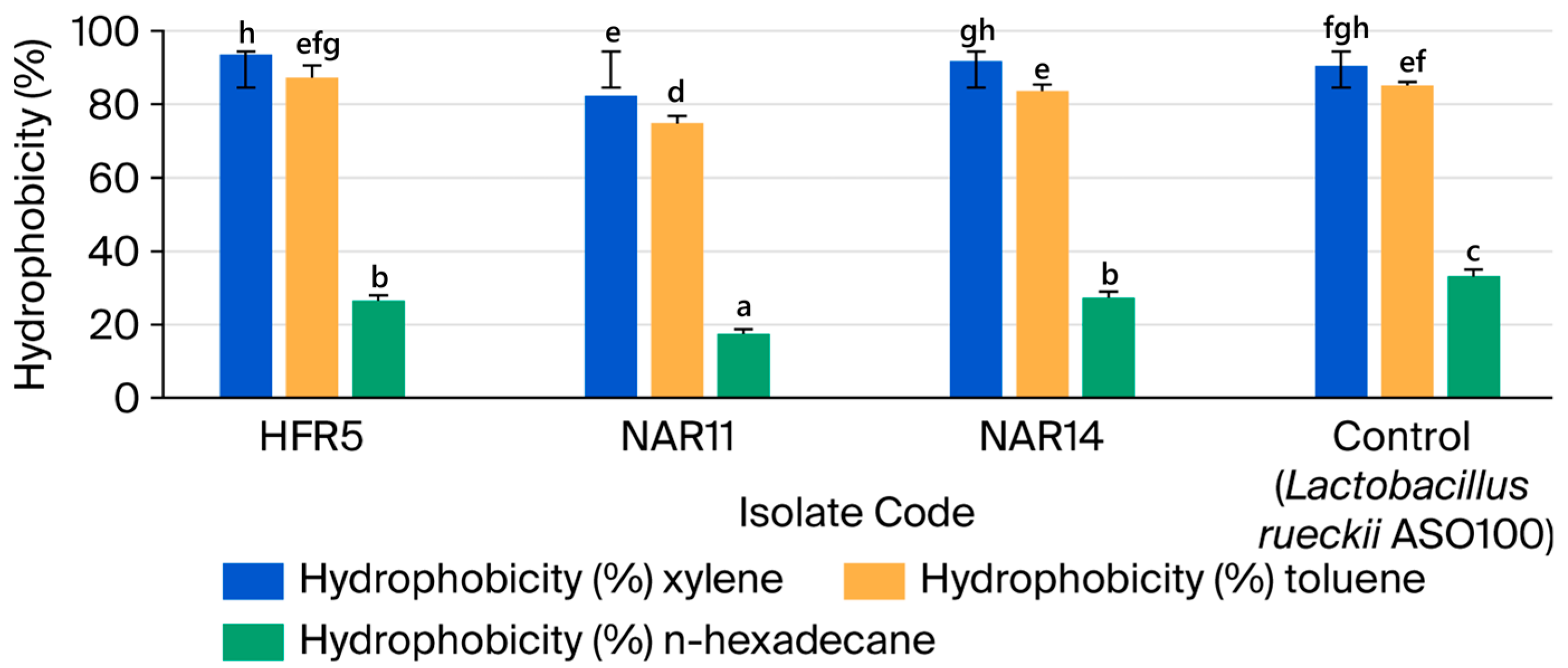
Cell colonization is a crucial characteristic of probiotic isolates. To mimic the capacity of the isolates to stick to the intestinal epithelium, we assessed their adherence to the hydrocarbons. The greatest cell hydrophobicity for LAB (HFR5) with xylene (93.5%) and NAR14 (91.7%) was observed, as illustrated in Figure 2, in contrast to toluene (87.2% and 88.6%), respectively. In xylene and toluene, the NAR11 LAB isolate exhibited reduced surface hydrophobicity. The nonpolar solvent (n-hexadecane) exhibited microbial adherence with a rating of 17.4% to 27.3%. Following a five-hour incubation period, strains NAR11 and HFR5 had greater percentages of auto-aggregation, which ranged from 90.1% to 95.3% (Figure 2).

3.2.4. LAB Isolates’ Aggregate Abilities

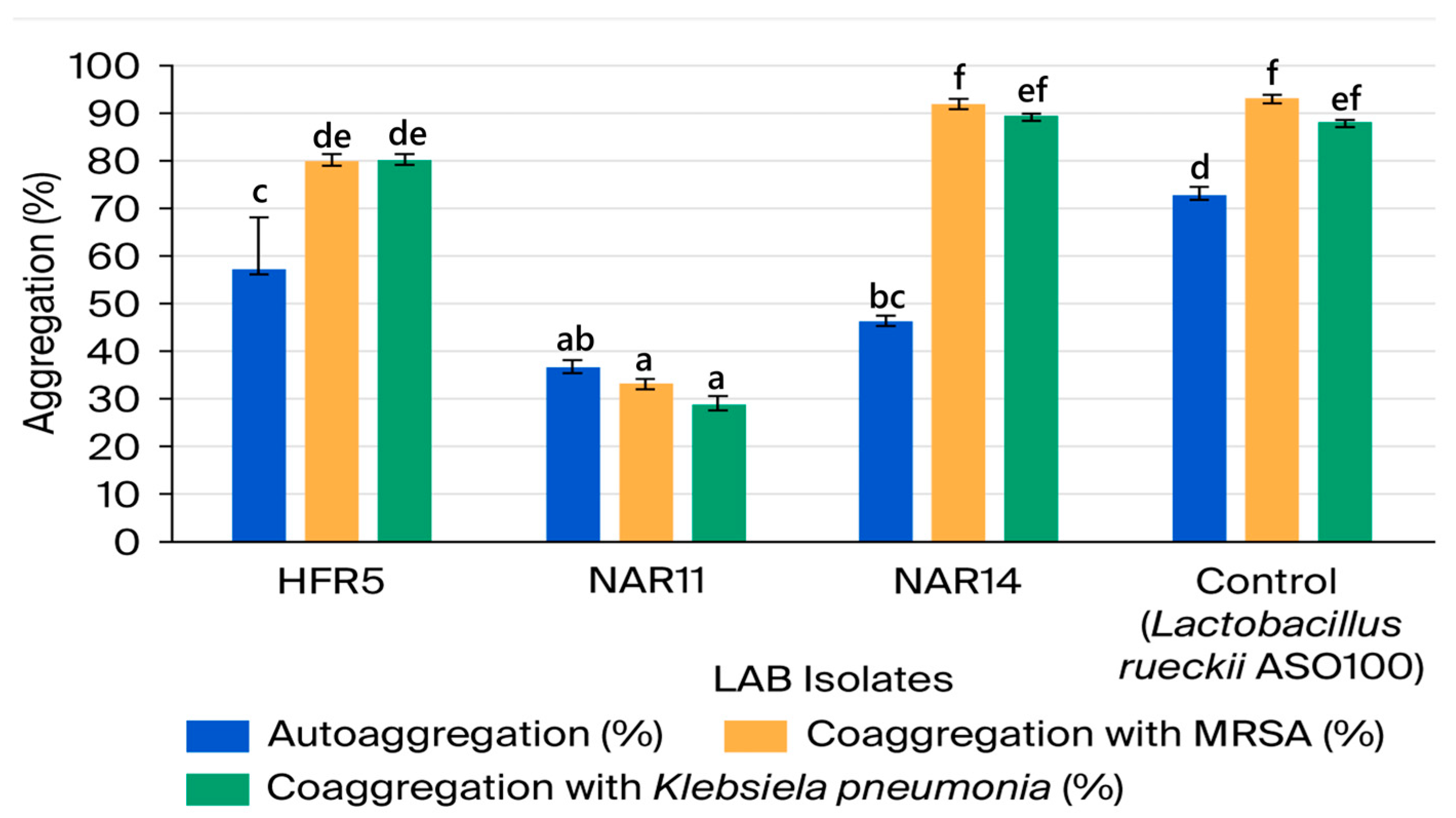
Figure 3 displays the findings demonstrating the capacities of the chosen LAB isolates for auto- and co-aggregation. The LAB isolates’ auto-aggregation ranged from 36.40 to 57.15%. In comparison to the other LAB isolates analyzed, isolate NAR11 had a very low capacity for auto-aggregation, while isolate HFR5 exhibited the highest proportion of auto-aggregation (57.15%). LAB isolate NAR14 had a high degree of co-aggregation ability with the two pathogens studied, exhibiting 91.87 and 89.35% for MRSA and K. pneumonia, respectively. We noticed a distinction between the tested pathogens and the LAB isolates.

3.3. Phenotypic and Genotypic Identification of LAB Isolates

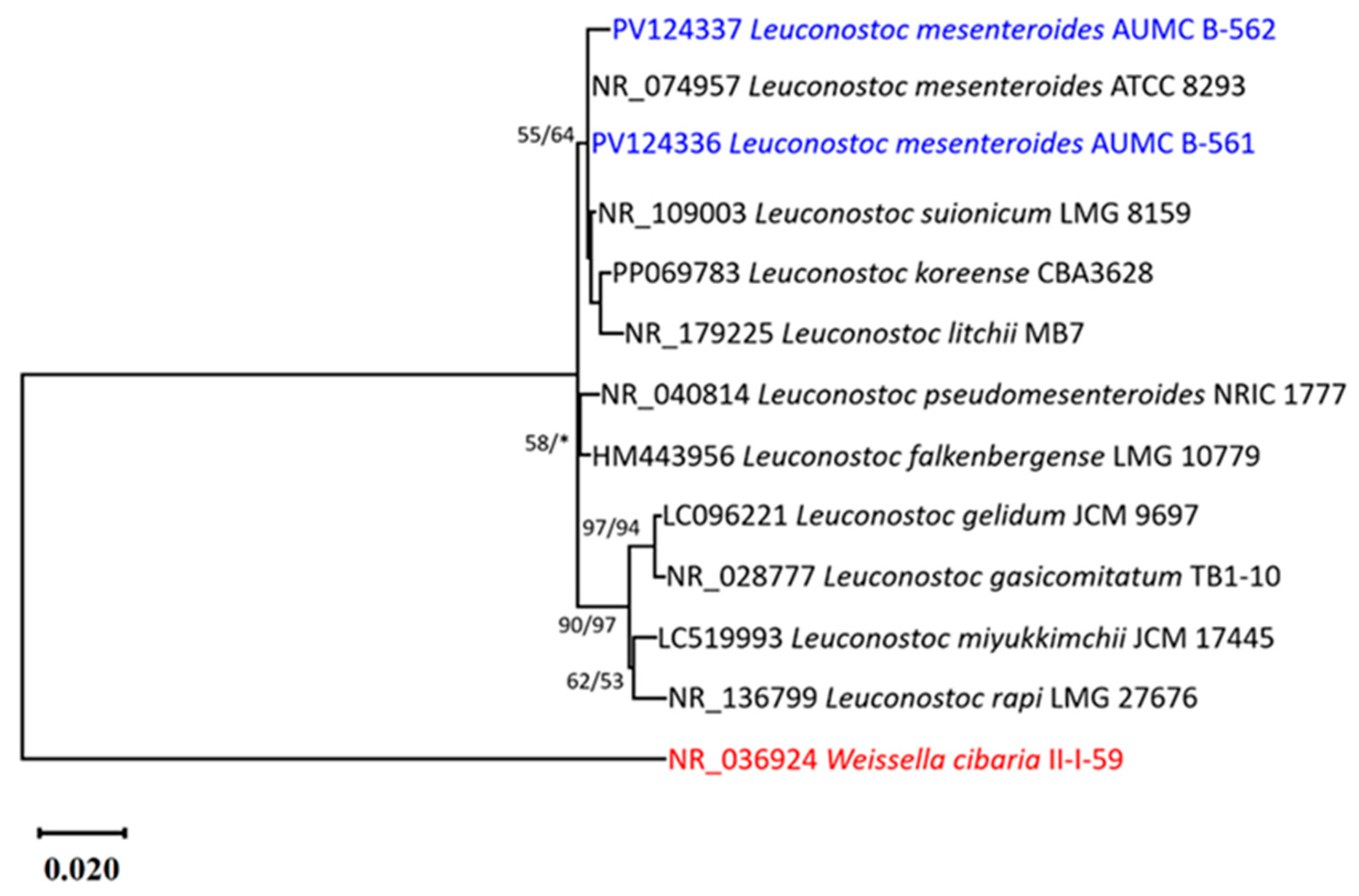
The two probiotic isolates (HFR5 and NAR14) that were chosen and were thought to be presumed LAB were found to belong to the Leuconostoc genus based on their coccoid cell shape, which was arranged in pairs or chains. The colonies were small, white, and smooth; they were able to grow up to a temperature of 10 °C, but did not grow at 45 °C, pH 9, 6.5% of NaCl. The complete 16S RNA dataset consisted of 13 species (Table S1). This study includes two sequences of Leuconostoc spp., ten sequences of closely related Leuconostoc mesenteroides obtained from GenBank, and one sequence of Weissella cibaria II-I-59, which serves as the root for the phylogenetic tree due to its 91.16% identity with Leuconostoc species. Of the maximum parsimony dataset of 1480 characters, including 1320 constant characters (without gaps or N), 148 variable characters were classified as parsimony-uninformative (11.2% of constant characters), and 36 characters were deemed parsimony-informative (2.7% of constant characters). Evolutionary history was deduced by utilizing the maximum likelihood approach and the Kimura two-parameter model, incorporating a discrete Gamma distribution (+G) with five rate categories, while presuming that a specific proportion of sites are evolutionarily invariant (K2+G+I). The optimal scoring ML tree, characterized by a final ML optimization likelihood value of 3184.85, a tree length of 221 steps, a consistency index of 0.658537, a retention index of 0.720000, and a composite index of 0.474146, encompassing all sites and parsimony-informative sites, was chosen to illustrate and analyze the phylogenetic relationships among taxa (Figure 4). The phylogenetic tree indicated that the bacterial isolates AUMC B-561 and AUMC B-562 consistently positioned on the clade containing the type species L. mesenteroides ATCC 8293, with support values of 55% ML and 64% MP. Consequently, the bacterial isolates AUMC B-561 and AUMC B-562 in this study were identified as L. mesenteroides. The 16SrRNA sequences of L. mesenteroides strains AUMC B-561 and AUMC B-562 in this study were submitted to GenBank as PV124336 and PV124337 (Figure 4).

3.4. Safety Assessment

3.4.1. Hemolytic and Gelatinase Activity

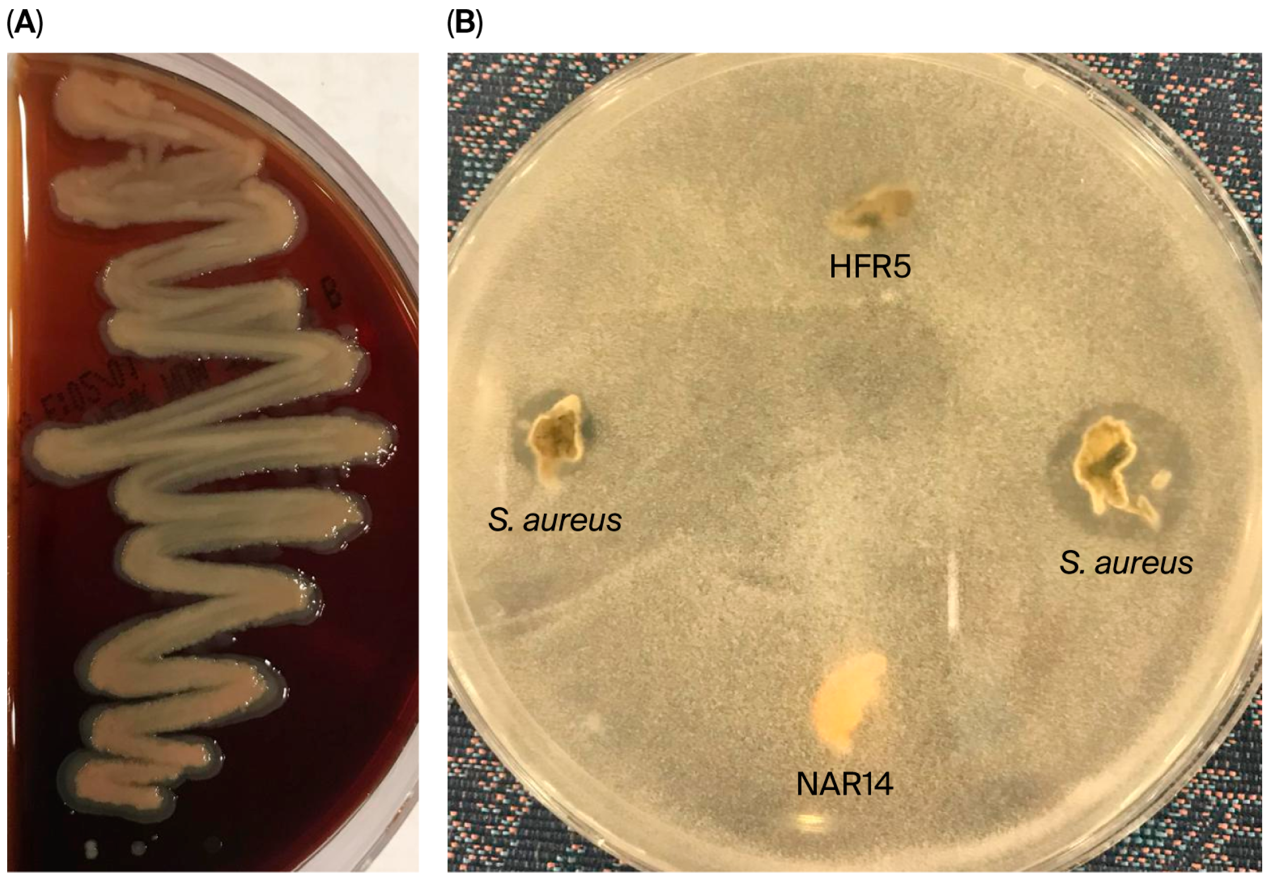
For probiotic L. mesenteroides isolates to be safe to use, they must not exhibit hemolytic or gelatinase activity. We verified the nonhemolytic nature and absence of gelatinase activity of the chosen L. mesenteroides isolates (Figure 5A,B) and their suitability for use as probiotics.

3.4.2. Antibiotic Susceptibility

Using a panel of seven antibiotics, the disc diffusion assay was used to determine antibiotic susceptibility. Among these are substances that hinder the creation of proteins, DNA, and cell walls. Table 5 displays the sizes of the inhibition zones (mm) for the antibiotics tested against probiotic bacteria. Our selected isolates were resistant to both vancomycin and kanamycin. Nevertheless, they were all responsive to erythromycin, penicillin, moxifloxacin, and cefotaxime. Furthermore, all bacteria exhibited only intermediate resistance to chloramphenicol.

4. Discussion

LAB from various sources, such as animal gastrointestinal tract (GIT), traditional fermented foods, and dairy products, have previously been examined for their probiotic qualities. In this study, probiotic qualities were assessed and defined for the LAB isolated from native camel milk. The enrichment phase of spontaneous fermentation was used to isolate the LAB, since raw camel milk does not contain a large amount of them, most likely because of the inhibitory effects of antimicrobial agents. According to Bouguerra et al. [53], camel milk is resistant to the growth of bacteria, particularly lactic flora, and its early acidification period is sluggish; however, this delay is compensated for by an extended incubation period. According to earlier publications [54,55], lactic acid bacteria were first enriched before being isolated from camel milk.
Despite the small number of LAB isolates examined in this work, they were specifically chosen to reflect unique morphological and biochemical traits of camel milk strains.
This study sets out to assess the probiotic potential of LAB isolated from camel milk. Probiotics were screened based on their qualities, such as their ability to withstand lysozymes, adhere to hydrocarbons, tolerate acidic pH, be resistant to bile salts, and exhibit antibacterial activity against pathogens. A crucial step in the selection of probiotics is determining their antibacterial activity, which helps to prevent infections and preserve food [56]. Six LAB isolates had an inhibitory effect against the tested strains of MRSA and K. pneumoniae. According to the World Health Organization (WHO) (2017) [57], MRSA and K. pneumoniae are high-priority multidrug-resistant bacteria that represent both Gram-positive and Gram-negative groups, which is why they were selected as test organisms for this investigation. To assess the antibacterial activity of the chosen probiotic isolates as part of their functional characterization, their inclusion provided rigorous and clinically relevant models. Because of nutrient competition and pH reduction, LAB has a suppressive effect on microorganisms [58]. Additionally, according to Champomier-Vergès et al. [58], LAB can produce a wide range of antimicrobial compounds, including hydrogen peroxide, ethanol, formic acid, acetoin, diacetyl, and bacteriocins. By generating CO2, the genus Leuconostoc can inhibit harmful aerobic bacteria and establish an anaerobic environment. Nonetheless, the build-up of carbon dioxide within the lipid bilayer has the potential to disrupt its permeability and impede the activity of decarboxylation enzymes [59]. The precise mechanisms causing this inhibition were not identified in the current study, despite the Leuconostoc isolates’ obvious antagonistic activity against the examined pathogens. Hydrogen peroxide, bacteriocins, and organic acids are some of the substances that might cause LAB inhibition. In order to distinguish the roles played by acids, hydrogen peroxide, and proteinaceous substances in the reported inhibitory effects, future research will employ targeted tests such as pH neutralization, catalase treatment, and protease treatment of cell-free supernatants. On the other hand, the ability of L. mesenteroides isolates to produce bacteriocins, specifically mesentericin-type peptides like mesentericin Y105, which have been shown to inhibit a wide variety of Gram-positive and Gram-negative pathogens, may account for their strong antagonistic activity against MRSA and Klebsiella pneumoniae. This discovery implies that because of their antibacterial properties, these isolates may be useful for application as probiotic supplements or natural biopreservatives in food systems. To verify the antimicrobial activity under in vivo or food model conditions and validate their functional applicability and safety, future research should focus on identifying and characterizing the genes encoding bacteriocins.
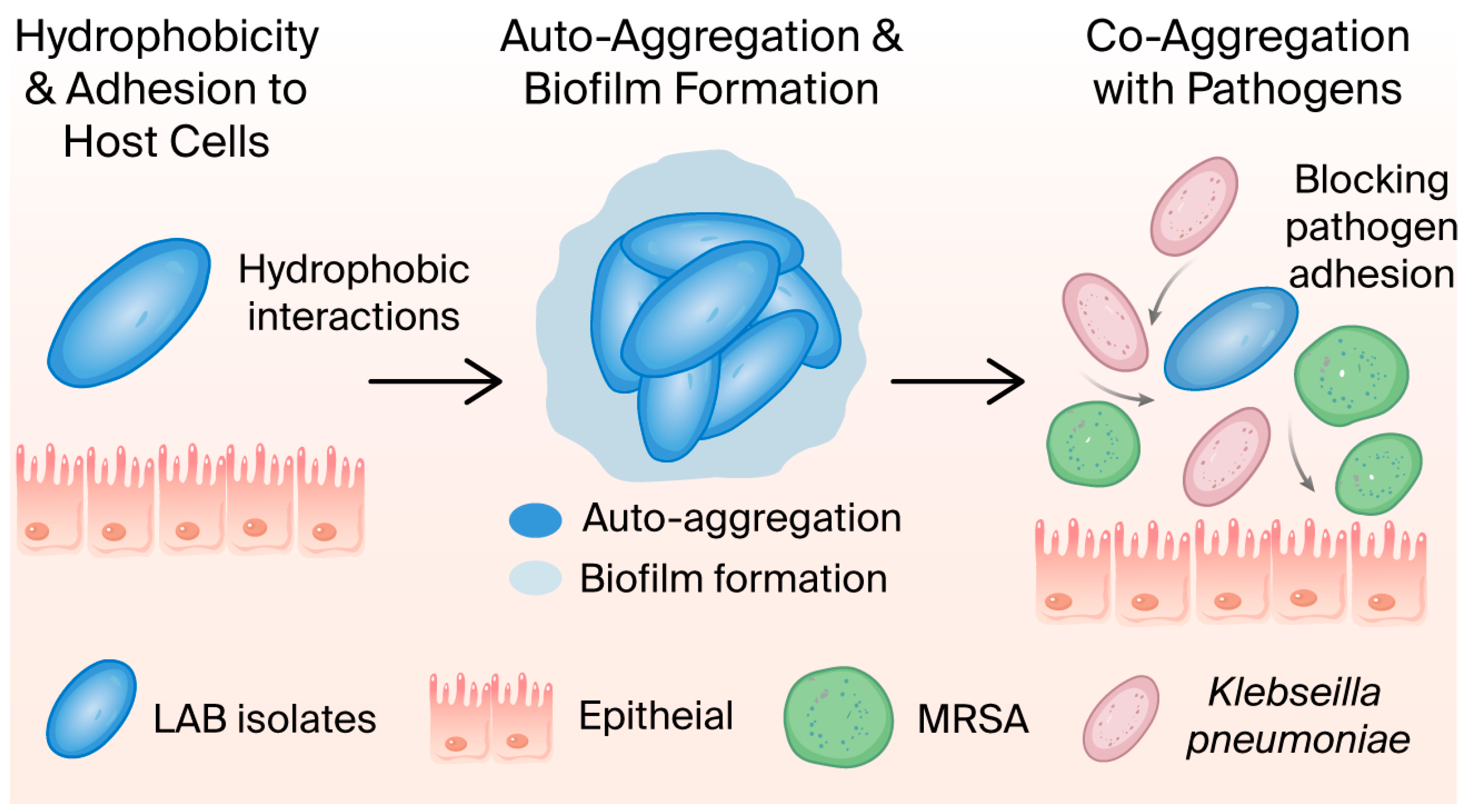
Another crucial factor in the selection of probiotic strains is their capacity to adhere to the gut epithelium. For a preliminary screening to identify isolates that might adhere, hydrophobicity and auto- or co-aggregation could be utilized [60]. According to Meena et al. [61], hydrophobicity may be the microbes’ initial point of contact with the host cells. Three isolates that possessed moderate to high hydrophobic characteristics were kept from the active strains. HFR5 and NAR14 exhibited the highest levels of cell hydrophobicity with xylene (93.5 and 91.7%) and toluene (87.2 and 83.1%), in that order. With the nonpolar solvent (n-hexadecane), the surface hydrophobicity was found to be lower, ranging from 17.4% to 27.3%. Hydrophobicity (H%) patterns varied amongst isolates for distinct hydrocarbons, indicating a correlation between hydrophobicity and strain-specific cell surface proteins. Comparable results were seen for L. animalis TSU 4 and L. gasseri TSU 3, with the former demonstrating significant hydrophobicity against both xylene and toluene and the latter only demonstrating hydrophobicity against xylene [62].
We also examined the cell-binding characteristics of our potential probiotic isolates, specifically auto-aggregation and co-aggregation. These two characteristics need to be considered when choosing possible probiotic strains. According to Del Re et al. [63], auto-aggregation, which occurs when the same microbial strains aggregate, and co-aggregation, which occurs when distinct microbial strains aggregate, promote bacterial attachment to the host GIT’s epithelial cells and inhibit pathogen colonization. The LAB under investigation had auto-aggregation rates of 36.44, 46.30, and 57.15% for NAR11, NAR14, and HFR5, respectively. According to Del Re et al. [63], auto-aggregation should be greater than 40% for all possible probiotic strains. Additionally, auto-aggregation facilitates LAB’s biofilm production, which encourages further colonization [64]. The study’s LAB isolates demonstrated isolate-specific co-aggregation capacity with the pathogens evaluated, with the HFR5 and NAR14 camel milk isolates demonstrating the highest co-aggregation ability. There are conflicting findings in the literature regarding the auto- and co-aggregation capabilities of LAB; strains of LAB isolated from milk in earlier investigations exhibited either little or no auto-aggregation [65,66]. High auto-aggregation was found by Puniya et al. [67]. Bacterial cell surfaces’ hydrophobicity, which is positively connected with adhesion effectiveness, indicates their capacity to interact with host epithelial cells and mucosal surfaces. This interaction is frequently facilitated by surface-associated proteins and exopolysaccharides. In the gastrointestinal tract, autoaggregation—the ability of cells belonging to the same strain to adhere to one another—allows biofilms to grow and persist, providing them with a competitive advantage over pathogens (Figure 6). On the other hand, co-aggregation describes how probiotic bacteria adhere to other microbial species, including pathogens, using complementary surface molecules. This may increase the removal of pathogens from the gut and limit their colonization [64,65,66,67]. Probiotics traveled via the stomach during food processing and then into the small intestine. After exposure to stomach acid and the proteolytic activity of pepsin in the stomach, viability may be lost [68]. According to our findings, two isolates (HFR5 and NAR14) were able to withstand and even thrive in the stomach’s acidic (pH 2) environment. When L. mesenteroides was isolated by Benmechernene et al. [18], the strain was shown to be able to live in conditions lower than pH 2. While the pH of the stomach varies from 1.5 to 6, pH 3 is generally thought to be ideal when choosing probiotics [56]. The strains that were tested are therefore thought to be acid-tolerant. Because bile salts cause membrane damage and DNA damage in the small intestine, they are thought to pose a threat to the survival of probiotics. Thus, prior to adopting lactic acid bacteria as probiotics, it is imperative to assess their resistance to bile salts [58]. Based on Shukla et al. [69], the physiological concentrations of human bile vary between 0.3% and 0.5% of bile salts; hence, our LAB isolates were tested in this study to see how long they could live at these concentrations. The findings show that there are significant strain-specific variations in the tested isolates’ resistance to bile salts. Strong bile resistance was demonstrated by the strain NAR14, which demonstrated remarkable survival at both low (0.1%) and high (0.5%) bile concentrations. This robustness raises the possibility of efficient gastrointestinal transit, which is a desirable quality in a probiotic.
The ability of L. mesenteroides isolates to endure bile salt exposure in the small intestine and gastric acidity is indicative of their capacity to tolerate these circumstances, which are essential preconditions for probiotic survival in vivo. Strong auto-aggregation capabilities and high surface hydrophobicity make it easier to adhere to intestinal epithelial cells, allowing for colonization and persistence in the gut environment. Furthermore, a mechanism of competitive exclusion, in which probiotics inhibit pathogen attachment and biofilm formation, is suggested by the observed co-aggregation with pathogens like MRSA and Klebsiella pneumoniae. Together, these functional characteristics suggest that the investigated isolates may successfully pass through the gastrointestinal tract and have positive effects on the host, which is in line with the physiological behavior anticipated of probiotics in vivo.
All of the LAB strains from this investigation that had previously survived the lysozyme-simulated gastric juice environment were also able to endure up to 0.3% bile salt. After 24 h of incubation, greater unviable counts were observed when the bile salt content rose to 0.4% and 0.5%. These outcomes supported those of Reuben et al. [35], who found that milk-derived LAB strains were tolerant of 0.1–0.5% bile salts.
Based on 16S rRNA gene sequence similarity, the isolates were identified as L. mesenteroides, recognizing that 16S rRNA analysis alone may not provide definitive species-level resolution within LAB. This work is consistent with previous investigations in which the same probiotic LAB species, L. mesenteroides, was successfully isolated from camel milk by various authors [18,53,70].
These isolates lack hemolysin and gelatinase, indicating their potential safety in food preparations. De Paula et al. [71] said that no incidences of infection from dairy products containing Leuconostoc spp. have been reported, demonstrating their safety. This shows that the isolates are safe to employ as starters and that they are suitable for industrial and biotechnological uses. This is consistent with the results of Colombo et al. [72], meeting specific pre-requisites of the proposed probiotic strain. The results showed that L. mesenteroides isolates were susceptible to four out of the seven antibiotics tested. All isolates are susceptible to cefotaxime, penicillin, moxifloxacin, and erythromycin. According to Morandi et al. [73], foodborne lactic acid bacteria are sensitive to penicillin and cephalosporins, but highly resistant to oxacillin. Our isolates of L. mesenteroides are naturally resistant to vancomycin. These findings concur with those of Hemme and Foucaud-Scheunemann [74], who claimed that certain features of its cell wall present D-lactate in the peptidoglycan rather than D-alanine. According to earlier reports, the vancomycin resistance seen in the Leuconostoc isolates is most likely intrinsic and chromosomally encoded [75]. A well-established trait of this genus that does not involve mobile genetic elements is the observed resistance of L. mesenteroides to vancomycin and kanamycin, which is generally regarded as intrinsic and non-transmissible. Since it does not entail horizontal gene transfer, such intrinsic resistance is typically regarded as suitable for applications connected to food and probiotics [76]. However, because they may spread to other bacteria, plasmid-borne or mobile resistance determinants pose a serious safety risk [77]. Even while our investigation lacked plasmid or genome profiling analyses to verify the genetic basis of resistance, contemporary regulatory frameworks, including those described by European Food Safety Authority (EFSA) [78], emphasize this distinction, which is crucial in determining the safety of probiotic strains.
Isolates HFR5 and NAR14 of L. mesenteroides showed similar or better acid and bile tolerance when compared to the commercial probiotic L. rhamnosus GG (ATCC 53103). At pH 2 and 0.3% bile salt, both isolates maintained survival rates of 75% and 74%, respectively. These values were comparable to or somewhat higher than those recorded for L. rhamnosus GG under the same test circumstances. Furthermore, the potential of HFR5 and NAR14 for intestinal adhesion and pathogen exclusion was supported by their auto-aggregation (>51%) and co-aggregation abilities with pathogens (85%). Their potential as domestic probiotic candidates derived from camel milk is further supported by their extensive antibacterial activity against MRSA and K. pneumoniae, which further shows functional equivalency to commercial probiotics.

5. Conclusions

Two strains of lactic acid bacteria, HFR5 and NAR14, that were isolated from Saudi Arabian camel milk, were found to be promising probiotic candidates in this investigation. Both isolates, which were identified as L. mesenteroides (GenBank accession numbers PV124336 and PV124337), showed excellent cell surface hydrophobicity and aggregation capabilities, good survivability in bile and acidic environments, and great antagonistic activity against MRSA and K. pneumoniae. Their satisfactory antibiotic susceptibility profiles and non-hemolytic, non-gelatinolytic behavior further bolster their safety for possible probiotic usage. These results offer fresh perspectives on camel milk-derived L. mesenteroides strains as beneficial probiotic resources, requiring more in vivo and genomic research to validate their safety and functional effectiveness. These results demonstrate that strains of L. mesenteroides derived from camel milk are useful probiotic resources; however, to guarantee their suitability for human use, future research should concentrate on genomic screening for virulent factors, mobile antibiotic resistance genes, and other genetic determinants of safety. Furthermore, to verify their long-term safety, stability in food formulations, and functional efficacy, thorough in vivo evaluations and clinical trials are required. Resolving these issues will increase their potential for usage in biotechnological applications, medicinal probiotics, and functional foods.

Supplementary Materials

The following supporting information can be downloaded at: https://www.mdpi.com/article/10.3390/microbiolres16120248/s1, Table S1: The bacterial strains that are part of this phylogenetic analysis are displayed together with their identification, percentage of similarity, and GenBank accession numbers.

Author Contributions

Conceptualization, S.M. and M.A.; methodology, S.M. and T.E.; software, S.M. and E.F.; validation, E.F., M.A. and T.E.; formal analysis, S.M. and A.-E.A.-A.; investigation, S.M.; resources, E.F.; data curation, M.A.; writing—original draft preparation, S.M.; writing—review and editing, M.A., T.E. and E.F.; visualization, S.M. and A.-E.A.-A.; supervision, S.M.; project administration, E.F.; funding acquisition, M.A. All authors have read and agreed to the published version of the manuscript.

Funding

This research received no external funding.

Institutional Review Board Statement

Not applicable.

Informed Consent Statement

Not applicable.

Data Availability Statement

The dataset generated and/or analyzed during the current study is available in the GenBank (https://www.ncbi.nlm.nih.gov): Leuconostoc mesenteroides strain AUMC B-561 16S ribosomal RNA gene, pa–Nucleotide–NCBI; accession number PV124336; and Leuconostoc mesenteroides strain AUMC B-562 16S ribosomal RNA gene, pa–Nucleotide–NCBI; accession number PV124337.

Acknowledgments

The authors acknowledge the support received from Ahmed Zia for helping with the data analysis.

Conflicts of Interest

The authors declare no conflicts of interest.

Abbreviations

The following abbreviations are used in this manuscript:
LABLactic acid bacteria
MRSAMethicillin-resistant Staphylococcus aureus
MRSde Man–Rogosa–Sharpe
CFUColony-forming unit
ODOptical density
MLMaximum likelihood
AICAkaike Information Criterion
MPMaximum parsimony
NCCLSNational Committee for Clinical Laboratory Standards
RBCRed blood cell

References

  1. Swelum, A.A.; El-Saadony, M.T.; Abdo, M.; Ombarak, R.A.; Hussein, E.O.S.; Suliman, G.; Alhimaidi, A.R.; Ammari, A.A.; Ba-Awadh, H.; Taha, A.E.; et al. Nutritional, antimicrobial and medicinal properties of Camel’s milk: A review. Saudi J. Biol. Sci. 2021, 28, 3126–3136. [Google Scholar] [CrossRef]
  2. BAHS. Basic Animal Husbandry Statistics. 2012. Available online: https://dahd.nic.in/related-links/publications-0 (accessed on 8 May 2025).
  3. Faye, B. The Camel today: Assets and potentials. Anthropozoologica 2014, 49, 15–24. [Google Scholar] [CrossRef]
  4. Alhassani, W.E. Camel milk: Nutritional composition, therapeutic properties, and benefits for human health. Open Vet. J. 2024, 14, 3164–3180. [Google Scholar] [CrossRef]
  5. Alebie, G.; Yohannes, S.; Worku, A. Therapeutic applications of camel’s milk and urine against cancer: Current development efforts and future perspectives. J. Cancer Sci. Ther. 2017, 9, 468–478. [Google Scholar] [CrossRef]
  6. Food and Agricultural Organization of the United Nations and World Health Organization. Joint FAO/WHO Working Group Report on Drafting Guidelines for the Evaluation of Probiotics in Food; Food and Agricultural Organization of the United Nations, Rome, Italy, 2002. Available online: https://isappscience.org/wp-content/uploads/2019/04/probiotic_guidelines.pdf (accessed on 27 March 2025).
  7. Albarrak, S.M. Evaluation of somatic cells count, antioxidants and antimicrobial proteins in milk samples obtained from different breeds of the dromedary camel. Int. J. Vet. Sci. 2023, 12, 462–469. [Google Scholar] [CrossRef]
  8. Mahmoudi, I.; Moussa, O.B.; Khaldi, T.E.M.; Kebouchi, M.; Soligot, C.; Le Roux, Y.; Hassouna, M. Functional in vitro screening of Lactobacillus strains isolated strains from Tunisian camel raw milk toward their selection as probiotic. Small Rumin. Res. 2016, 137, 91–98. [Google Scholar] [CrossRef]
  9. Sharma, A.; Lavania, M.; Singh, R.; Lal, B. Identification and probiotic potential of lactic acid bacteria from camel milk. Saudi J. Biol. Sci. 2021, 28, 1622–1632. [Google Scholar] [CrossRef]
  10. Tola, G.B. Medicinal, Physiological, and Nutritional Benefits of Camel Milk. Preprints 2024, 2024111552. [Google Scholar] [CrossRef]
  11. Albarrak, S.M.; Al-Sobayil, F.A. Evaluation of protective proteins and cytokines in milk and serum of healthy camels during the first two months postpartum. Int. J. Vet. Sci. 2024, 13, 401–406. [Google Scholar] [CrossRef]
  12. Konuspayeva, G.; Faye, B. Recent Advances in Camel Milk Processing. Animals 2021, 11, 1045. [Google Scholar] [CrossRef]
  13. El-Hanafy, A.A.; Saad, Y.M.; Alkarim, S.A.; Almehdar, H.A.; Alzahrani, F.M.; Almatry, M.A.; Uversky, V.N.; Redwan, E.M. Yield and Composition Variations of the Milk from Different Camel Breeds in Saudi Arabia. Sci 2023, 5, 2. [Google Scholar] [CrossRef]
  14. Yang, Z.; Ni, L.; Tian, W.; Chi, H. Screening and Identification of Goat-Milk-Derived Lactic Acid Bacteria with Bacteriocin-like Activity and Probiotic Potentials. Microorganisms 2023, 11, 849. [Google Scholar] [CrossRef]
  15. Asha, M.N.; Chowdhury, S.R.; Hossain, H.; Rahman, A.; Al Emon, A.; Tanni, F.Y.; Islam, R.; Hossain, M.; Rahman, M. Antibacterial potential of lactic acid bacteria isolated from raw cow milk in Sylhet district, Bangladesh: A molecular approach. Vet. Med. Sci. 2024, 10, e1463. [Google Scholar] [CrossRef]
  16. Akhmetsadykova, S.; Baubekova, A.; Konuspayeva, G.; Akhmetsadykov, N.; Faye, B.; Loiseau, G. Lactic acid bacteria biodiversity in raw and fermented camel milk. Afr. J. Food Sci. Technol. 2015, 6, 84–88. [Google Scholar] [CrossRef]
  17. Alhaj, O.A.; Jrad, Z.; Oussaief, O.; Jahrami, H.A.; Ahmad, L.; Alshuniaber, M.A.; Mehta, B.M. The characterization of Lactobacillus strains in camel and bovine milk during fermentation: A comparison study. Heliyon 2024, 10, e40162. [Google Scholar] [CrossRef]
  18. Benmechernene, Z.; Chentouf, H.F.; Bellil, Y.; Ghazi, F.; Quintela-Baluja, M.; Calo-Mata, P.; Barros-Velázquez, J. Technological Aptitude and Applications of Leuconostoc mesenteroides Bioactive Strains Isolated from Algerian Raw Camel Milk. BioMed Res. Int. 2013, 1, 418132. [Google Scholar] [CrossRef]
  19. Belkheir, K.; Laref, N. Autochthonous Lactic Acid Bacteria from Camelus dromedarius Milk. An Updated Systematic Review. Food Rev. Int. 2025, 41, 1576–1591. [Google Scholar] [CrossRef]
  20. Zheng, J.; Wittouck, S.; Salvetti, E.; Franz, C.M.A.P.; Harris, H.M.B.; Mattarelli, P.; O’Toole, P.W.; Pot, B.; Vandamme, P.; Walter, J.; et al. A taxonomic note on the genus Lactobacillus: Description of 23 novel genera, emended description of the genus Lactobacillus Beijerinck 1901, and union of Lactobacillaceae and Leuconostocaceae. Int. J. Syst. Evol. Microbiol. 2020, 70, 2782–2858. [Google Scholar] [CrossRef]
  21. Junior, W.J.L.; Sant’Ana, A.S.; Di Cagno, R.; Campanaro, S.; Gobbetti, M. Five years after the lactobacillus reclassification: Genomic and functional insights into Levilactobacillus, Lacticaseibacillus, Limosilactobacillus, Lactiplantibacillus, and Ligilactobacillus in fermented foods. Trends Food Sci. Technol. 2025, 163, 105118. [Google Scholar] [CrossRef]
  22. Alsulaimany, F.A.; Mohamed, M.M.; Al-Hejin, A.M.; Kabli, S. Molecular Identification of Goat’s Udder Microbes and Nutritional Value of Milk Using Dielectric Constants. Kafkas Univ. Vet. Fak. Derg. 2023, 29, 463–471. [Google Scholar] [CrossRef]
  23. Khelissa, S.; Chihib, N.-E.; Gharsallaoui, A. Conditions of nisin production by Lactococcus lactis subsp. lactis and its main uses as a food preservative. Arch. Microbiol. 2021, 203, 465–480. [Google Scholar] [CrossRef]
  24. Rezac, S.; Kok, C.R.; Heermann, M.; Hutkins, R. Fermented Foods as a Dietary Source of Live Organisms. Front. Microbiol. 2018, 9, 396129. [Google Scholar] [CrossRef]
  25. Younis, K.M.; Najem, A.H. Characterization and evaluation of probiotic strains isolated from different sources. Microbes Infect. Dis. 2025, (in press). [Google Scholar] [CrossRef]
  26. Ayivi, R.D.; Ibrahim, S.A. Lactic acid bacteria: An essential probiotic and starter culture for the production of yoghurt. Int. J. Food Sci. Technol. 2022, 57, 7008–7025. [Google Scholar] [CrossRef]
  27. Fijan, S.; Fijan, T. Current Trends in the Applications of Probiotics and Other Beneficial Microbes: Expanding Horizons. Appl. Microbiol. 2025, 5, 103. [Google Scholar] [CrossRef]
  28. Meral-Aktaş, H. Bacteriocin characterization of Enterococcus faecium isolates and evaluation of their in situ anti-Listerial activity in Beyaz cheese. Food Biosci. 2024, 61, 104741. [Google Scholar] [CrossRef]
  29. Liu, J.; Zhang, X.; Niu, J.; Han, Z.; Bi, C.; Mehmood, K.; Farraj, D.A.A.; Alzaidi, I.; Iqbal, R.; Qin, J. Complete genome of multi-drug resistant Staphylococcus Aureus in bovine mastitic milk in Anhui, China. Pak. Vet. J. 2023, 43, 456–462. [Google Scholar] [CrossRef]
  30. Tsakali, E.; Tsantes, A.G.; Houhoula, D.; Laliotis, G.P.; Batrinou, A.; Halvatsiotis, P.; Tsantes, A.E. The Detection of Bacterial Pathogens, including Emerging Klebsiella pneumoniae, Associated with Mastitis in the Milk of Ruminant Species. Appl. Sci. 2022, 13, 11484. [Google Scholar] [CrossRef]
  31. Alorainy, M.S.; Alhajuj, A.M.; Fathi, S.M. Biochemical identification, direct molecular detection, and antimicrobial agent resistance of Staphylococcus aureus isolated from raw camel milk in Al Madinah Region, Saudi Arabia. Int. J. Vet. Sci. 2023, 12, 740–745. [Google Scholar] [CrossRef]
  32. Jans, C.; Bugnard, J.; Njage, P.M.K.; Lacroix, C.; Meile, L. Lactic acid bacteria diversity of African raw and fermented camel milk products reveals a highly competitive, potentially health-threatening predominant microflora. LWT 2012, 47, 371–379. [Google Scholar] [CrossRef]
  33. Aljeldah, M.; Al Shammari, B.; Farrag, E.S.; Taha, E.M.; Mahmoud, S.Y. Prevalence of Multidrug-Resistant Methicillin-Resistant Staphylococcus aureus in Northeastern Saudi Hospitals. J. Pure Appl. Microbiol. 2022, 16, 1192–1199. [Google Scholar] [CrossRef]
  34. Al Yousef, S.A.; Mahmoud, S.Y.; Farrag, E.; Husseiny, S.; Bayoumi, F.; Ali, A. Clinical and Laboratory Profile of Urinary Tract Infections Associated with Extended Spectrum β-Lactamase Producing Escherichia coli and Klebsiella pneumoniae. Ann. Clin. Lab. Sci. 2016, 46, 393–400. [Google Scholar] [PubMed]
  35. Mahmoud, S.Y.M.; Atallah, A.A.; Badr, O.A.; Moustafa, M.M.A.; Esmael, A.; Ebrahim, N.; Aljeldah, M.; Al Shammari, B.; Alsafari, I.A.; Mohamed, S.A. Bioprospecting for Novel Probiotic Strains from Human Milk and Infants: Molecular, Biochemical, and Ultrastructural Evidence. Biology 2022, 11, 1405. [Google Scholar] [CrossRef]
  36. Schillinger, U.; Lücke, F.K. Antibacterial activity of Lactobacillus sake isolated from meat. Appl. Environ. Microbiol. 1989, 55, 1901–1906. [Google Scholar] [CrossRef]
  37. Kobierecka, P.A.; Wyszy’nska, A.K.; Aleksandrzak-Piekarczyk, T.; Kuczkowski, M.; Tuzimek, A.; Piotrowska, W.; Górecki, A.; Adamska, I.; Wieliczko, A.; Bardowski, J.; et al. In vitro characteristics of Lactobacillus spp. strains isolated from the chicken digestive tract and their role in the inhibition of Campylobacter colonization. Microbiol. Open 2017, 6, e00512. [Google Scholar] [CrossRef]
  38. Vinderola, C.; Reinheimer, J. Lactic acid starter and probiotic bacteria: A comparative ‘‘in vitro” study of probiotic characteristics and biological barrier resistance. Food Res. Int. 2003, 36, 895–904. [Google Scholar] [CrossRef]
  39. Reuben, R.; Roy, P.; Sarkar, S.; Alam, A.R.U.; Jahid, I. Characterization and evaluation of lactic acid bacteria from indigenous raw milk for potential probiotic properties. J. Dairy. Sci. 2020, 103, 1223–1237. [Google Scholar] [CrossRef]
  40. Armas, F.; Camperio, C.; Marianelli, C. In vitro assessment of the probiotic potential of Lactococcus lactis LMG 7930 against ruminant mastitis-causing pathogens. PLoS ONE 2017, 12, e0169543. [Google Scholar] [CrossRef] [PubMed]
  41. Axelsson, L. Lactic acid bacteria: Classification and physiology. In Lactic Acid Bacteria: Microbiological and Functional Aspects; CRC Press: Marcel Dekker, NY, USA, 2004; 66p. [Google Scholar]
  42. Abd-Alla, M.H.; El-Enany, A.W.E. Production of acetone-butanol-ethanol from spoilage date palm (Phoenix dactylifera L.) fruits by mixed culture of Clostridium acetobutylicum and Bacillus subtilis. Biomass Bioenergy 2012, 42, 172–178. [Google Scholar] [CrossRef]
  43. Sanger, F.; Air, G.M.; Barrell, B.G.; Brown, N.L.; Coulson, A.R.; Fiddes, J.C.; Hutchison, C.; Slocombe, P.M.; Smith, M. Nucleotide sequence of bacteriophage φx174 DNA. Nature 1977, 265, 687–695. [Google Scholar] [CrossRef]
  44. Katoh, K.; Standley, D.M. Mafft multiple sequence alignment software version 7: Improvements in performance and usability. Mol. Biol. Evol. 2013, 30, 772–780. [Google Scholar] [CrossRef]
  45. Criscuolo, A.; Gribaldo, S. BMGE (block mapping and gathering with entropy): A new software for selection of phylogenetic informative regions from multiple sequence alignments. BMC Evol. Biol. 2010, 10, 210. [Google Scholar] [CrossRef] [PubMed]
  46. Kumar, S.; Stecher, G.; Li, M.; Knyaz, C.; Tamura, K. Mega X: Molecular evolutionary genetics analysis across computing platforms. Mol. Biol. Evol. 2018, 35, 1547–1549. [Google Scholar] [CrossRef] [PubMed]
  47. Felsenstein, J. Confidence limits on phylogenies: An approach using the bootstrap. Evolution 1985, 39, 783–791. [Google Scholar] [CrossRef] [PubMed]
  48. Posada, D.; Crandall, K.A. Modeltest: Testing the model of DNA substitution. Bioinformatics 1998, 14, 817–818. [Google Scholar] [CrossRef]
  49. Liasi, S.A.; Azmi, T.I.; Hassan, M.D.; Shuhaimi, M.; Rosforizan, M.; Ariff, A.B. Antimicrobial activity and anti-biotic sensitivity of three isolates of Lactic acid bacteria from fermented product. Malays. J. Microbiol. 2009, 5, 33–37. [Google Scholar] [CrossRef]
  50. National Committee for Clinical Laboratory Standards [NCCLS]. Methods for Dilution Antimicrobial Susceptibility Tests for Bacteria that Grow Aerobically, 6th ed.; Clinical and Laboratory Standards Institute (CLSI): Wayne, PA, USA, 2003. [Google Scholar]
  51. Wang, Y.; Zhou, J.; Xia, X.; Zhao, Y.; Shao, W. Probiotic potential of Lactobacillus paracasei FM-LP-4 isolated from Xinjiang camel milk yoghurt. Int. Dairy J. 2016, 62, 28–34. [Google Scholar] [CrossRef]
  52. Ribeiro, S.C.; Coelho, M.C.; Todorov, S.D.; Franco, B.D.G.M.; Dapkevicius, M.L.E.; Silva, C.C.G. Technological properties of bacteriocin-producing lactic acid bacteria isolated from Pico cheese an artisanal cow’s milk cheese. J. Appl. Microbiol. 2014, 116, 573–585. [Google Scholar] [CrossRef]
  53. Bouguerra, A.; Harzallah, D.; Yasin, I.S.M.; Silvaraj, S.; Bakli, S.; Rabhi, N.E.H. Characterization of probiotic lactic acid bacteria isolated from Algerian camel milk in vitro. Int. J. Sci. Res. 2020, 76, 244–256. [Google Scholar] [CrossRef]
  54. Yateem, A.; Balba, M.T.; Al-Surrayai, T.; Al-Mutairi, B.; Al-Daher, R. Isolation of lactic acid bacteria with probiotic potential from camel milk. Int. J. Dairy Sci. 2008, 3, 194–199. [Google Scholar] [CrossRef]
  55. Abbas, M.M.; Mahasneh, A.M. Isolation of Lactobacillus strains with probiotic potential from camel’s milk. Afr. J. Microbiol. Res. 2014, 8, 1645–1655. [Google Scholar] [CrossRef]
  56. Azat, R.; Liu, Y.; Li, W.; Kayir, A.; Lin, D.; Zhou, W.; Zheng, X. Probiotic properties of lactic acid bacteria isolated from traditionally fermented Xinjiang cheese. J. Zhejiang Univ.-Sci. B 2016, 17, 597–609. [Google Scholar] [CrossRef]
  57. World Health Organization (WHO). WHO Publishes List of Bacteria for Which New Antibiotics are Urgently Needed; World Health Organization: Geneva, Switzerland, 2017. [Google Scholar]
  58. Champomier-Vergès, M.C.; Zagorec, M.; Fadda, S. Proteomics: A tool for understanding lactic acid bacteria adaptation to stressful environments. In Biotechnology of Lactic Acid Bacteria: Novel Applications; Mozzi, F., Raya, R.R., Vignolo, G.M., Eds.; Blackwell Publishing: Malden, MA, USA, 2010; pp. 57–72. [Google Scholar]
  59. Ouwehand, A.C.; Vesterlund, S. Antimicrobial components from lactic acid bacteria. In Lactic Acid Bacteria Microbiology and Functional Aspects, 3rd ed.; Salminen, S., von Wright, A., Ouwehand, A., Eds.; CRC Press: Marcel Dekker, NY, USA, 2004; pp. 375–395. [Google Scholar]
  60. Kokwe, N.H.; Tshabuse, F.; Swalaha, F.M. In Vitro Characterization and Safety Assessment of Streptococcus salivarius, Levilactobacillus brevis and Pediococcus pentosaceus Isolated from the Small Intestine of Broiler Breeders. Microorganisms 2025, 13, 1231. [Google Scholar] [CrossRef]
  61. Meena, K.K.; Joshi, M.; Rao, P.; Panwar, N.L.; Gupta, L.; Meena, S. Evaluation of probiotic properties of lactic acid bacteria isolated from sheep milk and its suitability as a starter in a milk-cereal-based beverage. Food Biomacromol. 2025, 2, 69–83. [Google Scholar] [CrossRef]
  62. Sahoo, T.K.; Jena, P.K.; Nagar, N.; Patel, A.K.; Seshadri, S. In vitro evaluation of probiotic properties of lactic acid bacteria from the gut of Labeo rohita and Catla catla. Probiotics Antimicrob. Proteins 2015, 7, 126–136. [Google Scholar] [CrossRef] [PubMed]
  63. Del Re, B.; Sgorbati, B.; Miglioli, M.; Palenzona, D. Adhesion, autoaggregation and hydrophobicity of 13 strains of Bifidobacterium longum. Lett. Appl. Microbiol. 2000, 31, 438–442. [Google Scholar] [CrossRef]
  64. Yao, P.; Mohd Esah, E.; Zhao, C. Regulatory mechanisms and applications of Lactobacillus biofilms in the food industry. Front. Microbiol. 2025, 15, 1465373. [Google Scholar] [CrossRef]
  65. Espeche, M.C.; Otero, M.C.; Sesma, F.; Nader-Macias, M.E. Screening of surface properties and antagonistic substances production by lactic acid bacteria isolated from the mammary gland of healthy and mastitic cows. Vet. Microbiol. 2009, 135, 346–357. [Google Scholar] [CrossRef]
  66. Espeche, M.C.; Pellegrino, M.; Frola, I.; Larriestra, A.; Bogni, C.; Nader-Macías, M.E.F. Lactic acid bacteria from raw milk as potentially beneficial strains to prevent bovine mastitis. Anaerobe 2012, 18, 103–109. [Google Scholar] [CrossRef] [PubMed]
  67. Puniya, M.; Ravinder, K.M.; Panwar, H.; Kumar, N. Screening of lactic acid bacteria of different origin for their probiotic potential. J. Food Process. Technol. 2016, 7, 545. [Google Scholar] [CrossRef]
  68. Han, S.; Lu, Y.; Xie, J.; Fei, Y.; Zheng, G.; Wang, Z.; Liu, J.; Lv, L.; Ling, Z.; Berglund, B.; et al. Probiotic Gastrointestinal Transit and Colonization After Oral Administration: A Long Journey. Front. Cell. Infect. Microbiol. 2021, 11, 609722. [Google Scholar] [CrossRef] [PubMed]
  69. Shukla, R.; Iliev, I.; Goyal, A. Leuconostoc mesenteroides NRRL B-1149 as probiotic and its dextran with anticancer properties. J. Biosci. Biotechnol. 2014, 3, 79–87. [Google Scholar]
  70. Ansari, F.; Pourjafar, H.; Samakkhah, S.A.; Mirzakhani, E. An overview of probiotic camel milk as a nutritional beverage: Challenges and perspectives. Food Sci. Nutr. 2024, 12, 6123–6141. [Google Scholar] [CrossRef]
  71. De Paula, A.; Jeronymo-Ceneviva, A.; Silva, L.; Todorov, S.; Franco, B.M.; Choiset, Y.; Haertlé, T.; Chobert, J.-M.; Dousset, X.; Penna, A. Leuconostoc mesenteroides SJRP55: A potential probiotic strain isolated from Brazilian water buffalo mozzarella cheese. Probiotics Antimicrob. Proteins 2014, 6, 186–197. [Google Scholar] [CrossRef]
  72. Colombo, M.; Nero, L.A.; Todorov, S.D. Safety profiles of beneficial lactic acid bacteria isolated from dairy systems. Braz. J. Microbiol. 2020, 51, 787–795. [Google Scholar] [CrossRef] [PubMed]
  73. Morandi, S.; Cremonesi, P.; Silvetti, T.; Brasca, M. Technological characterisation, antibiotic susceptibility and antimicrobial activity of wild-type Leuconostoc strains isolated from north Italian traditional cheeses. J. Dairy Res. 2013, 80, 457–466. [Google Scholar] [CrossRef]
  74. Hemme, D.; Foucaud-Scheunemann, C. Leuconostoc, characteristics, use in dairy technology and prospects in functional foods. Int. Dairy J. 2004, 14, 467–494. [Google Scholar] [CrossRef]
  75. Moreno, M.R.F.; Sarantinopoulos, P.; Tsakalidou, E.; De Vuyst, L. The role and application of enterococci in food and health. Int. J. Food Microbiol. 2006, 106, 1–24. [Google Scholar] [CrossRef]
  76. Gueimonde, M.; Sánchez, B.; de los Reyes-Gavilán, C.G.; Margolles, A. Antibiotic resistance in probiotic bacteria. Front. Microbiol. 2013, 4, 51661. [Google Scholar] [CrossRef]
  77. Fraqueza, M.J. Antibiotic resistance of lactic acid bacteria isolated from dry-fermented sausages. Int. J. Food Microbiol. 2015, 212, 76–88. [Google Scholar] [CrossRef]
  78. EFSA Panel on Additives and Products or Substances used in Animal Feed (FEEDAP). Guidance on the assessment of bacterial susceptibility to antimicrobials of human and veterinary importance. EFSA J. 2012, 10, 2740. [Google Scholar] [CrossRef]
Figure 1. Antagonistic activity of LAB isolates against Methicillin-resistant Staphylococcus aureus (MRSA) and Klebsiella pneumoniae using the agar diffusion method after incubating for 48 h at 37 °C. Bars are means of 3 replicates ± SE. Bars labeled with at least one different letter are significantly different according to Duncan’s multiple-range test.
Microbiolres 16 00248 g001
Figure 2. Hydrophobicity of LAB strains. Bars are means of 3 replicates ± SE. Bars labeled with at least one different letter are significantly different according to Duncan’s multiple-range test.
Microbiolres 16 00248 g002
Figure 3. Aggregation abilities of potential probiotic isolates from camel milk. Bars are means of 3 replicates ± SE. Bars labeled with at least one different letter are significantly different according to Duncan’s multiple-range test.
Microbiolres 16 00248 g003
Figure 4. Maximum likelihood phylogenetic tree generated from a heuristic search (1000 replications) of maximum likelihood/maximum parsimony analysis (ML/MP) of 16S rRNA sequences of bacterial strains in this study (in blue) compared to the most similar species in GenBank. Bootstrap support values less than 50% are asigned as (*) near the corresponding nodes. The tree is rooted to Weissella cibaria II-I-59 (in red).
Microbiolres 16 00248 g004
Figure 5. Safety assessment of L. mesenteroides isolates in vitro. (A) Hemolytic activity, (B) gelatinase.
Microbiolres 16 00248 g005
Figure 6. Suggested diagram for mechanisms of adhesion, auto-aggregation, and co-aggregation in probiotic bacteria.
Microbiolres 16 00248 g006
Table 1. Morphological properties of LAB isolates.
No.Isolate CodeCamel ColorCell MorphologyGram StainEndospore StainCatalase ProductionFacultatively Anaerobic or Microaerophilic
1HFR1BrownCocci+Non+
2HFR5BrownCocci+Non+
3HFR6BrownCocci+Non+
4HFR7BrownRods+Non
5HFR9BrownCocci+Non+
6HFR10BrownRods+Non
7NAR11BlackCocci+Non+
8NAR14BlackCocci+Non+
9NAR15BlackRods+Non
10NAR17BlackCocci+Non+
11NAR18BlackRods+Non
12NAR21BlackCocci+Non+
+: Positive reaction; −: Negative reaction.
Table 2. Lysozyme tolerance of lactic acid bacteria (LAB) isolates determined by the agar diffusion method under defined test conditions (0.2–2 mg mL−1 lysozyme, 37 °C, 24 h incubation).
Isolate CodeInhibition Zone Caused by Various Concentrations of Lysozyme (mg/mL)
0.10.20.40.60.812
HFR5
HFR6++++++++
NAR11
NAR14
NAR17++++++++
NAR21+++++++
Control (Lactobacillus rueckii ASO100)
−, no inhibition zone was observed; +, inhibition zone (<6 mm); ++, inhibition zone (6–10 mm); +++, inhibition zone (11–18 mm).
Table 3. Acid tolerance of lactic acid bacteria (LAB) isolates evaluated by the method of Kobierecka under defined test conditions (pH 2–5, 37 °C, 7 h incubation).
IsolateControl (Log10 CFU mL−1)pH 2pH 3pH 4pH 5
Log10 CFU mL−1Viability (%)Log10 CFU mL−1Viability (%)Log10 CFU mL−1Viability (%)Log10 CFU mL−1Viability (%)
HFR58.36.679.15 c7.185.53 e7.691.16 fg8.096.38 h
NAR117.82.1327.34 a5.773.11 b6.785.89 e7.191.02 fg
NAR148.56.171.76 b7.082.34 d7.689.40 f7.9393.33 g
* Control8.86.573.86 b7.585.22 e8.697.72 h8.798.48 h
* (Lactobacillus rueckii ASO100), Values are presented as mean ± standard deviation of three independent experiments (n = 3). For each value, followed by at least one different letter are significantly different according to Duncan’s multiple-range test at p ≤ 0.05.
Table 4. Bile salt tolerance of lactic acid bacteria (LAB) isolates determined under defined test conditions (0.1–0.5% bile salts, 37 °C, 7 h incubation).
IsolateControl (Log10 CFU mL−1)Concentration of Bile Salt (%)
0.10.20.30.40.5
Log10 CFU mL−1Viability (%)Log10 CFU mL−1Viability (%)Log10 CFU mL−1Viability (%)Log10 CFU mL−1Viability (%)Log10 CFU mL−1Viability (%)
HFR58.37.893.91 n7.1385.94 m6.476.70 k3.845.84 g2.0624.89 c
NAR117.86.887.17 m5.672.27 j4.051.27 h2.025.63 c1.3317.08 a
NAR148.58.296.47 o6.981.17 l6.1372.15 j3.3339.21 f2.428.23 d
* Control8.88.195.29 no7.386.27 m5.564.07 i3.0335.68 e1.6319.21 b
* (Lactobacillus rueckii ASO100), Values are presented as mean ± standard deviation of three independent experiments (n = 3). For each value, followed by at least one different letter are significantly different according to Duncan’s multiple-range test at p ≤ 0.05.
Table 5. Antibiotic susceptibility of L. mesenteroides isolates. Values are presented as mean ± standard deviation of three independent experiments (n = 3).
Antibiotic (µg/disc)SymbolInhibition Zone Diameter (mm)
L. mesenteroides (HFR5)L. mesenteroides (NAR14)
mmReactionmmReaction
KanamycinK13R14R
CefotaximeCTX23S25S
PenicillinP21S23S
VancomycinVA3R4R
MoxifloxacinMFX25S25S
ChloramphenicolC16I19I
ErythromycinE25S23S
R: resistant; I: intermediate; S: susceptible. The test result was based on NCCLS.
Disclaimer/Publisher’s Note: The statements, opinions and data contained in all publications are solely those of the individual author(s) and contributor(s) and not of MDPI and/or the editor(s). MDPI and/or the editor(s) disclaim responsibility for any injury to people or property resulting from any ideas, methods, instructions or products referred to in the content.