Article

Moringa Reduces Glucose Levels and Alters Wolbachia Abundance in Drosophila melanogaster

Biological Sciences Department, State University of New York at Plattsburgh, Plattsburgh, NY 12901, USA
*
Author to whom correspondence should be addressed.
Microbiol. Res. 2024, 15(3), 1870-1879; https://doi.org/10.3390/microbiolres15030125
Submission received: 23 July 2024 / Revised: 6 September 2024 / Accepted: 10 September 2024 / Published: 13 September 2024

Abstract

:
Moringa oleifera Lam. (moringa) is a plant native to India, used as a nutritional and medicinal supplement in many cultures around the world. Moringa has been linked to maintaining metabolic homeostasis and is often marketed as a weight loss supplement and a potential remedy for diseases such as diabetes. Here, we investigate how moringa, a ‘superfood’ with predicted protective effects against chronic diseases such as diabetes, influences the nutritional physiology and microbiome composition of the fruit fly Drosophila melanogaster. We administered moringa as a dietary supplement to Drosophila, and quantified key nutritional indices: glucose, triacylglyceride, and protein levels, and fly weight. We showed that dietary moringa supplementation significantly reduced fly glucose levels by up to ~30% and resulted in substantial restructuring of Drosophila microbiota composition, altering both gut and intracellular bacterial populations. The effect of moringa on fly glucose levels is specific because other nutritional indices, namely, triacylglyceride and protein levels and fly weight, were not significantly affected by dietary moringa supplementation. This study highlights the importance of moringa as a modulator of host glucose metabolism.

1. Introduction

The growing prevalence of metabolic diseases is a major global public health concern. Metabolic syndrome, an umbrella term defined by the presence of risk factors such as high blood pressure, high blood glucose levels, high blood triglycerides, and obesity, are important predictors of metabolic disease [1,2]. Metabolic syndrome is common in the United States and on the rise on all continents. About 1 in 3 adults in the United States and 1 in 4 adults worldwide have metabolic syndrome [3]. One of the main therapeutic strategies for combating metabolic syndrome and reducing the risk of cardiovascular disease is dietary modification. It is well established that diets play an important role in the development and progression of metabolic syndrome and subsequently metabolic diseases [4]. The mechanisms by which diet modules host metabolic disease are multifold, and include directly providing access to simple sugars that increase blood glucose and triglyceride levels and indirect modulation of changes to the gut microbiota through the provision of microbiota-accessible carbohydrates [5,6].
In recent times, the use of ‘superfoods’, an informal term used to describe natural foods that have high nutrient and low calorie content, to manage metabolic syndrome has increased exponentially worldwide due to their perceived medicinal and nutritional properties [7]. The medicinal applications of superfoods are wide and varied, ranging from managing glucose levels in diabetes to cancer treatment [7]. Nutritionally, superfoods are rich sources of proteins, microbiota-accessible carbohydrates, minerals, and vitamins. Due to their nutritionally dense nature, superfoods are important in the management of malnutrition worldwide [8,9]. Despite their widespread use, there is no consensus in the scientific community about their efficacy, and further research is needed to establish the molecular effects of superfoods on host physiology.
In this study we investigated the impact of Moringa oleifera Lam. (moringa) supplementation on host metabolism and microbiota composition. Moringa is native to India and is widely used in Africa and Asia as a medicinal and nutritional supplement. Moringa is a rich source of bioactive compounds [10] including chlorogenic acid and isothiocyanates, molecules that have been demonstrated as important for maintaining glucose homeostasis in mammalian systems by lowering blood plasma glucose [11] and increasing insulin sensitivity [12]. Moringa is also rich in protein and iron and has been valuable in the fight against malnutrition and anemia, especially in resource limited countries [13].
We used the fruit fly Drosophila melanogaster as a model system for this study. Drosophila is an excellent system to study the interactions between diet, microbiota, and host physiology. The gut microbiota of Drosophila is of low diversity, dominated by mostly acetic and lactic acid bacteria, and in some cases the intracellular bacterium Wolbachia. Drosophila offers a simplified model system to study the impact of dietary components on animal health.

2. Materials and Methods

2.1. Preparation of Flies and Diets

D. melanogaster (Canton S) stock cultures were maintained at 25 °C with a 12 h:12 h light:dark cycle on a yeast-glucose diet [100 g L−1 glucose (Sigma, St. Louis, MO, USA), 100 g L−1 inactive yeast (MP Biomedicals, Irvine, CA, USA), 13 g L−1 agar (Apex Bioresearch, Genesee Scientific, San Diego, CA, USA), and preservatives comprising 0.04% phosphoric acid (Fisher Scientific, Waltham, MA, USA) and 0.42% propionic acid (Fisher Scientific, Waltham, MA, USA)]. The moringa diet was created by adding 20 g L−1 (2% w/w) moringa leaf powder to the presterilized yeast-glucose diet. The moringa diet had the following constituents: 20 g L−1 moringa leaf powder, 100 g L−1 glucose (Sigma, St. Louis, MO, USA), 100 g L−1 inactive yeast (MP Biomedicals, Irvine, CA, USA), 13 g L−1 agar (Apex Bioresearch, Genesee Scientific, San Diego, CA, USA), and preservatives comprising 0.04% phosphoric acid (Fisher Scientific, Waltham, MA, USA) and 0.42% propionic acid (Fisher Scientific, Waltham, MA, USA). Moringa leaf powder was sourced from Ghana. Fresh moringa leaves were harvested from a 3-year-old moringa tree, washed three times in a 1% saline solution, and solar dried for a period of 4 days. The dried leaves were homogenized by pounding in a mortar to generate leaf powder. The leaf powder was dried at 50 °C for 30 min, packaged in sealed bags and transported to the United States. Moringa leaf powder packages were stored at 4 °C prior to use in experiments.

2.2. Experimental Design

Newly emerged flies were anesthetized using CO2, separated by sex, and transferred to tubes containing either the yeast glucose diet (control) or moringa. Four diet and sex groups were set up for the experiment: moringa female, control female, moringa male, and control male. Each group comprised three replicates of ten flies. All flies were incubated at 25 °C with a 12 h:12 h light: dark cycle for four days. The diet remained unchanged during the 4-day assay and the number of flies alive in each tube were scored daily. At the end of each experiment, living flies from each group were flash frozen, weighed using a Mettler-Toledo balance (0.1 mg accuracy), and stored at −80 °C for further analysis.

2.3. Nutritional Indices

Five flash frozen flies collected at the end of each feeding assay were used for quantification of glucose, triacylglycerol (TAG), and protein content as indices of fly nutritional status, as previously described [14,15]. Briefly, flies were homogenized in a 1.5 mL microcentrifuge tube with 125 µL TET buffer (10 mM Tris pH 8, 1 mM EDTA, 0.1% Triton X-100) and 100 μL lysis matrix D beads (MP Biomedicals, Irvine, CA, USA) in a Mini-Beadbeater (BioSpec Products, Bartlesville, OK, USA) at 4.0 M/s speed for 45 s. Homogenized flies were placed on ice and 10 µL of each homogenate was taken and diluted by adding 30 µL TET buffer. Diluted homogenized samples were immediately stored at −80 °C until subsequent protein quantification. The protein content of each sample was determined using the Bio-Rad DC kit (Bio-Rad Laboratories, Hercules, CA, USA). The remaining homogenate was incubated at 72 °C for 30 min to inactivate enzymes and stored at −80 °C until glucose and TAG quantification. Glucose and TAG quantification were performed using commercial kits, following the manufacturer’s instructions. Samples were thawed on ice and centrifuged at 15,000× g for 3 min. Glucose content was obtained using the Glucose (GO) Assay Kit (GAGO20, Sigma, St. Louis, MO, USA) and TAG content was quantified using the Free Glycerol Reagent (F6428, Sigma, St. Louis, MO, USA) and lipase [L9518, Sigma, St. Louis, MO, USA; 20 U/mL in lipase buffer, pH 7.5 (20 mM monobasic potassium phosphate, 2 mM magnesium chloride, 0.5 mM EDTA)]. Colorimetric readings of each assay were acquired using a BioTek Synergy H1 microplate spectrophotometer (Agilent Technologies, Inc., Santa Clara, CA, USA). Standards on each plate were used to derive the regression equation to quantify the content of interest. All nutritional indices are expressed as µg fly−1 fly.

2.4. Viable Bacteria Count

The total viable bacterial load in flies was obtained by plating homogenized flies on MRS media (Hardy Diagnostics, Santa Maria, CA, USA). Five flies from each diet and sex group were surface sterilized by washing in 70% ethanol and rinsing with sterile 1% NaCl. Surface sterilized flies were homogenized in a 1.5 mL microcentrifuge tube with 200 µL sterile PBS and 100 μL lysis matrix D beads (MP Biomedicals, Irvine, CA, USA) in a Mini-Beadbeater (BioSpec Products, Bartlesville, OK, USA) at 4.0 M/s speed for 45 s. The homogenized flies were diluted with 800 μL sterile PBS and serially diluted up to 10,000-fold. 100 μL of each dilution was spread plated on 1.5% MRS agar plates in triplicate using sterile glass beads. Inoculated plates were incubated at 25 °C for 4 days prior to counting colonies.

2.5. Microbiome Sample Preparation

Five flies from each diet and sex group were surface sterilized by washing in 70% ethanol and rinsing with sterile 1% NaCl. Surface sterilized flies were sent to Wright labs (Huntingdon, PA, USA) for microbial community DNA extraction and 16S rRNA library preparation and sequencing. Surface sterilized flies were homogenized for 10 min using the Genie Cell Disruptor (Scientific industries, Bohemia, NY, USA). DNA was extracted from the homogenized samples using the ZymoBIOMICS DNA/RNA Miniprep Kit (Zymo Research, Irvine, CA, USA) according to the manufacturer’s protocol and eluted using 50 µL of DNase/RNase free water. DNA concentrations for all extracts were quantified using an Invitrogen Qubit 4 Fluorometer and 1X Qubit dsDNA High Sensitivity Assay Kit (ThermoFisher Scientific, Waltham, MA, USA).

2.6. 16S rRNA Library Preparation and Sequencing

All 16S rRNA Illumina-tag PCR reactions were performed by following the Earth Microbiome Project’s protocol [16]. A negative control (PCR grade nuclease-free water) was processed in parallel with the samples for PCR amplification. PCR products were pooled, and gel was purified on a 2% agarose gel using the QIAquick Gel Purification Kit (Qiagen, Frederick, MD, USA). Before sequencing, the purified pool was quality checked using an Agilent 2100 BioAnalyzer and Agilent DNA High Sensitivity DNA kit (Agilent Technologies, Santa Clara, CA, USA). The purified pool was stored at −20 °C and sequenced using Illumina MiSeq v2 chemistry with paired-end 250 base pair reads.

2.7. Sequence Processing

Fastq sequence files were uploaded to the Nephele Pipeline [17] for quality checking and sequence processing. Sequence quality was determined using FastQC https://www.bioinformatics.babraham.ac.uk/projects/fastqc/ (accessed on 5 July 2024), and demultiplexed reads were processed with the DADA2 pipeline [18] to remove chimeras and resolve amplicon sequence variants (ASVs). Taxonomic assignments were obtained by using the RDP classifier trained on the SILVA 138.1 database [19]. SILVA 16S rRNA designations were verified via NCBI BLAST. In total, 916,323 reads were assigned to bacterial ASVs, with 32,350–123,804 reads per sample; the sequence coverage plots indicated saturation of bacterial diversity. Bacteria with less than 50 reads across the full dataset were excluded from downstream analysis.

2.8. Statistical Analysis

All statistical analyses were performed in R (version 3.6.3) (R Core Team, Vienna, Austria, 2019). Data visualizations were generated using the R package ggplot2 [20]. Statistical differences between diet and sex groups were investigated by one-way ANOVA followed by Tukey’s HSD post hoc test.

3. Results

3.1. Taxonomic Composition of Fly Bacterial Communities

The 16S rRNA gene reads amplified from homogenized surface sterilized flies were assigned to 160 ASVs (Table S1). The Drosophila microbiota was dominated by bacteria of the order Acetobacterales, accounting for 55–99% of the total bacterial reads per sample (Figure 1A). Most taxa occurred at generally low abundances, including members of the orders Lactobacillales, Sphingomonadales, Micrococcales, and Bacillales. Two ASVs were associated with the intracellular bacteria Wolbachia and Diplorickettsia (Table S1). Wolbachia ASVs were detected in ~92% of all fly samples and accounted for <1–44% of the total bacterial reads in each sample. Diplorickettsia ASVs were detected only in flies feeding on the control yeast glucose diet and accounted for less than 0.007% of the total fly reads.

3.2. Impact of Moringa on Fly Bacterial Communities

Drosophila fed on the diet supplemented with moringa over 4 days displayed increased levels of taxonomic diversity in both female and male flies, but these changes were not significant (Figure 1B). Feeding on moringa altered both gut and intracellular bacterial amplicon counts, but not significantly (Figure 2A,B). The impact of feeding on moringa was greatest on the most abundant bacterial taxon Acetobacter, with female and male flies feeding on moringa displaying a ~20 and 40% reduction in Acetobacter abundance respectively (Figure 2A). On the other hand, female flies feeding on moringa displayed up to a 300% increase in Wolbachia abundance and male flies displayed up to a 20% reduction in Wolbachia abundance on the moringa diet compared to the control diets (Figure 2B). Supplementing the fly diet with moringa, however, resulted in a significant reduction in the total viable bacteria count for both male and female flies feeding on moringa compared to the no-moringa controls (Figure 2C). There was up to ~96% reduction in viable microbiota in flies feeding on moringa compared to the no-moringa controls.

3.3. Impact of Moringa on Fly Glucose, Triacylglyceride, Protein Levels and Fly Weight

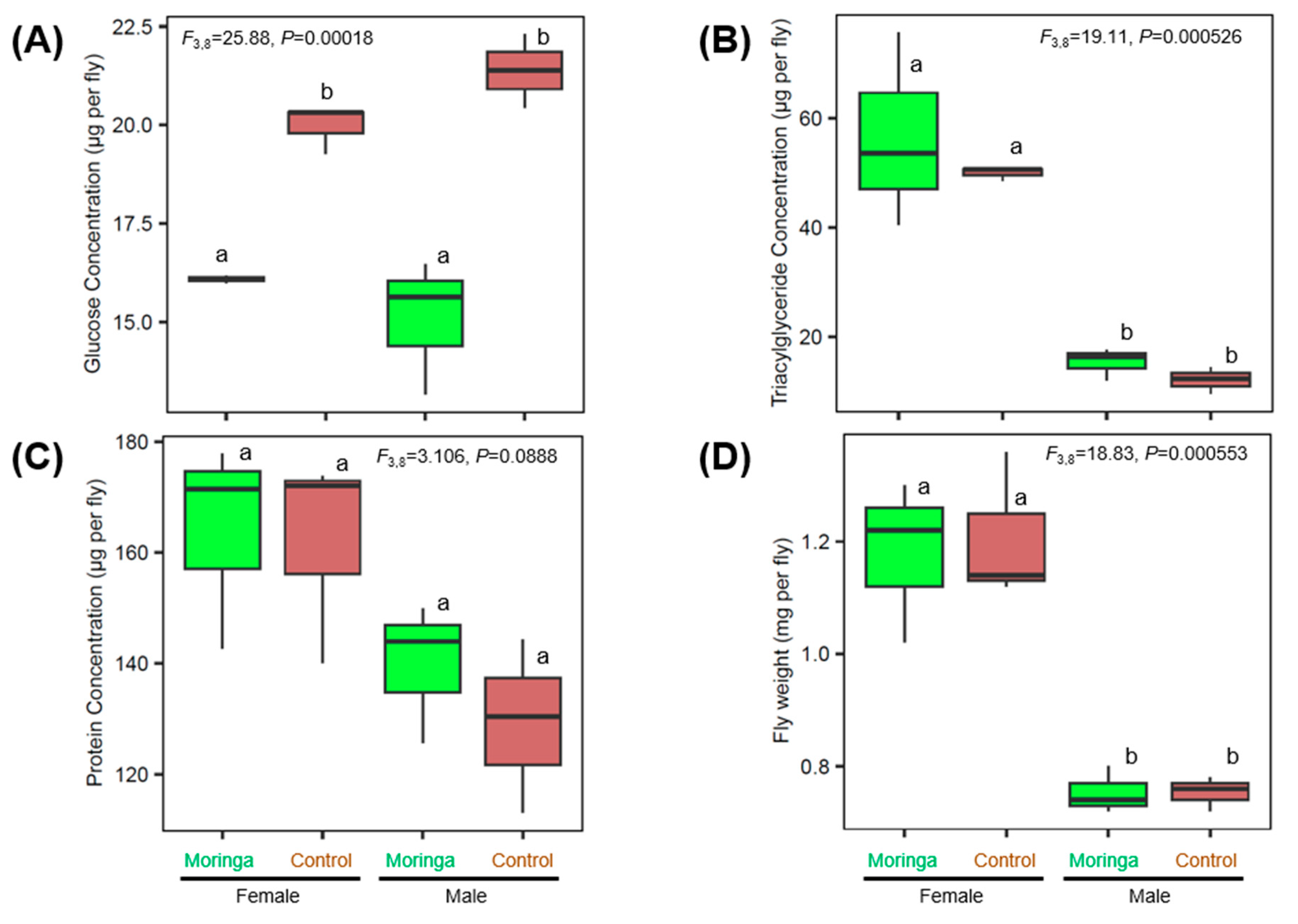
Supplementing the Drosophila diet with moringa resulted in significantly reduced glucose levels in both female and male flies, with the glucose content reducing by between 19 and 20% on average in flies fed on diets containing moringa relative to the moringa-free diet (Figure 3A). Further analyses revealed that the effect of moringa was specific to glucose, and not mirrored by any significant effect on the lipid (Figure 3B) or protein contents (Figure 3C) of the flies, or on fly weight (Figure 3D). In addition, we observed no difference in survival in both female and male female flies feeding on moringa or the moringa-free controls by the end of the 4-day incubation period (Table S2); there was no mortality recorded for both flies feeding on the moringa and control diets.

4. Discussion

Previous studies have demonstrated that the glucose and lipid content of humans and various animal models is significantly reduced by the inclusion of moringa in the host diet [21,22]. Many of these studies have focused only on changes to the metabolite profiles and/or gut microbial communities in response to dietary supplementation with moringa. To our knowledge, no studies have investigated the impact of dietary moringa on whole organism microbiome composition. In this study, we investigated the impact of moringa on whole organism microbiome composition and physiology using the fruit fly Drosophila melanogaster as a model system. We demonstrated that in addition to reducing fly glucose levels, moringa perturbed both the intracellular and gut microbial community composition of Drosophila. Specifically, supplementation of the Drosophila diet with moringa resulted in an increase in abundance of the intracellular bacterium Wolbachia and a decrease in Acetobacter counts. Although the changes in Acetobacter and Wolbachia amplicon counts were not significant, these observations raise two questions: (1) Why do Wolbachia counts increase and Acetobacter counts decrease in response to dietary moringa supplementation? and (2) What is the potential impact of the changes in microbiota composition on Drosophila physiology? Despite our observations being based on a relatively small number of flies (~5 flies per replicate), we predict that there are several factors that could give rise to changes to Wolbachia and Acetobacter counts in flies reared on a moringa diet.
It has been shown that iron availability influences Wolbachia abundance in insects [23]. As an intracellular bacterium, Wolbachia depends on Drosophila for iron [24,25], and Drosophila in turn depends on dietary supplies of iron for metabolism and growth [26]. The mechanisms by which Drosophila and Wolbachia share iron from the diet remains poorly understood, but current research suggests conflicting effects of sharing dietary iron on interactions between Drosophila and Wolbachia, with both antagonistic and beneficial outcomes anticipated depending on the concentration/availability of iron in the Drosophila diet [27,28]. Moringa is rich in iron [21], and we hypothesize that the presence of high levels of iron in moringa drives the increase in Wolbachia abundance in flies feeding on moringa. Other studies have demonstrated that Wolbachia infection in Drosophila buffers against stresses associated with low or high concentrations of iron in the diet [27], and we predict that on a moringa diet, Wolbachia titers increase to modulate iron homeostasis in the fly.
Previous research has shown that presence of Wolbachia has significant effects on the composition of the Drosophila microbiome [29,30,31], specifically reducing titers of dominant gut microbiota such as Acetobacter [30,31]. The mechanism for modulation of this change in gut microbiota by Wolbachia remains unresolved, but preliminary investigations have ruled out direct interactions between Wolbachia and the gut microbiome or the effect of Wolbachia mediated changes to fly immunity [30]. Ye et al. (2017) demonstrated that exposing Drosophila gut microbiota to antibiotics enhances Wolbachia density. In addition to being rich in iron, moringa contains a variety of bioactive substances, such as isothiocyanates, that have extensive antimicrobial properties against a variety of microorganisms [32,33]. We hypothesize that similar to the effects that commercial antibiotics have on Wolbachia density, the antimicrobial content of moringa drives increases in Wolbachia abundance in flies feeding on moringa.
The differences in Drosophila microbiota on a moringa diet could reflect influences unrelated to the iron or antimicrobial content of moringa or the impact of Wolbachia on gut microbiota composition. The increase in Wolbachia abundance in response to moringa supplementation was observed only in female flies in our study, raising the possibility that biological sex may affect the response to supplementation of the Drosophila diet with moringa. It is well established that male and female animals respond differently to dietary changes, but the biological cause of these dissimilarities remain to be elucidated [34,35,36]. Understanding the sex-based differential effects of moringa on host physiology is essential for developing precision dietary recommendations to manage metabolic diseases like diabetes.
What is the potential impact of moringa-driven changes in microbiota composition on Drosophila physiology? We hypothesize that the combined effect of diet and alterations to the host microbiota are important mediators of host glucose homeostasis. Like most animals, the Drosophila microbiome influences nearly all aspects of host physiology by competing with the host for ingested food and producing a diverse array of metabolic by-products [6,15,37,38,39,40]. The impact of microbial metabolic activity can directly modulate the nutrient availability to the host and produce specific metabolites that alter host signaling networks that regulate metabolism [41,42]. For example, Acetobacters have been demonstrated to reduce host lipid and sugar levels [14,41,43,44] by altering insulin-TOR signaling pathways via the production of acetic acid [41], and infection with Wolbachia has been demonstrated to increase glucose levels in flies by an unknown mechanism [39,45,46]. If previous reports show that Wolbachia infected flies generally have higher glucose levels than non-infected flies, why do we observe a decrease in fly glucose levels when Wolbachia counts increase in flies on the moringa diet? The answer to this question can be found in the literature on the links between iron-glucose metabolism and iron-Wolbachia interactions. The link between iron and glucose metabolism is well documented; specifically, iron plays an essential role as a cofactor for the enzymes and electron carriers necessary for fuel oxidation during respiratory glucose metabolism [47]. Iron also plays a direct and causal role in the progression of diabetes; in excess amounts, iron generates hydroxyl radicals that can cause β cell dysfunction and induce insulin resistance [47,48]. We predict that two mechanisms drive the concurrent increase in Wolbachia counts and decrease glucose levels. Firstly, the high iron content of moringa provides the cofactors necessary to increase the rate of glucose metabolism and this drives the significant reduction in host glucose levels. At the same time, as suggested by Kremer et al. (2009), we predict that Wolbachia titers increase to reduce the labile iron concentration and protect the Drosophila host from oxidative stress and cellular damage/death. A priority for future research is to quantify the impact of dietary moringa supplementation on Drosophila iron levels and to identify which dose and duration of treatment with moringa is the most effective for lowering blood glucose levels.
Altogether, our data reinforce the growing evidence that moringa plays important roles in modulating host metabolism, and specifically point to a physiological role for moringa in regulating glucose homeostasis in insects. Our findings reveal a novel mechanism by which moringa supplementation impacts host animals, leading to substantial modifications to intracellular bacterial numbers, which in turn indirectly affect gut microbiota composition and the physiology of the Drosophila host. These findings provide additional context for evaluating the impact of dietary supplements as potential therapeutics for managing metabolic diseases given the link between dietary supplementation and shifts in microbiome composition.

Supplementary Materials

The following supporting information can be downloaded at: https://www.mdpi.com/article/10.3390/microbiolres15030125/s1, Table S1. DADA2 read counts for ASVs associated with moringa vs. no-moringa Drosophila melanogaster; Table S2. The number of living and dead flies at the end of the 4-day assay.

Author Contributions

M.S., D.G., K.B. and N.Y.D.A. designed the study. M.S., D.G. and K.B. conducted experiments. M.S., D.G., K.B. and N.Y.D.A. performed data analysis. M.S., D.G. and K.B. wrote the first draft of the manuscript and revisions were made by all authors. All authors have read and agreed to the published version of the manuscript.

Funding

This study was funded in part by Cardinal Career Grants to Michaela Schaffer and Katherine Berge.

Institutional Review Board Statement

Not applicable.

Informed Consent Statement

Not applicable.

Data Availability Statement

The original contributions presented in the study are included in the Supplementary Material, further inquiries can be directed to the corresponding author. Raw sequence reads have been submitted to the NCBI Sequence Read Archive https://www.ncbi.nlm.nih.gov/sra/?term=PRJNA1139813 (accessed on 5 July 2024) (accession: PRJNA1139813).

Acknowledgments

This study used the Nephele platform from the National Institute of Allergy and Infectious Diseases (NIAID) Office of Cyber Infrastructure and Computational Biology (OCICB) in Bethesda, MD.

Conflicts of Interest

The authors declare no conflict of interest.

References

  1. Mohamed, S.M.; Shalaby, M.A.; El-Shiekh, R.A.; El-Banna, H.A.; Emam, S.R.; Bakr, A.F. Metabolic Syndrome: Risk Factors, Diagnosis, Pathogenesis, and Management with Natural Approaches. Food Chem. Adv. 2023, 3, 100335. [Google Scholar] [CrossRef]
  2. Samson, S.L.; Garber, A.J. Metabolic Syndrome. Endocrinol. Metab. Clin. N. Am. 2014, 43, 1–23. [Google Scholar] [CrossRef] [PubMed]
  3. Saklayen, M.G. The Global Epidemic of the Metabolic Syndrome. Curr. Hypertens. Rep. 2018, 20, 12. [Google Scholar] [CrossRef] [PubMed]
  4. Thomas, M.S.; Calle, M.; Fernandez, M.L. Healthy Plant-Based Diets Improve Dyslipidemias, Insulin Resistance, and Inflammation in Metabolic Syndrome. A Narrative Review. Adv. Nutr. 2023, 14, 44–54. [Google Scholar] [CrossRef]
  5. Sonnenburg, E.D.; Sonnenburg, J.L. Starving Our Microbial Self: The Deleterious Consequences of a Diet Deficient in Microbiota-Accessible Carbohydrates. Cell Metab. 2014, 20, 779–786. [Google Scholar] [CrossRef]
  6. Zmora, N.; Suez, J.; Elinav, E. You Are What You Eat: Diet, Health and the Gut Microbiota. Nat. Rev. Gastroenterol. Hepatol. 2019, 16, 35–56. [Google Scholar] [CrossRef]
  7. Hassoun, A.; Harastani, R.; Jagtap, S.; Trollman, H.; Garcia-Garcia, G.; Awad, N.M.H.; Zannou, O.; Galanakis, C.M.; Goksen, G.; Nayik, G.A.; et al. Truths and Myths about Superfoods in the Era of the COVID-19 Pandemic. Crit. Rev. Food Sci. Nutr. 2024, 64, 585–602. [Google Scholar] [CrossRef]
  8. Kunyanga, C.; Imungi, J.; Vellingiri, V. Nutritional Evaluation of Indigenous Foods with Potential Food-Based Solution to Alleviate Hunger and Malnutrition in Kenya. J. Appl. Biosci. 2013, 67, 5277. [Google Scholar] [CrossRef]
  9. Sokhela, H.; Govender, L.; Siwela, M. Complementary Feeding Practices and Childhood Malnutrition in South Africa: The Potential of Moringa Oleifera Leaf Powder as a Fortificant: A Narrative Review. Nutrients 2023, 15, 2011. [Google Scholar] [CrossRef]
  10. Vergara-Jimenez, M.; Almatrafi, M.; Fernandez, M. Bioactive Components in Moringa Oleifera Leaves Protect against Chronic Disease. Antioxidants 2017, 6, 91. [Google Scholar] [CrossRef]
  11. Karthikesan, K.; Pari, L.; Menon, V. Combined Treatment of Tetrahydrocurcumin and Chlorogenic Acid Exerts Potential Antihyperglycemic Effect on Streptozotocin-Nicotinamide-Induced Diabetic Rats. Gen. Physiol. Biophys. 2010, 29, 23–30. [Google Scholar] [CrossRef] [PubMed]
  12. Waterman, C.; Rojas-Silva, P.; Tumer, T.B.; Kuhn, P.; Richard, A.J.; Wicks, S.; Stephens, J.M.; Wang, Z.; Mynatt, R.; Cefalu, W.; et al. Isothiocyanate-rich Moringa oleifera Extract Reduces Weight Gain, Insulin Resistance, and Hepatic Gluconeogenesis in Mice. Mol. Nutr. Food Res. 2015, 59, 1013–1024. [Google Scholar] [CrossRef] [PubMed]
  13. Mushtaq, B.S.; Hussain, M.B.; Omer, R.; Toor, H.A.; Waheed, M.; Shariati, M.A.; Sergey, P.; Heydari, M. Moringa Oleifera in Malnutrition: A Comprehensive Review. Curr. Drug Discov. Technol. 2021, 18, 235–243. [Google Scholar] [CrossRef] [PubMed]
  14. Newell, P.D.; Douglas, A.E. Interspecies Interactions Determine the Impact of the Gut Microbiota on Nutrient Allocation in Drosophila Melanogaster. Appl. Environ. Microbiol. 2014, 80, 788–796. [Google Scholar] [CrossRef]
  15. Zhang, F.Q.; McMullen, J.G.; Douglas, A.E.; Ankrah, N.Y.D. Succinate: A Microbial Product That Modulates Drosophila Nutritional Physiology. Insect Sci. 2022, 29, 315–318. [Google Scholar] [CrossRef]
  16. Walters, W.; Hyde, E.R.; Berg-Lyons, D.; Ackermann, G.; Humphrey, G.; Parada, A.; Gilbert, J.A.; Jansson, J.K.; Caporaso, J.G.; Fuhrman, J.A.; et al. Improved Bacterial 16S rRNA Gene (V4 and V4-5) and Fungal Internal Transcribed Spacer Marker Gene Primers for Microbial Community Surveys. mSystems 2016, 1, e00009-15. [Google Scholar] [CrossRef]
  17. Weber, N.; Liou, D.; Dommer, J.; MacMenamin, P.; Quiñones, M.; Misner, I.; Oler, A.J.; Wan, J.; Kim, L.; Coakley McCarthy, M.; et al. Nephele: A Cloud Platform for Simplified, Standardized and Reproducible Microbiome Data Analysis. Bioinformatics 2018, 34, 1411–1413. [Google Scholar] [CrossRef]
  18. Callahan, B.J.; McMurdie, P.J.; Rosen, M.J.; Han, A.W.; Johnson, A.J.A.; Holmes, S.P. DADA2: High-Resolution Sample Inference from Illumina Amplicon Data. Nat. Methods 2016, 13, 581–583. [Google Scholar] [CrossRef]
  19. Quast, C.; Pruesse, E.; Yilmaz, P.; Gerken, J.; Schweer, T.; Yarza, P.; Peplies, J.; Glöckner, F.O. The SILVA Ribosomal RNA Gene Database Project: Improved Data Processing and Web-Based Tools. Nucleic Acids Res. 2012, 41, D590–D596. [Google Scholar] [CrossRef]
  20. Villanueva, R.A.M.; Chen, Z.J. Ggplot2: Elegant Graphics for Data Analysis (2nd ed.). Meas. Interdiscip. Res. Perspect. 2019, 17, 160–167. [Google Scholar] [CrossRef]
  21. Abdelazim, A.M.; Afifi, M.; Abu-Alghayth, M.H.; Alkadri, D.H. Moringa oleifera: Recent Insights for Its Biochemical and Medicinal Applications. J. Food Biochem. 2024, 2024, 1270903. [Google Scholar] [CrossRef]
  22. Owens, F.S.; Dada, O.; Cyrus, J.W.; Adedoyin, O.O.; Adunlin, G. The Effects of Moringa Oleifera on Blood Glucose Levels: A Scoping Review of the Literature. Complement. Ther. Med. 2020, 50, 102362. [Google Scholar] [CrossRef] [PubMed]
  23. Kremer, N.; Voronin, D.; Charif, D.; Mavingui, P.; Mollereau, B.; Vavre, F. Wolbachia Interferes with Ferritin Expression and Iron Metabolism in Insects. PLoS Pathog. 2009, 5, e1000630. [Google Scholar] [CrossRef] [PubMed]
  24. Gill, A.C.; Darby, A.C.; Makepeace, B.L. Iron Necessity: The Secret of Wolbachia’s Success? PLoS Negl. Trop. Dis. 2014, 8, e3224. [Google Scholar] [CrossRef] [PubMed]
  25. Jiménez, N.E.; Gerdtzen, Z.P.; Olivera-Nappa, Á.; Salgado, J.C.; Conca, C. A Systems Biology Approach for Studying Wolbachia Metabolism Reveals Points of Interaction with Its Host in the Context of Arboviral Infection. PLoS Negl. Trop. Dis. 2019, 13, e0007678. [Google Scholar] [CrossRef]
  26. Mandilaras, K.; Pathmanathan, T.; Missirlis, F. Iron Absorption in Drosophila Melanogaster. Nutrients 2013, 5, 1622–1647. [Google Scholar] [CrossRef]
  27. Brownlie, J.C.; Cass, B.N.; Riegler, M.; Witsenburg, J.J.; Iturbe-Ormaetxe, I.; McGraw, E.A.; O’Neill, S.L. Evidence for Metabolic Provisioning by a Common Invertebrate Endosymbiont, Wolbachia Pipientis, during Periods of Nutritional Stress. PLoS Pathog. 2009, 5, e1000368. [Google Scholar] [CrossRef]
  28. Newton, I.L.G.; Rice, D.W. The Jekyll and Hyde Symbiont: Could Wolbachia Be a Nutritional Mutualist? J. Bacteriol. 2020, 202, e00589-19. [Google Scholar] [CrossRef]
  29. Detcharoen, M.; Jiggins, F.M.; Schlick-Steiner, B.C.; Steiner, F.M. Wolbachia Endosymbiotic Bacteria Alter the Gut Microbiome in the Fly Drosophila Nigrosparsa. J. Invertebr. Pathol. 2023, 198, 107915. [Google Scholar] [CrossRef]
  30. Simhadri, R.K.; Fast, E.M.; Guo, R.; Schultz, M.J.; Vaisman, N.; Ortiz, L.; Bybee, J.; Slatko, B.E.; Frydman, H.M. The Gut Commensal Microbiome of Drosophila Melanogaster Is Modified by the Endosymbiont Wolbachia. mSphere 2017, 2, e00287-17. [Google Scholar] [CrossRef]
  31. Ye, Y.H.; Seleznev, A.; Flores, H.A.; Woolfit, M.; McGraw, E.A. Gut Microbiota in Drosophila Melanogaster Interacts with Wolbachia but Does Not Contribute to Wolbachia -Mediated Antiviral Protection. J. Invertebr. Pathol. 2017, 143, 18–25. [Google Scholar] [CrossRef] [PubMed]
  32. Fahey, J.W. Moringa oleifera: A Review of the Medicinal Potential. Acta Hortic. 2017, 1158, 209–224. [Google Scholar] [CrossRef]
  33. Romeo, L.; Iori, R.; Rollin, P.; Bramanti, P.; Mazzon, E. Isothiocyanates: An Overview of Their Antimicrobial Activity against Human Infections. Molecules 2018, 23, 624. [Google Scholar] [CrossRef] [PubMed]
  34. Maggi, A.; Della Torre, S. Sex, Metabolism and Health. Mol. Metab. 2018, 15, 3–7. [Google Scholar] [CrossRef] [PubMed]
  35. Maric, I.; Krieger, J.-P.; Van Der Velden, P.; Börchers, S.; Asker, M.; Vujicic, M.; Wernstedt Asterholm, I.; Skibicka, K.P. Sex and Species Differences in the Development of Diet-Induced Obesity and Metabolic Disturbances in Rodents. Front. Nutr. 2022, 9, 828522. [Google Scholar] [CrossRef]
  36. Pontifex, M.G.; Vauzour, D.; Muller, M. Sexual Dimorphism in the Context of Nutrition and Health. Proc. Nutr. Soc. 2024, 83, 109–119. [Google Scholar] [CrossRef]
  37. Ankrah, N.Y.D.; Barker, B.E.; Song, J.; Wu, C.; McMullen, J.G.; Douglas, A.E. Predicted Metabolic Function of the Gut Microbiota of Drosophila Melanogaster. mSystems 2021, 6, e01369-20. [Google Scholar] [CrossRef]
  38. Ankrah, N.Y.D.; Douglas, A.E. Nutrient Factories: Metabolic Function of Beneficial Microorganisms Associated with Insects. Environ. Microbiol. 2018, 20, 2002–2011. [Google Scholar] [CrossRef]
  39. Carneiro Dutra, H.L.; Deehan, M.A.; Frydman, H. Wolbachia and Sirtuin-4 Interaction Is Associated with Alterations in Host Glucose Metabolism and Bacterial Titer. PLoS Pathog. 2020, 16, e1008996. [Google Scholar] [CrossRef]
  40. Lesperance, D.N.; Broderick, N.A. Microbiomes as Modulators of Drosophila Melanogaster Homeostasis and Disease. Curr. Opin. Insect Sci. 2020, 39, 84–90. [Google Scholar] [CrossRef]
  41. Shin, S.C.; Kim, S.-H.; You, H.; Kim, B.; Kim, A.C.; Lee, K.-A.; Yoon, J.-H.; Ryu, J.-H.; Lee, W.-J. Drosophila Microbiome Modulates Host Developmental and Metabolic Homeostasis via Insulin Signaling. Science 2011, 334, 670–674. [Google Scholar] [CrossRef] [PubMed]
  42. Storelli, G.; Defaye, A.; Erkosar, B.; Hols, P.; Royet, J.; Leulier, F. Lactobacillus Plantarum Promotes Drosophila Systemic Growth by Modulating Hormonal Signals through TOR-Dependent Nutrient Sensing. Cell Metab. 2011, 14, 403–414. [Google Scholar] [CrossRef] [PubMed]
  43. Chaston, J.M.; Newell, P.D.; Douglas, A.E. Metagenome-Wide Association of Microbial Determinants of Host Phenotype in Drosophila Melanogaster. mBio 2014, 5, e01631-14. [Google Scholar] [CrossRef]
  44. Consuegra, J.; Grenier, T.; Akherraz, H.; Rahioui, I.; Gervais, H.; Da Silva, P.; Leulier, F. Metabolic Cooperation among Commensal Bacteria Supports Drosophila Juvenile Growth under Nutritional Stress. iScience 2020, 23, 101232. [Google Scholar] [CrossRef] [PubMed]
  45. Karpova, E.K.; Bobrovskikh, M.A.; Deryuzhenko, M.A.; Shishkina, O.D.; Gruntenko, N.E. Wolbachia Effect on Drosophila Melanogaster Lipid and Carbohydrate Metabolism. Insects 2023, 14, 357. [Google Scholar] [CrossRef]
  46. Zhang, H.-B.; Cao, Z.; Qiao, J.-X.; Zhong, Z.-Q.; Pan, C.-C.; Liu, C.; Zhang, L.-M.; Wang, Y.-F. Metabolomics Provide New Insights into Mechanisms of Wolbachia-Induced Paternal Defects in Drosophila Melanogaster. PLoS Pathog. 2021, 17, e1009859. [Google Scholar] [CrossRef]
  47. Simcox, J.A.; McClain, D.A. Iron and Diabetes Risk. Cell Metab. 2013, 17, 329–341. [Google Scholar] [CrossRef]
  48. Kaneto, H.; Katakami, N.; Matsuhisa, M.; Matsuoka, T. Role of Reactive Oxygen Species in the Progression of Type 2 Diabetes and Atherosclerosis. Mediators Inflamm. 2010, 2010, 453892. [Google Scholar] [CrossRef]
Figure 1. Impact of moringa diet supplementation on the composition and diversity of the bacterial community of D. melanogaster. (A) Relative abundance of bacterial orders present in D. melanogaster feeding on moringa versus the control diet. (B) Alpha diversity measurements for different treatment groups. Significantly different (p < 0.05) samples by Tukey’s HSD post hoc test are indicated by different letters. For each boxplot, the center line displays the median, and the lower and upper hinges correspond to the 25th and 75th percentiles. Moringa and control diets are represented by green and brown boxes, respectively.
Microbiolres 15 00125 g001
Figure 2. Response of gut and intracellular microbial communities to moringa supplementation. (A) Change to Acetobacter levels on a moringa diet. (B) Change to Wolbachia levels on a moringa diet. (C) Viable bacteria count on MRS agar plates. Significantly different (p < 0.05) samples by Tukey’s HSD post hoc test are indicated by different letters. For each boxplot, the center line displays the median, and the lower and upper hinges correspond to the 25th and 75th percentiles. Moringa and control diets are represented by green and brown boxes respectively.
Microbiolres 15 00125 g002
Figure 3. Effect of dietary supplementation with moringa on Drosophila nutritional indices (AC) and weight (D). Significantly different (p < 0.05) samples by Tukey’s HSD post hoc test are indicated by different letters. For each boxplot, the center line displays the median, and the lower and upper hinges correspond to the 25th and 75th percentiles. Moringa and control diets are represented by green and brown boxes, respectively.
Microbiolres 15 00125 g003
Disclaimer/Publisher’s Note: The statements, opinions and data contained in all publications are solely those of the individual author(s) and contributor(s) and not of MDPI and/or the editor(s). MDPI and/or the editor(s) disclaim responsibility for any injury to people or property resulting from any ideas, methods, instructions or products referred to in the content.