Article

Antagonistic Activity of Macrolepiota sp. CS185 against Post-Harvest Fungi of Fig Fruits (Ficus carica L.)

by
Guadalupe Gutiérrez-Soto
1,*,
Iosvany López-Sandin
1,*,
Jesús Salvador Hernández Ochoa
1,
Carlos Eduardo Hernadez-Luna
2,
Juan Francisco Contreras-Cordero
2 and
Carlos Alberto Hernández-Martínez
1
1
Laboratorio de Ciencias Naturales, Facultad de Agronomía, Francisco Villa S/N Col. Ex Hacienda El Canadá, Universidad Autónoma de Nuevo León, General Escobedo 66415, Mexico
2
Facultad de Ciencias Biológicas, Universidad Autónoma de Nuevo León, Pedro de Alba S/N., Ciudad Universitaria, San Nicolás de los Garza 66455, Mexico
*
Authors to whom correspondence should be addressed.
Microbiol. Res. 2024, 15(1), 371-384; https://doi.org/10.3390/microbiolres15010025
Submission received: 19 November 2023 / Revised: 3 January 2024 / Accepted: 9 January 2024 / Published: 7 March 2024

Abstract

:
Macrolepiota sp. CS185 is a basidiomycete with high potential as a biocontrol agent against various phytopathogenic fungi. Regardless of its pronounced potential as a post-harvest fungi biocontrol agent, its activity in tomato seedlings infected with Alternaria alternata has not been well studied. Thus, the present work aimed to evaluate the cultures and supernatants’ antagonistic activity against fig fruits’ post-harvest fungi and antifungal activity production kinetics. The culture antagonistic characteristics were assessed through multiple confrontations, the supernatant concentration effect, and the kinetics of antagonistic action. The multiple confrontations showed differences (p ≤ 0.05) among phytopathogens and over time, with Colletotrichum sp. 2 being the most susceptible. Based on the 9-day incubation profile, the treatment fractions supplemented with a 50% concentration of Macrolepiota sp. CS185 supernatants showed a higher inhibition percentage (%In). Except for Alternaria alternata 1 and 2, the rest of the isolates showed a similar decrease in antagonistic activity up to a certain extent over time. Among all tested strains, Colletotrichum sp. 2 was found with a higher susceptibility. Regarding the production kinetics of antagonistic activity, a triple interaction was observed between the phytopathogen, the age of the Macrolepiota sp. CS 185 culture, and incubation time. In addition, changes in the mycelium growth rate (p ≤ 0.05) along with the higher activity in the supernatants of 20 and 30 days were observed and suggested the production of multiple bioactive metabolites. These results indicate that Macrolepiota sp. CS185 produces antifungal metabolites at different times and could be a suitable candidate to control fig fruits’ post-harvest fungi issues.

1. Introduction

Continuous population growth has increased the current food demand worldwide, and to fulfill this ever-rising demand, intensifying agricultural production is in practice. However, incorrect production techniques and microbial attacks have been noticeable causes of significant food losses, mainly during post-harvest work [1]. It can be demonstrated in producing fruits and vegetables, where their quality and shelf life are significantly affected, generating losses close to 40% [2]. In the case of fruit trees, 50% of post-harvest damage is associated with microorganism activity [3]. Regardless of microbial spoilage, it is also imperative to mention other reasons that majorly contribute to this rise in food demand and supply. Fruits are nutritious sources with elevated nutritional content, particularly in the context of human dietary practices and health. This has led to a notable surge in their consumption patterns [4,5].
There has been growing interest in fig fruit (Ficus carica L.) due to its higher contents of total polyphenols, total flavonoids, and anthocyanins that provide a great antioxidant capacity [6,7,8]. However, fresh fig fruits are usually susceptible to pathophysiological disorders during post-harvest practices, such as softening and cracking of the skin, even under cold storage conditions. Therefore, they are highly perishable products in post-harvest with microbiological decomposition that results in an unpleasant taste and odor. This is why conservation methods are required to counteract their deterioration [9]. Some studies have reported the species associated with fruit deterioration, such as the genera of Alternaria, Aspergillus, Botrytis, Cladosporium, Eurotium, Fusarium, Geotrichum, Gloeosporium, Monilinia, Mucor, Penicillium, and Rhizopus [10,11,12]. Alternaria and Fusarium are notable for causing internal fruit rot and are the main fungal concerns in fig production [13].
The above has led to the search for alternative strategies that not only protect fig fruits from damage but also extend their shelf-life. Therefore, the current studies are mostly focused on evaluating of the effect of temperature [14,15], packaging in modified atmospheres [16], the use of chemical agents [17], radiation [18], and the development of coatings that preserve the quality attributes of the fruits [9]. The most used polymers are chitosan, sodium alginate, agar, and gum Arabic [9,19,20]. Interest has grown in developing hybrid coating materials with strong antimicrobial activities by combining different polymers with essential oils [21] or active compounds extracted from agricultural byproducts [22]. Other strategies include using coatings based on nopal mucilage with bactericidal activity [23] and zinc oxide nanoparticles [24] to preserve fruit quality and shelf-life.
Basidiomycetes are proven sources of bioactive metabolites. However, they have not been used in the post-harvest field [25,26]. Both organic extracts and supernatants obtained from carpophores or cultures of this group of fungi have shown antifungal activity and economic importance [27,28,29,30,31]. Thus, compounds such as Crinipellins A and I produced by the basidiomycete Crinipellis rhizomaticola have been identified, with activity against various phytopathogens, i.e., Magnaporthe oryzae, Colletotrichum coccodes, Botrytis cinerea, and Phytophthora infestans [32]. Strobilurins are a group of bioactive metabolites produced by several fungi and have been used to develop agricultural fungicides, e.g., β-methoxyacrylate [33,34]. Such fungicides have shown notable inhibitory activities against phytopathogens, such as Gibberella zeae, Sclerotinia sclerotiorum, and Rhizoctonia cerealis [35]. Likewise, Macrolepiota genus has shown great potential to produce bioactive compounds [36], along with other biological characteristics, such as antibacterial, antioxidant, anti-inflammatory, regulatory, antidepressant, and anticancer effects [37]. However, studies associated with their capacity as biocontrol agents are limited or lacking in the existing literature. For example, the native strain Macrolepiota sp. CS185 supernatants have antagonistic activity against Alternaria solani under greenhouse conditions in tomato seedlings [30]. Neither the antifungal activity production kinetics nor its effect on fungal isolates obtained from fruits under post-harvest conditions were studied. Therefore, the present work aimed to evaluate the cultures and supernatants’ antagonistic activity against fig fruits’ post-harvest fungi and antifungal activity production kinetics.
It is important to highlight that the genus Macrolepiota is a group of fungi characterized by smooth spores, a ring on the stem, and a large conical cap that often opens as it matures. These mushrooms usually have a tall, thin stem with a membranous mobile ring. The cap is white to brown, often with scales or spots. The slats are tight and free. Spores range in color from white to brown, green, and blue; the hymenophoral trama can be either regular or trabecular. The pileus covering’s structure (velar or pileipellicular) can be epithelioid, trichodermal, or cuticular. Some species of Macrolepiota are edible and valued for their flavor, but it is important to be careful when identifying mushrooms for consumption, as some species can be toxic. It is essential to consider all these morphological details when identifying fungi of the genus Macrolepiota to ensure accurate and safe identification [38,39].

2. Materials and Methods

2.1. Reagents

All the reagents and chemical compounds used were of reagent grade and obtained from Sigma-Aldrich (San Luis, MO, USA). Culture media were from BD (Becton, Dickinson and Company, Franklin Lakes, NJ, USA). All solutions were prepared using double distilled water in Laboratorios Monterrey, S.A. (Monterrey, Nuevo León, Mexico). Measurements of fungal mycelium growth were made with a digital vernier caliper of 150 mm with an accuracy of 0.01 mm.

2.2. Biological Material

All listed strains were kindly provided in their pure form by the Enzymology Laboratory of Facultad de Ciencias Biológicas, UANL. Briefly, four sub-strains of Alternaria alternata, i.e., A. alternata 1, A. alternata 2, A. alternata 3, and A. alternata 4, were used. Two sub-cultures of Colletotrichum sp. 1 and Colletotrichum sp. 2 obtained from the fruit of figs were acquired commercially. Macrolepiota sp. CS185, as an antagonist agent, was used as a positive control, whereas, in multiple confrontations, the Fusarium solani strain was used [30]. The above-listed strains were maintained periodically by reseeding every three months and preserved in YMGA medium (glucose 4 g L−1, malt extract 10 g L−1, yeast extract 4 g L−1, and agar 15 g L−1) [40]. The freshly prepared mixture of YMGA medium and potato dextrose agar (PDA) was used to develop fungal inoculums with five days of growth. From the periphery of reactive colonies, discs of 0.5 cm were taken to sow solid or liquid media.

2.3. Cell-Wall Degrading Enzymes Detection

The evaluation of the plant cell-wall component (DPCWC) enzymes profile, such as cellulases, xylanases, lignin-modifying enzymes (LME), pectinases, and amylases from phytopathogenic fungi, was conducted on solid medium. The base medium used was adapted from Sin et al. [41], with a composition of peptone (0.1%), yeast extract (0.01%), and agar (2.0%). Carboxymethylcellulose (CMC, 1%), xylan (1%), and pectin (0.5%) were used as carbon sources, added to the base medium for the detection of cellulases, xylanases, and pectin, respectively.
For the detection of amylases, 1% starch was used, while for the detection of LME activity, the Poly R-478 dye was used at 0.04% and 1% glucose as a carbon source. Laccase activity was revealed by the oxidation of syringaldazine (0.5%), which was added after sterilizing the medium [41]. Fungal growth was recorded daily. An iodine solution was used to reveal the activity of carbohydrolases on the third day. All assays were performed in quadruplicate. The diameter of the hydrolysis halo was measured to calculate the degradation index (DI) using Equation (1) [42]:
D I = h G  
where h corresponds to hydrolysis (mm) and G corresponds to fungal growth (mm).

2.4. Determination of Antagonistic Activity in Multiple Confrontations

Petri dishes were prepared with papa dextrose agar (PDA), which were inoculated with a 0.5 cm piece, taken from the Macrolepiota sp. CS185 culture periphery with five days of growth. The plates were incubated for 24 h at 28 °C and subsequently four post-harvest fungi were sown in the peripheries. As a negative control, the confrontation between the isolated post-harvest fungi was performed without the antagonist to rule out that inhibition may be due to nutrient depletion. The plates were incubated again at 28 °C to measure the mycelium radial growth every 24 h from the cylinder’s rim to the edge of the consolidated mycelium in the direction of the antagonist for four days. All trials were carried out in triplicate. The percentage inhibition (%In) was estimated using Equation (2):
% I n = Gc Ga Gc × 100  
where Gc corresponds to the growth of the phytopathogen in the control treatment, and Ga corresponds to the growth in confrontation with the basidiomycete.

2.5. Determination of the Antagonistic Activity of Cell-Free Supernatants

The antifungal potential of supernatants was determined using methodology described by Hernández et al. [30]. Briefly, around 200 mL of PDB medium was prepared, sterilized, and inoculated with three discs (each of 5 mm diameter). The above inoculated plates were incubated for 18 days at 28 °C under stirring conditions (150 rpm) in an orbital shaker from New Brunswick Scientific® (Edison, NJ, USA). Supernatants were recovered by filtration using the paper Whatman® N°1 and sterilized using 0.45 μm Luzeren cellulose ester membranes with the ultrafiltration system of Merck Millipore® (Burlington, MA, USA). Next, four flasks were prepared with PDA medium, using double-distilled water at 100, 85, 70, and 50%. These were sterilized at 121 °C for 15 min, cooled to 60 °C, and the sterilized supernatant was added to a final concentration of 0, 15, 30, and 50%. These mediums were poured into Petri dishes. They were inoculated with a 0.5 cm piece and incubated at 28 °C. The growth radius of the mycelium was measured daily for nine days. All treatments were carried out in triplicate. Equation (2) was used to calculate the growth percentage inhibition (%In). From the daily measurements of the growth of the colonies of the post-harvest isolates, the growth curves were obtained, allowing the determination of the mycelium growth rate (MGR) using the slope of the linear equation on the charts (Figures S1 and S2). The MGR of the treatments was used to estimate the relative percentage inhibition in the daily mycelium growth rate (RPI) by Equation (3):
R P I = MGRc MGRa MGRc × 100
where MGRc corresponds to the mycelium growth rate in the treatment control and MGRa corresponds to growth in confrontation with the basidiomycete antagonist.

2.6. Production Kinetics of Antifungal Activity

To evaluate the antagonistic activity, first, the supernatant suspension from cultures of Macrolepiota sp. CS185 was obtained. For said purpose, sixteen Erlenmeyer flasks containing 500 mL PDB medium were prepared, inoculated simultaneously, and incubated at 28 °C under shaking at 150 rpm. Two cultures were filtered at 0, 5, 10, 15, 20, 25, 30, and 40 days to recover the supernatants, which were frozen until later use. Subsequently, Petri dishes were prepared using a PDA medium enriched with 30% of each supernatant to evaluate the antagonistic effect as described above in Section 2.5. The media were inoculated with the respective phytopathogenic fungus, and 0.5 cm pieces were taken from the periphery of a culture with a growth of five days and placed in the center of the plates. These were incubated at 28 °C for eight days. The negative control comprised PDA medium without supernatant. The daily growth of the mycelium was recorded for the estimation of %In using Equation (2), and the growth curves and the determination of MGR and RPI were carried out using Equation (3). All treatments were recorded as a mean of five repetitions.

2.7. Statistical Analysis

For the analysis of the results of the evaluation of antifungal activity, a randomized block experimental design with a factorial arrangement of A × B was used. The data generated by the effect of the different sources of variation on the study variables were statistically contrasted through an analysis of variance (ANOVA) and a comparison of averages, according to the Tukey test to a degree of significance of 95% (p ≤ 0.05). InfoStat® statistical software was used in data analysis.

3. Results

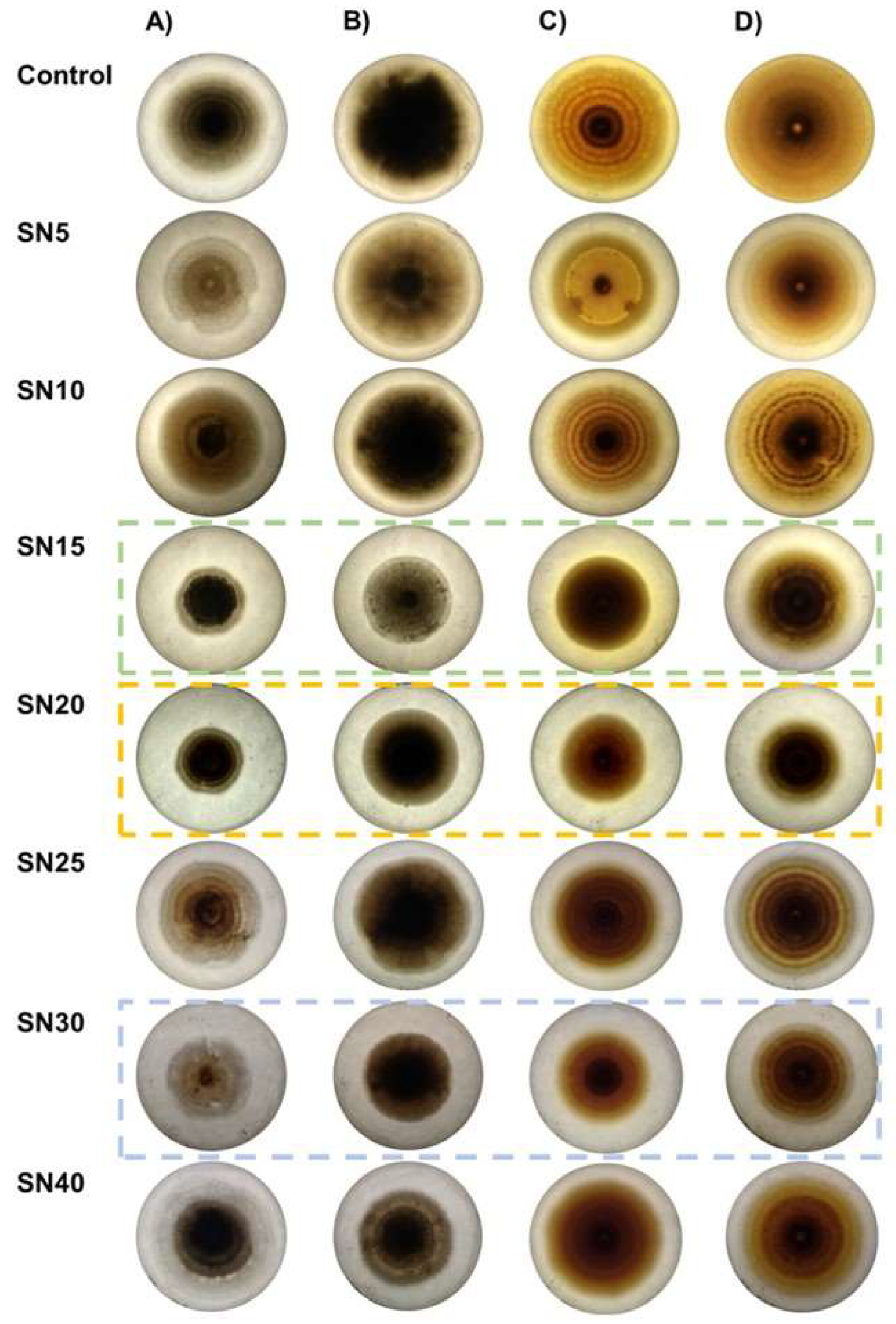
3.1. Determination of Antagonistic Activity in Multiple Confrontations

The comparison of means for %In of fungal radial growth is summarized in Table 1. Differences in statistical significance were observed in time and between isolates. In general, Colletotrichum isolates tended to be more susceptible (60–70%, p < 0.05) to Macrolepiota sp. CS 185 supernatants than Alternaria isolates (40–60%) and Fusarium (<50%) isolates. However, significant antagonistic activity was obtained at four days, except in Alternaria alternata 1. Colletotrichum sp. 2 was the most susceptible (73.3%), followed by Colletotrichum sp. 1 (68.8%), A. alternata 3 (66.9%), and A. alternata 4 (66%).
Likewise, the antagonistic activity increased in relation to the incubation time, showing the higher values at four days (except in A. alternata 1), with Colletotrichum sp. 2 being the most susceptible. It is worth mentioning that, despite the similarity of the behavior of the isolates of the same genus, the profile of degraders of plant cell-wall components was different (Table 2). All isolates were producers of cells and xylanases. Only Fusarium oxysporum showed pectinolytic activity, while Colletotrichum sp. 2 was the only one that presented laccase activity.

3.2. Determination of Antagonistic Activity in Multiple Confrontations

The effect of the tested concentrations for isolates of A. alternata and Colletotrichum spp. is shown in Figure 1. The higher %In was observed in treatments with the Macrolepiota sp. CS185 supernatant at 50%. At the same time, Colletotrichum isolates had a similar behavior (Figure 1E,F). The Colletotrichum sp. 2 isolate was the most susceptible, as observed in multiple confrontations.
On the other hand, A. alternata strains showed different inhibition patterns (Figure 1A–D), with Isolate 4 being the one with the higher susceptibility. It is worth mentioning that the antagonistic activity decreased linearly over time, except in the treatments with A. alternata 2 and A. alternata 3 (Figure 1B,C). The latter showed values of less than 30% inhibition in all treatments.
Figure S1 shows the growth curves for the different treatments for each isolate. From which the mycelium growth rate (MGR) and the relative percentage of inhibition (RPI) in daily growth rate were estimated. Concerning the MGR, A. alternata 1 and Colletotrichum sp. 2 were statistically equal (p < 0.05) in the control treatment (without supernatant), while A. alternata 2 had the lowest MGR (2.58 mm/d), reiterating the differences between strains of the same genus. The latter was the only isolate with the highest RPI value in the 30% supernatant treatment since the rest had the highest RPI values in the 50% treatment (Table 3).

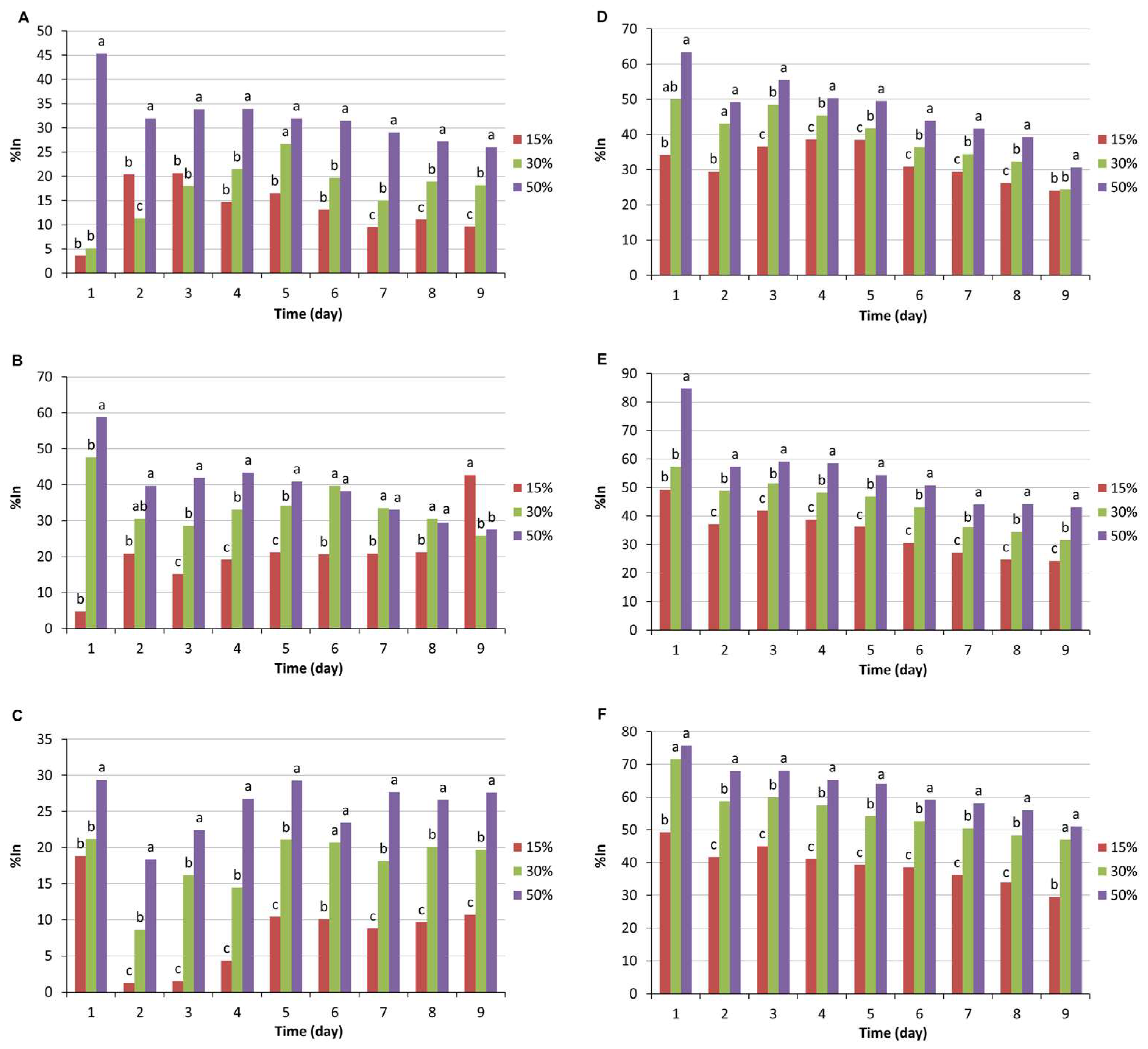
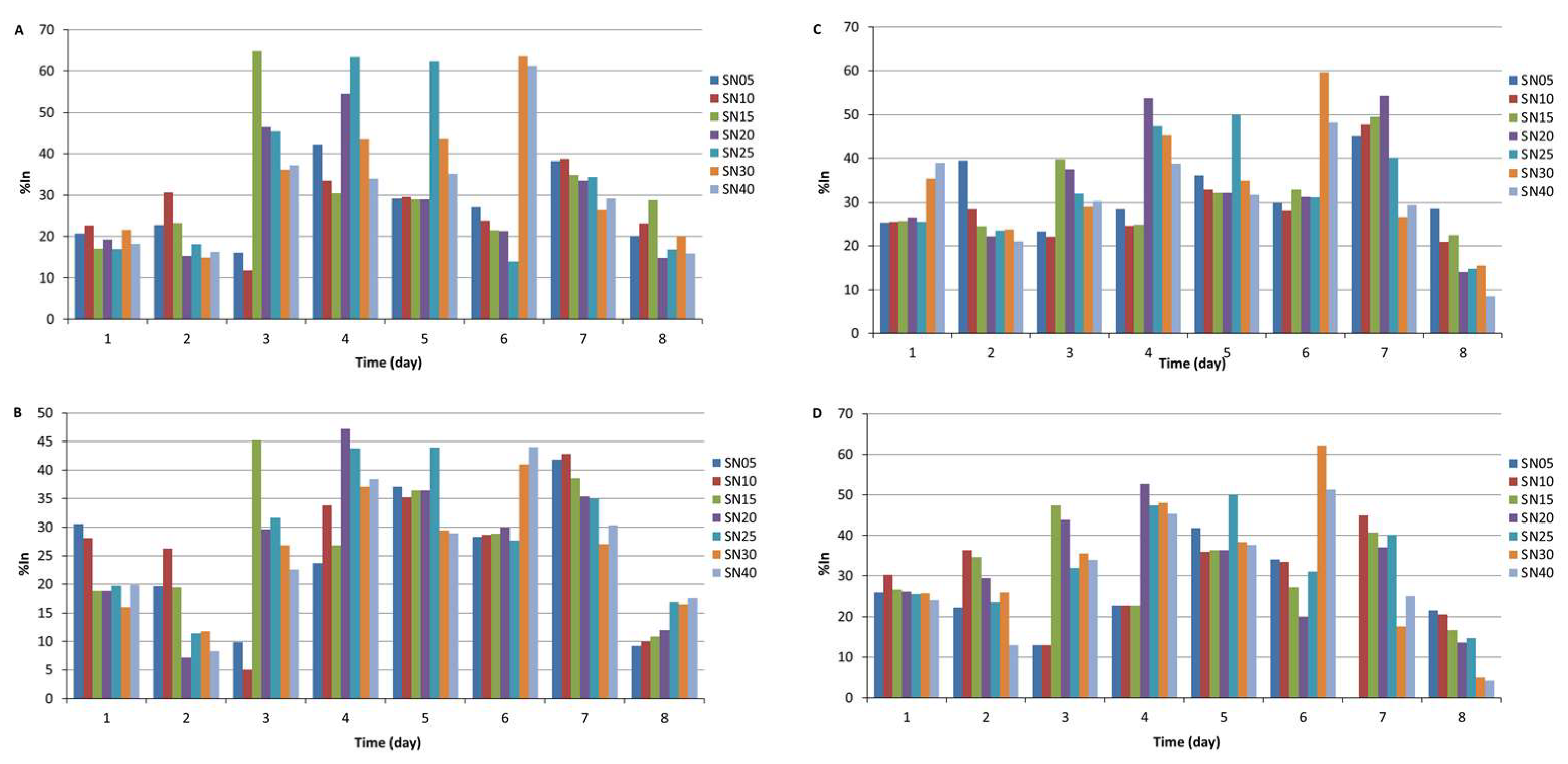
3.3. Production Kinetics of Antifungal Activity in Cell-Free Supernatants

To assess the antagonistic effect of the supernatants of Macrolepiota sp. CS185, the supernatants obtained from cultures with 18 days of incubation were used. To rule out that the decrease in antagonistic activity (concerning that obtained in the multiple confrontations) was associated with the age of the basidiomycete culture, an inhibition curve was performed with supernatants obtained from 0 to 40 days. From these, plates were prepared with 30% of each supernatant. In this phase, A. alternata 1, A. alternata 4, and Colletotrichum sp. 1 and 2 were selected considering the similarities in their growth rates.
An analysis of variance (Table S1) for the %In of supernatants from cultures with different incubation periods showed statistically significant differences (p ≤ 0.01) for the culture age, time, and isolated post-harvest fungi, indicating a three-way interaction between these factors. The effect of the culture age of Macrolepiota sp. CS185 on the production of antagonistic activity is shown in Figure 2.
A. alternata 1 (Figure 2A) showed a higher inhibitory activity between Days 3 and 6 of culture (>60%). However, it was not associated with a specific age of the supernatants, since at Day 3 of growth and with the supernatant obtained at Day 15 (SN15), a %In of 64.9% (p ≤ 0.01) was observed, whereas at Days 4 and 5 of culture, the highest antagonistic activity was reported in the supernatant (SN25) with 63.5 and 62.4%, respectively. Likewise, on Day 6, the supernatants, i.e., SN30 and SN40, presented the maximum inhibition with 63.6 and 61.2%, respectively. This could explain the low %In (<25%) observed in the previous phase with 18-day-old supernatants and with the same concentration (30%). A. alternata 4 (Figure 2B) showed a similar behavior of maximum antagonistic activity between Days 3 and 7 of culture, although this was also not attributed to a specific supernatant. However, the results for this isolate were lower (%In < 60%) than those observed for A. alternata 1, highlighting that both isolates presented the highest antagonistic activity with the supernatant SN25 at Day 6. In the case of Colletotrichum isolates, differences were also seen in the effect of antagonistic activity according to the age of the supernatant of Macrolepiota sp. CS185 (Figure 2C,D). The highest inhibition (>50%) of these isolates was between Days 4 and 7 but like that observed for Alternaria isolates with different supernatants. The higher antagonistic activity was seen on Day 6 with the supernatant SN30, with a %In of 60%. Figure S2 shows the growth curves of the phytopathogenic fungi cultivated in the media supplemented with supernatants of the Macrolepiota sp. CS185 at different times, in which it can be observed that the lowest growth rate was recorded in treatments SN20, SN25, and SN30. However, the RPI in the daily growth rate of these treatments was different (p < 0.05), except for A. alternata 1 (Table 4). A. alternata 4 had the lowest RPI values. In contrast, Colletotrichum sp. 1 showed the highest susceptibility under these conditions.
It is worth mentioning that the changes seen in the cultures of the different treatments suggest the production of multiple metabolites responsible for the antifungal activity (Figure 3). These two analyses enabled us to identify the days of the maximum output of the antifungal activity of Macrolepiota sp. CS185, in addition to the supernatants’ direct effect on the growth speed and colonial morphology. In addition, the antifungal activity may be due to more than one metabolite with different effects on the colonies and production times.

4. Discussion

According to the Ministry of Agriculture and Rural Development (SADER), Mexico is the third producer of figs in America, after Argentina and Peru. In 2022, the national production of figs was 11,500 tons [43].
Fungi are one of the leading causes of post-harvest losses in figs. Fungi can cause rot, spots, and other damage, making figs unacceptable for human consumption. Post-harvest losses due to fungi in figs can reach 50%, highlighting those caused by Alternaria alternata [44], mainly associated with the enzyme DPCWC production. For Alternaria alternata and Colletotrichum capsici, the production of cellulases and pectinases has been reported as a virulence factor [45]. In the case of post-harvest mushrooms obtained from figs, cellulase and xylanase activities predominated.
White rot basidiomycetes (WRB) are a group of fungi that stand out to produce ligninolytic enzymes [46], in addition to a wide variety of structurally diverse bioactive metabolites [47], among which can be found antimycobacterial, antiviral, antifungals, immunomodulators, and anticancer, to name a few [48]. However, the study of the production of these metabolites, their cellular targets, and their application is limited compared to what has been explored for ascomycetes and bacteria. Macrolepiota sp. CS185 has shown activity against phytopathogenic fungi such as Alternaria solani, Fusarium spp., Colletotrichum spp., and B. cinerea [30], which is why it was selected to evaluate its antagonistic potential against isolates of phytopathogenic fungi from fig fruit.
Thus, the %In results observed in Alternaria isolates were similar to those reported for Ganoderma lucidum (67.28%) and Lentinula edodes (57.46%) but higher than that described for Volvariella volvaceae (40.53%) [49]. In the case of Colletotrichum sp., the results obtained were higher than those reported by Priya et al. [28] for other basidiomycete genera such as Auricularia polytricha (53.70%), Coprinus comatus (40%), Ganoderma lucidum (54.81%), Volvariella volvacea (42.9%), Lentinus edodes (45.5%), Pycnoporus sanguineus (57.4%), Schizophyllum commune (47.8%), and Trametes versicolor (43.63%). Regarding the results observed with Fusarium isolates, they were lower than the inhibitory activity for basidiomycetes such as G. lucidum (60.3%) but like those reported for L. edodes (35.3%) [50]. Variations among fungal species may explain these results due to differences in their evolutionary origin. Interspecific hybridization results suggest that genetic exchange which leads to speciation can occur within a single species, in a complex mechanism that allows the dynamics of the evolution of phytopathogens [51].
Regarding the result of the antagonistic activity of cell-free supernatants to different concentrations, differences among Alternaria isolates can be explained by variability at the genetic level, mainly associated with fungicide-resistance genes [52,53]. This could also explain the observed differences in growth rate per day. Although all isolates showed linear growth behavior for the first seven days, the negative controls on isolates of Alternaria showed differences in the mycelium growth rate (MGR), ranging from 2.7 to 4.4 mm·d−1, while Colletotrichum controls showed similar values. However, the highest relative growth rate inhibition percent (RPI) was shown by Colletotrichum sp. 1. It is worth mentioning that the %In values in media supplemented with supernatants were lower than the results obtained in multiple confrontations. This behavior has been reported for other basidiomycetes that showed a higher antifungal activity on A. solani in dual confrontations than in media supplemented with organic fungi extracts [49]. V. volvaceae was the basidiomycete that showed the most significant loss with 54.13%, followed by A. polytricha (49.82%) and L. edodes (33.98%), while G. lucidum decreased by 21.11%, retaining most of its activity. This could be explained by interspecific interactions established among the cultures that promote the expression and production of enzymes and bioactive metabolites [54].
On the other hand, the differences in %In between the different supernatant times on the same phytopathogen can be explained by the production of more than one antifungal metabolite, since changes in colony pigmentation were observed between the different treatments. In this sense, it is worth mentioning that basidiomycetes are recognized for producing bioactive secondary metabolites mainly directed to traditional medicine [55]. However, little is exploited in the agricultural sector, despite the excellent chemical variety of these bioactive antifungal metabolites that can be of high (peptides) or low molecular weight (terpenoids, steroids, organic acids, etc.) and potential applications [56]. It is worth mentioning that the composition of the culture medium is a determining factor in the production of bioactive metabolites [57]. Thus, changes in the supernatants’ antifungal activity, in addition to possibly being associated with producing more than one metabolite, are induced by changes in the medium composition over time. This has been reported for the strains Trametes versicolor 353 and Ganoderma lucidum 162, where differences in antimicrobial activity were observed on different strains evaluated but did not show antifungal activity [58]. Likewise, in Ganoderma lipsiense CCIBt 2689, changes in antimicrobial activity and total phenol content were observed, demonstrating the effect of changes in the composition of the medium on the production of bioactive metabolites [59]. Thus, this could explain the differences observed between the genera of phytopathogens used in the present investigation.
The specificity observed and reported for metabolites produced by basidiomycetes could mean an advantage over broad-spectrum chemical fungicides that act non-specifically. Another advantage of cell-free supernatants is that they do not require complex processes to obtain them. They are compatible with agents conventionally used to prepare coatings, such as starch or pectin. Therefore, in future research, the metabolites responsible for the antagonistic activity, the effect of the composition of the medium on the production of bioactive metabolites, and their potential application in the preparation of fruit coatings will be characterized. To our knowledge, there are no reports on the identification of Macrolepiota antifungal bioactives or their application as biocontrol agents, except the Hernanez-Ochoa et al. [30] work, in which it was reported that antigenic activity could be associated with sesquiterpene lactones and quinones production. However, there are reports of sesquiterpenes with antifungal activity against plant pathogenic fungi produced by other basidiomycetes, such as Lactarius rufus [60]. Another fungus sesquiterpenoid producer with antifungal activity is Stereum complicatum [61].
To date, many sesquiterpenoids with diverse bioactivities have been reported, demonstrating the potential of the basidiomycetes as a promising source of novel bioactive compounds.

5. Conclusions

Cultures and supernatants of Macrolepiota sp. CS185 showed antagonistic activity on the isolated genus of Alternaria alternata and Colletotrichum sp. obtained from fig fruit, with the latter being the most susceptible. Regarding supernatant concentration, 50% was observed to produce the highest activity for most phytopathogenic isolates, which will require production studies to increase the production of metabolites. In this sense, the kinetics of the antagonistic activity for supernatants obtained at different culture times of Macrolepiota sp. CS185 showed that bioactivity can be related to more than one metabolite, and these have different effects on colony morphology. The foregoing suggests that Macrolepiota sp. CS185 supernatants can be used in coatings’ elaboration to control post-harvest fungi, since no cell-wall degrading enzymes were detected under these conditions.

Supplementary Materials

The following supporting information can be downloaded at: https://www.mdpi.com/article/10.3390/microbiolres15010025/s1, Figure S1: Growth curves with different concentrations of supernatant of Macrolepiota sp. CS185; Figure S2: Mycelial growth rate; Table S1: Mean squares of variance analysis for the percentage of inhibition of supernatants of different culture ages.

Author Contributions

Conceptualization, G.G.-S. and I.L.-S.; methodology, G.G.-S., J.S.H.O., C.E.H.-L. and C.A.H.-M.; software, I.L.-S.; validation, G.G.-S., I.L.-S., C.E.H.-L. and J.F.C.-C.; formal analysis, G.G.-S.; investigation, G.G.-S., I.L.-S. and J.S.H.O.; resources, G.G.-S.; data curation, I.L.-S.; writing—original draft preparation, G.G.-S.; writing—review and editing, I.L.-S.; visualization, I.L.-S.; supervision, G.G.-S.; project administration, G.G.-S. and I.L.-S.; funding acquisition, G.G.-S., J.F.C.-C. and C.A.H.-M. All authors have read and agreed to the published version of the manuscript.

Funding

This research was technically supported by the Universidad Autónoma de Nuevo León, with the approbation of project PAICYT CT1113-20.

Institutional Review Board Statement

Not applicable.

Informed Consent Statement

Not applicable.

Data Availability Statement

All data bases are disponible in Natural Science Laboratory of Facultad de Agronomía UANL.

Acknowledgments

The authors acknowledge the support received from the Universidad Autónoma de Nuevo León.

Conflicts of Interest

The authors declare no conflict of interest.

References

  1. Elik, A.; Yanik, D.K.; Istanbullu, Y.; Guzelsoy, N.A.; Yavuz, A.; Gogus, F. Strategies to reduce post-harvest losses for fruits and vegetables. Strategies 2019, 5, 29–39. [Google Scholar] [CrossRef]
  2. González-Estrada, R.; Blancas-Benítez, F.; Velázquez-Estrada, R.M.; Montaño-Leyva, B.; Ramos-Guerrero, A.; Aguirre-Güitrón, L.; Moreno-Hernández, C.; Coronado-Partida, L.; Herrera-González, J.A.; Rodríguez-Guzmán, C.A.; et al. Alternative Eco-Friendly Methods in the Control of Post-Harvest Decay of Tropical and Subtropical Fruits. In Modern Fruit Industry, 1st ed.; Kahramanoglu, I., Kafkas, N.E., Küden, A., Çömlekçioğlu, S., Eds.; IntechOpen: London, UK, 2019. [Google Scholar] [CrossRef]
  3. Carmona-Hernández, S.; Reyes-Pérez, J.J.; Chiquito-Contreras, R.G.; Rincon-Enriquez, G.; Cerdan-Cabrera, C.R.; Hernandez-Montiel, L.G. Biocontrol of postharvest fruit fungal diseases by bacterial antagonists: A review. Agronomy 2019, 9, 121. [Google Scholar] [CrossRef]
  4. Tzortzakis, N.; Chrysargyris, A. Postharvest ozone application for the preservation of fruits and vegetables. Food Rev. Int. 2017, 33, 270–315. [Google Scholar] [CrossRef]
  5. Ding, P.; Lee, Y.L. Use of essential oils for prolonging postharvest life of fresh fruits and vegetables. Int. Food Res. J. 2019, 26, 363–366. [Google Scholar]
  6. Arvaniti, O.S.; Samaras, Y.; Gatidou, G.; Thomaidis, N.S.; Stasinakis, A.S. Review on fresh and dried figs: Chemical analysis and occurrence of phytochemical compounds, antioxidant capacity and health effects. Food Res. Int. 2019, 119, 244–267. [Google Scholar] [CrossRef] [PubMed]
  7. Çalişkan, O.; Polat, A.A. Phytochemical and antioxidant properties of selected fig (Ficus carica L.) accessions from the eastern Mediterranean region of Turkey. Sci. Hortic. 2011, 128, 473–478. [Google Scholar] [CrossRef]
  8. Solomon, A.; Golubowicz, S.; Yablowicz, Z.; Grossman, S.; Bergman, M.; Gottlieb, H.E.; Altman, Z.K.; Flaishman, M.A. Antioxidant activities and anthocyanin content of fresh fruits of common fig (Ficus carica L.). J. Agric. Food Chem. 2006, 54, 7717–7723. [Google Scholar] [CrossRef] [PubMed]
  9. Paolucci, M.; Di Stasio, M.; Sorrentino, A.; La Cara, F.; Volpe, M.G. Active edible polysaccharide-based coating for preservation of fresh figs (Ficus carica L.). Foods 2020, 9, 1793. [Google Scholar] [CrossRef]
  10. Abbas, E.; Osman, A.; Sitohy, M. Biochemical control of Alternaria tenuissima infecting post-harvest fig fruit by chickpea vicilin. J. Sci. Food Agric. 2020, 100, 2889–2897. [Google Scholar] [CrossRef]
  11. Hedawoo, G.B.; Bijwe, H.V.; Maggirwar, R.C. Occurrence of Mycobiota Associated with Ficus carica L. Weather 2017, 14, 38. [Google Scholar]
  12. Villalobos, M.C.; Serradilla, M.J.; Martín, A.; Hernández-León, A.; Ruíz-Moyano, S.; de Guía Córdoba, M. Characterization of microbial population of breba and main crops (Ficus carica) during cold storage: Influence of passive modified atmospheres (MAP) and antimicrobial extract application. Food Microbiol. 2017, 63, 35–46. [Google Scholar] [CrossRef]
  13. Stover, E.; Aradhya, M.; Ferguson, L.; Crisosto, C.H. The fig: Overview of an ancient fruit. HortScience 2007, 42, 1083–1087. [Google Scholar] [CrossRef]
  14. Hayati, R.; Rahmawaty, M.; Lestari, T.N. Low temperature and duration on quality of fig fruit (Ficus carica L.). In IOP Conference Series: Earth and Environmental Science; IOP Publishing: London, UK, 2021; Volume 667, p. 012080. [Google Scholar] [CrossRef]
  15. Ertan, B.; Görücüoğlu, P.; Dağ, S.; Göçmez, A.; Aksoy, U.; Ertan, E. A research on the freezing conservation of ‘Sarılop’ figs. In Proceedings of the XXX International Horticultural Congress IHC2018: International Symposium on Strategies and Technologies to Maintain Quality 1275, Istanbul, Turkey, 12–16 August 2018; pp. 269–276. [Google Scholar]
  16. Martinez-Damian, M.T.; Omegar, C.A.; Oscar, C.A. Effect of modified atmosphere packaging on nutraceutical quality and overall appearance of figs stored at 1 °C. Not. Bot. Horti Agrobot. Cluj-Napoca 2020, 48, 2292–2305. [Google Scholar] [CrossRef]
  17. Irfan, P.K.; Vanjakshi, V.; Prakash, M.K.; Ravi, R.; Kudachikar, V.B. Calcium chloride extends the keeping quality of fig fruit (Ficus carica L.) during storage and shelf-life. Postharvest Biol. Technol. 2013, 82, 70–75. [Google Scholar] [CrossRef]
  18. Usberti, F.C.S.; Ferraz, A.C.D.O. UV-C radiation on fresh fig quality. Sci. Agric. 2020, 78, 1–5. [Google Scholar] [CrossRef]
  19. Chen, C.; Cai, N.; Chen, J.; Peng, X.; Wan, C. Chitosan-based coating enriched with hairy fig (Ficus hirta Vahl.) fruit extract for “Newhall” navel orange preservation. Coatings 2018, 8, 445. [Google Scholar] [CrossRef]
  20. Khaliq, G.; Mohamed, M.T.M.; Ali, A.; Ding, P.; Ghazali, H.M. Effect of gum arabic coating combined with calcium chloride on physico-chemical and qualitative properties of mango (Mangifera indica L.) fruit during low temperature storage. Sci. Hortic. 2015, 190, 187–194. [Google Scholar] [CrossRef]
  21. Saki, M.; ValizadehKaji, B.; Abbasifar, A.; Shahrjerdi, I. Effect of chitosan coating combined with thymol essential oil on physicochemical and qualitative properties of fresh fig (Ficus carica L.) fruit during cold storage. J. Food Meas. Charact. 2019, 13, 1147–1158. [Google Scholar] [CrossRef]
  22. Moccia, S.; La Cara, F.; Cervellera, C.; Russo, G.L.; Volpe, M.G. Active Edible Coating to Preserve Fresh Figs. Chem. Eng. Trans. 2021, 87, 181–186. [Google Scholar] [CrossRef]
  23. Allegra, A.; Sortino, G.; Inglese, P.; Settanni, L.; Todaro, A.; y Gallotta, A. La eficacia del recubrimiento comestible de mucílago de Opuntia ficus-indica en el mantenimiento poscosecha de la fruta ‘Dottato’ fig (Ficus carica L.). Envas. Aliment. Vida Útil. 2017, 12, 135–141. [Google Scholar]
  24. Lakshmi, S.J.; Roopa Bai, R.S.; Sharanagouda, H.; Ramachandra, C.T.; Nadagouda, S.; Nidoni, U. Effect of biosynthesized zinc oxide nanoparticles coating on quality parameters of fig (Ficus carica L.) fruit. J. Pharmacogn. Phytochem. 2018, 7, 10–14. [Google Scholar]
  25. Sidorova, I.; Voronina, E. Bioactive secondary metabolites of basidiomycetes and its potential for agricultural plant growth promotion. In Secondary Metabolites of Plant Growth Promoting Rhizomicroorganisms; Springer: Singapore, 2019; pp. 3–26. [Google Scholar]
  26. Schüffler, A. Secondary metabolites of basidiomycetes. In Physiology and Genetics; Springer: Cham, Switzerland, 2018; pp. 231–275. [Google Scholar]
  27. Fukushima-Sakuno, E. Bioactive small secondary metabolites from the mushrooms Lentinula edodes and Flammulina velutipes. J. Antibiot. 2020, 73, 687–696. [Google Scholar] [CrossRef]
  28. Priya, K.; Thiribhuvanamala, G.; Kamalakannan, A.; Krishnamoorthy, A.S. Antimicrobial activity of biomolecules from mushroom fungi against Colletotrichum capsici (Syd.) Butler and Bisby, the fruit rot pathogen of Chilli. Int. J. Curr. Microbiol. A Sci. 2019, 8, 1172–1186. [Google Scholar] [CrossRef]
  29. Dutta, S.; Woo, E.E.; Yu, S.M.; Nagendran, R.; Yun, B.S.; Lee, Y.H. Control of anthracnose and gray mold in pepper plants using culture extract of white-rot fungus and active compound schizostatin. Mycobiology 2019, 47, 87–96. [Google Scholar] [CrossRef] [PubMed]
  30. Hernández-Ochoa, J.S.; Levin, L.N.; Hernández-Luna, C.E.; Contreras-Cordero, J.F.; Niño-Medina, G.; Chávez-Montes, A.; López-Sandin, I.; Gutiérrez-Soto, G. Antagonistic potential of Macrolepiota sp. against Alternaria Solani as causal agent of early blight disease in tomato plants. Gesunde Pflanz. 2020, 72, 69–76. [Google Scholar] [CrossRef]
  31. Aqueveque, P.; Céspedes, C.L.; Becerra, J.; Aranda, M.; Sterner, O. Antifungal activities of secondary metabolites isolated from liquid fermentations of Stereum hirsutum (Sh134-11) against Botrytis cinerea (grey mould agent). Food Chem. Toxicol. 2017, 109, 1048–1054. [Google Scholar] [CrossRef]
  32. Han, J.W.; Oh, M.; Lee, Y.J.; Choi, J.; Choi, G.J.; Kim, H. Crinipellins A and I, two diterpenoids from the basidiomycete fungus Crinipellis rhizomaticola, as potential natural fungicides. Molecules 2018, 23, 2377. [Google Scholar] [CrossRef]
  33. Nofiani, R.; de Mattos-Shipley, K.; Lebe, K.E.; Han, L.C.; Iqbal, Z.; Bailey, A.M.; Cox, R.J. Strobilurin biosynthesis in Basidiomycete fungi. Nat. Commun. 2018, 9, 3940. [Google Scholar] [CrossRef]
  34. Iqbal, Z.; Han, L.C.; Soares-Sello, A.M.; Nofiani, R.; Thormann, G.; Zeeck, A.; Simpson, T.J. Investigations into the biosynthesis of the antifungal strobilurins. Org. Biomol. Chem. 2018, 16, 5524–5532. [Google Scholar] [CrossRef]
  35. Chen, L.; Zhu, Y.J.; Fan, Z.J.; Guo, X.F.; Zhang, Z.M.; Xu, J.H.; Song, Y.Q.; Yurievich, M.Y.; Belskaya, N.P.; Bakulev, V.A. Synthesis of 1, 2, 3-thiadiazole and thiazole-based strobilurins as potent fungicide candidates. J. Agric. Food Chem. 2017, 65, 745–751. [Google Scholar] [CrossRef]
  36. Erbiai, E.H.; Pinto da Silva, L.; Saidi, R.; Lamrani, Z.; Esteves da Silva, J.C.G.; Maouni, A. Chemical Composition, Bioactive Compounds and Antioxidant Activity of Two Wild Edible Mushrooms Armillaria mellea and Macrolepiota procera from Two Countries (Morocco and Portugal). Biomolecules 2021, 11, 575. [Google Scholar] [CrossRef] [PubMed]
  37. Adamska, I.; Tokarczyk, G. Possibilities of Using Macrolepiota procera in the Production of Prohealth Food and in Medicine. Int. J. Food Sci. 2022, 2022, 5773275. [Google Scholar] [CrossRef] [PubMed]
  38. UNAM. Hongos Comestibles de México. Repositorio Digital Multimedia Para la Determinación de Hongos Comestibles y Tóxicos de México. Available online: https://hongoscomestiblesytoxicos.ib.unam.mx/fichas/Macrolepiota_procera.html (accessed on 30 December 2023).
  39. Vellinga, E.C.; de Kok, R.P.; Bruns, T.D. Phylogeny and taxonomy of Macrolepiota (Agaricaceae). Mycologia 2003, 95, 442–456. [Google Scholar] [CrossRef] [PubMed]
  40. Hernández-Luna, C.E.; Gutiérrez-Soto, G.; Salcedo-Martínez, S.M. Screening for decolorizing basidiomycetes in Mexico. World J. Microbiol. Biotechnol. 2008, 24, 465–473. [Google Scholar] [CrossRef]
  41. Sin, M.K.; Hyde, K.D.; Pointing, S.B. Comparative enzyme production by fungi from diverse lignocellulosic substrates. J. Microbiol. 2002, 40, 241–244. [Google Scholar]
  42. Gutiérrez-Soto, G.; Medina-Gonzalez, G.E.; Treviño-Ramírez, J.E.; Hernández-Luna, C.E. Native macrofungi that produce lignin-modifying enzymes, cellulases, and xylanases with potential biotechnological applications. BioResources 2015, 10, 6676–6689. [Google Scholar] [CrossRef]
  43. SADER. 2022. Available online: https://www.gob.mx/agricultura/articulos/que-hay-detras-de-la-produccion-de-higo?idiom=es (accessed on 30 December 2023).
  44. Contreras Saavedra, S. Antifungal Activity of a Biodegradable Coating for Post-Harvest Handling of Ficus carica. Master’s Thesis, Instituto Politécnico Nacional, Mexico, Mexico, 2019. [Google Scholar]
  45. Anand, T.; Bhaskaran, R.; Karthikeyan, T.G.; Rajesh, M.; Senthilraja, G. Production of cell wall degrading enzymes and toxins by Colletotrichum capsici and Alternaria alternata causing fruit rot of chillies. J. Plant Prot. Res. 2008, 48, 438–451. [Google Scholar] [CrossRef]
  46. Brazkova, M.; Koleva, R.; Angelova, G.; Yemendzhiev, H. Ligninolytic enzymes in Basidiomycetes and their application in xenobiotics degradation. BIO Web Conf. 2022, 45, 02009. [Google Scholar] [CrossRef]
  47. Wang, Z.X.; Feng, X.L.; Liu, C.; Gao, J.M.; Qi, J. Diverse Metabolites and Pharmacological Effects from the Basidiomycetes Inonotus hispidus. Antibiotics 2022, 11, 1097. [Google Scholar] [CrossRef]
  48. Wang, Q.; Cao, R.; Zhang, Y.; Qi, P.; Wang, L.; Fan, S. Biosynthesis and regulation of terpenoids from basidiomycetes: Exploration of new research. AMB Express 2021, 11, 150. [Google Scholar] [CrossRef]
  49. Ragupathi, K.P.; Renganayaki, P.R.; Sundareswaran, S.; Kumar, S.M.; Kamalakannan, A. Mycomolecules against Alternaria solani causing Early blight of tomato. J. Entomol. Zool. Stud. 2020, 9, 101–104. [Google Scholar] [CrossRef]
  50. Thangaraj, P.; Subbiah, K.A.; Uthandi, S.; Amirtham, D. Antifungal volatiles from macrobasidiomycetes inhibits Fusarium oxysporum f. sp. lycopersici. Madras Agric. J. 2021, 108, 38–43. [Google Scholar] [CrossRef]
  51. Ikeda, K.; Park, P.; Nakayashiki, H. Cell biology in phytopathogenic fungi during host infection: Commonalities and differences. J. Gen. Plant Pathol. 2019, 85, 163–173. [Google Scholar] [CrossRef]
  52. Adachi, Y.; Watanabe, H.; Tsuge, T. Relationships between genetic polymorphisms and fungicide resistance within Alternaria alternata. Phytopathology 1996, 86, 1248–1254. [Google Scholar] [CrossRef]
  53. Alizadeh-Moghaddam, G.; Rezayatmand, Z.; Nasr-Esfahani, M.; Khozaei, M. Bio-genetic analysis of resistance in tomato to early blight disease, Alternaria alternata. Phytochemistry 2020, 179, 112486. [Google Scholar] [CrossRef] [PubMed]
  54. Flores, C.; Vidal, C.; Trejo-Hernández, M.R.; Galindo, E.; Serrano-Carreón, L. Selection of Trichoderma strains capable of increasing laccase production by Pleurotus ostreatus and Agaricus bisporus in dual cultures. J. Appl. Microbiol. 2009, 106, 249–257. [Google Scholar] [CrossRef]
  55. Frljak, J.; Mulabećirović, A.H.; Isaković, S.; Karahmet, E.; Toroman, A. Biological active components of selected medical fungi. Open J. Prev. Med. 2021, 11, 9–22. [Google Scholar] [CrossRef]
  56. Sivanandhan, S.; Khusro, A.; Paulraj, M.G.; Ignacimuthu, S.; Al-Dhabi, N.A. Biocontrol properties of basidiomycetes: An overview. J. Fungi 2017, 3, 2. [Google Scholar] [CrossRef]
  57. Shen, T.; Morlock, G.; Zorn, H. Production of cyathane type secondary metabolites by submerged cultures of Hericium erinaceus and evaluation of their antibacterial activity by direct bioautography. Fungal Biol. Biotechnol. 2015, 2, 8. [Google Scholar] [CrossRef]
  58. Mustafin, K.; Bisko, N.; Blieva, R.; Al-Maali, G.; Krupodorova, T.; Narmuratova, Z.; Saduyeva, Z.; Zhakipbekova, A. Antioxidant and antimicrobial potential of Ganoderma lucidum and Trametes versicolor. Turk. J. Biochem. 2022, 47, 483–489. [Google Scholar] [CrossRef]
  59. Costa, T.M.; Kaufmann, V.; Paganelli, C.J.; Siebert, D.A.; Micke, G.A.; Alberton, M.D.; Ballod Tavares, L.B.; de Oliveira, D. Kinetic identification of phenolic compounds and potential production of caffeic acid by Ganoderma lipsiense in solid-state fermentation. Bioprocess Biosyst. Eng. 2019, 42, 1325–1332. [Google Scholar] [CrossRef] [PubMed]
  60. Luo, D.Q.; Wang, F.; Bian, X.Y.; Liu, J.K. Rufuslactone, a new antifungal sesquiterpene from the fruiting bodies of the basidiomycete Lactarius rufus. J. Antibiot. 2005, 58, 456–459. [Google Scholar] [CrossRef] [PubMed]
  61. Perera, W.H.; Meepagala, K.M.; Wedge, D.E.; Duke, S.O. Sesquiterpenoids from culture of the fungus Stereum complicatum (Steraceae): Structural diversity, antifungal and phytotoxic activities. Phytochem. Lett. 2020, 37, 51–58. [Google Scholar] [CrossRef]
Figure 1. Percentage inhibition (%ln) at different concentrations of supernatants of Macrolepiota sp. CS185. (A) corresponds to Alternaria alternata 1, (B) A. alternata 2, (C) A. alternata 3, (D) A. alternata 4, (E) Colletotrichum sp. 1, (F) Colletotrichum sp. 2. a–c Different letters indicate the differences between the different concentrations (15, 30 and 50%) at each of the evaluated times.
Microbiolres 15 00025 g001
Figure 2. Production kinetics of the antagonistic activity of supernatants of Macrolepiota sp. CS185, where (A,B) correspond to isolated Alternaria alternata 1 and 2, respectively, while (C,D) to Colletotrichum sp. 1 and 2, respectively.
Microbiolres 15 00025 g002
Figure 3. Effect of supernatants on the phytopathogen colonial growth. Where SN corresponds to the supernatant of Macrolepiota sp. CS185 obtained on Days 0, 5, 10, 15 (green dashed line), 20 (yellow dashed line) 25, 30 (sky blue dashed line), and 40 of growth, (A) Alternaria alternata. 1, (B) A. alternata 4, (C) Colletotrichum sp. 1 and (D) Colletotrichum sp. 2 on Day 7 of incubation. Dashed lines highlight supernatants with the greatest effect on colonies’ growth or color.
Microbiolres 15 00025 g003
Table 1. Comparison of percentage inhibition means (%In) the radial growth of isolates obtained from fig fruits.
IsolateDayp-Value
1234
A. alternata 153.0 aA58.6 aA60.3 abA56.1 bcdA0.88
A. alternata 2 43.7 aA46.2 abA49.8 bcA52.4 cdA0.13
A. alternata 3 38.8 abC48.0 abBC62.9 abAB66.9 abcA0.003
A. alternata 4 51.9 aB53.6 abAB62.9 abAB66.0 abcA0.02
Colletotrichum sp. 155.2 aB57.8 aB63.5 abAB68.8 abA0.005
Colletotrichum sp. 2 63.1 aC63.7 aBC72.0 aAB73.3 aA0.01
Fusarium oxysporum15.3 bcC27.0 bcBC46.6 bcAB52.9 cdA0.005
F. solani0.0 cB8.1 cB30.7 cA43.2 dA0.0003
p-value0.0010.0020.0010.001
%In = percentage inhibition; a–d Different letters in the same column denote statistically significant differences (Tukey, p ≤ 0.05). A–C Different letters in the same row denote statistically significant differences (Tukey, p ≤ 0.05).
Table 2. Degradation rate (DR) of the cell-wall degrading enzymes produced by post-harvest fungi.
Isolate *CellulasesXylanasesPectinasesLaccaseLME
DR (%)
Alternaria alternata 1 1.07 bc0.96 a0.00 b0.00 b0.00
A. alternata 2 1.16 ab0.75 c0.00 b0.00 b0.00
A. alternata 3 1.26 a0.78 bc0.00 b0.00 b0.00
A. alternata 4 1.12 abc0.98 a0.00 b0.00 b0.00
Colletotrichum sp. 10.983 cd0.71 c0.00 b0.00 b0.00
Colletotrichum sp. 2 1.17 ab0.92 ab0.00 b2.32 a0.00
Fusarium oxysporum0.91 d0.81 bc1.10 a0.00 b0.00
F. solani1.22 a0.77 c0.00 b0.00 b0.00
p-value0.0010.0010.0010.001 sd
DR = enzyme degradation rate; * = post-harvest fungi isolated from figs. LME = lignin modifying enzymes. a–d Different letters in the same column denote statistically significant differences (Tukey, p ≤ 0.05).
Table 3. Mycelium growth rates and relative percentage of growth inhibition at different concentrations of supernatants.
Post-Harvest Fungi * 0%15%30%50%
MGR
(mm/d)
RPI
(%)
MGR
(mm/d)
RPI
(%)
MGR
(mm/d)
RPI
(%)
MGR
(mm/d)
RPI
(%)
A. alternata 14.44 aA0 aD3.99 aB10.01 eC3.62 aC18.47 eB3.30 aD25.71 dA
A. alternata 22.58 eA0 aD2.25 eB12.83 dC1.96 eD23.83 cdA2.06 cdC19.99 eB
A. alternata 32.80 dA0 aD2.49 cB11.01 deC2.19 dC21.81 dB2.02 cdD27.98 dA
A. alternata 44.00 bA0 aD3.05 bB23.83 bC3.02 bB24.61 cB2.75 bC31.30 cA
Colletotrichum sp. 13.02 cA0 aD2.42 dB19.92 cC2.17 dC28.05 bB1.88 dD37.65 bA
Colletotrichum sp. 24.34 aA0 aD3.09 bB28.83 aC2.42 cC44.17 aB2.20 cD49.27 aA
* = isolated from fig fruits, MGR = Mycelium growth rate, RPI = relative percentage of inhibition in daily growth rate, mm/d = millimeters per day. a–e Different letters in the same column denote statistically significant differences (Tukey, p ≤ 0.05). A–D Different letters denote statistically significant differences in MGR or RPI values between isolates (Tukey, p ≤ 0.05).
Table 4. Mycelium growth rates and relative percentage of growth inhibition in media supplement with supernatants of different culture days.
TtA. alternata 1A. alternata 4Colletotrichum sp. 1Colletotrichum sp. 2
MGR
(mm/d)
RPI
(%)
MGR
(mm/d)
RPI
(%)
MGR
(mm/d)
RPI
(%)
MGR
(mm/d)
RPI
(%)
Control *4.50 a0.00 d4.56 a0.00 c4.88 a0.00 e4.67 a0.00 e
SN054.970.57 d4.95 a0.00 c4.16 ab14.87 d4.56 a2.48 de
SN103.87 ab22.57 b4.66 a0.00 c3.92 bc19.72 c4.49 a3.94 d
SN153.25 b34.94 a3.40 a12.33 a3.01 d38.27 a3.10 a33.66 a
SN203.25 b34.94 a3.40 a12.33 a3.01 d38.27 a3.10 a33.66 a
SN253.35 b32.91 a3.40 a12.33 a3.55 bcd27.27 b3.55 a24.05 b
SN303.39 b32.08 a4.11 a9.97 b3.06 cd37.29 a3.03 a35.21 a
SN404.57 ab8.62 c5.11 a0.00 c4.01 ab17.79 c3.84 a17.86 c
Tt = treatments with Macrolepiota sp. CS185 supernatant of different culture days. * = negative control (without supernatant; 0%). GMR = mycelium growth rate. RPI = relative percentage inhibition in daily growth rate, mm/d = millimeters per day. a–e Different letters in the same column denote statistically significant differences (Tukey, p ≤ 0.05).
Disclaimer/Publisher’s Note: The statements, opinions and data contained in all publications are solely those of the individual author(s) and contributor(s) and not of MDPI and/or the editor(s). MDPI and/or the editor(s) disclaim responsibility for any injury to people or property resulting from any ideas, methods, instructions or products referred to in the content.

