Article

Design and Development of Molecular Beacon-Based Real-Time PCR Assays to Identify Clostridioides difficile Types of Main Evolutionary Clades

by
Enrico Maria Criscuolo
,
Fabrizio Barbanti
and
Patrizia Spigaglia
*
Department of Infectious Diseases, Istituto Superiore di Sanità, Viale Regina Elena 299, 00161 Roma, Italy
*
Author to whom correspondence should be addressed.
Microbiol. Res. 2024, 15(1), 354-370; https://doi.org/10.3390/microbiolres15010024
Submission received: 21 December 2023 / Revised: 20 February 2024 / Accepted: 29 February 2024 / Published: 5 March 2024

Abstract

:
C. difficile infection (CDI) has an important impact on both human and animal health. The rapid detection and monitoring of C. difficile PCR-ribotypes (RTs) cause of CDI is critical to control and prevent this infection. This study reports the first application of the Molecular Beacon (MB)-based real-time PCR method in genotyping important C. difficile RTs of the main evolutionary clades. The cdtR gene was used as target and the cdtR sequences were analyzed after extraction from deposited genomes or were obtained after sequencing from strains of different origin. cdtR alleles were identified after sequence comparisons and MB-based real-time PCR assays were developed to discriminate them. In total, 550 cdtR sequences were compared, 38 SNPs were found, and five different cdtR alleles were identified. In total, one or two alleles were associated to the RTs grouped in the same evolutionary clade. A MB-based real-time assay was designed for each allele and for optimized testing of the C. difficile strains. The results obtained demonstrated that the MB-based real-time PCR assays developed in this study represent a powerful, original, and versatile tool to identify C. difficile types/clades and to monitor changes in the population structure of this important pathogen.

1. Introduction

Clostridioides difficile is a Gram-positive, spore-forming, anaerobic bacillus that is recognized as the main cause of diarrhea associated to antibiotics in the hospital environment [1,2,3,4,5,6,7]. The main virulence factors of C. difficile are the toxin A (TcdA) and the toxin B (TcdB) [6,8,9]. Some C. difficile types also produce a third toxin, denominated binary toxin (CDT) [6,8,9]. C. difficile infection (CDI) is characterized by a wide range of symptoms, from mild diarrhea and colitis to severe pseudomembranous colitis and toxic megacolon, that is often life-threatening for patients [10]. In the last two decades, the epidemiology of CDI has evolved. In fact, recent studies indicated an increased incidence of CDI in the community (CA-CDI) [11]. Recent surveillance data showed an incidence of 101.3 cases per 100,000 persons in the United States, with 51.2% being CA-CDI and 50.1% being healthcare associated (HA-CDI), while the mean incidence of CDI in Europe is 3.48 cases per 10,000 patient days, with 60.9% of cases recognized as HA-CDI and 32.7% accounting for CA-CDI cases [11].
Interestingly, recent epidemiological changes have led to the international spread of C. difficile ribotypes (RTs), which has an important impact on global public health. These RTs, such as RT 027, RT 014/020, RT 078, and more recently, RT 023, are associated to a higher occurrence and severity of infection in humans [4,6,7,12,13,14], but they have also been identified as a cause of infection in animals and are isolated in food and environments [4,5,6,7,9,13,15]. Different domestic and wild animal species can be colonized and infected by C. difficile, including food-producing animals [4,16,17,18]. In general, although the pathological lesions observed in animals with CDI are similar to those described in humans [9], clinical manifestations can vary among the different animal species [4,16]. Among food-producing animals, CDI prevalence has been reported globally in both swine (mean value 43%, range 0–100%) and cattle (mean value 14%, range 0.5–56.4%) [19]. In particular, CDI prevalence and mortality is very high in neonatal piglets with rates that can reach up to 100% and 50%, respectively [4,16,18,19,20,21]. For these reasons, CDI not only has a significant impact on the health of the animals but, in the case of food-production animals, also a relevant economic burden for industries [19].
Some emergent RTs have been reported as the cause of both CDI in the community [4,5,13,14,15] and in animals [4,9,15,16]. In fact, although C. difficile RT 027 has been known as the main cause of HA-CDI worldwide [1,2,6,7,10], it is also isolated in animals, food, and the environment [4,9,15,22]. More recently, RT 023 has emerged and spread in Europe and United Kingdom (UK) [12]. This RT is rarely detected in hospitals and is more frequently isolated from CA-CDI cases, animals, and environments [9,15,23].
RT 014 and RT 078 are considerably important from a One Health point of view [4,17]. In fact, strains belonging to these RTs are commonly identified as the cause of CA-CDI and infections in animals [4,24,25]. Furthermore, recent studies support an inter-species transmission of the strains RT 014 and RT 078, suggesting a possible zoonotic transfer between animals and humans, mediated by contaminated food and the environment [4,5,17,24,25,26,27,28].
C. difficile RTs that are genetically related show similar sequences of conserved genes [29] and they may be considered a phylogenetic lineage. For example, the recently emerged RT 019, RT 036, RT 176, and RT 181, together with RT 027, constitute the RT 027 lineage [8]. Similarly, RT 078 and the RT 126, RT 033, RT 045, RT 066, and RT 288 constitute the RT 078 lineage [28,29,30]. Strains of this lineage have a relevance from the One Health point of view, since they are associated with human and animal CDI, have large open-pan genomes, and are resistant to numerous antibiotics used in human and veterinary medicine [25].
Genomic analysis has demonstrated a high heterogeneity in the C. difficile population structure, and eight evolutionary clades have been recognized based on their genetic relatedness [17,31]. Among these clades, five (C1-C5) include toxigenic RTs of clinical relevance [4,31]. In particular, the C1 groups several types, including RT 014, while the C2 includes the RT 027 lineage, the C3 is represented by RT 023, and the RT 078 lineage belongs to the C5 [7,12,28,32,33]. Furthermore, the C4 includes RT 017 (a TcdA-negative, TcdB-positive type, endemic in Asia) [3,31] and three cryptic clades (C-I, C-II, C-III) that group some clinical and environmental RTs, but they are still poorly characterized [3,31].
The rapid identification of emerging types of clinical and One Health importance requires the constant monitoring of the C. difficile population to effectively prevent and control CDI. Several methods, based on a single genetic regions analysis, macro-analysis of a whole genome, or sequencing, are available to differentiate C. difficile strains and to identify emergent types [3,17,33,34]. In particular, the use of the real-time PCR, a semi-automated fast method, allows a rapid and easy detection of genetic mutations through an oligonucleotide probe [35,36,37,38,39], allowing us to identify, type, and characterize pathogens [40,41,42,43]. Different kinds of probes can be used in the real-time PCR method, but Molecular Beacon (MB) probes have been found to be superior in detecting single nucleotide polymorphisms (SNPs) compared to other probes, in terms of specificity and selectivity [37,38,39,44,45,46].
In C. difficile, the cdtR gene is located on a 6 kb chromosomic element CdtLoc, together with the cdtA and the cdtB genes that encodes for the two subunits of the binary toxin CDT, produced by highly virulent RTs, such as RT 027 and RT 078 [3,47]. The CdtR is a response regulator of 30 kDa, belonging to the LytTR family [3,47] that upregulates the CDT, and it has also been observed that the CdtR upregulates the TcdA and the TcdB in strains of RT 027 [47,48,49]. Due to its intrinsic variability, the cdtR has been considered a candidate for typing purposes [28,29,48,50]. In fact, Bouvet et al. [48] found a variability in the sequence of this gene and, more recently, Janezic et al. [51] associated different cdtR sequences with different phylogenetic clades. Although the integration of CdtLoc is stable between the gene CD26020 and the trpS gene in the C. difficile genome, some differences among the phylogenetic clades have been observed [51]. In fact, a full-length CdtLoc has been found in strains belonging to the clades C2, C3, and C5, while full length or truncated forms of the CdtLoc (4.2 kb) were observed in strains from the clade C1 [3,47,51]. In the full-length CdtLoc, the cdtA, cdtB, and cdtR genes are present, while in the truncated CdtLoc, the cdtR gene is intact, but the cdtA and cdtB genes are present as fragments or pseudogenes. The CdtLoc is not present in the RTs belonging to the C4 and in non-toxigenic strains where it is replaced by a 68 bp sequence [3,47,51].
In this study, a large number of cdtR sequences were obtained from genomes deposited in the public databases and from a selection of C. difficile strains collected by the National Public Health of Italy (Istituto Superiore di Sanità—ISS), after the cdtR sequencing. The C. difficile genomes and strains included in this study were from different sources and belonged to RTs of the main evolutionary clades C1, C2, C3, and C5, known to be of clinical and One Health importance. The cdtR sequences were analyzed and compared with the aim to evaluate the presence of specific SNPs associated to the different RTs/clades and to develop MB-based real-time PCR assays able to correctly and rapidly identify them.

2. Materials and Methods

2.1. Study Design Overview

The cdtR sequences analyzed in this study were extracted from assembled genomes and raw genome sequences of C. difficile strains deposited in the Genome List of National Center for Biotechnology Information (NCBI) “www.ncbi.nlm.nih.gov/genome” (accessed on 20 December 2023) or in the European Nucleotide Archive website “www.ebi.ac.uk/ena/browser/home” (accessed on 20 December 2023). An additional number of cdtR sequences were obtained from C. difficile strains selected among the ISS collection after sequencing.
C. difficile genomes and strains were selected to include a representative number of cdtR sequences for each of the prevalent RTs of the main evolutionary clades, C1, C2, C3, and C5. All of the cdtR sequences included in this study were aligned and compared to evaluate the presence of SNPs to use as targets for the different RTs/clades and, consequently, to design the related MB probes and MB-based real-time PCR assays.

2.2. Selection of C. difficile Genomes and Genomes Mapping

In the selection of genomes, the collection date and the origin were considered, if available, including genomes from human, animal, food, and environmental strains. The raw genome sequences were mapped with the Geneious R9.1 software (Biomatters Inc., Boston, MA, USA) using the cdtR sequence of the C. difficile strain 630 as reference. The cdtR sequence of 747 bp was extracted from each mapped genome. The quality of the cdtR sequences extracted from the raw genome sequences was evaluated using the Geneious R9.1 software.

2.3. C. difficile Strains Selection, Culture, and Genomic DNA Extraction

In total, 142 C. difficile strains were selected from the ISS collection (Table S1). The selected strains were isolated between 2006 and 2022 and were from 93 patients and 49 animals. In particular, 60 strains belonged to C1 (3 RT 001, 6 RT 012, 13 RT 014, 10 RT 018, 11 RT 020, 9 RT 106, and 8 RT 607), 12 strains belonged to C2 (1 RT 019, 8 RT 027, 1 RT 153, and 2 RT 181), 3 strains belonged to C3 (2 RT 023 and 1 RT 212), and 67 strains belonged to C5 (12 RT 033, 3 RT 045, 31 RT 078, 19 RT 126, 1 RT 127, and 1 RT 620).
C. difficile strains were inoculated onto blood agar plates supplemented with 5% sheep blood, 5 mg/L hemin, and 0.5 mg/L vitamin K1, and were incubated in an anaerobic chamber (90% N2, 5% H2 and 5% CO2) at 35 °C for 24 h. The bacterial colonies were successively inoculated in 10 mL Brain Heart Infusion broth (Oxoid Ltd., Basingstoke, UK), incubated O/N, and stored at −20 °C after growth. Bacterial chromosomal DNA was extracted using the Wizard Genomic DNA Purification kit (Promega Co., Madison, WI, USA). Genomic DNA was quantified using the Nanodrop spectrophotometer (Thermo Fisher Scientific, Waltham, MA, USA) and the DNA concentration was adjusted to 10 ng/μL for each sample.

2.4. cdtR Gene Amplification

The cdtR gene was amplified using the genomic DNAs as templates and the primer couple cdtR-F (5′-AGCATAAATATACCTTAATTCTAAC-3′) and cdtR-R (5′-TCGAATCATAATCTAGGAACA-3′). These primers were designed using the Primer3 implemented on Geneious R9.1 software in the conserved external regions at the 5′-end and at 3′-end of the gene to amplify a fragment of 1046 bp containing the entire cdtR sequence of 747 bp. The PCR program consisted of 94 °C for 5′, 30 cycles at 94 °C for 30″, 51 °C for 30″, 72 °C for 1′, and a final step at 72 °C for 5′, followed by cooling at 4 °C. The amplified DNA fragments were analyzed using the Chromas 2.6.6 software (Technelysium, Tewantin, Australia).

2.5. Analysis of the cdtR Sequences and Design of Real-Time PCR Primers and Probes

A multiple alignment of the cdtR nucleotide sequences was performed with the Geneious R9.1 software, set with a global alignment with free end gaps and a cost matrix of 65% similarity, to identify the cdtR alleles. The obtained aligned cdtR sequences were translated into amino acid sequences using Geneious R9.1 software.
Primers for the MB-based real-time PCR assays were designed using the Primer3, implemented on the Geneious R9.1 software, while the MB probes were designed using the Beacon Designer 8 software (Primer Biosoft International, Palo Alto, CA, USA), following the rules reported in Table 1.
The low GC content of the C. difficile genome and the cdtR gene was taken into consideration in both the primers and probes design [33,48,55]. The secondary structures of the oligoes were evaluated with the Geneious R9.1 software and confirmed with the DNAMan 5.2.10 software (Lynnon Biosoft, San Ramon, CA, USA) [56].
The designed MB probes were labelled at the 5′-end with 6-Carboxyfluorescein (FAM) as a fluorophore (Excitation 495 nm and Emission 515 nm) and at the 3′-end with the Black Hole Quencher-1TM (BHQ-1), according to the MB probe rules design [52,53,56]. Primers and probes were designed to work at the PCR annealing temperature of 48 °C.

2.6. MB-Based Real-Time PCR Assays

Real-time PCR assays were performed on the LightCycler 480 Instrument II (Roche Diagnostic Co., Indianapolis, IN, USA) using the LightCycler 480 Probes Master mix kit.
The real-time PCR assays outputs were analyzed with the LightCycler 480 software version 1.5 using the Absolute Quantification 2nd Derivative Maximum method, which plotted the raw fluorescence intensity as a function of the PCR cycle. To determine if a designed MB probe was able to discriminate a specific cdtR allele from the others, the raw fluorescence intensities of positive and negative samples were compared at the end of the designed real-time PCR assay for that allele. The means of the raw fluorescence intensity values for both positive and negative samples were calculated for each PCR cycle of each real-time PCR assay with the respective standard deviation values. For each real-time PCR assay, the data were normalized between 0 and 1 and plotted in relative fluorescence units (RFU) [44]. To obtain the data in RFU, the means of raw fluorescence intensity values were calculated for both positive and negative samples at each real-time PCR cycle and divided by the respective mean raw fluorescence maximum intensity value obtained at the 40th cycle.

3. Results

3.1. Characteristics of C. difficile Strains and Origin of the cdtR Sequences

In total, 550 cdtR sequences were analyzed (Table 2); 75.5% were from human C. difficile strains, 21.3% from animal strains, and 2.7% from food and environmental strains, while 0.5% were from an unknown origin.
In detail, 39 cdtR sequences were extracted from assembled genomes deposited in the NCBI database between 1980 and 2020; thirty-one (79.5%) were from human C. difficile strains, five (12.8%) from animal strains, and three (7.7%) from food/environment. A total of 469 cdtR sequences were extracted after the mapping of raw genome sequences deposited in the ENA; 291 (78.9%) were from human strains, 63 (17.1%) from animal strains, 12 (3.2%) from food/environmental strains, and 3 (0.8%) from strains of an unknown origin. Among the 142 cdtR sequences from the selected ISS C. difficile strains, 93 (65.5%) were from humans and 49 (34.5%) were from animals. These strains belonged to 27 different RTs, 79.5% were positive for the TcdA, the TcdB and the CDT, 12.7% were positive for the TcdA and the TcdB, and 7.8% (RT 033 and RT 288) were only positive for the CDT (Table 2). In general, among the 550 cdtR sequences analyzed, 70 (12.7%) were extracted from strains belonging to ten different RTs belonging to the C1 clade, 33 (6%) were from strains belonging to six different RTs belonging to the C2 clade, nine (1.6%) were from strains belonging to three different RTs belonging to the C3 clade, and 438 (79.6%) were from strains belonging to eight different RTs belonging to the C5 clade (Table 2).
In the C1 clade, 78.6% (55/70) of the cdtR sequences were from human strains (RT 001, RT 002, RT 012, RT, 014, RT 018, RT 020, RT 056, RT 087, RT 106, and RT 607) and 21.4% (15/70) were from animals (RT 001, RT 012, RT, 014, RT 020, and RT 106) (Table 2). A total of 84.8% (28/33) of the cdtR sequences of strains belonging to the C2 clade were from humans (RT 019, RT 027, RT 036, RT 153, RT 176, and RT 181), the 3% (1/33) were from animals (RT 027), the 6.1% (2/33) were from food/environment (RT 027), and 6.1% (2/33) were from unknown sources (RT 019 and RT 153). The 66.7% (6/9) of cdtR sequences from strains belonging to C3 had a human origin (RT 023, RT 063, and RT 212) and the 33.3% (3/9) had a food/environment origin (RT 023), while the 74.4% (326/438) of the cdtR sequences of strains grouped in C5 were from humans (RT 033, RT 045, RT 078, RT 126, RT 127, RT 288, and RT 620), 23.1% (101/438) were from animals (RT 033, RT 045, RT 078, RT 126, RT 127, RT 288, and RT 620), 2.3% (10/438) were from food/environment (RT 033 and RT 078) and 0.2% (1/438) were from an unknown origin (RT 193).

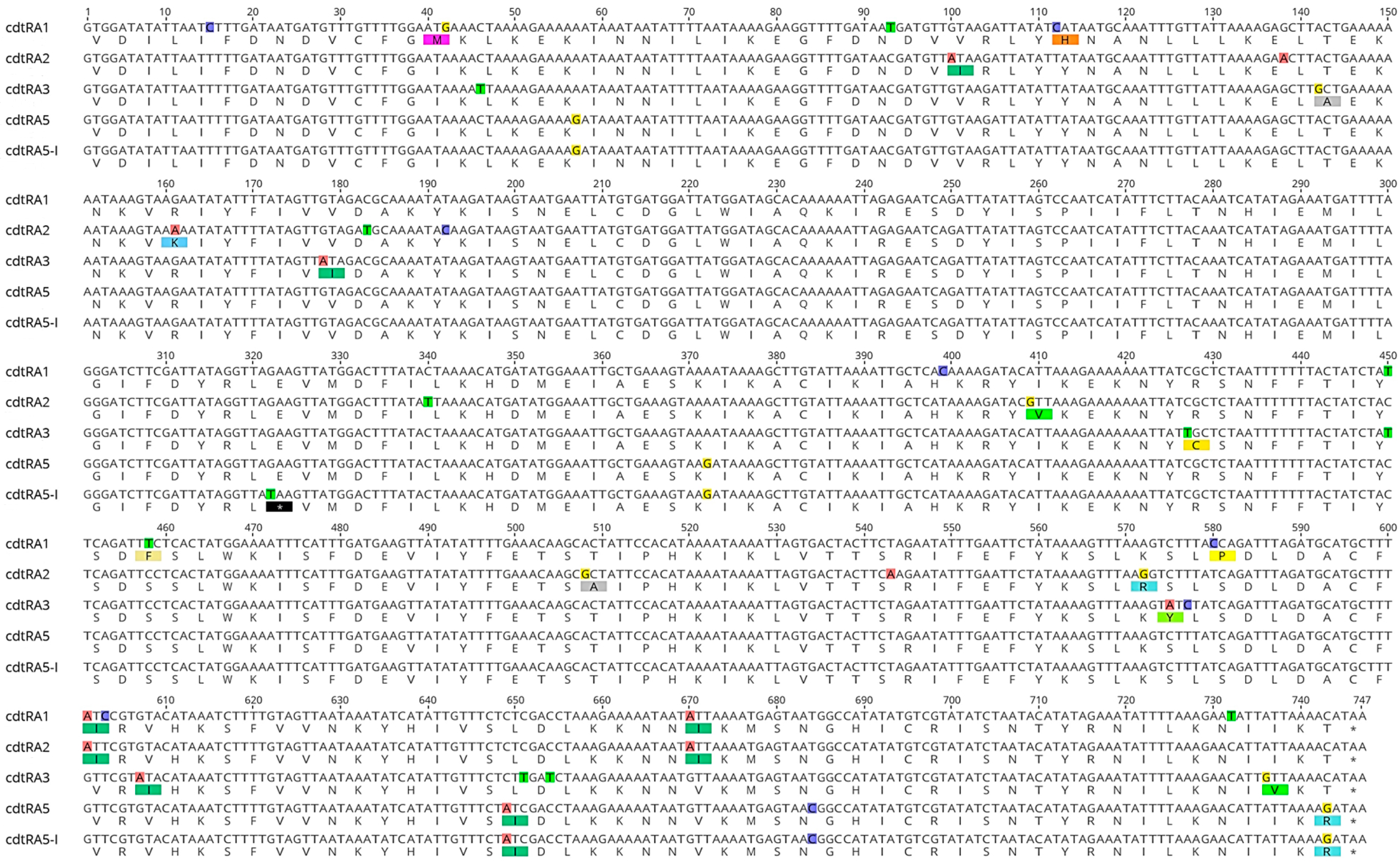
3.2. cdtR Sequences Alignment and Analysis

Overall, five different alleles (arbitrarily denominated cdtRA1, cdtRA2, cdtRA3, cdtRA5, and cdtRA5-I) were identified after the multiple alignments of the 550 cdtR sequences included in this study (Table 2, Figure 1 and Figure 2). Interestingly, a unique cdtR allele was associated to the RTs grouped in the same clade, except for the RTs of C5 that were associated to two different cdtR alleles. In particular, cdtRA1 was found in the RTs belonging to C1, cdtRA2 in the RTs belonging to C2, cdtRA3 in the RTs belonging to C3, and both cdtRA5 and cdtRA5-I were found in the RTs belonging to C5; the first allele in the RT 033, RT 045, RT 127, and RT 288, and the second allele in the RT 078, RT 193, and RT 620. Interestingly, strains RT 126 that belonged to C5 showed the cdtRA5 (12/100) or the cdtRA5-I (88/100) (Table 2).
The GC content slightly varied among the five alleles: 22.8% for cdtRA1, 22.4% for cdtRA2, 22.1% for cdtRA3, 23% for cdtRA5, and 22.9% for allele cdtRA5-I.
A total of 38 SNPs were identified after the multiple alignments of the cdtR sequences (Figure 2). A unique SNPs distribution was associated to each of the five cdtR alleles identified in the study (Figure 2). The analysis of the five CdtR-deduced amino acid sequences indicated that the 38 SNPs determined 17 different silent synonymous mutations with no amino acid substitutions, one SNP in a non-sense mutation (stop codon), and 19 in amino acid substitutions, of which 11 were non-synonymous and conservative and eight were non-synonymous and non-conservative (2/8 in the CdtRA1, and 1/8 in the CdtRA2, 3/8 in the CdtRA3, 1/8 in the CdtRA5, and 1/8 in the CdtRA5-I) (Figure 2).

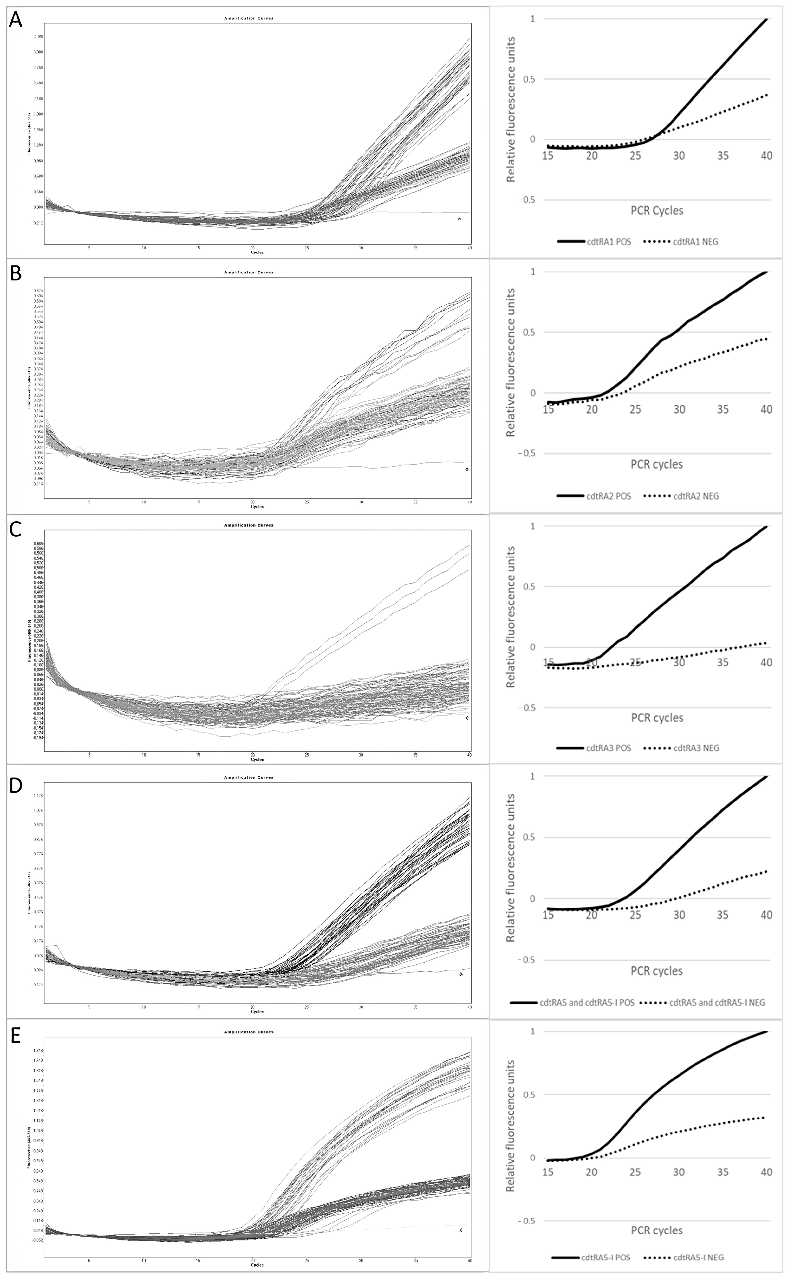
3.3. Optimization of the MB-Based Real-Time PCR Assays

Among the 38 SNPs identified, six specific SNPs were chosen as targets for the MB-based real-time PCR assays (Figure 1 and Table 3).
A single SNP was chosen as the target for cdtRA1, cdtRA2, and cdtRA5-I, and two SNPs were chosen as a target for cdtRA3 (Figure 1 and Table 3). The cdtRA5 and the cdtRA5-I differed for the SNP 322T, chosen as a target for the cdtRA5-I. For this reason, the SNP 372G that is common between cdtRA5 and cdtRA5-I but is absent in the other cdtR alleles was considered as a target for a second MB-based real-time assay to discriminate strains with a cdtRA5 allele, that resulted negative for the SNP 322T (Table 3). Target SNPs were chosen to be in the middle of the amplicon, while primer pairs were designed in the flanking conserved regions around the SNPs (Figure 1). In total, five sets of primer pairs, five MB probes, and five separate MB-based real-time PCR assays (denominated according to the considered cdtR allele detected) were developed to identify the different target SNPs and to work in monoplex (Table 3).
The thermocycling conditions and the concentrations of reagents for the five real-time PCR assays were optimized by testing 88 of the selected 142 strains from the ISS collection. In particular, 37 strains of clade C1 were analyzed (three to RT 001, four RT 012, six RT 014, seven RT 018, six RT 020, five RT 106, and six RT 607), 12 strains of C2 (one RT 019, eight RT 027, one RT 153, and two RT 181), 3 strains of C3 (two RT 023 and one RT 212), and 36 strains of C5 (seven RT 033, two RT 045, sixteen RT 078, ten RT 126, and one RT 620) (Table 2 and Table 4). The PCR mix for one sample consisted of 10 μL of LightCycler 480 Probes Master 2x concentrated, 5 μL of primer–probe mix (1.5 μL of each primer, 0.75 μM and 0.3 μL of probe 0.15 μM), 1.7 μL of PCR grade water, and 5 μL of DNA concentrated at 10 ng/μL.
The data, obtained from real-time PCR assays and reported as outputs from LightCycler 480 in raw fluorescence intensity, were normalized in the respective relative fluorescence units (RFU) to evaluate the capability of each probe to detect the target allele (Table 5 and Figure 3).
The raw fluorescence intensity observed in each MB-based real-time PCR assay allowed us to discriminate between the strains that were positive and negative for the targeted cdtR allele (Table 5). Considering the signals obtained, the 40th PCR cycle was considered as the cycle of reference to discriminate between the strains that were positive and negative for each cdtR allele. As far as the data obtained from normalization in RFU, the normalized mean signal yielded by negative strains in each real-time PCR assay was lower than half of the normalized mean signal yielded by positive strains (Figure 3 and Table 5).

4. Discussion

The heterogeneous structure of the C. difficile population reflects the genomic complexity of this pathogen [3,17,31,33,55]. C. difficile genomic evolution is associated to the acquisition of virulence and antibiotic resistance determinants [3,17,33,51,57,58,59], with an ongoing worldwide emergence of new highly virulent strains and types [4,8,58], which highlights the pressing need for a clinical and epidemiological surveillance of this bacterium.
Molecular methods represent a major advance in differentiating C. difficile strains and in establishing their evolutionary relationships, as well as in tracking outbreaks and the emergence of new, highly virulent C. difficile types [3,11,60,61,62].
In the last decade, CDI cases have increased worldwide, particularly in the community [4,5,13,14,15]. Whole genome sequencing (WGS) has shown that C. difficile strains from humans, animals, food, and the environment may be genetically closely related or indistinguishable, and that certain types of C. difficile are potentially transmitted between animals and humans [3,4,9,17,23,24,25,28,29,31,33,57,60,63]. For these reasons, genetic analysis has been increasingly associated with a One Health approach to CDI for a better control and prevention of this infection [4,17,33,60,61].
Although WGS represents the ideal approach to improve pathogen surveillance and provides precise data for a fast identification of outbreaks, this methodology still presents problems for its applicability due to elevated costs, execution time, and personnel expertise required [3,44,61,64]. Real-time PCR is an established, semi-automated, fast method used to detect and monitor pathogenic bacteria and viruses for both diagnostics and surveillance purposes [37,65].
Technologies that combine the real-time PCR with the use of fluorescent probes have been demonstrated to be a useful and rapid tool to detect SNPs in a specific target [36,37,38,43,45,46,52,62]. In particular, the MB probes show a better specificity and superiority in detecting SNPs than linear probes, like TaqMan, and have a less complex design compared to Scorpion probes [36,37,38,39,44,46,52,54].
The real-time PCR technique has rarely been reported for C. difficile analysis and the literature is limited to the applicability of this method in CDI diagnosis [66,67,68,69,70]. In this study, for the first time, the MB-based real-time PCR method was used for a rapid and easy genotyping of human, animal, and food/environmental C. difficile strains belonging to different RTs and evolutionary clades, using the cdtR gene as target.
The variability in the sequence of cdtR gene, previously observed by other authors [48,51], was confirmed in this study with the identification and characterization of five different cdtR alleles. Furthermore, previous investigations on a possible association between different cdtR sequences and different evolutionary clades, undertaken by Janezic et al. [51], were expanded in this study, with the association of the cdtR alleles identified to specific RTs and clades. Interestingly, the strains belonging to the same clade showed the same cdtR sequence, although they belonged to different RTs and they were isolated in different years and from different sources (humans, animals, and food/environment). This observation suggests that the nucleotide sequence of the different cdtR alleles is conserved over time, in line with previous studies demonstrating the conserved structure of the CdtLoc [51,71].
Interestingly, the analysis of the amino acid sequences deduced from each cdtR allele showed variations that might affect the structure/function of the CdtR protein. In fact, the amino acid sequence of the CdtRA3 presents three non-synonym and non-conservative missense substitutions, Thr48Ala, Cys143Arg, and Ser192Tyr, located in the N-terminal receiver domain, in the region that connects the N-terminal to the C-terminal, and in the C-terminal DNA binding domain, respectively, all regions are important for the correct functionality of CdtR [71,72]. In addition, the sequences of the CdtRA5-I show the already described nonsense substitution Glu108Stop, resulting in the production of a truncated CdtR protein (107 amino acids instead of 248 amino acids) [28,48,49,71]. In the present study, the CdtRA5-I was found in all of the strains RT 078 that have been demonstrated to produce the CDT, despite the non-functional CdtR [72]. The CdtRA5 shows an amino acid sequence identical to CdtRA5-I, but it does not present a stop codon, resulting in a complete protein, with a non-synonymous and non-conservative substitution in the DNA binding domain (Thr248Arg). Noteworthy, the deduced sequence of CdtRA2 shows one non-synonymous non-conserved substitution (Thr170Ala) in the C-terminal DNA binding domain, and six non-synonymous conserved substitution, two of which were also found in CdtRA1, potentially affecting the CdtR function in both RT 027 and RTs of C1.
In general, the results obtained indicated that the cdtR gene is a stable target for C. difficile genotyping. The analysis of the cdtR sequences included in this study indicate that the nucleotide sequence of each cdtR allele is conserved over time, in line with previous studies demonstrating the conserved structure of the entire CdtLoc [51,71]. In fact, strains belonging to the same clade showed the same cdtR allele, although they belonged to different RTs, and they were isolated in different years, from different sources (humans, animals, and food/environment), and in different countries. The only exception was represented by strains RT 126, in which the cdtRA5-I was prevalently found (88%), while the cdtRA5 was observed only in 12 Australian strains [28].
Interestingly, 147 of the genomes included in this study were from clonal strains RT 078, isolated by Knetsch et al. from humans, animals, food, and environment [26]. The data obtained by the authors support a C. difficile transmission between Dutch farmers and pigs, with several possible direct and indirect routes of transmission, including a common environmental source. The other 155 genomes included in the present study were from strains belonging to C5 (65 from strains RT 126, 43 from strains RT 127, 26 from strains RT 033, 19 from strains RT 078, and 2 from strains RT 288) that Knight et al. demonstrated to be involved in intra- and inter-species long-range transmission across geographical boundaries [28].
The design and the development of the MB-based real-time assays were adapted to the intrinsic structure of the cdtR gene. In fact, due to the low GC content, an accurate selection of the nucleotide regions to design primers and probes was necessary, together with a correct balance between the length and the melting temperature of each oligo. PCR mixes and thermocycling settings were also precisely optimized in order to develop monoplexed assays that could work at the same conditions. The design of the MB probe for each cdtR allele was adapted to the SNPs distribution. In particular, the MB probe designed for the cdtRA1 allele worked well, although the targeted SNP was not optimally centered in the loop of the probe, as suggested by some authors [38,52,53]. Moreover, the MB probe designed for the cdtRA3 allele, targeting two different SNPs, yielded a higher signal for the positive samples compared to the other probes used in this study, indicating that a MB probe detecting more than one SNP may show very high specificity.
Considering the results obtained in RFU, the discriminating capability of the different MB probes designed was similar, with only slight variations. All of the five MB-based real-time PCR assays developed in this study were able to successfully detect the five different cdtR alleles identified, as confirmed by the cdtR sequencing performed on the selected C. difficile strains that were tested during the optimization of the assays.
The main limitation of the present study is represented by the impossibility to detect both strains/RTs belonging to the clade C4 and non-toxigenic strains, for the absence of the cdtR gene in their genomes. For this reason, our further research will be focused on sequence analysis and a comparison of strains belonging to C4 to find conserved genes and identify a potential target for a specific MB-based real-time PCR assay. Other future developments are represented by the possibility to design a multiplexed MB-based real-time assay, using different probes labeled with different fluorophores, and/or use crude DNAs as a template in order to reduce reagents and the costs of analysis.
In conclusion, in this study the MB-based real-time PCR method has been used to genotype C. difficile for the first time. The five monoplex MB-based real-time PCR assays, developed using the cdtR gene as a target, succeeded in identifying the different main evolutionary clades C1, C2, C3, and C5, and the RTs grouped in each of these clades. These MB-based real-time PCR assays represent a powerful, original, and versatile tool to detect, differentiate, and track the main C. difficile RTs/clades involved in human and animal CDI. The proposed assays may be used in epidemiological and phylogenetic studies, offering the possibility to explore the geographic distribution and the evolutionary dynamics of important C. difficile RTs/clades at a local and national/international level. Considering that the One Health approach has been assuming a crucial role in CDI surveillance, the proposed MB-based real-time PCR method may be also employed to evaluate the epidemiology of C. difficile across different species and environments, to elucidate the possible routes of transmission and potential sources of infection. Future improvements and the resolution of the current limitations will allow an expansion of the potential applications of the proposed method, including CDI diagnosis and control.

Supplementary Materials

The following supporting information can be downloaded at: https://www.mdpi.com/article/10.3390/microbiolres15010024/s1; Table S1: C. difficile genomes and strains included in the study.

Author Contributions

Conceptualization, E.M.C. and P.S.; methodology, E.M.C. and F.B.; software, E.M.C. and F.B.; validation, E.M.C.; formal analysis, E.M.C. and F.B.; investigation, E.M.C. and F.B.; data curation, E.M.C. and F.B.; writing—original draft preparation, E.M.C. and P.S.; writing—review and editing, P.S.; supervision, P.S. All authors have read and agreed to the published version of the manuscript.

Funding

This research received no external funding.

Institutional Review Board Statement

Not applicable.

Informed Consent Statement

Not applicable.

Data Availability Statement

Data are contained within the article or Supplementary Material.

Conflicts of Interest

The authors declare no conflicts of interest.

References

  1. Kuijper, E.J.; Coignard, B.; Tüll, P.; Poxton, I.; Brazier, J.; Duerden, B.; Delmée, M.; Mastrantonio, P.; Gastmeier, P.; Barbut, F.; et al. Emergence of Clostridium difficile-Associated Disease in North America and Europe. Clin. Microbiol. Infect. 2006, 12, 2–18. [Google Scholar] [CrossRef]
  2. Loo, V.G.; Poirier, L.; Miller, M.A.; Oughton, M.; Libman, M.D.; Michaud, S.; Bourgault, A.-M.; Nguyen, T.; Frenette, C.; Kelly, M.; et al. A Predominantly Clonal Multi-Institutional Outbreak of Clostridium difficile–Associated Diarrhea with High Morbidity and Mortality. N. Engl. J. Med. 2005, 353, 2442–2449. [Google Scholar] [CrossRef]
  3. Elliott, B.; Androga, G.O.; Knight, D.R.; Riley, T.V. Clostridium difficile Infection: Evolution, Phylogeny and Molecular Epidemiology. Infect. Genet. Evol. 2017, 49, 1–11. [Google Scholar] [CrossRef]
  4. Lim, S.C.; Knight, D.R.; Riley, T.V. Clostridium difficile and One Health. Clin. Microbiol. Infect. 2020, 26, 857–863. [Google Scholar] [CrossRef]
  5. Hensgens, M.P.M.; Keessen, E.C.; Squire, M.M.; Riley, T.V.; Koene, M.G.J.; De Boer, E.; Lipman, L.J.A.; Kuijper, E.J. Clostridium difficile Infection in the Community: A Zoonotic Disease? Clin. Microbiol. Infect. 2012, 18, 635–645. [Google Scholar] [CrossRef]
  6. Fatima, R.; Aziz, M. The Hypervirulent Strain of Clostridium difficile: NAP1/B1/027—A Brief Overview. Cureus 2019, 11, e3977. [Google Scholar] [CrossRef]
  7. He, M.; Miyajima, F.; Roberts, P.; Ellison, L.; Pickard, D.J.; Martin, M.J.; Connor, T.R.; Harris, S.R.; Fairley, D.; Bamford, K.B.; et al. Emergence and Global Spread of Epidemic Healthcare-Associated Clostridium difficile. Nat. Genet. 2013, 45, 109–113. [Google Scholar] [CrossRef]
  8. Spigaglia, P.; Mastrantonio, P.; Barbanti, F. Antibiotic Resistances of Clostridium difficile. Adv. Exp. Med. Biol. 2018, 1050, 137–159. [Google Scholar] [CrossRef]
  9. Keessen, E.C.; Gaastra, W.; Lipman, L.J.A. Clostridium difficile Infection in Humans and Animals, Differences and Similarities. Vet. Microbiol. 2011, 153, 205–217. [Google Scholar] [CrossRef]
  10. Loo, V.G.; Bourgault, A.-M.; Poirier, L.; Lamothe, F.; Michaud, S.; Turgeon, N.; Toye, B.; Beaudoin, A.; Frost, E.H.; Gilca, R.; et al. Host and Pathogen Factors for Clostridium difficile Infection and Colonization. N. Engl. J. Med. 2011, 365, 1693–1703. [Google Scholar] [CrossRef]
  11. Liu, C.; Monaghan, T.; Yadegar, A.; Louie, T.; Kao, D. Insights into the Evolving Epidemiology of Clostridioides difficile Infection and Treatment: A Global Perspective. Antibiotics 2023, 12, 1141. [Google Scholar] [CrossRef]
  12. Shaw, H.A.; Preston, M.D.; Vendrik, K.E.W.; Cairns, M.D.; Browne, H.P.; Stabler, R.A.; Crobach, M.J.T.; Corver, J.; Pituch, H.; Ingebretsen, A.; et al. The Recent Emergence of a Highly Related Virulent Clostridium difficile Clade with Unique Characteristics. Clin. Microbiol. Infect. 2020, 26, 492–498. [Google Scholar] [CrossRef]
  13. De Roo, A.C.; Regenbogen, S.E.; De Roo, A.C.; Regenbogen, S.E. Clostridium difficile Infection: An Epidemiology Update. Clin. Colon Rectal Surg. 2020, 33, 49–57. [Google Scholar] [CrossRef]
  14. Goorhuis, A.; Bakker, D.; Corver, J.; Debast, S.B.; Harmanus, C.; Notermans, D.W.; Bergwerff, A.A.; Dekker, F.W.; Kuijper, E.J. Emergence of Clostridium difficile Infection Due to a New Hypervirulent Strain, Polymerase Chain Reaction Ribotype 078. Clin. Infect. Dis. 2008, 47, 1162–1170. [Google Scholar] [CrossRef]
  15. Kachrimanidou, M.; Tzika, E.; Filioussis, G. Clostridioides (Clostridium) difficile in Food-Producing Animals, Horses and Household Pets: A Comprehensive Review. Microorganisms 2019, 7, 667. [Google Scholar] [CrossRef]
  16. Weese, J.S. Clostridium (Clostridioides) difficile in Animals. J. Vet. Diagn. Investig. 2020, 32, 213–221. [Google Scholar] [CrossRef]
  17. Mitchell, M.; Nguyen, S.V.; MacOri, G.; Bolton, D.; McMullan, G.; Drudy, D.; Fanning, S. Clostridioides difficile as a Potential Pathogen of Importance to One Health: A Review. Foodborne Pathog. Dis. 2022, 19, 806–816. [Google Scholar] [CrossRef]
  18. Spigaglia, P.; Barbanti, F.; Faccini, S.; Vescovi, M.; Criscuolo, E.M.; Ceruti, R.; Gaspano, C.; Rosignoli, C. Clostridioides difficile in Pigs and Dairy Cattle in Northern Italy: Prevalence, Characterization and Comparison between Animal and Human Strains. Microorganisms 2023, 11, 1738. [Google Scholar] [CrossRef]
  19. Hain-Saunders, N.M.R.; Knight, D.R.; Bruce, M.; Riley, T.V. Clostridioides difficile Infection and One Health: An Equine Perspective. Environ. Microbiol. 2022, 24, 985–997. [Google Scholar] [CrossRef]
  20. Uzal, F.A.; Navarro, M.A.; Asin, J.; Boix, O.; Ballarà-Rodriguez, I.; Gibert, X. Clostridial Diarrheas in Piglets: A Review. Vet. Microbiol. 2023, 280, 109691. [Google Scholar] [CrossRef]
  21. Songer, J.G.; Uzal, F.A. Clostridial Enteric Infections in Pigs. J. Vet. Diagn. Investig. 2005, 17, 528–536. [Google Scholar] [CrossRef]
  22. Goyal, M.; Hauben, L.; Pouseele, H.; Jaillard, M.; De Bruyne, K.; van Belkum, A.; Goering, R. Retrospective Definition of Clostridioides difficile PCR Ribotypes on the Basis of Whole Genome Polymorphisms: A Proof of Principle Study. Diagnostics 2020, 10, 1078. [Google Scholar] [CrossRef]
  23. Blau, K.; Berger, F.K.; Mellmann, A.; Gallert, C. Clostridioides difficile from Fecally Contaminated Environmental Sources: Resistance and Genetic Relatedness from a Molecular Epidemiological Perspective. Microorganisms 2023, 11, 2497. [Google Scholar] [CrossRef]
  24. Knight, D.R.; Riley, T.V. Genomic Delineation of Zoonotic Origins of Clostridium difficile. Front. Public Health 2019, 7, 164. [Google Scholar] [CrossRef]
  25. Knight, D.R.; Squire, M.M.; Collins, D.A.; Riley, T.V. Genome Analysis of Clostridium difficile PCR Ribotype 014 Lineage in Australian Pigs and Humans Reveals a Diverse Genetic Repertoire and Signatures of Long-Range Interspecies Transmission. Front. Microbiol. 2017, 7, 2138. [Google Scholar] [CrossRef]
  26. Knetsch, C.W.; Connor, T.R.; Mutreja, A.; van Dorp, S.M.; Sanders, I.M.; Browne, H.P.; Harris, D.; Lipman, L.; Keessen, E.C.; Corver, J.; et al. Whole Genome Sequencing Reveals Potential Spread of Clostridium difficile between Humans and Farm Animals in the Netherlands, 2002 to 2011. Eurosurveillance 2014, 19, 20954. [Google Scholar] [CrossRef] [PubMed]
  27. Tsai, C.S.; Hung, Y.P.; Lee, J.C.; Syue, L.S.; Hsueh, P.R.; Ko, W.C. Clostridioides difficile Infection: An Emerging Zoonosis? Expert Rev. Anti. Infect. Ther. 2021, 19, 1543–1552. [Google Scholar] [CrossRef]
  28. Knight, D.R.; Kullin, B.; Androga, G.O.; Barbut, F.; Eckert, C.; Johnson, S.; Spigaglia, P.; Tateda, K.; Tsai, P.J.; Riley, T.V. Evolutionary and Genomic Insights into Clostridioides difficile Sequence Type 11: A Diverse Zoonotic and Antimicrobial-Resistant Lineage of Global One Health Importance. mBio 2019, 10, 1–17. [Google Scholar] [CrossRef]
  29. Kurka, H.; Ehrenreich, A.; Ludwig, W.; Monot, M.; Rupnik, M.; Barbut, F.; Indra, A.; Dupuy, B.; Liebl, W. Sequence Similarity of Clostridium difficile Strains by Analysis of Conserved Genes and Genome Content Is Reflected by Their Ribotype Affiliation. PLoS ONE 2014, 9, e86535. [Google Scholar] [CrossRef]
  30. Knetsch, C.W.; Terveer, E.M.; Lauber, C.; Gorbalenya, A.E.; Harmanus, C.; Kuijper, E.J.; Corver, J.; van Leeuwen, H.C. Comparative Analysis of an Expanded Clostridium difficile Reference Strain Collection Reveals Genetic Diversity and Evolution through Six Lineages. Infect. Genet. Evol. 2012, 12, 1577–1585. [Google Scholar] [CrossRef] [PubMed]
  31. Knight, D.R.; Imwattana, K.; Kullin, B.; Guerrero-Araya, E.; Paredes-Sabja, D.; Didelot, X.; Dingle, K.E.; Eyre, D.W.; Rodríguez, C.; Riley, T.V. Major Genetic Discontinuity and Novel Toxigenic Species in Clostridioides difficile Taxonomy. eLife 2021, 10, e64325. [Google Scholar] [CrossRef]
  32. Imwattana, K.; Knight, D.R.; Kullin, B.; Collins, D.A.; Putsathit, P.; Kiratisin, P.; Riley, T.V. Clostridium difficile Ribotype 017–Characterization, Evolution and Epidemiology of the Dominant Strain in Asia. Emerging Microbes and Infections. Emerg. Microbes Infect. 2019, 8, 796–807. [Google Scholar] [CrossRef]
  33. Knight, D.R.; Elliott, B.; Chang, B.J.; Perkins, T.T.; Riley, T.V. Diversity and Evolution in the Genome of Clostridium difficile. Clin. Microbiol. Rev. 2015, 28, 721–741. [Google Scholar] [CrossRef]
  34. Knetsch, C.W.; Lawley, T.D.; Hensgens, M.P.; Corver, J.; Wilcox, M.W.; Kuijper, E.J. Current Application and Future Perspectives of Molecular Typing Methods to Study Clostridium difficile Infections. Eurosurveillance 2013, 18, 20381. [Google Scholar] [CrossRef]
  35. Rodríguez, A.; Rodríguez, M.; Córdoba, J.J.; Andrade, M.J. Design of Primers and Probes for Quantitative Real-Time PCR Methods. Methods Mol. Biol. 2015, 1275, 31–56. [Google Scholar] [CrossRef] [PubMed]
  36. Prajapati, G.K.; Kumar, A.; Wany, A.; Pandey, D.M. Molecular Beacon Probe (MBP)-Based Real-Time PCR. Methods Mol. Biol. 2023, 2638, 273–287. [Google Scholar] [CrossRef]
  37. Arya, M.; Shergill, I.S.; Williamson, M.; Gommersall, L.; Arya, N.; Patel, H.R.H. Basic Principles of Real-Time Quantitative PCR. Expert Rev. Mol. Diagn. 2005, 5, 209–219. [Google Scholar] [CrossRef]
  38. Edwards, K.J.; Logan, J.M.J. Mutation Detection by Real-Time PCR. In Real-Time PCR: Current Technology and Application; Caister Academic Press: Poole, UK, 2009; pp. 185–210. [Google Scholar]
  39. Wang, C.; Yang, C.J. Application of Molecular Beacons in Real-Time PCR. In Molecular Beacons; Nature Publishing Group: London, UK, 2014; pp. 45–59. [Google Scholar] [CrossRef]
  40. Lager, M.; Mernelius, S.; Löfgren, S.; Söderman, J. Real-Time PCR Typing of Escherichia coli Based on Multiple Single Nucleotide Polymorphisms-a Convenient and Rapid Method. Clin. Lab. 2016, 62, 349–355. [Google Scholar] [CrossRef]
  41. Greig, D.R.; Hickey, T.J.; Boxall, M.D.; Begum, H.; Gentle, A.; Jenkins, C.; Chattaway, M.A. A Real-Time Multiplex PCR for the Identification and Typing of Vibrio cholerae. Diagn. Microbiol. Infect. Dis. 2018, 90, 171–176. [Google Scholar] [CrossRef]
  42. Birdsell, D.N.; Vogler, A.J.; Buchhagen, J.; Clare, A.; Kaufman, E.; Naumann, A.; Driebe, E.; Wagner, D.M.; Keim, P.S. TaqMan Real-Time PCR Assays for Single-Nucleotide Polymorphisms Which Identify Francisella tularensis and Its Subspecies and Subpopulations. PLoS ONE 2014, 9, 223. [Google Scholar] [CrossRef]
  43. Ben Shabat, M.; Mikula, I.; Gerchman, I.; Lysnyansky, I. Development and Evaluation of a Novel Single-Nucleotide-Polymorphism Real-Time PCR Assay for Rapid Detection of Fluoroquinolone-Resistant Mycoplasma bovis. J. Clin. Microbiol. 2010, 48, 2909–2915. [Google Scholar] [CrossRef]
  44. Dikdan, R.J.; Marras, S.A.E.; Field, A.P.; Brownlee, A.; Cironi, A.; Hill, D.A.; Tyagi, S. Multiplex PCR Assays for Identifying All Major Severe Acute Respiratory Syndrome Coronavirus 2 Variants. J. Mol. Diagn. 2022, 24, 309–319. [Google Scholar] [CrossRef]
  45. Täpp, I.; Malmberg, L.; Rennel, E.; Wik, M.; Syvänen, A.C. Homogeneous Scoring of Single-Nucleotide Polymorphisms: Comparison of the 5′-Nuclease TaqMan® Assay and Molecular Beacon Probes. Biotechniques 2000, 28, 732–738. [Google Scholar] [CrossRef]
  46. Wattiau, P.; Fretin, D. Real-Time PCR Typing of Single Nucleotide Polymorphism in DNA Containing Inverted Repeats. Biotechniques 2006, 41, 544–546. [Google Scholar] [CrossRef]
  47. Carter, G.P.; Lyras, D.; Allen, D.L.; Mackin, K.E.; Howarth, P.M.; O’Connor, J.R.; Rood, J.I. Binary Toxin Production in Clostridium difficile Is Regulated by CdtR, a LytTR Family Response Regulator. J. Bacteriol. 2007, 189, 7290–7301. [Google Scholar] [CrossRef] [PubMed]
  48. Bouvet, P.J.M.; Popoff, M.R. Genetic Relatedness of Clostridium difficile Isolates from Various Origins Determined by Triple-Locus Sequence Analysis Based on Toxin Regulatory Genes tcdC, tcdR, and cdtR. J. Clin. Microbiol. 2008, 46, 3703–3713. [Google Scholar] [CrossRef]
  49. Lyon, S.A.; Hutton, M.L.; Rood, J.I.; Cheung, J.K.; Lyras, D. CdtR Regulates TcdA and TcdB Production in Clostridium difficile. PLoS Pathog. 2016, 12, e1005758. [Google Scholar] [CrossRef]
  50. Monot, M.; Eckert, C.; Lemire, A.; Hamiot, A.; Dubois, T.; Tessier, C.; Dumoulard, B.; Hamel, B.; Petit, A.; Lalande, V.; et al. Clostridium difficile: New Insights into the Evolution of the Pathogenicity Locus. Sci. Rep. 2015, 5, 15023. [Google Scholar] [CrossRef]
  51. Janezic, S.; Dingle, K.; Alvin, J.; Accetto, T.; Didelot, X.; Crook, D.W.; Borden Lacy, D.; Rupnik, M. Comparative Genomics of Clostridioides difficile Toxinotypes Identifies Module-Based Toxin Gene Evolution. Microb. Genom. 2020, 6, mgen000449. [Google Scholar] [CrossRef]
  52. Marras, S.A.E.; Kramer, F.R.; Tyagi, S. Genotyping SNPs with Molecular Beacons. Methods Mol. Biol. 2003, 212, 111–128. [Google Scholar]
  53. Vet, J.A.M.; Marras, S.A.E. Design and Optimization of Molecular Beacon Real-Time Polymerase Chain Reaction Assays. Methods Mol. Biol. 2005, 5, 15023. [Google Scholar] [CrossRef]
  54. Kolpashchikov, D.M. An Elegant Biosensor Molecular Beacon Probe: Challenges and Recent Solutions. Scientifica 2012, 2012, 928783. [Google Scholar] [CrossRef]
  55. Sebaihia, M.; Wren, B.W.; Mullany, P.; Fairweather, N.F.; Minton, N.; Stabler, R.; Thomson, N.R.; Roberts, A.P.; Cerdeño-Tárraga, A.M.; Wang, H.; et al. The Multidrug-Resistant Human Pathogen Clostridium difficile Has a Highly Mobile, Mosaic Genome. Nat. Genet. 2006, 38, 779–786. [Google Scholar] [CrossRef]
  56. Marras, S.A.E. Selection of Fluorophore and Quencher Pairs for Fluorescent Nucleic Acid Hybridization Probes. Methods Mol. Biol. 2006, 335, 3–16. [Google Scholar] [CrossRef] [PubMed]
  57. Knetsch, C.W.; Kumar, N.; Forster, S.C.; Connor, T.R.; Browne, H.P.; Harmanus, C.; Sanders, I.M.; Harris, S.R.; Turner, L.; Morris, T.; et al. Zoonotic Transfer of Clostridium difficile Harboring Antimicrobial Resistance between Farm Animals and Humans. J. Clin. Microbiol. 2018, 56, e01384-17. [Google Scholar] [CrossRef] [PubMed]
  58. Imwattana, K.; Rodríguez, C.; Riley, T.V.; Knight, D.R. A Species-Wide Genetic Atlas of Antimicrobial Resistance in Clostridioides difficile. Microb. Genom. 2021, 7, 000696. [Google Scholar] [CrossRef]
  59. Dingle, K.E.; Elliott, B.; Robinson, E.; Griffiths, D.; Eyre, D.W.; Stoesser, N.; Vaughan, A.; Golubchik, T.; Fawley, W.N.; Wilcox, M.H.; et al. Evolutionary History of the Clostridium difficile Pathogenicity Locus. Genome Biol. Evol. 2014, 6, 36–52. [Google Scholar] [CrossRef]
  60. Markovska, R.; Dimitrov, G.; Gergova, R.; Boyanova, L. Clostridioides difficile, a New “Superbug”. Microorganisms 2023, 11, 845. [Google Scholar] [CrossRef] [PubMed]
  61. Abad-Fau, A.; Sevilla, E.; Martín-Burriel, I.; Moreno, B.; Bolea, R. Update on Commonly Used Molecular Typing Methods for Clostridioides difficile. Microorganisms 2023, 11, 1752. [Google Scholar] [CrossRef]
  62. Tümmler, B. Molecular Epidemiology in Current Times. Environ. Microbiol. 2020, 22, 4909–4918. [Google Scholar] [CrossRef]
  63. Knight, D.R.; Squire, M.M.; Riley, T.V. Nationwide Surveillance Study of Clostridium difficile in Australian Neonatal Pigs Shows High Prevalence and Heterogeneity of PCR Ribotypes. Appl Environ. Microbiol. 2015, 81, 119–123. [Google Scholar] [CrossRef] [PubMed]
  64. Chrysostomou, A.C.; Aristokleous, A.; Rodosthenous, J.H.; Christodoulou, C.; Stathi, G.; Kostrikis, L.G. Detection of Circulating SARS-CoV-2 Variants of Concern (VOCs) Using a Multiallelic Spectral Genotyping Assay. Life 2023, 13, 304. [Google Scholar] [CrossRef] [PubMed]
  65. Artika, I.M.; Dewi, Y.P.; Nainggolan, I.M.; Siregar, J.E.; Antonjaya, U. Real-Time Polymerase Chain Reaction: Current Techniques, Applications, and Role in COVID-19 Diagnosis. Genes 2022, 13, 2387. [Google Scholar] [CrossRef] [PubMed]
  66. Jia, X.; Wang, Y.; Zhang, W.; Li, W.; Bai, L.; Lu, J.; Ma, C.; Wu, Y. A Rapid Multiplex Real-Time PCR Detection of Toxigenic Clostridioides difficile Directly from Fecal Samples. 3 Biotech 2023, 13, 54. [Google Scholar] [CrossRef] [PubMed]
  67. Kilic, A.; Alam, M.J.; Tisdel, N.L.; Shah, D.N.; Yapar, M.; Lasco, T.M.; Garey, K.W. Multiplex Real-Time PCR Method for Simultaneous Identification and Toxigenic Type Characterization of Clostridium difficile from Stool Samples. Ann. Lab. Med. 2015, 35, 306–313. [Google Scholar] [CrossRef]
  68. Kouhsari, E.; Douraghi, M.; Barati, M.; Yaseri, H.F.; Talebi, M.; Abbasian, S.; Moqarabzadeh, V.; Amirmozafari, N. Rapid Simultaneous Molecular Stool-Based Detection of Toxigenic Clostridioides difficile by Quantitative TaqMan Real-Time PCR Assay. Clin. Lab. 2019, 65, 461–469. [Google Scholar] [CrossRef]
  69. Jayaratne, P.A.; Monkman, L.; Broukhanski, G.; Pillai, D.R.; Lee, C. Real-Time Polymerase Chain Reaction Method for Detection of Toxigenic Clostridium difficile from Stools and Presumptive Identification of NAP1 Clone. Diagn. Microbiol. Infect. Dis. 2013, 75, 121–123. [Google Scholar] [CrossRef]
  70. Bélanger, S.D.; Boissinot, M.; Clairoux, N.; Picard, F.J.; Bergeron, M.G. Rapid Detection of Clostridium difficile in Feces by Real-Time PCR. J. Clin. Microbiol. 2003, 41, 730–734. [Google Scholar] [CrossRef]
  71. Metcalf, D.S.; Scott Weese, J. Binary Toxin Locus Analysis in Clostridium difficile. J. Med. Microbiol. 2011, 60, 1137–1145. [Google Scholar] [CrossRef]
  72. Bilverstone, T.W.; Minton, N.P.; Kuehne, S.A. Phosphorylation and Functionality of CdtR in Clostridium difficile. Anaerobe 2019, 58, 103–109. [Google Scholar] [CrossRef]
Figure 1. Positions of the primers and probes designed. Forward primers are in dark green, reverse primers are in light green and probes’ loops in red (stems are not shown). The different deoxyribonucleotides in single nucleotide polymorphisms position are highlighted in different colors (Geneious R9.1 software).
Microbiolres 15 00024 g001
Figure 2. Multiple alignment of the cdtR nucleotidic and deduced amino acid sequences (Geneious R9.1 software). Different colors highlight the single nucleotide polymorphisms and the amino acid substitutions.
Microbiolres 15 00024 g002
Figure 3. Molecular Beacon based real-time PCR results. (A) cdtRA1 assay; (B) cdtRA2 assay; (C) cdtRA3 assay; (D) cdtRA5 and cdtRA5-I assay; (E) cdtRA5-I assay. On the left side: LightCycler 480 outputs. On the right side: normalized mean value in Relative Fluorescence Units. Signals of controls containing no DNA are labeled with a *.
Microbiolres 15 00024 g003
Table 1. Parameters for primer and Molecular Beacon probes design.
ParameterPCR ProductPrimersMB ProbesMB StemMB LoopReferences
GC
content
optimal 21%optimal 23%70–100% [35,37,38,44,52,53]
Tm 1 Optimal 48 °C.
Tm of primers should not differ > 2 °C
3–7 °C > Ta 2 PCR>Ta PCR>Tm stem[35,37,38,44,52,53]
Length100–200 bp.
Primers and probe distance: 20–50 bp
15–30 bp
(optimal 25 bp)
40 bp
(linear)
7 bp26 bp[35,37,38,44,52,53]
Poly-G/C Avoided [35,38,44,52,53,54]
Self-
dimers
AvoidedMBs have a secondary structure [35,38,44,52,53,54]
Primer-
dimers
AvoidedProbe or primer
dimers should be avoided
[35,38,44,52,53,54]
G at 5′-end Avoided [35,38,44,52,53,54]
1 Tm: melting temperature; 2 Ta: annealing temperature.
Table 2. Origin and characteristics of C. difficile strains included in the study.
CladecdtR
Allele
RT 1Toxin A 2Toxin B 2Binary
Toxin CDT 2
cdtR Gene Sequences fromTotal cdtR
Sequences (%)
GenomesISS Strains
HumanAnimalFood/EnvironmentUnknownHumanAnimal
C1cdtRA1001++2 215 (0.9)
002++1 1 (0.2)
012++1 517 (1.3)
014++1 8514 (2.5)
018++1 10 11 (2)
020++1 6512 (2.2)
056++1 1 (0.2)
087++1 1 (0.2)
106++1 6310 (1.8)
607++ 8 8 (1.5)
C2cdtRA2019+++ 11 2 (0.4)
027+++1412 8 25 (4.5)
036+++1 1 (0.2)
153+++ 11 2 (0.4)
176+++1 1 (0.2)
181+++ 2 2 (0.4)
C3cdtRA3023+++2 3 2 7 (1.3)
063+++1 1 (0.2)
212+++ 1 1 (0.2)
C5cdtRA5033+1955 4841 (7.5)
045+++2 215 (0.9)
127+++3310 144 (8)
288+11 2 (0.4)
126+++66 12 (2.2)
cdtRA5-I126+++663 12788 (16)
078+++165415 1417242 (44)
193+++ 1 1 (0.2)
620+++11 1 3 (0.5)
322681539349550
1 RT: PCR-ribotype. 2 +: strain positive for the considered toxin; −: strain negative for the considered toxin.
Table 3. Set of primers and probes used in the real-time PCR assays.
cdtR
Alleles
Primers and
Probes
SequenceNameSNPsAmplicon Size
cdtRA1Forward5′-GGGATCTTCGATTATAGGTTA-3′cdtR-F1399C176 bp
Reverse5′-GAAATTTTCCATAGTGAGGA-3′cdtR-R1
Probe 15′-FAM-CGCGATCAGCTTGTATTAAAATTGCTCACAAAAGATCGCG-BHQ1-3′cdtR-MB-1
cdtRA2Forward5′-GATGAAGTTATATATTTTGAAAC-3′cdtR-F2543A119 bp
Reverse5′-AAGCATGCATCTAAATCTG-3′cdtR-R2
Probe5′-FAM-CGCGATCAGTGACTACTTCAAGAATATTTGAATGATCGCG-BHQ1-3′cdtR-MB-2
cdtRA3Forward5′-CAGATTTAGATGCATGCTT-3′cdtR-F3651T and 654T136 bp
Reverse5′-CTATATGTATTAGATATACGAC-3′cdtR-R3
Probe5′-FAM-CGCGATCCATATTGTTTCTCTTGATCTAAAGAAGATCGCG-BHQ1-3′cdtR-MB-3
cdtRA5
and
cdtRA5-I
Forward5′-GGGATCTTCGATTATAGGTTA-3′cdtR-F1372G176 bp
Reverse5′-GAAATTTTCCATAGTGAGGA-3′cdtR-R1
Probe5′-FAM-CGCGATCTTGCTGAAAGTAAGATAAAAGCTTGTGATCGCG-BHQ1-3′cdtR-MB-5
cdtRA5-IForward5′-GAATCAGATTATATTAGTCCAAT-3′cdtR-F5-I322T153 bp
Reverse5′-AGCAATTTTAATACAAGCTTTTA-3′cdtR-R5-I
Probe5′-FAM-CGCGATCCGATTATAGGTTATAAGTTATGGACTGATCGCG-BHQ1-3′cdtR-MB-5-I
1 The sequence of each Molecular Beacon probe is reported with the stem sequences highlighted in yellow and the targeted SNPs highlighted in red.
Table 4. Thermocycling settings of the Molecular Beacon-based real-time PCR assay developed in the study.
Program CyclesAnalysis ModeTarget (°C)Acquisition ModeHold (hh:mm:ss)Ramp Rate (°C/s)Acquisition (per °C)
pre-incubation1None95None00:05:004.4
amplification40Quantification95None00:00:104.4
48Single00:00:151.55
72None00:00:014.4
cooling1None40None00:00:101.5
Table 5. Molecular Beacon-based real-time assays results at 40th PCR cycle.
cdtR
Allele Assay
Range of F 1 (40th Cycle)Mean F ± STD 2 (40th Cycle)RFU 3
PositiveNegativePositiveNegativePositiveNegative
cdtRA12.22–3.350.79–1.542.75 ± 0.351 ± 0.0510.3
cdtRA20.47–0.610.15–0.320.55 ± 0.050.24 ± 0.0510.4
cdtRA30.49–0.570–0.180.56 ± 0.040.02 ± 0.0510
cdtRA50.84–1.160.14–0.360.98 ± 0.090.22 ± 0.0810.25
cdtRA5-I1.39–1.790.11–0.551.59 ± 0.120.51 ± 0.1010.3
1 F: raw fluorescence intensity; 2 STD: Standard Deviation; 3 RFU: relative fluorescence units.
Disclaimer/Publisher’s Note: The statements, opinions and data contained in all publications are solely those of the individual author(s) and contributor(s) and not of MDPI and/or the editor(s). MDPI and/or the editor(s) disclaim responsibility for any injury to people or property resulting from any ideas, methods, instructions or products referred to in the content.

