Article

Fungal Endophytes of Moringa (Moringa oleifera L.), Neem (Azadirachta indica) and Lavender (Lavandula angustifolia) and Their Biological Control of Fusarium Wilt of Banana

by
Bonface M. Nthuku
1,2,
Esther W. Kahariri
3,
Johnson K. Kinyua
2 and
Evans N. Nyaboga
1,*
1
Department of Biochemistry, University of Nairobi, P.O. Box 30197, Nairobi 00100, Kenya
2
Department of Biochemistry, Jomo Kenyatta University of Agriculture and Technology, P.O. Box 62000, Nairobi 00200, Kenya
3
Department of Biological Sciences, Karatina University, P.O. Box 1957, Karatina 10101, Kenya
*
Author to whom correspondence should be addressed.
Microbiol. Res. 2023, 14(4), 2113-2132; https://doi.org/10.3390/microbiolres14040143
Submission received: 15 October 2023 / Revised: 9 December 2023 / Accepted: 11 December 2023 / Published: 15 December 2023

Abstract

:
Fusarium wilt, caused by Fusarium oxysporum f. sp. cubense (Foc), significantly affects the productivity of the banana crop in the field. Currently, there are no effective control measures available, and therefore, there is an urgent need to develop novel strategies to control the spread of the disease. Biological control is a promising strategy for the management and control of the disease. The aim of this study was to identify fungal endophytes associated with Moringa (Moringa oleifera), Neem tree (Azadirachta indica) and Lavender (Lavandula angustifolia) and their antifungal activities against Fusarium oxysporum f. sp. cubense tropical race 4 (Foc TR4). We isolated 69 fungal endophytes from different tissues of M. oleifera, A. indica and L. angustifolia and screened for antifungal activity against Foc TR4. Six fungal endophytes exhibited highest antagonistic activity against Foc TR4 based on dual-culture assays. Based on morphology and ITS gene sequence analysis, the selected six endophytes were identified to be related to Alternaria alternata (MB7 and NR3), Neofusicoccum parvum (LB1), Fusarium oxysporum (LR1), Talaromyces amestolkiae (MB14) and Alternaria tenuissima (NB6). The culture filtrates of the six fungal endophytes (LB1, LR1, MB7, MB14, NB6 and NR3) exhibited more than 50% inhibition of mycelial growth of Foc TR4 in vitro and were producers of β-1,3-glucanase. The six fungal endophytes showed biocontrol efficacy against Fusarium wilt in pot experiments. The findings from this study demonstrate that fungal endophytes LB1, LR1, MB7, MB14, NB6 and NR3 should be explored as biocontrol agents and biofertilizers in banana production.

1. Introduction

Banana (Musa spp.) is a staple food for more than 400 million people worldwide. It is also an important cash and trading fruit in the international fruit market [1,2]. According to the World Food and Agricultural Organization, banana ranks as the fourth largest food crop after rice, wheat and maize, with more than 140 million metric tons produced in 2021 [3]. However, diseases in bananas have become more common and frequent, resulting in epidemics with huge economic losses [4]. Among the diseases, Fusarium wilt is one of the most destructive diseases affecting banana production worldwide [4,5].
Fusarium wilt (Panama disease) of banana is a major constraint to the production of banana worldwide. The causal agent for the disease is the soil-borne fungus Fusarium oxysporum f. sp. cubense (Foc) [6]. The Foc strains are classified into four races according to the host range of banana, with race 4 later being divided into subtropical race 4 (STR4) and tropical race 4 (TR4) [7]. Among the Foc races, TR4 is the most destructive and threatens the production of Cavendish cultivars, plantain and other important cooking and ripening banana genotypes [8,9]. The fungus leads to the wilting of banana plants as it invasively penetrates and infects the vascular bundle system of the plant [10]. The pathogen damages the xylem and chloroplast tissues of the infected plants with leaves starting to wilt, starting with the oldest to the youngest [11], and eventually the entire plant succumbs to the disease. The fungal pathogen latently survives in the soil for decades and this means that where the infected rhizomes are present in a banana field, the likelihood of entire crop failure is very high [12]. Such a failure causes enormous economic losses to farmers and other industry stakeholders [13].
Improving banana production requires better management of Fusarium wilt in order to decrease the yield losses. Fusarium wilt is difficult to manage because Foc chlamydospores persist in the soil for many years, as well as in the presence of weeds, which act as reservoirs for the dissemination of the pathogen [14]. Several disease management approaches have been used to prevent and control Fusarium wilt in bananas [15]. Among these approaches, the use of synthetic chemical fungicides (such as prochloraz and propiconazole) is the most adopted method in the field but only shows limited efficacy against Foc TR4 [16,17]. The use of chemical fungicides in agro-ecosystems for plant disease management for a long time can lead to fungal resistance and pose a risk to the environment and human health. Other management practices that have been utilized are the development of resistant varieties, field sanitation, crop rotation, application of biological agents and organic amendments and the use of cover crops [12]. Among these methods, biological control has currently received attention as an effective and sustainable strategy to manage plant diseases and improve crop productivity [18].
Biological control (including biocontrol microbes or microbial metabolites and natural plant products) is an environment friendly approach that can be used as an alternative to chemical control due to its low toxicity, safety and high efficiency. Many biological isolates, such as Trichoderma sp., Bacillus, Streptomyces sp., and Pseudomonas, have been studied and demonstrated to be effective against Foc TR4 [19,20,21,22]. Plant extracts have also shown great potential in limiting pathogen development and have been reported to provide new and unique compounds for the development of novel agricultural products [23]. Subsequently, plants harbor endophytes which may provide potent antimicrobial agents that are better than the plant extracts. Endophytes are naturally occurring microorganisms integrally associated with their hosts and have major roles in enhanced stress tolerance and protection against pathogens [24].
Many medicinal plants and herbs have been recognized as a source of endophytes with novel metabolites for the control of phytopathogenic fungi. Moringa oleifera, a traditional medicinal plant, is known for its antifungal, antimicrobial and antioxidant properties [25,26,27]. Recently, these plants have attracted increasing attention for their bioprotection against fungal pathogens infecting crop plants [26,27]. The Neem plant (Azadirachta indica) is a rich repository of more than 300 secondary metabolites and has been frequently used as a natural pesticide [28]. Azadirachta indica (Neem) leaves, bark and roots are known for their anti-inflammatory, antimalarial, antifungal, antibacterial, antiviral, antioxidant and anticarcinogenic properties [29,30,31]. The Lavender plant (Lavandula angustifolia) is a popular herb, has a long history of medicinal use and exhibits antioxidant, antifungal and antibacterial properties [32,33,34]. The aforementioned properties of M. oleifera, A. indica and L. angustifolia demonstrate that the endophytic fungi associated with these plants have the potential to protect crop plants against fungal pathogens. However, there is limited information on the endophytic fungi from these medicinal plants, especially their antifungal activities.
The aim of this study was to identify fungal endophytes associated with different parts of M. oleifera, Neem tree (A. indica) and Lavender (L. angustifolia) and determine their biocontrol efficacies against Fusarium oxysporum f. sp. cubense tropical race 4 (Foc TR4) using the susceptible banana cultivar Grand Naine.

2. Materials and Methods

2.1. Collection of Plant Material

One hundred and twenty (120) healthy root, bark, leaf and flower tissues of Moringa (Moringa oleifera), Neem (Azadirachta indica) and Lavender (Lavandula angustifolia) plants were collected from the forest at the University of Nairobi, Chiromo Campus, Nairobi, Kenya. The plant tissues were packaged in sterile transparent zip-lock polypropylene bags and transported in ice boxes to the Microbiology Laboratory at the Department of Biochemistry, University of Nairobi, and analysis was conducted within 3 h of collection.

2.2. Isolation of Fungal Endophytes

Fungal endophytes were isolated from fresh tissues of the root, bark, leaves and pods of M. oleifera, A. indica and L. angustifolia plants. To remove microbes and particles on the surfaces, the samples were washed with running tap water. The samples were then sterilized with 75% ethanol for 1 min and 1.5% sodium hypochlorite for 5 min and then rinsed three times in sterile distilled water. The sterilized samples were cut into small pieces (0.5 cm–1 cm in length), and six pieces for each sample were placed on potato dextrose agar (PDA) supplemented with antibiotics (100 µg/mL penicillin and 100 µg/mL streptomycin sulphate). The inoculated samples were incubated in the dark at 28 ± 1 °C. After 6–8 days of growth, the emerging hyphae of the endophytic fungi on each tissue were sub-cultured and purified by transferring segments of the hyphae into fresh PDA in Petri dishes. The recovered and purified fungal endophyte isolates were then cultured on PDA slants and stored at 4 °C.

2.3. Dual-Culture Assay to Identify Fungal Endophytes with Antifungal Activity (Pre-Screening)

Fungal endophytes from different tissues of M. oleifera, A. indica and L. angustifolia plants were evaluated for their antifungal activity against Foc TR4. Foc TR4 isolate was originally obtained from the Kenya Plant Health Inspectorate Service (KEPHIS) and stored at the Department of Biochemistry, University of Nairobi, where the isolates had been maintained on PDA. The Foc TR4 isolates were confirmed using Foc-TR4-specific primers, FocTR4-F (5′-CACGTTTAAGGTGCCATGAGAG-3′) and FocTR4-R (5′-CGCACGCCAGGACTGCCTCGTGA-3′) as described by Dita et al. [35], and an expected amplified PCR product of approximately 463 bp was obtained. Each fungal endophyte isolate and Foc TR4 was co-cultivated in a Petri dish containing PDA. The Foc TR4 was pre-cultured on PDA and then the fungal endophytes were introduced after 3 days. An approximately 6 mm diameter disk of fungal endophyte was placed on the four edges (0.5 cm from the corner) of the Petri dish containing the PDA and a disk of Foc TR4 pathogen was placed at the center of the plate and incubated at 28 ± 2 °C for 7–10 days until the Foc TR4 pathogen covered the entire plate in the control plate. The inhibition of mycelia growth of Foc TR4 by the fungal endophytes was assessed by measuring the zone of inhibition. After 10 days for incubation at 28 ± 2 °C, the colony diameter of Foc TR4 pathogen was measured and compared to the control plate. The percent inhibition rate (I) was calculated as I (%) = [(DC − DE)/DC] × 100, where I = percentage inhibition of Foc TR4 pathogen growth by the fungal endophytes, DC = the colony diameter of Foc TR4 in the control plate and DE = the colony diameter of Foc TR4 in a plate that co-cultured with endophytic fungi. Differences in the inhibition capacity between different fungal endophytes were then used to classify the isolates based on the fungal antagonistic activity.

2.4. Pathogenicity Test of Fungal Endophytes to Banana Cultivar Grand Naine

The six fungal endophytes with the highest antagonistic activity against Foc TR4 were inoculated to 4-week-old banana plants of cultivar Grand Naine to determine if they were pathogenic. Spore suspension of the selected 6 fungal endophytes for inoculating banana plantlets were produced in 250 mL Erlenmeyer flasks containing 125 mL of sterile potato dextrose broth (PDB) medium. Mycelia blocks of each fungal endophyte were cut from 1-week-old culture on PDA and aseptically transferred to PDB. Three flasks containing non-inoculating PDB were used as control. Flasks were incubated in a rotary shaker at 120 rpm at 28 ± 2 °C for 1 week to allow for fungal sporulation and to disperse the spores throughout PDB medium. The spores were harvested by filtering the spore suspension through the cheese muslin cloth to remove the mycelia fragments. The spore density of sporulating fungi was estimated using the hemocytometer and the suspension was adjusted a final spore concentration of 106 spores/mL. The fungal endophytes were inoculated by drenching the base of the banana plantlets with 25 mL spore suspension (106 spores per mL) of 1-week-old culture, and the inoculation was repeated after 2 weeks. The banana plantlets were evaluated for the presence of leaf yellowing and rhizome discoloration. The experiment used a randomized complete block design (RCBD) with three replications (blocks). Each treatment consisted of 5 banana plantlets within each block for each of the fungal endophytes.

2.5. Morphological Characterization of the Antagonistic Endophytic Fungi

Fungal endophytes were grown in Petri dishes containing PDA at ambient temperature until sporulation. The fungal endophyte cultures were examined visually for macroscopic (morphology, size, coloration of the mycelium and agar medium) characteristics. Semi-permanent slides were prepared and microscopic observations were carried out using a compound microscope. The microscopic features such as spore structures, kinds of sporogenesis and the morphology of sporogenous hyphae were observed and recorded. Based on morphological and microscopic features, the isolates were identified at least up to the genus level following the descriptions and keys given by standard mycological manuals [36,37].

2.6. Molecular Identification of Antagonistic Fungal Endophytes

The mycelia of antagonistic fungal endophytes were grown on PDA in Petri plates. After 7 days of growth, the mycelia were scraped and placed in a 2 mL Eppendorf tube containing sterile beads. Fungal DNA was extracted using the cetyltrimethylammonium bromide (CTAB) method as described by Doyle and Doyle [38], with some modifications such as the addition of 10% sodium dodecyl sulphate (SDS) in the extraction buffer. The extracted DNA samples were dissolved in 70 µL of Tris-EDTA (TE) buffer (10 mM Tris-HCl and 1 mM EDTA). DNA samples were treated with RNase (0.6 mg/mL RibonucleaseA), followed by incubation of the samples in a water bath at 37 °C for 30 min. The quality and quantity of the extracted DNA samples were estimated using a UV spectrophotometer and by resolving the DNA samples on 1% agarose gel in 1× Tris-Borate-EDTA (TBE) buffer. The DNA was stored at −20 °C until use.
The extracted DNA was subjected to PCR amplification of the ITS region of rRNA gene. The universal primers ITS1 (5′-TCCGTAGGTGAACCTGCGG-3′) and ITS4 (5′-TCCTCCGCTTATTGATATGC-3′) were used for amplification. PCR reactions were performed in a total volume of 20 µL and each reaction contained 12.5 µL of 2× Taq PCR Master mix, 0.5 µL each of 10 mM ITS1 and ITS4 primers, 2 µL of 50 ng of DNA, and 9.5 µL sterile double-distilled water. The PCR thermocycling conditions were 94 °C for 3 min, followed by 35 cycles of 94 °C for 40 s, 54 °C for 50 s, and 72 °C for 60 s and a final extension at 72 °C for 10 min. The PCR products were verified in 1.5% (w/v) agarose gel electrophoresis in 1× TBE buffer. The gels were visualized under a UV transilluminator (BioRad, Hercules, CA, USA). Amplicons were cleaned using a gel clean-up kit (Applied Biosystems, Thermo Fischer Scientific, Waltham, MA, USA) and sent for Sanger sequencing at the University of Nairobi Center of Excellence in HIV Medicine (CoEHM, Nairobi, Kenya) using a 3730s DNA analyzer (Thermo Fischer, USA). Sequencing of the amplicons was performed in both the forward and reverse directions using the same primers used for their amplification.
The sequences obtained for each endophytic fungus were matched with the sequences from the database using the Basic Local Alignment Search Tool (BLAST) of the US National Center for Biotechnology Information (NCBI). The sequences were aligned using CLUSTAL W version 2.1 software packaged with MEGA 7.0 under default settings. The phylogenetic trees were reconstructed by neighbor-joining in MEGA 7.0 software. The robustness of the internal branches was also assessed with 1000 bootstrap replications. The ITS sequences of the endophytic fungal isolates were deposited in the NCBI GenBank and accession numbers were obtained.

2.7. Effect of Culture Filtrates of Antagonistic Endophytic Fungi on In Vitro Growth of Foc TR4

The culture filtrates of fungal endophytes with the highest antagonistic activity from dual-culture assays were further evaluated against the growth of Foc TR4 in vitro. To prepare the culture filtrates, a 1 cm2 disc of mycelial disc agar was cut from the pure culture of selected endophytic fungi and inoculated in 40 mL sterilized potato dextrose broth (PDB) in 50 mL falcon tubes and incubated at 25 ± 2 °C under orbital shaking at 120 rpm for 10 days. Thereafter, the culture broths were filtered through syringe filters and the culture filtrates were tested for their antagonistic effect on in vitro growth of Foc TR4.
To ascertain the effect of the culture filtrate of fungal endophytes on the growth of Foc TR4, PDA was prepared, autoclaved, distributed into 50 mL falcon tubes (45 mL per falcon tube) and maintained in a water bath at 47 °C. Then, 5 mL of each culture filtrate was added into the falcon tubes. The mixtures were then individually distributed in Petri dishes and allowed to solidify. Approximately 5 mm diameter, 7-day-old mycelia discs of Foc TR4 were inoculated at the center of the Petri dishes and incubated at 25 ± 2 °C for 7 days. Each test was performed in triplicate and the diameters of the cultivated Foc TR4 discs were measured after 7 days. The antagonistic activity of the culture filtrates was measured in terms of percentage of Foc TR4 mycelial growth inhibition (MGI) and calculated using the following formula: MGI = (dc − dt/dc) ×100, where dc = mean diameter of control (untreated) and dt = mean diameter of those treated with culture filtrate.

2.8. Analysis of Activities of Endo- and Exo-glucanases

2.8.1. Determination of Endo-β-1,3-Glucanase Activity

Approximately 500 µL of fungal endophyte extract was mixed with 500 µL of the substrate (1% CMC prepared in 50 mM sodium acetate buffer pH 5) and incubated in a water bath at 60 °C for 30 min. All procedures were carried out on ice. The quantity of glucose produced was assayed using the Dinitrosalicylic Acid Test (DNS) [38]. Included was a control prepared with heat-denatured stock crude enzyme extract (100 °C for 60 min) and substrate (1% CMC prepared in 50 mM sodium acetate buffer pH 5). One milliliter of the endophytic fungi crude enzyme extract mixed with substrate and 1 mL of DNS reagent were pipetted into a test tube and the mixture was boiled at 95 °C for 15 min until the solution turned brown before the addition of 333 µL of 40% Rochelle salt (sodium potassium tartrate). The contents of the test tube were cooled, serial dilutions were prepared with distilled water and absorbances were taken at 540 nm using a digital spectrophotometer. Using the established glucose standard curve, the amount of glucose produced was calculated and values obtained were used to determine the enzyme activity. One enzyme unit (U) was defined as the amount of enzyme producing 1 µmol of sugar, measured as glucose per minute per mL.

2.8.2. Determination of Exo-β-1,3-Glucanase Activity

A total of 500 µL of fungal endophyte extract was mixed with 500 µL of the substrate (1% microcrystalline cellulose (MMC) (Avicel) prepared in 50 mM sodium acetate buffer pH 5) and incubated in a water bath at 60 °C for 30 min. The quantity of glucose produced was assayed with a Dinitrosalicylic Acid Test (DNS) as described by Miller [39]. Using the established glucose standard curve, the amount of glucose produced was calculated and values obtained were used to determine the enzyme activity. One enzyme unit (U) was defined as the amount of enzyme producing 1 µmol of sugar, measured as glucose per minute per mL.

2.9. Evaluation of Antifungal Activities of Selected Endophytic Fungi against Foc in the Greenhouse

The selected six fungal endophytes (LB1, LR1, MB7, MB14, NB6 and NR3) with the highest antagonistic activity were evaluated for biocontrol and growth promotion efficacies in the greenhouse. Each of the fungal endophytes and Foc TR4 were separately cultivated in sterile millet grains in sterile plastic containers for two weeks. Each fungal endophyte was evaluated individually for its antagonistic activity against Foc TR4. Eight-week-old banana plantlets with 5–6 leaves were selected to conduct the pot experiment. The plantlets were planted in polythene bags containing sterile soil mixed with Foc TR4 and each of the selected endophytic fungi. Plantlets treated with sterile water were used as controls. The experiment was set up in a randomized complete block design (RCBD) with three replications (blocks). Each treatment consisted of 5 banana plantlets within each block for each of the tested fungal endophytes. After treatments/inoculation, the pots with banana plantlets were placed randomly on a holding track in a greenhouse at 25–28 °C and 70% relative humidity for further observation. Disease development was evaluated 8 weeks after inoculation, when Fusarium wilt symptoms were observed in control plantlets treated with sterile water. The corms of the banana plantlets were cut to detect the degree of infection of Foc TR4. The rhizomes were cut in halves using the handle-sliced (manual) method to score the extent of discoloration. The rhizome discoloration index was scored using rating scale described by Mak et al. [40]. The rating scale is presented in Table 1. The average score from five banana plantlets was used to evaluate the plant response to Foc TR4 inoculation.
At the end of the experiment (8 weeks after treatment/inoculation) plant growth promotion parameters of the banana plants, including plant height (cm) and number of leaves, were measured and recorded. Plant height was measured from the ground to the intersection of the two petioles at the top by using a tape measure. The number of leaves was recorded as the number of green leaves per plantlet. Data were analyzed using GenStat® Executable Statistical Analysis Software (18th edition version 1.0) with analysis of variance (ANOVA). Significant differences between treatments were determined using Fisher’s protected least-square significance difference.

3. Results

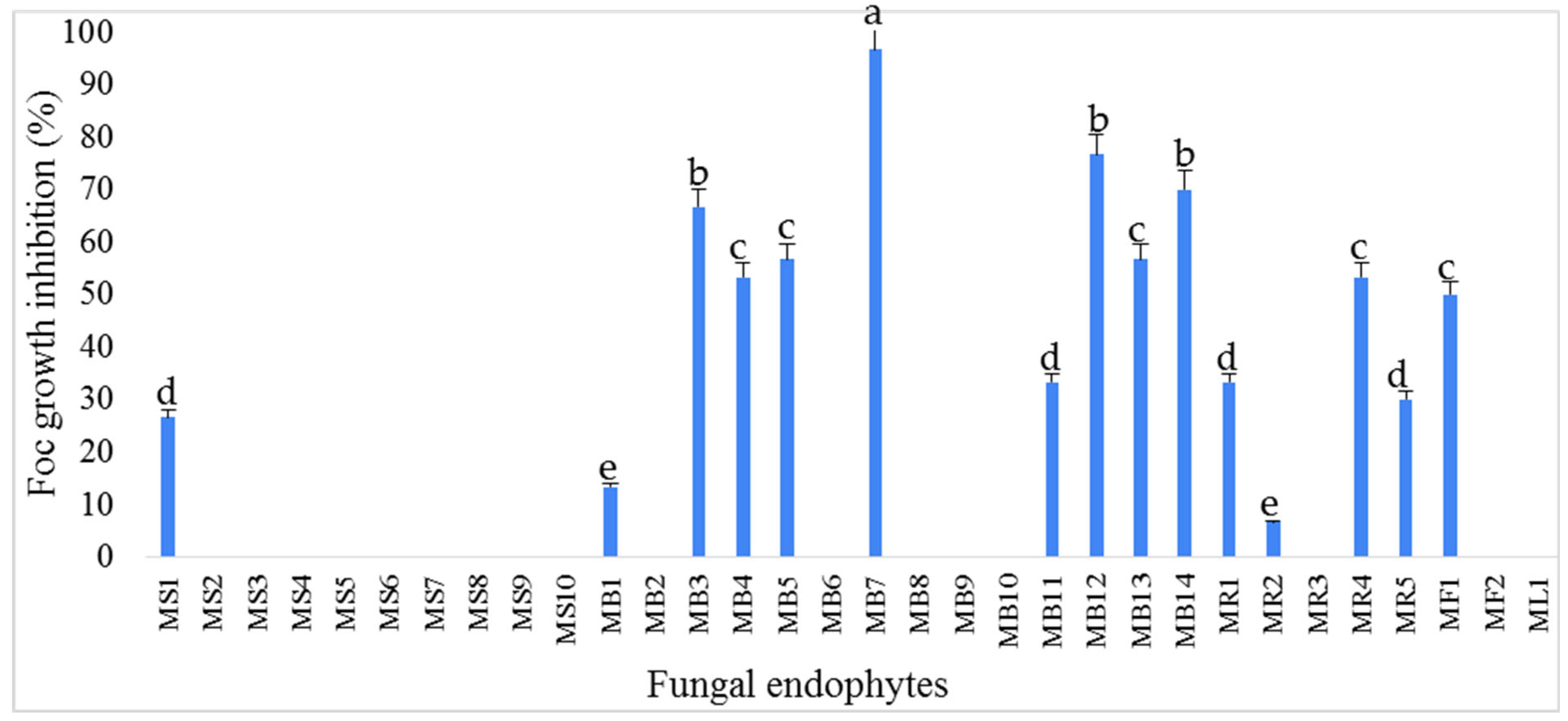
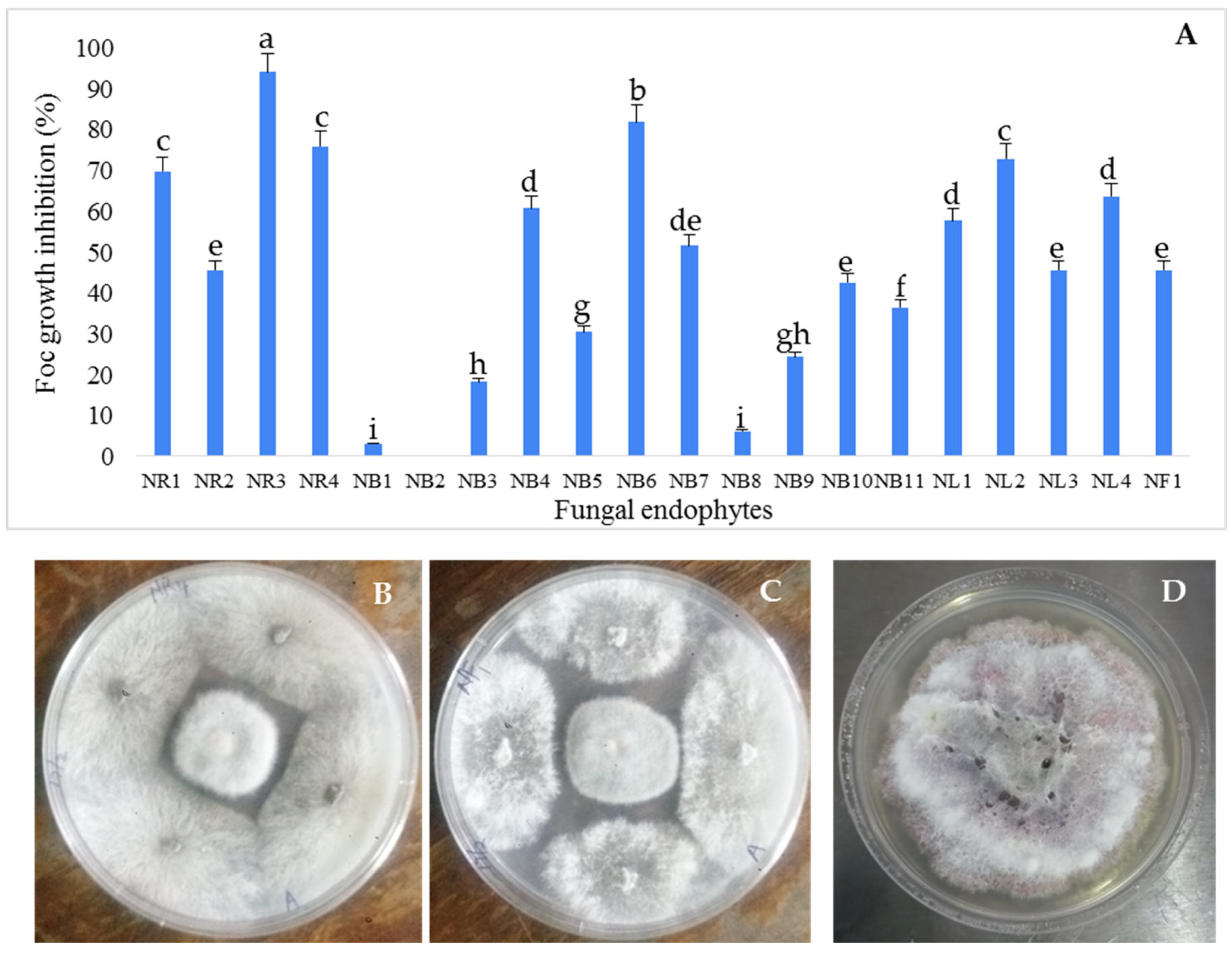
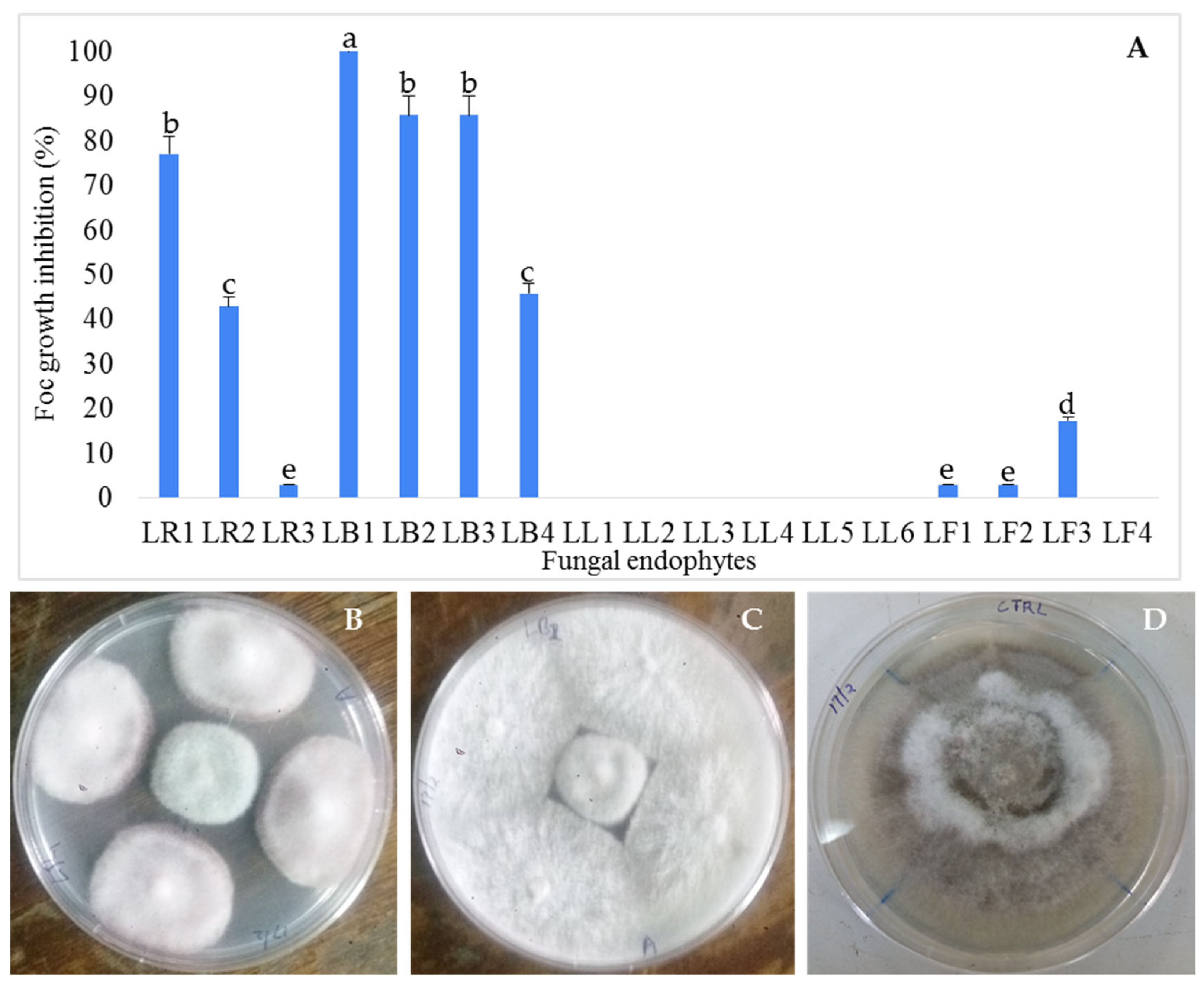
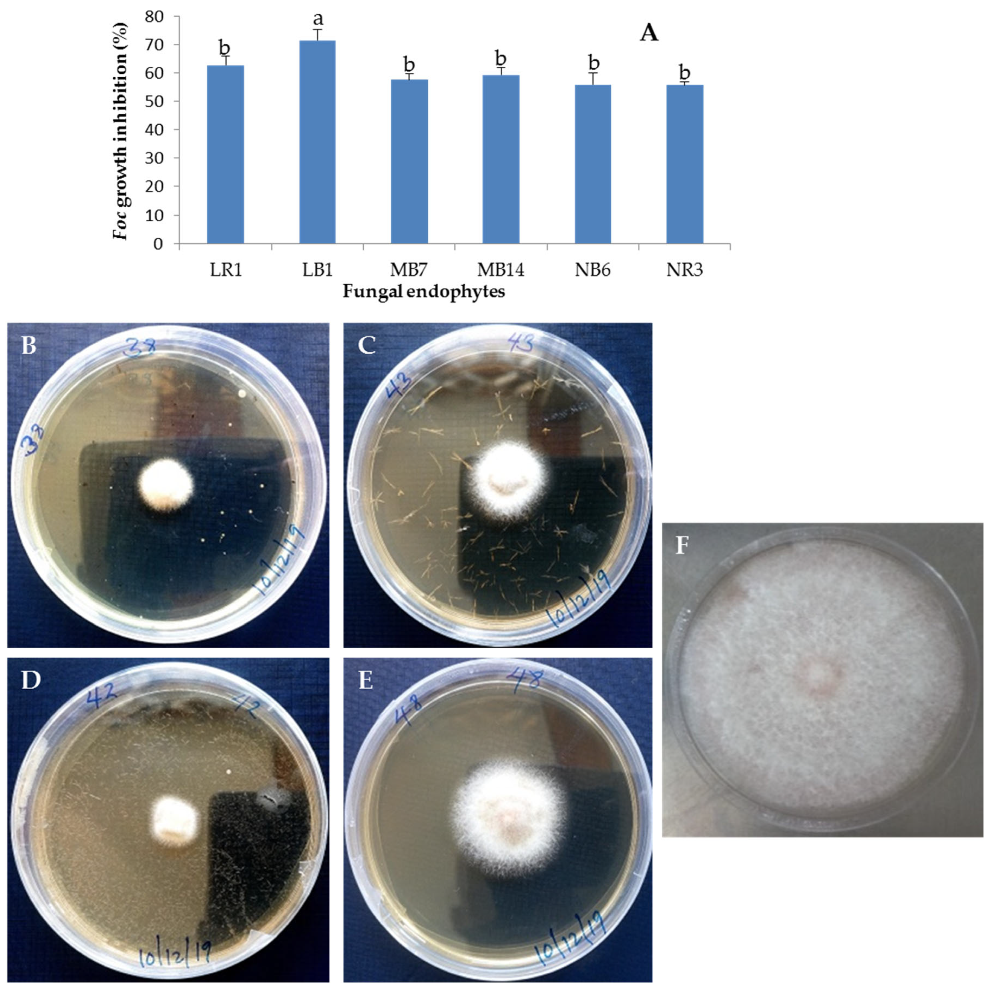
3.1. Isolation and Antifungal Activity Testing of the Endophytic Fungi Isolates

A total of sixty-nine fungal endophytes were isolated and purified, of which thirty-two, twenty and seventeen were isolated from the bark, root, flowers, leaves and seeds/pods of M. oleifera, A. indica and L. angustifolia plants, respectively, based on their ability to grow on potato dextrose agar (PDA) (Supplementary Table S1).
The isolated fungal endophyte isolates were tested for antagonistic activity against Foc TR4 using a dual-culture assay. Figure 1 shows the growth of Foc TR4 isolate in the treatments with fungal endophytes from M. oleifera and controls. The ANOVA showed significant differences in the colony growth and the percentage of Foc TR4 inhibition among the fungal endophyte isolates in the dual-culture assay, which ranged from 0% (MS2–MS10, MB2, MB6, MB8–MB10, MR3, MF2 and ML1) to 96.67% (MB7) (Figure 1). Among the fungal endophyte isolates, seven showed antagonistic activity against Foc TR4 with inhibition rates of more than 50% (Figure 1). Isolates MB7 and MB14 were selected as representatives of fungal endophytes from M. oleifera for further characterization and investigation of antifungal activity.
The in vitro co-cultivation assay revealed that 95% of the fungal endophyte isolates inhibited the mycelial growth of Foc TR4, but the degree of inhibition varied among the endophytes. Nine endophytic fungi isolates showed greater than 50% inhibition against Foc TR4. Isolate NR3 recorded the highest inhibition rate percentage (I%) against Foc TR4, with 93.94% (Figure 2). This was followed by isolates NB6 and NR4 with I% of 81.82% and 75.76%, respectively. Isolates NR3 and NB6, with the highest I% against Foc TR4, were selected as representatives of fungal endophytes from A. Indica for further characterization and investigation of antifungal activity.
The percentage antagonism (growth inhibition percentage) based on the dual-culture assay was calculated for each fungal endophyte isolate from L. angustifolia and is presented in Figure 3. The growth inhibition rate percentage (I%) ranged from 0 to 100%. Growth inhibition of greater than 50% against Foc TR4 was recorded by 23.53% of the fungal endophytes using dual culture. Isolate LB1 recorded the highest inhibition percentage (I%) against Foc TR4 with 100% (Figure 3). This was followed by isolates LB2, LB3 and LR1 with I% of 85.71%, 85.71% and 77.14%, respectively. LB1 and LR1 were selected as representatives of fungal endophytes from L. angustifolia for further characterization and investigation of antifungal activity.

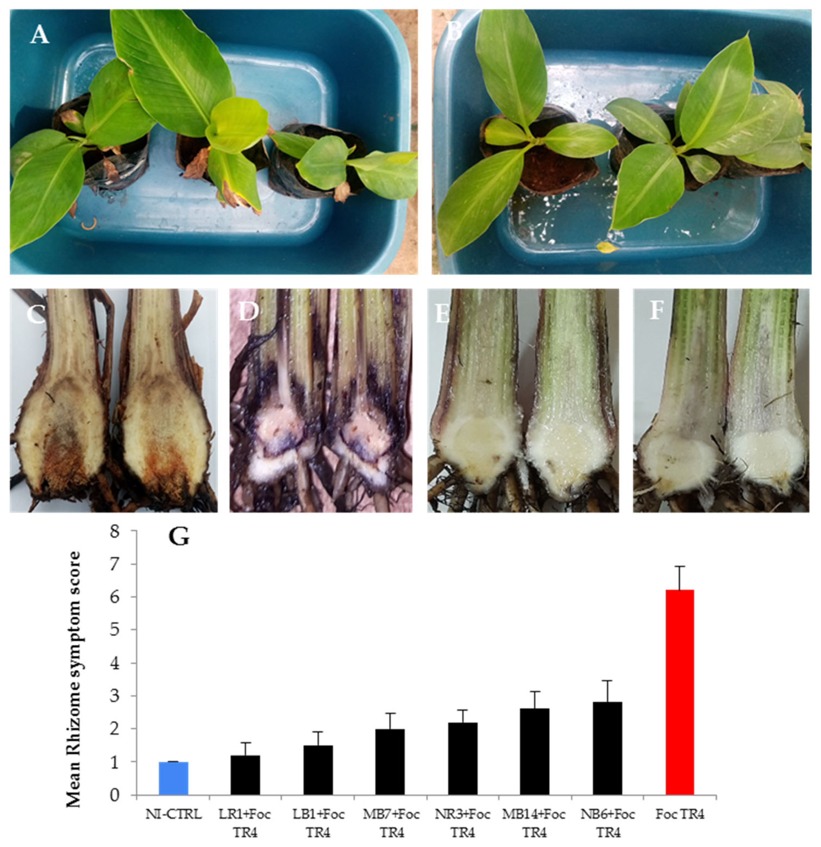
3.2. Pathogenicity of Selected Fungal Endophytes Banana Cultivar Grand Naine

All six fungal endophytes did not cause disease in all banana plantlets of the Grand Naine cultivar used. The rhizomes of the banana plantlets were all healthy, with no discoloration and necrosis.

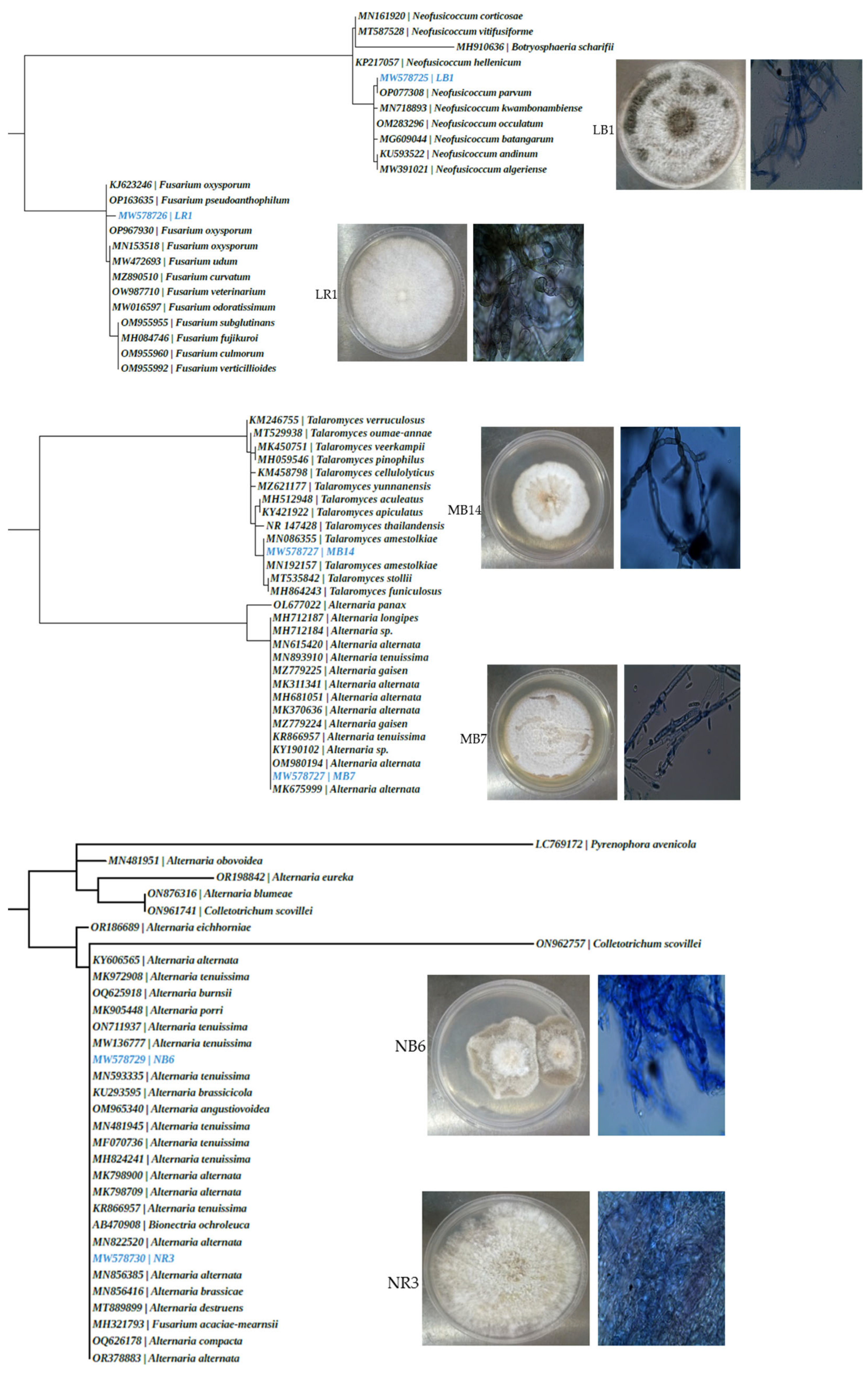
3.3. Identification of Selected Fungal Endophytes with Antifungal Activity

For the identification of the six selected fungal endophytes, morphological and molecular techniques were used. Six fungal endophyte isolates (MB7, MB14, LB1, LR1, NB6 and NR3) with the highest antagonistic activity were identified on the basis of colony and spore characteristics (Table 2 and Figure 4) and ITS sequence analysis. According to the microscopic observation, the isolate LB1 presented branched septate hyphae, and the conidia were fusiform and ellipsoidal with an ovoid base (Figure 4). NB6 was also branched, but the conidia were short-chained, elliptical, and beaked with transverse septate (Figure 4). The colony and spore characteristics of the six fungal endophytes are presented in Table 2.
PCR amplification was performed using the ITS1/4 primer, which successfully amplified products of approximately 550 bp. The ITS sequence lengths of the six antagonistic fungal endophyte isolates were in the approximate range of 500–600 bp. The similarities between the antagonistic isolates and reference sequences in the NCBI database (accessed on 17 July 2023) were all above 99%. The maximum identities for each isolate exceeded 99% with an E-value of 0 and the gene sequences of the 6 isolates were submitted to the GenBank database and the accession numbers were obtained (Table 3). The results of the ITS analysis showed that the fungal endophytes MB7 and MB14 were most similar to Alternaria alternata (with 100% identity) and Talaromyces amestolkiae (with 99.71% identity), respectively, while LB1 and LR1 were most similar to Neofusicoccum parvum and Fusarium oxysporum, respectively, both with 100% identity. Fungal endophytes NB6 and NR3 were most similar to Alternaria tenuissima and Alternaria alternata, respectively, both with 100% identity. Detailed descriptions of the antagonistic endophytic fungi isolates, with their respective codes, GenBank accession numbers and similar sequence homologs are summarized in Table 3. The phylogenetic trees (Figure 4) were generated using the sequences from the nucleotide BLAST results. The phylogenetic trees from the neighbor-joining method revealed that the two fungal endophytes, MB7 and NR3, were related to Alternaria alternata and the four other fungal endophytes LB1, LR1, MB14 and NB6 were related to Neofusicoccum parvum, Fusarium oxysporum, Talaromyces amestolkiae and Alternaria tenuissima, respectively.

3.4. Antifungal Activity of Culture Filtrates of Selected Fungal Endophytes against Foc TR4

The antifungal activity of the culture filtrates of the six selected fungal endophyte isolates was further tested using the mycelial growth method. The antifungal activities of the culture filtrates of fermentation products of the fungal endophytes are shown in Figure 5. The six fungal endophytes showed significant antifungal activity against Foc TR4. The culture filtrates at a concentration of 50 mg/mL significantly inhibited the growth of Foc TR4. Culture filtrates from all six fungal endophytes exhibited more than 50% inhibition against Foc TR4. However, fungal endophyte LB1 exhibited a significantly (F = 139.5745, p < 0.001) high-percentage inhibition compared to the other fungal endophytes. The differential inhibitory activity of the culture filtrates demonstrates variations in the metabolite production by the fungal endophytes.

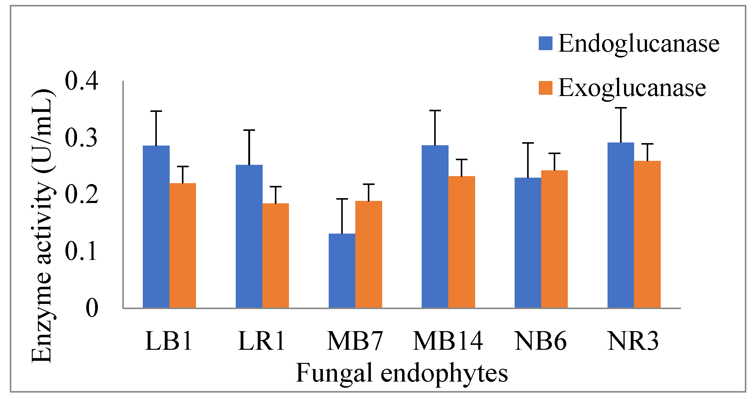
3.5. Production of Glucanases by the Antagonistic Endophytic Fungi

The endo- and exo-β-1,3-glucanase activities in six endophytic fungi cultured in media containing glucose were evaluated due to their relevance in mycoparasitism mechanisms. The results showed that the six endophytic fungi (MB7, MB14, LB1, LR1, NB6 and NR3) could produce endo-β-1,3-glucanase and exo-β-1,3-glucanase (Figure 6). The ANOVA test demonstrated that there was a significant (p < 0.001) difference between endoglucanase and exoglucanase activities in each of the fungal endophytes. There was a significant (p > 0.001) difference in endoglucanase and exoglucanase activities in fungal endophytes LB1, MB14, NB6 and NR3.

3.6. Evaluation of Selected Fungal Endophytes on Improving Plant Resistance against Foc TR4

Pot experiments were performed to test whether the fermentation broth for Alternaria alternata (MB7 and NR3), Neofusicoccum parvum (LB1), Fusarium oxysporum (LR1), Talaromyces amestolkiae (MB14) and Alternaria tenuissima (NB6) can enhance plant resistance to Foc TR4. At 8 weeks after Foc TR4 inoculation, chlorotic symptoms were observed in control banana plants treated with water (Figure 7A). No chlorotic symptoms (Figure 7B) were observed in the leaves of plants treated with fungal endophytes Alternaria alternata (MB7 and NR3), Neofusicoccum parvum (LB1), Fusarium oxysporum (LR1), Talaromyces amestolkiae (MB14) and Alternaria tenuissima (NB6). After cutting the corms, we observed that the corms of the control plantlets treated with sterile water exhibited brownish-black decay symptoms due to Foc TR4 (Figure 7C), while healthy whitish corms were observed in endophyte-crude-extract-treated (LB1 + TR4, LR1 + TR4, MB7 + TR4, MB14 + TR4, NB6 + TR4 and NR3 + TR4) plantlets (Figure 7E,F). A rhizome score of 1 or 2, which indicated no discoloration in the rhizome at 8 weeks after inoculation, was observed in plantlets treated with fungal endophyte LB1, LR1 and MB7 crude extracts (Figure 7G).
To assess the effect of selected fungal endophytes on plant growth, plant height and number of leaves were measured at 8 weeks after treatment. The banana plantlets treated with each of the fungal endophytes showed a significantly enhanced growth rate compared to the non-treated plantlets. The six endophytic fungi significantly increased the plant height and the number of leaves of the banana plantlets (Table 4). Significant increase in plant height in Grand Naine banana plantlets was recorded by all the six fungal endophytes treatments as compared to the non-treated control (NI-CTRL) and the control plants inoculated with Foc TR4 pathogen. The minimum plant height was recorded by plantlets inoculated with Foc TR4 pathogen compared to the non-treated control (NI-CTRL) (Table 4). A remarkable increase in the number of leaves was recorded in fungal endophytes LB1 and NR3, respectively, as compared to the non-treated control (NI-CTRL) and control plantlets inoculated with Foc TR4 (Table 4).

4. Discussion

Medicinal plants and herbs are receiving a lot of attention currently and their extracts possess various metabolites that have the potential to destroy many plant-disease-causing pathogens or inhibit their growth [41,42,43]. The present study was performed to understand the association of fungal endophytes with M. oleifera, A. indica and L. angustifolia and the biocontrol potential of the fungal endophytes against phytopathogen Foc TR4. Evidence shows that fungal endophytes isolated from native or endemic plants may hold significant interest for their bioactive compound production and have been shown to inhibit the growth of pathogens [44]. In this study, the three plant species harbored a rich diversity of fungal endophytes from different plant tissues. The fungal interactions between the endophytes and Foc TR4 in the co-cultivation assay showed that some of the fungal endophytes are potential candidates for bioprospecting.
The dual-culture assay was used in the current study as a standard confrontation test to assess the inhibitory action of the biocontrol agents [45]. The fungal endophytes showed variable antagonistic activity against Foc TR4 based on dual-culture assays. This demonstrated that the antifungal activities of fungal endophytes against Fusarium wilt were significantly different. This is consistent with previous findings that endophytic fungi isolated from different host plant species or tissues showed different antifungal activities against pathogens [46,47]. Fungal endophytes can act as biocontrol agents to antagonize pathogens through antibiosis, mycoparasitism and competition [48]. In this study, the dual-culture assay showed in vitro antagonistic activity against Foc TR4 through physical contact of mycelia, and they can inhibit the growth of pathogens by competing for living space and nutrients in the medium. Therefore, the mechanism possessed by the fungal endophytes in the current study may be similar to other fungal endophytes with known antagonistic activity and inhibits pathogens through competition, antibiosis and/or mycoparasitism [48,49].
Our results supported that the selected fungal endophytes LB1, LR1, MB7, MB14, NB6 and NR3 from M. oleifera, A. indica and L. angustifolia had robust in vitro antifungal activity against Foc TR4. In this study, morphological characteristics and ITS gene sequences were used to identify the six fungal endophytes. The ITS gene region reveals high richness and diversity, and therefore, it is commonly used in molecular studies of fungi [50]. In this study, ITS sequences demonstrated that the fungal endophytes were of the genera Talaromyces, Fusarium, Neofusicoccum and Alternaria. Morphological and molecular approaches revealed that the two isolates MB7 and NR3 were identified as Alternaria alternata and the four other isolates LB1, LR1, MB14 and NB6 were identified as Neofusicoccum parvum, Fusarium oxysporum, Talaromyces amestolkiae and Alternaria tenuissima, respectively.
The fungal endophytes isolated in this study have also been reported from other plants in previous studies. Neofusicoccum parvum has been demonstrated to produce functional secondary metabolites, including cyclohexenones, melleins, myrtucommulones, naphthalenones, naphthoquinones, phenols, alcohols and sesquiterpenes [51]. These secondary metabolites have been shown to have antimicrobial activities against pathogenic bacteria and fungi. Fusarium oxysporum is an abundant fungus in the environment and can also survive in many plant species. Some F. oxysporum strains are pathogenic and are associated with vascular wilt diseases affecting a wide range of plant hosts [52]. However, many other F. oxysporum strains are non-pathogenic as they only penetrate the roots but do not invade the vascular system to cause disease [53]. Non-pathogenic populations of Fusarium species have been shown to confer suppression of Fusarium wilt in soils [52]. Talaromyces amestolkiae is a ubiquitous heterothallic fungus yielding numerous types of structurally diverse active secondary metabolites [54]. In addition to plant growth promotion, these metabolites have been shown to possess significant α-glucosidase inhibitory activity [54]. The abundance of bioactive secondary metabolites in T. amestolkiae makes the endophyte an ideal candidate for the development and application of endophytic biocontrol agents. A. alternata and A. tenuissima are widespread fungi with a broad host range, frequently isolated from plants as both an endophyte and a pathogen. The endophytes produce a variety of bioactive secondary metabolites including polyketides, quinones, and terpenes [55]. These compounds exhibit antibacterial, antioxidant, and enzyme-inhibitory activities [56]. In addition, these metabolites have been shown to be effective insecticides and herbicides as well as enhancers of plant immunity against plant pathogens [57].
Production of antimicrobial compounds in the form of non-volatiles is one of the main mechanisms used by antagonists for the control of pathogens. From our findings, the culture filtrates of the six fungal endophytes displayed a potent inhibition of mycelial growth. Based on these results, we speculate that the fungal endophytes A. alternata, N. parvum, F. oxysporum, T. amestolkiae and A. tenuissima demonstrated antifungal activity against Foc TR4, possibly by producing non-volatile metabolites. The inhibition of Foc TR4 mycelial growth demonstrated that the fungal endophytes produced fungistatic metabolites/ non-volatile compounds in the filtrates. The varying degrees of antifungal activity displayed by the culture filtrates of the fungal endophytes could be due to the number and concentration of the active metabolites produced [58]. Secondary metabolites extracted from endophytic fungi (Nigrospora spp. and Fusarium spp.) isolated from the leaves, roots and seeds of M. oleifera have been reported to have strong antimicrobial activities against pathogenic bacteria and fungi [59]. Secondary metabolites derived from fungal endophytes have been reported to comprise classes of compounds such as steroids, xanthones, phenols, isocoumarins, perylene derivatives, quinines, furandiones, terpenoids, depsipeptides and cytochalasins, which are identified as possessing antifungal activities [59,60]. Some of the specific antifungal compounds reported to be produced by fungal endophytes include 1,5-hydroxy benzofuranone, aurantiamine, benzylpyridine B, pavetanin, fumitremorgin B and verruculogen [59,60]. In the study by Kaur et al. [61,62], the exploration of the endophytic fungus Chaetomium globosum of Moringa oleifera showed the chloroformic extracts from the fungus have antimutagenic activity against mutagen 2-aminofluorene (2-AF). Patil et al. [63] established that endophytic fungi isolated from A. indica produce secondary metabolites that possess antagonistic activities. The fungal endophytes Cladosporium sp. and Curvularia sp. isolated from A. indica have proteolytic, cellulolytic, amylolytic and lipase activities that are responsible for the suppression of the growth of pathogenic microbes. Phenolic acids extracted from fungal endophytes isolated from Lavandula spp. have also been shown to inhibit the growth of fungal and bacterial pathogens [64]. Endophytic fungi such as Talaromyces sp. and Fusarium sp. have the potential to produce different types of bioactive secondary metabolites and are supported by studies of other medicinal plants and herbs [65,66]. Many species of Alternaria isolated as endophytes produce antifungal compounds [67]. In a study by Prieto et al. [68], T. amestolkiae showed strong hydrolytic activity. Some β-glycosidases produced by T. amestolkiae catalyze the transfer of sugar molecules through glycosylation and degrade plant biomass by secreting complex extracellular enzyme mixtures, which play an important role in destroying the cell structure of plant pathogens.
The cell walls of fungi are predominantly composed of β-1,3-glucan and chitin [69]. The enzymatic assays in this study showed that all six fungal endophytes produced β-1,3-glucanase but with variations in their activities. The fungal endophytes exhibited different antifungal activities, probably due to the variations in the β-1,3-glucanase production capacity. The production of glucanases by the six fungal endophytes plays a vital role in the inhibition of Foc TR4 growth as the secretion of these enzymes breaks down glucans and cellulose, which are the structural components of the Foc TR4 cell wall. The activity of glucanases and other enzymes such as proteases have been reported to be responsible for the breakdown of the components of the cell wall of Foc TR4, thus inhibiting hyphae growth [70]. These enzymes may act indirectly by releasing fragments of cell walls that may act as elicitors of a defense reaction such as the occurrence of phytoalexin accumulation. The secretion of these extracellular enzymes by the fungal endophytes could be one of the antifungal mechanisms. Higher activity of β-glucanase and chitinase was confirmed in F. oxysporum f. sp. cubense race-1-tolerant cultivars and hybrids of bananas [71]. Previous reports by Migheli et al. [72] on the biocontrol of P. debaryanum by Arthrobacter spp. elucidated the role of glucanase and protease in the lysis of mycelium.
Beta-1,3-glucanases from fungal endophytes have been reported to be involved in an alternative defense mechanism, where degradation of the hyphal cell wall of invading fungi releases β-1,3-glucan oligosaccharides, which serve as elicitors. Upon recognition by plant surveillance systems, these β-1,3-glucan oligosaccharides act as microbe-associated molecular patterns (MAMPs) triggering the activation of signaling cascades resulting in a wide range of localized and systemic defense responses [69]. For example, upon Foc TR4 infection, endophytic fungi β-1,3-glucanases are able to release β-glucan elicitors from the fungal wall, which trigger the production of antimicrobial compounds such as phytoalexins, as described previously in rice and soybean [73]. However, the exact mechanism of recognition and mode of action of pathogen-derived β-glucan elicitors is not fully understood, and it seems to be dependent not only on the plant species and the origin of the β-glucan elicitor but also on the glycan structure, where length, branching pattern and presence of chemical modifications have been shown to be relevant [74].
The greenhouse tests in which the banana plantlets were primed with endophytic fungi revealed that the plantlets treated with the endophytes did not show chlorosis and rhizome discoloration. The greenhouse trials demonstrated the positive effects of fungal endophytes Neofusicoccum parvum, Fusarium oxysporum, Talaromyces amestolkiae and Alternaria tenuissima in suppressing the growth of Fusarium wilt caused by Foc TR4. Banana growth promotion in terms of increased plant height and number of leaves was observed after treatments with fungal endophytes Neofusicoccum parvum, Alternaria alternata, Fusarium oxysporum, Talaromyces amestolkiae and Alternaria tenuissima. The fungal endophytes from Fusarium sp., Talaromyces sp. and Alternaria sp. induce the production of secondary metabolites in plants [65,66]. The produced compounds have been reported to be involved in plant protection from pathogenic microbes [75]. The in vivo analysis of the biocontrol activity demonstrated that the fungal endophytes inhibited Foc TR4 growth in banana. It seems that the fungal endophytes in this study used the same mechanisms after deployment on banana plants, thereby suppressing the growth of Foc TR4, and enhanced the morphological growth traits of plants. The infection method used to screen the banana plants with Fusarium wilt was the millet grain method, which favors spore multiplication and fungus survival [76]. The foliage of the banana plants did not display wilting, chlorosis, emerging leaf malformation, rhizome discoloration and plant death due to the resistance to Foc TR4 conferred by the fungal endophytes [77].
The fungal endophytes isolated in the current study and demonstrated to have biocontrol potential in the management of Fusarium wilt in banana have also been reported in previous studies from other plants. Fungal endophyte Alternaria alternata has been isolated from infected water hyacinth leaves in India and demonstrated biocontrol potential against water hyacinth (Eichhornia crassipes [Mart.] Solms) [78]. Fusarium oxysporum has been isolated from suppressive soil in South Africa and used as a biocontrol against Fusarium oxysporum f. sp cubense, the causal agent of Fusarium wilt of banana [79]. Talaromyces amestolkiae has been isolated from soil samples and demonstrated to be a useful biocontrol against Fusarium oxysporum f. sp cubense, the causal agent of Fusarium wilt of banana [80]. Alternaria tenuissima isolated from rice has been confirmed to possess biocontrol potential against Bipolaris oryzae, the causal agent of rice brown spot disease [81].

5. Conclusions

The findings from this study highlight the potential of M. oleifera, A. indica and L. angustifolia fungal endophytes in the biocontrol of Foc TR4, the causal agent of Fusarium wilt of banana. The fungal endophytes Neofusicoccum parvum LB1, Fusarium oxysporum LR1, Alternaria alternata MB7, Alternaria alternata NR3, Talaromyces amestolkiae MB14 and Alternaria tenuissima NB6 showed high antagonistic activity against Foc TR4. Our study indicates that the six fungal endophytes are potential biological control agents to combat Fusarium oxysporum f. sp. cubense in banana. There is also a need to evaluate the performance of the candidates under field conditions. Further studies of the secondary metabolites produced by the fungal endophytes are required for the development of biological agents against Foc TR4.

Supplementary Materials

The following supporting information can be downloaded at: https://www.mdpi.com/article/10.3390/microbiolres14040143/s1. Figure S1: Isolated fungal endophytes from plant tissues of Moringa oleifera, Azadirachta indica and Lavandula angustifolia. Table S1: Fungal endophytes isolated from different parts of Moringa (Moringa oleifera), Neem (Azadirachta indica) and Lavender (Lavandula angustifolia).

Author Contributions

Conceptualization, B.M.N., E.W.K., J.K.K. and E.N.N.; Methodology, B.M.N., E.W.K., J.K.K. and E.N.N.; Investigation, B.M.N.; Data Analysis, B.M.N. and E.N.N.; Supervision, B.M.N., J.K.K. and E.N.N.; Writing—Original Draft Preparation, B.M.N.; Writing—Review and Editing, B.M.N., E.W.K., J.K.K. and E.N.N. All authors have read and agreed to the published version of the manuscript.

Funding

This research received no external funding.

Institutional Review Board Statement

Not applicable.

Informed Consent Statement

Not applicable.

Data Availability Statement

The nucleotide sequences of the ITS region for six isolates obtained in this study were deposited in Genebank through the online submission portal and were assigned the following accession numbers: MW578725, MW578726, MW578727, MW578728, MW578729 and MW578730.

Acknowledgments

The authors wish to acknowledge the Department of Biochemistry, University of Nairobi, for providing facilities for research.

Conflicts of Interest

The authors declare no conflict of interest.

References

  1. Pua, E.C.; Davey, M.R. Banana. In Transgenic Crops V; Springer: Berlin/Heidelberg, Germany, 2007; Volume 60, pp. 3–34. [Google Scholar]
  2. Dash, P.K.; Rai, R. Translating the Banana Genome to Delineate Stress Resistance, Dwarfing, Parthenocarpy and Mechanisms of Fruit Ripening. Front. Plant Sci. 2016, 7, 1543. [Google Scholar] [CrossRef] [PubMed]
  3. FAOSTAT. FAO Statistical Database. 2021. Available online: http://www.fao.org/ (accessed on 30 May 2022).
  4. Drenth, A.; Kema, G. The Vulnerability of Bananas to Globally Emerging Disease Threats. Phytopathol 2021, 111, 2146–2161. [Google Scholar] [CrossRef] [PubMed]
  5. Dale, J.; James, A.; Paul, J.; Khanna, H.; Smith, M.; Peraza-Echeverria, S.; Garcia-Bastidas, F.; Kema, G.; Waterhouse, P.; Mengersen, K.; et al. Transgenic Cavendish bananas with resistance to Fusarium wilt tropical race 4. Nat. Commun. 2017, 8, 1496. [Google Scholar] [CrossRef] [PubMed]
  6. Pegg, K.G.; Moore, N.Y.; Bentley, S. Fusarium wilt of banana in Australia: A review. Austral. J. Agric. Res. 1996, 47, 637–650. [Google Scholar] [CrossRef]
  7. Nguyen, T.V.; Tran-Nguyen, L.T.T.; Wright, C.L.; Trevorrow, P.; Grice, K. Evaluation of the efficacy of commercial disinfectants against Fusarium oxysporum f. sp. cubense race 1 and tropical race 4 propagules. Plant Dis. 2019, 103, 721–728. [Google Scholar]
  8. Souza, I.F.; Napoleão, T.H.; de Sena, K.X.; Paiva, P.M.; de Araújo, J.M.; Coelho, L.C. Endophytic microorganisms in leaves of Moringa oleifera collected in three localities at Pernambuco State, Northeastern Brazil. Brit. Microbiol. Res. J. 2016, 13, 1. [Google Scholar] [CrossRef]
  9. Chen, A.; Sun, J.; Matthews, A.; Armas-Egas, L.; Chen, N.; Hamill, S.; Mintoff, S.; Tran-Nguyen, L.T.T.; Batley, J.; Aitken, E.A.B. Assessing variations in host resistance to Fusarium oxysporum f sp. cubense Race 4 in Musa species, with a Focus. on the subtropical race 4. Front. Microbiol. 2019, 10, 1062. [Google Scholar] [CrossRef]
  10. Ploetz, R.C. Fusarium Wilt of Banana. Phytopathology 2015, 105, 1512–1521. [Google Scholar] [CrossRef]
  11. Dong, X.; Xiong, Y.; Ling, N.; Shen, Q.; Guo, S. Fusaric acid accelerates the senescence of leaf in banana when infected by Fusarium. World J. Microbiol. Biotechnol. 2014, 30, 1399–1408. [Google Scholar] [CrossRef]
  12. Dita, M.; Barquero, M.; Heck, D.; Mizubuti, E.S.G.; Staver, C.P. Fusarium wilt of banana: Current knowledge on epidemiology and research needs toward sustainable disease management. Front. Plant Sci. 2018, 9, 1468. [Google Scholar] [CrossRef]
  13. Gang, G.; Bizun, W.; Weihong, M.; Xiaofen, L.; Xiaolin, Y.; Chaohua, Z.; Jianhong, M.; Huicai, Z. Biocontrol of Fusarium wilt of banana: Key influence factors and strategies. Afr. J. Microbiol. Res. 2013, 7, 4835–4843. [Google Scholar]
  14. Pegg, K.G.; Coates, L.M.; O’Neill, W.T.; Turner, D.W. The Epidemiology of Fusarium Wilt of Banana. Front. Plant Sci. 2019, 10, 1395. [Google Scholar] [CrossRef] [PubMed]
  15. Jamil, F.N.; Tang, C.N.; Saidi, N.B.; Lai, K.S.; Baharum, N.A. Fusarium wilt in banana: Epidemics and management strategies. In Horticultural Crops; Baimey, H.K., Hamamouch, N., Kolombia, Y.A., Eds.; IntechOpen: London, UK, 2019; pp. 117–142. [Google Scholar]
  16. Siamak, S.B.; Zheng, S. Banana Fusarium wilt (Fusarium oxysporum f. sp. cubense) control and resistance, in the context of developing wilt-resistant bananas within sustainable production systems. Hortic. Plant J. 2018, 4, 208–218. [Google Scholar] [CrossRef]
  17. Cannon, S.; Kay, W.; Kilaru, S.; Schuster, M.; Gurr, S.J.; Steinberg, G. Multi-site fungicides suppress banana Panama disease, caused by Fusarium oxysporum f. sp. cubense Tropical Race 4. PLoS Pathog. 2022, 18, e1010860. [Google Scholar] [CrossRef] [PubMed]
  18. Bubici, G.; Kaushal, M.; Prigigallo, M.I.; Gómez-Lama Cabanás, C.; Mercado-Blanco, J. Biological Control Agents Against Fusarium Wilt of Banana. Front. Microbiol. 2019, 10, 616. [Google Scholar] [CrossRef] [PubMed]
  19. Lv, N.; Tao, C.; Ou, Y.; Wang, J.; Deng, X.; Liu, H.; Shen, Z.; Li, R.; Shen, Q. Root-associated antagonistic Pseudomonas spp. contributes to soil suppressiveness against banana Fusarium wilt disease of banana. Microbiol. Spect. 2023, 11, e0352522. [Google Scholar] [CrossRef] [PubMed]
  20. Tao, C.; Wang, Z.; Liu, S.; Lv, N.; Deng, X.; Xiong, W.; Shen, Z.; Zhang, N.; Geisen, S.; Li, R.; et al. Additive fungal interactions drive biocontrol of Fusarium wilt disease. New Phytol. 2023, 238, 1198–1214. [Google Scholar] [CrossRef]
  21. Xiang, D.; Yang, X.; Liu, B.; Chu, Y.; Liu, S.; Li, C. Bio-priming of banana tissue culture plantlets with endophytic Bacillus velezensis EB1 to improve Fusarium wilt resistance. Front. Microbiol. 2023, 14, 1146331. [Google Scholar] [CrossRef]
  22. Zhu, Z.; Wu, G.; Deng, R.; Hu, X.; Tan, H.; Chen, Y.; Tian, Z.; Li, J. Spatiotemporal biocontrol and rhizosphere microbiome analysis of Fusarium wilt of banana. Commun. Biol. 2023, 6, 27. [Google Scholar] [CrossRef]
  23. Jamiołkowska, A. Natural compounds as elicitors of plant resistance against diseases and new biocontrol strategies. Agronomy 2020, 10, 173. [Google Scholar] [CrossRef]
  24. Jaber, L.R.; Alananbeh, K.M. Fungal entomopathogens as endophytes reduce several species of Fusarium causing crown and root rot in sweet pepper (Capsicum annuum L.). Biol. Control 2018, 126, 117–126. [Google Scholar] [CrossRef]
  25. Onsare, J.G.; Kaur, H.; Arora, D.S. Antimicrobial activity of Moringa oleifera from different locations against some human pathogens. Acad. J. Med. Plants 2013, 1, 80–91. [Google Scholar]
  26. Ke, Y.; Chen, Z.; Ma, J.B.; Huang, X.M.; Zeng, S.R. Studies of isolation of endophytic fungi from Moringa sp. and antibiotic active substance produced by the Aspergillus sp. ly14. J.-Hunan Agric. Univ. 2006, 32, 521. [Google Scholar]
  27. Zhao, J.H.; Zhang, Y.L.; Wang, L.W.; Wang, J.Y.; Zhang, C.L. Bioactive secondary metabolites from Nigrospora sp. LLGLM003, an endophytic fungus of the medicinal plant Moringa oleifera Lam. World J. Microbiol. Biotechnol. 2012, 28, 2107–2112. [Google Scholar] [CrossRef]
  28. Nicoletti, M.; Murugan, K. Neem the tree of 21st century. Pharmacologyonline 2013, 3, 115–121. [Google Scholar]
  29. Kumar, D.; Rahal, A.; Malik, K.J. Neem extract. In Nutraceuticals. Efficacy, Safety and Toxicity; Ramesh, C., Ed.; Gupta: Chennai, India, 2016; pp. 585–597. [Google Scholar]
  30. Wylie, M.R.; and Merrell, D.S. The Antimicrobial Potential of the Neem Tree Azadirachta indica. Front. Pharmacol. 2022, 13, 891535. [Google Scholar] [CrossRef] [PubMed]
  31. Hossain, M.A.; Al-Toubi, W.A.S.; Weli, A.M.; Al-Riyami, Q.A.; Al-Sabahi, J.N. Identification and characterization of chemical compounds indifferent crude extracts from leaves of Omani neem. J. Taibah Uni. Sci. 2013, 7, 181–188. [Google Scholar] [CrossRef]
  32. Robu, S.; Aprotosoaie, A.C.; Miron, A.; Cioancǎ, O.; Stǎnescu, U.; Hǎncianu, M. In vitro antioxidant activity of ethanolic extracts from some Lavandula species cultivated in Romania. Farmacia 2012, 60, 394–401. [Google Scholar]
  33. Turgut, A.C.; Emen, F.M.; Canbay, H.S.; Demirdöğen, R.E.; Çam, N.; Kılıç, D.; Yeşilkaynak, T. Chemical characterization of Lavandula angustifolia Mill. which is a phytocosmetic species and investigation of its antimicrobial effect in cosmetic products. J. Turk. Chem. Soc. Sect. A Chem. 2016, 4, 283–298. [Google Scholar] [CrossRef]
  34. Zuzarte, M.; Gonçalves, M.J.; Cavaleiro, C.; Cruz, M.T.; Benzarti, A.; Marongiu, B.; Maxia, A.; Piras, A.; Salgueiro, L. Antifungal and anti-inflammatory potential of Lavandula stoechas and Thymus herba-barona essential oils. Ind. Crops Prod. 2013, 44, 97–103. [Google Scholar] [CrossRef]
  35. Dita, M.A.; Waalwijk, C.; Buddenhagen, I.W.; Souza, M.T., Jr.; Kema, G.H.J. A molecular diagnostic for tropical race 4 of the banana fusarium wilt pathogen. Plant Pathol. 2010, 59, 348–357. [Google Scholar] [CrossRef]
  36. Ellis, M.B. Dematiaceous Hyphomycetes; Commonwealth Mycological Institute: London, UK, 1971; 608p. [Google Scholar]
  37. Gilman, J. A manual of soil fungi. Soil. Sci. 1957, 84, 183. [Google Scholar] [CrossRef]
  38. Doyle, J.J. Isolation of plant DNA from fresh tissue. Focus 1990, 12, 13–15. [Google Scholar]
  39. Miller, G.L. Use of Dinitrosalicylic Acid Reagent for Determination of Reducing Sugar. Anal. Chem. 1959, 31, 426–428. [Google Scholar] [CrossRef]
  40. Mak, C.; Mohamed, A.A.; Liew, K.W.; Ho, Y.W. Early screening technique for Fusarium wilt resistance in banana micropropagated plants. In Banana Improvement: Cellular, Molecular Biology, and Induced Mutations; Swennen, R., Jain, M.S., Eds.; Science Publishers, Inc.: Enfield, NH, USA, 2004; pp. 219–227. [Google Scholar]
  41. Yadav, S.; Goswami, P.; Mathur, J. Evaluation of fungicidal efficacy of Moringa oleifera Lam. leaf extract against Fusarium wilt in wheat. J. Nat. Pestic. Res. 2023, 4, 100034. [Google Scholar] [CrossRef]
  42. Ahmadu, T.; Ahmad, K.; Ismail, S.I.; Rashed, O.; Asib, N.; Omar, D. Antifungal efficacy of Moringa oleifera leaf and seed extracts against Botrytis cinerea causing gray mold disease of tomato (Solanum lycopersicum L.). Braz. J. Biol. 2021, 81, 1007–1022. [Google Scholar] [CrossRef]
  43. Hassanein, N.M.; Abou Zeid, M.A.; Youssef, K.A.; Mahmoud, D.A. Efficacy of Leaf Extracts of Neem (Azadirachta indica) and Chinaberry (Melia azedrach) Against Early Blight and Wilt Diseases of Tomato. Aust. J. Basic. Appl. Sci. 2008, 2, 763–772. [Google Scholar]
  44. Vidal, A.; Parada, R.; Mendoza, L.; Cotoras, M. Endophytic Fungi Isolated from Plants Growing in Central Andean Precordillera of Chile with Antifungal Activity against Botrytis cinerea. J. Fungi 2020, 6, 149. [Google Scholar] [CrossRef]
  45. Pakdaman, B.S.; Goltapeh, E.M.; Soltani, B.M. Toward the quantification of confrontation (dual culture) test: A case study on the biological control of Pythium aphanidermatum with Trichoderma asperelloides. J. Biofertil. Biopestic. 2013, 4, 1000137. [Google Scholar] [CrossRef]
  46. Bae, H.; Roberts, D.P.; Lim, H.S. Endophytic Trichoderma isolates from tropical environments delay disease onset and induce resistance against Phytophthora capsici in hot pepper using multiple mechanisms. Mol. Plant Microbe Interact. 2011, 24, 336–351. [Google Scholar] [CrossRef]
  47. Saxena, A.; Raghuwanshi, R.; Singh, H.B. Elevation of defense network in chilli against Colletotrichum capsici by phyllospheric Trichoderma strain. J. Plant Growth Regul. 2016, 35, 377–389. [Google Scholar] [CrossRef]
  48. Thambugala, K.M.; Daranagama, D.A.; Phillips, A.J.L.; Kannangara, S.D.; Promputtha, I. Fungi vs. fungi in biocontrol: An overview of fungal antagonists applied against fungal plant pathogens. Front. Cell Infect. Microbiol. 2020, 10, 604923. [Google Scholar] [CrossRef]
  49. Hamzah, T.N.T.; Lee, S.Y.; Hidayat, A.; Terhem, R.; Faridah-Hanum, I.; Mohamed, R. Diversity and Characterization of Endophytic Fungi Isolated from the Tropical Mangrove Species, Rhizophora mucronata, and Identification of Potential Antagonists Against the Soil-Borne Fungus, Fusarium solani. Front. Microbiol. 2018, 9, 1707. [Google Scholar] [CrossRef]
  50. Liu, J.; Yu, Y.; Cai, Z.; Bartlam, M.; Wang, Y. Comparison of ITS and 18S rDNA for estimating fungal diversity using PCR-DGGE. World J. Microbiol. Biotechnol. 2015, 31, 1387–1395. [Google Scholar] [CrossRef] [PubMed]
  51. Salvatore, M.M.; Alves, A.; Andolfi, A. Secondary metabolites produced by Neofusicoccum species associated with plants: A Review. Agriculture 2021, 11, 149. [Google Scholar] [CrossRef]
  52. Fravel, D.; Olivain, C.; Alabouvette, C. Fusarium oxysporum and its biocontrol. New Phytol. 2003, 157, 493–502. [Google Scholar] [CrossRef] [PubMed]
  53. Michielse, C.B.; Rep, M. Pathogen profile update: Fusarium oxysporum. Mol. Plant Pathol. 2009, 10, 311–324. [Google Scholar] [CrossRef]
  54. Lei, L.-R.; Gong, L.-Q.; Jin, M.-Y.; Wang, R.; Liu, R.; Gao, J.; Liu, M.-D.; Huang, L.; Wang, G.-Z.; Wang, D.; et al. Research advances in the structures and biological activities of secondary metabolites from Talaromyces. Front. Microbiol. 2022, 13, 984801. [Google Scholar] [CrossRef]
  55. Li, F.; Lin, S.; Zhang, S.; Pan, L.; Chai, C.; Su, J.C.; Zhang, Y. Modified fusicoccane-type diterpenoids from Alternaria brassicicola. J. Nat. Prod. 2020, 83, 1931–1938. [Google Scholar] [CrossRef]
  56. Tian, L.L.; Ren, H.; Xi, J.M.; Fang, J.; Zhang, J.Z.; Wu, Q.X. Diverse anti-inflammation and anti-cancer polyketides isolated from the endophytic fungi Alternaria sp. MG1. Fitoter. 2021, 153, 105000. [Google Scholar] [CrossRef]
  57. Shi, Z.Z.; Miao, F.P.; Fang, S.T.; Liu, X.H.; Yin, X.L.; Ji, N.Y. Sesteralterin and tricycloalterfurenes A–D: Terpenes with rarely occurring frameworks from the marine-alga-epiphytic fungus Alternaria alternata k21-1. J. Nat. Prod. 2017, 80, 2524–2529. [Google Scholar] [CrossRef] [PubMed]
  58. Eze, P.M.; Nnanna, J.C.; Okezie, U.; Buzugbe, H.S.; Abba, C.C.; Chukwunwejim, C.R.; Okoye, F.B.C.; Esimone, C.O. Screening of metabolites from endophytic fungi of some Nigerian medicinal plants for antimicrobial activities. EuroBiotechnol. J. 2019, 3, 10–18. [Google Scholar] [CrossRef]
  59. Mwanga, Z.; Mvungi, E.; Tibuhwa, D. Antimicrobial Activities of Endophytic Fungi Secondary Metabolites from Moringa oleifera (Lam). Tanzan. J. Sci. 2019, 45, 463–476. [Google Scholar]
  60. Xu, K.; Li, X.-Q.; Zhao, D.-L.; Zhang, P. Antifungal Secondary Metabolites Produced by the Fungal Endophytes: Chemical Diversity and Potential Use in the Development of Biopesticides. Front. Microbiol. 2021, 12, 689527. [Google Scholar] [CrossRef] [PubMed]
  61. Kaur, N.; Arora, D.S. Prospecting the antimicrobial and antibiofilm potential of Chaetomium globosum an endophytic fungus from Moringa oleifera. AMB Expr. 2020, 10, 206. [Google Scholar] [CrossRef]
  62. Kaur, N.; Arora, D.S.; Kalia, N.; Kaur, M. Bioactive potential of endophytic fungus Chaetomium globosum and GC–MS analysis of its responsible components. Sci. Rep. 2020, 10, 18792. [Google Scholar] [CrossRef] [PubMed]
  63. Patil, M.G.; Pagare, J.; Patil, S.N.; Sidhu, A.K. Extracellular enzymatic activities of endophytic fungi isolated from various medicinal plants. Int. J. Curr. Microbiol. App. Sci. 2015, 4, 1035–1042. [Google Scholar]
  64. Xiong, T.; Xie, S.; Xue, Y.; Bai, R.; Feng, J.; Guan, Z.; Liao, X.; Cai, Y. Isolation of endophytic fungi of phenolic acids in lavender and the identification of its products. J. Food Sci. Biotechnol. 2018, 37, 250–255. [Google Scholar]
  65. Liu, F.; Cai, X.; Yang, H.; Xia, X.; Guo, Z.; Yuan, J.; Li, M.; She, Z.; Lin, Y. The bioactive metabolites of the mangrove endophytic fungus Talaromyces sp. ZH-154 isolated from Kandelia candel (L.) druce. Planta Med. 2010, 76, 185–189. [Google Scholar] [CrossRef]
  66. Yang, H.; Li, F.; Ji, N. Alkaloids from an algicolous strain of Talaromyces sp. Chin. J. Oceanol. Limnol. 2016, 34, 367–371. [Google Scholar] [CrossRef]
  67. Root-Bernstein, M.; Valenzuela, R.; Huerta, M.; Armesto, J.; Jaksic, F. Acacia caven nurses endemic sclerophyllous trees along a successional pathway from silvopastoral savanna to forest. Ecosphere 2017, 8, e01667. [Google Scholar] [CrossRef]
  68. Prieto, A.; de Eugenio, L.; Mendez-Liter, J.A.; Nieto-Dominguez, M.; Murgiondo, C.; Barriuso, J.; Bejarano-Munoz, L.; Martinez, M.J. Fungal glycosyl hydrolases for sustainable plant biomass valorization: Talaromyces amestolkiae as a model fungus. Int. Microbiol. 2021, 24, 545–558. [Google Scholar] [CrossRef] [PubMed]
  69. Fesel, P.H.; Zuccaro, A. β-glucan: Crucial component of the fungal cell wall and elusive MAMP in plants. Fungal Genet. Biol. 2016, 90, 53–60. [Google Scholar] [CrossRef] [PubMed]
  70. Schoffelmeer, E.A.M.; Klis, F.M.; Sietsma, J.H.; Cornelissen, B.J.C. The Cell Wall of Fusarium oxysporum. Fungal Genet. Biol. 1999, 27, 275–282. [Google Scholar] [CrossRef] [PubMed]
  71. Kavino, M.; Kumar, N.; Damodaran, T.; Harish, S.; Saravanakumar, D. Biochemical markers as a useful tool for the early identification of Fusarium oxysporum f. sp. cubense, race 1 resistance banana clones. Arch. Phytopathol. Pflanzenschutz. 2009, 42, 1069–1078. [Google Scholar] [CrossRef]
  72. Migheli, Q.; Friard, O.; Del Tedesco, D.; Musso, M.R.; Gullino, M.L. Stability of transformed antagonistic Fusarium oxysporum strains in vitro and in soil microcosms. Mol. Ecol. 1996, 5, 641–649. [Google Scholar] [CrossRef]
  73. Yamaguchi, T.; Yamada, A.; Hong, N.; Ogawa, T.; Ishii, T.; Shibuya, N. Differences in the Recognition of Glucan Elicitor Signals between Rice and Soybean: β-Glucan Fragments from the Rice Blast Disease Fungus Pyricularia Oryzae That Elicit Phytoalexin Biosynthesis in Suspension-Cultured Rice Cells. Plant Cell 2000, 12, 817–826. [Google Scholar]
  74. Wanke, A.; Malisic, M.; Wawra, S.; Zuccaro, A. Unraveling the Sugar Code: The Role of Microbial Extracellular Glycans in Plant-Microbe Interactions. J. Exp. Bot. 2021, 72, 15–35. [Google Scholar] [CrossRef]
  75. Wink, M. Functions and Biotechnology of Plant Secondary Metabolites. In Annual Plant Reviews, 2nd ed.; Blackwell Publishing Ltd.: Hoboken, NJ, USA, 2010; Volume 39. [Google Scholar]
  76. Smith, L.J.; Smith, M.K.; Tree, D.; O’Keefe, D.; Galea, V.J. Development of a small-plant bioassay to assess banana grown from tissue culture for consistent infection by Fusarium oxysporum f. Sp. Cubense. Austral. Plant Pathol. 2008, 37, 171–179. [Google Scholar] [CrossRef]
  77. García-Bastidas, F.A.; Van der Veen, A.J.T.; Nakasato-Tagami, G.; Meijer, H.J.G.; Arango-Isaza, R.E.; Kema, G.H.J. An Improved Phenotyping Protocol for Panama Disease in Banana. Front. Plant Sci. 2019, 10, 1006. [Google Scholar] [CrossRef]
  78. Babu, R.M.; Sajeena, A.; Seetharaman, K.; Vidhyasekaran, P.; Rangasamy, P.; Som Prakash, M.; Raja, A.S.; Biji, K.R. Host range of Alternaria alternata—A potential fungal biocontrol agents for water hyacinth in India. Crop Prot. 2002, 21, 1083–1085. [Google Scholar] [CrossRef]
  79. Nel, B.; Steinberg, C.; Labuschagne, N.; Viljoen, A. The potential of non-pathogenic Fusarium oxysporum and other biological control organisms for suppressing Fusarium wilt of banana. Plant Pathol. 2006, 55, 217–223. [Google Scholar] [CrossRef]
  80. Tian, Y.; Zhao, Y.; Fu, X.; Yu, C.; Gao, K.; Liu, H. Isolation and identification of Talaromyces SP. strain Q2 and its biocontrol mechanisms involved in the control of Fusarium wilt. Front. Microbiol. 2021, 12, 724842. [Google Scholar] [CrossRef] [PubMed]
  81. Motlagh, M.R.S.; Mohammadian, S. Identification of non-pathogenic fungi of rice and the evaluation of their effect on biological control of Bipolaris oryzae, the causal agent of rice brown spot disease in vitro. J. Agric. For. 2017, 63, 291. [Google Scholar]
Figure 1. Inhibition rates of 32 fungal endophyte isolates from Moringa oleifera against Fusarium oxysporum f. sp. cubense (Foc) tropical race 4 (TR4) after 7 days of confrontation (dual) culture. The data in the figure are mean ± SD. The percentage inhibition values are the means ± SD of three replications (5 Petri dishes for each replication). Different lowercase letters indicate significant differences at p < 0.001.
Microbiolres 14 00143 g001
Figure 2. (A) Inhibition rates of 20 endophytic fungi isolates from Azadirachta indica against Fusarium oxysporum f. sp. cubense tropical race 4 (Foc TR4) after 7 days of confrontation (dual) culture. The data in the figure are mean ± SD of three replications (5 Petri dishes for each replication). Different lowercase letters indicate significant differences at p ˂ 0.001. Photos (B,C) display the inhibitory effect of fungi endophytes NR4 and NF1, respectively, based on dual-culture assay on the potato dextrose agar (PDA) medium; and (D) is the growth of the control (Foc TR4) on PDA medium.
Microbiolres 14 00143 g002
Figure 3. (A) Inhibition rates of 17 endophytic fungi isolates from Lavandula angustifolia against Fusarium oxysporum f. sp. cubense tropical race 4 (Foc TR4) after 7 days of confrontation (dual) culture. The data in the figure are mean ± SD of three replications (5 Petri dishes for each replication). Different lowercase letters indicate significant differences at p ˂ 0.001. Photos (B,C) display the inhibitory effect of fungi endophytes LR1 and LB2, respectively, based on dual-culture assay on the potato dextrose agar (PDA) medium, and (D) is the growth of the control (Foc TR4) on PDA medium.
Microbiolres 14 00143 g003
Figure 4. Phylogenetic relationships of the fungal endophytes based on internal transcribed spacer (ITS) sequences of the endophytic fungi. Neighbor-joining trees were constructed using Mega6. Pictures on the right side are a colony and spore-chain morphology. Fungal endophytes MB7 and NR3 were identified as Alternaria alternata and LB1, LR1, MB14 and NB6 were identified as Neofusicoccum parvum, Fusarium oxysporum, Talaromyces amestolkiae and Alternaria tenuissima, respectively. The blue fonts represent the fungal endophytes LB1, LR1, MB7, MB14, NB6 and NR3.
Microbiolres 14 00143 g004
Figure 5. The inhibitory effects of culture filtrates (crude extracts) from fermentation products of endophytic fungi. (A) Inhibition of growth of Foc TR4 by the culture filtrates of the endophytic fungi (LR1, LB1, MB7, MB14, NB6 and NR3). Different lowercase letters indicate significant differences at p < 0.001. The data in the figure are mean ± SD of three replications (5 Petri dishes for each replication). Photos (BE) display the inhibitory effect of culture filtrates of endophytic fungi MB7, MB14, LB1 and LR1, respectively, on the growth of Foc TR4, and (F) represents the growth of the control (Foc TR4) on PDA medium without culture filtrate.
Microbiolres 14 00143 g005
Figure 6. Enzymatic activities of endoglucanase and exoglucanase produced by the six selected fungal endophytes. Values show the mean ± SD of three replicates. The error bar indicates the standard deviation. Fungal endophytes MB7 and NR3 were identified as Alternaria alternata and LB1, LR1, MB14 and NB6 were identified as Neofusicoccum parvum, Fusarium oxysporum, Talaromyces amestolkiae and Alternaria tenuissima, respectively.
Microbiolres 14 00143 g006
Figure 7. Effect of crude extracts of fungal endophytes on improving banana resistance to Fusarium wilt on cultivar Grand Naine. Banana plantlets (A) treated in tap water as a control, showing chlorotic symptoms; (B) treated in fungal endophyte LR1 crude extract, showing no chlorotic symptoms. (C,D) banana corm symptom after being inoculated with Foc TR4 (as a control); (E) banana corm of plantlets treated with fungal endophyte MB7 crude extract and after being inoculated with Foc TR4; (F) banana corm of plantlets treated with fungal endophyte LR1 crude extract and after being inoculated with Foc TR4; (G) effect of Foc TR4 on banana plantlets treated with fungal endophyte crude extracts (internal symptoms were assessed by using a scoring system based on the percentage of rhizome discoloration [40]). Data are expressed as mean ± standard deviation of three replications (five plantlets for each replication). Fungal endophytes MB7 and NR3 were identified as Alternaria alternata and LB1, LR1, MB14 and NB6 as Neofusicoccum parvum, Fusarium oxysporum, Talaromyces amestolkiae and Alternaria tenuissima, respectively. NI-CTRL represents non-treated control plants.
Microbiolres 14 00143 g007
Table 1. Rhizome discoloration index used in determining the response of plant response to Foc TR4, as described previously by Mak et al. [40].
Rhizome Discoloration IndexDescription of the Symptom
1No discoloration of the tissue in the stellar region of the rhizome and the surrounding region
2No discoloration of the tissue in the stellar region of the rhizome. Discoloration of the junctions of root and rhizome
3Trace up to 5% of the stellar region discolored
46 to 20% of the stellar region discolored
521 to 50% of the stellar region discolored
6More than 50% of the stellar region discolored
7The entire rhizome stele discolored
8The plant is completely dead
Table 2. Morphological characteristics and growth rate of fungal endophyte colonies and microscopic features of characterized fungal endophytes.
IsolateColony Size (mm)Colony Appearance Colony StructureGrowth Rate (Fast/Slow)Colony Color on PDAHyphaeConidiophoresConidia
LB162CottonyCircular flatFast growingGreyish whiteSeptate, branchedSeptate, simple, smooth-walledSeptate, fusiform, ellipsoidal, with ovoid base
LR147.5Cottony Circular raisedFast growingWhiteSeptate, non-branchedSeptate with short and thick phialidesSeptate, slightly curved, thick, ovoid, tapered and slightly hooked
MB770.5WoollyCircular flatFast growing WhiteSeptate, filamentousSeptate, branchedHave transverse and longitudinal septations, appear singly
-Ovoid, darkly pigmented, muriform and smooth
MB1467.5FloccoseCircular raisedSlow growingWhite, red in the centerSeptate, branched, hyalineSeptate, solitary, simple or branched, smooth-walledSmooth-walled, single-celled, ellipsoidal
NB673.5CottonyElliptical raisedFast growingGrey, black in the centerSeptate, branchedSeptate, elongated and simple, non-branched.Short-chained, elliptical, beaked, septate transversely
NR373.5CottonyCircular raisedFast growingGreyish whiteSeptate, elongated Septate, elongatedOvoid, septate, muriform, smooth, long-chained, short-beaked
Table 3. Molecular identification of six antagonistic fungal endophytes based on BLAST analysis of ITS sequences.
Isolate
Code
GenBank Accession
Number of the Isolate
Closest Match of the ITS Sequence
(Accession Number)
Query
Cover
Maximum
Identity (%)
Identity of the Isolate
(Closest Related Species)
LB1MW578725Neofusicoccum parvum (OP077308)100%100%Neofusicoccum parvum
LR1MW578726Fusarium oxysporum (KJ623246)100%100%Fusarium oxysporum
MB7MW578727Alternaria alternata (MK675999)100%100%Alternaria alternata
MB14MW578728Talaromyces amestolkiae (MN192157)100%99.71%Talaromyces amestolkiae
NB6MW578729Alternaria tenuissima (MW136777)100%100%Alternaria tenuissima
NR3MW578730Alternaria alternata (MN822520)100%100%Alternaria alternata
Table 4. Plant-growth-promoting effect (plant height and number of leaves) of banana plantlets with and without fungal endophyte inoculation.
TreatmentPlant Height (cm)Number of Leaves
NI-CTRL27.11 ± 1.23 c9.2 ± 0.51 c
LB131.91 ± 1.42 bc15.5 ± 1.11 a
LR131.87 ± 1.31 bc12.43 ± 0.32 b
MB734.61 ± 1.77 b11.34 ± 1.43 b
MB1434.98 ± 1.12 b12.52 ± 0.81 b
NB633.79 ± 1.9 b12.44 ± 0.73 b
NR338.51 ± 1.61 a16.12 ± 0.77 a
Foc TR4 21.32 ± 1.73 d7.48 ± 0.33 d
LB1 + Foc TR432.32 ±1.47b c14.22 ± 0.43 ab
LR1 + Foc TR431.53 ± 1.92 bc13.16 ± 1.12 b
MB7 + Foc TR434.92 ± 1.11 b12.64 ± 0.37 b
MB14 + Foc TR433.96 ± 1.59 b12.42 ± 0.94 b
NB6 + Foc TR434.29 ± 2.1 b13.32 ± 0.27 b
NR3 + Foc TR438.12 ± 0.87 a15.56 ± 0.61 a
Data are expressed as mean ± standard deviation of three replications (five plantlets for each replication). The experiment was carried out under greenhouse conditions. Fungal endophytes MB7 and NR3 were identified as Alternaria alternata and LB1, LR1, MB14 and NB6 were identified as Neofusicoccum parvum, Fusarium oxysporum, Talaromyces amestolkiae and Alternaria tenuissima, respectively. NI-CTRL represents non-treated control plants. Ranking determined by Tukey’s test for post hoc analysis in SAS software, and means ± standard deviation followed by different lowercase letters within the same column indicate that they are significantly different based on variance with the least significant difference test at p < 0.05 according to Fisher’s least significance test.
Disclaimer/Publisher’s Note: The statements, opinions and data contained in all publications are solely those of the individual author(s) and contributor(s) and not of MDPI and/or the editor(s). MDPI and/or the editor(s) disclaim responsibility for any injury to people or property resulting from any ideas, methods, instructions or products referred to in the content.

